Detailed Review Paper on Amphibian Metamorphosis Assay ...
106
Unclassified ENV/JM/MONO(2004)17 Organisation de Coopération et de Développement Economiques Organisation for Economic Co-operation and Development 22-Oct-2004 ___________________________________________________________________________________________ _____________ English - Or. English ENVIRONMENT DIRECTORATE JOINT MEETING OF THE CHEMICALS COMMITTEE AND THE WORKING PARTY ON CHEMICALS, PESTICIDES AND BIOTECHNOLOGY OECD SERIES ON TESTING AND ASSESSMENT Number 46 DETAILED REVIEW PAPER ON AMPHIBIAN METAMORPHOSIS ASSAY FOR THE DETECTION OF THYROID ACTIVE SUBSTANCES JT00172273 Document complet disponible sur OLIS dans son format d'origine Complete document available on OLIS in its original format ENV/JM/MONO(2004)17 Unclassified English - Or. English
-
Upload
khangminh22 -
Category
Documents
-
view
1 -
download
0
Transcript of Detailed Review Paper on Amphibian Metamorphosis Assay ...
Unclassified ENV/JM/MONO(2004)17 Organisation de Coopération et de Développement Economiques Organisation for Economic Co-operation and Development 22-Oct-2004 ________________________________________________________________________________________________________ English - Or. English ENVIRONMENT DIRECTORATE JOINT MEETING OF THE CHEMICALS COMMITTEE AND THE WORKING PARTY ON CHEMICALS, PESTICIDES AND BIOTECHNOLOGY
OECD SERIES ON TESTING AND ASSESSMENT Number 46 DETAILED REVIEW PAPER ON AMPHIBIAN METAMORPHOSIS ASSAY FOR THE DETECTION OF THYROID ACTIVE SUBSTANCES
JT00172273 Document complet disponible sur OLIS dans son format d'origine Complete document available on OLIS in its original format
EN
V/JM
/MO
NO
(2004)17 U
nclassified
English - O
r. English

ENV/JM/MONO(2004)17
2
OECD Environment Health and Safety Publications
Series on Testing and Assessment
No. 46
DETAILED REVIEW PAPER ON AMPHIBIAN METAMORPHOSIS ASSAY
FOR THE DETECTION OF THYROID ACTIVE SUBSTANCES
Environment Directorate ORGANISATION FOR ECONOMIC CO-OPERATION AND DEVELOPMENT
Paris June 2004

ENV/JM/MONO(2004)17
3
Also published in the Series on Testing and Assessment:
No. 1, Guidance Document for the Development of OECD Guidelines for Testing of Chemicals (1993; reformatted 1995)
No. 2, Detailed Review Paper on Biodegradability Testing (1995)
No. 3, Guidance Document for Aquatic Effects Assessment (1995)
No. 4, Report of the OECD Workshop on Environmental Hazard/Risk Assessment (1995)
No. 5, Report of the SETAC/OECD Workshop on Avian Toxicity Testing (1996)
No. 6, Report of the Final Ring-test of the Daphnia magna Reproduction Test (1997)
No. 7, Guidance Document on Direct Phototransformation of Chemicals in Water (1997)
No. 8, Report of the OECD Workshop on Sharing Information about New Industrial Chemicals Assessment (1997)
No. 9, Guidance Document for the Conduct of Studies of Occupational Exposure to Pesticides during Agricultural Application (1997)
No. 10, Report of the OECD Workshop on Statistical Analysis of Aquatic Toxicity Data (1998)
No. 11, Detailed Review Paper on Aquatic Testing Methods for Pesticides and industrial Chemicals (1998)
No. 12, Detailed Review Document on Classification Systems for Germ Cell Mutagenicity in OECD Member Countries (1998)
No. 13, Detailed Review Document on Classification Systems for Sensitising Substances in OECD Member Countries 1998)
No. 14, Detailed Review Document on Classification Systems for Eye Irritation/Corrosion in OECD Member Countries (1998)
No. 15, Detailed Review Document on Classification Systems for Reproductive Toxicity in OECD Member Countries (1998)
No. 16, Detailed Review Document on Classification Systems for Skin Irritation/Corrosion in OECD Member Countries (1998)

ENV/JM/MONO(2004)17
4
No. 17, Environmental Exposure Assessment Strategies for Existing Industrial Chemicals in OECD Member Countries (1999)
No. 18, Report of the OECD Workshop on Improving the Use of Monitoring Data in the Exposure Assessment of Industrial Chemicals (2000)
No. 19, Guidance Document on the Recognition, Assessment and Use of Clinical Signs as Humane Endpoints for Experimental Animals used in Safety Evaluation (1999)
No. 20, Revised Draft Guidance Document for Neurotoxicity Testing (in approval)
No. 21, Detailed Review Paper: Appraisal of Test Methods for Sex Hormone Disrupting Chemicals (2000)
No. 22, Guidance Document for the Performance of Out-door Monolith Lysimeter Studies (2000)
No. 23, Guidance Document on Aquatic Toxicity Testing of Difficult Substances and Mixtures (2000)
No. 24, Guidance Document on Acute Oral Toxicity Testing (2001)
No. 25, Detailed Review Document on Hazard Classification Systems for Specifics Target Organ Systemic Toxicity Repeated Exposure in OECD Member Countries (2001)
No. 26, Revised Analysis of Responses Received from Member Countries to the Questionnaire on Regulatory Acute Toxicity Data Needs (2001)
No 27, Guidance Document on the Use of the Harmonised System for the Classification of Chemicals Which are Hazardous for the Aquatic Environment (2001)
No 28, Guidance Document for the Conduct of Skin Absorption Studies (2004)
No 29, Guidance Document on Transformation/Dissolution of Metals and Metal Compounds in Aqueous Media (2001)
No 30, Detailed Review Document on Hazard Classification Systems for Mixtures (2001)
No 31, Detailed Review Paper on Non-Genotoxic Carcinogens Detection: The Performance of In-Vitro Cell Transformation Assays (draft)

ENV/JM/MONO(2004)17
5
No. 32, Guidance Notes for Analysis and Evaluation of Repeat-Dose Toxicity Studies (2000)
No. 33, Harmonised Integrated Classification System for Human Health and Environmental Hazards of Chemical Substances and Mixtures (2001)
No. 34, Guidance Document on the Development, Validation and Regulatory Acceptance of New and Updated Internationally Acceptable Test Methods in Hazard Assessment (in preparation)
No. 35, Guidance notes for analysis and evaluation of chronic toxicity and carcinogenicity studies (2002)
No. 36, Report of the OECD/UNEP Workshop on the use of Multimedia Models for estimating overall Environmental Persistence and long range Transport in the context of PBTS/POPS Assessment (2002)
No. 37, Detailed Review Document on Classification Systems for Substances Which Pose an Aspiration Hazard (2002)
No. 38, Detailed Background Review of the Uterotrophic Assay Summary of the Available Literature in Support of the Project of the OECD Task Force on Endocrine Disrupters Testing and Assessment (EDTA) to Standardise and Validate the Uterotrophic Assay (2003)
No. 39, Guidance Document on Acute Inhalation Toxicity Testing (in preparation)
No. 40, Detailed Review Document on Classification in OECD Member Countries of Substances and Mixtures Which Cause Respiratory Tract Irritation and Corrosion (2003)
No. 41, Detailed Review Document on Classification in OECD Member Countries of Substances and Mixtures which in Contact with Water Release Toxic Gases (2003)
No. 42, Guidance Document on Reporting Summary Information on Environmental, Occupational and Consumer Exposure (2003)
No. 43, Draft Guidance Document on Reproductive Toxicity Testing and Assessment (in preparation)
No. 44, Description of Selected Key Generic Terms Used in Chemical Hazard/Risk Assessment (2003) No. 45, Guidance Document on the Use of Multimedia Models for Estimating Overall Environmental Persistence and Long-range Transport (2004)

ENV/JM/MONO(2004)17
6
No. 46, Detailed Review Paper on Amphibian Metamorphosis Assay for the Detection of Thyroid Active Substances (2004)
© OECD 2003 Applications for permission to reproduce or translate all or part of this material should be made to: Head of Publications Service, OECD, 2 rue André-Pascal, 75775 Paris Cedex 16, France

ENV/JM/MONO(2004)17
7
ABOUT THE OECD
The Organisation for Economic Co-operation and Development (OECD) is an intergovernmental organisation in which representatives of 30 industrialised countries in North America, Europe and the Asia and Pacific region, as well as the European Commission, meet to co-ordinate and harmonise policies, discuss issues of mutual concern, and work together to respond to international problems. Most of the OECD’s work is carried out by more than 200 specialised committees and working groups composed of member country delegates. Observers from several countries with special status at the OECD, and from interested international organisations, attend many of the OECD’s workshops and other meetings. Committees and working groups are served by the OECD Secretariat, located in Paris, France, which is organised into directorates and divisions. The Environment, Health and Safety Division publishes free-of-charge documents in nine different series: Testing and Assessment; Good Laboratory Practice and Compliance Monitoring; Pesticides and Biocides; Risk Management; Harmonisation of Regulatory Oversight in Biotechnology; Safety of Novel Foods and Feeds; Chemical Accidents; Pollutant Release and Transfer Registers; and Emission Scenario Documents. More information about the Environment, Health and Safety Programme and EHS publications is available on the OECD’s World Wide Web site (http://www.oecd.org/ehs/). This publication was produced within the framework of the Inter-Organisation Programme for the Sound Management of Chemicals (IOMC).
The Inter-Organisation Programme for the Sound Management of Chemicals (IOMC) was established in 1995 following recommendations made by the 1992 UN Conference on Environment and Development to strengthen co-operation and increase international co-ordination in the field of chemical safety. The participating organisations are FAO, ILO, OECD, UNEP, UNIDO, UNITAR and WHO. The World Bank and UNDP are observers. The purpose of the IOMC is to promote co-ordination of the policies and activities pursued by the Participating Organisations, jointly or separately, to achieve the sound management of chemicals in relation to human health and the environment.

ENV/JM/MONO(2004)17
8
This publication is available electronically, at no charge.
For this and many other Environment, Health and Safety publications, consult the OECD’s
World Wide Web site (www.oecd.org/ehs/)
or contact:
OECD Environment Directorate, Environment, Health and Safety Division
2 rue André-Pascal
75775 Paris Cedex 16 France
Fax: (33-1) 45 24 16 75
E-mail: [email protected]

ENV/JM/MONO(2004)17
9
TABLE OF CONTENTS
ABOUT THE OECD ........................................................................................................................ 7
PREAMBLE ................................................................................................................................... 12
ACKNOWLEDGMENTS .............................................................................................................. 15
EXECUTIVE SUMMARY............................................................................................................. 16
INTRODUCTION .......................................................................................................................... 18
1.1 Rationale and Objectives of the Amphibian Metamorphosis Assays................................ 18 1.2 Methods Used in this Analysis .......................................................................................... 19 1.3 Acronyms and Definitions ................................................................................................. 19
OVERVIEW AND SCIENTIFIC BASIS OF AMPHIBIAN METAMORPHOSIS ASSAYS (ENDOCRINE CONTROL OF THE THYROID AXIS)............................................................... 21
2.1 The Endocrine System ....................................................................................................... 21 2.2 The Thyroid and Thyroid Hormone (TH).......................................................................... 23 2.3 Neuroendocrine Control of the Thyroid ............................................................................ 25
2.3.1 Pituitary Regulation of the Thyroid .......................................................................... 25 2.3.2 Hypothalamic Regulation of the Pituitary ................................................................ 27
2.4 Impact of Other Hormones on Metamorphosis ................................................................. 27 2.4.1 Corticoids .................................................................................................................. 27 2.4.2 Gonadal Steroids ....................................................................................................... 29 2.4.3 Prolactin and Other Hormones.................................................................................. 29
2.5 Morphological Changes during Metamorphosis................................................................ 31 2.6 Biochemical Changes during Metamorphosis ................................................................... 33 2.7 Production of Thyroid Hormone and Mechanism of Thyroid Hormone Action............... 36 2.8 Anticipated Sites of EDC Impact on the Thyroid Axis ..................................................... 40 2.8 Anticipated Sites of EDC Impact on the Thyroid Axis ..................................................... 41
DESCRIPTION OF CANDIDATE ENDPOINTS REFLECTIVE OF THYROID DYSFUNCTION ............................................................................................................................ 42
3.1 Whole Organism Tests....................................................................................................... 42 3.1.1 Morphological Measures........................................................................................... 42 3.1.2 Thyroid Pathology..................................................................................................... 43 3.1.3 Biochemical Measures .............................................................................................. 44 3.1.4 Molecular Biomarkers............................................................................................... 45
3.2 Organ and Cell Culture ...................................................................................................... 49 3.3 Receptor and Protein Binding Assays ............................................................................... 49

ENV/JM/MONO(2004)17
10
RESPONSE TO THYROID AGONISTS AND ANTAGONISTS................................................ 50
4.1 Chemicals with known thyroid activity ............................................................................. 50 4.2 Endpoint Sensitivity to Thyroid Stimulation and Inhibition ............................................. 50 4.3 Gender Differences ............................................................................................................ 51 4.4 Species Sensitivity ............................................................................................................. 51
CULTURE AND HANDLING OF TEST SPECIES ..................................................................... 52
5.1 Anurans.............................................................................................................................. 52 5.1.1 Pipids......................................................................................................................... 52 5.1.2 Ranids........................................................................................................................ 57 5.1.3 Hyperoliids................................................................................................................ 59
5.2 Urodeles ............................................................................................................................. 59 5.3 Species Selection Criteria .................................................................................................. 60 5.4 Strengths and Weaknesses of Candidate Species .............................................................. 60
EXPERIMENTAL DESIGN CONSIDERATIONS FOR AMPHIBIAN METAMORPHOSIS ASSAYS ......................................................................................................................................... 63
6.1 Exposure Window.............................................................................................................. 63 6.1.1 Developmental Stage ................................................................................................ 63 6.1.2 Duration .................................................................................................................... 64
6.2 Route of Administration .................................................................................................... 65 6.2.1 Water ......................................................................................................................... 65 6.2.2 Oral (Food)................................................................................................................ 65 6.2.3 Parenteral .................................................................................................................. 66
6.3 Test Chemical Selection .................................................................................................... 66 6.4 Dose Selection ................................................................................................................... 67 6.5 Statistical Considerations................................................................................................... 67
6.5.1 Sample Size: Ensuring Adequate Test Specimens.................................................... 68 6.5.2 Endpoints .................................................................................................................. 69
CANDIDATE PROTOCOLS ......................................................................................................... 70
7.1 16-day Metamorphic Climax Assay .................................................................................. 70 7.2 Xenopus Metamorphosis Assay (XEMA) .......................................................................... 71 7.3 Prometamorphosis Assay in Xenopus................................................................................ 72
RECOMMENDED PROTOCOL AND ADDITIONAL DATA NEEDS...................................... 73
8.1 Rationale ............................................................................................................................ 73 8.2 Candidate Test Species ...................................................................................................... 73 8.3 Protocol Recommendations ............................................................................................... 74
8.3.1 Exposure Window..................................................................................................... 74 8.4 Endpoints ........................................................................................................................... 76 8.4 Endpoints ........................................................................................................................... 77 8.5 Test Chemicals................................................................................................................... 78 8.6 Data Gaps........................................................................................................................... 79
IMPLEMENTATION CONSIDERATIONS ................................................................................. 80
9.1 Animal Welfare Considerations......................................................................................... 80

ENV/JM/MONO(2004)17
11
9.2 Recommended Equipment/Capabilities............................................................................. 80 9.2.1 Laboratory Capabilities............................................................................................. 80 9.2.2 Test Organisms and Diet........................................................................................... 82 9.2.3 Test Materials............................................................................................................ 82 9.2.4 Quality Assurance and Quality Control .................................................................... 82
9.3 Recommendations for Prevalidation Studies..................................................................... 83
LIST OF TABLES Table 1-1. Acronyms and Definitions........................................................................................................ 20 Table 2-1. Comparative Larval Anuran Stages.......................................................................................... 24 Table 2-2. Genes Regulated by Thyroid Hormone in Anurans1 ................................................................ 35 Table 2-3. Catalog of Endpoints Potentially Useful in Measuring Thyroid Disruption Based on Specific
Modes of Action..................................................................................................................... 41 Table 4-1. Thyroid Pathways and Relevant Chemicals ............................................................................. 50 Table 5-1. Comparison of Nutritional Constitution of Standardized Xenopus Diets................................. 57 Table 5-2. Strengths and Weaknesses of Species Evaluated for Testing................................................... 62 Table 6-1. Test Chemicals used in Various Preliminary/Prevalidation Amphibian Metamorphosis Assays.
................................................................................................................................................ 67 Table 8-1. Advantages and Disadvantages of the Two Pipid Species ....................................................... 74 Table 8-2. Thyroid Related Modes of Action and Possible Endpoints...................................................... 78
LIST OF FIGURES
Figure 2-1: Endocrine Control of Amphibian Metamorphosis ................................................................ 27 Figure 2-2: Generalized Use Cycle in Anuran Tadpole .......................................................................... 38 Figure 2-3: Generalized Biosynthetic Pathway of Thyroid Hormone (TH) ............................................ 39 Figure 2-4: Interaction Between Thyroid Hormone (TH) and TH Receptor (TR) .................................. 41 Figure 5-1: Phylogenetic Relationships between Xenopus and Rana ..................................................... 57 Figure 8-1: Schematic of Methodology for Prevalidation Optimization of Pre/Prometamorphosis and Prometamorphosis Assays using Xenopus...................................................................... 76

ENV/JM/MONO(2004)17
12
PREAMBLE
In 1998, a Task Force on Endocrine Disrupter Testing and Assessment (EDTA) was established at the request of OECD member countries. The EDTA Task Force is a Special Activity of the Test Guidelines Programme and its main objectives are to:
• identify the needs and prioritize the development of new and enhanced guidelines for the detection and characterization of endocrine disrupting chemicals;
• develop a harmonized testing strategy for the screening and testing of endocrine disrupters;
• manage validation work for newly developed and enhanced Test Guidelines as appropriate; and,
• provide practical tools for sharing testing results and assessments.
The need for new and updated test methods to detect and characterise endocrine disrupting chemicals has been expressed by the Task Force for the assessment of human health effects and environmental effects. At early meetings of the EDTA Task Force, it appeared that existing OECD Test Guidelines would insufficiently cover for endocrine-related effects, especially for the environment. Member countries decided to list test methods which could potentially cover effects of chemicals on the reproductive system (estrogen agonists/antagonists and androgen agonists/antagonists) and on the development (thyroid system), and proposed enhancements where needed. The amphibian metamorphosis assay was one that was thought to be a promising test method for thyroid effects.
This Detailed Review Paper (DRP) is intended to provide the current state-of-the-knowledge in the area of amphibian metamorphosis with the view to use amphibian metamorphosis as a model for the detection of chemicals affecting the thyroid axis in vertebrates. Amphibian metamorphosis is dependent on the thyroid axis which orchestrates a diverse and well-understood program resulting in measurable physiological and biochemical changes in post-embryonic morphogenesis, selective cell death, and measurable anatomical restructuring in free-living larvae in most anurans. Several tests have been developed worldwide; they consist in short-term morphological, biochemical, and molecular-based assays with different principles, methods and techniques recommended. The DRP has been written to provide a summary of the literature up to 2003, and an overview of existing approaches relevant for the standardisation and validation of an amphibian metamorphosis assay.
The amphibian metamorphosis assay, regardless of the different approaches, is based on the principle that metamorphosis is a period of substantial morphological changes under the control of the thyroid axis, which are developmentally comparable to post-embryonic organogenesis in mammals. Metamorphosis in the anuran class is separated into three distinct periods, premetamorphosis, prometamorphosis, and metamorphic climax. Each of the three sequences is the occasion of specific morphological changes, influenced by biochemical and molecular signals under the control of thyroid. Three primary morphological changes occur during metamorphosis, 1) resorption or regression of tissue or organ systems that have primary function only in the larval life stage, 2) remodeling of larval organ systems to their adult
ENV/JM/MONO(2004)17
13
form, which are suitable only for the adult, and 3) de novo development of tissues in the adult that are not required by the larvae.
The thyroid axis represents one potential target for environmental chemicals, and many different thyroid disruption pathways are possible. The objective of the Amphibian metamorphosis assay is to provide an indication of whether a chemical substance acts as a thyroid disrupter in amphibians, thus in vertebrates, through the measurement of defined endpoints. It is true that from an evolutionary standpoint, amphibians are distinctively separated phylogenetically from other vertebrates. However, specific aspects of the thyroid axis are conserved amongst most chordates at both the morphological and molecular levels. The conserved nature of the thyroid axis enhances the ability to use amphibian, particularly anuran, as a general model for evaluating thyroid disruption that can be extrapolated to other vertebrate species. Our working hypothesis is that amphibians can be used to screen for thyroid disrupting chemicals as a representative chordate. The anuran model will be used as a reference in this DRP.
The content of this review is used, together with on-going works in OECD member countries, to define the parameters (principle, methods and techniques) that will be used in the amphibian metamorphosis assay. The assay, as agreed by member countries, then needs to be validated to establish its relevance (i.e. whether the test is meaningful and useful for the intended purpose) and its reliability (i.e. reproducibility of test results over time within and among laboratories).
General principles for the conduct of validation studies have been defined following the OECD Stockholm Conference on Validation and Regulatory Acceptance of New and Updated Test Methods in Hazard Assessment. The OECD draft Guidance Document No. 34 describes these guiding principles and addresses the important steps and aspects that must be considered prior to and during the validation process. They include: (i) the definition of the test method and related issues (e.g., purpose, predictions, endpoints, limitations), (ii) the design and conduct of the initial phase of the validation process leading to the optimisation of the test method (often referred to as the pre-validation), (iii) the design and conduct of the broader, multi-laboratory follow-up validation work, based on the outcomes of the initial phases and aiming at accumulation of data on the relevance and reliability of the test method, and (iv) the overall data evaluation and subsequent validation study conclusion, keeping in mind the requirements of regulatory authorities for submission of information relating to new or modified test procedures. It also discusses the need for and the extent of an independent evaluation, or peer review, of validated test methods.
The planning and conduct of a validation study should be undertaken on a case-by-case basis since there may be several ways of assessing the validity of the method. As described in the draft Guidance Document No. 34, the validation process is sufficiently flexible so that it can be applied equally well to a wide variety of tests and procedures. The flexibility also applies regardless of whether tests are for health or environmental effects. Flexibility is also encouraged on issues such as the amounts of information required at each phase, the number of chemicals tested, when and to what extent to use blind testing, and the number of laboratories participating.
A Validation Management Group for Ecotoxicity Testing (VMG-eco) has been established at the OECD level to supervise the planning and conduct of experimental work in fish, birds, amphibians and invertebrates. This VMG-eco reports back to the Task Force on Endocrine Disrupters Testing and Assessment (EDTA). To discuss the technical details of the assay, an Ad hoc Expert group on Amphibian Testing was created in 2002 and met for the first time in June 2003 to discuss ans compare the exisitng approach and prepare a proposal for initial validation work to the VMG-eco.
The assay, once validated, is intended to be used as a short-term assay within the overall testing strategy for the detection and assessment of potential endocrine disrupters.

ENV/JM/MONO(2004)17
14
The U.S. Environmental Protection Agency took the lead in preparing the initial version of this Detailed Review Paper for their national programme on endocrine disrupters. A draft of this document was circulated for comments in September 2002 to OECD member countries. Comments were received from several internationally recognised experts (see acknowledgement section). A revised draft taking into account comments received served as the basis for the present OECD Detailed Review Paper. Dr. Daniel Pickford, the delegate from the United Kingdom to the OECD Ad hoc Expert Group on Amphibian Testing, assisted the Secretariat in making the final version of the DRP.

ENV/JM/MONO(2004)17
15
ACKNOWLEDGMENTS
The OECD Secretariat would like to acknowledge the contributions of national experts who provided extensive comments on the draft version of this detailed review paper:
• Laboratoire de Physiologie Générale et Comparée, Muséum National d'Histoire Naturelle (France),
• Leibniz-Institute of Freshwater Ecology and Inland Fisheries (Germany),
• Irish Health and Safety Authority (Ireland),
• Laboratorio di Tossicologia Comparata ed Ecotossicologia (Italy),
• Towa Kagaku Company Limited (Japan),
• U.S. EPA; Office of Science Coordination & Policy (United States),
• Syngenta Crop Protection AG,
• Rohm & Haas Company; Toxicology Department,
• International Council on Animal Protection in OECD Programmes.

ENV/JM/MONO(2004)17
16
EXECUTIVE SUMMARY
i) Concerns regarding both the presence of endocrine disruptors in food, water, or other environmental media and the potential risk they pose to humans and wildlife have been growing in recent years. So far much attention has been dedicated to chemicals interfering with the normal functioning of the reproductive system. The thyroid axis which regulates development in vertebrates is also an area of interest and merits further research with the view to develop a relevant and reliable test method for the detection of chemicals acting as thyroid agonists or antagonists. The Amphibian Metamorphosis Assay is a short-term test with morphological, biochemical, and molecular-based elements designed to evaluate the effects of chemicals on the thyroid axis. The Detailed Review Paper (DRP) explains the scientific basis of the Assay, describes candidate endpoints reflective of thyroid dysfunction and their sensitivity to thyroid stimulation and inhibition, proposes test species and species selection criteria, defines the experimental design considerations, reviews candidate protocols with the view to make recommendations on protocol parameters and finally defines the additional data needs.
ii) The rationale and objectives of the Amphibian Metamorphosis Assay, together with an explanation on the methodology used for the review are reminded in Section 1: Introduction.
iii) During metamorphosis in amphibians, certain tissues are resorbed, some are remodeled, and some are created to form an adult organism capable of surviving in a different habitat. Thyroid axis control of the metamorphosis in amphibians is highly complex and involves the central nervous system (CNS), hypothalamus, pituitary gland, thyroid gland, thyroid hormone (TH) transport proteins, thyroid receptors (TR), and transcriptional elements. Although highly complex, two principles remain constant, 1) metamorphic events are triggered by TH, and 2) tissue responsiveness to TH is based on selective response based on TH interaction with TR. Overall, the thyroid axis is a potential target of EDC action. Production and transport of TH, TH binding to receptor, morphological and biological cascade of events occurring during metamorphosis, and overview of anticipated disruption pathways by chemicals are the subject of Section 2: Overview and scientific basis of amphibian metamorphosis assays (endocrine control of the thyroid axis).
iv) Endpoints indicative of disruption of the thyroid axis exist at various levels of biological complexity and method sophistication for their measurement. Each of these levels is described in vivo and in vitro to a lesser extent in Section 3: Description of candidate endpoints reflective of thyroid dysfunction. Endpoints originally considered noteworthy included: development staging, limb development, tail resorption, and thyroid morphology and pathology. Methods for biochemical measurement of TH, as well as deiodinase, were evaluated. These methods included conventional radioimmunoassay (RIA), enzyme-linked immunosorbent assay (ELISA) techniques, and LC/GC-MS methods. Finally, molecular techniques designed to biomark thyroid function or dysfunction, including transgenic whole animal lines, transfected amphibian cell culture lines, receptor binding and carrier protein assays, and gene expression assays (differential display, RNase Protection Assay [RPA], reverse transcription-polymerase chain reaction [RT-PCR], and gene array technologies) were reviewed.

ENV/JM/MONO(2004)17
17
v) Following the overview of possible endpoints, an attempt is made to define their sensitivity to thyroid stimulation and inhibition. The different thyroid pathways are laid down, consideration is given to chemicals known to have an action on the sequence of events along the thyroid axis and to possible ways to measure their action through identified endpoints. This is the subject of Section 4: Response to thyroid agonists and antagonists.
vi) Test species that can be used in amphibian metamorphosis assays are then listed and characterised in Section 5: Culture and handling of test species, with a view to define those which are more amenable to the metamorphosis assay for its use as a screen for thyroid disruption.
vii) All parameters relevant to the amphibian metamorphosis assays, including possible exposure periods, exposure duration and routes and other variables to consider are described in Section 6: Experimental design considerations for amphibian metamorphosis assays.
viii) Several assays have been developed to address the objectives announced in the introduction. Each approach is reviewed in Section 7: Candidate protocols. Amphibian metamorphosis assays could potentially consist of whole organism exposure tests, histological analysis, biochemical (hormone) analyses, or molecular assays designed to screen substances that might adversely disturb thyroid function. Whole organism tests reviewed in this paper include a short-term metamorphic climax test with Xenopus sp., a metamorphosis test with Xenopus sp (XEMA), and a prometamorphosis test with Xenopus sp.
ix) The following Section 8: Recommended protocol and additional data needs is inspired by exisiting candidate protocols, and aims at defining how they could be optimized to finally be merged into a single protocol taking advantage of existing experience and data and defining the additional data needs. The primary data gaps that exist at this point include understanding of what responses may be induced at both organismal and suborganism-levels by establishing thyroid axis agonists and antagonists; which endpoints will link the effects induced as a thyroid-based mechanism; the time course of the responses; the sensitivity of the measurement endpoints; and the point at which a molecular change constitutes a valid marker of thyroid disruption. Finally, the dynamic range of thyroid axis homeostasis and its relationship to gross morphological, molecular, biochemical, and histological endpoints need to be determined.
ix) Important aspects to consider a part from the protocol itself are reviewed in Section 9: Implementation considerations. The path forward into prevalidation of the proposed assay should be divided into a phased-set of activities, proposed in Section 9. The OECD Ad hoc Expert Group on Amphibian Testing met for the first time in June 2003 in Duluth (MN) in the United States to review existing approaches and establish an action plan for the prevaliation work. This prevalidation work was underway at the time of the finalization of the Detailed Review Paper. The Expert Group will meet again in June 2004 to review the outcome of the initial phase of the experimental work which has taken place in three laboratories and will propose further validation work with the use of a unified protocol and additional chemicals to be tested in a greater number of laboratories, as appropriate.

ENV/JM/MONO(2004)17
18
INTRODUCTION
1.1 Rationale and Objectives of the Amphibian Metamorphosis Assays
1. Metamorphosis is a period of substantial morphological change in which an organism alters its mode of living and occurs in all major chordate groups with the exception of amniotes (Dent, 1968; Just et al., 1981). In fact, metamorphosis is developmentally comparable to post-embryonic organogenesis in mammals (Tata, 1993). Three primary characteristics define metamorphosis, 1) change in non-reproductive structures between a post-hatch or larval state and sexual maturity, 2) form of the larvae enable it to occupy a unique ecological niche different from that used by the adult life stage, and 3) the morphological changes that occur at the conclusion of larval development depend on some environmental stimulus, either external (i.e., temperature or food supply), or internal (hormonal changes). Each of the three classes of amphibians, anurans, urodeles, and caecilians, undergo metamorphosis, although not all species within each class metamorphose. For example, obligatory neotenic urodeles do not metamorphose, and reproduce as an aquatic “adult larvae”.
2. Three primary morphological changes occur during metamorphosis, 1) resorption or regression of tissue or organ systems that have primary function only in the larval life stage, 2) the remodeling of larval organ systems to their adult form, which are suitable only for the adult, and 3) de novo development of tissues in the adult that are not required by the larvae. These changes are most marked in anuran species, and less obvious in urodeles and caecilians. In each of the three classes of amphibians, metamorphosis is controlled by thyroid hormone (TH), although less is currently known about the role of TH in the metamorphosis of caecilian species. Amphibian metamorphosis has been most widely studied in anurans, primarily due to the dramatic nature of metamorphosis and the ease in use of anuran species in research. However, within the anurans, of which are nearly 4,000 species (Stebbins and Cohen, 1995) metamorphosis has only been reasonably well studied in three species, Xenopus laevis (South African clawed frog), Rana catesbeiana (bull frog), and R. pipiens (Northern Leopard frog).
3. Anuran metamorphosis is separated into three distinct periods, premetamorphosis, prometamorphosis, and metamorphic climax (Etkin, 1964; Etkin 1968; and Dodd and Dodd, 1976). Premetamorphosis refers to a period of embryonic and early larvae development that takes place without thyroid hormone. Some advanced morphological developments occur during this stage including hind limb bud development. More specific morphogenesis, such as differentiation of the toes and rapid growth (elongation) of the hind limbs, occurs during prometamorphosis. Biochemically, prometamorphosis is characterized by rising concentrations of endogenous TH. The final period is metamorphic climax in which a surge of TH triggers the final processes associated with metamorphosis, including forelimb development and resorption of the tail. Drastic internal transformations at the organ system, tissue, and biochemical levels are also taking place during prometamorphosis and metamorphic climax.
4. Tata (1998) described amphibian metamorphosis as a unique model for studying thyroid axis function. In most vertebrates, THs have a profound influence on post- development and growth, and thyroid-regulated metamorphosis in anurans is an excellent and tractable model of post-embryonic development. Evaluation of the influence of the thyroid axis on fetal development in mammals is

ENV/JM/MONO(2004)17
19
complicated by a myriad of maternal factors that modulate the action of TH. In contrast, anuran metamorphosis is a well-characterised process involving physiological and biochemical changes, morphogenesis, selective cell death, and anatomical restructuring in a free living organism that is accessible for experimental research.
5. While amphibians are phylogenetically very distant from humans, as with other hormone systems, there is a high level of evolutionary conservation of the thyroid system among vertebrates, at both the morphological and molecular levels. While there is obviously divergence in sequence of genes and protein components of the thyroid, and some differences in regulatory interactions (see subsequent sections) the basic components and functions of the thyroid hormones system are held in common between disparate vertebrate taxa. This evolutionarily conserved nature of the vertebrate thyroid system enhances the ability to use an amphibian, particularly anurans, as a general model for evaluating thyroid disruption that can be extrapolated to other vertebrate species, including humans.
6. To date, the debate on endocrine disruptors has mostly revolved around gonadal steroids including estrogens and androgens, because of controversy regarding their possible link to infertility, breast cancer, and lower sperm counts. Thus, the thyroid has received comparatively little attention. Brucker-Davis (1998) recently reviewed the effects of synthetic chemicals in the environment on thyroid function. This review confirms the hypothesis of thyroid disruption by environmental chemicals in wildlife and supports the need for human population and laboratory animal studies on compounds already identified as thyroid disruptors. In this review, Brucker-Davis (1998) described the effects of over 40 pesticides and 45 industrial chemicals on the thyroid axis.
7. This DRP considers the use of several potential amphibian species in the development of amphibian metamorphosis assays that will achieve the above-stated goals in the most effective and efficient manner possible. In the context of the present DRP, discussion of different species will not be limited to frogs; however, it should be noted that the majority of the currently available literature exists in the frog domain. Considering the intended use of amphibian metamorphosis assays as a screening test, the most substantial discussion will be given to those species whose life history and laboratory adaptability are most amenable to use in this light.
1.2 Methods Used in this Analysis
8. A detailed description of the methods employed for the literature search (e.g., key words, databases, and results) is provided in Appendix A. After key papers were identified, retrieved, and read for content, pertinent information was synthesized to create this DRP. In addition to the literature review, interviews with experts were conducted to obtain the current views and opinions regarding promising assays, methods, procedures, and measurement endpoints that hold promise for developing amphibian metamorphosis assays to identify chemicals that affect (i.e., inhibit or enhance) thyroid activity. The results of the interviews are found in Appendix B.
1.3 Acronyms and Definitions
9. The following are acronyms and definitions of terms used in the DRP.

ENV/JM/MONO(2004)17
20
Table 1-1: Acronyms and Definitions
ACTH Adenocortropin Hormone ASTM American Society for Testing and Materials cDNA Complimentary Deoxyribonucleic Acid CNS Central Nervous System CRF Corticotropin Releasing Factor CV Coefficient of Variation DIT Diiodotyrosine DRP Detailed Review Paper EACs Endocrine-active Chemicals EDCs Endocrine-disrupting Chemicals EDs Endocrine Disruptors ELISA Enzyme-linked Immunosorbent Assay FETAX Frog Embryo Teratogenesis Assay-Xenopus HAES Hyperolius Argus Endocrine Disruption Screen MBS Moderately Buffered Solution MIT Monoicotyrosine mRNA Messenger Ribonucleic Acid NF Developmental stage according to Nieuwkoop and Faber (1994) NOAEC No-observed Adverse Effect Concentration OECD Organization for Economic Cooperation and Development PCR Polymerase Chain Reaction; RT-PCR: semi-quantitative reverse-transcription
PCR; qPCR: quantitative real-time PCR RIA Radioimmunoassay Rnase Ribonuclease RPA Ribonuclease Protection Assay RXR Retinoic Acid X Receptor TH Thyroid Hormone TR Thyroid Receptor TRH Thyroid Receptor Element TSH Thyroid Stimulating Hormone TUNEL Terminal Deoxynucleotidyl Transferase-mediated dUTP-biotin Nick-end Labeling T3 3,3',5–triiodothyronine T4 Thyroxine XEMA Xenopus Metamorphosis Assay

ENV/JM/MONO(2004)17
21
OVERVIEW AND SCIENTIFIC BASIS OF AMPHIBIAN METAMORPHOSIS ASSAYS (ENDOCRINE CONTROL OF THE THYROID AXIS)
2.1 The Endocrine System
10. The endocrine system, also referred to as the hormone system, consists of glands and secretory cells located throughout the body, hormones that are synthesized and secreted by the glands into the bloodstream, receptors in the various target organs, and tissues that recognize and respond to the hormones. Normal function of the endocrine system, therefore, contributes to homeostasis (the body’s ability to maintain itself in the presence of external and internal changes) and to the body’s ability to control and regulate reproduction, development, and/or behavior. The function of the system is to regulate a wide range of biological processes, including control of blood sugar (through the hormone insulin from the pancreas); growth and function of reproductive systems (through the hormones testosterone and estrogen and related components from the testes and ovaries); regulation of metabolism (through the hormones cortisol from the adrenal glands and thyroxin from the thyroid gland); development of the brain and the rest of the nervous system (estrogen and thyroid hormones); and development of an organism from conception through adulthood and old age. An endocrine system is found in nearly all animals, including mammals, nonmammalian vertebrates (e.g., fish, amphibians, reptiles, and birds), and invertebrates (e.g., snails, lobsters, insects, and other species).
11. As summarised by Hayes (2000), the function of the amphibian endocrine system is reasonably consistent with vertebrate hormonal axes, with several exceptions. As in most vertebrate endocrine systems, tropic hormones are released from the pituitary as the result of pituitary stimulation by releasing factors secreted by the hypothalamus (Hayes, 1997a). External environmental stressors and input from the central nervous system influence hypothalamic activity. The fundamental difference between hypothalamic control over thyrotrope production (TSH) and release from the pituitary in mammals and amphibians is that thyrotropin releasing hormone (TRH) does not appear to mediate this process in amphibians. Rather, release of TSH from the pituitary, and ultimately TH from the thyroid, is controlled by corticotropin releasing hormone (CRH) which also provides negative feedback at the pituitary level (Denver, 1993; Denver, 1997a; Denver, 1998; Denver and Licht, 1989; and Ganecedo et al., 1992).
12. Various inter-relationships between glucocorticoids, gonadal steroids, and the thyroid axis have been found to occur in developing amphibians (Roth, 1948; Frieden and Naile, 1955; Jaffe, 1981; Kobayashi, 1958; Kikuyama et al., 1983; Krug et al., 1983; Leatherland, 1985; Galton, 1990; Gray and Janssens, 1990; Leloup-Hatey et al., 1990; Hayes et al., 1993; Kikuyama et al., 1993; Hayes, 1995a; Hayes, 1995b, Hayes, 1997b, and Hayes, 2000). These endocrine pathway interactions are described in more detail in the follow sections. In summary, TH interactions with gluctocorticoids include: 1) TH-induced production of corticoids by the interrenal gland, and 2) increased titers of T3 via conversion from T4. Both processes increase the activity of the thyroid axis. In contrast, sex steroids repress the activity of the thyroid axis directly opposite to the effect of the corticoids. TH interaction with gonadal steroid hormones include: 1) inhibition of T4 to T3 conversion, 2) establishment of a negative feedback mechanism at the pituitary level, ultimately slowing the production and secretion of TH. In addition, numerous hormone interactions with the thyroid axis may occur at the receptor level, including: 1)

ENV/JM/MONO(2004)17
22
corticoid enhancement of TH activity by facilitating binding to the TR (Niki et al., 1981; Suzuki and Kikuyama, 1983); 2) TH facilitation of steroid receptor induction in anurans (Hayes, 1997b), and 3) induction of TR synthesis by T3 (Rabelo and Tata, 1993; Rabelo et al., 1994; Tata, 1994; Ulisse and Tata, 1994).
13. Anthropogenic compounds, as well as naturally occurring chemicals, have the potential to disrupt the endocrine system of animals, including humans (Colborn and Clement, 1992). Among the anthropogenic contaminants suspected to interfere with vertebrate and invertebrate endocrine systems are the persistent, bioaccumulative organic compounds including pesticides, industrial chemicals, as well as, some metals (Brucker-Davis, 1998). It is suspected that wildlife populations are already adversely affected by these compounds. Lister and Van der Kraak (2002) and McMaster et al. (2001) have summarized the potential impacts of EDCs in various wildlife which include, but may not be limited to: 1) thyroid dysfunction in birds, amphibians, and fish; 2) decreased fertility in birds, amphibians, fish, shellfish, and mammals; 3) decreased hatching success in birds, fish, alligators, and turtles; 4) gross birth defects in birds, amphibians, fish, and turtles; 5) metabolic abnormalities in birds, fish, and mammals; 6) behavioral abnormalities in birds; 7) demasculinization and feminization of male fish, amphibians, birds, and mammals; 8) defeminization and masculinization of female fish, amphibians, alligators, and birds; 9) and compromised immune system in birds and mammals.
14. The term “endocrine disruption” and the hypothesis that such agents exist in the environment that affect reproduction and development date back to the late 1980s (Colborn and Clement, 1992; Kavlock et al., 1996). These authors described such effects in fish-eating birds, alligators, Great Lakes mink, frogs, invertebrates, and humans. They suggested that these chemicals served as agonists or antagonists to endogenous endocrine hormonal axes to disrupt the hormonal control of homeostasis, cellular differentiation, embryonic growth, and development, and notably included effects on reproductive organs and reproductive function. These agents were called hormonally active agents (HAAs), endocrine-active chemicals (EACs), endocrine-disrupting chemicals (EDCs), or most popularly “endocrine disruptors” (EDs) (EDSTAC, 1998).
15. Reduced growth, reproductive dysfunction, abnormal behavior, and abnormal development from exposure to a variety of natural and anthropogenic chemicals in invertebrates, fish, amphibian, reptilian, avian, and mammalian species have been recently demonstrated (Lister and Van der Kraak, 2002; McMaster et al., 2001). Although EDCs are now thought to adversely affect development, reproduction, and general homeostasis in a wide variety of different taxa, several other issues complicate the evaluation of EDCs in vertebrate animals: 1) the chemicals of concern may have entirely different effects on the embryo, fetus, or perinatal organism than on the adult; 2) the effects are most often manifested in offspring, not in the exposed parent; 3) the timing of exposure in the developing organism is crucial in determining its character and future potential; and 4) although critical exposure occurs during embryonic development, obvious manifestations might not occur until maturity (Kavlock et al., 1996). It is also possible to have differing effects of the same compound in different species or tissues, possibly due to differences in receptors, or due to different modes of action, especially in organisms of different developmental stages where specific mechanisms may be on or off.
16. The primary intent of the present DRP is to derive a protocol for measuring the thyroid disruption capacity of chemical substances as a component of a larger EDC Screening Strategy. However, it should be noted that metamorphosis and, in some cases, thyroid function can be influenced by a combination of other biotic and abiotic factors beyond the realm of chemical stressors. These factors include temperature, water availability, crowding, light, diet, and environmental iodine levels (Dodd and Dodd, 1976). Amphibian larvae respond to changes in these factors through high levels of plasticity in the phenotypes (Stearns, 1989). Some factors that inhibit growth when present during premetamorphic stages are also capable of inducing rapid metamorphosis when present during prometamorphosis. These factors

ENV/JM/MONO(2004)17
23
include crowding, resource limitation, habitat desiccation, and predation (Denver, 1997a; Denver, 1998). Temperature also affects the rate of metamorphosis such that greater temperatures stimulate the rate of metamorphosis (Hayes et al., 1993), whereas lower temperatures slow down TH-induced metamorphosis (Dodd and Dodd, 1976). The effects of temperature may be due to reduction in TH binding at the tissue level, changes in neuroendocrine control of TH synthesis, or more generalized effects on metabolism (Tata, 1972; Dodd and Dodd, 1976). Biotic factors, which alter the rate of metamorphosis, such as the synergistic effects of corticosteroids on TH-induced metamorphosis, must also be considered. Overall, it must be understood that the link between the thyroid axis and metamorphosis can be influenced by several different forms of extraneous factors as occurs in many other developmental processes.
17. The following sections provide information on a battery of potential amphibian metamorphosis assays designed to screen for thyroid disruption. Initially, methods in four candidate amphibian species (i.e., X. laevis, X. tropicalis, R. pipiens, and Hyperolius sp.) will be discussed. This document puts forward the relevant principles, methods, and techniques recommended for an initial protocol(s), and identifies issues that might require pre-validation studies to adequately address. The ultimate outcome will be a standardized transferable protocol that can be used to screen chemicals in a regulatory arena to determine their potential of being an EDC that could negatively affect the thyroid axis in amphibians.
2.2 The Thyroid and Thyroid Hormone (TH)
18. The importance of the thyroid axis in inducing metamorphosis dates back to the early 1900s (Gudernatsch, 1912; and Kendall, 1915; Kendall, 1919). Ultimately, these studies provided the prelude to the isolation of two endogenous THs and demonstration that TH is responsible for inducing amphibian metamorphosis. Today amphibian metamorphosis and thyroid axis function in anuran species is reasonably well understood. However, like many developmental processes in higher animals, metamorphosis is regulated by several other hormonal pathways, including hormones from the pituitary and the adrenal glands which implicates the necessity of other neuroendocrine pathways. A review by Shi (2000) summarizes the current understanding of amphibian metamorphosis and the role of various endocrine systems, including the thyroid axis.
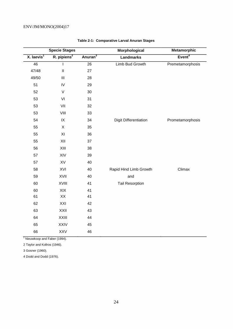
19. To facilitate the description of the morphological development of the thyroid, a comparison between Xenopus development and development in Rana is provided in Table 2-1. The thyroid gland in most amphibians develops during late embryogenesis (Dodd and Dodd, 1976; Regard, 1978). In X. laevis, the thyroid develops from a pharyngeal epithelial ridge around NF stage 35 (Nieuwkoop and Faber, 1994). Following division of the thyroid, follicular development is first present by NF stage 44. A functional thyroid gland with numerous follicles is present by NF stage 53. Follicular development continues resulting in growth of the gland throughout prometamorphosis. Concurrently, TH synthesis and secretion into the circulatory system increases in preparation for metamorphosis and peaks with a surge at the onset of metamorphic climax. After metamorphosis is complete, the thyroid gland regresses in size and reduced levels of circulating TH are present. Two naturally-occurring TH: 1) 3,5,3’,5’-tetraiodothyronine (T4 or thyroxine), and 2) 3,5,3’-triiodothyronine (T3) have been found in anuran species. Based on nearly one hundred years of research, the affect of TH on amphibian metamorphosis is no longer debated, although research in understanding the functional mechanisms and interaction with other hormonal pathways continues today (Gudernatsch, 1912; Allen, 1916; Allen, 1929; White and Nichol, 1981; Tata, 1968; Dodd and Dodd, 1976; Brown et al., 1995; Shi, 2000).
ENV/JM/MONO(2004)17
24
Table 2-1: Comparative Larval Anuran Stages
Specie Stages Morphological Metamorphic
X. laevis1 R. pipiens2 Anuran3 Landmarks Event4
46
I 26 Limb Bud Growth Premetamorphosis
47/48 II 27
49/50 III 28
51 IV 29
52 V 30
53 VI 31
53 VII 32
53 VIII 33
54 IX 34 Digit Differentiation Prometamorphosis
55 X 35
55 XI 36
55 XII 37
56 XIII 38
57 XIV 39
57 XV 40
58 XVI 40 Rapid Hind Limb Growth Climax
59 XVII 40 and
60 XVIII 41 Tail Resorption
60 XIX 41
61 XX 41
62 XXI 42
63 XXII 43
64 XXIII 44
65 XXIV 45
66 XXV 46 1 Nieuwkoop and Faber (1994).
2 Taylor and Kollros (1946).
3 Gosner (1960).
4 Dodd and Dodd (1976).

ENV/JM/MONO(2004)17
25
2.3 Neuroendocrine Control of the Thyroid
20. It is now reasonably well understood that synthesis of TH in the thyroid is under the direction of complex neuroendocrine pathways. TH, in turn, completes a complicated feedback loop at the central nervous system (CNS), hypothalamus, and pituitary levels. These interactions form a complex pathway referred to as the hypothalamus-pituitary-thyroid axis (Shi, 2000). This axis is summarized in Figure 2-1.
2.3.1 Pituitary Regulation of the Thyroid
21. The pituitary hormone thyrotropin (or thyroid stimulating hormone [TSH]), produced and secreted by the par distalis region of the pituitary gland, is primarily responsible for inducing the production and release of TH from the thyroid gland (Shi, 2000). TSH production and release is controlled via negative feedback at the pituitary level (Dodd and Dodd, 1976; White and Nicoll, 1981; Kikuyama et al., 1993; Kaltenbach, 1996; Denver, 1996). Although traditional measures of plasma TSH have not been successful in amphibians due to a lack of sensitivity in the assay, Sakai et al. (1991) found that both purified frog and purified bovine TSH stimulated the release of T4 from the thyroid gland. In hypophysectomized X. laevis tadpoles, Dodd and Dodd (1976) estimated TSH levels in crude pituitary extracts during development using radioiodine uptake. This work suggested that TSH was detectable at trace levels during prometamorphosis (Nieuwkoop and Faber [NF] stage 56), but increased markedly at the onset of metamorphic climax (NF stage 59). In these studies, a decrease in pituitary TSH levels at stage 61 followed by a spike in pituitary TSH at stage 62 was found. Thus, increasing levels of TSH occur during metamorphosis when TH is required. Coincidently, the drop in pituitary TSH production occurs simultaneously with peak TH levels and appears to be the result of increased release of TSH from the pituitary. An understanding of this process at the molecular level has been acheived as the result of the production of complementary DNAs (cDNAs) coding for TSH in X. laevis (Buckbinder and Brown, 1993). Buckbinder and Brown (1993) essentially found that messenger RNA (mRNA) levels during metamorphosis indicated that TSH genes were activated around mRNA stage 53, immediately prior to the first stage in which pituitary TSH levels are detectable. TSH levels peak at approximately NF stages 58 or 59, and drop to appreciably lower levels toward the conclusion of metamorphosis (Dodd and Dodd, 1976; Shi, 2000). TSH gene repression subsequent to stage 59 coincides with high levels of plasma TH. This finding is consistent with a TH-induced negative feedback loop at the pituitary or hypothalamic levels. Interestingly, Dodd and Dodd (1976) and Kikuyama et al. (1993) found a relatively high degree of homology between anuran TSH cDNA and mammalian species. Recently, Okada et al. (2004) developed a RIA for TSH.

ENV/JM/MONO(2004)17
26
Figure 2-1: Endocrine Control of Amphibian Metamorphosis
Modified from Shi (2000).
(+/-)
(+/-)
(-)
ACTH (+)
TH (+)
Thyroid Receptor
Metamorphosis
Corticosteroids (+)
(+)
(+/-)
CNS
Hypothalamus
Pituitary
External Stressors
Prolactin (-)
CRF (+) TRH (?)
TSH (+)
Dopamine (-)
Thyroid Interrenal Glands
Feed Back Loops

ENV/JM/MONO(2004)17
27
2.3.2 Hypothalamic Regulation of the Pituitary
22. The influence of the hypothalamus on metamorphosis is mediated through induction of the release of TSH from the pituitary. TRH is responsible for inducing the secretion of TSH from the pituitary in a similar pathway found in most mammals (Shi, 2000). Historically, the importance of the hypothalamus in the control of metamorphosis has been demonstrated by hypothalectomy, pituitary transplant to a remote part of the body, or providing an impermeable barrier between the hypothalamus and the pituitary gland in frogs (Dodd and Dodd, 1976; White and Nicoll, 1981; Kikuyama et al., 1993; Kaltenbach, 1996, Denver, 1996). High concentrations of TRH have been detected in the brain and skin of R. pipiens (Jackson and Reichlin, 1977). Further, in X. laevis and R. catesbeiana brain tissue, TRH levels have been found to increase throughout metamorphosis and metamorphic climax (King and Miller, 1981; Bray and Sicard, 1982; Millar et al., 1983; Balls et al., 1985; Mimnagh et al., 1987). However, a paradoxical relationships appears to exist between TRH and the rate of metamorphosis (Shi, 2000). More specifically, TRH is readily capable of inducing the release of TSH from the anuran pituitary. However, most experiments have not shown that administration of TRH accelerates metamorphosis (Dodd and Dodd, 1976; White and Nicoll, 1981; Denver and Licht, 1989; Kikuyama et al., 1993; Kaltenbach, 1996; Denver, 1993; 1996; 1998).
23. An important clue to the TRH paradox was uncovered by Denver and co-workers (Denver, 1988; and Denver and Licht, 1989) when these investigators found that mammalian corticotropin-releasing factor (CRF) stimulates the release of TSH. In mammals, CRF is responsible for inducing the secretion of ACTH. Further experimentation demonstrated that mammalian CRF is also capable of accelerating ACTH release from frog pituitaries (Tonon et al., 1986; Gracia-Navarro et al., 1992). Interestingly, ACTH does not induce the thyroid to produce TH (Sakai et al., 1991). CRF is now thought to act directly on the pituitary gland, stimulating the release of TSH (Denver 1988; Denver and Licht, 1989; and Jacobs and Kuhn, 1992). Because CRF is capable of raising TH levels in anurans and accelerating metamorphosis, and because the use of anti-CRF antibodies or CRF receptor antagonists slows metamorphosis; CRF appears to function as the mammalian surrogate of TRH and orchestrates regulation of the the anuran pituitary at the hypothalamic level (Rivier et al., 1984; Gancedo et al., 1992; Denver, 1993; 1997b). Anuran CRF genes in X. laevis are relatively homologous to mammalian CRF (ca. 93%) (Stenzel-Poore et al., 1992; Shi, 2000). CRF gene expression and the presence of CRF-expressing cells in the hypothalamus of X. laevis have not only been identified, but found to be TH-dependent (Verhaert et al., 1984; Olivereau et al., 1987; Gonzalez and Lederis, 1988, Carr and Norris, 1990; Stenzel-Poore et al., 1992). These findings generally agree with the suggestion by Denver et al. (1997) that a hypothalamic feedback loop at the pituitary level (Carr and Norris, 1990). Overall, the primary significance of this research is that CRF, not TRH, is the primary hypothalamic releasing hormone responsible ultimately for the induction of metamorphosis (Carr and Norris, 1990; Denver, 1996; Denver et al., 1997; Shi, 2000).
2.4 Impact of Other Hormones on Metamorphosis
2.4.1 Corticoids
24. In general, the relative importance and capacity of corticosteroids in enhancing TH-induced metamorphosis in amphibians has been purported by several sets of investigators (Kaltenbach, 1985; Kikuyama et al., 1993; and Hayes, 1997a). In amphibians, the interregnal gland is responsible for the production of corticosteroids and receives direct input from the hypothalamus via adrenocorticotropin (ACTH). In turn, two primary corticoids are produced and secreted by the anuran interrenal gland: 1) corticosterone, and 2) aldosterone (Cartensen et al., 1961; Macchi and Phillips, 1966; and Kikuyama et al., 1993; Shi, 2000). Interestingly, several investigators have demonstrated that the major corticoid levels in plasma in metamorphosing anurans follow the pattern of rising plasma TH levels in metamorphosing
ENV/JM/MONO(2004)17
28
tadpoles (Jaffe, 1981; Krug et al., 1983; Jolivet-Jaudet and Leloup-Hatey, 1984; Kikuyama et al., 1986; Kikuyama et al., 1993; Hayes, 1997a). Experimental evidence supporting the role of corticoid hormones in the induction of metamorphosis range from basic fundamental studies to complex experiments.
25. For example, Kaltenbach (1985) and Kikuyama et al. (1983) found that exogenous administration of corticoids via the culture media enhanced tail resorption of premetamorphic tadpoles. Similar responses in cultured anuran tails have also been noted as the result of exogenous corticoid (Kikuyama et al., 1983; Hayes et al., 1993; Hayes and Wu, 1995a; Hayes and Wu, 1995b; Hayes, 1997a). Several different tissues in the metamorphosing anuran appear to be responsive to the impact of corticoids on TH action including: 1) the limbs (Galton, 1990; Kikuyama et al., 1993; Hayes, 1997a), and 2) skin (Shimizu-Nishikawa and Miller, 1992). Further, corticoid receptor sites have been identified in the metamorphosing anuran tail and determined to be important in the control of metamorphosis (Woody and Jaffe, 1984; Yamamoto and Kikuyama, 1993).
26. As an alternative to exogenous corticoid supplementation, the influence of inhibiting the synthesis of endogenous corticoids on metamorphic processes was also evaluated (Kikuyama et al., 1982). In essence, results from these studies indicate that inhibition of corticoid synthesis using Amphenone B is capable of reducing the efficicacy of exogenous TH supplementation to thiourea-induced thyroid repressed amphibians. This study suggests that TH and corticoids work in concert to influence amphibian metamorphosis.
27. A study by Hayes (1997a) suggests that corticoids may operate under a dual mode of action based on the stage of anuran metamorphosis. Based on these studies (Hayes, 1997a), corticoids appear to slow development during early embryogenesis. Prior to and during the early stages of prometamorphosis, endogenous TH levels are low. As TH levels begin to rise with the onset of metamorphosis, corticoids enhance the capacity of TH to induce metamorphosis, although a clear mechanism is not yet known. Much of the evidence supporting the role of corticoids in amphibian metamorphosis is based on in vitro studies involving cell and organ cultures. Based on these studies collectively, corticoids appear to exert negative feedback at the pituitary and hypothalic levels in anurans (Denver and Licht, 1989; Galton, 1990; Nishikawa et al., 1992; Shimizu-Nishikawa and Miller, 1992; Gancedo, et al., 1992; Denver, 1993; Schneider and Galton, 1995; Tata, 1997; Hayes, 1997a).
28. The influence of corticoids on TH-induced metamorphic events has also been observed at the cell and molecular level (Galton, 1990; Kikuyama et al., 1993; Hayes, 1997a). For example, maturation of the skin which occurs during the metamorphic transition of the larvae to an adult involves the expression of adult keratin genes in the epidermis of X. laevis. Under normal physiological conditions, up-regulation is controlled by TH. However, corticoids have also been shown to potentiate the response of these genes to TH. Current research suggests that corticoids act through a nuclear receptor, the glucocorticoid receptor (GR). The GR appears to be similar to classical nuclear-based steroid receptors which essentially belong to the same superfamily of receptors that includes TH receptors (Evans, 1988; Green and Chambon, 1988; Mangelsdorf et al., 1995). Thus, as with most steroid hormones, corticoid effects are induced at the transcriptional level.
29. In summary, the synthesis and secretion of endogenous corticoids are under the direct or indirect control of TH, ACTH, and CRF. Based on the work of Hayes (1997a), CRF appears to have dual functions, stimulating the release of both TSH (thyrotropes) and ACTH (corticotropes) from two different regions of the pituitary (Denver and Licht, 1989). Conversely, the role of TRH in metamorphosis which is the primary thyrotrope in most mammals, is currently thought to be insignificant (Shi, 2000). Overall, physiological synthesis and secretion of corticoids play an important role in anuran metamorphosis.

ENV/JM/MONO(2004)17
29
2.4.2 Gonadal Steroids
30. Unlike corticoids, the role of gonadal steroids on metamorphosis is significantly less clear. Based on an early study by Frieden and Naile (1955) in Bufo bufo, estrone enhanced the effect of T4 on metamorphosis. However, the results of this study have not been demonstrated by other investigators. Rather, the majority of historical studies indicate that estradiol and testosterone antagonize the effects of T4 in R. temporaria (Roth, 1941; Roth, 1948) and inhibit larval development in R. pipiens, X. laevis, and B. boreas (Richards and Nace, 1978; Gray and Janssens, 1990; Hayes et al., 1993) in vivo. Hayes et al. (1993) found that at 22° C, testosterone and estradiol had no effect on growth or size at metamorphosis, although testosterone induced precocious forelimb emergence. However, at 27° C, testosterone and estradiol inhibited growth and development, but did not alter the time to forelimb emergence. However, Gray and Janssens (1990) also found that gonadal steroids did not inhibit the resorption of cultured whole tails in vitro. These results suggest that an inhibitory action of gonadal steroids most likely does not occur at the TR level. Gray and Janssens (1990) and Hayes (1997a) suggest that gonadal steroids most likely act at the hypothalamic-pituitary-thyroid axis level. Hayes (1997a) further hypothesized that the most likely mechanism of gonadal steroid inhibition of metamorphosis occurs through the down-regulation of TH levels, and potentially by up-regulating prolactin levels, which as described below also is capable of inhibiting metamorphosis.
31. Several other investigators have evaluated the effects of gonadal steroids on thyroid axis homeostasis and function; and implications on larval growth, development, and metamorphosis (Jacobs et al., 1988; Vandorpe and Kuhn, 1989; Hayes et al., 1993). Jacobs et al. (1988) found that plasma concentrations of T4 were significantly raised following IV administration of synthetic luteinizing hormone-releasing hormone (LHRH) in ranids. These investigators concluded that this stimulatory effect was mediated through the hypophysis and suggested a possible correlation between the gonadal axis and thyroid axis. Vandorpe and Kuhn (1989) evaluated the effect of estradiol implants in female Rana ridibunda on plasma TH levels and 5'-monodeiodination activity in kidney homogenates in vitro. These investigators found that plasma T3 and TH levels, and the in vitro T3 production in kidney homogenates were significantly decreased, suggesting that estradiol may repress the thyroid axis. Other investigators have evaluated the influence of TH on gonadal steroid activity during metamorphosis (Rabelo et al., 1994; Cohen and Kelley, 1996; Robertson and Kelley, 1996). Rabelo et al. (1994) found that T3 enhanced the precocious activation of vitellogenin genes by estradiol in X. laevis during advanced metamorphosis between NF stages 58-64. Cohen and Kelley (1996) found that androgen-induced cell proliferation in the developing larynx of X. laevis is controlled by TH. These investigators determined that although TH was not required for androgen receptor (AR) mRNA expression is the larynx, cellular proliferation was enhanced by TH, both in vitro and in vivo. Further, Robertson and Kelley (1996) concluded that while gonadal differentiation is independent of TH, androgen-sensitive larangeal development, including sexual dimorphism, require exposure to endogenous TH.
2.4.3 Prolactin and Other Hormones
32. Similar to the effect of corticoids on metamorphosis (Hayes, 1997a), prolactin also appears to exert a bimodal effect on development and maturation of amphibians (Shi, 2000). However, in the case of prolactin, the response is opposite that of corticoids which are capable of inhibiting early development and potentiating TH-induced metamorphosis (Hayes, 1997a). In contrast, prolactin is currently thought to stimulate development during embryogenesis and premetamorphosis, but inhibit the maturation events associated with metamorphosis. In fact, several investigators (Etkin and Lehrer, 1960; Dodd and Dodd, 1976; White and Nichol, 1981; Kikuyama et al., 1993; Denver, 1996) have elaborated on the capacity of prolactin to serve as an apparent growth stimulator in amphibians during premetamorphosis, while also inhibiting metamorphosis in anuran species. Also, in contrast to the effect of corticoids on anuran metamorphosis, prolactin is capable of exerting its inhibitory influence on metamorphosis in vitro (tail

ENV/JM/MONO(2004)17
30
explants) (Dodd and Dodd, 1976; Tata et al., 1991). These results suggest that the inhibitory effects of prolactin on metamorphosis could be mediated at the TR level rather than endocrine regulatory level (Leloup and Buscaglia, 1977). In fact, Tata and coworkers demonstrated that prolactin is capable of inhibiting induction of the TR beta genes by TH (Baker and Tata, 1992; Tata, 1997). Wakao et al. (1994) and Han et al. (1997) have also suggested that prolactin inhibits the function of the TH-TR complex.
33. Anuran prolactin, which was originally difficult to isolate due to the low plasma levels, was first isolated from bullfrogs (Shi, 2000). Cloned amphibian prolactin was subsequently found to be relatively homologous to mammalian prolactin (Yamamoto and Kikuyama, 1981; Yasuda et al., 1991; Takahashi et al., 1990; Buckbinder and Brown, 1993). Prolactin in anuran species is produced in the distal lobe of the pituitary gland (Yamamoto et al., 1986; Tanaka et al., 1991). Prolactin production and secretion is under tight stimulatory and inhibitory control at the hypothalamic level (Kaltenbach, 1996; Shi, 2000). Prolactin is transported to various target tissues through the plasma. Low plasma prolactin levels have been dectected during pre- and prometamorphic stages. However, prolactin levels appear to rise to peak levels late in metamorphic climax (Clemons and Nicoll, 1977; Yamamoto and Kikuyama, 1982; Yamamoto et al., 1986). Interestingly, TRH serves as the primary prolactin-releasing hormone in amphibians, whereas, dopamine serves as the primary neurological inhibitor of prolactin release. Thus, rather than stimulating the release of TSH (as in mammals), TRH induces the release of prolactin and CRF induces the release of TSH.
34. Originally, prolactin was thought to serve as an “amphibian juvenile hormone” (Shi, 2000) similar to that found in insects. However, work by Baker and Tata (1992) and Tata (1997) suggest that the upregulation of prolactin during metamorphosis indicates there is an alternative function during metamorphosis which differs appreciably from the mechanism by which juvenile hormones act (Riddiford, 1996). In essence, gene expression profiles for prolactin expression at both genomic and proteonomic levels suggest an alternative role for prolactin in the control of metamorphic events. Since many of the morphological changes during metamorphosis, such as intestinal remodeling, hind limb digit differentiation, forelimb emergence, and tail resorption (Leloup and Buscaglia, 1977; Nieuwkoop and Faber, 1994), occur at different developmental stages with differing TH levels, prolactin may play a significant role in the coordination of TH-induced amphibian metamorphosis. Further, hypothalamic factors (TRH) do not affect prolactin gene expression during the early stages of metamorphosis, but is upregulation during metamorphic climax (Shi, 2000). De novo synthesis of prolactin appears to be controlled by TH. Buckbinder and Brown (1993) found that inhibition of TH synthesis with the classical anti-thyroid drug methimazole repressed prolactin gene expression. Conversely, treatment of tadpoles with T3 leads to precocious upregulation of prolactin. Unlike the other hormones and hormonal factors, the anti-metamorphic effect of prolactin appears to be exerted at the tissue level rather than in the brain. It is currently thought that prolactin inhibits TH activity at the thyroid hormone receptor (TR) level. Although this model needs further confirmation, it appears that prolactin gene products interfere with TH binding to TR, thus blocking the action of TH.
35. The current hypothesis regarding the role of prolactin during metamorphosis is that prolactin appears to control the high concentrations of TH present during metamorphic climax so that sequential transformation of different tissues can be systematically coordinated (Shi, 2000). This potential role is significant in tadpole-frog transformation, since different tissues/organ systems require differing TH levels at different times during metamorphosis. For prolactin to exert this effect, it must act directly at the tissue level and relatively early in the TH signal transduction process (Leloup and Buscaglia, 1977). Tata and coworkers recently demonstrated that prolactin is capable of inhibiting induction of the TR beta genes by TH (Baker and Tata, 1992; Tata, 1997). Wakao et al. (1994) and Han et al. (1997) have also suggested that prolactin inhibits the function of the TH-TR complex.
ENV/JM/MONO(2004)17
31
36. Another hypothesis is that prolactin interacts with a membrane bound receptor that initiates a cascade of biochemical events that give rise to transcription factors known as Stats (signal transducers and activators of transcription). Interaction of Stats and TR leads to the inhibition of TR and thus, blocks TH-induced metamorphosis (Kanamori and Brown, 1992). Based on this model, the effects of prolactin on TH action are tissue-dependent, because receptor and Stat levels likely differ in the different cell types. This may provide a method of coordinating systematic transformation of different tissues during metamorphosis. Overall, this discussion demonstrates the importance of other related hormonal systems in the control of metamorphosis.
37. More recently, Farre-Young et al, (2000) demonstrated that TRβ/RXR transfected cell lines (HEK293) significantly repressed prolactin-dependent Stat5a- or Stat5b-induced reporter gene expression. Further, over-expression of the TRβ/RXR complex resulted in increased nuclear localization of Stat5a. These investigators concluded that TRβ/RXR modified the subcellular distribution of transcriptional activator Stat5a, thus providing regulation over this prolactin-mediated signaling pathway.
38. Melatonin, produced by the anuran pineal gland and somatostatin appear to be capable of altering anuran metamorphosis (Shi, 2000). Both factors inhibit TSH secretion and are capable of retarding metamorphosis (Denver, 1996). Melatonin may also inhibit metamorphosis through the induction of prolactin pathways (Rose and Rose, 1998). On the contrary, gonadotropin-releasing hormone (GnRH) has been found to increase TH levels in axolotls and frogs, thus has the potential of accelerating metamorphosis (Shi, 2000). Limited information is available on these and other hormones and the understanding of their roles in metamorphosis remains unclear.
2.5 Morphological Changes during Metamorphosis
39. Morphological changes that occur during amphibian metamorphosis have been extensively described and various reviews exist regarding these drastic changes in anatomy (Dodd and Dodd, 1976; Hourdry and Dauca, 1977; Gilbert and Frieden, 1981; Fox, 1983; Balls and Bownes, 1985; Yoshizato, 1989). Essentially, three primary changes in tadpoles take place during metamorphosis in order to transform almost all of the tadpole organs to their adult form (Shi, 2000). The first change involves complete destruction or digestion of tadpole-specific organs. The most obvious example of such a resorption process is the loss of the tail during metamorphic climax. The second change involves de novo development of new tissues from newly produced and proliferated cells. As with many embryological processes these newly produced, but unspecified cells, subsequently differentiated cell lines ultimately leading to tissue morphogenesis (i.e., digits of the hind limbs). Finally, restructuring of existing organ systems, such as the liver, lungs, and intestine into their adult forms occurs. These processes occur to allow the metamoph to adapt to a new terrestrial environment or adulthood in species remaining aquatic. For the sake of brevity, only morphological features that are relevant to the development of amphibian metamorphosis assays will be discussed in this DRP.
40. Of the organ systems resorbed during metamorphosis, two systems degenerate completely; the tail and the gills. Of these two organ systems, resorption of the tail has been the most widely studied (Dodd and Dodd, 1976; Houdry and Dauca, 1977; Gilbert and Frieden, 1981; Fox, 1983; Balls and Bownes, 1985; Yoshizato, 1989). All tissues that comprise the tadpole tail are resorbed during metamorphosis, including an epidermis, connective tissue, muscular tissue, blood vessels, and the notochord (Shi, 2000). Typically, tail resorption begins at the onset of metamorphic climax around NF stage 58-60 with the loss of cross-striations of the myofibrils and disintegration of sub-cellular structures, such as the mitochondria (Weber, 1964; Dodd and Dodd, 1976) and concludes around stage 65 to 66 with the complete disintegration of the tail fin.

ENV/JM/MONO(2004)17
32
41. Condensation and histolysis primarily contribute to tail resorption (Yoshizato, 1989). The loss of tail length during metamorphic climax results from condensation. Water loss, in turn, causes alteration in the organization of the cellular organization of the tail tissue resulting in compaction of the cells and extracellular matrix (Frieden, 1961; Lapiere and Gross, 1963; and Yoshizato, 1989; Yoshizato, 1996). Extensive studies have focused on histolysis as a potential mechanism of condensation, but the specific mechanism has not yet been clearly identified (Kerr et al., 1974; Kinoshita et al., 1985; Yoshizato, 1989). The cells of the tail undergo programmed cell death or apoptosis, and the extracellular matrix is degraded by various enzymes. This process is consistent with apoptosis in other vertebrates. The tail is genetically pre-determined to resorb, requiring only sufficient levels of TH to initiate the process.
42. The classic example of de novo development is the limbs. Hind limb buds are first visible in X. laevis at stage 46 and continue to grossly develop until stage 54 without the assistance of TH. However, between stages 54 and 58, TH levels rise and induce the differentiation of the limb bud cells allowing morphogenesis to form the specific features of the hind limb and toes. The forelimb develops in a similar pattern with the undifferentiated limb bud developing without endogenous TH.
43. The majority of organ systems are present in both the tadpole and the adult anuran; however, most require some form of modification in the adult (Shi, 2000). The liver undergoes little overt morphological change, however extensive biochemical changes take place during metamorphosis. Fine structural changes in liver cells, including increases in the size of mitochondria, endoplasmic reticulum, and Golgi complexes during early and intermediate stages of metamorphosis; and increases in the number of nucleoli and heterochromatic nature of the nuclei during the latter stages of metamorphosis increase the biosynthetic capacity of the liver during metamorphosis. On a biochemical level, hepatocytes within the liver convert from ammonotelic to ureotelic metabolism (Atkinson, 1994; Atkinson et al., 1996; Chen et al., 1994), see also paragraph 46. The nervous system is also restructured to accommodate adult physiology (Kollros, 1981; Fox, 1983; Gona et al., 1988; Tata, 1993, Shi, 2000). At the gross morphological level, changes in the shape of the diencephalon, medulla, and restructuring of neurons in the cerebellum occur during metamorphosis (Gona et al., 1988). At the neuronal level, one of the most dramatic changes includes the genetically programmed regression of the various specialized cells. These cells include a specialized pair of giant neurons, Mauthner cells and Rohon-Beard neurons, which regress or disappear completely, respectively, during metamorphosis (Hughes, 1957; Moulton et al., 1968). In a recent study by Cohen et al. (2001), these investigators that found the antiapoptotic protein Xr11 prolonged survival of Rohon-Beard neurons and reduced morphological change to Mauthner cells. However, Xr11 was not effective in controlling the alterations and ultimate disappearance of other neurons, including spinal cord motor neurons. On the contrary, Purkinje cells, lateral motor column neurons, and the dorsal root ganglia neurons, further differentiate during metamorphosis (Hoskins, 1990). The tadpole intestine is proportionately longer, but more simplistic than the adult anuran intestine, which resembles most vertebrate intestines in terms of structure and function (Shi, 2000). The tadpole intestine is comprised of a single epithelial layer surrounded by thin layers of muscle and connective tissue (McAvoy and Dixon, 1977; Kordylewski, 1983; Ishizuya-Oka and Shimozawa 1987; Shi and Ishizuya-Oka, 1996). Remodeling of the intestine during metamorphosis creates a shorter, but substantially more sophisticated epithelium with numerous microvilli, brush boarders, and ultimately, substantially increased luminal surface area. Thus, the increased microstructure of the intestine developed during metamorphic remodelling effectively increases the effectiveness and efficiency of absorption in the intestine reducing the need for the proportional length found in the tadpole intestine.
44. The role of TH in primary and secondary sexual differentiation has only recently been investigated (Hayes, 1997a). Administration of goitrogens, such as thiourea, which block TH production, resulted in skewed sex ratios (100% female) in X. laevis (Hayes, 1997a; Hayes, 1998). In addition, TH has also been shown to directly induce the testosterone receptor in the larynx of developing male X. laevis (Cohen and Kelley, 1996; Robertson and Kelley, 1996). In the sexually dichromatic anuran, H. argus,
ENV/JM/MONO(2004)17
33
administration of estradiol induces female coloration in both male and female specimens (Hayes, 1997a). However, when estradiol is administered concurrently with thiourea, the skewing toward female coloration characteristics does not occur. Hayes (1997a) found that when thiourea, classical TH synthesis inhibitor, is co-administered with testosterone, induction of gular pouch development does not occur. However, gular pouch development is induced when testosterone is administered alone (Hayes, 1997a).
2.6 Biochemical Changes during Metamorphosis
45. The cellular and biochemical changes that occur in anurans during metamorphosis can be divided into at least six general areas: 1) molecular and biochemical activities associated with programmed cell death or apoptosis, 2) shift from ammonotelism to ureotelism (see 46.), 3) increase in serum protein levels, 4) changes in hemoglobin isoforms, 5) alterations in digestive enzymes, and 6) changes in the respiratory system (Shi, 2000). The role of apoptosis in the selective resorption of tadpole tissues was discussed in the previous section. Many of the genes involved in programmed cell death have been isolated and characterized largely due to genetic studies in the nematode Caenorhabditis elegans, which is a useful model organism for understanding fundamental processes which are conserved in higher animals such as amphibians and mammals. At least three execution genes, seven engulfment genes, and one degradation gene are involved in apoptosis in C. elegans, which represent the three primary processes in selective cell death (Yuan et al., 1993; Alnemri et al., 1996; Cryns and Yuan, 1998). Genes participating in execution of apoptosis and subsequent steps are most likely common in biological organisms where cell death takes place (Ellis and Horovitz, 1986). Homologs to the C. elegans and mammalian genes are currently being studied in amphibians. On the other hand, signal transduction genes participating in the early steps leading to apoptosis, such as induction by TH, may vary in different species. A primary feature of apoptosis involves fragmentation of chromatin which is exploited as a means of evaluating apoptosis by TUNEL (terminal deoxynucleotidyl transferase-mediated dUTP-biotin nick-end labeling). TUNEL is capable of marking apoptosis prior to the fragmentation of the nucleus and the cytoplasm (Gavrieli et al., 1992). The initiation of apoptosis is dependent on TH, and increasing concentrations of T3 (5 to10 nM) induce an increasing response, demonstrating dose-dependence. Classical inhibitors of apoptosis, including ATA and Z-VAD, are capable of inhibiting epithelial apoptosis (Su et al., 1997; Shi et al., 1989). Apoptotic bodies in the tail can be observed as early as NF stage 59 in X. laevis (Shi, 2000).
46. Anuran tadpoles primarily excrete nitrogen waste in the form of ammonia (ammonetelism) during premetamorphosis (Munro, 1953). During the onset of metamorphic climax, ammonia excretion decreases and urea (ureotelism) excretion increases. In most juvenile anurans, at least 75% of nitrogen waste is comprised of urea (Brown et al., 1959). X. laevis represents a primary exception to the excretory conversion to ureotelism. Since X. laevis maintain an aquatic life history as an adult, it primarily excretes ammonia under normal conditions (Munro, 1953). Transient increases in urea during prometamorphosis are typically detected (Underhay and Baldwin, 1955). However, restrictions in water supply induce a drastic increase in urea excretion, suggesting that X. laevis has similar excretory conversion capabilities as the terrestrial anurans (Balinsky et al, 1961). During the process of tail resorption, upregulation of a series of proteases, acid and alkaline hydrolases, and ribonucleases occurs (Shi, 2000). Upregulation of the degradative enzymes is regionally specific in the tail. For example, acid phosphatase levels in the dorsal fin region, which resorbs first in this process, are dramatically elevated in this region during this time. Similar lysosomal hydrolases are upregulated in the intestine during re-modeling. In each case, TH has been shown to control up and down-regulation of these degradative enzymes during metamorphosis (Table 2-2).
47. In most anuran species, serum protein levels dramatically increase during metamorphosis (Shi, 2000). The ratio of serum albumin to globins is markedly elevated during TH-induced metamorphosis. Other plasma proteins that increase during metamorphosis include, ceruloplasmin, transferrin, and carbonic anhydrase (Inaba and Frieden, 1967; Frieden and Just, 1970; Wise 1970). The increase in serum proteins
ENV/JM/MONO(2004)17
34
is thought to play an adaptive role as the tadpole transforms into the frog. In the case of albumin, the biochemical properties of this critical protein not only fulfill osmotic requirements, but also provide circulatory transport needs associated with the terrestrial living habitat (Frieden, 1968; Weber, 1967; Broyles, 1981).
48. Anurans undergo changes in hemoglobin synthesis during development similar to mammals and birds (Shi, 2000). However, in anurans, only one primary change occurs as opposed to other animals, which typically undergo several changes in synthesis patterns. In this case, complete replacement of globulin chains occurs (Weber, 1996). Hemoglobin in tadpoles is independent of pH and has a markedly greater affinity for oxygen than frog hemoglobin, which has lower oxygen binding affinity and is subject to the Bohr Effect (decreased affinity with decreasing pH) (McCutcheon, 1936; Riggs, 1951; Frieden, 1961). The greater affinity of tadpole hemoglobin for oxygen most likely allows for adaptation to low oxygen environments characteristic of the aquatic tadpole habitat. Changes in hemoglobin structure occur around metamorphic climax, although larval hemoglobin persists for some time in metamorphosed frogs to allow for adequate adaptation to the change in the environment (Just and Atkinson, 1972; Weber et al., 1991). Adult frogs require hemoglobin with lower oxygen affinity to facilitate terrestrial life, which has more rapid and extensive oxygen requirements (Bennett and Frieden, 1962; Dodd and Dodd, 1976).

ENV/JM/MONO(2004)17
35
Table 2-2: Genes Regulated by Thyroid Hormone in Anurans1
Gene Name Tissue Source Gene Name Tissue Source
Tail 1/3 Tail2, brain3 N Limb5
Tail 7 Tail2 H Limb5
IU 16/33 Intestine4 J Limb5
Tail 8/9 Tail2, intestine4 Carbamyl-phosphate synthase I
Liver6, 7, 8
TR Beta Tail2, intestine4 Arginosuccinate synthase Liver6
Tail 11 Tail2 Arginosuccinate lyase Liver9
Tail 14 Tail2, intestine4 Arginase Liver9, 10, 11, 12
Tail 13 Tail2 Ornithine carbamyltransferase
Liver7
Tail D Tail2 N-CAM Liver13
Tail C Tail2 Albumin Liver14, 15
Tail 14 Tail2 a 1-mircoglobulin Liver16
Tail 15 Tail2, brain3 Myosin heavy chains Limb5, 17, 18
Xh20 Brain3 Tropomyosin Limb17
Xh1 Brain3 M-line protein Limb19
IU22 Intestine4 a-Actinin Limb19
IU24 Intestine4 Keratins Epidermis20, 21
IU12 Intestine4 Magainin Skin22
IU27 Intestine4 Trypsin Pancreas23
P Limb5 Intestinal fatty acid-binding protein
Intestine24
B Limb5 Mdr (multidrug resistence) Intestine25
I Limb5 Gelatinase A Intestine26
M Limb5 Collagenase-4 Tail27
1 Modified from Shi (2000). 15 Moskaitis et al. (1989). 2 Wang and Brown (1993) 16 Kawahara et al. (1997). 3 Denver et al. (1997). 17 Dhanarajan et al. (1988). 4 Shi and Brown (1993). 18 Sachs et al. (1997). 5 Buckbinder and Brown (1992). 19 Dhanarajan and Atkinson (1981). 6 Morris (1987). 20 Mathisen and Miller (1989). 7 Helbing et al. (1992). 21 Ketola-Pirie and Atkinson (1988, 1990). 8 Galton et al. (1991). 22 Reilly et al. (1994). 9 Iwase et al. (1995). 23 Shi and Brown (1990). 10 Xu et al. (1993). 24 Shi and Hayes (1994). 11 Helbing and Atkinson (1994). 25 Zucker et al. (1997). 12 Atkinson et al. (1994). 26 Patterton et al. (1995). 13 Levi et al. (1990). 27 Stolow et al. (1996). 14 Schultz et al. (1988).

ENV/JM/MONO(2004)17
36
49. In addition to an increase in plasma proteins, the metabolic capacity of the liver markedly changes in the metamorphosing anuran. During metamorphosis, drastic increases in nucleic acid and protein synthesis result in large increases in several enzymes including, catalase, uricase, phosphatases, and the urea cycle enzymes (Shi, 2000). Of these enzymes, the urea cycle has been the most widely studied. Upregulation of these enzymes has been observed during anuran metamorphosis and induced by TH as the direct result of de novo protein synthesis (Brown and Cohen, 1958; Brown et al., 1959; Paik and Cohen, 1960; Cohen, 1970; Dodd and Dodd, 1976). Although each of the four primary enzymes associated with the urea cycle increase at least several fold, the mitochondrial enzyme carbamyl phosphate synthetase actually increases nearly 30-fold during metamorphic climax. Cytosolic arginase, catalyzes the conversion of arginine to urea waste and ornithine (Figure 2-2) increases nearly 5-fold in activity in the liver of metamorphing anurans.
2.7 Production of Thyroid Hormone and Mechanism of Thyroid Hormone Action
50. The primary active THs, T4 and 3,3’, 5–triiodothyronine (T3), are synthesized directly in the thyroid gland (Shi, 2000). Metabolic conversion of T4 to T3, however can occur in other tissues (Fox, 1983; Dodd and Dodd, 1976). TH synthesis is initiated by up-regulation of the thyroglobulin gene in the thyroid, which consequently produces thyroglobulin, the precursor of T4. An intricate set of post-translational modifications, including iodination and condensation of the tyrosine residue to produce T4, is then required (Figure 2-3). T4 can either be secreted into the plasma from the thyroid gland, or directly converted to T3 in the thyroid by 5’-deiodinase. Both T4 and T3 can be selectively inactivated by 5-deiodinases by converting either TH to T2 or reverse T3, respectively. This allows different tissues to possess different ratios of T3 to T4 depending on their specific requirements. St. Germain and Galton (1997) located two different 5-deiodinases in anurans that have different enzymatic properties and tissue distributions. Differing deiodinases have been isolated and cloned in R. catesbeiana (Davey et al., 1995; Becker et al., 1995) and X. laevis (St. Germain, 1994). Each different isoform was found to have distinctly different regulation patterns in different tissues, thus supporting the hypothesis of TH level regulation at the tissue level.
51. The action of TH during development is regulated at many different levels, due in part to the presence of numerous TH binding proteins in the plasma, cytosol, and nucleus. Since many of the proteins with which TH interacts are cytosolic and many of the effects occur at a non-genomic level, it was originally thought that TH acted through cytosolic actions (Davis and Davis, 1996). However, today more evidence exists that TH acts at the nuclear level mediating gene regulation via nuclear-based TR (Tata and Widnell, 1966; Tata, 1967; Oppenheimer, 1979). TH secreted from the thyroid is carried in the plasma to various tissues by various serum TH hormone binding proteins. At least nine cytosolic and plasma proteins are known to transport TH, although several are more significant factors (Shi, 2000).

ENV/JM/MONO(2004)17
37
Figure 2-2: Generalized Urea Cycle in Anuran Tadpole
Citrulline
ornithine
ARGINASE
Urea
Arginine
Modified from Shi (2000).
FIGURE 3-2 GENERALIZED UREA CYCLE IN ANURAN TADPOLE
ARGINOSUCCINATE LYASE
Argininosuccinate
ARGININOSUCCINATE SYNTHETASE
Citrulline
ornithine
ARGINASE
Urea
Arginine
Modified from Shi (2000).
ARGINOSUCCINATE LYASE
Argininosuccinate
ARGININOSUCCINATE SYNTHETASE

ENV/JM/MONO(2004)17
38
Figure 2-3: Generalized Biosynthetic Pathway of Thyroid Hormone (TH)
Modified from Shi (2000).
Thyroid Receptor (TR)
I [plasma ]
Peroxidase/Tyrosine Iodinase
MIT/DIT-Thyroglobulin (TG)
T4-TG
T3-TG
T4 or T3 Transporter Protein [plasma ]
Thyroid
ENV/JM/MONO(2004)17
39
52. In the serum, TH immediately encounters serum binding proteins, the most notable of which is transthyretin (Yamauchi et al., 1993), which transport TH to the target tissues where TH enters the cytosol (Jorgensen, 1978; Barsano and DeGroot, 1983; Galton, 1983; Benvenga and Robbins, 1993). Cellular uptake mechanisms are not well understood. T3 and T4 are relatively hydrophobic at physiological pH (Shi, 2000). Thus, passive diffusion through the cell membrane is a possible route. However, some evidence suggests that a carrier-mediated transport process involving translocation of both the TH transporter and TH is possible (Blondeau et al., 1988; Oppenheimer et al., 1987; Robbins, 1992; Ribeiro et al., 1996; Benvenga and Robbins, 1993). Within the cytoplasm, TH interacts with a separate group of multifunctional proteins, collectively referred to as CTHBP (cytoplasmic TH binding proteins) (Cheng, 1991). It is presently unclear whether the TH-CTHBP complex is required for activation of the nuclear TRs, or whether it only provides a means of transport to the TR.
53. Diploid vertebrate animals, including X. tropicalis, possess two TR genes (TR alpha and TR beta) (Lazar, 1993). X. laevis, which is oligotetraploid, possess four TR genes, two TR alpha and two TR beta (Mangelsdorf, et al., 1995). Alternative splicing of the TR beta transcripts gives rise to two different isoforms in higher vertebrates and four different isoforms in X. laevis (Brooks et al., 1989; Yaoita et al., 1990). TRs belong to the superfamily of nuclear hormone receptors, including glucocorticoid, estrogen, vitamin D, and retinoic acid receptors (Evans, 1988; Tsai and O’Malley, 1994; Yen and Chin, 1994). In general, the TR contains five different binding domains, A/B, C, D, E, F (amino to carboxy terminus), which are reasonably consistent within this class of nuclear receptors (Shi, 2000). The amino terminus (A/B domain) of the TR alpha A/B and TR beta-B2 TR isoforms specifically contains the AF-1 domain, which appears to be involved in T3-independent recruitment of specific co-activators. Thus, ligand-independent activation of transcription by at least the TR beta-B2 isoform may be mediated by the binding of specific co-factors to the AF-1 region of the A/B domain (Obertse-Berghaus et al., 2000; Yang and Privalsky, 2001). DNA binding occurs in domain C. The C domain is highly conserved amongst nuclear receptors. Domain D is the variable hinge region which contains a nuclear localization signal and influences both DNA binding and transactivation through co-repressor binding (Giguere et al., 1986; Godowski et al., 1988; Hollenberg and Evans, 1988; Picard and Yamamoto, 1987; Guiochon-Mantel et al., 1989; Zechel et al., 1994; Lee and Mahdavi, 1993; Uppaluri and Towle, 1995; Puzianowska-Zunicka et al., 1997). Domains E and F are the ligand (hormone) binding and transactivation domains. The carboxy terminus, or region F, contains the AF-2 domain. The AF-2 domain has been found to be a binding site for specific co-activators containing the LXXLL motif of liganded TR (Obertse-Berghaus et al., 2000; Heery et al., 1997; Langlois et al., 1997). The conceptual model for transcriptional regulation by TR is illustrated in Figure 2-4. TR is presumed to form a heterodimer with the retinoic acid X receptor (RXR). The heterodimer binds to the TH response element in a target gene. In the absence of TH, the heterodimer represses gene transcription, most likely through the recruitment of a co-repressor complex (Horlein et al., 1995; Chen and Evans, 1995).

ENV/JM/MONO(2004)17
40
Figure 2-4: Interaction between Thyroid Hormone (TH) and TH Receptor (TR)
TH [i ]
Modified from Shi (2000).
Cell
Gene Expression
TR/RXR Receptor
Nucleus
TH-TR/RXR
TH [free ]
TH
TH [bound ]
transportprotein
5-deiodinase

ENV/JM/MONO(2004)17
41
2.8 Anticipated Sites of EDC Impact on the Thyroid Axis
54. Based on the previous discussion, EDCs could potentially affect the thyroid axis at three levels, 1) CNS (including pituitary and hypothalamus), 2) thyroid, and 3) TR. More specifically, specific modes of actions of thyroid axis disruptors could potentially include alteration of TH synthesis, TH transport, TH elimination, neuro-endocrine (H-P) axis regulation, and TR expression and/or function. A summary of potential modes/sites of EDC action on the thyroid axis, in relation to endpoints possibly useful in measuring thyroid disruption, is provided in Table 2-3. The effect at the pituitary level is complex since it may involve thyrotropes (TSH), corticotropes (ACTH), and lactotropes (prolactins). In addition to the thyroid gland, the interrenal gland may also be a site of EDC action, which could potentially impact metamorphosis. Thus, the impact of potential EDCs on metamorphosis may occur at multiple different levels. The complexity of metamorphosis and control by the neuroendocrine system must be strongly considered in the design of appropriate test methods. Since the objective of the test method is to screen for thyroid axis disruption, a rapid, high throughput biochemical measurement test or molecular test would likely be more advantageous. However, alone, biochemical measurements and molecular tests might not provide adequate information on the effect of the EDC on metamorphosis at the whole organism level. Incorporation of a high-throughput biochemical or molecular assay within a short-term morphological method would therefore be advantageous. It is crucial that the methodology used demonstrates diagnostic power by distinguishing between non-thyroid and thyroid-related delays in developmental progress.
Table 2-3: Catalog of Endpoints Potentially Useful in Measuring Thyroid Disruption Based on Specific Modes of Action1
Mode of Action
General Measures Specific Endpoint
TH Synthesis
TH Transport
TH Action
Neuroendocrine (H-P) TR
Morphology Developmental Stage
+ ? +? + +
Hind Limb Differentiation
+ ? +? + +
Histology Thyroid ++ ? +? ++ + Pituitary + ? +? + Biochemical T3/T4 + +? ++ Liver Enzymes - +? +? +? +? Molecular Biology TR beta + ? + - ++ TSH + ? ? + ? CRF + ? ? + ? UDPGTP - + ? ?
1Based on discussions of Group 1 at “Uses of Amphibian in Assessing Endocrine Disruption”, Duluth, MN, June 2003. ++=method(s) demonstrated and endpoint potentially useful, +=methods somewhat demonstrate and endpoint potentially useful, +?=endpoint potentially useful, but method not demonstrated, ?=utility of endpoint unknown, -=not potentially useful endpoint.

ENV/JM/MONO(2004)17
42
DESCRIPTION OF CANDIDATE ENDPOINTS REFLECTIVE OF THYROID DYSFUNCTION
3.1 Whole Organism Tests
55. It is generally accepted that as the function of the vertebrate endocrine system is coordination of physiological processes, test designed to detect disruption of hormonal function must both incorporate the potential for feedback effects, and demonstrate the impact of a chemical at the level of the intact organism. These considerations cannot currently be addressed with in vitro tests of biochemical activity alone. A variety of morphological, biochemical, and molecular endpoints may be applied to tests involving exposure of intact organisms to test substances, as described below.
3.1.1 Morphological Measures
56. Protocols ultimately developed to morphologically detect thyroid impairment may include any applicable endpoint discussed in the following sections, and should not necessarily be limited to one endpoint. Different morphological endpoints, as discussed, have utility at different stages of larval development (e.g. limb development earlier than tail resorption). Current protocols for amphibian metamorphosis using Xenopus laevis generally use ‘developmental stages’, for which a well-defined sequence has been characterized (Nieuwkoop and Faber, 1994). Other tables, though less refined, are also available for Ranid species. Use of developmental stages integrates multiple aspects of morphological development, and is less sensitive to bias resulting from operator error or the potential for differential effects of toxicants on morphological development. It should however be noted that asynchronous or atypical development can occur in response to chemical exposure, which complicates developmental staging based on morphology.
3.1.1.1 Tail Resorption.
57. Tail resorption occurs in anuran species during metamorphic climax and is triggered by a surge of TH. Metamorphic climax in X. laevis occurs roughly over a 16-18 day period and incorporates stages 58 to 66. Tail resorption can be monitored during culture using digital photography and measuring the tail lengths with a scanning digitizer. A rate of tail resorption can be determined from the mean length data obtained. Abnormal tail resorption can also be monitored during this process. Specimens can be selectively preserved throughout the process of tail resorption and evaluated morphologically. In terms of culture, different approaches may be considered. For example, the specific process of tail resorption can be monitored by culturing tadpoles at relatively low densities such as in multiple replicates for 16 to 18 d from stage 58 to 66 during which time the rate of tail resorption is measured (Fort et al., 2000). Low densities of ca. one organism per 500 mL of test solution are required to achieve consistent maximum growth rates in culture. However, this approach does not consider the metamorphic events that lead up to metamorphic climax. In addition, natural inherent variation in tail resorption can be observed due to differing levels of endogenous TH between organisms.
ENV/JM/MONO(2004)17
43
58. Based on this information, several factors relating to the measurement of tail resorption and the interpretation of the results require consideration. First, the rate of tail resorption is naturally variable in whole organism culture (Fort et al., 2000; Fort et al., 2001b), which reduces the sensitivity and predictability of this endpoint. Second, this process occurs in the later stage of metamorphosis, when the thyroid is fully active and at its peak early in the climatic period.
3.1.1.2 Limb Development.
59. As previously discussed, early hind limb bud development (emergence) occurs prior to thyroid activity in the developing anuran tadpole. However, hind limb differentiation and forelimb development occur during the prometamorphic phase of metamorphosis. Thyroid dysfunction impairs the process of limb differentiation, but not limb bud emergence. Selecting an exposure window that encompasses hind limb differentiation would require exposure from stages 54 to 60. The longer-term test format described by the German investigators, incorporates limb differentiation, by way of using developmental stage as the morphological endpoint. The only standardized test method that evaluates limb development was evaluated by Fort and Stover (1996) and Fort et al. (1997) using X. laevis. However, this modified FETAX assay (ASTM, 1998) evaluated only hind limb development, initiating exposure at an early blastula stage and completing exposure around 30 days at stage 54. Thus, this design did not incorporate TH-dependent limb growth and differentiation.
60. The morphology, rate, and extent of limb differentiation can be monitored in a similar manner as described for tail resorption. However, in this case the occurrence of abnormal limb development, including asymmetrical differentiation, should be monitored, thus requiring additional specimens for histological examination, CAT scan, and x-ray analysis.
3.1.1.3 Skin Development
61. During metamorphosis, substantial changes to the skin in terms of protein structure, keritinization, and pigmentation occur. Changes in skin structure have already been discussed; however, changes in pigmentation also occur that change a transparent tadpole such as X. laevis to a frog with pigmented, non-transparent skin. Classical thyroid inhibitors, such as thiourea, also inhibit pigmentation by blocking melanin synthesis. An evaluation of melanin distribution in skin structure can be evaluated in Xenopus under normal light microscopy. Unlike larval skin, metamorph skin possesses well-formed melanocytes containing a relatively dense distribution of melanin. Staining is only required to evaluate the neurological status of the pigmentation process. For evaluation of epidermal structure and keritinization, a standard eosin or hemotoxylin/eosin can be effectively used. Immunohistochemical techniques can be used to distinguish the presence of specific proteins that mark the newly developing frog skin. Therefore, skin maturation is a potential valuable endpoint. Like the endpoints previously discussed, this endpoint is best served with a battery of other metamorphosis-based morphological endpoints in a longer-term exposure design.
3.1.2 Thyroid Pathology
62. In most cases, thyroid dysfunction, such as goiter or myxedema, manifests changes in the morphometry of the thyroid. For example, tadpoles exposed to the goitrogen methimizole develop enlarged thyroid glands that are visible under low magnification in the transparent Xenopus tadpole (Fort et al., 2000). In this case, the thyroid gland can be digitally photographed and the size quantified by photodigitization. Some EDCs may induce myxedema, or a shrinking of the thyroid gland (Wollman, 1980). Again, myxedema can also be quantified using the methods discussed for goiter. Distinct histopathology is also associated with goiter, including a thickened capsule wall, shrinking of capillaries, fusion of the follicles, increased follicular size, and infiltration of connective tissue (partitions) entering the
ENV/JM/MONO(2004)17
44
lobe from the capsule (Wollman, 1980). A possible framework for of analysis for thyroid histopathology is given in Hooth et al. (2001). The histological techniques are relatively simple, using thin sections of the thyroid gland and standard light microscopy of hemotoxylin/eosin stained tissue. Use of thyroid morphometry and pathology, particularly in Xenopus, where the thyroid gland is fully visible in the intact specimen, should be included in the analysis of thyroid function.
63. Specimens for histological examination can be selectively preserved in Bouin’s Solution prior to preparation. Following tissue processing, including decalcification if needed, the tissue sample can be embedded in paraffin. Microtome sectioning (4-5 µm) or step sectioning (30-32 µm between steps) can be performed prior to hemotoxylin-eosin staining. The histological examination could include changes in the gland, including hypertrophy of follicular cells, hyperplasia of thyroid follicles, size of the follicle, and degree of colloid accumulation. Use of digital photographs can be used to illustrate changes and provide a means for outside peer examination. In addition to traditional light microscopic procedures, electron microscopy (EM), particularly scanning EM (SEM) or scanning transmission EM (STEM), can be considered as a potentially useful diagnostic tool, though it is unlikely these techniques could practically or economically be applied in the context of a screening assay.
3.1.3 Biochemical Measures
3.1.3.1 Corticotropin Releasing Factor and Thyroid Stimulating Hormone
64. As previously discussed, CRF, produced and secreted by the hypothalamus, stimulates the production and secretion of both ACTH and TSH from the pituitary. Although the structure of CRF is known (41 amino acid peptide), there is currently no diagnostic test, such as a RIA or ELISA that has been readily used. The development of such a test for CRF is possible.
65. In humans, TSH is measured by high affinity RIA, and in a clinical setting, TSH levels are used in combination with TH levels in diagnosis and management of thyroid dysfunction. As with CRF, an amphibian RIA (or ELISA) for TSH could be developed, considering the similarities in structure between mammals and amphibians, and used in the current context of the amphibian metamorphosis assays, in combination with measurement of more downstream hormones and factors.
3.1.3.2 Thyroid Hormones
66. Measurement of TH, specifically T4 and T3, produced by the thyroid provides a valuable measure of thyroid status during metamorphosis. Both T4 and T3 can be measured using conventional serum or tissue RIAs. Both serum and tissue (whole brains, thyroid, or carcass following cardiac puncture to obtain serum) should be considered. Three different methodologies of TH analysis are currently being evaluated, 1) high sensitivity RIA, 2) ELISA, and 3) liquid chromatography/gas chromatography with mass selective detection (LC/GC-MS). The former two techniques are reasonably well established in mammals (Ekins, 1999; Baiser et al., 2000), though only the first (RIA) has been used in amphibian studies (Galton et al., 1991). Specific ELISAs would need to be developed and validated for measurement of T3/T4 in amphibian tissue matrices. The chromatographic technique is not as well established (Moller et al., 1983; De Brabandere et al., 1998), but use in clinical diagnostics for thyroid function indicates significant promise, because it may be able to simultaneously analyze monoiodotyrosine (MIT), diiodotyrosine (DIT), reverse T3, T3, and T4. In some cases it will be important to measure the concentration of free T4 and T3 in relation to the transport protein-bound TH, since the majority of TH is protein bound (Baiser et al., 2000).
67. At this point, conventional RIA analysis methods of TH are adequate, though for some laboratories the use of radioisotopes may be problematic. Consequently, research effort should be focused

ENV/JM/MONO(2004)17
45
on development and validation of sensitive non-isotopic ELISA methods. Regardless of method, quality assurance (QA) measures associated with ELISA and RIA analyses should include an evaluation of cross-reactivity with other hormones or similar substances, evaluation of linearity using standard curves, and the use of standard additions to assess recoveries. Moreover, the sensitivity and reliability of these techniques will be an important consideration, given that TH levels will not be high in a prometamorphic assay, and the magnitude of changes in TH that may be induced by test chemicals may be small. Overall, TH analysis will be an important component of the amphibian metamorphosis assays. However, the most beneficial use of TH analysis will be in combination with the histological, morphological, and molecular test methods used. It is unlikely, that TH analysis alone will provide sufficient information to be a stand-alone measure of thyroid dysfunction.
3.1.3.3 Iodothyronine Deiodinase
68. Two deiodinase isoforms are present in most anurans. One isoform, type II, catalyzes the conversion of T4 to T3 in the thyroid and various target tissues, whereas the other isoform, type III, selectively inactivates T3 and T4 by converting them to T2 and reverse T3 (Huang et al., 1999). It is thought that type III deiodinase in anurans (X. laevis) is responsible for protecting the tissues from circulating TH. Koopdonk-Kool et al. (1993) developed a method for measuring deiodinase activity by measuring the conversion of [125I]T3 to T2. In most cases deiodinase activity is not considered in evaluating thyroid function; however differences in tissue levels of T4 and T3 can in some cases be explained by differing deiodinase activities. Further work will be required to fully determine the usefulness of deiodinase measurement in evaluating thyroid dysfunction.
3.1.3.4 Thyroid Hormone Transport Proteins and Thyroid Hormone Receptors
69. TH synthesis and secretion, TH transport by carrier proteins, and TH binding to TR constitutes the primary thyroid axis pathway. Thus, measurement of TH transport proteins and TR provide a measure of TH activity and responsiveness (Tata, 1999). These components of the thyroid axis can be measured by most practicable methods of evaluating these processes; such methods appear to be quantifying changes in protein levels during metamorphosis. Conventional ELISA is suitable for the analysis of specific transport proteins, such as transthyretin (Yamauchi et al., 1993), while immunohistochemical analysis of TR in the whole organism can be used to regionally quantify the presence of TR in a metamorphosing tadpole. Evaluating gene expression by RT-PCR or RPA also represents a suitable method for evaluating the up and down regulation of TR or TH binding proteins.
3.1.3.5 Clinical Tests of Thyroid Function
70. Aside from analytically measuring pituitary and TH levels, associated regulatory enzymes of the thyroid axis, and TH transport proteins; several classical methods of measuring thyroid function in higher animals, including humans, could be considered. These classical assays include, radioactive I (125I) uptake by the thyroid and T3 resin tests, which measure thyroglobulin binding protein among a host of others (Thomson, 1974). None of these tests have been specifically adapted for use in amphibians due to the advent of more sophisticated molecular techniques. Therefore, it is unlikely that the classical methods of measuring thyroid dysfunction in humans will be more effective in measuring thyroid disruption in amphibians, than the methods already described or the approaches discussed in the following section.
3.1.4 Molecular Biomarkers
71. Three general molecular approaches for measuring TH-induced metamorphosis are currently being evaluated: single gene expression assays (e.g. qPCR), multiple gene expression assays (e.g. microarray analysis), and somatic or germinal transgenesis of relevant reporter gene constructs. Use of

ENV/JM/MONO(2004)17
46
these molecular approaches in conjunction with a validated in vivo assay has the potential to provide useful information on the mode of action of any chemicals that alter metamorphosis at the morphological level.
3.1.4.1 Single Gene Expression Assays
72. Measuring the expression of specific individual genes that are activated or inactivated during metamorphosis has the potential to make amphibian metamorphosis assays more mechanistically informative. For example, genes that code for TR beta or the urea cycle enzyme, arginase, could be selectively monitored for activity during metamorphosis. Among the most intriguing TH responsive genes that could be evaluated during prometamorphosis are TR beta or stromelysin-3 (ST3). ST3 is expressed in frogs, mice, and humans during development, in tissues were selective cell death takes place (Bassett et al., 1990; Lefebvre et al., 1992). Results from these investigators suggest that ST3 is involved in both apoptosis and cell migration, both of which are involved in limb definition, intestinal remodeling, and tail resorption. Furthermore, ST3 is ubiquitous in Xenopus tadpoles, and exogenous TH induces the precocious activation of ST3 (Wang and Brown, 1993; Shi and Brown, 1993; Patterton et al., 1995). In vivo, the developmental expression of ST3 mRNA correlates strongly with organ-specific metamorphosis. For example, in the tail ST3 mRNA is low until stage 62, when expression increases and apoptosis is detected by TUNEL (Kerr et al., 1974). ST3 mRNA levels are appreciably lower in the elongating hind limb until stage 54 to 56 when the interdigital cells degenerate to form the toes. Therefore, like TR beta, TH-responsive ST3-expression is a strong candidate for use in molecular screening of thyroid function.
73. Several techniques are currently available to measure the expression of single genes e.g. ribonuclease protection assay (RPA), semi-quantitative reverse-transcription PCR (RT-PCR) and real-time or quantitative PCR (qPCR). RPA is now rather outdated and has been superceded by RT-PCR methodologies.
74. RT-PCR methodologies for specifically measuring TR beta gene expression changes, as the result of exposure to potential EDCs in Rana catesbeiana tail biopsies, were developed by Veldhoen and Helbing (2001). RT-PCR analysis of ST3 or other relevant gene activity during prometamorphosis could also be considered. RT-PCR technology is based on the construction of cDNA from isolated RNA using reverse transcriptase. The cDNA is amplified by means of corresponding cDNA primer fragments (e.g. TR beta, TR alpha, arginase, ST3). The amplified DNA products are then separated on an agarose gel and the amplified DNA bands quantitatively analyzed using densitometry. RT-PCR techniques have been used to measure the induction of vitellogenin genes in Xenopus as the result of exposure to the weakly estrogenic compound bisphenol A (Kloas et al., 1999; Levy et al., 2004). Work by Veldhoen and Helbing (2001) using Rana catesbeiana tadpoles demonstrates that quantitative analysis of single gene activity, such as TR beta, is feasible. If the TH-response genes selected for evaluation are ubiquitous, other tissues could be sampled besides the tail, including the hind limb, using a similar biopsy approach.
75. Real time or quantitative PCR techniques (qPCR) use control template amplicons to provide a standard curve against which to quantify sample amplification curves. This methodology offers greater convenience, efficiency and sensitivity, and is likely to be the method of choice for measuring expression of single (i.e. known) genes. The method has been used by Crump et al. (2002b), Helbing et al. (2003), and Veldhoen et al. (2002) for analysis of TH-dependent gene expression in the tail of Rana catesbeiana.
3.1.4.2 Multiple Gene Expression Assays
76. TH-induced metamorphosis involves the regulation of a multitude of genes in tissues involved in TH homeostasis aswell as in target tissues undergoing metamorphic remodelling. Consequently, measuring changes in multiple genes in response to exogenous agents has the potential to lend greater mechanistic specificity to an amphibian metamorphosis assay.
ENV/JM/MONO(2004)17
47
77. One approach to screening differential activity of multiple genes is differential display (Liang and Pardee, 1992). This technique involves synthesizing cDNA from a subset of the compared mRNA population by using a partially degenerate primer. In this case, the subset might include mRNA specifically expressed during critical phases of metamorphosis. A subset of the cDNA is amplified using polymerase chain reaction (PCR) with the appropriate 5’-primer. The amplified products from two samples are then displayed on a sequencing gel. Bands of differing intensities between the two samples are derived from mRNAs of different quantities. These bands can be cut out of the gel, eluted into buffer, and PCR amplified. The amplified products can then be used for in situ hybridization to confirm the regulation of the specific genes. Crump et al. (2002a) evaluated the effects of octylphenol, an estrogenic EDC, and UV-B radiation on hypothalamic gene expression, using differential display as a diagnostic tool to screen for candidate genes that were differentially expressed in R. pipiens. These investigators found that NcK, ash, phospholipase C gamma banding protein, brain angiogenesis inhibitor-3 in tadpoles; and GAD 67, cytochrome C oxidase, and brain angiogenesis inhibitors -2 and -3 in metamorphs were differentially expressed. Overall, differential display analysis is a reasonably well-developed technique for measuring multiple gene activity. However, quantification and interpretation of results from differential display analysis can be difficult, and the technique may therefore be difficult to apply in the context of a screening test.
78. A more recently developed technique for evaluating multiple gene activity involves the use of gene arrays. DNA micro- and macro- arrays are powerful tools in the analysis of differential gene expression. The cDNA micro-arrays are capable of profiling gene expression patterns of tens-of-thousands of genes in a single experiment. In this technique, DNA targets, in the form of 3' expressed sequence tags (ESTs), are arrayed onto glass slides or membranes and probed with fluorescent- or radio-labeled cDNAs (Duggan et al., 1999). TH-mediated gene expression patterns have been identified in the rat (Witzel et al., 2001) and X. laevis (Denver et al., 1997) using cDNA expression arrays and a slightly different technique, PCR-based subtractive hybridization. Recent studies using this approach have identified sets of T3-responsive genes in various tadpole tissues, including the tail (Wang and Brown, 1993; Brown et al., 1996; Crump et al., 2002b; Veldhoen et al., 2002; Helbing et al., 2003), hind limb (Buckbinder and Brown, 1992), brain (Denver et al., 1997), and intestine (Shi and Brown, 1993). These analyses have demonstrated that some early TH-response genes are common to all tadpole tissues, whereas others are tissue specific. Genes identified by Denver et al. (1997) to be up-regulated during prometamorphosis and metamorphic climax include xh4, xh7, xh15, xh6, xh1, TR beta, and bZIP. Targeted gene array constructs could therefore be developed to evaluate the effect of potential EDCs on multiple TH-dependent gene expression pathways (e.g. including TR beta, TR alpha, arginase, ST3, and other related genes).
79. Gene array technology offers advantages over differential display (e.g. in terms of interpretation), and over RT-PCR, through its ability to simultaneously measure expression of multiple genes of interest. Its potential utility in this arena will therefore depend on the availability of genome sequence for relevant species (e.g. Xenopus, Rana), and speed at which the technique becomes more affordable. However, the technique is technically demanding and its suitability for quantification of gene expression is limited: the technique is predominantly used for discovery of differentially expressed genes of interest for further investigation, and quantification by other techniques (e.g. Real Time RT-PCR). However, the potential power of gene array technology as a diagnostic tool in the future cannot be over-looked.
3.1.4.3 Transgenic Strains
80. A physiological means of studying gene function is through gene knockout and transgenic lines. To date, no gene knockouts have been developed in amphibians. However, two methods of developing transgenic lines have been developed using amphibian species. The first approach involves the nuclear transplantation of somatic nuclei into an enucleated oocyte. Once the transplantation is complete, the

ENV/JM/MONO(2004)17
48
oocyte is fertilized. Kroll and Gerhart (1994) used this approach to transfect a gene of interest into a X. laevis tissue culture cell line. Successfully transfected nuclei are then microinjected into newly fertilized embryos. However, this approach has not been largely successful in growing embryos beyond a young larval stage. Thus, the use of transfected somatic nuclei transgenesis is not well suited for studying metamorphosis.
81. The inability to raise the transgenic specimen beyond early larval stages prompted Kroll and Amaya (1996) to develop the second approach that uses undifferentiated sperm cell nuclei. This approach entails the insertion of a gene or genes with appropriate promoters in a plasmid. The plasmid is then linearized using restriction enzymes. The linearized plasmid and X. laevis sperm cell nuclei are mixed in a high-speed extract made from X. laevis eggs. A short incubation period allows decondensation of the nuclei to occur, allowing plasmid incorporation into the chromatin. The transfected sperm nuclei are then microinjected into oocytes at a rate of one nucleus per egg. Although the efficiency is still rather low, the techniques compare favorably to similar approaches developed for the mouse and zebrafish. The information available on many genes involved in metamorphosis will eventually allow construction of transgenic lines that model the expression of metamorphosis-related genes, using tissue-specific promoters. Development of transgenic lines expressing novel TH-inducible gene sets (i.e.TR beta and related TREs) is therefore possible, and these transgenic models have the potential to provide in vivo bioassays of TH-dependent gene expression, in a context where mechanistic information can be linked to effects at higher levels of biological organization (e.g. biochemistry, morphology). However, the complexity and time required to create a transgenic line makes it somewhat less attractive than the gene expression assays.
82. Adaptation of these techniques to X. tropicalis, a diploid organism with a shorter lifecycle, further increases their feasibility. A transgenic line exploiting TH/TR response elements could be developed. Activation of these response elements by the action of an exogenous EDC could be marked by a marker protein. For example, Oofusa et al. (2001) evaluated the activity of the thyroid responsive element (TRE) using X. laevis carrying a transgene containing the 5' upstream region of the TR beta A1 gene and a green fluorescent protein (EGFP) gene. EGFP expression was then monitored throughout the entire premetamorphic, prometamorphic, and metamorphic climax periods. TR beta expression was found as early as neurula stage at low levels, with low activity during the remainder of premetamorphosis, culminating at metamorphic climax. A similar transgenic Xenopus line could be developed to measure TR beta expression and the influence of potential EDCs on gene expression. Furlow and Brown (1999) identified a novel leucine zipper transcriptional factor (TH/bZIP) that is induced by TH during metamorphosis in transgenic X. laevis. Two genomic TH/bZIP genes regulated by an adjoining DR+4 TRE have been found in X. laevis. The effect of potential EDCs on up and down regulation of TH/bZIP could also be measured using an EGFP marker.
83. A compound that interferes with metamorphosis at the morphological level through disruption of TH homeostasis must ultimately influence transcription of TH-responsive genes. Therefore, in accordance with the work of Luze et al. (1998), Ulisse (1996), and Rowe et al. (2002), it is possible that an in vivo reporter gene bioassay created from either somatic or germinal transgenesis of a TRE-bearing construct could be used to evaluate thyroid axis disruption in a quantifiable and rapid manner. Such an assay would integrate effects of a test chemical at multiple sites on the thyroid axis, and could be combined with apical endpoints such that mechanistic information can be linked to effects at higher levels of biological organization (e.g. biochemistry, morphology). However, a significant amount of work will be required to optimize reporter constructs and to assess and minimise signal leakage (non-TH-dependent reporter activity). The technical practicalities of performing somatic gene transfer as an adjunct to a high throughput screening assay are questionable, and potential future adoption of this type of approach is likely to be dependent on establishment of stable lines through germinal transgenesis.

ENV/JM/MONO(2004)17
49
3.2 Organ and Cell Culture
84. Consistent TH-induced tail resorption is relatively easy to monitor in organ cultures (Weber, 1967; Tata et al., 1991). Whole tail cultures could be used to evaluate TH agonists and antagonists downstream from the thyroid gland. More specifically, tail culture assays could potentially evaluate TH interaction with the TR, TR activation of the TREs, and the cascade of molecular events associated with selective cell death in the tail. Similar transfected cell culture lines could also be established to measure the influence of EDCs on TREs and associated gene products (Denver et al., 2002).
3.3 Receptor and Protein Binding Assays
85. Since EDC binding to the TR and plasma TH transport proteins represents an additional process, which may be affected by EDCs, a receptor or transport protein binding assay could also be considered. Currently, TR binding assays, which measure the relative binding affinities of potential EDCs to the TR, are available and are being evaluated (Burkhart et al., personal communication). In addition, a transthyretin binding assay has also been developed for the study of TH transport (Denver et al., personal communication). However, this assay technique has not been evaluated for use in evaluating EDC activity on the thyroid axis.

ENV/JM/MONO(2004)17
50
RESPONSE TO THYROID AGONISTS AND ANTAGONISTS
4.1 Chemicals with known thyroid activity
86. As noted in section 3.8, a number of processes in the thyroid axis could be targeted by endocrine disrupters, in general; neuroendocrine, synthesis, transport, transformation, elimination, receptor binding, and post-receptor events. A variety of chemicals is known which exhibit activity relative to these modes of action, and are listed in table 4.1, below.
Table 4-1: Thyroid Pathways and Relevant Chemicals
Mode of action Chemicals which have an action Synthesis PTU, Methimazole, Perchlorate Transport Research need – not known Elimination Research need – not known Thyroid Receptor NH3, T4, T3, brominated flame retardants (e.g. tetrachlorobisphenol) Neuroendocrine Corticosteriod (research need) Transformation IOP
4.2 Endpoint Sensitivity to Thyroid Stimulation and Inhibition
87. Whole organismal endpoints, including tail resorption, limb emergence, and skin development, will likely exhibit differential sensitivity to thyroid stimulation or inhibition. It is possible to speculate which of the morphological endpoints will be the most and least sensitive as the result of EDC exposure. It is anticipated that tail resorption would be relatively less sensitive to pertubation than other tissues remodeled during metamorphosis. The rationale for this hypothesis is that endogenous TH is at its peak during metamorphic climax. Thus, alteration of metamorphic events during this period would require substantial change in TH homeostasis compared to prometamorphosis when TH concentrations are appreciably lower. Of these morphological events and corresponding TH levels, hind limb differentiation is more sensitive to TH-induction than resorption of the tail, as the hind limb reaches full differentiation well in advance of the tail at the completion of the prometamorphic phase under lower endogenous TH levels. Likewise, the biochemical and molecular endpoints will confer a different degree of sensitivity compared to the morphological endpoints. This sensitivity depends on the biochemical, molecular, and morphological endpoints selected. The anticipated order of endpoint sensitivity would most likely be expressed as molecular tests (greatest sensitivity) > biochemical measurements > histological endpoints >> morphological endpoints. Although the morphological endpoints tend to be less sensitive than the other tests, they provide an understanding of an actual physical outcome in the organism and how that relates to changes in biochemical molecular activities. However, the diagonistic capability of the single morphological endpoint is generally poor as they are not capable of categorizing chemicals as to their mode of action and whether or not the changes are the result of thyroidal or non-thyroidal mechanisms.

ENV/JM/MONO(2004)17
51
4.3 Gender Differences
88. Although metamorphosis in amphibians is not dependent upon the sex of the animal, general endocrinological differences between male and female specimens may confer a different sensitivity to the EDCs. Gonadal steroids and prolactin could potentially be factors. However, sexual development (some cases primary, and all cases secondary) in anurans occurs after metamorphosis is complete and is at least partially dependent on successful TH-induced metamorphosis (Hayes, 1997a). Therefore, the influence of gender differences on the induction of TH disruption will need to be evaluated further, particularly in the case of simultaneous precocious sexual development.
4.4 Species Sensitivity
89. Since the mechanisms involved in metamorphosis are reasonably well-conserved, especially among anurans, a large difference in response is not necessarily expected due to phylogenetic differences. Variance in sensitivity may result from differences in life history strategies that confer differing lengths of metamorphosis, and thus, different exposure regimes. Little is currently known about species responsiveness and sensitivity differences amongst species.

ENV/JM/MONO(2004)17
52
CULTURE AND HANDLING OF TEST SPECIES
5.1 Anurans
5.1.1 Pipids
5.1.1.1 Xenopus Laevis
90. The primitive family pipidae of the order Anura consists of only three genera, two of which are native to Africa and one native to South America. Xenopus fossils have been dated back to the Cretaceous period. Pipidae are characterized as being purely aquatic and lacking tongues (Aglossa). The phylogenetic relationship between the species discussed in this section is described in Figure 5-1. The South African clawed frog (X. laevis) represents the most widely distributed species of the fourteen species in the Genus Xenopus. X. laevis is native to Africa south of the Sahara desert. Their natural habitat in Africa primary includes murky ponds and puddles. X. laevis is known to move from pond to pond during the rainy season and is capable of burrowing in the mud for several months at a time during droughts.
91. Adult X. laevis males and females are ca. 5 to10 cm and 10 to 15 cm in length, respectively. Larvae reach a maximum length of approximately 60 mm before onset of metamorphic climax. X. laevis is sexually dimorphic. Sexually mature females are substantially larger than males of the same general age. In addition, females possess an enlarged cloaca from which eggs emanate during breeding. Males possess thick black nuptial pads on their forearms. Cytogenetically, X. laevis is oligotetraploid.
92. Adult Xenopus, embryos, and tadpoles can be obtained from several commercial vendors who specialize in the rearing and distribution of these frogs. To block outdoor lighting cues, Xenopus are best maintained in rooms with no external light sources using a 12 hour laboratory light/dark cycle. Adults are housed separately by sex in tanks in either a static or flow-through system. Since Xenopus live naturally in static environments, care is required when using flow-through culture systems so that the flow does not disturb the frogs. Grates cover the tanks to prevent the frogs from jumping out of the aquaria. Generally, a minimum water depth of 5 to10 cm is required. At this depth, the optimal number of adults that could be housed together is four per 5 to10 L of water. Prior to use, adult culture water is dechlorinated using activated carbon filters. Standard measures of water quality are routinely checked to ensure adequacy and consistency and include temperature (18 to 20°C), pH, alkalinity, hardness, specific conductance, residual chlorine, ammonia, heavy metals, and total organic carbon. Several diets have been used to culture Xenopus adults including ground beef liver, frog brittle (Nasco, Ft. Atkinson, WI), trout starter, and salmon starter diet (Zeigler Brothers, Inc., Gardners, PA). A comparison of the nutritional breakdown of each standardized diet is provided in Table 5-1. Although beef liver is the classic diet for Xenopus, the salmon or trout starter diets have received more attention lately because it is a standardized diet, and the frogs consume it similar to the liver. Adults should be fed at least three times per week. Within 2-4 hours after feeding the culture tanks should be cleaned to remove uneaten and regurgitated food. Each of the diets proposed for use are relatively high in protein with fish meal contributing the primary source of protein. Soy meal is present in a relatively low quantity in each diet. The influence of soy products that potentially contain phytoestrogens will need to be examined further (Kupferberg, 1997). However, years of culture data documenting normal development and growth, sexual maturation, sex ratios, and reproductive
ENV/JM/MONO(2004)17
53
performance supports the use of any one of these diets (Fort, personal communications). Although low soy diets are not generally available for amphibians, customized diets may be obtained from several different vendors.
93. Adult X. laevis are susceptible to several diseases in culture. The most noteworthy diseases are the skin invading capillarid, Capillaria xenopodus, and the bacteria Aeromonus (red leg). Capillarids include parasitic round worms, including nematodes (Tinsley and Kobel, 1996). Adults may be treated with ivermectin or tetracycline for these diseases. Ivermectin should be administered by sc injection into the dorsal lymph sac at a dose of ca. 2 µg/g body weight once per week until the infection has been treated. Tetracycline may be administered once or twice daily for 7d by either parenteral (sc) or enteral routes (intubation). In either case, a dose of ca. 0.2 mg/g body weight has been relatively effective in controlling early stages of bacterial infections (Bantle et al., 1998).
94. Breeding of the adult frogs is induced by injecting commercially obtained human chorionic gonadotropin (hCG) into the dorsal lymph sac. Injecting the frogs with two doses of hCG ranging 1 to 5 hours apart is performed. Male frogs receive 500 to 800 U (two equal injections) and the female frogs receive 750 to 1,000 U (primer dose of ca. 200 U in first injection) hCG injected into the dorsal lymph sac. Once the injections are complete the frogs are placed in a breeding tank with a false bottom to allow collection of the deposited embryos. Frogs may be bred in dechlorinated tap water or Frog Embryo Teratogenesis Assay – Xenopus (FETAX) Solution, a reconstituted water medium for the culture of Xenopus. X. laevis is capable of producing 1,500 to 2,000 fertilized embryos per breeding (Dawson et al., 1992; ASTM, 1998; Bantle et al., 1998). Adults that have been bred require a one to two month refractory period. Females are typically productive in the lab for 2 to 3 years, whereas the males are productive for 3 to 5 years under normal healthy conditions.
95. Embryos are collected by gently flushing them from the bottom of the breeding tank. Samples of the embryos can be removed and examined under a dissecting microscope to ensure fertilization indicated by cleavage and the formation of blastomers. The remaining organisms are placed in a flask or beaker for sorting. To ease in handling, some investigators use 2% (w/v) cysteine (pH 8.1) to remove the jelly coat. Embryos and tadpoles are reared in dechlorinated tap water or FETAX Solution (see Section 10.1.1). After the embryos have reached stage 47 (ca. 4.5 d), feeding is required. Strained baby food green beans or peas, boiled Romaine lettuce (organically grown), a well-mixed slurry of salmon or trout starter (Zeigler Bros., Inc., Gardners, PA), or a blended mixture of TetraFin (Tetra Sales, Blacksburg, VA,), Spirulina algae discs (The Wardley Corporation, Secaucus, N.J.), Silver Cup Trout Starter (Nelson & Sons Inc., Murray Utah), along with live brine shrimp (Bio-Marine Brand, Bio-Marine Inc., Hawthorne CA) is used to feed the developing tadpoles (Fort, personal communications with EPA). Generally, ca. 250 mg food per tadpole daily is provided in a slurry. As tadpoles reach metamorphosis, the rate of feeding is increased. Feeding is temporarily terminated during metamorphic climax when tail resorption is occurring, as food is not required during this stage. For static cultures, only partial replacement of culture water, including excess food material, is recommended, especially during metamorphosis, as the larvae become sensitive to more drastic environmental changes. Flow-through culture systems may also be used effectively to culture tadpoles and may be more effective in controlling buildup of waste products. Care must be taken with flow-through systems that adequate feeding time is maintained and food is not substantially lost. These factors must be evaluated on a case-by-case basis, depending on the rate of system flow. Essentially, feeding practices should be as close to ad libitum as possible, which can be determined by the presence or absence of feeding behavior. Measurement of developmental rates should be performed and generally follow the time frame established by Nieuwkoop and Faber (1994). Culture densities are a critical factor in successfully raising tadpoles through metamorphosis in a normal period of time (2 to 3 months). Generally, tadpoles grow faster and more successfully in small groups. The ideal density is 2 to 3 metamorphic-age tadpoles per 500 mL per culture water. Since this is often not practicable, slightly greater densities can be used during earlier development, followed by segregation of the tadpoles by stage

ENV/JM/MONO(2004)17
54
into smaller groups at a later stage of development prior to metamorphosis. Xenopus larvae are transparent until metamorphosis is complete which allows visualization of the developing and changing organ systems.
5.1.1.2 Xenopus Tropicalis
96. X. (Silurana) tropicalis is a close relative to X. laevis and resides naturally in the southern tip of Africa. Compared to X. laevis, which has been studied for over 100 years, use of X. tropicalis in research has been fairly recent. However, two primary aspects of X. tropicalis development have attracted researchers: a diploid genome, and a relatively short life cycle. The life cycle for X. tropicalis is roughly 4 to 5 months, whereas, the life cycle in X. laevis can run from 1.5 to 2 years. X. tropicalis is capable of producing 1,500 to 3,000 fertilized embryos per breeding. This shorter lifecycle may offer logistical advantages in terms of establishing transgenic lines, though initial development of transgenics may not be any easier in X. tropicalis than in X. laevis (see section 7.3.2). Addtionally, some investigators have reported that larval development in X. tropicalis exhibits greater synchrony, i.e. less variation in time taken to reach a given developmental stage (Rowe et al. 2002). This could lend X. tropicalis further advantages for a metamorphosis in terms of the predictability of its developmental kinetics.
97. Because of the phylogenetic closeness of these two Xenopus species, many techniques of animal husbandry, breeding, and tadpole rearing are similar. Further, many of the molecular probes developed historically for X. laevis can be also used with X. tropicalis. Cytogenetically, X. laevis is tetraploid, containing duplicated gene copies of which many are non-functional. This cytogenetic organization complicates creating transgenic lines and analyzing gene regulation. X. tropicalis utilizes a smaller diploid genome comprised of twenty chromosomes, with about 1.7 x 10⁹ bp, compared to X. laevis which has thirty-six chromosomes with ca. 3.1 x 10⁹ bp. X. tropicalis is the only diploid species in the Xenopus genus and a re-evaluation of morphological data and molecular evidence has conclusively shown that X. tropicalis is monophyletic with the rest of the Xenopus family. The remainder of this section will be devoted to a discussion of fundamental differences between the two Xenopus species in culturing techniques.
98. Grainger et al. (2000) have identified three critical factors in successful X. tropicalis husbandry: diet, density, and temperature. Salmon pellets as described for X. laevis, except smaller in size (1/32”) serve as a standardized diet, although live food, including blackworms can be used in combination with the salmon diet. The recommended density for X. tropicalis is one frog per liter of water. Froglets and adults are cultured at temperatures of roughly 24 to 25°C. Prolonged exposure to water temperatures below 22°C may cause an increased susceptibility to disease. Recent findings by Reed et al. (2000) have isolated both chytrid and Chlamydia pneumoniae fungal infections in X. tropicalis. The former pathogen has been identified in many native anuran species and represents a serious problem for laboratory cultures.
99. X. tropicalis do not display the extreme sexual dimorphism observed in X. laevis. Thus sexing X. tropicalis can be challenging. Several criteria can be used to sex these frogs including 1) cloacal protrusion, 2) body shape, 3) body size, and 4) nuptial pads. However, none of the characteristics are inherently obvious. Breeding is induced by hCG using a small primer dose of ca. 15 U 1 to 2 days prior to mating followed by a final injection of 100 U 3 to 4 hours prior to mating. Although the embryos are smaller in size, they are collected and treated similarly to methods used with X. laevis. Removal of the jelly coat typically requires a 1 min. incubation in 2% (w/v) cysteine (pH 8.1). Since X. tropicalis generally require lower salt concentrations in their media than do X. laevis, Grainger et al. (2000) recommend the use of 1/9 modified Barth’s saline (MBS). 100% MBS consists of 88 mM NaCl, 1 mM KCl, 2.4 mM NaHCO3, 15 mM Hepes NaOH (pH adjusted to 7.6), 0.3 mM Ca(Na3)2, 0.41 mM CaCl2, and 0.82 mM MgSO4 per L of solution. After several days it is recommended that the MBS be diluted 1:20, and the tadpoles finally placed into water with no additional salt supplementation. FETAX Solution is also effective in culturing X. tropicalis embryos and tadpoles (Song et al., 2003; Fort et al., in press).

ENV/JM/MONO(2004)17
55
100. Raising tadpoles appears to be the most challenging aspect of X. tropicalis husbandry. Minor changes in temperature, salinity, or pH can rapidly kill large quantities of growing tadpoles. Grainger et al. (2000) recommend an initial tadpole density of 12 tadpoles/L. Partial removal and full renewal practices effectively double the water volume, and thus decrease the culture density. Tadpoles are fed four times daily with small amounts of SERA Micron (Sera USA, Poulsbo, WA).
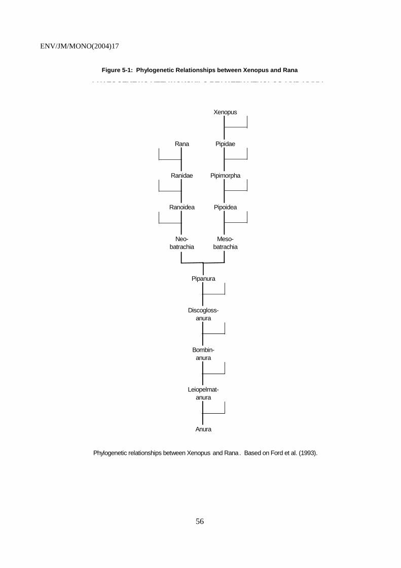
ENV/JM/MONO(2004)17
56
Figure 5-1: Phylogenetic Relationships between Xenopus and Rana
Xenopus
Rana Pipidae
Ranidae Pipimorpha
Ranoidea Pipoidea
Neo- Meso-batrachia batrachia
Pipanura
Discogloss-anura
Bombin-anura
Leiopelmat-anura
Anura
PHYLOGENETIC RELATIONSHIPS BETWEEN XENOPUS AND RANA
Phylogenetic relationships between Xenopus and Rana . Based on Ford et al. (1993).

ENV/JM/MONO(2004)17
57
Table 5-1: Comparison of Nutritional Constitution of Standardized Xenopus Diets
Nutritional Breakdown (%)
Diet Protein2 Fat Fiber Moisture Ash Soy3
Salmon Starter4 55.0 15.0 1.0 12.0 10.0 2.8-5.5
Frog Brittle5 44.2 6.0 2.0 NR 11.0 8.4-8.8
Tetra Fin6 42.0 8.0 2.0 6.5 NR NA
Trout Starter3 50.0 15.0 1.0 12.0 10.0 2.5-5.0
5.1.2 Ranids
5.1.2.1 Rana Pipiens.
101. The Northern Leopard Frog (R. pipiens) is native to a large portion of North America, including Canada (Canada’s Aquatic Environments, 2002; Northern Leopard Frog, Rana Pipiens, 2002). The only regions within the U.S. where R. pipiens is not located are in the south-eastern, south-central, and western portions of the country. R. pipiens is found in marshes, meadows, and ponds in the U.S. and Canada. Adult R. pipiens often migrate far from water; however, they must return to water to breed. Breeding season ranges from mid-March to the first part of April. Egg masses may be as large as 6,000. In the laboratory, metamorphosis is complete within 3-4 months post-fertilization, and sexual maturity is achieved in roughly 2 years (Duellman and Trueb, 1994).
NR = Not reported. NA = Not available or proprietary information. 1 Major source of protein = pesticide free fish meal. 2 Expressed as % of total protein. 3 Ziegler Brothers, Gardners, PA. Ingredients: Fish meal, dehulled soybean meal, corn gluten meal, fish oil, wheat flour, blood meal, brewers dried yeast, soy lecithin, yeast culture, vitamin A acetate, vitamin D3 supplement, dl-alpha tocopheryl acetate (vitamin E supplement), vitamin B12 supplement, riboflavin supplement, niacin, calcium pantothenate, menadione sodium bisulfite complex (source of vitamin K activity), folic acid, thiamine mononitrate, pyridoxine hydrochloride, biotin, choline chloride, manganese, proteinate, zinc proteinate, copper proteinate, calcium iodate, iron proteinate, cobalt proteinate, calcium carbonate, sodium selenite, L-ascorbyl-2-polyphosphate (source of vitamin C), ethoxyquin. 4 NASCO, Fort Atkinson, WI. Ingredients: Fish meal, meat meal, soybean meal, corn meal, wheat flour, dried yeast, distillers solubles, whey, wheat germ meal, salt, diacalcium phosphate. Vitamin supplements: Vitamin A (14,000 IU/Kg), vitamin D (5,000 IU/Kg), vitamin E (88 IU/Kg), vitamin B12 (0.04 mg/Kg), folic acid 1.5 mg/Kg), thiamine (4.0 mg/Kg), riboflavin (9.0 mg/Kg), pantothenote acid (11.0 mg/Kg), niacin (60.0 mg/Kg), choline (1,550 mg/Kg), calcium (2.0%), phosphorus (1.6%), sodium chloride (1.2%), magnesium (2.0%), linoleic acid, (2.8%), iron ( 300 mg/Kg), copper (10.0 mg/Kg), cobalt (2.2 mg/Kg), manganese (9.0 mg/Kg), zinc (100 mg/Kg), iodine (48.0 mg/Kg), pyridoxine (8.8 mg/Kg). 5 Tetra, Blackburg, VA. Ingredients: Fish meal, ground brown rice, dried yeast, shrimp meal, feeding oat meal, wheat gluten, soybean oil, fish oil, corn gluten, algae meal, sorbitol, potato protein, lecithin, ascorbic acid, inositol, niacin, 1-ascorbyl-2-polyphosphate (vitamin C), A-tocopherol-acetate (source of vitamin E), d-calcium pantothenate, riboflavin-5-phosphate, thiamin, mononitrate, menadione sodium bisulfite complex, folic acid, pyridoxine hydrochloride, vitamin A, cyanocobalamin, cholecalciferol, manganese sulfate, zinc sulfate, ferrous sulfate, cobalt sulfate, artificial colors, ethoxyquin and citric acid as preservatives.

ENV/JM/MONO(2004)17
58
102. Adult R. pipiens may be maintained in the laboratory under appropriate conditions (Ankley et al., 1998b). Unlike the pipidae, which are purely aquatic, a protected outdoor aquatic and terrestrial environment needs to be provided for longer-term cultures of ranid species (>4 weeks). Short-term adult cultures (<4 weeks) can be maintained in smaller exposure chambers. Small enclosures (ca. 1 or 2 m2) containing a small tub of water and moistened sphagnum moss can be used to house adult R. pipiens. Alternatively, a stainless steel culture rack in which the housing chambers slide in and out of the rack can be used. Since ranids feed primarily on living food, their diet consists of crickets, larval flies, or black worms. Food should be provided daily at an amount that is commensurate with consumption by the specimen. Fresh water should also be provided daily. Sexual dimorphism is not readily apparent in R. pipiens, thus other factors including gravidity should also be considered. The primary dimorphic characteristic is the length of the second toe of the forelimb.
103. Breeding R. pipiens currently requires artificial fertilization techniques, although methods for laboratory simulated natural breeding are under investigation (Ankley et al., 1998b). Artificial fertilization requires the injection of a large dose of female pituitary extract or fresh reconstituted pituitary into the dorsal lymph sac of the female a day or two prior to artificial fertilization. Some investigators have used LHRH to induce super-ovulation, but in general, the use of pituitary extract has been most successful. Males are sacrificed immediately prior to the artificial fertilization, and the testes are quickly removed, minced, and homogenized to produce a concentrated sperm solution. Eggs can be stripped from the properly prepared female, by gently squeezing the specimen along the dorsal flanks in an anterior-posterior direction. The stripped eggs are placed directly into a glass Petri dish and the concentrated sperm solution is poured over the eggs. Fertilization is monitored, and the developing embryos are separated into different test dishes. Newer techniques are currently being developed and evaluated which involve the use of simulated forced hibernation. These techniques involve the use of temperature-controlled incubators which drop the temperature over a set period of time to induce hibernation in the winter (over-wintering) for a predetermined amount of time (ca. 30 days), followed by a ramp of increasing temperatures to simulate the onset of Spring or breeding season. Although hormonal treatment including dopamine (prior to over-wintering) and LHRH (immediately prior to breeding) are required, this process is designed to induce amplexus. Thus, artificial fertilization is not required by this technique. More work will be required to fully demonstrate this process and standardize the methods.
104. R. pipiens embryos develop slower than their pipid counterparts. Developing embryos (unhatched) are protected by a thick jelly coat and do not hatch for 7-10 days. Embryos are cultured in spring water or dechlorinated tap water with the optimal pH near 8 at a temperature of 18-20ºC. Larvae are generally fed an algal diet, although the larval diets previously described for the other anuran species have also been used. Developing ranid larvae can be maintained in either static or flow-through systems although close attention to water quality is required.
5.1.2.3 Rana Temporaria
105. The European or Common frog, Rana temporaria is found throughout Great Britain, Europe and Northern Asia, ranging as far north as the Artic Circle in some areas of Scandinavia. Throughout its range it is found in damp habitat, commonly in cool upland forest and wet meadows, and is the most common frog in upland lakes. Breeding takes place in February and March in southern and lowland areas, but can be as late as June at high latitudes and altitudes. Eggs are laid in gelatinous masses, about 400 per female, and take between 30 and 40 days to hatch, though this may be considerably quicker in laboratory conditions at higher temperatures. Metamorphosis proceeds much as in Rana pipiens, taking 3-4 months to complete in the wild, though in laboratory conditions this can be reduced to 50-60 days. Sexual maturity is reached after about 3 years in the wild (Duellman and Trueb, 1994).

ENV/JM/MONO(2004)17
59
106. Procedures for rearing R. temporaria in the laboratory are likely to be much the same as those appropriate for R. pipiens: the same problems of live feeding and artificial fertilization for induced breeding apply. Techniques developed to induce amplexus (e.g. simulated hibernation) may therefore also be applicable to R. temporaria. Chronic larval development studies have been conducted with this species to investigate endocrine disruption, in both semi-field and flow-through conditions (Bogi et al. 2003; Pickford et al. 2002). While some alterations to standard protocols for Xenopus are needed (e.g. feeding regime, arrangements for emergence at metamorphic climax), these studies indicate that R. temporaria could be used as a test species in metamorphosis assays for further hazard characterisation with an ecologically relevant species.
5.1.2.4 Rana Rugosa
107. This dark-brown or grayish-brown Ranid is found in Korea, North-Eastern China, Southern parts of Siberia and throughout Japan (though probably introduced in Okinawa and Hokkaido), where it is known as the wrinkled or warty frog owing to the rough and warty skin on their backs. Adults reach 4-6 cm in length (snout-vent) and tadpoles reach a total length of 5-7cm. They usually spend much time near water, and inhabit rice fields, ditches, ponds, swamps and small streams. R. rugosa breed in May to August, females depositing small egg masses on suitable substrate such as rice plant, weed or stone. Tadpoles hatch in 4-5 days, and metamorphose in autumn. R. rugosa is quite amenable to laboratory study, as it can be induced to breed easily, laying in the region of 700-2000 eggs per spawning, and the window of larval sexual differentiation is well defined. Moreover, the genetics of this species are interesting in that there is geographical variation in sex chromosome systems, such that laboratory crosses of ZZ male frogs with XX females generates all-male (XZ) male offspring (Ohtani et al 2000). The XEMA amphibian metamorphosis assay has already been successfully adapted to R. rugosa.
5.1.3 Hyperoliids
108. Native to Kenya, Hyperolius spp., commonly referred to as reed frogs, are unique in undergoing ontogenic color change during juvenile maturation (Hayes, 1997b). Generally, ontogenic color change in maturing anurans and sexually-based dichromatitism is unusual in most anuran species. Male and female H. argus metamorphose with a bright green dorsum. However, females ultimately develop brownish dorsal coloration with white spots (Hayes, 1997b). Mature males are also distinguished by the development of gular pouches.
5.2 Urodeles
109. The urodeles (e.g., salamanders, newts, axolotls) are indigenous to many locations in North America and other locations across the globe. Breeding season for the spotted and tiger salamander (Ambystoma maculatum and A. tigrinum) is typically in January and February in the warmer regions and March and April in the colder regions (Carolina Biological Supply Company, 1993; Canada’s Aquatic Environments, 2002). A. maculatum and A. tigrinum egg masses rarely contain greater than 100 to 200 embryos. These eggs are large (ca. 2.5 to 3 mm) for amphibian eggs and are protected by a massive jelly coat. Hatching typically requires 3 to 4 weeks with metamorphosis occurring in about 3 months. Other than the resorption of external gills, morphological changes during metamorphosis in urodeles are reasonably minor. Following metamorphosis, Ambystoma initiates a terrestrial lifestyle and returns to the water 1 to 2 years later to breed. Newts utilize a similar life strategy, although reproductively mature newts that return to water from terrestrial life often remain aquatic for the remainder of their lives. Some urodeles, such as Necturus, are neotenic (obligatory) and retain their larval characteristics throughout adulthood.

ENV/JM/MONO(2004)17
60
110. Little standardized information is currently available on urodele husbandry, breeding, and culture. Studies using these species have been conducted using the techniques described for the anuran species (Caldwell, et al., 1980; Petranka and Sih, 1987). Adult terrestrial salamanders require cool temperatures (18 to 20< C) and high humidity, which may be provided in a terrestrial aquarium (Caldwell, et al., 1980; Petranka and Sih, 1987). A water source and daily misting of the natural substrates (leaves, rocks, moss, small tree branch) maintain high humidity. Salamanders are fed mealworms, crickets, whiteworms, earthworms, and some soft-bodied insects. Unlike frogs, salamanders consume water orally and require fresh drinking water. Embryos and larvae can be cultured in spring water, pond water, or dechlorinated tap water using partial renewal techniques. Larvae are fed the same food described for the anuran species. Although less information is available on the culture requirements for urodeles, it is necessary to evaluate this order of amphibians, since urodeles represent an important group of amphibians.
5.3 Species Selection Criteria
111. Considering that the objectives in selecting an amphibian species are to: 1) develop a short-term screening assay for thyroid disruption, and to 2) ultimately develop a longer term assay that includes advanced developmental and reproductive endpoints, the minimal attributes of a test species are described in the following eight criteria:
1. The species must be amenable to continuous culture in the laboratory;
2. Reproduction in the laboratory must be routine throughout the year, using either naturally occurring reproduction or through the use of some type of hormonal induction;
3. Larvae must be able to be routinely reared to predetermined developmental stages;
4. The developmental rate for the interval included in any test must be relatively fast so that the effects are observed quickly, thus minimizing test duration and test costs;
5. The endpoints which constitute the test data that will be used for regulatory or pre-regulatory action need to be supported by a sufficient knowledge base that indicates that they are relevant to the question at hand.
In addition to the minimal criteria noted above, it would be highly beneficial for the subject species to have additional information relevant to the following areas:
6. Genetic information, including gene sequences of thyroid axis-related genes and some knowledge of the genetic programs associated with TH dependent processes;
7. Biochemical information on the endocrine axis, particularly of the hypothalamus-pituitary-thyroid (HPT) axis; and
8. Metabolism information, especially as it relates to TH homeostasis.
5.4 Strengths and Weaknesses of Candidate Species
112. In summary, Xenopus sp. represents a significant advantage in the wealth of information available ranging from culturing to molecular biology, and their ease of use in the laboratory. Its ubiquity in use and acceptance in the scientific research community as a model for development makes it an excellent candidate for the amphibian metamorphosis assays. It should be noted, however, that substantially more information is available for X. laevis than for X. tropicalis. The primary advantages of using X. tropicalis lie primarily in the potential for developing transgenic lines (see section 4.1.1.2), the
ENV/JM/MONO(2004)17
61
rate of development, and the robust developmental kinetics of metamorphic change. However, it should be noted that from a practical standpoint, the smaller egg size of X. tropicalis imparts greater difficulty in creation of founder transgenic animals (F0) than in X. laevis. Rowe et al. (2002) suggest that X. tropicalis display extremely consistent kinetics associated with metamorphic change. On the contrary, X. laevis show greater developmental variability. Since the objective of the assay is to screen for chemical thyroid axis disruptors, the origin of the species is not a primary factor in selection. Next to Xenopus sp., there is some literature available on the native American Rana sp. metamorphic processes and culturing practices. However, the use of Rana in the laboratory (husbandry, breeding, and rearing tadpoles) is more difficult than for Xenopus. The most literature available on metamorphosis is for R. catesbeiana (bull frog). However, use of R. catesbeiana in the laboratory for metamorphosis studies is impractical due to the long length of development, thus warranting the consideration of R. pipiens.
113. The only potential advantage to the inclusion of the urodeles sp. to the list of potential candidate species for the amphibian metamorphosis assays is that they represent a different amphibian order (Caudata), and some differences in sensitivities may exist between Caudata and Anura. However, little literature is available on the use of urodeles, and they are generally more difficult to work with in the laboratory. A description of the general strengths and weaknesses of each species is provided in Table 5-2.

ENV/JM/MONO(2004)17
62
Table 5-2: Strengths and Weaknesses of Species Evaluated for Testing
Species Strengths Weaknesses
South African clawed frog (Xenopus laevis)
- Wealth of literature base on all aspects of development, reproduction, metamorphosis, including molecular models
- Ease of culture in lab - husbandry
- Many laboratories are familiar with culture and testing
- Breeds naturally and repeatedly with hormone stimulus
- High productivity from breeding and rapid development
- Good transgenic capacity
- Mapped genome
- Time to metamorphosis - ca. 60 d
- Relatively long life cycle with respect to sexual maturity
- distribution in sub-saharan Africa, limited relevance to temperate Northern Hemisphere environments
- Oligotetraploid genome
South African clawed frog (Xenopus tropicalis) [compared to X. laevis only]
- Relatively short life cycle
- Rapid development
- Diploid genome
- Good transgenic capacity
- Capacity to develop inbred lines
- Mapped genome
- Tight metamorphosis kinetics
- Time to metamorphosis - ca. 35-40 d
- distribution in sub-saharan Africa, limited relevance to temperate Northern Hemisphere environments
- More challenging animal husbandry
- Less information currently available, data base
- Disease susceptibility
- Availability
Northern Leopard Frog (Rana pipiens)
- Native species
- Reasonable database
- Relatively short metamorphic period for native species
- Terrestrial and aquatic life phase
- More difficult animal husbandry and breeding
- Limited testing experience
Hyperolius sp.
- External endpoints - Gender dichromatitism
- Straight-forward end points
- Suitable animal husbandry
- Connection to sexual development (gonadotrophs)
- Limited database
- African distribution
- Availability
- Does not directly measure thyroid dysfunction
Urodeles
- Represent different Order (non-frog amphibian)
- Native species
- Terrestrial and aquatic life phase
- Limited testing experience
- More difficult animal husbandry and breeding
- Limited database

ENV/JM/MONO(2004)17
63
EXPERIMENTAL DESIGN CONSIDERATIONS FOR AMPHIBIAN METAMORPHOSIS ASSAYS
6.1 Exposure Window
6.1.1 Developmental Stage
114. The process of metamorphosis can generally be divided into three phases: premetamorphosis, prometamorphosis, and climax, which are well defined in terms of developmental stage in Xenopus laevis. Premetamorphosis is the interval of development that proceeds from hatch to stage 54. This stage is characterized primarily by growth in the absence of a functional thyroid gland and consequently this development is considered TH independent. This is supported by the fact that inhibition of TH synthesis during premetamorphosis permits development up to stage 54, but not beyond. In fact, continuous exposure to TH synthesis inhibitors during premetamorphosis will maintain the organism at stage 54 indefinitely. At stage 54, prometamorphosis begins, which is characterized by the onset of thyroid gland function, rising levels of TH, and the process of TH-dependent morphogenesis. Prometamorphosis continues through approximately stage 60, and many tissues reach their respective metamorphic fate during this phase (e.g., limbs). At stage 60, metamorphic climax begins which is characterized by an abrupt rise in TH synthesis and the dramatic morphogenetic events, including tail resorption, which complete the transition from the larval to the juvenile phase at stage 66.
115. In the case of the amphibian metamorphosis assays, the aim is to exposure the test organism during developmental stages that will be most sensitive to disruption on the thyroid hormone axis. Given the ontogeny of development of the thyroid axis in Xenopus, possible exposure scenarios include 1) exposure from late premetamorphosis through metamorphic climax, 2) exposure from prometamorphosis through metamorphic climax, 3) exposure during metamorphic climax only and 4) exposure during prometamorphosis only.
116. The first scenario is most inclusive since it covers most of the developmental phase in which the thyroid is active. However, the length of an assay could easily be in the order of 35 d in X. laevis. Exposure from prometamorphosis to metamorphic climax provides a similar advantage in that it covers most of the significant events occurring during metamorphosis, but still requires approximately 28 d in X. laevis.
117. A metamorphic climax assay can effectively be performed over a 16 d period in X. laevis. The assay is relatively quick compared to the other exposure scenarios, however, it is only capable of evaluating the final processes of metamorphosis. Since many morphological metamorphic programs are already underway during metamorphic climax and endogenous TH is at its greatest level, exposure during metamorphic climax may not provide a sensitive or realistic estimation of the effect of a given test substance on the thyroid axis.
118. An assay that includes only the prometamorphic period could be performed in roughly 14 d in X. laevis. This approach would benefit from greater sensitivity to TH antagonists as endogenous TH levels are

ENV/JM/MONO(2004)17
64
much lower than during metamorphic climax, and developmental endpoints are therefore expected to exhibit greater sensitivity to TH disruption.
119. However, it can be argued that exposure only during prometamorphosis may limit the sensitivity of such and assay to weak agonist activities that would only be manifest in the absence of endogenous TH. The use of a pre/prometamorphic exposure protocol provides an interesting scenario in that it covers a transitional developmental period in which the thyroid is acquiring activity. Effects of inhibition that precede endogenous production of TH could be enhanced because there is no TH pool to promote TH-dependent development in the absence of nascent synthesis. Conversely, effects of stimulation could be enhanced since there is no TH at the initial stages of the study.
120. To address the issues of necessary length of exposure and optimal developmental period, Tietge et al. (US EPA Mid Continent Ecology lab, Duluth, MN, personal communication) have examined the effects of methimazole, perchlorate, and propylthiouracil on development of stage 51 and 54 organisms for periods of up to 14 d. Based on these studies, exposure of stage 51 or 54 larvae for 14 to 21 days were capable of detected developmental delay based on apical morphological endpoints. Further, histological markers of thyroid impairment were observed substantially earlier than the conclusion of the exposure period. Utilization of stage 51 specimens confirmed a slight increase in sensitivity. However, further examination of the differential sensitivity conferred by beginning exposure with stage 51 or 54 and the duration of exposure needs to be performed.
121. While some exposure during premetamorphosis may provide for increased sensitivity to weak thyroid agonists (i.e. acting in the absence of endogenous signal), it is questionable how much benefit would result from extended exposure prior to late premetamorphosis. Moreover, including a significant component of premetamorphosis potentially renders the test sensitive to developmental toxicants affecting thyroid gland development through non-thyroidal mechanisms. This could reduce the specificity of the test.
6.1.2 Duration
122. In a general sense, an increase in exposure time often results in increased sensitivity of an organism to a given toxicant. However, the maintenance of longer-term exposures is costly and can result in unexpected interruptions in exposure as a result of test-substance behaviour in water or equipment malfunction. Chemical analysis of the exposure solutions and cleaning the exposure system to maintain high dissolved oxygen concentrations, especially in the presence of organic solvents used as carriers, add significantly to the time and effort in maintaining a long-term exposure. Therefore, the exposure duration of a study needs to encompass an appropriate time of exposure necessary to elicit an effect, but not beyond this necessary time frame, thereby controlling costs and potential exposure interruptions. In the current context, the only rationale for extending the exposure period to earlier stages of development than are expected to be sensitive to the MOA of interest would be when the physicochemical nature of the test substance is such that it has extremely low water solubility, and increased exposure time is required to reach effective tissue concentrations during prometamorphosis.
123. In terms of a test that encompasses prometamophic stages of development, reduction of the test duration could result in reduced sensitivity to some MOA’s. For example, evidence suggests that monodeiodinase inhibitors (e.g. IOP), which can disrupt TH homeostasis, only manifest their effects in the context of significant levels of TH (Galton 1989). For a metamorphosis assay to detect agents exhibiting this MOA, it may therefore be necessary to include late-prometamorphic stages (i.e. up to stages 58-60), when endogenous TH levels are sufficiently high for effects on monodeiodinase to be manifest (e.g. on hind-limb development. Similarly, terminating a prometamorphic assay too early may have implications with regard to sensitivity of other biochemical endpoints (e.g. TH levels, TRβ). These issues need to be
ENV/JM/MONO(2004)17
65
addressed in prevalidation studies by appropriate comparisons of exposure windows with appropriate test chemicals and relevant endpoints.
6.2 Route of Administration
6.2.1 Water
124. Water exposure is the most common route to expose larval and metamorphic amphibians to EDCs. The delivery of a toxicant in water at different concentrations is reasonably well established for most aquatic test species, although considerations for static-renewal and flow-through exposures must be made. Although many amphibians, including those previously discussed in this review paper, prefer a static environment (Dawson et al., 1992), the difficulties associated with longer-term static renewal exposure studies necessitate the use of a flow-through design (ASTM, 1998; Ankley et al., 1998b). The primary drive of flow through methods is efficacious exposure. That is, exposure to chemicals which are prone to hydrolysis, photolysis, bio-degradation, volatility, and those with relatively high log Kow must be conducted using flow through methods. In addition, benchtop stability studies should be conducted prior to conducting a study, in the absence of the above information. Problems associated with a static-renewal exposure system are cost of maintaining a long-term static renewal exposure study, maintaining a consistent aqueous concentration during the study, and stress caused to test organisms through the physical disruption of water replacement. Flow-through exposure systems using a variety of mechanical approaches have been successfully used by many laboratories (Greenhouse, 1976; Pickford et al., 2003), and while due consideration must be given to the biological needs of the test organism, there is currently no evidence to suggest that flow-through conditions are deleterious to developing Xenopus larvae.
125. Exposure concentrations should be measured no less than weekly during the exposure under flow-through conditions. Under static-renewal conditions analysis should rather be performed every 48 h. However, definite specifications of exposure method and frequency of the test substance analysis will be dependent on the stability of the substance in the test system. Test substances with rapid degradation rates will require flow-through exposure with more frequent analysis of test substance concentration. Since test substance concentrations tend to decrease over time in mature culture environments, careful attention to maintain a consistent concentration will also be required. The OECD Guidance Document on Aquatic Toxicity Testing of Difficult Substances and Mixtures (OECD, 2000) provides a more detailed overview of necessary considerations and possible measures for adequate handling of difficult substances.
126. The greatest challenge in an aqueous exposure system is solubilizing the test substance in water. Organic solvents are often used as co-solvents to assist in delivering a test substance into water, which requires the maintenance of a solvent control. Organic solvents also enhance bacterial growth in the test system, which increases maintenance time during the exposure, and which may impact dissolved oxygen concentration. Organic solvents may also act interactively or non-interactively with the toxicant to increase (additivity, synergism or potentiation) or decrease (antagonism) its effect. In some cases, saturator columns have been used to eliminate the use of solvents.
6.2.2 Oral (Food)
127. Patyna et al. (1999) recommends that hydrophobic compounds with log P values >5 should be administered via food. Although dosing adult amphibians via food has been accomplished, little work has been performed using this route of administration in tadpoles (Fort et al., 2001a). Given that Xenopus larvae are filter-feeders, developing methods for measuring food intake will be an area for future research if the use of dietary exposure is to be adequately controlled. Dosing the commercial salmon diet is practicable, although homogenization of the food after spiking the test substance is challenging and obtaining a homogeneous mixture is often quite difficult. Dosing live food items, such as worms, has been

ENV/JM/MONO(2004)17
66
performed, but it is also quite difficult to obtain a consistent diet and differentiate between effects from the diet and effects from toxicants that leach from the diet into the culture water (Fort et al., 2001a). In a large-scale screening program like EDSP, oral dosing is probably not as practicable as aqueous exposure, unless required due to limitation in aqueous solubility.
6.2.3 Parenteral
128. Intramuscular or intravenous administration of a toxicant in a premetamorphic tadpole (stage 47-48) is possible. However, the technique is technically difficult and the environmental relevance is questionable since it is difficult to determine or estimate effective environmental concentrations. Thus, as with oral dosing, parenteral routes of administration are not practicable for high volume screening programs.
6.3 Test Chemical Selection
129. A number of chemicals with agonist/antagonist activity against various targets on the hypothalamic—pituitary-thyroid axis have been tested in preliminary and pre-validation studies by various groups. The results of these tests are summarized in table 6.1. Candidate chemicals for use as control substances in further development, validation, and qualtity control of an Amphibian metamorphosis assay are indicated.

ENV/JM/MONO(2004)17
67
Table 6-1: Test Chemicals used in Various Preliminary/Prevalidation Amphibian Metamorphosis Assays.
Chemicals Germany Japan United States PTU + + + ETU + + Zineb - Amitrole + T4 + + + T3 + IOP + + Methimazole + Perchlorate + Estradiol - Β-trenbolone -
TBBA -
Shaded cells indicate the chemical as been tested by the respective member country, +/- indicates activity/lack of activity, respectively.
indicates chemicals currently recommended for use as positive/negative controls (1st meeting of Expert Group on Amphibian Testing, OECD, Duluth 2003).
6.4 Dose Selection
130. Dose levels for amphibian metamorphosis assays should be selected with the use of range-finding data, unless other reliable data are readily available. Selected concentrations should be less than lethal levels and less than the maximum water soluble concentration.
131. For screening purposes, at least three widely spaced concentration levels would be appropriate depending on the assay endpoints considered. The treatment levels can be separated by up to an order of magnitude. A definitive amphibian metamorphosis assay study should be conducted with at least five treatment levels, and the treatment levels should be separated by approximately a factor of two. At least one of the treatment levels should be below the no-observed adverse effect concentration (NOAEC). Concentrations should be selected to produce an adequate concentration-response curve for the endpoints measured during the study.
6.5 Statistical Considerations
132. The objective of the amphibian metamorphosis assays is to provide the most sensitive and specific screen of toxicity associated with thyroid disruption for potential EDCs. Thus, the assay must be biologically sensitive, have minimal variability associated with exposure concentration throughout the duration of the test, and be statistically powerful. Biological sensitivity is a function of the choice of species tested, the relevance of the endpoints measured to species survival, and the route and duration of the chemical exposure. Design-associated variability in dose exposure is a function of exposure route and duration, chemical stability and purity within the testing environment, and the testing protocol. The power of a statistical inference is a function of the inherent variability in response; design-associated variability; the degrees of freedom and the source of variability for testing; and the estimation process and decision criteria. Other areas in this section have discussed biological sensitivity; this subsection will focus on design-associated variability and statistical power.
133. Ideally, an experimental design incorporates randomness, independence, and replication (Cochran and Cox, 1957). Randomness is used to remove noise, independence is used to extend the inferences made, and replication provides a measure of variability for testing (Chapman et al., 1996).

ENV/JM/MONO(2004)17
68
Randomization of: 1) experimental containers within a testing environment, 2) treatment application to experimental containers, and 3) application of organisms to experimental containers allows one to incorporate the variability associated with the environmental conditions, the containers, and the organisms equally across all treatments. Thus, when the difference between treatments, meaning the variability associated with experimental environment, experimental containers, and organisms being treated, are removed, only the effect of the treatment remains.
134. Independence of treatment application, including the creation of the treatment, incorporates the variability associated with someone else, somewhere else making and applying the same treatment. Thus, the inferences associated with the treatment response are extended to someone else repeating the experiment. The random sample of organisms from a given population actually limits the inference to that population. However, one can evaluate the stability of the inherent variability of the population over time. An experimental unit is defined as the group of material to which a treatment is applied independently in a single trial of the experiment (Cochran and Cox, 1957). Replication of experimental units for each treatment provides a measure of all the necessary sources of variability needed to extend the inference across time and space. A reduction in the sources of variability that are truly independent constrains the inference (Hurlbert, 1984). Thus, if only one solution of each treatment is made and then divided between replicates, the source of variation associated with making the treatment is not included in the variability for testing, and the inference is limited. It can be argued that this variability is nuisance noise, too small to be of concern, and costly to include. Therefore, if this source of variability is not included, it should at least be acknowledged. The variability between replicate experimental units may also include noise that was not randomized out due to a poor randomization or variable measurement error. These sources of variability can be reduced without loss to inference.
135. Statistical power is the probability of rejecting the null hypothesis (of equal means) when the alternative is true (i.e., detecting a difference when there is a difference). Statistical power is a function of the variability between replicate experimental units (i.e., within a treatment), the number of replicate experimental units, the size of the type I error, and the percentage of difference one wishes to detect. The latter three components can be controlled; however, the variability in response is inherent in the organism being tested. Thus, the choice of which species should be tested and the relevant endpoints measured should include a comparison of inherent variability or CVs (coefficient of variation=standard deviation/mean x 100%). In terms of power, high CVs have low power for detecting small-scale differences. Power can be increased by increasing the number of replicates. The choice of the test species and endpoints with the least inherent variability, by default, requires the least replication for a given level of power and, thus, are more cost effective.
6.5.1 Sample Size: Ensuring Adequate Test Specimens
136. Typical practical considerations for sample size are based on the number of endpoints to be collected and whether the specimen must be sacrificed to collect the data. With long-term dosing protocols, a dose response is expected (i.e., over some specified range of doses there will be varying intensity of endpoint response that is significantly different from un-dosed or control specimen). Additionally, it is assumed that at some dose there will be no difference between the dosed and un-dosed specimen. In order to statistically determine the appropriate sample size, the inherent variability of the endpoint must be measured, according to the desired statistical resolution, and the power of predictability determined. No current guidelines are available for longer-term exposure assays with amphibians. However, the length of the prospective assays and the natural variability associated with metamorphosis will require larger sample sizes than used for short-term partial life-cycle fish tests. As a useful guide, 100 fish larvae per replicate has been a standard sample size for starting a long-term exposure for regulatory purposes. Due to density issues when raising amphibians, a greater number of replicates with less specimens will be required (i.e., 5 replicates of 20 specimen per concentration). However, before sample

ENV/JM/MONO(2004)17
69
size and replicate requirements can be determined for amphibian metamorphosis assays, formal statistical power analysis will be required.
6.5.2 Endpoints
137. The NOAEC has been used to evaluate data from longer-term amphibian exposure studies, typically when the differences between exposure levels are no greater than a factor of approximately 3. An Amphibian Metamorphosis Assay battery will generate multiple NOAEC values if multiple endpoints are selected.
138. Even though the NOAEC is widely used, it should not be relied on as the sole indicator of low toxicity. The largest dose for which statistical differences have failed to be detected is a direct function of the power of the test. It is also conceivable that short-term range finding experiments will have difficulty in predicting the location of an NOAEC. Using ECx calculations are an appropriate alternate for estimating doses associated with low toxicity. However, care must be taken not to estimate an ECx value that is more sensitive than the data and experimental design will allow. Ultimately, the data may simply indicate impairment of thyroid axis function.
ENV/JM/MONO(2004)17
70
CANDIDATE PROTOCOLS
139. The following protocols have been used in relevant previous studies on amphibian metamorphosis. These studies explored several options for amphibian metamorphosis assays and served to inform the process of identifying actual candidate protocols outlined in section 8.
7.1 16-day Metamorphic Climax Assay
140. The 16-day Metamorphic Climax Assay is conducted during the final period of metamorphosis, which is most prominently marked by the resorption of the tail and the development of the forelimb. As originally proposed by Fort et al. (2000), this assay primarily quantitatively evaluated the rate of tail resorption in X. laevis, although maturation of the skin and forelimb development were noted anecdotally. Due to higher than acceptable levels of variability in the rate of tail resorption, the assay has been modified somewhat to increase robustness. However, this variability is primarily due to the over-whelming influence of the endogenous TH peak that occurs during this developmental window. Thus, it is anticipated that this stage would be relatively insensitive to TR agonists since the system is fully stimulated by the TH cascade. Conversely, this stage may also be insensitive to TR antagonists as the endogenous TH surge initiates a cascade of gene activity that is insensitive to most synthesis inhibitors (Brown et al., 1996).
141. In terms of the original assay design, larvae are cultured in FETAX Solution (ASTM, 1998), a reconstituted water medium suitable for the culture of X. laevis embryos and larvae. FETAX Solution consists of 625 mg NaCl, 96 mg NaHCO3, 75 mg MgSO4, 60 mg CaSO4 H2O, 30 mg KCl, and 15 mg CaCl2 per L of solution. Larvae are fed the supernatant of groundSalmon Starter diet (Silver Cup tadpole starter, Xenopus 1®, Dexter, MI) slurry prepared by blending ca. 6 g diet/L FETAX Solution. Larvae were fed ca. 2 mL of the slurry/organism, twice per day, starting after day 4 through metamorphosis. FETAX Solution or one-half strength FETAX Solution may be used for the culture of X. tropicalis. Twenty stage 58 larvae are placed in each of four 10-L vessels containing varying constant concentrations of the toxicants. Stock solutions were prepared in FETAX Solution. Dilutions are also prepared in FETAX Solution. Five test concentrations are tested in quadruplicate, using a flow-through delivery/diluter system. A solid phase concentrator may be used when necessary to maximize the water solubility of highly hydrophobic test materials. Four separate vessels containing 20 larvae each are exposed to FETAX solution alone. Treatment and control dishes contain a total of 8 L of solution. The pH of the test solutions is maintained between 7.0 to 8.0. Generally, culture temperature for X. laevis larvae is maintained between 21-23° C with a 12 h light : 12 h dark photoperiod. The light intensity should be maintained between 61-139 lumens using fluorescence lights. Fort et al. (2000) used a culture temperature of 21±0.5ºC.
142. If a static renewal design is used, renewal must be performed daily unless degradation of the test substance is minimal over a long period of time. Waste, excess food material, and dead embryos and larvae are removed daily regardless of the exposure design and numbers recorded. Generally, the use of a flow-through exposure design is preferred. Staging is performed during the renewal process (Nieuwkoop and Faber, 1994). Tests are terminated once the larvae reach stage 66 for the evaluation of tail resorption. At the completion of the exposure, larvae are fixed in 3% (w/v) formalin, pH = 7.0, and the gross effects on limb development and skin maturation noted. Limb defect assessment is aided by the use of a dissecting microscope.
ENV/JM/MONO(2004)17
71
143. Digital images are captured using a high-resolution color digital video camera. A computer with image processing software is used to digitize the tail length at developmental stages 58 to 66. A ruler videotaped with the larvae is used to monitor image distortion and calibrate the length-measuring program to ensure accurate measurements of the larvae. Tail lengths are measured using digitizing software. The mean tail lengths at day 14 for each concentration of each test material evaluated are corrected for the starting tail length at d 0. Statistical comparisons of the control and exposure treatments and determination of NOAEC values are performed using ANOVA. Isotonic regression of monotonic data is performed to determine median inhibitory (IC50) or median stimulatory (SC50) data.
144. As originally described, the primary drawback to this approach, aside from questionable sensitivity, is that it relied merely on gross morphological endpoints. As previously discussed, in order to distinguish between thyroidal- and non-thyroidal-based changes in developmental rates, other biochemical and molecular endpoints need to be incorporated into the test protocol that can establish mechanistic links to the observed morphological effects. Based on this information, a modified morphological test based on the original metamorphic climax assay that incorporates a more sensitive stage of the metamorphic period, such as early prometamorphosis, that is capable of utilizing biochemical and molecular endpoints will be more appropriate.
7.2 Xenopus Metamorphosis Assay (XEMA)
145. This protocol has been developed by Kloas et al., IGB, Berlin, Germany. The test is a 28 day morphological assay, with exposure of Xenopus laevis larvae in static renewal system and assessment of length (total and tail) and developmental stage at days 7, 14, 21 and 28. Test design typically incorporates 5 test chemical concentrations, a ‘negative’ control (75 mg/l PTU), and a positive control (1 µg/l T4). The test is initiated with larvae at stage 48-50, which in controls typically proceed to late prometamorphic/early climax (stages 58-60).
146. The test is run at 22°C, 12:12 light:dark photoperiod, in a reconstituted water medium ( 2.5 g of the commercial salt “Tropic Marine Export” are dissolved in 10 L distilled water), which is aerated with airstones. Spawning is induced 14 days prior to the beginning of the test, and eggs and developing tadpoles are kept in 40 L tanks until stage 48/50, which time the tadpoles are transferred into 10 L-glass aquaria for pre-test acclimation. Thirty (30) tadpoles are placed in each aquarium, with two (test concentrations) or three replicate test vessels per treatment group. During the test, tadpoles are fed daily (including weekends) a commercial fry food (SERA Micron), 200 mg/d/aquarium from day 0 – 5 and 300 mg/d/aquarium from day 6 – 28. Treatment solutions are changed out completely and renewed with fresh test medium three times a week (Monday, Wednesday, Friday). New stock solutions of all test substances are freshly prepared each week. Developmental stage, whole body length and tail length are determined for each tadpole at the beginning of the exposure (day 0) and again at day 7, 14, 21 and 28, during the changeover of test solution. Developmental stages are determined according to Nieuwkoop and Faber (1956) by using a dissection microscope. It is not necessary to anesthetize the tadpoles for this procedure.
147. As such, the XEMA represents a ‘full prometamorphic’ assay that also incorporates a significant portion of the premetamorphic developmental period. The inclusion of premetamorphic development may lend this assay sensitivitiy to weak agonists, as it incorporates a significant window in which endogenous TH levels are low, but larvae may respond to exogenous thyroid active chemicals through the TH receptor system. This responsiveness is confirmed by results from an international pre-validation exercise: T4 reliably stimulated a small but significant acceleration of development.

ENV/JM/MONO(2004)17
72
148. Additional endpoints that could be used in conjunction with this assay include hindlimb differentiation, thyroid histology and RT-PCR analysis of TSH β-subunit and TR β genes (Opitz et al., 2002).
7.3 Prometamorphosis Assay in Xenopus
149. Work by Fort and Bantle (Fort Environmental Laboratories, OK, US / Oklohoma State University, OK, US, unpublished data) has indicated that developmental stages prior to those incorporated in the tail resporption assays, around stages 52 to 54, were capable of responding to thyroid agonists and antagonists. Although tail resorption was used as a morphological marker of metamorphosis in this study, it is not unreasonable that hind limb development would follow the same pattern, only with increased sensitivity. Investigators at the US EPA Mid Continent Ecology lab, Duluth, MN, compared 8 and 14 day exposures to perchlorate, starting with either stage 51 (late premetamorphosis) or stage 54 (early prometamorphosis). They concluded that the test was more sensitive overall (morphology and thyroid histology) with 14 day exposure, and that inclusion of late premetamorphosis did not improve the assay. A late premetamorphosis (stage 51) to early prometamorphosis (stage 54) exposure scenario which also is completed in ca. 14 d could also be considered. Culturing and measurement methodology cited in Section 7.1 would apply to this assay design. Proposals for a short-term prometamorphic assay and Xenopus Gene expression assay, description of a Xenopus metamorphosis assay (XEMA) for assessment of biological effects caused by thyroid disruption in X. laevis tadpoles, and additional references, provided by technical experts from Germany, are presented in appendices C-D, respectively.

ENV/JM/MONO(2004)17
73
RECOMMENDED PROTOCOL AND ADDITIONAL DATA NEEDS
8.1 Rationale
150. Further optimization of an amphibian metamorphosis assay must be focused on development of a mechanistically informative test method that can adequately test the hypothesis “exposure to a test chemical causes changes in the homeostasis and action of thyroid hormone on the thyroid axis” in a relatively rapid, economical screening format.
151. Exposure window needs to be optimised to provide adequate sensitivity to agonist and antagonist activity, in the minimum test duration possible. Morphological endpoints will be necessary to demonstrate biological relevance at the level of the whole organism, and may be further refined in prevalidation studies. Biochemical and molecular endpoints have been identified which have the potential to render this test more mechanistically informative.
8.2 Candidate Test Species
152. Principal candidates for test species for the amphibian metamorphosis assay are X. laevis and X. tropicalis. While these species are not native to many OECD countries, and may have limited ecological relevance to temperate northern hemisphere environments, these species are highly suited to laboratory testing and at the current time represent the most pragmatic choice for a test that is aimed at detecting thyroid activity per se. Higher tier hazard characterisation of active chemicals may well require use of other, more ecologically relvant species.
153. Advantages and disadvantage of X. laevis and X. tropicalis are presented in table 8.1. It is expected that prevalidation work will continue with X. laevis. However, as X. tropicalis husbandry methods are optimized, genomic resources for X. tropicalis increase and mechanistically informative molecular endpoints are developed, it is possible that X. tropicalis will supercede X. laevis in this context.

ENV/JM/MONO(2004)17
74
Table 8-1: Advantages and Disadvantages of the Two Pipid Species
Aspect considered X. laevis X. tropicalis (Silurana)
Life cycle, development
Relatively long life cycle with respect to sexual maturity (approx 1.5 yrs to sex maturity)
Short life cycle, approximately 4 months to sex maturity
Distribution in sub-saharan Africa, limited relevance to temperate Northern Hemisphere environments
Larval development Approx. 60 days to metamorphosis Possibly shorter (35-40 days)
Developmental kinetics maybe more precise (less asynchrony)
Laboratory culture and husbandry
Easy, much experience in scientific community
Reproduction induced by administration of gonadotrophic hormones
Some aspects of husbandry more challending e.g. early larval development
Potentially greater disease susceptibility
Available data on development, reproduction, metamorphosis
Substantial Less information currently available
Genome and transgenic potential
Oligotetraploid genome - mapped
Not suitable for knockouts
Good transgenic capability
Longer to establish stable lines
Diploid genome - mapped
Suitable for knockouts
Good transgenic potential (potentially more demanding technically)
Less time to develop stable lines
Availability Commercially available Less well established – stocks will need to be developed
8.3 Protocol Recommendations
8.3.1 Exposure Window
154. Given the arguments presented in previous sections and data from several groups working in this area, an optimum exposure window will encompass prometamorphic development, and possible a portion of late premetamorphic development. A prometamorphosis assay would therefore start with larvae at stage 54 and expose for 14 days, with control larvae reaching approximately stage 58-59 (i.e. start of metamorphic climax). Inclusion of premetamorphic stages would require initiation of the test with larvae at stage 51-52, and 21 day exposure, after which larvae would again be reaching stages 58-59. It is possible that inclusion of some premetamorphic stages would render the test more sensitive to weak agonist activity.
155. However, it should be born in mind that the longer such a test runs, and the earlier in development exposure is initiated, the greater the variability in developmental stage will be by the end. Thus, a longer exposure window to encompass premetamorphic stages may have impacts on statistical power and experimental design ramifications. These considerations may to some extend be offset, if more precise developmental kinetics in X. tropicalis are confirmed.
156. In summary, the recommended exposure protocol will involve a ca. 21-/14-day pre-/prometamorphosis assay with X. laevis/tropicalis initiated at either stage 51 or 54 and concluded at stage

ENV/JM/MONO(2004)17
75
58-59. Tests should ideally be performed in flow-through exposure systems, with regular verification of test concentrations. Use of static-renewal systems may be acceptable on a case-case basis, dependent the physicochemical properties of the test substance and adequate analytical chemistry support. On at least days 8 and 14 (and possibly 21), specimens should be digitally photographed to document morphological development and stage. Hind limb development can be measured using a scanning digitizer. Specimens should be randomly selected for histological examination of the thyroid and samples should be collected for analysis of mechanistically informative biochemical and molecular endpoints. See Figure 8-1 for schematic of the approach for prevalidation optimization of amphibian metamorphosis assay, and Appendix E for proposal for validation phase 1 of the amphibian metamorphosis assay.
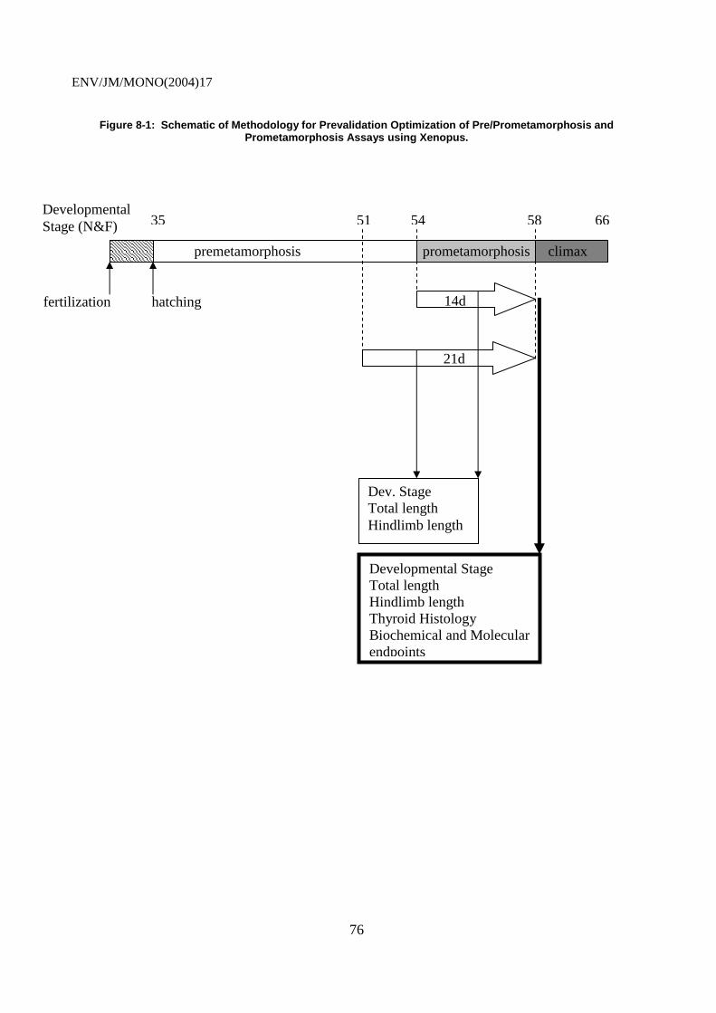
ENV/JM/MONO(2004)17
76
Figure 8-1: Schematic of Methodology for Prevalidation Optimization of Pre/Prometamorphosis and Prometamorphosis Assays using Xenopus.
fertilization
35
hatching
58 66 54 51
21d
14d
climax prometamorphosis premetamorphosis
Developmental Stage (N&F)
Dev. Stage Total length Hindlimb length
Developmental Stage Total length Hindlimb length Thyroid Histology Biochemical and Molecular endpoints

ENV/JM/MONO(2004)17
77
8.4 Endpoints
157. Endpoints which confer diagnostic specificity are needed in order to accurately determine whether or not the thyroid pathway is the target of a test chemical. This is needed, not only to properly characterize the chemical for potential regulatory action, but to provide a basis for interspecies and interchemical extrapolation. The molecular and biochemical endpoints suggested in this document require additional research and development, conducted as part of prevalidation exercises, in order to assess their relative sensitivity and reliability and relevance. Their successful implementation will reduce the time and cost associated with conducting an assay, and will improve the quality and utility of the data.
158. Endpoints assessed during prevalidation studies should include core endpoints (e.g. developmental stage, total length, thyroid histopathology, TRb and TSH mRNA expression levels). These will be augmented by optional endpoints (e.g. hindlimb length, thyroid hormone concentration, pituitary histopathology, mRNA expression of other TH-dependent genes) as research indicates they are sensitive and mechanistically informative. Thus, a battery of endpoints will be developed, and further validation exercises will provide information necessary for selection of a core set of endpoints for any subsequent guideline. Table 8-2 lists core and optional endpoints for morphology, biochemistry and molecular biology, and indicates which MOA’s these endpoints are likely to be sensitive for.

ENV/JM/MONO(2004)17
78
Table 8-2: Thyroid Related Modes of Action and Possible ndpoints.
MOA
Possible endpoints
Synthesis - I- uptake - TPO inhibition
Transport - TTR displacement
Elimination - deiodination - conjugation (UDPGT)
Neuroendocrine HP axis
TR - agonism - antagonism
Morphology: • Dev. Stage • Hind limb
+1 +2
?1 ? 2
+?1
+1
+1
Histology: • Thyroid • Pituitary
++1 +2
? 1 ? 2
+?1
++1 +2
+1
Biochemistry : • T3/T4 • Enzymes
(D2/D3)
+2
+2
++2 +?
+?
Mol. Biology: • TRβ mRNA • TSHβ
mRNA • CRF • UDPGT
+1 +1
+
? 1 ? 1 ? ?
+1 ? 1
+
+1 +1
+1
+ endpoints able to measure effects on the associated thyroid related mode of action; 1 core endpoints of the frog metamorphosis assay proposed by the Amphibian Expert Group; 2 optional endpoints of the frog metamorphosis assay proposed by the Amphibian Expert Group.
8.5 Test Chemicals
159. A number of test chemicals have been assessed in various approaches to an amphibian metamorphosis assay. Comparison of effects observed at the 1st OECD Ad Hoc Expert meeting on Amphibian testing is summarized in table 6-1. PTU and T4 are suggested as preferred agonist and antagonist reference chemicals for further prevalidation studies.

ENV/JM/MONO(2004)17
79
8.6 Data Gaps
160. The primary objective in identifying data gaps is to prioritize and apply resources to areas of uncertainty so as to reduce this uncertainty through research. As the OECD program for implementation of tests for detection of ED activity progresses, several critical questions must be addressed.
161. The effects of thyroid agonists and antagonists on apical morphological changes during anuran metamorphosis are reasonably well understood. However, the relationship between changes in biochemical and molecular biomarkers of TH-axis disruption and effects at the level of the whole organism is not as obvious. When does a molecular change constitute a valid indication of thyroid perturbation? To understand with confidence at what point molecular changes are an indicator of thyroid disruption, the results must be shown to be related to an upstream or downstream response within the thyroid axis. If molecular changes, such as inhibition of TR beta mRNA synthesis, can be linked to a histological, biochemical, or possibly even an apical morphological change in metamorphic processes, this relationship can be addressed. Work will be required to determine the threshold of molecular change that results in a physiological change in thyroid status. However, once this relationship and set-point are determined, the molecular assays, like gene arrays and RT-PCR techniques, will be extremely valuable. It will be critical to evaluate the sensitivity and specifity of these biochemical, and in particular molecular endpoints in the context of an assay that assessed apical effects (i.e. morphology).
162. A variety of molecular and biochemical endpoints, including transgenic approaches and genomic/microarray technology, have been identified which may be useful in elucidating MOA of chemicals exhibiting activity in this type of assay. A considerable amount of further research will be required to establish which of these endpoints are most useful and most sensitive. Use of more sensitive endpoints may enable reduction of exposure duration of the test, but this must be weighed against the potential for false negatives, though exclusion of certain critical/sensitive windows (e.g. premetamorphic stages for weak agonists, late prometamorphosis for effects on target tissue TH metabolism).
163. Furthermore, while currently favoured endpoints and protocols may be highly informative for disruption central TH homeostasis (i.e. effects on central hypothalamic-pituitary-thyroid control axis), these approaches may be less suitable for detecting peripheral effects on local TH metabolism in target tissues. While there is currently a paucity of information on environmental chemicals exhibiting this type of activity, addressing the potential and need for these types of assays (see appendix C for German proposals) is a critical further research need.
164. Histopathology of the thyroid gland, already shows considerable potential as a sensitive measure of TH-synthesis inhibition. Further work is required to establish the most reliable and sensitive endpoints for measuring thyroid pathology (e.g. volume, epithelial cell height), and to agree on standardized scoring systems.
165. Finally, while there is much to commend the use of an amphibian metamorphosis assay to test for thyroid activity, to avoid redundancy in a testing program, future prevalidation work should be performed in consultation with other expert groups working on assays that may offer utility in this regard. Test compound choice must be conducted with reference to chemicals used in bird, fish and mammalian assay development that may exhibit TH activity (e.g. 20-day pubertal rat assay). Only in this way can the relative sensitivity of different assays to TH-active chemicals be established, and ultimately such comparisons are necessary to establish the competence of an amphibian metamorphosis assay to act as a surrogate for other vertebrates, including mammals, in testing for TH activity.
ENV/JM/MONO(2004)17
80
IMPLEMENTATION CONSIDERATIONS
9.1 Animal Welfare Considerations
166. Although in vitro and molecular test methods of evaluating specific aspects of thyroid axis disruption, including reporter gene assays, TR and transport protein binding assays, gene arrays, and other gene expression techniques are emerging and appear promising, an in vivo based metamorphosis assay is considered to be the most sensitive and practicable approach for use in a screening battery at the present time. Additional discussion of the attributes and short-comings of current in vitro and molecular approaches in relation to the in vivo methods is provided in the WA 4-7 DRP on thyroid hormone disruption.
167. In accordance with current animal welfare guidelines, the test protocols developed and research conducted throughout prevalidation phases will comply with requirements stipulated in the Animal Welfare Act and documented in the “Guide for the Care and Use of Laboratory Animals,” prepared by the Committee on Care and Use of Laboratory Animals of the Institute of Laboratory Resources, National Research Council (NIH, 1985). Under this animal welfare program, consideration should be given to the reduction, replacement, or refinement in the use of animals in research. Use of amphibians in research does reduce the use of higher vertebrate animals, including mammals. Care is required to ensure that the number of organisms used and pain and distress induced in research animals are minimized. Laboratories participating in prevalidation and validation studies should have an “Assurance of Compliance with Public Health Service (PHS) Policy on Humane Care and Use of Laboratory Animals” on file with PHS and EPA. This process requires the establishment of an active Institutional Animal Care and Use Committee (IACUC) and the use of Animal Care and Use Protocols (ACUPs) for the studies performed.
9.2 Recommended Equipment/Capabilities
9.2.1 Laboratory Capabilities
168. Laboratories generally qualified to perform research and conduct testing associated with the prevalidation and validation of the amphibian metamorphosis assays should have staff toxicologists, chemists, biologists, and biochemists that specialize in freshwater ecotoxicology, trace-contaminant analysis/metals chemistry, organic chemistry, environmental forensics, ecological risk assessment support, bioassay method development, endocrine activity assessment, and ecotoxicological research in amphibians.
9.2.1.1 Amphibian Laboratory
169. General aquatic toxicology laboratory facilities are required to conduct amphibian bioassays. Bioassay laboratories should provide adequate space for studies requiring flowing and static freshwater. Aquatic animal isolation facilities should be available to conduct moderate and high hazard work, with space for a variety of tank configurations for physiological, biochemical, and molecular studies on amphibians. Large tubs can be used to maintain pathogen-free, genetically characterized aquatic animals for studies of this nature.

ENV/JM/MONO(2004)17
81
170. A freshwater distribution system should be available to supply purified freshwater to the laboratories. In addition to freshwater, high-quality, ASTM Type II purified reagent-grade water can be obtained from a separate reverse osmosis-deionization polishing system. A system of pumps can be used to provide a continuous supply of dechlorinated freshwater to experimental tanks, as needed. Since some amphibian studies involve static exposure, freshwater carboys can be stationed throughout the laboratories for these studies. An emergency generator should be available to ensure continuous freshwater supply and other essential seervices in the event of electrical failure. Reserve tanks are required to provide freshwater to the wet laboratories for up to 72 h in the event of failure of the pumps.
171. Holding and breeding facilities for amphibians at a variety of different ages should be provided in these laboratories. All water leaving the laboratory from experimental test systems should be passed through a treatment system prior to release into the sewer system. Static water can be collected and analyzed for test substances prior to release into the sewer system. This includes pathogen disinfection and hazardous waste collection for the controlled access rooms.
172. Animal test facilities should be generally organized and operated following PHS guidelines on care and use as previously described in Section 12.1. In addition to the general requirements, flow-through diluter systems, solid phase concentrators, and other relevant delivery systems may be required. Aeration and dissolved oxygen and pH monitoring equipment may also be necessary.
9.2.1.2 Analytical Laboratories
173. Additional laboratories, including general chemistry, biochemistry/molecular biology, and histopathology are useful and may be required. Instrumentation and supplies might include an array of chromatography and metals instrumentation, digestion systems, analytical balances, sonicators, freezers, refrigerators, drying ovens, ash furnaces, centrifuges (ultra and low/mid-low speeds) with rotors, microfuges, spectrophotometers (double beam) with spectroscopy software, spectrophotometers with gel scanner, absorbance monitors with flow cells, fraction collectors and syringe pumps for fractionating gradients, power supplies, UV transilluminator and digital camera system, micro injection equipment (with pipette puller) for frog oocyte injection experiments, fume hoods, laminar flow hoods, Polytron tissue homogenizers, hot-cold circulator baths, water baths, incubators, and aquatic monitoring equipment (including pH, conductivity, and dissolved oxygen meters, thermometers, and titration systems) for measuring routine water quality (pH, conductivity, D.O., temperature, alkalinity, hardness, ammonia-nitrogen, and residual chlorine).
174. The following are descriptions of the specialized laboratories that might be required or considered useful: 1) QA Laboratory for receipt of samples and sample preparation for analysis; 2) General Wet Chemistry Laboratory for general water quality analysis, analysis of physical properties for water and sediment, and preparation of sample extracts; 3) Chromatography and Mass Spectroscopy Laboratory for conducting specialized cleanup procedures and analyses of test materials and organic contaminants using microprocessor-controlled high-resolution gas chromatographs (GC) with FID/PID, N/P, ECD, and mass selective (GC-MS) detectors and high-performance liquid chromatograph/ion chromatograph (HPLC/IC) with variable-wavelength ultraviolet detector, and pulsed electrochemical detector, and fraction collector for specialized sample preparation; 4) Metals Laboratory for preparation and analysis of samples for metals using atomic absorption (AA) spectrophotometers equipped with graphite furnace and flame capabilities, and autosamplers and inductively coupled plasma (ICP) spectroscopes; and 5) Biochemical/Molecular Laboratory for evaluation of gene activity, creation of transgenic lines, analysis of hormone levels, and histopathology using water baths, thermocyclers (PCR), blotting and DNA imaging equipment, electrophoresis equipment, power supplies, transfusion equipment, ELISA and RIA equipment (including scintillation counters), and histological equipment (including microtomes, fluorescence and light microscopes, and electron microscopes).

ENV/JM/MONO(2004)17
82
9.2.2 Test Organisms and Diet
175. The procurement of animals and feed should include requirements for chain of custody of animals during shipping and documentation of any available feed analysis, feed storage recommendations, and expiration dates so that feed quality can be monitored. Formulated diets which batch identification should be used, and should be analysed on a regular basis for a standard panel of organic pesticides and metals. Animal shippers should be requested to document conditions of animals and environmental parameters (temperature) at the time of shipping for comparison with conditions encountered at the time of receipt. In some cases, it might be important to include QA requirements for a minimum/maximum thermometer or temperature strip in the cooler at the time of shipping.
9.2.3 Test Materials
9.2.3.1 Standards and Reference Materials
176. Standards and reference materials should be traceable to the National Institute of Standards and Technology (NIST) or other nationally recognized standard (e.g., American Society for Testing and Materials or ASTM). The traceability should be documented by a certificate or label that verifies this traceability.
9.2.3.2 Reagents, Chemicals, and Solutions
177. The procurement of reagents, chemicals, and solutions should include requirements for shipping stocked inventory materials with the longest period to the expiration date (i.e., the freshest material) possible, with specified lot numbers. When large quantities of materials are purchased, procurement should require obtaining materials from the same lot to minimize variability. In some cases where extremely high purity material is requested, a request for purity documentation may be necessary.
178. Procurement procedures should require that a manufacturer’s recommended expiration date be provided with every standard material. If manufacturer’s expiration dates are not provided, the laboratory should assign an appropriate expiration date, based on professional judgement and in consideration of the shelf life for similar materials at similar concentrations.
179. A chemical inventory system-lab information management system (CIS-LIMS), with requirements for logging in reagents, chemicals, and solutions into the associated chemical management system of the company-wide health and safety program, should also be considered.
9.2.3.3 Sample Tracking Capabilities and Criteria
180. Chemicals should be immediately coded and entered into appropriate logs. A sample custodian should supervise the chain of custody log-in. All items under chain of custody should be inventoried, and the sample custodian should examine the sample container(s) to ensure that the sample seals are intact and the sample containers have not been damaged.
9.2.4 Quality Assurance and Quality Control
9.2.4.1 Specimen and Data Storage Facilities
181. Sample specimens should be stored in appropriate locations (freezers, refrigerators, walk-in coolers, etc.) based on temperature and light requirements, until time for disposal. Repository chemicals should be stored as per manufacturer’s recommendation prior to purity testing.

ENV/JM/MONO(2004)17
83
182. Data storage while tests are in progress should be maintained by the Quality Assurance Unit (QAU) under “restricted access” conditions. These areas include record file cabinets, record storage rooms, and the GLP data archive areas. Post-project data archives should be maintained in a locked, limited access room, free from environmental or pest damage or degradation. Since validation studies will be conducted under the GLP guidelines, post-project files should be retained for 10 years (5 years for non-GLP studies).
9.2.4.2 Data Management
183. The following areas associated with data handling and management should be considered by participating laboratories: 1) statistical design and measurement – experimental designs, statistical models, statistical analysis strategies, and measurement protocols; 2) data collection and management – automated and manual data collection planning, scheduling, and implementation systems and protocols and data acquisition, data transfer, data processing, and data storage and retrieval systems and protocols; and 3) data analysis and presentation – statistical analyses, systems, and methodologies and data visualization systems and techniques, and QA review.
9.2.4.3 Facility GLP Requirements (Validation Phase Only)
184. The QAU, under the direction of the QAU manager, should maintain all GLP QA documents, including the GLP guidance document, health and safety plan, relevant animal welfare plans and licenses, master schedule log, standard operating procedures (SOPs), study protocols, training records, staff experience information, and GLP document forms.
9.3 Recommendations for Prevalidation Studies
185. One of the primary objectives of the prevalidation studies will be to address the Data Gaps identified in Section 8.6. In order to address these gaps, prevalidation studies should be divided into a phased-set of activities. The first phase should focus on final definition and development of the following recommended endpoints associated with the X. laevis prometamorphosis assay protocol. The most significant work needs to be performed in the final development of the molecular endpoints. Use of TR beta single gene expression from tissue biopsy samples using RT-PCR analysis is reasonably well-developed. However, TH-inducible gene arrays need to be constructed for evaluating multiple gene activity. The latter approach appears to be the most powerful at this time.
186. As for histological, morphological, and biochemical endpoints, preliminary protocols should be prepared for use in prevalidation studies. A general protocol describing how the exposure is to be performed and how data from each endpoint are to be collected in an integrated format is also recommended. During this phase, a set of three known thyroid agonists (e.g., thyroxin) and antagonists (i.e., perchlorate, propylthiouracil, and amiodarone) should be tested. Once results with each endpoint are collected, analyzed, and reviewed, revised protocols should be prepared in the third phase. An additional set of three test chemicals which have anecdotal thyroid disruption capacity and perhaps one that has no information (fourth phase) regarding thyroid axis activity should then be evaluated using the revised protocol. Following analysis of the data, the revised protocol should again be reviewed and revised, if necessary, and a Final Protocol developed (fifth phase) for use in interlaboratory GLP validation studies.

ENV/JM/MONO(2004)17
84
REFERENCES
1. Allen, BM, 1916, The results of extirpation of the anterior lobe of the hypophysis and of the thyroid of Rana pipiens larvae: Science, v. 44, p. 755-758.
2. Allen, BM, 1929, The influence of the thyroid gland and hypophysis upon growth and development of amphibian larvae: Quarterly Review of Biology, v. 4, p. 325-352.
3. Alnemri, ES, DJ Livingston, DW Nicholson, G Salvesen, NA Thornberry, WW Wong, and J Yuan, 1996, Human ICE/CED-3 protease nomenclature: Cell, v. 87, p. 171.
4. American Society for Testing and Materials, 1998, Standard Guide for Conducting the Frog Embryo Teratogenesis Assay – Xenopus (FETAX), ASTM E1439-91: Annual Book of ASTM Standards, v. 11.05, p. 826-836, ASTM, Philadelphia.
5. Ankley, G, et al., 1998a, Overview of a workshop on screening methods for detecting potential (anti-) estrogenic/androgenic chemicals in wildlife: Environmental Toxicology and Chemistry, v. 17, p. 68-87.
6. Ankley, GT, JE Tietge, DL Defoe, DM Jensen, GW Holcombe, EJ Durhan, and SA Diamond, 1998b, Effects of Ultraviolet Light and Methoprene on Survival and Development of Rana pipiens: Environmental Toxicology and Chemistry, v. 17, p. 2530-2542.
7. Atkinson, BG, 1994, Metamorphosis: Model systems for studying gene expression in postembryonic development: Developmental Genetics, v. 15, p. 313-319.
8. Atkinson, BG, C Helbing, and Y Chen, 1994, Reprogramming of gene expression in the liver of Rana catesbeiana tadpoles during metamorphosis, In LI Gilbert, JR Tata, and BG Atkinson (eds), Perspectives in Comparative Endocrinology: Ottawa, Canada, National Research Council of Canada, p. 416-423.
9. Atkinson, BG, C Helbing and Y Chen, 1996, Reprogramming of genes expressed in amphibian liver during metamorphosis, in LI Gilbert, JR Tata and BG Atkinson (eds), Metamorphosis: Postembryonic Reprogramming of Gene Expression in Amphibian and Insect Cells: New York, Academic Press, p. 539-566.
10. Baiser, WV, J Hertoghe, and W Eeckhaut, 2000, Thyroid Insufficiency. Is TSH Measurement the Only Diagnostic Tool?: Journal of Nutritional and Environmental Medicine, v. 10, p. 105-113.
11. Baker, BS and JR Tata, 1992, Prolactin prevents the autoinduction of thyroid hormone receptor mRNAs during amphibian metamorphosis: Developmental Biology, v. 149, p. 463-467.
12. Balinsky, JB, MM Cragg, and E Baldwin, 1961, The adaptation of amphibian waste nitrogen excretion to dehydration: Comprehensive Biochemistry and Physiology, v. 3, p. 236-244.

ENV/JM/MONO(2004)17
85
13. Balls, M, and M Bownes, 1985, Metamorphosis: Oxford, England, Claredon Press.
14. Balls, M, RH Clothier, JM Rowles, NA Kiteley, and GW Bennett, 1985, TRH distribution levels, and significance during the development of Xenopus laevis, in B Balls and M Bownes (eds), Metamorphosis: Oxford, England, Claredon Press, p. 260-272.
15. Bantle, JA, JN Dumont, RA Finch, G Linder, and DJ Fort, 1998, Atlas of Abnormalities: A Guide to the Performance of FETAX, Second Ed.: Stillwater, OK, Oklahoma State University Press.
16. Barsano, CP, and L De Groot, 1983, Nuclear cytoplasmic interrelationships, in J Oppenheimer and H Samuels (eds), Molecular basis of thyroid hormone action: New York, Academic Press, p. 139-177.
17. Basset, P, JP Bellocq, C Wolf, I Stoll, P Hutin, JM Limacher, OL Podhajcer, MP Chenard, MC Rio, and P Chambon, 1990, A novel metalloproteinase gene specifically expressed in stromal cells of breast carcinomas: Nature, v. 348, p. 699-704.
18. Becker, KB, MJ Schneider, JC Davey and VA Galton, 1995, the type III 5-deiodinase in Rana catesbeiana tadpoles is encoded by a thyroid hormone-responsive gene: Endocrinology, v. 136, p. 4424-4431.
19. Bennett, TP, and E Frieden, 1962, Metamorphosis and biochemical adaptation in amphibia, in M Florkin and HS Mason (eds), Comparative Biochemistry Vol 4: New York, Academic Press, p. 483-556.
20. Benvenga, S, and J Robbins, 1993, Lipoprotein-thyroid hormone interactions: Trends in Endocrinology and Metabolism, v. 4. P. 194-198.
21. Blondeau, J-P, J Osty, and J Francon, 1988, Characterization of the thyroid hormone transport system of isolated hepatocytes: Journal of Biology and Chemistry, v. 263, p. 2685-2692.
22. Bögi C, Schwaiger J, Ferling H, Mallow U, Steineck C, Sinowatz F, Kalbfus W, Negele RD, Lutz I, Kloas W (2003) Endocrine effects of environmental pollution on Xenopus laevis and Rana temporaria. Environmental Research 93(2): 195-201.
23. Boutin, JM, C Jolicoeur, H Okamura, J Gagnon, M Edery, M Shirota, D Banville, I Dusanter Fourt, J Djiane, and PA Kelly, 1988, Cloning and expression of the rat prolactin receptor, a member of the growth hormone/prolactin receptor gene family: Cell, v. 53, p. 69-77.
24. Bray, T, and RE Sicard, 1982, Correlation among the changes in the levels of thyroid hormones, thyrotropin and thyrotropin-releasing hormone during the development of Xenopus laevis: Experimental Cell Biology, v. 50, p. 101-107.
25. Brooks, AR, G Sweeney, and RW Old, 1989, Structure and functional expression of a cloned Xenopus thyroid hormone receptors: Nucleic Acid Research, v. 17, p. 9395-9405.
26. Brown, GW Jr., and PP Cohen, 1958, Biosynthesis of urea in metamorphosing tadpoles, in W McElroy and B Glass (eds), The Chemical Basis of Development: Baltimore, MD, Johns Hopkins Press, p. 495-513.
27. Brown, GW Jr., WR Brown and PP Cohen, 1959, Comparative biochemistry of urea synthesis: Journal of Biological Chemistry, v. 234, p. 1775-1780.
ENV/JM/MONO(2004)17
86
28. Brown, DD, Z Wang, A Kanamori, B Eliceiri, J D Furlow, and R Schwartzman, 1995, Amphibian metamorphosis: A complex program of gene expression changes controlled by the thyroid hormone: Recent Prog. Hormone Research, v. 50, p. 309-315.
29. Brown, DD, Z Wang, J D Furlow, A Kanamori, RA Schwartzman, BF Remo, and A Pinder, 1996, The thyroid hormone-induced tail resorption program during Xenopus laevis metamorphosis: Proc. Natl. Acad. Sci. USA, v. 93, p. 1924-1929.
30. Broyles, RH, 1981, Changes in the blood during amphibian metamorphosis, in L I Gilbert and E Frieden (eds), Metamorphosis: A Problem in Developmental Biology, 2nd Ed: New York, Plenum Press, p. 461-490.
31. Brucker-Davis, F, 1998, Effects of Environmental Synthetic Chemicals on Thyroid Function: Thyroid, v. 8, p. 827-846.
32. Buckbinder, L and DD Brown, 1992, Thyroid hormone-induced gene expression changes in the developing frog limb: J. Biol. Chem., v. 267, p. 25786-25791.
33. Buckbinder, L and DD Brown, 1993, Expression of the Xenopus laevis prolactin and thyrotropin genes during metamorphosis: Proceedings of the National Academy of Science USA, v. 90, p. 3820-2824.
34. Caldwell, JP, JH Thorp, and TO Jervey, 1980, Predator-Prey Relationships Among Larval Dragonflies, Salamanders, and Frogs: Oecologia, v. 46, p. 285-289.
35. Canada’s Aquatic Environments, 2002, http://www.aquatic.uoguelph.ca/amphibians/index.htm.
36. Carolina Biological Supply Company, 1993, Reptiles and Amphibians Care and Culture, p. 4-5,15.
37. Carr, JA, and DO Norris, 1990, Immunohistochemical localization of corticotropin releasing factor- and arginine vasotocin-like immunoreactivities in the brain and pituitary of the American bullfrog (Rana catesbeiana) during development and metamorphosis: Gen. Comp. Endocrinology, v. 87 p. 180-188.
38. Cartensen, H, ACJ Burgers, and CH Li, 1961, Demonstration of aldosterone and corticosterone as the principal steroids formed in incubates of adrenals of the American bullfrog (Rana catesbeiana) and stimulation of their production by mammalian adrenocorticotropin: General and Com. Endocrinology, v. 1, p. 37-50.
39. Chapman, PF, M Crane, J Wiles, F Noppert, E McIndoe, 1996, Improving the quality of statistics in regulatory ecotoxicity tests: Ecotoxicology, v. 5, p. 169-186.
40. Chen, JD, and RM Evans, 1995, A transcriptional co-repressor that interacts with nuclear hormone receptors: Nature, v. 377, p. 454-457.
41. Chen, Y, H Hu, and BG Atkinson, 1994, Characterization and expression of C/EBP-like genes in the liver of Rana catesbeiana tadpoles during spontaneous and thyroid hormone-induced metamorphosis: Developmental Genetics, v. 15, p. 366-377.
42. Cheng, S-Y, 1991, in SY Wu (ed), Thyroid Hormone Metabolism. Regulation and Clinical Implications: Boston, Blackwell Scientific Publications, p. 145-166.
ENV/JM/MONO(2004)17
87
43. Clemons, GK, and CS Nicoll, 1977, Effects of antisera to bullfrog prolactin and growth hormone on metamorphosis of Rana catesbeiana tadpoles: General Comp. Endocrinology, v. 31, p. 495-497.
44. Cochran, WG, and GM Cox, 1957, Experimental Design, New York NY, John Wiley & Sons, Inc.
45. Coen, L, D du Pasquier, S Le Mevel, S Brown, J Tata, A Mazabraud, and BA Demeneix, 2001, Xenopus Bcl-XL selectively protects Rohon-Beard neurons from metamorphic degeneration: PNAS, v. 98, p. 7869-7874.
46. Cohen, PP, 1970, Biochemical differentiation during amphibian metamorphosis: Science, v. 168, p. 533-543.
47. Cohen, MA, and DB Kelley, 1996, Androgen-induced proliferation in the developing larynx of Xenopus laevis is regulated by thyroid hormone: Dev. Biol., v. 178, p. 113-123.
48. Colborn T, and C Clement (eds), 1992, Chemically-Induced Alterations in Sexual and Functional Development: The Wildlife/Human Connection: Princeton, NJ, Princeton Scientific Publishing Co, Inc.
49. Crump, D, D Lean, and VL Trudeau, 2002a, Octylphenol and UV-B radiation alter larval development and hypothalamic gene expression in the leopard frog (Rana pipiens): Environ. Health Perspect., v. 110(3), p. 277-284.
49a. Crump, D, K Werry, N Veldhoen, G van Aggelen, and CC Helbing, 2002b, Exposure to the herbicide acetochlor alters thyroid hormone-dependent gene expression and metamorphosis in Xenopus laevis: Environ. Health Perspect., v. 110, p. 1199-1205.
50. Cryns, V, and J Yuan, 1998, Proteases to die for: Genes Development, v. 12, p. 1551-1470.
51. Davey, JC, KB Becker, MJ Schneider, DL St. Germani, and VA Galton, 1995, Cloning of a cDNA for the type II iodothyronine deiodinase: Journal of Biological Chemistry, v. 270, p. 26786-26789.
52. Davis, PJ, and FB Davis, 1996, Nongenomic actions of thyroid hormone: Thyroid v. 6, p. 407-504.
53. Dawson, DA, TW Schultz, and EC Shroeder, 1992, Laboratory care and Breeding of the African Clawed Frog: Lab Animal, v. 21, p. 31-35.
54. De Brabandere, VI, P Hou, D Stockl, LM Thienpont, AP De Leenheer, 1998, Isotope dilution-liquid chromatography/electrospray ionization-tandem mass spectrophotometry for the determination of serum thyroxine as a potential reference method: Rapid Communications in Mass Spectrophotometry, v. 16, p. 1099-1103.
55. Debouch C and PN Goodfellow, 1999, DNA microarrays in drug discovery and development: Nat. Genet., v. 21, p. 48-50.
56. Dent, JN, 1968, Survey of amphibian metamorphosis, in W Etkin and L I Gilbert (eds), Metamorphosis: A Problem in Developmental Biology: New York, Appleton-Crofts, p. 271-311.
57. Denver, RJ, 1988, Several hypothalamic peptides stimulate in vitro thyrotropin secretion by pituitaries of anuran amphibians: General Comp. Endocrinology, v. 72, p. 383-393.

ENV/JM/MONO(2004)17
88
58. Denver, RJ, 1993, Acceleration of anuran amphibian metamorphosis by corticotropin releasing hormone-like peptides: General and Comp. Endocrinology, v. 91, p. 38-51.
59. Denver, RJ, 1996, Neuroendocrine control of amphibian metamorphosis, in IL Gilbert, JR Tata, and BG Atkinson (eds), Metamorphosis: Postembryonic Reprogramming of Gene Expression in Amphibian and Insect Cells: New York, Academic Press, p. 433-464.
60. Denver, RJ, 1997a, Environmental stress as a developmental cue: Corticotropin-releasing hormone is a proximate mediator of adaptive phenotypic plasticity in amphibian metamorphosis: Hormones Behavior, v. 31, p. 169-179.
61. Denver, RJ, 1997b, Proximate mechanisms of phenotypic plasticity in amphibian metamorphosis: American Zoology, v. 37, p. 172-184.
62. Denver, RJ, S Pavgi, and Y-B Shi, 1997, Thyroid hormone-dependent gene expression program for Xenopus neural development: Journ.Biol.Chem., v. 272, no. 13, p. 8179-8188.
63. Denver, RJ, 1998, Hormonal correlates of environmentally induced metamorphosis in the western spadefoot toad Scaphiopus hammondii: General and Comp. Endocrinology, v. 119, p. 326-336.
64. Denver, RJ, KA Glennemeier, and GC Boorse, 2002, Endocrinology of Complex Lifecycles: Amphibian Hormones, Brain and Behavior: Elserier Science, v. 2, p. 469-513.
65. Denver, RJ, and P Licht, 1989, Neuropeptide stimulation of thyrotropin secretion in the larval bullfrog: Evidence for a common neuroregulator of thyroid and interregnal activity during metamorphosis: Journal of Experimental Zoology, v. 252, p. 101-104
66. Deuchar, EM, 1972, Xenopus laevis and Developmental Biology: Biol Rev, v. 47, p. 37-112.
67. Deuchar, EM, 1975, Xenopus: The South African Clawed Frog: New York, Wiley.
68. Dhanarajan, ZC, and BG Atkinson, 1981, Thyroid hormone induces synthesis and development of anuran tadpole hind limbs: Detection and quantitation of M-line protein and �-actinin synthesis: Developmental Biology, v. 82, p. 317-328.
69. Dhanarajan, ZC, PA Merrifield, and BG Atkinson, 1988, Thyroid hormone induces synthesis and accumulation of tropomyosin and myosin heavy chain in limb buds of premetamorphic tadpoles: Biochemistry and Cell Biology, v. 66, p. 724-734.
70. Dodd, MHI, and JM Dodd, 1976, The biology of metamorphosis, in B Lofts (ed), Physiology of Amphibia: New York, Academic Press, p. 467-599.
71. Duellman, WE, and L Trueb, 1994, Biology of Amphibians: New York, McGraw-Hill.
72. Duggan, DJ, M Bittner, Y Chen, P Meltzer, and JM Trent, 1999, Expression profiling using cDNA microarrays: v. 21, p. 10-14.
73. Ekins, RP, 1999, Immunoassay, DNA Analysis, and Other Ligand Binding Assay Techniques: From Electropherograms to Multiplexed, Ultrasensitive Microarrays on a Chip: Journal Chemical Education, v. 76, p. 769-780.

ENV/JM/MONO(2004)17
89
74. Ellis, HM, and HR Horovitz, 1986, Genetic control of programmed cell death in the nematode C. elegans: Cell, v. 44, p. 817-829.
75. Endocrine Disruptor Screening and Testing Advisory Committee (EDSTAC), 1998, Final Report from the Endocrine Disruptor Screening and Testing Advisory Committee, August, 1998, http://www.epa.gov/oppintr/opptendo/finalrpt.htm.
76. Etkin, W, 1964, Metamorphosis, in JA Moore (ed), A Problem in Developmental Biology: New York, Appleton.
77. Etkin, W, 1968, Hormonal control of amphibian metamorphosis, in W Etkin and LI Gilbert (eds), Metamorphosis, a Problem in Developmental Biology: New York, Appleton, p. 313-348.
78. Etkin, W, and R Lehrer, 1960, Excess growth in tadpoles after transplantation of adenohypophysis: Endocrinology (Baltimore), v. 67, p. 457, 466.
79. Evans, RM, 1988, The steroid and thyroid hormone receptor superfamily: Science, v. 240, p. 889-895.
80. Favre-Young, H, F Dif, F Roussille, BA Demeneix, and PA Kelly, 2000, Cross-talk between signal transducer and activator of transcription (stat5) and thyroid hormone receptor- 1 (TR 1) signaling pathways: Molecular Endocrinology, v. 14, p. 1411-1424.
81. Federal Register, 1998a, Endocrine Disruptor Screening Program: Notice by U.S. Environmental Protection Agency (USEPA), 63 FR 42852-42855, August 11, 1998.
82. Federal Register, 1998b, Endocrine Disruptor Screening Program: Statement of Policy and Priority-Setting Workshop: Notice by U.S. Environmental Protection Agency (USEPA), 63 FR 71541-71568, December 28, 1998.
83. Food Quality Protection Act (FQPA), 1996, Food Quality Protection Act of 1996: U.S. Public Law 104-170, 21 U.S.C. 46a(p), Section 408(p), 110 STAT.1489, August 3, 1996.
84. Ford, LS, and Cannatella, 1993, The major classes of frogs: Herpetological Monographs, v. 7, p. 94-117.
85. Fort, DJ, and EL Stover, 1997, Development of short-term, whole embryo assays to evaluate detrimental effects on amphibian limb development and metamorphosis using X. laevis, in FJ Dwyer, TR Doane, and ML Hinman (eds), Environmental Toxicology and Risk Assessment: Modeling and Risk Assessment: Philadelphia, PA, American Society for Testing and Materials, v. 6 ASTM STP 1317, p. 376-390.
86. Fort, DJ, and EL Stover, 1996, Effect of low-level copper and pentachlorophenol exposure on various early life stages of Xenopus laevis, in DA Bengtson and DS Henshel (eds), Environmental Toxicology and Risk Assessment: Biomarkers and Risk Assessment: Philadelphia, PA, American Society for Testing and Materials, v. 5 ASTM STP 1306, p. 188-203.
87. Fort, DJ, EL Stover, JA Bantle, JN Dumont, and RA Finch, 2001a, Evaluation of a reproductive toxicity assay using Xenopus laevis: boric acid, cadmium, and ethylene glycol monomethyl ether: J. Appl. Toxicol., v. 21, p. 41-52.

ENV/JM/MONO(2004)17
90
88. Fort, DJ, RL Rogers, JH Thomas, BO Buzzard, AM Noll, and CD Spaulding, in press, The comparative sensitivity of Xenopus tropicalis and Xenopus laevis as test species for the FETAX model: J. Appl. Toxicol.
89. Fort, DJ, RL Rogers, RR Paul, MF Miller, P Clark, EL Stover, J Yoshioko, R Rolland, F Quimby, SA Sower, KL Reed, and KJ Babbitt, 2001b, Effects of pond water, sediment, and sediment extract samples from new hampshire usa, on early Xenopus development and metamorphosis: comparison to native species: J. Appl. Toxicol., v. 21, p. 199-209.
90. Fort, DJ, RL Rogers, LA Morgan, MF Miller, PA Clark, JA White, RR Paul, and EL Stover, 2000, Preliminary validation of a short-term morphological assay to evaluate adverse effects on amphibian metamorphosis and thyroid function using Xenopus laevis: J. Appl. Toxicol., v. 20, p. 419-425
91. Fox, H, 1983, Amphibian morphogenesis: Clifton, NJ, Humana Press.
92. Frieden, E, 1961, Biochemical adaptation and anuran metamorphosis: American Zoology, v. 1, p. 115-149.
93. Frieden, E, 1968, Biochemistry of amphibian metamorphosis, in W Etkin and LI Gilbert (eds), Metamorphosis: A Problem in Developmental Biology: New York, Appleton, p. 349-398.
94. Frieden, E, and JJ Just, 1970, Hormonal responses in amphibian metamorphosis, in G Litwack (ed), Action of Hormones on Molecular Processes Vol 1: New York, Academic Press, p. 1-53.
95. Frieden, E, and B Naile, 1955, Biochemistry of Amphibian Metamorphosis: I Enhancement of induced metamorphosis by glucocorticoids: Science, v. 121, p. 37-39.
96. Furlow, JD and DD Brown, 1999, In vitro and in vivo analysis of the regulation of a trascription factor gene by thyroid hormone during Xenopus laevis metamorphosis: Mol. Endo., v. 13, no. 12, p. 2076-2089.
97. Galton, VA, 1983, Thyroid hormone action in amphibian metamorphosis, in JH Oppenheimer and HH Samuels (eds), Molecular Basis of Thyroid Hormone Action: New York, Academic Press, p. 445-483.
98. Galton, VA, 1990, Mechanisms underlying the acceleration of thyroid hormone-induced tadpole metamorphosis by corticosterone: Endocrinology (Baltimore), v. 127, p. 2997-3002.
99. Galton, VA, CM Morganelli, MJ Schneider, and K Yee, 1991, The role of thyroid hormone in the regulation of hepatic carbamyl phosphate synthetase activity in Rana catesbeiana: Endocrinology, v. 129, p. 2298-2304.
100. Gancedo, B, I Corpas, AL Alonso-Gomez, MJ Delgado, G Morreale de Escobar, and M Alonso-Bedate, 1992, Corticotropin-releasing factor stimulates metamorphosis and increases thyroid hormone concentration in prometamorphic Rana catesbeiana: Endocrinology, v. 87, p. 6-13.
101. Gavereli, Y., Y Sherman, and SA Ben-Sasson, 1992, Identification of programmed cell death in suite via specific labeling of nuclear DNA fragmentation: Journal of Cell Biology, v. 119, p. 493-501.
102. Giguere, V, SM Hollenberg, MG Rosenfeld, and RM Evans, 1986, Functional domains of human glucocorticoid receptor: Cell, v. 46, p. 645-652.

ENV/JM/MONO(2004)17
91
103. Gilbert, LI, and E Frieden, 1981, Metamorphosis: A Problem in Developmental Biology, 2nd edition: New York, Plenum Press.
104. Godowski, PJ, Picard D, and KR Yamamoto, 1988, Signal transduction and transcriptional regulation by glucocorticoid receptor-LexA fusion proteins: Science, v. 241, p. 812-816.
105. Gona, AG, NJ Uray, and KF Hauser, 1988, Neurogenesis of the frog cerebellum, in ED Pollack, and HD Bibb (eds), Developmental Neurobiology of the Frog: New York, Alan R. Liss, p. 255-276.
106. Gonzalez, GC, and K Lederis, 1988, Sauvagine-like and corticotropin-releasing factor-like imunoreactivity in the brain of the bullfrog (Rana catesbeiana): Cell Tissue Research, v. 253, p. 29-37.
107. Gosner, KL, 1960, A simplified table for staging anuran embryos and larvae with notes on identification: Herpetology, v. 16, p. 183-190.
108. Gracia-Navarro, F, M Lamacz, M Tonon, and H Vaudry, 1992, Pituitary adenylate cyclase-activating polypeptide stimulates calcium mobilization in amphibian pituitat cells: Endocrinology, v. 131, p. 1069-1074.
109. Grainger, R, E Amaya, D DeSimone, R Harland, and R Keller, 2000, X. tropicalis Amphibian Model for Vertebrate Developmental Genetics, http://minerva.acc.virginia.edu/%7Edevelbio/trop.
110. Gray, KM, and PA Janssens, 1990, Gonadal hormones inhibit the induction of metamorphosis by thyroid hormones in Xenopus laevis tadpoles in vivo, but not in vitro: Gen. Comp. Endocrinol., v. 77, p. 202-211.
111. Gray, MA, 1997, Reproductive success of Japanese medaka (Oryzias latipes exposed to 4-tert-octylphenol: Canadian Technical Report of Fisheries and Aquatic Sciences, p. 7-8.
112. Green, S, and P Chambon, 1988, Nuclear receptors enhance our understanding of transcription regulation: Trends Genet, v. 4, p. 309-313.
113. Greenhouse, G, 1976, Effects of Pollutants on Eggs, Embryos, and Larvae of Amphibian Species: Aerospace medical research Laboratory, Aerospace Medical Division, Air Force Systems Command, Wright-Patterson Air Force Base, Ohio, AMRL-TR-76-31, National Technical Information Service, ADA025403.
114. Gudernatsch, JF, 1912, Feeding experiments on tadpoles. I. Influence of specific organs given as food on growth and differentiation: a contribution to the knowledge of organs with internal secretion: Archives of Entwicklungsmech. Org., v. 35, p. 457-483.
115. Guide for the Care and Use of Laboratory Animals, Revised 1985, National Institute of Health (NIH) Publication 86-23, Research Triangle Park, NC.
116. Guiochon-Mantel, A, H Loosfelt, P Lescop, S Sar, M Atger, M Perrot-Applanat, and E Milgrom, 1989, Mechanism of nuclear localization of the progesterone receptor: evidence for interaction between monomers: Cell, v. 57, p. 1147-1154.
117. Han, Y, D Watling, NC Rogers, and GR Stark, 1997, JAK2 and STAT5, but not JAK1 and STAT1, are required for prolactin-induced �-lactoglobulin transcription: Molecular Endocrinology, v. 11, p. 1180-1188.

ENV/JM/MONO(2004)17
92
118. Hayes, TB, 1995a, An histological examination of the effects of corticosterone in larvae of the western toad, Bufo boreas (Anura: Bufonidae), and the Oriental fire bellied toad, Bombina orientalis (Anura: Discoglossidae): J Morphol, v. 226, p. 297-307.
119. Hayes, TB, 1995b, Interdependence of corticosterone and thyroid hormones in larval growth and development in the western toad (Bufo boreas). I. Thyroid hormone dependent and independent effects of corticosterone on growth and development: J Exp Zool, v. 271, p. 95-102.
120. Hayes, TB, 1997a, Steroid as potential modulators of thyroid hormone activity in anuran metamorphosis: American Zoology, v. 37, p. 185-194.
121. Hayes, TB, 1997b, Hormonal mechanisms as potential constraints on evolution: Examples from the Anura: Am. Zool., v. 37, p. 482-490.
122. Hayes, TB, 1998, Endocrine disruptors in amphibians: potential impacts and the usefulness of amphibian screens for detecting endocrine disrupting compounds: Sci J, v. 68. p. 557-568.
123. Hays, TB, 1999, Hyperolius argus endocrine screen test: Regents of the University of California, Oakland, CA: Published under Patent Cooperation Treaty, International publication No. WO 99/47181.
124. Hayes, TB, 2000, Endocrine Disruption in Amphibians, in DW Sparling, G Linder, and CA Bishop (eds), Ecotoxicology of Amphibians and Reptiles: Pensacola, FL, SETAC Press, p. 573-593.
125. Hayes, TB, R Chan, and P Licht, 1993, Interaction of temperature and steroids on larval growth, development and metamorphosis in a toad (Bufo boreas): Journal of Experimental Zoology, v. 266, p. 206-215.
126. Hayes, TB, and KP Menendez, 1999, The Effect of Sex Steroids on Primary and Secondary Sex Differentiation in the Sexually Dichromatic Reedfrog (Hyperolius argus: Hyperolidae) from the Arabuko Sokoke Forest of Kenya: General and Comparitve Endocrinology, v. 115, p. 188-199.
127. Hayes, TB, and TH WU, 1995a, Interdependence of corticosterone and thyroid hormones in larval growth and development in the western toad (Bufo boreas). II. Regulation of corticosterone and thyroid hormones: Journal of Experimental Zoology, v. 271, p. 103-111.
128. Hayes, TB, and TH Wu, 1995b, the role of corticosterone in anuran metamorphosis and its potential role in stress-induced metamorphosis: Proceedings of the 17th Conference of the European Society of Comparative Endocrinology: Netherlands Journal of Zoology, v. 45, p. 107-109.
129. Heery, DM, E Kalkoven, S Hoare, and MG Parker, 1997, A signature motif in transcriptional co-activators mediates binding to nuclear receptors: Nature, v387, p. 733-736.
130. Helbing, CC, and BG Atkinson, 1994, 3, 5, 3’-triiodothyronine-induced carbamyl-phosphate synthetase gene expression is stabilized in the liver of Rana catesbeiana tadpoles during heat shock: Journal of Biology and Chemistry, v. 269, p. 11743-11750.
131. Helbing, CC, G Gergely, and BG Atkinson, 1992, Sequential up-regulation of thyroid hormone β receptor, ornithine transcarbamylase, and carbamyl phosphate synthetase mRNAs in the liver of Rana catesbeiana tadpoles during spontaneous and thyroid hormone-induced metamorphosis: Developmental Genetics, v. 13, p. 289-301.

ENV/JM/MONO(2004)17
93
131a. Helbing, CC, K Werry, D Crump, D Domanski, N Veldhoen, and CM Bailey, 2003, Expression profiles of novel Thyroid Hormone-responsive genes and proteins in the tail of Xenopus laevis tadpoles undergoing precocious metamorphosis: Mol. Endocrinol. v. 17, p. 1395-1409.
132. Hollenberg, SM, and RM Evans, 1988, Multiple and cooperative transactivation domains of human glucocorticoid receptor: Cell, v. 55, p. 899-906.
133. Holtfreter, J, 1931, Uber die Aufzucht isolierter Teile des Amphibien Keimes II: Arch. F. Ent. Mech., v. 124, p. 404-465.
133a. Hooth, MJ, AB DeAngelo, MH George, ET Gaillard, GS Travlos, GA Boorman, and DC Wolf, 2001, Subchronic sodium chlorate exposure in drinking water results in a concentration-dependent increase in rat thyroid follicular cell hyperplasia: Toxicologic Pathology, v. 29(2), p. 250-259.
134. Horlein, AJ, AM Naar, J Torchia, B Gloss, R Kurokawa, A Ryan, Y Kamei, M Soderstrom, CK Glass, and MG Rosenfeld, 1995, Ligand-independent repression by the thyroid hormone receptor mediated by a nuclear receptor co-repressor: Nature, v. 377, p. 305-404.
135. Hoskins, SG, 1990, Metamorphosis of the amphibian eye: Journal of Neurobiology, v. 21, p. 970-989.
136. Hourdry, J, and M Dauca, 1977, Cytological and cytochemical changes in the intestinal epithelium during anuran metamorphosis: International Review of Cytology, v. 5, p. 337-385.
137. Huang, M, N Marsh-Armstrong, and DD Brown, 1999, Metamorphosis is inhibited in transgenic Xenopus laevis tadpoles that overexpress type III deiodinase: Proceedings of the National Academy of Science USA, v. 96, p. 962-967.
138. Hughes, AFW, 1957, The development of the primary sensory system in Xenopus laevis (Daudin): J Anat, v. 91, p. 323-338.
139. Hurlbert, SH, 1984, Pseudoreplication and the Design of Ecological Field Experiments: Ecological Monographs, p. 187-211.
140. Inaba, T, and E Frieden, 1967, Changes in ceruloplasmin during anuran metamorphosis: Journal of Biological Chemistry, v. 242, p. 4789-4796.
141. Interagency Coordinating Committee on the Validation of Alternative Methods (ICCVAM), 2000, ICCVAM Authorization Act of 2000: U.S. Public Law 106-545, 114 STAT.2721, signed into law December 19, 2000.
142. Ishizuya-Oka, A, and A Shimozawa, 1987, Development of connective tissue in the digestive tract of the larval and metamorphosing Xenopus laevis: Ant. Anz. Jena., v. 164, p. 81-93.
143. Iwase, K, K Yamauchi, and K Ishikawa, 1995, Cloning of cDNAs encoding argininosuccinate lyase and arginase from Rana catesbeiana liver and regulation of their mRNAs during spontaneous and thyroid hormone-induced metamorphosis: Biochim. Biophys. Acta., v. 1260, p. 139-146.
144. Jackson, IMD, and S Reichlin, 1977, Thyrotropin-releasing hormone: Abundance in the skin of the frog, Rana pipiens: Science, v. 198, p. 414-415.

ENV/JM/MONO(2004)17
94
145. Jacobs, GF, MP Goyvaertws, G Vandorpe, AM Quaghebeur, and ER Kuhn, 1988, Luteinizing hormone-releasing hormone as a potent stimulator of the thyroidal axis in ranid frogs: Gen Comp. Endocrinology, v 70(2), p. 274-283.
146. Jacobs, GFM, and ER Kuhn, 1992, Thyroid hormone feedback regulation of the secretion of bioactive thyrotropin in the frog: Gen. Comp. Endocrinology, v. 88, p. 415-423.
147. Jaffe, RC, 1981, Plasma concentration of corticocosterone during Rana catesbeiana tadpole metamorphosis: Gen. Comp. Endocrinology, v. 44, p. 314-318.
148. Jolivet-Jaudet, G, and J Leloup-Hatey, 1984, Variations in aldosterone and corticosterone plasma levels during metamorphosis in Xenopus laevis tadpoles: Gen. Comp. Endocrinology, v. 56, p. 59-65.
149. Jorgensen, EC, 1978, Thyroid hormones and analogs. II. Structure-activity relationships, in CH Li (ed), Hormonal Proteins and Peptides: New York, Academic Press, p. 107-199.
150. Just, JJ, and BG Atkinson, 1972, Hemoglobin transition in the bullfrog Rana catesbeiana during spontaneous and induced metamorphosis: Journal of Experimental Zoology, v. 182, p. 271-280.
151. Just, JJ, J Kraust-Just, and DA Check, 1981, Survey of chordate metamorphosis, in LI Gilbert and E Frieden (eds), Metamorphosis: A Problem in Developmental Biology: New York, Plenum Press, p. 265-326.
152. Kaltenbach, JC, 1985, Amphibian metamorphosis: Influence of thyroid and steroid hormones, in B Lofts and WN Holmes (eds), Current Trends in Comparative Endocrinology: Hong Kong, Hong Kong University Press, p. 533-534.
153. Kaltenbach, JC, 1996, Endocrinology of amphibian metamorphosis, in LI Gilbert, JR Tata and BG Atkinson (eds), Metamorphosis: Postembryonic Reprogramming of Gene Expression in Amphibian and Insect Cells: New York, Academic Press, p. 403-431.
154. Kanamori, A, and DD Brown, 1992, The regulation of thyroid hormone receptor �
155. Genes by thyroid hormone Xenopus laevis: Journal of Biological Chemistry, v. 267, p. 739-745.
156. Kavlock, RJ, GP Daston, C DeRosa, P Fenner-Crisp, LE Gray, S Kaattari, G Lucier, M Luster, MJ Mac, C Maczka, R Miller, J Moore, R Rolland, G Scott, DM Sheehan, T Sinks, and HA Tilson, 1996, Research needs for the risk assessment of health and environmental effect of endocrine disruptors: A report of the USEPA-sponsered workshop: Environ Health, Perspect 104, p. 715740.
157. Kawahara, A, A Hikosaka, T Sasado, and K Hirota, 1997, Thyroid hormone-dependent repression of � 1-microglobulin/bikunin precursor (AMBP) gene expression during amphibian metamorphosis: Developmental Genes and Evolution, v. 206, p. 355-362.
158. Kendall, EC, 1915, The isolation in crystalline form of the compound containing iodine which occurs in the thyroid: Its chemical nature and physiological activity: Transactions of the Association of American Physicians, v. 30, p[. 420-449.
159. Kendall, EC, 1919, Physiological action of the thyroid hormone: American Journal of Physiology, v. 49, p. 137-137.

ENV/JM/MONO(2004)17
95
160. Kerr, JFR, B Harmon, and J Searle, 1974, An electron-microscope study of cell eletion in the anuran tadpole tail during spontaneous metamorphosis with special reference poptosis of striated muscle fibres: Journal of Cell Science, v. 14, p. 571-585.
161. Ketola-Pirie, CA, and BG Atkinson, 1988, 3, 3’, 5-triiodothyronine-induced differences in water-soluble protein synthesis in primary epidermal cell cultures from the hind limb of premetamorphic Rana catesbeiana tadpoles: Gen. Comp. Endocrinology, v. 69, p. 197-204.
162. Ketola-Pirie, CA, and BG Atkinson, 1990, Thyroid hormone-induced differential synthesis of water-insoluble proteins in epidermal cell cultures from the hind limb of Rana catesbeiana tadpoles in Stages XII-XV and XVI-XIX: Gen. Comp. Endocrinology, v. 79, p. 275-282.
163. Khan J, LH Saal, ML Bittner, Y Chen, JM Trent, and PS Meltzer, 1999, Expression profiling in cancer using cDNA microarrays: Electrophoresis, v. 20 (2), p. 223-229.
164. Kikuyama, S, K Niki, M Mayumi, and K Kawamura, 1982, Retardation of thyroxine-induced metamorphosis by Amphenone B in toad tadpole: Journal of Endocrinology, v. 29, p. 659-662.
165. Kikuyama, S, K Niki, M Mayumi, R Shibayama, M Nishikawa, and N Shitake, 1983, Studies on corticoid action on the toad tadpoles in vitro: Gen. Comp. Endocrinology, v. 52, p. 395-399.
166. Kikuyama, S, MR Suzuki, and S Iwamuro, 1986, Elevation of plasma aldosterone levels of tadpoles at metamorphic climax: Gen. Comp. Endocrinology, v. 63, p. 186-190.
167. Kikuyama, S, K Kawamura, S Tanaka, and K Yamamoto, 1993, Aspects of amphibian metamorphosis: hormonal control: Int. Rev. Cytology, v. 145, p. 105-148.
168. King, JA, and RP Miller, 1981, TRH, GH-RIH, and LH-RH in metamorphosing Xenopus laevis: Gen. Comp. Endocrinology, v. 44, p. 20-27.
169. Kinoshita, T, F Sasaki, and K Watanabe, 1985, Autolysis and heterolysis of the epidermal cells in anuran tadpole tail regression: Journal of Morphology, v. 185, p. 269-275.
170. Kloas, W, I Lutz, R Einspanier, 1999, Amphibians as Models to Study Endocrine Disruptors: II. Estrogenic Activity of Environmental Chemical in vitro and in vivo: Science Tot Environ, v. 225, p. 59-68.
171. Kobayashi, H, 1958, Effect of deoxycorticosterone acetate on metamorphosis induced bythyroxine in anuran tadpoles: Endocrinology, v. 62, p. 371-377.
172. Kollros, JJ, 1981, Transitions in the nervous systems during amphibian metamorphosis, in LI Gilbert and E Frieden (eds), Metamorphosis: A Problem in Developmental Biology, 2nd edition: New York, Plenum Press, p. 445-459.
173. Koopdonk-Kool, JM, MC van Lopik-Peterse, GJ Veenboer, TJ Visser, CH Schoenmakers, and JJ de Vijlder, 1993, Quantification of Type III Iodothyronine Deiodinase Activity Using Thin-Layer Chromatography and Phosphor Screen Autoradiography: Anal. Biochem., v. 214, p. 329-331.
174. Kordylewski, L, 1983, Light and electron microscopic observation of the development of intestinal musculature in Xenopus: Anat. Forsch. v. 97, p. 719-734.

ENV/JM/MONO(2004)17
96
175. Kroll, KL, and E Amaya, 1996, Transgenic X. laevis embryos from sperm nuclear transplantations reveal FGF signaling requirements during gastrulation: Development, v. 122, p. 3173-3183.
176. Kroll, KL, and JC Gehart, 1994, Transgenic X. laevis embryos from eggs transplanted with nuclei of transfected cultures cells: Science, v. 266, p. 650-653.
177. Krug, EC, KV Honn, J Battista, and CS Nicoll, 1983, Corticosteroids in serum of Rana catesbeiana during development and metamorphosis: Gen. Comp. Endocrinology, v. 52, p. 232-241.
178. Kupferberg, SJ, 1997, The role of larval diet in anuran metamorphosis: Amer Zool, v. 37, p. 146-159.
179. Langlois, MF, K Zanger, T Monden, JD Safer, AN Hollenberg, and FE Wondisford, 1997, A unique role of the beta-2 thyroid hormone receptor isoform in negative regulation by thyroid hormone. Mapping of a novel amino-terminal domain important for ligand-independent activation: Journal Biological Chemistry, v. 272, p. 24927-24933.
180. Lapiere, CM and J Gross, 1963, Animal collagenase and collage metabolism, in RF Sognnaes (ed), Mechanisms of Hard Tissue Destruction: Washington DC, American Association for the Advancement of Science, p. 663-694.
181. Lazar, MA, 1993, Thyroid hormone receptors: Multiple forms, multiple possibilities: Endocrine Review, v. 14, p. 184-193.
182. Leatherland, JF, 1985, Effects of 17b-estradiol and methyl testosterone on the activity of the thyroid gland in rainbow trout, Salmo gairdneri Richardson: Gen Comp Endocrinol, v. 60, p. 343-352.
183. Lee, Y and V Mahdavi, 1993, The D domain of the thyroid hormone receptor � 1 specifies positive and negative transcriptional regulation functions: Journal of Biological Chemistry, v. 268, P. 2021-2028.
184. Lefebvre, O, C Wolf, J-M Limacher, P Hutin, C Wendling, M LeMeur, P Basset, and M-C Rio, 1992, The breast cancer-associated stromelysin-3 gene is expressed during mouse mammary gland apoptosis: Journal of Cell Biology, v. 119, p. 997-1002.
185. Leloup, J, and M Buscagalia, 1977, La triiodothyronine: hormone de la metamorphose des amphibiens: C. R. Academy of Science, v. 284, p. 2261-2263.
186. Leloup-Hatey, J, M Buscagalia, G Jolivet-Jaudet, Leloup J, 1990, Interrenal function during metamorphosis in anuran larvae, in W Hanke (ed), Biology and physiology of amphibians: New York, Gustav Fishcer Verlag, p. 139-154.
187. Levi, G, F Broders, D Dunon, GM Edleman, and JP Thiery, 1990, Thyroxine-dependent modulations of the expression of the neural cell adhesion molecule N-CAM during Xenopus laevis metamorphosis: Development, v. 109, p. 681-692.
187a. Levy, G, I Lutz, A Krüger, and W Kloas, 2004, Bisphenol A induces feminization in Xenopus laevis tadpoles: Environ Res, v. 94, p. 102-111.
188. Liang, P, and AB Pardee, 1992, Differential display of eukaryotic messenger RNA by means of the polymerase chain reaction: Science, v. 257, p. 967-971.

ENV/JM/MONO(2004)17
97
189. Lister, A, and G Van Der Kraak, 2002, Modulation of goldfish testicular testosterone production in vitro by tumor necrosis factor alpha, interleukin-1beta, and macrophage conditioned media: J Exp Zool, v. 292, p. 477-486.
190. Luze, AD, L Sachs, and B Demeneix, 1993, Thyroid hormone-dependent transcriptional regulation of exogenous genes transferred into Xenopus tadpole muscle in vivo: Proc. Natl. Acad. Sci., v. 90, p. 7322-7326.
191. Macchi, IA, and JG Phillips, 1966, In vitro effect of adrenocorticotropin on corticoid secretion in the turtle, snake, and bullfrog: General and Compositional Endocrinology, v. 6, p. 170-182.
192. Maclatchy DL, DG Cyr, and J Eales, 1986, Estradiol-17b depresses T4 to T3 conversion in rainbow trout: Am Zool, v. 26, p. 24A.
193. Mangelsdorf, DJ, C Thummel, M Beato, P Herrlich, G Schutz, K Umesono, B Blumberg, P Kastner, M Mark, P Chambon, and RM Evans, 1995, The nuclear receptor superfamily: The second decade: Cell, v. 83, p. 835-839.
194. Mathisen, PM, and L Miller, 1989, Thyroid hormone induces constitutive keratin gene expression during Xenopus laevis development: Molecular and Cellular Biology, v. 9, p. 1823-1831.
195. McAvoy, JW, and KE Dixon, 1977, Cell proliferation and renewal in the small intestinal epithelium of metamorphosing and adult Xenopus laevis: Journal of Experimental Zoology, v. 202, p. 129-138.
196. McCutcheon, FH, 1936, Hemoglobin function during the life history of the bullfrog: Journal of Cellular and Comp. Physiology, v. 8, p. 63-81.
197. McMaster, ME, JJ Jardine, GT ankley, WH Benson, MS Greeley Jr, TS Gross, LJ Guillette Jr, DL MacLatchy, EF Orlando, GJ Van Der Kraak, and KR Muntkittrick, 2001, An interlaboratory study on the use of steroid hormones in examining endocrine disruption: Environ Toxicol Chem, v. 20, p. 2081-2087.
198. Millar, RP, S Nicolson, JA King, and GN Louw, 1983, Functional significance of TRH in metamorphosing and adult anurans, in EC Griffiths and GW Bennett (eds), Thyrotropin-Releasing Hormone: New York, Raven, p. 217-227.
199. Mimnagh, KM, JL Bolaffi, NM Montgomery, and JC Kaltenbach, 1987, Thyrotropin-releasing hormone (RRH): Immunohistochemical distribution in tadpole and frog brain: General Comp. Endocrinology, v. 66, p. 394-404.
200. Moller, B, I Bjorkhem, O Falk, O Lantto, A Larsson, 1983, Identification of thyroxine in human breast milk by gas chromatography-mass spectrometry: Journal Endocrinol. Metab., v. 56, p. 30-34.
201. Morris Jr., SM, 1987, Thyroxine elicits divergent changes in mRNA levels of two urea cycle enzymes and one gluconeogenic enzyme in tadpole liver: Arch Biochemistry and Biophysiology, v. 259, p. 144-148.
202. Moskaitis, JE, TD Sargen, LH Smith Jr., RL Pastori, and DR Schoenberg, 1989, Xenopus laevis serum albumin: sequence of the complementary deoxyribonucleic acids encoding the 68-and 74-kilodalton peptides and the regulation of albumin gene expression by thyroid hormone during development: Molecular Endocrinology, v. 3, p. 464-473.

ENV/JM/MONO(2004)17
98
203. Moulton, JM, A Jurand, and H Fox, 1968, A cytological study of Mauthner’s cells in Xenopus laevis and Rana temporaria during metamorphosis: J Embryol Exp Morphol, v. 19, p. 415-431.
204. Munro, AF, 1953, The ammonia and urea excretion of different species of amphibian development: Journal of Biochemistry, v. 54, p. 29-36.
205. National Academy of Sciences (NAS), 1999, Hormonally Active Agents in the Environment: Washinton, DC, National Academy Press.
206. National Institute of Environmental Health Sciences (NIEHS), 1997, Validation and Regulatory Acceptance of Toxicological Test Methods: A Report of the ad hoc Interagency Coordinating Committee on the Validation of Alternative Methods (ICCVAM): NIH Publication No. 97-3981, National Institute of Environmental Health Sciences.
207. Nieuwkoop, PD, and J Faber, 1994, Normal Table of Xenopus laevis: New York and London, Garland Publishing.
208. Niki, K, K Yoshizato, and S Kikuyama, 1981, Augmentation of nuclear binding capacity of triiodothyronine be aldosterone in tadpole tail: Proc Japan Acad, v. 57, p. 271-275.
209. Nishikawa, A, K Shimizu-Nishikawa, and L Miller, 1992, Spatial, temporal and hormonal regulation of epidermal keratin expression during development of the frog, Xenopus laevis: Developmental Biology, v. 151, p. 145-153.
210. Northern Leopard Frog, Rana pipiens, 2002, http://allaboutfrogs.org/info/species/leopard.html
211. Nuwaysir EF, M Bittner, J Trent, JC Barrett, and CA Afshari, 1999, Microarrays and toxicology: the advent of toxicogenomics: Mol. Carcinog., v. 24 (3), p. 153-159.
212. OECD (Organization for Economic Co-operation and Development), 1996, Report of the OECD Workshop on harmonization of validation and acceptance criteria for alternative toxicological test methods: Paris Organization for Economic Co-operation and Development, p. 60.
213. Oberste-Berghaus, C, K Zanger, K Hashimoto, RN Cohen, AN Hollenberg, and FE Wondisford, 2000, Thyroid hormone-independent interaction between the thyroid hormone receptor 2 amino terminus and coactivators: Journal of Biological Chemistry, v. 275, p. 1787-1792.
213a. OECD (Organization for Economic Co-operation and Development), 2000, Guidance Document on Aquatic Toxicity Testing of Difficult Substances and Mixtures: OECD Series on Testing and Assessment, No. 23, 53 pages. http://www.olis.oecd.org/olis/2000doc.nsf/LinkTo/env-jm-mono(2000)6
214. OECD (Organization for Economic Co-operation and Development), 2001, Task force on endocrine disrupters testing and assessment (EDTA) of the test guidelines programme report of validation management group eco1: OECD.
215. Ohtani H, Miura I, Ichikawa Y, 2000, Effects of Dibutyl Phthalate as an Environmental Endocrine Disruptor on Gonadal Sex Differentiation of Genetic Males of the Frog Rana rugosa. Environmental Health Perspectives v. 108, p. 1189-1193.
215a. Okada, R, Yamamoto, K, Koda, A, Ito, Y, Hayashi, H, Tanaka, S, Hanaoka, Y, Kikuyama, S, 2004, Development of radioimmunoassay for bullfrog thyroid-stimulating hormone (TSH): Effects of

ENV/JM/MONO(2004)17
99
hypothalamic releasing hormones on the release of TSH from the pituitary in vitro: General and Comparative Endocrinology v. 135, p. 42-50.
216. Olivereau, M, F Vandesande, E Boucique, F Ollevier, and JM Oliveau, 1987, Immunocytochemical localization and spatial relation to the adenohypophysis of a somatostatin-like and a corticotropin-releasing factor-like peptide in the brain of the four amphibian species: Cell Tissue Research, v. 274, p. 317-324.
217. Oofusa, K, O Tooi, A Kashiwagi, K Kashiwagi, Y Kondo, Y Watanabe, T Sawada, K Fujikawa, and K Yoshizato, 2001, Expression of thyroid hormone receptor beta a gene assayed by transgenic Xenopus laevis carrying its promoter sequences: Mol. Cell Endocrinol., v. 18, p. 97-110.
218. Oppenheimer, JH, 1979, Thyroid hormone action at the cellular level: Science, v. 203, p. 971-979.
219. Oppenheimer, JH, HL Schwartz, CN Mariash, WB Kinlaw, NCW Wong, and HC Freake, 1987, Advances in our understanding of thyroid hormone action at the cellular level: Endocrinological Review, v. 8, p. 288-308.
220. Paik, W, and PP Cohen, 1960, Biochemical studies on amphibian metamorphosis. I. The effect of thyroxine on protein synthesis in the tadpole: Journal of Gen. Physiology, v. 43, p. 683-696.
221. Patterton, D, WP Hayes, and Y-B Shi, 1995, Transcriptional activation of the matrix metalloproteinase gene stromelysin-3 coincides with thyroid hormone-induced cell death during frog metamorphosis: Developmental Biology, v. 167, p. 252-262.
222. Patyna, PJ, RA Davi, TF Parkerton, RP Brown, KR Brown, KR Cooper, 1999, A proposed multigeneration protocol for Japanese medaka (Oryzias latipes) to evaluate effects of endocrine disruptors: Science of the Total Environment, v. 233, p. 211-220.
223. Pearson, AK, TB Hayes, P Licht, 1998, Immunochemical identification of thyrotropes and gonadotropes in the pars distalis and pars tuberalis of the toad (Bufo boreas) with reference to ontogenetic changes: Gen Comp Endocrinol, v. 16, p. 107-109.
224. Petranka, JW, and A Sih, 1987, Habitat Duration, Length of Larval Period, and the Evolution of a Complex Life Cycle of a Salamander, Ambystoma Texanum: Evolution, v. 41, p. 1347-1356.
225. Picard, D, and KR Yamamoto, 1987, Two signals mediate hormone-dependent nuclear localization of the glucocorticoid receptor: Journal of EMBO, v. 6, p. 3333-3340.
226. Pickford DB, Pounds NA, Woods CB, Hetheridge MJ and Hutchinson TH (2002) Effects of 17�-ethynylestradiol on larval development of Rana temporaria in flow-through conditions. 23rd Annual Meeting of the Society of Environmental Toxicology and Chemistry, Salt Lake City, USA 17-20 November 2002.
227. Puzianowska-Kuznicka, M, S Damjanovski, and Y-B Shi, 1997, Both thyroid hormone and 9-cis retinoic acid receptors are required to efficiently mediate the effects of thyroid hormone on embryonic development and specific gene regulation in Xenopus laevis: Molecular Cell Biology, v. 17, 4738-4749.
228. Puzianowska-Zunicka, M, and Y-B Shi, 1996, Nuclear factor I as a potential regulator during post-embryonic organ development: Journal of Biological Chemistry, v. 271, p. 6273-6282.

ENV/JM/MONO(2004)17
100
229. Rabelo, EML, BS Baker, and JR Tata, 1994, Interplay between thyroid hormone and estrogen in modulating expression of their receptor and vitellogenin genes during Xenopus metamorphosis: Mech Dev, v. 45, p. 49-57.
230. Rabelo, EML, and JR Tata, 1993, Thyroid hormone potentiates estrogen activation of vitellogenin genes and autoinduction of estrogen receptor in adult Xenopus hepatocytes: Mol Cell Endocrinol, v. 96, p. 37-44.
231. Reed, KD, GR Ruth, JA Meyer, and SK Shukla, 2000, Chlamydia pneumoniae Infection in a Breeding Colony of African Clawed Frogs (Xenopus tropicalis): Emerging Infectious Diseases, v. 6, p. 196-199.
232. Regard, E, 1978, Cytophysiology of the amphibian thyroid gland through larval development and metamorphosis: Int. Rev. Cytology, v. 52, p. 81-118.
233. Reilly, DS, N Tomassini, and M Zasloff, 1994, Expression of magainin antimicrobial peptidegenes in the developing granular glands of Xenopus skin and induction by thyroid hormone: Developmental Biology, v. 162, 123-133.
234. Ribiero, RCJ, RR Cavelieri, N Lomri, CM Rahmaoui, JD Baxter, and BF Scharschmidt, 1996, Thyroid hormone export regulates cellular hormone content and response: Journal of Biological Chemistry, v. 271, p. 17147-17151.
235. Richards, CM, and GW Nace, 1978, Gynogenetic and hormonal sex reversal used in tests of the XX-XY hypothesis of sex determination in Rana pipiens: Growth, v. 42, p. 319-331.
236. Riddiford, LM, 1996, Molecular aspects of juvenile hormone action in insect metamorphosis, in LI Gilber, JR Tata, and BG Atkinson (eds), Metamorphosis: Postembryonic Reprogramming of Gene Expression in Amphibian and Insect Cells: New York, Academic Press, p. 223-251.
237. Riggs, A, 1951, The metamorphosis of hemoglobin in the bullfrog: Journal of General Physiology, v. 35, p. 23-40.
238. Rivier, J, C Rivier, and W Vale, 1984, Synthetic competitive antagonists of corticotropin-releasing factor: Effect on ACTH secretion in the rat: Science, v. 224, p. 889-891.
239. Robbins, J, 1992, Thyroxine transport and the free hormone hypothesis: Endocrinology, v. 131, p. 546-547.
240. Robertson, JC, and Kelley, DB, 1996, Thyroid hormone controls the onset of androgen sensitivity in the developing larynx of Xenopus laevis: Dev. Biol., v. 176, p. 108-123.
241. Rose, MF, and SR Rose, 1998, Melatonin accelerates metamorphosis in Xenopus laevis: J Pineal Research, v. 24, p. 90-95.
242. Roth, P, 1941, Action antogoniste du propionate de testosterone dans la metamorphose experimentale des batraciens provoquee par thyroxine: Bull. Mus, Natl. Hist. Nat. (Paris), v. 13, p. 500-502.
243. Roth, P, 1942, Les antagonists de la thyroxine dans la metamorphose des Batraciens anoures. La diiodotyrosine, le propionate de testosterone et le benzoate d’oestradiol: Bull Mus Nat Hist Nat (Paris), v. 14, p. 408-415.

ENV/JM/MONO(2004)17
101
244. Roth, P, 1948, Sur l’action antagoniste des substances oestrogenes dans la metamorphose experimentale des amphibiens (3e note): Bull. Mus. Natl. Hist. Natl. (Paris), v. 20, p. 408-415.
245. Rowe, I, L Coen, K Le Blay, S Le Mevel, and BA Demeneix, 2002, Autonomous regulation of muscle fibre fate during metamorphosis in Xenopus tropicalis: Dev. Dyn., v. 224, p. 381-390.
246. Sachs, LM, B Andallah, A Hassan, G Levi, A De Luze, JC Reed, and BA Demeneix, 1997, Tail regression, apoptosis and thyroid hormone regulation of myosin heavy chain isoforms in Xenopus tadpoles: Molecular Cell Endocrinology, v. 131, p. 211-219.
247. Sakai, M, Y Hanoaka, S Tanaka, H Hayashi, and S Kikuyama, 1991, Thyrotropic activity of various adenohypophysis hormones of the bullfrog: Zool. Sci., v. 8, p. 929-934.
248. Schneider, MJ, and VA Galton, 1995, Effect of glucocorticoids on thyroid hormone action in cultured red blood cells from Rana catesbeiana tadpoles: Endocrinology, v. 136, p. 1435-1440.
249. Schultz, JJ, MP Price, and E Frieden, 1988, Triidothyronine increases translatable albumin messenger RNA in Rana catesbeiana tadpole liver: Journal of Experimental Zoology, v. 247, p. 69-76.
250. Shi, Y-B, 2000, Amphibian metamorphosis from morphology to molecular biology: New York, Wiley-Liss Press.
251. Shi, Y-B, and DD Brown, 1990, Development and thyroid hormone dependent regulation of pancreatic genes in Xenopus laevis: Genes and Development, v. 4, p. 1104-1113.
252. Shi, Y-B, and DD Brown, 1993, The earliest changes in gene expression in tadpole intestine induced by thyroid hormone: Journal of Biological Chemistry, v. 268, p. 20312-20317.
253. Shi, Y-B, and WP Hayes, 1994, Thyroid hormone dependent regulation of the intestinal fatty acid-binding protein gene during amphibian metamorphosis: Developmental Biology, v. 161, p. 48-58.
254. Shi, Y-B, and A Ishizuya-Oka, 1996, Biphasic intestinal development in amphibians: Embryogenesis and remodeling during metamorphosis: Current Topics in Developmental Biology, v. 32, p. 205-235.
255. Shi, YF, BM Sahai, and DR Green, 1989, Cyclosporin A inhibits activation-induced cell death in T-cell hybridomas and thymocytes: Nature, v. 339, p. 625-626.
256. Shi, Y-B, Y Su, Q Li, and S Damjanovski, 1998, Autoregulation of thyroid hormone receptor genes during metamorphosis: Roles in apoptosis and cell proliferation: Int. Journal of Developmental Biology, v. 42, p. 107-116.
257. Shimizu-Nishikawa, K, and L Miller, 1992, Hormonal regulation of adult type keratin gene expression in larval epidermal cells of the frog Xenopus laevis: Differentiation, v. 49, p. 77-83.
258. Song, MO, DJ Fort, DL McLaughlin, RL Rogers, JH Thomas, BO Buzzard, AM Noll, and NK Myers, 2003, Evaluation of Xenopus tropicalis as an alternative test organism for frog embryo teratogenesis assay - Xenopus (FETAX): Drug Chem. Toxicol., v. 26, p. 177-189.
259. Stearns, S, 1989, The evolutionary significance of phenotypic plasticity: Bioscience, v. 39, p. 436-445.

ENV/JM/MONO(2004)17
102
260. Stebbins, RC, and NW Cohen, 1995, A Natural History of Amphibians: Princeton University Press, Princeton, NJ.
261. Stenzel-Poore, MP, KA Heldwein, P Stenzel, S Lee, and WW Vale, 1992, Characterization of the genomic corticotropin-releasing factor (CRF) gene from Xenopus laevis: Two members of the CRF family exist in amphibians: Molecular Endocrinology: V. 6, p. 1716-1724.
261. Stolow, MA, DD Bauzon, J Li, T Segwick, VC-T Liang, QA Sang, and Y-B Shi, 1996, Identification and characterization of a novel ollagenase in Xenopus laevis: Possible roles during frog development: Mol. Biol. Cell, v. 7, p. 1471-1483.
262. St. Germain, DL, 1994, Iodothyronine deiodinases: Trends Endocrinology and Metabolism, v. 5, p. 36-42.
263. St. Germain, DL, and VA Galton, 1997, The deiodinase family of selenoproteins: Thyroid, v. 7, p. 655-658.
264. Su, Y, Y Shi, M Stolow, and Y-B Shi, 1997, Thyroid hormone induces apoptosis in primary cell cultures of tadpole intestine: cell type specificity and effects of extracellular matrix: Journal of Cell Biology, v. 139, p. 1533-1543.
265. Suzuki MR, and S Kikuyama, 1983, Corticoides augement nuclear binding capacity for triiodothyronine in bullfrog tadpole tail fins: Gen Comp Endocrinol, v. 52, p. 272-278.
266. Takahashi, N, K Yoshihama, S Kikuyama, K Yamamoto, K Wakabayashi, and Y Kato, 1990, Molecular cloning and nucleotide sequence analysis of complementary DNA for bullfrog prolactin: Journal of Molecular Endocrinology, v. 5, p. 281-287.
267. Tanaka, S, M Sakai, MK Park, and K Kurosumi, 1991, Differential appearance of the subunits of glycoprotein hormones (LH, FSH, and TSH) in the pituitary of bullfrog (Rana catesbeiana) larvae during metamorphosis: Gen. Comp. Endocrinology, v. 84, p. 318-327.
268. Tata, JR, 1967, The formation and distribution of ribosomes during hormone induced growth and development: Journal of Biochemistry, v. 104, p. 1.
269. Tata, JR, 1968, Early metamorphic competence of Xenopus larvae: Developmental Biology, v. 18, p. 415-440.
270. Tata, JR, 1972, Hormonal regulation of metamorphosis: Symp. Soc. Exp. Biol., v. 25, p. 163-181.
271. Tata, JR, 1993, Gene expression during metamorphosis: An ideal model for post-embryonic development: BioEssays, v. 15, p. 239-248.
272. Tata, JR, 1994, Gene expression during post-embryonic development: metamorphosis as a model: Proc Indian Nat Sci Acad Part B, Biol Sci, v. 60, p. 287-301.
273. Tata, JR, 1996, Amphibian metamorphosis: an exquisite model for hormonal regulation of postembryonic development in vertebrates: Develop Growth Dif, v. 38, p. 223-231.
274. Tata, JR, 1997, How hormones regulates programmed cell death during amphibian-metamorphosis, in Y-B Shi, Y Shi, Y Xu, and DW Scott (eds), Programmed Cell Death: New York, Plenum Press, p. 1-11.

ENV/JM/MONO(2004)17
103
275. Tata, JR, 1998, Amphibian metamorphosis as a model for studying the developmental actions of thyroid hormone: Cell Res., v. 8, p. 259-272.
276. Tata, JR, 1999, Amphibian metamorphosis as a model for studying the developmental actions of thyroid hormone: Biochim., v. 81, p. 359-366.
277. Tata, JR, A Kawhara, and BS Baker, 1991, Prolactin inhibits both thyroid hormone-induced morphogenesis and cell death in cultured amphibian larval tissues: Developmental Biology, v. 146, p. 72-80.
278. Tata, JR, and CC Widnell, 1966, Ribonucleic acid synthesis during the early action of thyroid hormones: Journal of Biochemistry, v. 88, p. 604.
279. Taylor, AC, and JJ Kollros, 1946, Stages in the normal development of Rana pipiens larvae: Anat. Rec., v. 93, p. 7-23.
280. Thomson, JA, 1974, Clinical Tests of Thyroid Function, Westport, Connecticut, Technomic Publishing Company, Inc.
281. Tinsley, RC, 1996, Infections and defense: Parasites of Xenopus, In: The Biology of Xenopus: (RC Tinsley and HR Kobel, eds), Zoological Society of London, Clarendon Press, Oxford, UK, p. 233-251.
282. Tonon, M-C, P Cuet, M Lamacz, S Jegou, J Cote, L Gouteux, N Ling, G Pelletier, and H Vaudry, 1986, Comparative effects of corticotropin-releasing factor, arginien vasopressin, and related neuropeptides on the secretion of ACTH and -MSH by frog anterior pituitary cells and neurointermediate lobes in vitro: Gen. Comp. Endocrinology, v. 61, p. 438-445.
283. Tsai, M-J, and BW O’Malley, 1994, Molecular mechanisms of action of steroid/thyroid receptor superfamily members: Ann. Rev. Biochem., v. 63, p. 451-486.
284. Ulisse, S, G Esslemont, BS Baker, VKK Chatterjee, and JR Tata, 1996, Dominant-negative mutant thyroid hormone receptors prevent transcription from Xenopus thyroid hormone receptor gene promoter in response to thyroid hormone in Xenopus tadpoles in vivo: Proc. Natl. Acad. Sci., v. 93, p. 1205-1209.
285. Ulisse S, and JR Tata, 1994, Thyroid hormone and glucocorticoid independently regulate the expression of estrogen receptor in male Xenopus liver cells: Molec Cell Endocrinol, v. 105, p. 45-53.
286. Underhay, EE, and W Baldwin, 1955, Nitrogen excretion in the tadpoles of Xenopus laevis daudin: Journal of Biochemistry, v. 61, p. 544-547.
287. United States Environmental Protection Agency, 1997, Special Report on Environmental Endocrine Disruption: An Effects Assessment and Analysis: EPA/630/R-96/012.
288. United States Environmental Protection Agency, 1998, Research Plan for Endocrine Disruptors: EPA/600/R-98/087.
289. Uppaluri, R, and HC Towle, 1995, Genetic dissection of thyroid hormone receptor : identification of mutations that separate hormone binding and transcriptional activation: Molecular Cell Biology, v. 15, p. 1499-1512.

ENV/JM/MONO(2004)17
104
290. Vandorpe, G, ER Kuhn, 1989, Estradiol-17 beta silastic implants in female Rana ridibunda depress thyroid hormone concentrations in plasma and the in vitro 5'-monodeiodination activity of kidney homogenates: Gen Comp. Endocrinology, v. 76(3), p. 341-345.
291. Veldhoen, N, C Helbing, 2001, Detection of environmental endocrine-disruptor effects on gene expression in live Rana catesbeiana tadpoles using a tail fin biopsy technique: Environ. Toxicol. and Chem., v. 20, No. 12, p. 2704-2708.
292. Veldhoen, N, D Crump, K Werry, and CC Helbing, 2002, Distinctive gene profiles occur at key points during natural metamorphosis in the Xenopus laevis tadpole tail: Developmental Dynamics, v. 225, p. 457-468.
293. Verhaert, P, S Marivoet, F Vandesande, and A De Loof, 1984, Localization of CRF immunoreactivity in the central nervous system of three vertebrate and one insect species: Cell and Tissue Research, v. 238, p. 49-53.
294. Wakao, H, F Gouilleux, and B Groner, 1994, Mammary gland factor (MGF) is a novel member of the cytokine regulated transcription factor gene family and confers the PRL response: Journal of EMBO, v. 13, p. 2182-2191.
295. Wang, Z, and DD Brown, 1993, Thyroid hormone-induced gene expression program for amphibian tail resorption: Journal of Biological Chemistry, v. 268, p. 16270-16278.
296. Weber, R, 1964, Ultrastructural changes in regressing tail muscles of Xenopus laevis at metamorphosis: Journal of Cell Biology, v. 22, p. 481-487.
297. Weber, R, 1967, Biochemistry of amphibian metamorphosis, in R Weber (ed), The Biochemistry of Animal Development, Vol 2: New York, Academic Press, p. 227-301.
298. Weber, R, 1996, Switching of global genes during anuran metamorphosis, in LI Gilbert, JR Tata, and BG Atkinson (eds), Metamorphosis: Postembryonic of Gene Expression in Amphibian and Insect Cells: New York, Academic Press, p. 567-597.
299. Weber, R, B Blum, and PR Muller, 1991, The switch from larval to adult globin gene expression in Xenopus laevis is mediate by erythroid cells from distinct compartments: Development, v. 112, p. 1021-1029.
300. Weitzel, JM, C Radtke, and HJ Seitz, 2001, Two thyroid hormone-mediated gene expression patterns in vivo identified by cDNA expression arrays in rat: Nucleic Acids Res., v. 29, No. 24, p. 5148-5155.
301. White, BA, and CS Nicoll, 1981, Hormonal control of amphibian metamorphosis, in LI Gilbert and E Frieden (eds), Metamorphosis: A Problem in Developmental Biology: New York, Plenum Press, p. 363-396.
302. Wise, RW, 1970, An immunochemical comparison of tadpole and frog hemoglobins: Comp. Biochem. Physiol., v. 32, p. 89-95.
303. Wollman, SH, 1980, Structure of the Thyroid Gland, in M De Vissher (ed), The Thyroid Gland, New York, Raven Press, p. 1-19.

ENV/JM/MONO(2004)17
105
304. Woody, CJ, and RC Jaffe, 1984, Binding of dexamethasone by hepatic, intestine and tail fin cytosol in Rana catesbeiana tadpoles during spontaneous and induced metamorphosis: Gen. Comp. Endocrinology, v. 54, p. 194-202.
305. Xu, Q, BS Baker, and JR Tata, 1993, Developmental and hormonal regulation of the Xenopus liver-type arginase gene: Eur. Journal of Biochemistry, v. 211, p. 891-898.
306. Yamamoto, K and S Kikuyama, 1981, Purification and properties of bullfrog prolactin: Endocrinology Jpn., v. 28, p. 59-64.
307. Yamamoto, K, and S Kikuyama, 1982, Radioimmunoassay of prolactin in plasma of bullfrog tadpoles: Endocrinology Jpn., v. 29, p. 159-167.
308. Yamamoto, K, and S Kikuyama, 1993, Binding of aldosterone by epidermal cytosol in the tail of bullfrog larvae: Gen. Comp. Endocrinology, v. 89, p. 283-290.
309. Yamamoto, K, K Niinuma, and S Kikuyama, 1986, Synthesis and storage of prolactin in the pituitary gland of bullfrog tadpoles during metamorphosis: Gen. Comp. Endocrinology, v. 62, p. 247-253.
310. Yamauchi, K, T Kasahara, H Hayashi, and R Horiuchi, 1993, Purification and characterization of a 3, 5, 3’-L-triiodothyroninie-specific binding proteins from bullfrog tadpole plasma: A homolog of mammalian transthyretin: Endocrinology, v. 132, p. 2254-2261.
311. Yang, Z, and ML Privalsky, 2001, Isoform-specific transcriptional regulation by thyroid hormone receptors: hormone-independent activation operates through a steroid receptor mode of coactivator interaction: Molecular Endocrinology, v. 15, p. 1170-1185.
312. Yaoita, Y, Y-B Shi, and DD Brown, 1990, Xenopus laevis α and β Thyroid hormone receptors: Proceedings of the National Academy of Sciences USA, v. 87, p. 7090-7094.
314. Yasuda, A, K Yamaguchi, T Kobayashi, K Yamamoto, S Kikuyama, and H Kawauchi, 1991, The complete amino acid sequence of prolactin from the bullfrog, Rana catesbeiana: Gen. Comp. Endocrinology, v. 83, p. 218-226.
315. Yen, PM, and WW Chin, 1994, New advances in understanding the molecular-mechanisms of thyroid hormone action: Trends in Endocrinology Metabolism, v.5, p. 65-72.
316. Yoshizato, K, 1989, Biochemistry and cell biology of amphibian metamorphosis with a special emphasis on the mechanism of removal of larval organs: Int. Rev. Cytology, v. 119, p. 97-149.
317. Yoshizato, K, 1996, Cell death and histolysis in amphibian tail during metamorphosis, in LI Gilber, JR Tata, and BG Atkinson (eds), Metamorphosis: Postembryonic Reprogramming of Gene Expression in Amphibian and Insect Cells: New York, Academic Press, p. 647-671.
318. Yuan, J, S Shaham, S Ledoux, HM Eliss, and HR Horvitz, 1993, The C. elegans cell death gene ced-3 encodes a protein similar to mammalian interleukin-1 beta-converting enzyme: Cell, v. 75, p. 641-652.
319. Zechel, C, X-Q Shen, J-Y Chen, Z-P Chen, P Chambon, and H Groenmeyer, 1994, The dimerization interfaces formed between the DNA binding domains of RXR, RARA and TR determine the binding

ENV/JM/MONO(2004)17
106
specificity and polarity of the full-length receptors to direct repeats: Journal of EMBO, v. 13, p. 1425-1433.
320. Zucker, SN, G Catillo, and SB Horwitz, 1997, Down-regulation of the mdr gene by thyroid hormone during Xenopus laevis development: Molecular and Cellular Endocrinology, v. 129, p. 73-81.