An in vitro breast cell immortalisation assay
8
MOLECULAR AND CELLULAR BIOLOGY, Jan. 1995, p. 425–432 Vol. 15, No. 1 0270-7306/95/$04.0010 Copyright q 1995, American Society for Microbiology Spontaneous In Vitro Immortalization of Breast Epithelial Cells from a Patient with Li-Fraumeni Syndrome JERRY W. SHAY, 1 * GAIL TOMLINSON, 2 MIECZYSLAW A. PIATYSZEK, 1 AND LAUREN S. GOLLAHON 1 Department of Cell Biology and Neurosciences 1 and Harold C. Simmons Comprehensive Cancer Center, 2 University of Texas Southwestern Medical Center at Dallas, Dallas, Texas 75235-9039 Received 2 August 1994/Returned for modification 30 August 1994/Accepted 25 October 1994 Individuals with germ line mutations in the p53 gene, such as Li-Fraumeni syndrome (LFS), have an increased occurrence of many types of cancer, including an unusually high incidence of breast cancer. This report documents that normal breast epithelial cells obtained from a patient with LFS (with a mutation at codon 133 of the p53 gene) spontaneously immortalized in cell culture while the breast stromal fibroblasts from this same patient did not. Spontaneous immortalization of human cells in vitro is an extremely rare event. This is the first documented case of the spontaneous immortalization of breast epithelial cells from a patient with LFS in culture. LFS patient breast stromal fibroblasts infected with a retroviral vector containing human papillomavirus type 16 E7 alone were able to immortalize, whereas stromal cells obtained from patients with wild-type p53, similarly infected with human papillomavirus type 16 E7, did not. The present results indicate a protective role of normal pRb-like functions in breast stromal fibroblasts but not in breast epithelial cells and reinforces an important role of wild-type p53 in the regulation of the normal growth and development of breast epithelial tissue. Familial cancer syndromes with germ line mutations, such as the dominantly inherited p53 mutations present in Li-Frau- meni syndrome (LFS), have helped to illustrate the important role of tumor suppressor genes in the development of human cancers (3, 30, 31, 35, 36, 37, 52). The p53 gene is presently considered to be one of the most frequently mutated genes in human cancer (19, 22, 28, 29, 47, 56, 58), and the functional effects of mutations in evolutionarily conserved regions of the p53 phosphoprotein are currently a subject of intense study (24, 25, 28, 43, 46, 59–63). While a complete understanding of wild-type p53 function is not yet known, it is generally believed that perturbations of wild-type p53 function may lead to genomic instability and permit the expansion of the pool of proliferating cells, which leads to a cascade of additional mu- tations, increasing the probability of neoplastic transformation. The discovery of the importance of the tumor suppressor gene p53, and the identification of germ line mutations in p53 in LFS-affected families, has led to a growing awareness of the cancer risk to such families. Even though rare bone and soft tissue sarcomas are relatively common in families affected by LFS, other, more frequently occurring forms of cancer other than breast cancer (such as colorectal carcinoma) are not over- represented. Among women in families affected by LFS, breast tumors are the most prevalent cancer (afflicting at least 50%), with 28% of the breast cancers diagnosed before age 30 and 89% diagnosed before age 50 (21, 31, 37). A molecular expla- nation for the specifically increased incidence of breast cancer, particularly early-onset breast cancer, in families affected by LFS relative to other forms of cancer has not yet been eluci- dated (20, 41). We and others have shown that spontaneous immortaliza- tion of human cells in vitro (a cell culture term for unlimited proliferative capacity of cells) is an extremely rare event (23, 32, 50) requiring alteration or mutations in several genes which are normally involved in the regulation of cellular senescence (16, 42, 57). It has been suggested that cellular immortalization is a critical and perhaps rate-limiting step in the development of most human cancers (18, 50). It has previously been re- ported (1, 2, 45, 49) that the expression of viral oncoproteins such as large T antigen of simian virus 40 (SV40) and E6/E7 of high-risk strains of human papillomavirus (HPV) can cause human breast epithelial cells to immortalize at a much higher frequency than fibroblasts. In cell culture, while the stromal fibroblasts require abrogation of both p53 and retinoblastoma (pRb)-like functions to become immortalization competent, human breast epithelial cells appear to require abrogation only of p53 (1, 2, 45, 49). In either case, alteration of p53 in breast epithelial cells or p53 and a pRb-like function in breast stromal cells is only the first of two stages that need to be altered for cells to become immortal. While abrogation of this first stage (mortality stage 1 [M1]) generally results in extension of the in vitro life span, a second step, referred to as crisis or mortality stage 2 (M2), represents a condition in which most cells cease proliferation again. In normal human somatic cells there is a gradual loss of the ends of chromosomes (telomeres), a process known as the telomere end replication problem (8, 14, 17). The loss of telo- meric repeats in vitro and in vivo continues during the period between M1 and M2 (8, 48, 49). At M2 the telomeres reach a critically short length, resulting in destabilization of chromo- somes and cessation of cell proliferation. It has been proposed that only if telomerase is reexpressed (an enzyme activity that adds DNA hexameric TTAGGG sequences to telomeres) does an immortalized cell line arise out of M2 (8, 9, 26, 46). Once a cell overcomes crisis (M2), telomerase appears to stabilize telomere length and permit indefinite cell division. This study addresses the molecular basis for the increased frequency of immortalization-competent human breast epithe- * Corresponding author. Mailing address: Department of Cell Biol- ogy and Neurosciences, University of Texas Southwestern Medical Center at Dallas, 5323 Harry Hines Blvd., Dallas, TX 75235-9039. Phone: (214) 648-3282. Fax: (214) 648-8694. Electronic mail address: [email protected]. 425
-
Upload
sacklerinstitute -
Category
Documents
-
view
2 -
download
0
Transcript of An in vitro breast cell immortalisation assay

MOLECULAR AND CELLULAR BIOLOGY, Jan. 1995, p. 425–432 Vol. 15, No. 10270-7306/95/$04.0010Copyright q 1995, American Society for Microbiology
Spontaneous In Vitro Immortalization of Breast Epithelial Cellsfrom a Patient with Li-Fraumeni Syndrome
JERRY W. SHAY,1* GAIL TOMLINSON,2 MIECZYSLAW A. PIATYSZEK,1
AND LAUREN S. GOLLAHON1
Department of Cell Biology and Neurosciences1 and Harold C. Simmons Comprehensive Cancer Center,2
University of Texas Southwestern Medical Center at Dallas, Dallas, Texas 75235-9039
Received 2 August 1994/Returned for modification 30 August 1994/Accepted 25 October 1994
Individuals with germ line mutations in the p53 gene, such as Li-Fraumeni syndrome (LFS), have anincreased occurrence of many types of cancer, including an unusually high incidence of breast cancer. Thisreport documents that normal breast epithelial cells obtained from a patient with LFS (with a mutation atcodon 133 of the p53 gene) spontaneously immortalized in cell culture while the breast stromal fibroblasts fromthis same patient did not. Spontaneous immortalization of human cells in vitro is an extremely rare event. Thisis the first documented case of the spontaneous immortalization of breast epithelial cells from a patient withLFS in culture. LFS patient breast stromal fibroblasts infected with a retroviral vector containing humanpapillomavirus type 16 E7 alone were able to immortalize, whereas stromal cells obtained from patients withwild-type p53, similarly infected with human papillomavirus type 16 E7, did not. The present results indicatea protective role of normal pRb-like functions in breast stromal fibroblasts but not in breast epithelial cells andreinforces an important role of wild-type p53 in the regulation of the normal growth and development of breastepithelial tissue.
Familial cancer syndromes with germ line mutations, such asthe dominantly inherited p53 mutations present in Li-Frau-meni syndrome (LFS), have helped to illustrate the importantrole of tumor suppressor genes in the development of humancancers (3, 30, 31, 35, 36, 37, 52). The p53 gene is presentlyconsidered to be one of the most frequently mutated genes inhuman cancer (19, 22, 28, 29, 47, 56, 58), and the functionaleffects of mutations in evolutionarily conserved regions of thep53 phosphoprotein are currently a subject of intense study(24, 25, 28, 43, 46, 59–63). While a complete understanding ofwild-type p53 function is not yet known, it is generally believedthat perturbations of wild-type p53 function may lead togenomic instability and permit the expansion of the pool ofproliferating cells, which leads to a cascade of additional mu-tations, increasing the probability of neoplastic transformation.The discovery of the importance of the tumor suppressor genep53, and the identification of germ line mutations in p53 inLFS-affected families, has led to a growing awareness of thecancer risk to such families. Even though rare bone and softtissue sarcomas are relatively common in families affected byLFS, other, more frequently occurring forms of cancer otherthan breast cancer (such as colorectal carcinoma) are not over-represented. Among women in families affected by LFS, breasttumors are the most prevalent cancer (afflicting at least 50%),with 28% of the breast cancers diagnosed before age 30 and89% diagnosed before age 50 (21, 31, 37). A molecular expla-nation for the specifically increased incidence of breast cancer,particularly early-onset breast cancer, in families affected byLFS relative to other forms of cancer has not yet been eluci-dated (20, 41).We and others have shown that spontaneous immortaliza-
tion of human cells in vitro (a cell culture term for unlimitedproliferative capacity of cells) is an extremely rare event (23,32, 50) requiring alteration or mutations in several genes whichare normally involved in the regulation of cellular senescence(16, 42, 57). It has been suggested that cellular immortalizationis a critical and perhaps rate-limiting step in the developmentof most human cancers (18, 50). It has previously been re-ported (1, 2, 45, 49) that the expression of viral oncoproteinssuch as large T antigen of simian virus 40 (SV40) and E6/E7 ofhigh-risk strains of human papillomavirus (HPV) can causehuman breast epithelial cells to immortalize at a much higherfrequency than fibroblasts. In cell culture, while the stromalfibroblasts require abrogation of both p53 and retinoblastoma(pRb)-like functions to become immortalization competent,human breast epithelial cells appear to require abrogation onlyof p53 (1, 2, 45, 49). In either case, alteration of p53 in breastepithelial cells or p53 and a pRb-like function in breast stromalcells is only the first of two stages that need to be altered forcells to become immortal. While abrogation of this first stage(mortality stage 1 [M1]) generally results in extension of the invitro life span, a second step, referred to as crisis or mortalitystage 2 (M2), represents a condition in which most cells ceaseproliferation again.In normal human somatic cells there is a gradual loss of the
ends of chromosomes (telomeres), a process known as thetelomere end replication problem (8, 14, 17). The loss of telo-meric repeats in vitro and in vivo continues during the periodbetween M1 and M2 (8, 48, 49). At M2 the telomeres reach acritically short length, resulting in destabilization of chromo-somes and cessation of cell proliferation. It has been proposedthat only if telomerase is reexpressed (an enzyme activity thatadds DNA hexameric TTAGGG sequences to telomeres) doesan immortalized cell line arise out of M2 (8, 9, 26, 46). Once acell overcomes crisis (M2), telomerase appears to stabilizetelomere length and permit indefinite cell division.This study addresses the molecular basis for the increased
frequency of immortalization-competent human breast epithe-
* Corresponding author. Mailing address: Department of Cell Biol-ogy and Neurosciences, University of Texas Southwestern MedicalCenter at Dallas, 5323 Harry Hines Blvd., Dallas, TX 75235-9039.Phone: (214) 648-3282. Fax: (214) 648-8694. Electronic mail address:[email protected].
425

lial cells, by testing the hypothesis that LFS patient breastepithelial cells containing a germ line p53 mutation wouldspontaneously immortalize at a relatively high frequency butfibroblasts from the same patient should only rarely immortal-ize (because of a pRb-like function preventing abrogation ofM1). Previously, it was reported that skin fibroblasts derivedfrom members of two separate LFS-affected families immor-talized in cell culture at a very low frequency (4). However,efforts to reproduce these findings using the same primaryfibroblasts were unsuccessful even though loss of the wild-typep53 allele occurred after long-term culture (33). In addition,Maclean et al. (34) originally were unable to observe sponta-neous immortalization of fibroblasts obtained from other LFS-affected families, even though more recently they have suc-ceeded (43a).
MATERIALS AND METHODS
Cells and culture. Primary tumor and adjacent normal tissue samples wereobtained from a 31-year-old LFS patient undergoing surgery for breast cancer.The normal breast tissue was enzymatically digested by a combination of hya-luronidase and collagenase to separate breast epithelial and ductal tissue (or-ganoids) from stromal cellular components (primarily adipocytes) (55). Afterdispersion, organoid clusters were cultured in serum-free medium (53) (MEBMfrom Clonetics Corp., San Diego, Calif.) supplemented with 0.4% bovine pitu-itary extract (Hammond Cell Tech, Alameda, Calif.), 5 mg of insulin (Sigma, St.Louis, Mo.) per ml, 10 ng of epidermal growth factor (Collaborative Research,Bedford, Mass.) per ml, 0.5 mg of hydrocortisone (Sigma) per ml, 5 mg oftransferrin per ml, and 25 mg of gentamicin (Sigma) per ml to select for growthof epithelial cells (HME50; Fig. 1a). The medium was changed every 2 to 3 days.To select for the growth of stromal fibroblasts (HMS50; Fig. 1b), cells weregrown in a 4:1 mixture of Dulbecco modified Eagle medium and medium 199containing 15% iron-supplemented calf serum (Hyclone Laboratories, Logan,Utah) supplemented with 5 mg of insulin per ml and 0.5 mg of hydrocortisone(Sigma) per ml. Epithelial cells were continuously subcultured when near or atconfluence, and the cumulative population-doubling level was recorded. Epithe-lial cells with a typical cobblestone morphology grew out of the organoids inMEBM and expressed cytokeratin 14 (a basal cell marker), cytokeratin 18 (aluminal cell marker), and involucrin (a marker associated with keratinizingsquamous epithelium) (55). The epithelial cells growing in these conditionsappeared to be from a stem cell population capable of differentiating into anumber of different pathways. Breast epithelial cells obtained from milk appearto have more of a luminal cell phenotype (expressing cytokeratin 19), as is thecase for the majority of breast tumors, with only a small subset showing someevidence of basal markers (54).Retroviral vectors and transfection. Retroviral vectors consisted of the parent
vector pLXSN (obtained from A. D. Miller) or pLXSN containing the genes forHPV type 16 (HPV16) E6, HPV16 E7, or both (designated HPV16 E6/E7) underthe transcriptional regulation of the Moloney murine leukemia virus promoter-enhancer sequences (obtained from D. Galloway). These vectors also contain thegene conferring neomycin resistance under the transcriptional regulation of theSV40 promoters. Recombinant viruses were generated in the amphotrophicpackaging line PA317 according to previously described procedures (38). Plas-mid DNA was transfected into Psi-2 or PE501 cells by calcium phosphate pre-cipitation. Viral supernatants derived from the Psi-2 cells were used to infectPA317 cells to generate clones containing unrearranged proviral copies ofpLXSN, HPV16 E6, HPV16 E7, or HPV16 E6/E7. PA317 clones which had viraltiters of approximately 3 3 104 to 5 3 104 PFU/ml (15) were selected on G418(1 mg/ml). Medium containing released viruses produced from confluent dishesof each clone was filtered (0.4-mm pore size) and used to infect human mammaryepithelial cells and stromal cells as previously described (49). In brief, cellsgrowing in 100-mm-diameter plates were approximately 30 to 50% confluent theday prior to infection. On the day of infection, the medium was removed andreplaced with medium containing helper-free viral supernatant (preventing fur-ther spread of the vector after initial infection) in the presence of 2 to 4 mg ofPolybrene (Gibco/BRL, Gaithersburg, Md.) per ml. After 12 to 16 h the mediumwas replaced with fresh medium lacking viral supernatant. The next day the cellswere split in a series of dilutions into several plates for isolation of clones andthen selected on G418 for approximately 2 weeks (Gibco/BRL). Breast stromalcells were selected on 600 to 800 mg of G418 per ml in medium containing serum,while breast epithelial cells, which are more sensitive to G418, were selected on50 to 100 mg of G418 per ml in serum-free medium. Infection frequencies of 10to 25% were common, and donor age did not appear to alter this result as longas the cells were replication competent. These frequencies were determined bydividing the number of G418-resistant colonies by the number of colonies grow-ing in the absence of selection.Mutation analysis. Single-strand conformation polymorphism (SSCP) analysis
(40) was used to screen for mutations of the p53 gene in cells and tissues from
the proband and other patients. Exons 5 to 9 of p53 were PCR amplified withprimers flanking coding regions (6). [a-32P]dCTP was incorporated into the PCRin order to obtain radiolabeled PCR products. Amplified products were treatedwith formamide, heated to 958C, subjected to electrophoresis through 5% poly-acrylamide gels, and visualized by direct autoradiography. A shift in electro-phoretic mobility, suggestive of a change in conformation due to sequencevariation, was confirmed by cloning the amplified fragments into M13 vectorsand DNA sequencing.Telomerase assays. The one-tube PCR-based telomerase assay is schemati-
cally presented in Fig. 2 and is based on the technique as originally described(26). The assay is performed in two steps: (i) telomerase-mediated extension ofan oligonucleotide primer (TS), which serves as a substrate for telomerase, and(ii) PCR amplification of the resultant product (an incremental 6-nucleotidesingle-stranded DNA ladder) with the oligonucleotide primer pair TS (forward)and CX (reverse).Details of the method are as follows. For cells in culture, pellet 100,000 cells
(3,000 3 g in a 1.5-ml microcentrifuge tube for 6 min [Eppendorf centrifuge]) inculture medium. Carefully remove the supernatant, and quickly store the pelletat 2808C. Washing the pellet is not necessary. Lyse the cells with 200 ml ofice-cold lysis buffer consisting of 0.5% 3-[(3-cholamidopropyl)-dimethyl-ammo-nio]-1-propanesulfonate (CHAPS), 10 mM Tris-HCl (pH 7.5), 1 mM MgCl2, 1mM ethylene glycol-bis(b-aminoethyl ether)-N,N,N9,N9-tetraacetic acid (EGTA),10% glycerol, 5 mM b-mercaptoethanol, 0.1 mM AEBSF [4-(2-aminoethyl)-benzenesulfonyl fluoride] (ICN Biomedical Inc., Aurora, Ohio), and leave them
FIG. 1. The breast organoids obtained by overnight digestions with enzymesconsist of epithelial, myoepithelial, stromal, and stem cells which are placedeither in MCDB170 medium to select for the growth of mammary epithelial cells(a) or in Dulbecco modified Eagle medium-medium 199 with iron-supplementedbovine calf serum to select for the growth of stromal cells (b). Initially all typesof cells grow in defined growth medium, but with continuous cell culture theepithelial cells predominate. The ductules of the human mammary gland arelined by a layer of luminal epithelial cells surrounded by a layer of basal ormyoepithelial cells. The epithelial cells which grow out from the organoids havea cuboidal, cobblestone-like appearance (a) and are keratin positive (data notshown; see reference 55), whereas the stromal cells (b) are more fusiform,elongated, and vimentin positive (data not shown; see reference 55).
426 SHAY ET AL. MOL. CELL. BIOL.

on ice for 30 min. Centrifuge the lysate at 16,000 3 g for 20 min at 148C. Collect160 ml of supernatant into an Eppendorf tube, making sure that no traces of celldebris from pellet are withdrawn; flash-freeze the supernatant in liquid nitrogen;and then store it at 2808C. Generally 2 ml of each lysate is analyzed, which isequivalent to approximately 1,000 cells. Modifications of this procedure arerequired for analysis of primary tumor material. Each tissue sample of 50 to 100mg of frozen (2808C) tissue is first washed in ice-cold washing buffer (10 mMHEPES [N-2-hydroxyethylpiperazine-N9-2-ethanesulfonic acid]-KOH [pH 7.5],1.5 mM MgCl2, 10 mM KCl, 1 mM dithiothreitol) and then homogenized in 200ml of ice-cold lysis buffer in Kontes tubes with matching disposable pestles(VWR, Vineland, N.J.) rotated at 450 rpm by a drill. After 25 min of incubationon ice, the lysate is centrifuged at 16,000 3 g for 20 min at 48C, and thesupernatant is rapidly frozen in liquid nitrogen and stored at 2808C. The con-centration of protein is measured with the bicinchoninic acid protein assay kit(Pierce Chemical Co., Rockford, Ill.), and an aliquot of the extract containing 6mg of protein is used for each telomerase assay.An appropriate amount of extract is assayed in 50 ml of reaction mixture
containing 50 mM each deoxynucleoside triphosphate, 344 nM TS primer (59-AATCCGTCGAGCAGAGTT-39), 0.5 mM T4 gene 32 protein (U.S. Biochemi-cals, Cleveland, Ohio), [a-32P]dCTP, [a-32P]TTP, and 2 U of Taq polymerase(Gibco/BRL) in a 0.5-ml tube which contains the CX primer (59-CCCTTACCCTTACCCTTACCCTAA-39) at the bottom sequestered by a wax barrier (Am-pliwax; Perkin-Elmer, Foster City, Calif.). After 30 min of incubation at roomtemperature for telomerase-mediated extension of the TS primer, the reactionmixture is heated at 908C for 90 s to inactivate telomerase and subjected to 31PCR cycles of 948C for 30 s, 508C for 30 s, and 728C for 45 s (Fig. 2). As a control,5 ml of extract is incubated with 1 mg of RNase (5Prime33Prime, Boulder,Colo.) for 20 min at 378C prior to the telomerase assay. The PCR products areelectrophoresed on a 10% acrylamide gel as previously described (26). Sincehuman telomerase is processive, during the initial 30 min of incubation in thepresence of the TS primer, various numbers of hexameric repeats are added toit and when subsequently amplified yield a 6-bp DNA incremental ladder. Ex-tracts from tissues not containing telomerase do not extend the TS primer (26).Gel electrophoresis and immunoblotting. Cell extracts were prepared accord-
ing to published protocols (11) and analyzed for protein concentration (bicin-choninic acid protein assay; Pierce). Proteins were separated in one dimension bysodium dodecyl sulfate (SDS)-polyacrylamide gel electrophoresis on 8% gelswith 4% stacking gels using a minigel apparatus (Mini Protean II System; Bio-Rad, Richmond, Calif.). Immunoblotting, incubation, and developing proce-dures followed the protocol for chemiluminescence detection of proteins (5) asmodified by Gillespie and Hudspeth (13). Briefly, after electrophoresis, gels weretransferred to charged nylon (Nytran from Schleicher & Schuell, Keene, N.H.) orpolyvinylidene difluoride membranes (Immobilon P from Millipore, Bedford,Mass.) and incubated with a primary antibody (anti-p53 clone PAb1801; Onco-gene Science Inc., Manhasset, N.Y.) followed by a secondary antibody conju-gated to alkaline phosphatase. The blot was then placed in an assay buffer
containing methoxyspiroyl phenyl phosphate for 5 min, blotted on filter paper,and exposed to X-ray film.Immunoprecipitation procedures were modified from those of Zhang et al.
(62, 63). Briefly, treated cells were washed with phosphate-buffered saline (PBS),incubated for 2 to 4 h in methionine- and cysteine-free medium, and thenmetabolically labelled with 200 mCi of [35S]methionine per ml for 4 h at 378C ina 5% CO2 incubator. Cells were rinsed with PBS, placed for 1 h at 48C in lysisbuffer (150 mM NaCl, 0.5% Nonidet P-40, 5 mM EDTA, 20 mM Tris-HCl [pH8.0], 10 mM dithiothreitol, and 2.5 mM phenylmethylsulfonyl fluoride [Sigma]),and immunoprecipitated with anti-p53 antibodies, PAb240, PAb1620, andPAb1801 (Oncogene Science Inc.). Lysates were precleared with Protein-G Plusagarose and immunoprecipitated overnight at 48C. Samples were then run on anSDS–10% acrylamide gel, dried, and exposed to Fuji X-ray film.Metaphase spread analysis. Cultures were incubated with 0.01 mg of colcemid
(Gibco/BRL) per ml for 4 h. After collection, cells were incubated for 1 h at 378Cin 0.067 M KCl and then fixed in 3:1 methanol-glacial acetic acid. Cell suspen-sions were dropped onto slides, and the resulting chromosome spreads werestained in 4% Giemsa stain (Sigma). Chromosomes were counted from 25randomly chosen spreads per clone.Fluctuation analysis. The frequency of escape from crisis (i.e., immortaliza-
tion frequency of HME50 clones and HMS50 clones expressing HPV16 E7) wasestimated by an approach based on what is essentially a fluctuation analysis aspreviously described (45, 48). Clones were expanded several population dou-blings before crisis into multiple series in several sizes of culture vessels at aconstant cell density. Each series was subsequently maintained as a separateculture, so that at the end of the experiment the fraction of each series that gaverise to an immortal cell line could be determined. Using different sizes of vesselspermitted setup of series which contained a different number of cells per dishwhile maintaining a constant culture environment (cells per square centimeter).Cultures were split at or just prior to confluence. Once cells reached crisis, theywere split at least once every 3 weeks until virtually no surviving cells remainedor the culture had immortalized. Stromal fibroblast clones were subcultivated at6,667 cells per cm2, and mammary epithelial cells were subcultivated at 5,000cells per cm2. When too few cells were obtained, all of the cells were put backinto culture in a single dish. Mammary epithelial and stromal fibroblasts wereconsidered immortal if they expressed telomerase or if vigorous growth occurredafter crisis during two subcultivations in which 1,000 cells were seeded into a50-cm2 dish and allowed to proliferate for 3 weeks for each cycle.Immortalization is expressed as the number of immortal lines per number of
culture series. Frequency is expressed as the probability of obtaining an immortalcell line based on the total number of cells plated at each passage (not per celldivision) and is calculated by dividing the total number of independent immor-talization events by the total number of cells plated. For example, if one main-tained nine series at a minimum population size of 106 cells per dish, for a totalpool size of 9 3 106, and three immortalization events were observed, this wouldyield a frequency of 3 divided by 9 3 106, or 3.3 3 1027.
FIG. 2. Diagram of the PCR-based telomerase assay. PCR amplification of telomerase extension products is as detailed by Kim et al. (26). Telomerase synthesizestelomeric repeats [(TTAGGG)n] onto the nontelomeric oligonucleotide (TS) which serves as a telomerase substrate. Such telomerase products are specifically amplifiedby PCR using the downstream primer CX [59-(CCCTTA)3CCCTAA-39] and the upstream primer TS. As is illustrated in this figure, a single-tube assay is accomplishedby initially separating the CX primer from the rest of the reaction mix by a wax barrier. The CX primer in the photograph in this figure was labelled at the 59 end withfluorescein to illustrate its sequestration below the wax barrier.
VOL. 15, 1995 SPONTANEOUS IMMORTALIZATION OF BREAST EPITHELIAL CELLS 427

RESULTS
The frequency of in vitro spontaneous immortalization of anLFS patient’s normal epithelial cells (HME50; Fig. 1a) wascompared with that of breast stromal fibroblast cells (HMS50;Fig. 1b) derived from the same patient (Table 1). Both LFS-affected (HMS50) and normal (HMS31 and HMS32) stromalcells were infected shortly after isolation with the defectiveretrovirus (pLSXN) expressing HPV16 E6/E7 (as a positivecontrol for immortalization), HPV16 E7 alone (15), or HPV16E6 alone or the control vector pLXSN (38) lacking HPV16inserted sequences (as a negative control) and cultured alongwith control (untransfected) populations of LFS patient stro-mal fibroblasts.The results of these experiments confirmed our hypothesis
that breast epithelial cells from a patient with LFS can spon-taneously immortalize (Table 1). While no spontaneous im-mortalization of the LFS patient control fibroblasts HMS50(zero of six clones), HMS31 (zero of six clones), and HMS32(zero of six clones) was observed, we did observe spontaneousimmortalization of LFS patient breast epithelial cells (HME50) incell culture (four of nine cultures) which followed a period ofcrisis (analyzed positively by the telomerase activity assay; Fig.2). Breast epithelial cells containing wild-type p53 (HME31and HME32) did not spontaneously immortalize (0 of 24 and0 of 6 clones, respectively). Additionally, the immortalizationof LFS patient HMS50 fibroblasts expressing HPV16 E7 alonewas observed (two of six clones), but that of HPV16 E7-ex-pressing normal breast stromal cells (HMS31, zero of sixclones; HMS32, zero of six clones) was not. In these experi-ments, after infection of the retroviral vector and G418 selec-tion, individual clones were isolated and maintained separatelyto determine if immortalization occurred. All immortalizedclones were thus likely to be of independent origin. While mostclones did not immortalize under these experimental condi-tions, the clones that did immortalize went through a period ofcrisis that varied in time for each of the individual clones (insome instances lasting several months). The probability of ob-taining an immortalization event (in immortalization-compe-tent clones) is generally proportional to the number of cellsmaintained at the time of crisis.Figure 3a illustrates the LFS-affected family pedigree along
with SSCP data (Fig. 3b) from the primary breast tumor dem-onstrating a p53 alteration. DNA sequencing (data not shown)confirmed that this alteration was a codon 133 mutation (Metto Thr [M133T]). This same p53 mutation was previously re-ported (27) in a large LFS-affected family pedigree also char-acterized by the frequent occurrence of very early-onset breastcancer. It has been reported that while some p53 mutations donot affect the wild-type p53 protein conformation, the p53mutation M133T does (27, 52). SSCP analysis of DNA from
FIG. 3. (a) Pedigree of an LFS-affected family. The arrow indicates the31-year-old proband from which breast tissue was obtained to establish epithelialand stromal cell cultures. This family has at least three generations of transmis-sion of breast cancer (B) and one individual with osteogenic sarcoma (O.S.).Circles, females; squares, males. Numbers indicate the ages at which individualspresented with cancer (solid symbols); slashes indicate death from the cancer. (b)SSCP analysis of peripheral blood mononuclear cells obtained from the probandin panel a (lane 2). Sequence analysis of PCR-amplified fragments of p53 usingprimers flanking exon 5 indicate that there is an alteration at codon 133 of thep53 gene. Other, unrelated breast cancer patients (lanes 1, 3, 5, and 6) do nothave the p53 alteration, but an additional member of this family does (lane 4).
TABLE 1. Spontaneous immortalization of breast epithelial cellsobtained from a patient with LFS containing a mutant p53allele (HME50) but not in breast epithelial cells (HME31
and HME32) containing wild-type p53a
Clone (n) p53alleles
No. of immortalized clones expressing:
pLXSN(vector only)
HPV16E6
HPV16E7
HPV16E6/E7
HME50 (9) 1/2 4 NDb ND NDHME31 (24) 1/1 0 4 0 7HME32 (6) 1/1 0 1 0 2HMS50 (6) 1/2 0 0 2 3HMS31 (6) 1/1 0 0 0 2HMS32 (6) 1/1 0 0 0 2
a Five of the six HMS50 stromal fibroblasts senesced around population dou-bling 40 to 50, while one clone exhibited extended growth but then senesced atpopulation doubling 68. This extended in vitro growth was not observed inHMS31 and HMS32 stromal fibroblasts. Immortalization occurred in LFS pa-tient HMS50 cells expressing HPV16 E7 and containing mutant p53 but not inHPV16 E7-expressing HMS31 and HMS32 cells which contain wild-type p53.b ND, not done.
428 SHAY ET AL. MOL. CELL. BIOL.
peripheral blood mononuclear cells (Fig. 3b, lane 4) fromaffected relatives in this family indicates that this p53 mutationis likely to underlie the high frequency of early-onset breastcancer in this family. Cancer incidence in this family was tracedthrough three generations. Nine of eighteen women presentedwith breast cancer (the majority before age 40), and one malewho presented with osteogenic sarcoma died at the age of 19.The breast tumor and adjacent normal tissue were analyzed
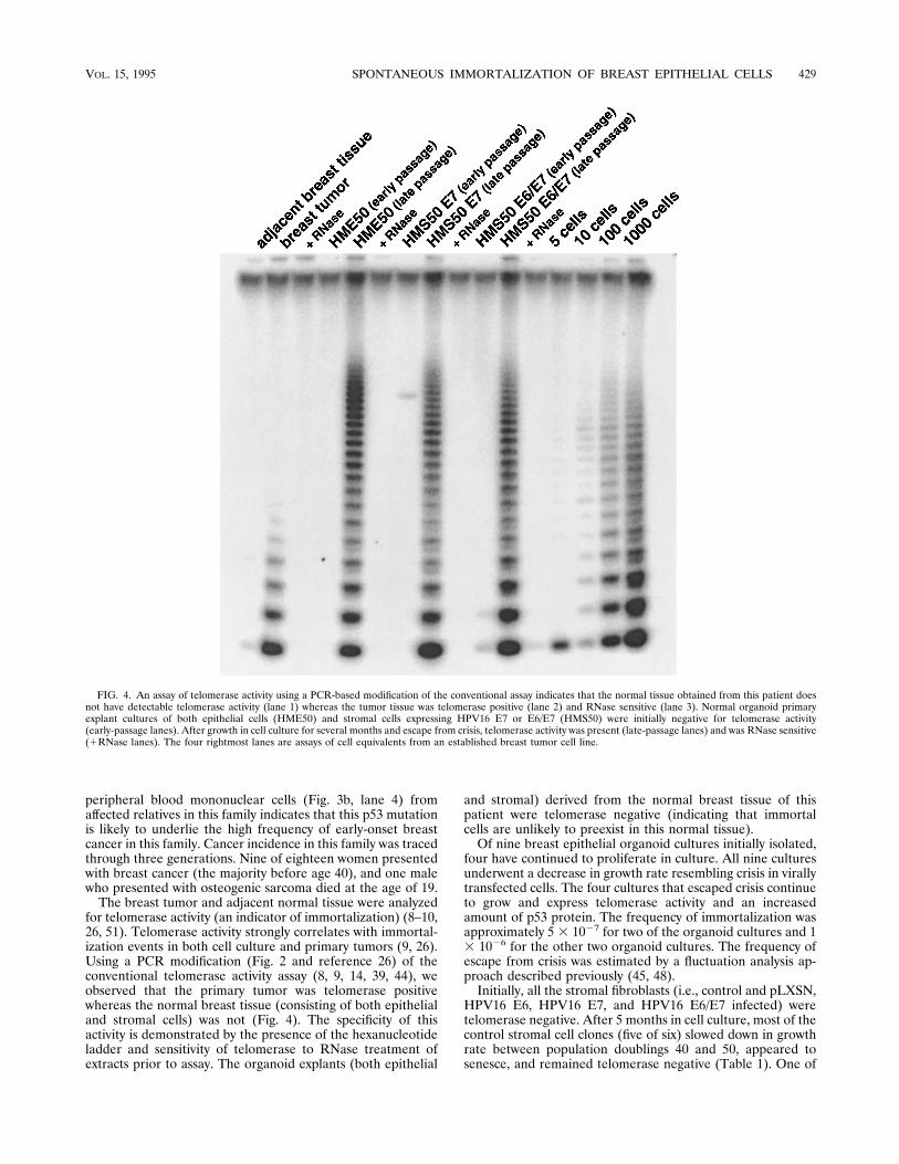
for telomerase activity (an indicator of immortalization) (8–10,26, 51). Telomerase activity strongly correlates with immortal-ization events in both cell culture and primary tumors (9, 26).Using a PCR modification (Fig. 2 and reference 26) of theconventional telomerase activity assay (8, 9, 14, 39, 44), weobserved that the primary tumor was telomerase positivewhereas the normal breast tissue (consisting of both epithelialand stromal cells) was not (Fig. 4). The specificity of thisactivity is demonstrated by the presence of the hexanucleotideladder and sensitivity of telomerase to RNase treatment ofextracts prior to assay. The organoid explants (both epithelial
and stromal) derived from the normal breast tissue of thispatient were telomerase negative (indicating that immortalcells are unlikely to preexist in this normal tissue).Of nine breast epithelial organoid cultures initially isolated,
four have continued to proliferate in culture. All nine culturesunderwent a decrease in growth rate resembling crisis in virallytransfected cells. The four cultures that escaped crisis continueto grow and express telomerase activity and an increasedamount of p53 protein. The frequency of immortalization wasapproximately 5 3 1027 for two of the organoid cultures and 13 1026 for the other two organoid cultures. The frequency ofescape from crisis was estimated by a fluctuation analysis ap-proach described previously (45, 48).Initially, all the stromal fibroblasts (i.e., control and pLXSN,
HPV16 E6, HPV16 E7, and HPV16 E6/E7 infected) weretelomerase negative. After 5 months in cell culture, most of thecontrol stromal cell clones (five of six) slowed down in growthrate between population doublings 40 and 50, appeared tosenesce, and remained telomerase negative (Table 1). One of
FIG. 4. An assay of telomerase activity using a PCR-based modification of the conventional assay indicates that the normal tissue obtained from this patient doesnot have detectable telomerase activity (lane 1) whereas the tumor tissue was telomerase positive (lane 2) and RNase sensitive (lane 3). Normal organoid primaryexplant cultures of both epithelial cells (HME50) and stromal cells expressing HPV16 E7 or E6/E7 (HMS50) were initially negative for telomerase activity(early-passage lanes). After growth in cell culture for several months and escape from crisis, telomerase activity was present (late-passage lanes) and was RNase sensitive(1RNase lanes). The four rightmost lanes are assays of cell equivalents from an established breast tumor cell line.
VOL. 15, 1995 SPONTANEOUS IMMORTALIZATION OF BREAST EPITHELIAL CELLS 429

the stromal cell clones grew until population doubling 68, didnot express telomerase, and did not immortalize. However,two of the six HPV16 E7-expressing and three of the sixHPV16 E6/E7-expressing HMS50 stromal cell clones immor-talized with a frequency of approximately 33 1027 continue togrow vigorously (.100 population doublings) and most haveacquired the ability to express telomerase activity (immortal-ized) that is RNase sensitive. Interestingly, one clone ofHMS50 stromal cells expressing HPV16 E7 is presently beyondpopulation doubling 130 and does not express telomerase.Similar results have been observed in some SV40 large Tantigen immortalized human fibroblasts (26), but at present wedo not have a molecular understanding of this phenomenon.The breast stromal fibroblasts obtained from patients under-going mammoplasty for hypermastia (HMS32) and for prophy-lactic mastectomy (HMS31) neither spontaneously immortal-ized nor immortalized when expressing HPV16 E7 (Table 1).However, they did immortalize when expressing HPV16 E6/E7(Table 1) or SV40 large T antigen (45, 55).While it is difficult to accurately quantitate relative telo-
merase activity levels in each sample, we analyzed extractsfrom different numbers of cell equivalents of a telomerase-positive breast tumor cell line. The final three lanes of Fig. 4illustrate that we can detect telomerase activity from as few as10 to 100 cell equivalents (1 to 10% [by volume] of an extractof 103 cells). The primary breast tumor (second lane), thoughpositive for telomerase, has a slightly less processive hex-anucleotide ladder than the late-passage immortalized stromaland epithelial cell lines illustrated in Fig. 4. This could beexplained by the fact that the primary breast tumor is a mixtureof stromal cells (telomerase negative) and epithelial carcinomacells (telomerase positive). In addition, during the early pas-sages after an immortalization event has occurred in cell cul-ture, there is often a weak telomerase signal which generallyincreases within several passages (data not shown). While it ispossible that expression of telomerase activity increases withtime, we believe that it is much more likely that in the earlystages after immortalization there are still many mortal (te-lomerase-negative) cells mixed in the population with a fewimmortalized (telomerase-positive) cells.Results of Western blot (immunoblot) analysis of protein
extracts from representative epithelial and stromal cellsprobed with antibodies (recognizing both wild-type and mutantp53 [PAb1801]) are illustrated in Fig. 5. Protein extracts ofbreast tumor tissue from this LFS patient express more p53than do those of the adjacent normal breast tissue. The or-ganoid breast epithelial cultures (HME50) initially express lowlevels of p53 but appear to increase in total p53, a changewhich is presumed to be due to an increase in the abundanceof mutant conformation of p53 as part of the immortalizationprocess in cell culture (as previously reported [12, 33]). Thisindicates that while the HME50 breast epithelial cells in cul-ture initially contain both mutant and wild-type p53 alleles, theallele containing the wild-type p53 appears to become inacti-vated (perhaps by mutation of the endogenous wild-type allele,by the loss of the wild-type p53 alleles, or by ectopic expressionof a dominant-negative p53 allele). The breast stromal cells(HMS50) transfected with HPV16 E7 also initially express lowlevels of p53, but similarly to the breast epithelial cells, as partof the immortalization process in cell culture, the mutant p53protein levels increase. Thus, loss of wild-type p53 functionlikely permits increased cell proliferation, ultimately resultingin immortalization. With time, the mortal cells stop dividingand/or die while the immortalized cells increase and dominatethe population, leading to a stronger telomerase activity signal.We are not sure that there is a second p53 mutation in the
primary tumor, since by SSCP analysis the wild-type p53 signalis still present. This is likely due, in part, to the presence ofconnective tissue stromal cells mixed in with the primary tu-mor. In order to determine if wild-type p53 was expressed inthe immortalized epithelial cells, we metabolically labelledHME50 cells pre- and postimmortalization with [35S]methi-onine and then immunoprecipitated them using p53 antibodiesthat recognize wild-type p53 conformation (PAb1620), mutantp53 conformation (PAb240), or both the mutant and wild-typep53 conformations (PAb1801) (7, 61–63). While we could de-tect both a wild-type and mutant p53 conformation in early-passage HME50 cells (population doubling 22), only a strongermutant conformation signal in late passage (population dou-bling .55) cells was detected (data not shown). This indicatesthat the mutant conformation of p53 increases as part of theimmortalization process but does not exclude the possibilitythat a small amount of wild-type p53 remained undetectable byimmunoprecipitation.From our previous studies (45), we predicted that those
clones which were able to remain near diploid were the mostlikely to immortalize. Chromosome analysis of metaphasespreads indicated that there was a higher fraction of breastepithelial clones remaining near diploid in three clones thatspontaneously immortalized compared with three clones thatdid not spontaneously immortalize (Table 2). This was alsotrue for the LFS patient breast stromal cells expressing HPV16E7 and E6/E7 (Table 2).
DISCUSSION
This is the first report documenting spontaneous immortal-ization of human breast epithelial cells obtained from patientswith LFS, although it has been reported that skin fibroblastcells from LFS patients can spontaneously immortalize (4).Even though we did not observe spontaneous immortalizationof LFS patient stromal cells in the present study, we did ob-serve immortalization of LFS patient stromal cells expressingHPV16 E7. These results indicate that wild-type p53 is impor-tant in regulating cellular senescence in breast epithelial cellsand also suggest an important role for both p53 and a pRb-likefunction in the regulation of senescence of breast stromal cells.Previously it has been reported that LFS patient skin fibro-
blast cells immortalize spontaneously at a very low frequency
FIG. 5. Western blot analysis of p53 in protein extracts from the tissue andcells obtained from an LFS patient by using PAb1801 antibody, which recognizesboth wild-type and mutant conformations of p53. Levels of p53 in the tumor aremuch higher than in the adjacent normal tissue from which epithelial and stromalcells were obtained. LFS patient epithelial and stromal cells in early passage alsohave low levels of expression of p53, while the epithelial cells that spontaneouslyimmortalized and the stromal cells expressing HPV16 E7 that immortalized haveincreased expression of p53. The stromal cells that immortalized with HPV16E6/E7 do not have increased expression of p53, since the E6 protein of HPV16facilitates degradation of p53.
430 SHAY ET AL. MOL. CELL. BIOL.

(4), but this work has been difficult to confirm (33, 34). In a yetunpublished study (43a), stromal fibroblasts of an LFS patientappeared to senesce at 42 population doublings, but after sev-eral months of maintenance in the senescent state, cell prolif-eration which was associated with a loss of the wild-type p53allele recommenced. One of these clones appeared to sponta-neously immortalize even though after an additional 30 popu-lation doublings the rest of the clones again ceased prolifera-tion (similar to crisis in SV40-transformed cells) and did notimmortalize. These results and those in the present reportsuggest that wild-type p53 is important in maintenance of DNAstability and that loss of wild-type p53 function may be asso-ciated with a breakdown in cell growth control (loss of cellularhomeostasis), causing increased proliferation ultimately result-ing in immortalization, generally by the reactivation of telo-merase activity. While our studies indicate that loss of p53function may be sufficient to allow breast epithelial cells toimmortalize, in the fibroblast lineages loss of p53 functionalone may not be sufficient to obtain immortalization, and it isonly after the additional loss of a pRb-like function that thesecells become immortalization competent. Irrespective, loss ofp53 function in breast epithelial cells or p53 and pRb-likefunction in stromal cells is only the first of two stages that mustbe overcome for cells to immortalize (46, 50). Thus, it is notsurprising that fibroblasts obtained from LFS patients are dif-ficult to immortalize and that even with the complete loss ofwild-type p53 immortalization was not observed (33).The implications of these findings are potentially important,
not only because they concern LFS patients’ risks of develop-ing cancer but also because they indicate the important role ofnormal p53 in protecting human breast epithelial cells fromimmortalizing and progressing to malignant carcinoma. Thesestudies may also provide one reason for the high frequency ofbreast cancer in LFS-affected families. While alterations in p53appear to be a central factor for the development of breastcancer in LFS patients, tissue-specific changes (perhaps relatedto differentiation) are also likely to be important, since LFS-affected families do not have a high incidence of sporadiccolorectal cancer although such cancers are associated with ahigh prevalence of p53 mutations (22). Finally, the spontane-ously immortalized breast epithelial cell lines obtained in thepresent study may be useful in the elucidation of additionalcritical steps in the development of breast cancer.
ACKNOWLEDGMENTS
This study was supported by research grants CA50195 and CA64871from the National Institutes of Health (J.W.S.); USAMR grantDAMD-94-J-4077 (J.W.S.); Geron Corp., Menlo Park, Calif. (J.W.S.);
the Children’s Cancer Research Fund (G.T.); the American Society ofClinical Oncology (G.T.); and the Susan G. Komen Breast CancerFoundation (G.T. and J.W.S.).We thank Woodring Wright, Joe Goldstein, Jennifer Cuthbert, John
Minna, Arnold Levine, and Curtis Harris for valuable discussions.
REFERENCES
1. Band, V., S. Dalal, L. Delmolino, and E. J. Androphy. 1993. Enhanceddegradation of p53 protein in HPV-6 and BPV-1 E6-immortalized humanmammary epithelial cells. EMBO J. 12:1847–1852.
2. Band, V., J. A. De Caprio, L. Delmolino, V. Kulesa, and R. Sager. 1991. Lossof p53 protein in human papillomavirus type 16-immortalized human mam-mary epithelial cells. J. Virol. 65:6671–6676.
3. Birch, J. M., A. L. Hartley, K. J. Tricker, J. Prosser, A. Condie, A. M. Kelsey,M. Harris, P. H. M. Jones, A. Binchy, D. Crowther, A. W. Craft, O. B. Eden,D. G. R. Evans, E. Thompson, J. R. Mann, J. Martin, E. L. D. Mitchell, andM. F. Santibanez-Koref. 1994. Prevalence and diversity of constitutionalmutations in the p53 gene among 21 Li-Fraumeni families. Cancer Res.54:1298–1304.
4. Bischoff, F. Z., S. O. Yim, S. Pathak, G. Grant, M. J. Siciliano, B. C.Giovanella, L. C. Strong, and M. A. Tainsky. 1990. Spontaneous abnormal-ities in normal fibroblasts from patients with Li-Fraumeni cancer syndrome:aneuploidy and immortalization. Cancer Res. 50:7979–7984.
5. Bronstein, I., J. C. Voyta, K. G. Lasseri, O. Murphey, B. Edwards, and L. J.Kricka. 1990. Rapid and sensitive detection of DNA in Southern blots withchemiluminescence. BioTechniques 8:310–314.
6. Buchman, V. L., P. M. Chumakov, N. N. Ninkina, O. P. Samarina, and G. P.Georgiev. 1988. A variation in the structure of the protein-coding region ofthe human p53 gene. Gene 70:245–252.
7. Chen, J.-Y., W. D. Funk, W. E. Wright, J. W. Shay, and J. D. Minna. 1993.Heterogeneity of transcriptional activity of mutant p53 proteins and p53DNA target sequences. Oncogene 8:2159–2166.
8. Counter, C. M., A. A. Avilion, C. E. LeFeuvre, N. G. Stewart, C. W. Greider,C. B. Harley, and S. Bacchetti. 1992. Telomere shortening associated withchromosome instability is arrested in immortal cells which express telomer-ase activity. EMBO J. 11:1921–1929.
9. Counter, C. M., H. W. Hirte, S. Bacchetti, and C. B. Harley. 1994. Telo-merase activity in human ovarian carcinoma. Proc. Natl. Acad. Sci. USA91:2900–2904.
10. deLange, T. 1994. Activation of telomerase in a human tumor. Proc. Natl.Acad. Sci. USA 91:2882–2885.
11. Dunbar, B. S. 1987. Two-dimensional electrophoresis and immunologicaltechniques, p. 335–341. Plenum Press, New York.
12. Frebourg, T., M. Sadelain, Y.-S. Ng, J. Kassel, and S. H. Friend. 1994. Equaltranscription of wild-type and mutant p53 using bicistronic vectors results inthe wild-type phenotype. Cancer Res. 54:878–881.
13. Gillespie, P. G., and A. J. Hudspeth. 1991. Chemiluminescence detection ofproteins from single cells. Proc. Natl. Acad. Sci. USA 88:2563–2567.
14. Greider, C. W. 1990. Telomeres, telomerase and senescence. Bioessays 12:363–369.
15. Halbert, C. L., G. W. Demers, and D. A. Galloway. 1992. The E6 and E7genes of human papillomavirus type 6 have weak immortalizing activity inhuman epithelial cells. J. Virol. 66:2125–2134.
16. Hara, E., H. Tsurui, A. Shinozaki, S. Nakada, and K. Oda. 1991. Coopera-tive effect of antisense-Rb and antisense-p53 oligomers on the extension oflife span in human diploid fibroblasts. Biochem. Biophys. Res. Commun.179:528–534.
17. Harley, C. B., A. B. Futcher, and C. W. Greider. 1990. Telomeres shortenduring aging of human fibroblasts. Nature (London) 345:458–460.
18. Harris, C. C. 1987. Human tissues and cells in carcinogenesis research.Cancer Res. 47:1–10.
19. Harris, C. C. 1993. p53: at the crossroads of molecular carcinogenesis andrisk assessment. Science 262:1980–1981.
20. Harris, J. P., M. E. Lippman, and V. Veronesi. 1992. Breast cancer. N. Engl.J. Med. 327:319–328.
21. Hartley, A. L., J. M. Birch, H. B. Marsden, and M. Harris. 1986. Breastcancer risk in mothers of children with osteosarcoma and chondrosarcoma.Br. J. Cancer 54:819–823.
22. Hollstein, M., D. Sidransky, B. Vogelstein, and C. C. Harris. 1991. p53mutations in human cancers. Science 253:49–53.
23. Huschtscha, L. I., and R. Holiday. 1983. Limited and unlimited growth ofSV40-transformed cells from human diploid MRC-5 fibroblasts. J. Cell Sci.63:77–99.
24. Kastan, M. B., Q. Zhan, W. S. El-Deiry, F. Carrier, T. Jacks, W. V.Walsh, B. S. Plunkett, B. Vogelstein, and A. J. Fornace. 1992. A mammaliancell cycle checkpoint pathway utilizing p53 and GADD45 is defective inataxia-telangiectasia. Cell 71:587–597.
25. Kern, S. E., J. A. Pietenpol, S. Thiagalingam, A. Seymour, K. W. Kinzler, andB. Vogelstein. 1992. Oncogenic forms of p53 inhibit p53-regulated geneexpression. Science 256:827–830.
26. Kim, N.-W., M. A. Piatyszek, K. R. Prowse, P. Ho, G. Coviello, C. Harley,
TABLE 2. Chromosome analysis of human mammary epithelial(HME) and human mammary stromal (HMS)
cells from an LFS patient
Clone name Immortal-ized
Range ofchromosomes/metaphasea
Median no.of chromo-somes
% Diploidmetaphases
HME50-5 Yes 44–123 47 60HME50-8 Yes 38–101 46 68HME50-9 Yes 41–107 49 55HME50-3 No 42–99 59 42HME50-6 No 32–155 75 20HME50-7 No 37–174 78 36HMS50-E7-5 Yes 41–113 47 72HMS50-E7-pop Yes 38–102 46 84HMS50-E6/E7-2 Yes 40–133 46 68
a Based on counts from 25 metaphase spreads.
VOL. 15, 1995 SPONTANEOUS IMMORTALIZATION OF BREAST EPITHELIAL CELLS 431

M. D. West, W. E. Wright, S. Weinrich, and J. W. Shay. Specific associationof human telomerase activity with immortal cells and cancer. Science, inpress.
27. Law, J. C., L. C. Strong, A. Chidambaram, and R. E. Ferrell. 1991. A germline mutation in exon 5 of the p53 gene in an extended cancer family. CancerRes. 51:6385–6387.
28. Levine, A. J. 1993. 11th Ernst Klenk Lecture. The p53 tumor suppressor geneand product. Biol. Chem. 374:227–235.
29. Levine, A. J., J. Momand, and C. A. Finlay. 1991. The p53 tumor suppressorgene. Nature (London) 351:49–53.
30. Li, F. P., and J. F. Fraumeni, Jr. 1969. Soft tissue sarcomas, breast cancer,and other neoplasms. A familial syndrome. Ann. Intern. Med. 71:747–752.
31. Li, F. P., J. F. Fraumeni, Jr., J. J. Mulvihill, W. A. Blattner, M. G. Dreyfus,M. A. Tucker, and R. W. Miller. 1988. A cancer family syndrome in twenty-four kindreds. Cancer Res. 48:5358–5362.
32. Linder, S., and H. Marshall. 1990. Immortalization of primary cells by DNAtumor viruses. Exp. Cell Res. 191:1–7.
33. Livingstone, L. R., A. White, J. Sprouse, E. Livanos, T. Jacks, and T. D.Tlsty. 1992. Altered cell cycle arrest and gene amplification potential accom-pany loss of wild-type p53. Cell 70:923–935.
34. Maclean, K., E. M. Rogan, N. J. Whitaker, A. C.-M. Chang, P. B. Rowe, L.Dalla-Pozza, G. Symonds, and R. R. Reddel. 1994. In vitro transformation ofLi-Fraumeni syndrome fibroblasts with SV40 large T antigen mutants. On-cogene 9:719–725.
35. Malkin, D. 1990. p53 and the Li-Fraumeni syndrome. Cancer Genet. Cyto-genet. 66:83.
36. Malkin, D., K. W. Jolly, N. Barbier, A. T. Look, S. H. Friend, M. C. Geb-hardt, T. I. Anderson, A. L. Borresen, F. P. Li, J. Garber, et al. 1992.Germline mutations of the p53 tumor-suppressor gene in children and youngadults with second malignant neoplasms. N. Engl. J. Med. 326:1309–1315.
37. Malkin, D., F. P. Li, L. C. Strong, J. F. Fraumeni, C. E. Nelson, D. H. Kim,J. Kassell, M. A. Gryka, F. Z. Bischoff, M. A. Tainsky, and S. H. Friend.1990. Germ line p53 mutations in a familial syndrome of breast cancer,sarcomas, and other neoplasms. Science 250:1233–1238.
38. Miller, A. D., and G. J. Rosman. 1989. Improved retroviral vectors for genetransfer and expression. BioTechniques 7:980–990.
39. Morin, G. B. 1989. The human telomere terminal transferase is a ribonucle-oprotein that synthesizes TTAGGG repeats. Cell 59:521–529.
40. Orita, M., H. Iwahana, H. Kanazawa, K. Hayashi, and T. Sekiya. 1989.Detection of polymorphisms of human DNA by gel electrophoresis as single-strand conformation polymorphisms. Proc. Natl. Acad. Sci. USA 86:2766–2770.
41. Osteen, R. T., and K. H. Karnell. 1994. The National Cancer Data Basereport on breast cancer. Cancer 73:1994–2000.
42. Radna, R. L., Y. Caton, K. K. Jha, P. Kaplan, G. Li, F. Traganos, and H. L.Ozer. 1989. Growth of immortal simian virus 40 tsA-transformed humanfibroblasts is temperature dependent. Mol. Cell. Biol. 9:3093–3096.
43. Raycroft, L., H. Y. Wu, and G. Lozano. 1990. Transcriptional activation bywild-type but not mutants of the p53 antioncogene. Science 249:1049–1051.
43a.Reddell, R. (Children’s Medical Research Institute, Westmead, New SouthWales, Australia). Personal communication.
44. Shay, J. W., D. Brasiskyte, M. Ouellete, M. A. Piatyszek, H. Werbin, Y. Ying,and W. E. Wright. 1994. Methods for analysis of telomerase and telomeres.
Methods Mol. Genet. Genes Chromosome Anal. Part C 5:263–280.45. Shay, J. W., B. A. Van der Haegen, Y. Ying, and W. E. Wright. 1993. The
frequency of immortalization of human fibroblast and mammary epithelialcells transfected with SV40 large T-antigen. Exp. Cell Res. 209:45–52.
46. Shay, J. W., and H. Werbin. 1993. Toward a molecular understanding of theonset of human breast cancer: a hypothesis. Br. Cancer Res. Treat. 25:83–94.
47. Shay, J. W., H. Werbin, W. Funk, and W. E. Wright. 1992. Cellular andmolecular advances in elucidating p53 function. Mutat. Res. 277:163–171.
48. Shay, J. W., and W. E. Wright. 1989. Quantitation of the frequency ofimmortalization of normal diploid fibroblasts by SV40 large T-antigen. Exp.Cell Res. 184:109–118.
49. Shay, J. W., W. E. Wright, D. Brasiskyte, and B. A. Van Der Haegen. 1993.E6 of human papilloma virus 16 can overcome the M1 stage of immortal-ization in human mammary epithelial cells but not in human fibroblasts.Oncogene 8:1407–1413.
50. Shay, J. W., W. E. Wright, and H. Werbin. 1991. Defining the molecularmechanisms of human cell immortalization. Biochim. Biophys. Acta 1072:1–7.
51. Shay, J. W., W. E. Wright, and H. Werbin. 1993. Loss of telomeric DNAduring aging may predispose cells to cancer. Int. J. Oncol. 3:559–563.
52. Srivastava, S., S. Wang, Y. A. Tong, K. Pirollo, and E. H. Chang. 1993.Several mutant p53 proteins detected in cancer-prone families with Li-Fraumeni syndrome exhibit transdominant effects on the biochemical prop-erties of the wild-type p53. Oncogene 8:2449.
53. Stampfer, M. R., R. C. Hallowes, and A. J. Hackett. 1980. Growth of normalhuman mammary cells in culture. In Vitro (Rockville) 16:415–425.
54. Taylor-Papadimitriou, J., F. Berdichevsky, B. D’Souza, and J. Burchell.1993. Human models of breast cancer. Cancer Surv. 16:59–78.
55. Van Der Haegen, B. A., and J. W. Shay. 1993. Immortalization of humanmammary epithelial cells by SV40 large T-antigen involves a two-step mech-anism. In Vitro Cell. Dev. Biol. 29:180–182.
56. Vogelstein, B., and K. W. Kinzler. 1992. p53 function and dysfunction. Cell70:523–526.
57. Wright, W. E., O. M. Pereira-Smith, and J. W. Shay. 1989. Reversiblecellular senescence: a two-stage model for the immortalization of normalhuman diploid fibroblasts. Mol. Cell. Biol. 9:3088–3092.
58. Wright, W. E., and J. W. Shay. 1992. Telomere positional effects and theregulation of cellular senescence. Trends Genet. 8:193–197.
59. Yin, Y., M. A. Tainsky, F. Z. Bischoff, L. C. Strong, and G. M. Wahl. 1992.Wild-type p53 restores cell cycle control and inhibits gene amplification incells with mutant p53 alleles. Cell 70:937–948.
60. Zambetti, G. P., J. Bargonetti, K. Walker, C. Prives, and A. J. Levine. 1992.Wild-type p53 mediates positive regulation of gene expression through aspecific DNA sequence element. Genes Dev. 6:1143–1152.
61. Zauberman, A., Y. Barak, N. Ragimov, N. Levy, and M. Oren. 1993. Se-quence-specific DNA binding by p53: identification of target sites and lack ofbinding to p53-MDM2 complexes. EMBO J. 12:2799–2808.
62. Zhang, W., W. D. Funk, W. E. Wright, J. W. Shay, and A. B. Deisseroth.1993. DNA binding and transcriptional activation by mutant p53 proteins.Oncogene 8:2555–2559.
63. Zhang, W., J. W. Shay, and A. Deisseroth. 1993. Inactive p53 mutants mayenhance the transcriptional activity of wild-type p53. Cancer Res. 53:4772–4775.
432 SHAY ET AL. MOL. CELL. BIOL.