Dendrimer modified biochip for detection of 2, 4, 6 trinitrotoluene on SPR immunosensor: Fabrication...
7
Sensors and Actuators B 137 (2009) 403–409 Contents lists available at ScienceDirect Sensors and Actuators B: Chemical journal homepage: www.elsevier.com/locate/snb Dendrimer modified biochip for detection of 2,4,6 trinitrotoluene on SPR immunosensor: Fabrication and advantages Praveen Singh a,d,∗,1 , Takeshi Onodera a , Yutaka Mizuta a , Kiyoshi Matsumoto b , Norio Miura c , Kiyoshi Toko a a Department of Electronics, Graduate School of Information Science and Electrical Engineering, Kyushu University, 744 Motooka, Fukuoka 819-0395, Japan b Graduate School of Agriculture, Kyushu University, Fukuoka 812-8581, Japan c Art, Science and Technology Centre for Cooperative Research, Kyushu university, Kasuga-shi, Fukuoka 816-8580, Japan d Biophysics and Electron Microscopy Section, Indian Veterinary Research Institute, Izatnagar 243122, Bareilly, UP, India article info Article history: Received 13 June 2007 Received in revised form 2 December 2008 Accepted 12 December 2008 Available online 25 December 2008 Keywords: TNT SPR biosensor Competitive assay Dendrimer G4 abstract This paper describes the fabrication and evaluation of new sensor chip technology based on applica- tion of polyamidoamine (PAMAM) dendrimer generation 4 (G4) for enhancing surface loading capacity and efficiency for selective and sensitive detection of explosive molecule trinitrotoluene (TNT) under competitive inhibition assay format. The fabrication process of a nano-scale biosensor chip surface was optimized at various steps involving thiol layer, cross-linker, dinitro phenylated-keyhole limpet hemo- cyanin (DNP-KLH) protein conjugate as ligand. The top ligand layer is supported by underlying 11-amino 1-undecanethiol hydrochloride (AUT) self-assembled monolayer (SAM) and PAMAM dendrimer G4. Bis- sulfosuccinimidyl suberate (BS 3 ) was used as homobifunctional crosslinker to react with amine groups at both the ends. PAMAM dendrimer activation was achieved over 11-amino 1-undecanethiol SAMs over gold chip. PAMAM sensor surfaces scored many advantages over the planer surfaces such as enhanced antibody immobilization, allowing the use of lower concentrated antibody to work under the regime of lower detection limit besides stable and robust surface leading to longer life. Bioactive thin films were fabricated over gold chip via layer-by-layer self-assembly methods. Biomolecular interaction between DNP-KLH conjugate surface and specific TNP-gly-KLH mouse IgG antibody was monitored through surface plasmon resonance (SPR) based real-time optical transducer. The quantitation of TNT on this bioactive surface was done using the competitive inhibition immunoassay. The DNP-KLH surface biosensor has shown a detection limit of 110ppt for TNT molecule. This TNT specific biosensor holds the promise to be most sensitive, easy to regenerate, highly stable, low response loss sensor surface under competitive assay format. A 12 s injection of 10 mM glycine–HCl solution or 50 mM NaOH solution was found ade- quate for regeneration of DNP-KLH surface for repeated use. DNP-KLH sensor platform was checked for its repeatability and storability. © 2009 Elsevier B.V. All rights reserved. 1. Introduction The world community is besieged with serious challenge to contain the terrorism, to enhance the safety in air travel and to combat environmental pollution being caused by unexploded ord- nance (UXO) containing trinitrotoluene (TNT) [1,2]. The detection of explosive molecules at parts per trillion levels or below is the urgent necessity for creating safety in passengers travel, cargo han- ∗ Corresponding author at: Japan Society for Promotion of Science, R. No. 459, West 2 4F, 744 Motooka Nishi-ku, Fukuoka 810-0395, Japan. Tel.: +81 92 8023729; fax: +81 92 8023770. E-mail addresses: [email protected], [email protected] (P. Singh). 1 The author is presently under deputation to Japan Society for Promotion of Science, Tokyo through JSPS-PDF Program at Kyushu University, Japan. dling, containing trafficking in explosives and checking the rise of global terrorism. TNT, the main component of explosive products serves as an indicator for existence of these explosives. Current physical detection methods are less selective, unreli- able and have poor accuracy. Emerging sensor technologies such as ground penetrating radar [3] suffer from low sensitivity, accuracy and false negative as it is unable to distinguish the patterns obtained from plastic landmines. The upgraded piezo-sensors [4] are useful at high concentration level in spite of inadequacies such as incon- sistency, frequent errors and selectivity. Dogs are very effective in detecting the explosives substances. Dogs need extensive training, trainer and unfit for round the clock security protocol. TNT detection by rapid fluorescent quenching [5] with analyte on porous polymer films appears to be promising. In this method, fluorescent attenu- ation depends on several factors that need to be taken care of on practical application. Micro-cantilever sensors [6] can measure the 0925-4005/$ – see front matter © 2009 Elsevier B.V. All rights reserved. doi:10.1016/j.snb.2008.12.027
-
Upload
independent -
Category
Documents
-
view
0 -
download
0
Transcript of Dendrimer modified biochip for detection of 2, 4, 6 trinitrotoluene on SPR immunosensor: Fabrication...
Di
PNa
b
c
d
a
ARRAA
KTSCD
1
ccnou
Wf
S
0d
Sensors and Actuators B 137 (2009) 403–409
Contents lists available at ScienceDirect
Sensors and Actuators B: Chemical
journa l homepage: www.e lsev ier .com/ locate /snb
endrimer modified biochip for detection of 2,4,6 trinitrotoluene on SPRmmunosensor: Fabrication and advantages
raveen Singha,d,∗,1, Takeshi Onoderaa, Yutaka Mizutaa, Kiyoshi Matsumotob,orio Miurac, Kiyoshi Tokoa
Department of Electronics, Graduate School of Information Science and Electrical Engineering, Kyushu University, 744 Motooka, Fukuoka 819-0395, JapanGraduate School of Agriculture, Kyushu University, Fukuoka 812-8581, JapanArt, Science and Technology Centre for Cooperative Research, Kyushu university, Kasuga-shi, Fukuoka 816-8580, JapanBiophysics and Electron Microscopy Section, Indian Veterinary Research Institute, Izatnagar 243122, Bareilly, UP, India
r t i c l e i n f o
rticle history:eceived 13 June 2007eceived in revised form 2 December 2008ccepted 12 December 2008vailable online 25 December 2008
eywords:NTPR biosensorompetitive assayendrimer G4
a b s t r a c t
This paper describes the fabrication and evaluation of new sensor chip technology based on applica-tion of polyamidoamine (PAMAM) dendrimer generation 4 (G4) for enhancing surface loading capacityand efficiency for selective and sensitive detection of explosive molecule trinitrotoluene (TNT) undercompetitive inhibition assay format. The fabrication process of a nano-scale biosensor chip surface wasoptimized at various steps involving thiol layer, cross-linker, dinitro phenylated-keyhole limpet hemo-cyanin (DNP-KLH) protein conjugate as ligand. The top ligand layer is supported by underlying 11-amino1-undecanethiol hydrochloride (AUT) self-assembled monolayer (SAM) and PAMAM dendrimer G4. Bis-sulfosuccinimidyl suberate (BS3) was used as homobifunctional crosslinker to react with amine groupsat both the ends. PAMAM dendrimer activation was achieved over 11-amino 1-undecanethiol SAMs overgold chip. PAMAM sensor surfaces scored many advantages over the planer surfaces such as enhancedantibody immobilization, allowing the use of lower concentrated antibody to work under the regime oflower detection limit besides stable and robust surface leading to longer life. Bioactive thin films werefabricated over gold chip via layer-by-layer self-assembly methods. Biomolecular interaction betweenDNP-KLH conjugate surface and specific TNP-gly-KLH mouse IgG antibody was monitored through surfaceplasmon resonance (SPR) based real-time optical transducer. The quantitation of TNT on this bioactive
surface was done using the competitive inhibition immunoassay. The DNP-KLH surface biosensor hasshown a detection limit of 110 ppt for TNT molecule. This TNT specific biosensor holds the promise tobe most sensitive, easy to regenerate, highly stable, low response loss sensor surface under competitiveassay format. A 12 s injection of 10 mM glycine–HCl solution or 50 mM NaOH solution was found ade-DNP-bility
quate for regeneration ofits repeatability and stora
. Introduction
The world community is besieged with serious challenge toontain the terrorism, to enhance the safety in air travel and to
ombat environmental pollution being caused by unexploded ord-ance (UXO) containing trinitrotoluene (TNT) [1,2]. The detectionf explosive molecules at parts per trillion levels or below is thergent necessity for creating safety in passengers travel, cargo han-∗ Corresponding author at: Japan Society for Promotion of Science, R. No. 459,est 2 4F, 744 Motooka Nishi-ku, Fukuoka 810-0395, Japan. Tel.: +81 92 8023729;
ax: +81 92 8023770.E-mail addresses: [email protected], [email protected] (P. Singh).
1 The author is presently under deputation to Japan Society for Promotion ofcience, Tokyo through JSPS-PDF Program at Kyushu University, Japan.
925-4005/$ – see front matter © 2009 Elsevier B.V. All rights reserved.oi:10.1016/j.snb.2008.12.027
KLH surface for repeated use. DNP-KLH sensor platform was checked for.
© 2009 Elsevier B.V. All rights reserved.
dling, containing trafficking in explosives and checking the rise ofglobal terrorism. TNT, the main component of explosive productsserves as an indicator for existence of these explosives.
Current physical detection methods are less selective, unreli-able and have poor accuracy. Emerging sensor technologies such asground penetrating radar [3] suffer from low sensitivity, accuracyand false negative as it is unable to distinguish the patterns obtainedfrom plastic landmines. The upgraded piezo-sensors [4] are usefulat high concentration level in spite of inadequacies such as incon-sistency, frequent errors and selectivity. Dogs are very effective indetecting the explosives substances. Dogs need extensive training,
trainer and unfit for round the clock security protocol. TNT detectionby rapid fluorescent quenching [5] with analyte on porous polymerfilms appears to be promising. In this method, fluorescent attenu-ation depends on several factors that need to be taken care of onpractical application. Micro-cantilever sensors [6] can measure the
4 Actua
eaAs
vsAflelffoRTfta
sdbpTgciTgs
atcdCdo[trwDputptfifd
2
2
ppacCfS
04 P. Singh et al. / Sensors and
xtremely small forces caused by molecular attachment and offerdaptation to sensing platform suitable for multiplexed sensing.lthough cantilever sensors are sensitive, they suffer from intrinsicelectivity problem.
Immunochemical techniques used to detect TNT exist in aariety of formats from competitive ELISA, fiber optic biosen-ors, sol–gel trapped assays, microcapillary immunosensors [7–9].nalytical method such as displacement immunoassay [10] usinguorescent labeled antigen is well suited for trace detection ofxplosives. This method requires a fluorescent-labeled antigen, soimiting its generic nature and becoming cumbersome, not suitableor on-site detection of explosive molecules. On the other hand, sur-ace plasmon resonance (SPR) biosensors are evanescent field basedptical sensors using thin gold film for sensing applications [11–15].apid, inexpensive, reliable and sensitive methods for detection ofNT and similar compounds are therefore urgently needed. For pastew years, we are concentrating on development of highly sensi-ive SPR immunosensor for TNT and trinitrophenol detection usingntigen-antibody based reaction methods [16–18].
The present study demonstrates the fabrication procedure ofensor chip that enhances the sensitivity and efficiency for TNTetection, the key elements of high affinity antigen–antibody-ased immunosensor. The chip is designed to function as a sensorlatform on SPR system for a competitive, label-free detection ofNT. The use of densely packed, well oriented and robust SAMsenerated by organosulfur compounds on gold chip has been suc-essfully demonstrated in many previous studies [19–22]. The basicdea behind this report was to enhance the anti-TNT antibody (Ab-NT) loading on the surface and to increase binding efficiency onold chip surface, besides selecting a ligand that will have structuralimilarity but less affinity for Ab-TNT.
Polyamidoamine (PAMAM) dendrimers have been recognizeds promising building blocks of molecularly organized nanos-ructures. This work uses highly branched macromoleculesomposed of a PAMAM–amidoethylethanol dendrimer, 1,12-iaminododecane core with four dendron arms radiating from it.rooks and Ricco[23] and others [23–25] reported fabricated den-rimer monolayers on metallic surfaces and shown higher amountf protein ligand on surface for interaction study. Benters et al.26] demonstrated that a dendrimer linker system on surface leadso very high immobilization efficiency for DNA oligomers andemarkable high stability during repeated regeneration cycles. Itas reported [27] that multistep immobilization of proteins andNA on chemically prepared gold films using dendrimers, increasedroteins attachment capacity of the chip. Another advantage ofsing this molecule is that chemical reactivity [28,24] of func-ional group used for forming a stable link is higher than whenresent in other macromolecules. In this paper, development ofhree-dimensional dendrimer modified gold chip surface, havingrmly stable DNP-KLH protein matrix as biorecognition element
or an efficient, cost-effective and highly sensitive immunoassay, isescribed.
. Experimental
.1. Materials
The following chemicals were used as received from sup-liers: 11-amino 1-undecanethiol hydrochloride (AUT) wasurchased from Dojindo laboratory, Kumamoto, Japan. PAMAM-
midoethylethanolamine dendrimer, 1,12-diaminododecaneore (Generation 4 with 64 surface primary amino groups,760H1524N250O188 FW 17173.79) in 10% (w/w) in methanol, manu-actured by Dendrintic Nano Technolgies, USA was purchased fromigma–Aldrich, St. Louis, USA. The solution was stored at −20 ◦C
tors B 137 (2009) 403–409
and used as received. Bis(sulfo succinimidyl) suberate (BS3), waspurchased from Pierce Technology, Rockford, USA. Upon receipt theproduct was store desiccated at 4 ◦C. Monoclonal anti-TNT antibody(mAb A1.1.1), raised in mouse against TNP-gly-KLH as immunogen,was purchased from Strategic Biosolutions, USA. 21.8 ppm TNTsolution in milli Q water was purchased from Chugoku KayakuCo. Ltd., Japan. Dinitro phenylated-keyhole limpet hemocyanin(DNP-KLH) was purchased from Cosmo Bio, Ltd., Japan. All aqueoussolutions were prepared from Milli Q (18 M�-cm resistivity) deion-ized water obtained from Milli-Q-system (Millipore Corporation,USA). KH2PO4 (99% purity) and Na2PO4 were purchased from WakoChemicals, Japan. NaCl and KCl of 99.5% purity were purchasedfrom Kanto Chemicals, Japan. TRIS extra pure was purchased fromICN Biomedicals, Ohio, USA.
2.2. Surface chemistries
SIA Kit from Biacore, Uppsala, Sweden was used for fabricationof multi-step monolayered formation of various films on surface.Initially, the sensor chip Au packet is allowed to equilibrate at roomtemperature, so that gold chip is released from gel easily and thereshould not be any condensation on the surface. The chip is removedfrom adhesive by fine tweezer lifting it by holding 2 mm edgeof chip. This sensor chip is first washed in acetone for 10 min inultrasonic cleaner, followed by washing in ethanol and 2-propanolfor 2 min each. Thereafter, the chip is further washed in solution(NH3:H2O2:H2O (18.2 M�-cm)) in 1:1:5 (v/v/v) ratio on hot plateat 90 ◦C for 20 min [29]. Upon removal from this solution, chip wasrinsed with deionized water for a while, followed by drying in highpurity nitrogen gas. Now the gold chip surface is ready for thiolsmonolayer fabrication.
The thiols coating of gold surface is done in 1 mM AUT for18–22 h at room temperature. The longer soaking in AUT-ethanolsolution will help in crystallization of amino-terminated SAM overgold surface. This procedure makes a thin coating of SAM containingamino group at the outer end of parallel oriented, high density SAMs[21,22]. Upon removal of chip from thiol–ethanol, the gold substrateis rinsed in excess ethanol, and blown dry in nitrogen gas. ThisAUT modified gold substrate was activated by bis-sulfosuccinimidylsuberate for covalent linking of amine group through formationof amide bond on both sides of linker. BS3 is water-soluble ester,having sodium salt at both terminuses; it shows high reactivitytowards primary amines. BS3 possess a charged group at its sur-face, making it very useful for cell-surface protein cross-linking.BS3 is used for formation of three-dimensional PAMAM den-drimer generation 4 polymer layers through succinimidyl linkergroup.
AUT modified SAMs surface was incubated in 10 mM BS3 in PBSpH 7.2 (2.7 mM KCL, 137 mM NaCl, 4.3 mM Na2HPO4·7H2O, 1.4 mMKH2PO4) at room temperature. Amine-terminated SAMs are usu-ally used to achieve a stable immobilization of proteins by covalentbonding in the esterification of N-hydroxy succinimide (NHS) ester.After 45 min soaking in BS3, substrate is rinsed with Milli-Q waterand nitrogen blown dry. Subsequently the substrates were incu-bated overnight (16–20 h) in a 10% (w/w) methanolic solution ofthe PAMAM dendrimers at 22 ◦C. Upon removal from dendrimersolution, the substrates were washed repeatedly with methanol toremove excess dendrimers and blown dry with nitrogen stream.
For covalent attachment of proteins on prepared surface, thedendrimer-coated substrates were incubated in 10 mM BS3 in PBSpH 7.2 for 45 min, rinsed with Milli-Q water, and dried under nitro-
gen, followed by immediate dipping of these gold substrates into200 ppm DNP-KLH in PBS pH 7.2 protein solutions for 2 h at roomtemperature. Protein treated substrates were washed in PBS for5–10 min in ultrasonic cleaner and rinsed in MQ water and thennitrogen dried.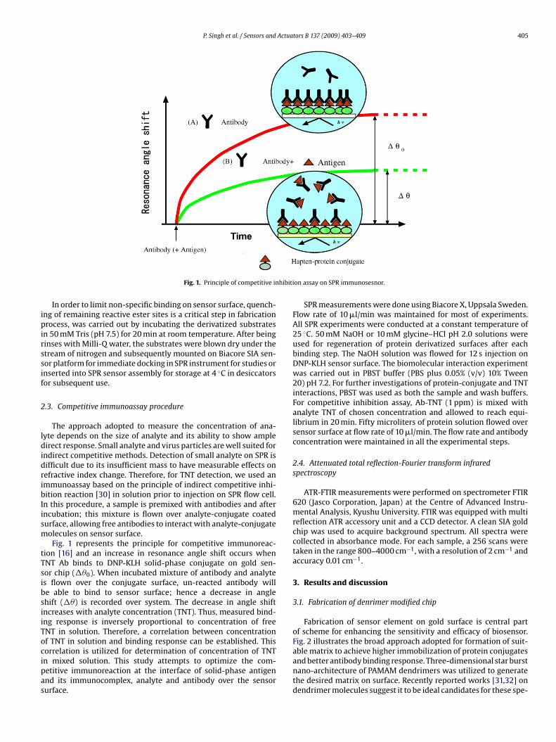
P. Singh et al. / Sensors and Actuators B 137 (2009) 403–409 405
hibiti
ipirssif
2
ldidribIism
tTsibsiiTocipas
Fig. 1. Principle of competitive in
In order to limit non-specific binding on sensor surface, quench-ng of remaining reactive ester sites is a critical step in fabricationrocess, was carried out by incubating the derivatized substrates
n 50 mM Tris (pH 7.5) for 20 min at room temperature. After beinginses with Milli-Q water, the substrates were blown dry under thetream of nitrogen and subsequently mounted on Biacore SIA sen-or platform for immediate docking in SPR instrument for studies ornserted into SPR sensor assembly for storage at 4 ◦C in desiccatorsor subsequent use.
.3. Competitive immunoassay procedure
The approach adopted to measure the concentration of ana-yte depends on the size of analyte and its ability to show ampleirect response. Small analyte and virus particles are well suited for
ndirect competitive methods. Detection of small analyte on SPR isifficult due to its insufficient mass to have measurable effects onefractive index change. Therefore, for TNT detection, we used anmmunoassay based on the principle of indirect competitive inhi-ition reaction [30] in solution prior to injection on SPR flow cell.
n this procedure, a sample is premixed with antibodies and afterncubation; this mixture is flown over analyte-conjugate coatedurface, allowing free antibodies to interact with analyte-conjugateolecules on sensor surface.Fig. 1 represents the principle for competitive immunoreac-
ion [16] and an increase in resonance angle shift occurs whenNT Ab binds to DNP-KLH solid-phase conjugate on gold sen-or chip (��0). When incubated mixture of antibody and analytes flown over the conjugate surface, un-reacted antibody wille able to bind to sensor surface; hence a decrease in anglehift (��) is recorded over system. The decrease in angle shiftncreases with analyte concentration (TNT). Thus, measured bind-ng response is inversely proportional to concentration of freeNT in solution. Therefore, a correlation between concentrationf TNT in solution and binding response can be established. This
orrelation is utilized for determination of concentration of TNTn mixed solution. This study attempts to optimize the com-etitive immunoreaction at the interface of solid-phase antigennd its immunocomplex, analyte and antibody over the sensorurface.on assay on SPR immunosesnor.
SPR measurements were done using Biacore X, Uppsala Sweden.Flow rate of 10 �l/min was maintained for most of experiments.All SPR experiments were conducted at a constant temperature of25 ◦C. 50 mM NaOH or 10 mM glycine–HCl pH 2.0 solutions wereused for regeneration of protein derivatized surfaces after eachbinding step. The NaOH solution was flowed for 12 s injection onDNP-KLH sensor surface. The biomolecular interaction experimentwas carried out in PBST buffer (PBS plus 0.05% (v/v) 10% Tween20) pH 7.2. For further investigations of protein-conjugate and TNTinteractions, PBST was used as both the sample and wash buffers.For competitive inhibition assay, Ab-TNT (1 ppm) is mixed withanalyte TNT of chosen concentration and allowed to reach equi-librium in 20 min. Fifty microliters of protein solution flowed oversensor surface at flow rate of 10 �l/min. The flow rate and antibodyconcentration were maintained in all the experimental steps.
2.4. Attenuated total reflection-Fourier transform infraredspectroscopy
ATR-FTIR measurements were performed on spectrometer FTIR620 (Jasco Corporation, Japan) at the Centre of Advanced Instru-mental Analysis, Kyushu University. FTIR was equipped with multireflection ATR accessory unit and a CCD detector. A clean SIA goldchip was used to acquire background spectrum. All spectra werecollected in absorbance mode. For each sample, a 256 scans weretaken in the range 800–4000 cm−1, with a resolution of 2 cm−1 andaccuracy 0.01 cm−1.
3. Results and discussion
3.1. Fabrication of denrimer modified chip
Fabrication of sensor element on gold surface is central partof scheme for enhancing the sensitivity and efficacy of biosensor.Fig. 2 illustrates the broad approach adopted for formation of suit-
able matrix to achieve higher immobilization of protein conjugatesand better antibody binding response. Three-dimensional star burstnano-architecture of PAMAM dendrimers was utilized to generatethe desired matrix on surface. Recently reported works [31,32] ondendrimer molecules suggest it to be ideal candidates for these spe-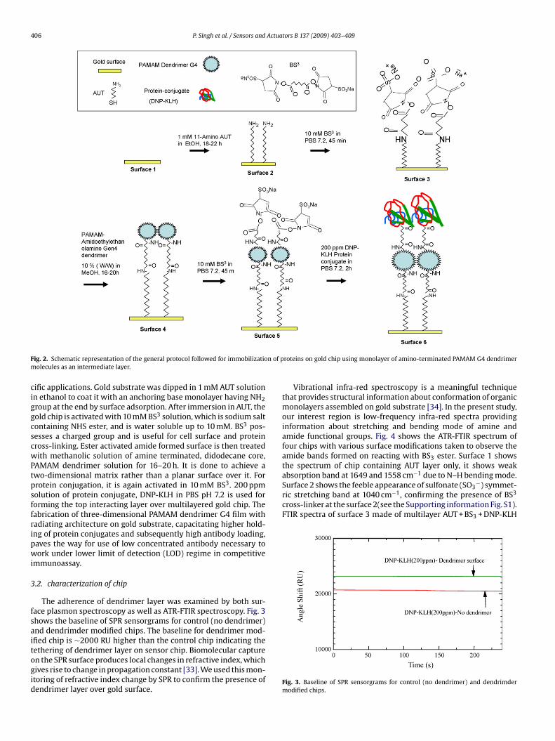
406 P. Singh et al. / Sensors and Actuators B 137 (2009) 403–409
F n of pm
ciggcscwPtpsffripwi
3
fsaitogid
absorption band at 1649 and 1558 cm due to N–H bending mode.Surface 2 shows the feeble appearance of sulfonate (SO3
−) symmet-ric stretching band at 1040 cm−1, confirming the presence of BS3
cross-linker at the surface 2(see the Supporting information Fig. S1).FTIR spectra of surface 3 made of multilayer AUT + BS3 + DNP-KLH
ig. 2. Schematic representation of the general protocol followed for immobilizatioolecules as an intermediate layer.
ific applications. Gold substrate was dipped in 1 mM AUT solutionn ethanol to coat it with an anchoring base monolayer having NH2roup at the end by surface adsorption. After immersion in AUT, theold chip is activated with 10 mM BS3 solution, which is sodium saltontaining NHS ester, and is water soluble up to 10 mM. BS3 pos-esses a charged group and is useful for cell surface and proteinross-linking. Ester activated amide formed surface is then treatedith methanolic solution of amine terminated, didodecane core,
AMAM dendrimer solution for 16–20 h. It is done to achieve awo-dimensional matrix rather than a planar surface over it. Forrotein conjugation, it is again activated in 10 mM BS3. 200 ppmolution of protein conjugate, DNP-KLH in PBS pH 7.2 is used fororming the top interacting layer over multilayered gold chip. Theabrication of three-dimensional PAMAM dendrimer G4 film withadiating architecture on gold substrate, capacitating higher hold-ng of protein conjugates and subsequently high antibody loading,aves the way for use of low concentrated antibody necessary toork under lower limit of detection (LOD) regime in competitive
mmunoassay.
.2. characterization of chip
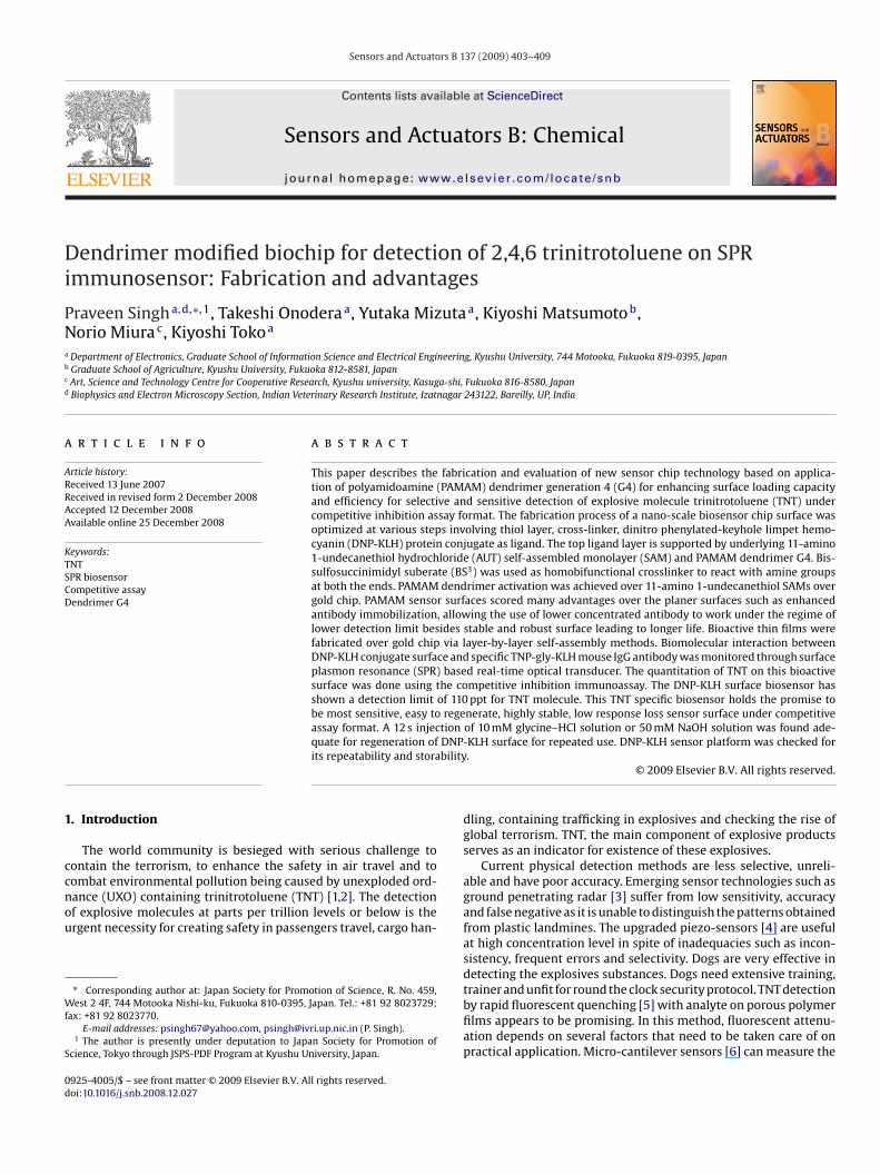
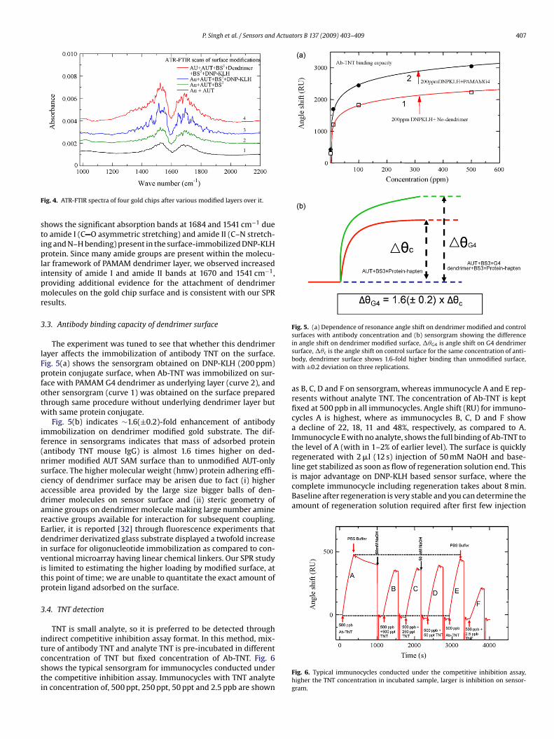
The adherence of dendrimer layer was examined by both sur-ace plasmon spectroscopy as well as ATR-FTIR spectroscopy. Fig. 3hows the baseline of SPR sensorgrams for control (no dendrimer)nd dendrimder modified chips. The baseline for dendrimer mod-fied chip is ∼2000 RU higher than the control chip indicating the
ethering of dendrimer layer on sensor chip. Biomolecular capturen the SPR surface produces local changes in refractive index, whichives rise to change in propagation constant [33]. We used this mon-toring of refractive index change by SPR to confirm the presence ofendrimer layer over gold surface.roteins on gold chip using monolayer of amino-terminated PAMAM G4 dendrimer
Vibrational infra-red spectroscopy is a meaningful techniquethat provides structural information about conformation of organicmonolayers assembled on gold substrate [34]. In the present study,our interest region is low-frequency infra-red spectra providinginformation about stretching and bending mode of amine andamide functional groups. Fig. 4 shows the ATR-FTIR spectrum offour chips with various surface modifications taken to observe theamide bands formed on reacting with BS3 ester. Surface 1 showsthe spectrum of chip containing AUT layer only, it shows weak
−1
Fig. 3. Baseline of SPR sensorgrams for control (no dendrimer) and dendrimdermodified chips.
P. Singh et al. / Sensors and Actuators B 137 (2009) 403–409 407
F
stiplipmr
3
lFpfotw
if(nscadarEdivitp
3
itcsti
Fig. 5. (a) Dependence of resonance angle shift on dendrimer modified and controlsurfaces with antibody concentration and (b) sensorgram showing the difference
line get stabilized as soon as flow of regeneration solution end. Thisis major advantage on DNP-KLH based sensor surface, where thecomplete immunocycle including regeneration takes about 8 min.Baseline after regeneration is very stable and you can determine theamount of regeneration solution required after first few injection
ig. 4. ATR-FTIR spectra of four gold chips after various modified layers over it.
hows the significant absorption bands at 1684 and 1541 cm−1 dueo amide I (C O asymmetric stretching) and amide II (C–N stretch-ng and N–H bending) present in the surface-immobilized DNP-KLHrotein. Since many amide groups are present within the molecu-
ar framework of PAMAM dendrimer layer, we observed increasedntensity of amide I and amide II bands at 1670 and 1541 cm−1,roviding additional evidence for the attachment of dendrimerolecules on the gold chip surface and is consistent with our SPR
esults.
.3. Antibody binding capacity of dendrimer surface
The experiment was tuned to see that whether this dendrimerayer affects the immobilization of antibody TNT on the surface.ig. 5(a) shows the sensorgram obtained on DNP-KLH (200 ppm)rotein conjugate surface, when Ab-TNT was immobilized on sur-
ace with PAMAM G4 dendrimer as underlying layer (curve 2), andther sensorgram (curve 1) was obtained on the surface preparedhrough same procedure without underlying dendrimer layer butith same protein conjugate.
Fig. 5(b) indicates ∼1.6(±0.2)-fold enhancement of antibodymmobilization on dendrimer modified gold substrate. The dif-erence in sensorgrams indicates that mass of adsorbed proteinantibody TNT mouse IgG) is almost 1.6 times higher on ded-rimer modified AUT SAM surface than to unmodified AUT-onlyurface. The higher molecular weight (hmw) protein adhering effi-iency of dendrimer surface may be arisen due to fact (i) higherccessible area provided by the large size bigger balls of den-rimer molecules on sensor surface and (ii) steric geometry ofmine groups on dendrimer molecule making large number amineeactive groups available for interaction for subsequent coupling.arlier, it is reported [32] through fluorescence experiments thatendrimer derivatized glass substrate displayed a twofold increase
n surface for oligonucleotide immobilization as compared to con-entional microarray having linear chemical linkers. Our SPR studys limited to estimating the higher loading by modified surface, athis point of time; we are unable to quantitate the exact amount ofrotein ligand adsorbed on the surface.
.4. TNT detection
TNT is small analyte, so it is preferred to be detected throughndirect competitive inhibition assay format. In this method, mix-
ure of antibody TNT and analyte TNT is pre-incubated in differentoncentration of TNT but fixed concentration of Ab-TNT. Fig. 6hows the typical sensorgram for immunocycles conducted underhe competitive inhibition assay. Immunocycles with TNT analyten concentration of, 500 ppt, 250 ppt, 50 ppt and 2.5 ppb are shownin angle shift on dendrimer modified surface, ��G4 is angle shift on G4 dendrimersurface, ��c is the angle shift on control surface for the same concentration of anti-body, dendrimer surface shows 1.6-fold higher binding than unmodified surface,with ±0.2 deviation on three replications.
as B, C, D and F on sensorgram, whereas immunocycle A and E rep-resents without analyte TNT. The concentration of Ab-TNT is keptfixed at 500 ppb in all immunocycles. Angle shift (RU) for immuno-cycles A is highest, where as immunocycles B, C, D and F showa decline of 22, 18, 11 and 48%, respectively, as compared to A.Immunocycle E with no analyte, shows the full binding of Ab-TNT tothe level of A (with in 1–2% of earlier level). The surface is quicklyregenerated with 2 �l (12 s) injection of 50 mM NaOH and base-
Fig. 6. Typical immunocycles conducted under the competitive inhibition assay,higher the TNT concentration in incubated sample, larger is inhibition on sensor-gram.

408 P. Singh et al. / Sensors and Actuators B 137 (2009) 403–409
Ft
oc
dcfiaeiacdws
FaD
Table 1TNT competitive SPR.
Analyte TNTTNT antibody mAb A1.1.1Protein-conjugate DNP-KLHInhibition concentration (IC50) ng/mla ∼4.0 ± 1.4LOD (ppt) pg/mlb 110 ± 50 (S.D.)
a
ig. 7. Typical immunoassay conducted on one of the prepared surface reported inhis study. This immunoassay indicates the limit of detection as 60 ppt.
n surface. Surface needs little more regeneration solution if thehip is used after long storage period.
Fig. 7 shows the results of an immunoassay conducted onendrimer modified surface as bound percentage of Ab-TNT andoncentration of analyte. This is a typical immunoassay per-ormed on one of the prepared surface reported in this study. Thismmunoassay indicates the limit of detection as 60 ppt, which ist 12% reduced response level than no analyte response consid-ring the 3 standard deviation above the background. Inside boxn Fig. 8 shows the curve depicting dependence of binding rate vs.nalyte concentration on same immunoassay. Binding rate was cal-
ulated as ıR/ıt at t = 0, where R is angle shift in resonance unituring the ıt interval of time [33]. Binding rate is equal to rate athich Ab-TNT associates at beginning before the initiation of dis-ociation from surface. Binding rate vs. concentration curve shows
ig. 8. (a) Response of Ab-TNT against repeated regeneration on DNP-KLH surfacend (b) response of Ab-TNT against repeated regeneration on dendrimer modifiedNP-KLH surface.
Inhibition concentration (IC50): concentration based on 50% inhibition of Abbinding. Error bars represent mean of triplicate samples with an average CV ∼4%.
b LOD calculated based on mean of triplicate surfaces prepared at different time,±3 S.D. above background.
the limit of detection reduced to 32 ppt level at 12% inhibition from60 ppt equilibrium level. Fig. 8 shows that IC50 is ∼3 ppb and detec-tion range is up to 7 ppb. DNP-KLH matrix based biosensors haveshown advantageous properties with regards to sensitivity, sta-bility, regeneration, storage life and measurement cycle [18], thisdendrimer modified DNP-KLH biosensors further reduced the limitof detection and provided more stability for repeated use.
Table 1 shows the summary of results of competitive SPR exper-iments. The present study uses DNP-KLH protein conjugate onlyon dendrimer modified and unmodified surfaces and Ab-TNT fromBiosolutions named mAb A1.1.1. Limit of detection (LOD) is a calcu-lated mean (110 ppt) of triplicate measurements on three differentchip surfaces with standard deviation (S.D.) of ±50. The high S.D.in LOD is the outcome of small variation in chip surface condi-tions owing to the variations in preparation conditions and runningbuffer condition.
3.5. Comparison of dendrimer modified surface with DNP-KLHonly surface
LOD was found to be 150 ppt for TNT analyte over DNP-KLHsurface with no underlying dendrimer layer under competitiveinhibition assay format as reported by us [18]. Dendrimer modi-fied DNP-KLH biosensor has shown a detection limit of 110 ppt asreported in Table 1, which is significant improvement over the samesurface without dendrimer layer. LOD on the basis of binding rate isfurther reduced to 32 ppt level for this assay without any recourseto sensitivity enhancement step. This may be the results of easieraccessibility of binding site on dendrimer modified surface archi-tecture with increased activity of modified surface. The attempt wasmade to evaluate the surface response after repeated regenerationsteps employing fixed concentration Ab-TNT in each injection cycle.
Fig. 8(a) shows the 500 ppb Ab-TNT response over DNP-KLH sur-face without underlying dendrimer monolayer against repeatedregeneration with 50 mM NaOH. The graph shows that responseincreases in first 4–5 Ab-TNT injections with intermittent few lowresponses before stabilizing in later injections at higher RU. Fig. 8(b)shows the 500 ppb Ab-TNT response over dendrimer derivatizedDNP-KLH surface and is distinct from Fig. 8(a). Ab-TNT responseincreases initially before stabilizing at higher RU in later injections.Standard deviation in sensor response (RU) is up to ∼3% on den-drimer based sensor surface, where as it is ∼4% on surface withoutdendrimer G4. Dendrimer surface score over unmodified surface interms of response stability, sensitivity and lower detection limit.
4. Conclusions
We have successfully demonstrated the fabrication and evalua-tion of new sensor chip technology based on competitive inhibition
assay and application for PAMAM dendrimer G4 for enhancingsurface loading capacity and efficiency for selective and sensi-tive detection of explosive molecule TNT. The fabrication processof a nano-scale biosensor chip surface was optimized at varioussteps involving thiol layer, cross-linking of molecules and DNP-KLH
Actua
plSd
SitassduKTms1aTtd
A
(ftoWKo
A
t
R
[
[[
[
[
[
[
[
[
[[[[[[[
[
[[
[
[[[[
P. Singh et al. / Sensors and
rotein conjugate as ligand supported by AUT self assembled mono-ayer (SAM). Dendrimer derivatized surface was characterized byPR spectroscopy as well as ATR-FTIR for confirmation of anchoredendrimer layer.
It is also demonstrated that dendrimer monolyers on reactiveAMs offer many advantages as a biomolecular interface for achiev-ng high antibody loading density, surface stability etc. Bioactivehin films were fabricated over gold chip via layer-by-layer selfssembly methods. Biomolecular interaction between substratepecific TNP-gly-KLH mouse Ig antibody and DNP-KLH conjugateurface was monitored through SPR based real-time optical trans-ucer. The quantitation of TNT on this bioactive surface was donesing the competitive inhibition assay immunoassay. The DNP-LH surface biosensor has shown a detection limit of 110 ppt forNT molecule. This TNT specific biosensor holds the promise to beost sensitive, easy to regenerate, highly stable, low response loss
ensor surface under competitive assay format. A 12 s injection of0 mM glycine–HCl solution or 50 mM NaOH solution was founddequate for regeneration of DNP-KLH surface for repeated use.hese advantages are likely to originate from the structural fea-ures of the dendrimer monolayers such as the surface exposure oferivatized DNP-KLH ligand and a corrugated surface.
cknowledgements
Financial support from Japan Society for Promotion of ScienceJSPS) Japan to Praveen Singh (P.S.), in the form of JSPS-PDF fororeign researcher is gratefully acknowledged. P.S. is also thankfulo ICAR, New Delhi, India for grant of study leave for this collab-rative research work. This work was partially supported by JST.e are indebted to the Centre of Advanced Instrumental Analysis,
yushu University for helping us in the measurement and analysisf ATR-FTIR scans.
ppendix A. Supplementary data
Supplementary data associated with this article can be found, inhe online version, at doi:10.1016/j.snb.2008.12.027.
eferences
[1] P. Julicher, E. Mussenbrock, R. Renneberg, K. Cammann, Anal. Chim. Acta 315(1995) 279–287.
[2] E.H. Lan, B. Dunn, J.I. Zink, Chem. Mater. 12 (2000) 1874–1878.[3] C.J. Cumming, C. Aker, M. Fisher, M. Fox, M.J.L. Grone, D. Reust, M.G. Rockly, T.M.
Swager, E. Towers, V. Williams, IEEE Trans. Geosci. Remote Sensing 39 (2001)1119–1128.
[4] K. Toko, Biomimetic Sensor Technology, 1st ed., Cambridge University Press,2000.
[5] J.S. Yang, T.M. Swager, J. Am. Chem. Soc. 120 (1998) 11864–11873.[6] T. Thundat, Nanosensors get practical, SPIE’s Mag. (2005) 25–26, February.[7] I.B. Bakaltcheva, F.S. Ligler, C.H. Patterson Jr., L.C. Shriver-lake, Anal. Chim. Acta
399 (1999) 13–20.[8] M. Altstein, A. Bronshtein, B. Glattstein, A. Zeichner, T. Tamiri, J. Almog, Anal.
Chem. 73 (2001) 2461–2467.[9] P.T. Charles, A.W. Kusterbeck, Biosens. Bioelectron. 14 (1999) 387–396.10] S.Y. Rabbany, W.J. Lane, W.A. Marganski, A.W. Kusterbeck, F.S. Ligler, J. Immunol.
Methods 246 (2002) 69–77.11] J. Homola, S. Yee, G. Gauglitz, Sens. Actuators B 54 (1999) (1999) 3–15.
12] A. Larsson, J. Angbrant, J. Ekeroth, P. Mansson, B. Liedberg, Sens. Actuators B:Chem. 113 (2006) 730–748.13] Q. Yu, S. Chen, A.D. Taylor, J. Homola, B. Hock, S. Jiang, Sens. Actuators B 107
(2005) 193–201.14] A.N. Naimushin, S.D. Soelberg, D.K. Nguyen, L. Dunlap, D. Barthollomew, J.
Elkind, J. Melendez, C. Furlong, Biosens. Bioelectron. 17 (2002) 573–584.
tors B 137 (2009) 403–409 409
15] T.M. Chinowsky, J.G. Quinn, D.U. Bartholomew, R. Kaiser, J.L. Elkind, Sens. Actu-ators B 6954 (2003) 1–9.
16] D. Ravi Shankaran, K.V. Gobi, K. Matsumoto, T. Imato, K. Toko, N. Miura, Sens.Actuators B 100 (2004) 450–454.
[17] K. Matsumoto, A. Torimaru, S. Ishitobi, T. Sakai, H. Ishikawa, K. Toko, N. Miura,T. Imato, Talanta, 68 (2005) 305–311.
18] P. Singh, T. Onodera, Y. Mizuta, K. Matsumoto, N. Miura, K. Toko, Sens. Mater. 19(2007) 261–273.
19] J. Lahiri, L. Isaacs, B. Grzybowski, J.D. Carbeck, G.M. Whitesides, Langmuir 15(1999) 7186–7198.
20] M. Riepl, K. Enander, B. Liedberg, Langmuir 18 (2002) 7016–7023.21] R.G. Nuzzo, D.L. Allara, J. Am. Chem. Soc. 105 (1983) 4481–4483.22] A. Ulman, Chem. Rev. 96 (1996) 1533–1554.23] R.M. Crooks, A.J. Ricco, Accounts Chem. Res. 31 (1998) 219–227.24] M.Y. Hong, D. Lee, H.C. Yoon, H.S. Kim, Bull. Kor. Chem. Soc. 24 (2003) 1197–1202.25] J.G. Worden, Q. Dai, Q. Huo, Chem. Commun. (2006) 1536–1538.26] R. Benters, C.M. Niemeyer, D. Drutschmann, D. Blohm, D. Wohrle, Nucleic Acids
Res. 30 (2002) (2002) 1–7e10.27] S.S. Mark, N. Sandhyarani, C. Zhu, C. Campagnolo, C.A. Batt, Langmuir 20 (2004)
6808–6817.28] P. Singh, Bioconjugate Chem. 9 (1998) 54–63.29] T. Park, J. Choo, M. Lee, Y.S. Kim, E.K. Lee, H.S. Lee, Anal. Sci. 20 (2004) 1255–
1258.30] N. Miura, H. Higobashi, G. Sakai, A. Takeyasu, T. Uda, N. Yamazoe, Sens. Actuators
B 13–14 (1993) 188–191.31] A. Rar, M. Curry, J.A. Barnard, Tribol. Lett. 12 (2002) 87–94.32] R. Benters, C.M. Niemeyer, D. Wohrle, Chem. Biochem. 3 (2001) 686–694.33] J. Homola, Anal. Bioanal. Chem. 377 (2003) 528–539.34] P.E. Laibinis, G.M. Whitesides, D.L. Allara, Y.T. Tao, A.N. Parikh, R.G. Nuzzo, J. Am.
Chem. Soc. 113 (1991) 7151–7167.
Biographies
Praveen Singh had obtained his PhD degree from Department of Physics, IndianInstitute of Technology Delhi, New Delhi India in 2001 under the supervision of Prof.R.K. Puri and Prof. T.C. Goel. He had worked as scientist (physics) at CAZRI Jodh-pur from 1996 to 1999, then moved to Indian veterinary research Institute (IVRI),Bareilly, UP, India, where he is senior scientist (physics) since 2005 in Biophysics andElectron microscopy section. Currently, he is on deputation to department of elec-tronics, Kyushu University, Fukuoka, Japan under JSPS-PDF for foreign researcher. Hehas published 20 original research articles in journals and books. His research inter-ests encompass radar absorbing materials, magnetic field stimulation for fractureaugmentation, biosensors for explosive and biological substances.
Takeshi Onodera received his BA degree from Toyama University of InternationalStudies, Toyama, Japan, in 1996. He received his MEd degree in technology educa-tion and PhD degree in engineering from Kanazawa University in 1998 and 2001,respectively. He is now an assistant professor at Kyushu University. He is a memberof the Institute of Electrical Engineers of Japan, the Electrochemical Society of Japan.
Yutaka Mizuta received his BSc degree in organic chemistry from Waseda University,Japan, in 2000. He works for the Central Customs Laboratory. Since 2006, he has beenengaged in the development of SPR based immunosensors for explosives detectionat Kyushu University as a cooperative researcher.
Kiyoshi Matsumoto has been a professor at Kyushu University since 1989. Hereceived his DAgr degree from the Department of Food Science and Technology,Kyushu University, Japan. He has published more than 150 original papers and 50reviews. His current research concentrates on the development of enzyme sensorsfor food quality control and the preparation of new antibodies for the developmentof SPR biosensors.
Norio Miura has been a professor at Kyushu University since 1999. He received DEngdegree in 1980 from the Department of Applied Chemistry, Kyushu University, Japan.He has published more than 300 original papers and has nearly 120 patents. Hiscurrent research concentrates on the development of high-performance chemicalsensors as well as other electrochemical functional devices such as selective Oxygenseparator and electrochemical capacitor.
Kiyoshi Toko received his BE, ME, and PhD degrees, all in electrical engineering,from Kyushu University, Japan, in 1975, 1977, and 1982, respectively. He is now aprofessor at the Graduate School of Information Science and Electrical Engineeringof Kyushu University. He is a member of the Japan Society of Applied Physics, theInstitute of Electrical Engineers of Japan and the Japanese Association for the Studyof Taste and Smell.

![CSE 168 [Spr 20], Lecture 17: Image-Based Rendering](https://static.fdokumen.com/doc/165x107/633317da69509937270211a2/cse-168-spr-20-lecture-17-image-based-rendering.jpg)