Decreased Brain Volume in Adults with Childhood Lead Exposure
Upload
independentCategory
view
2download
0
Ž .Molecular Brain Research 65 1999 34–43
Research report
Decreased GAP-43rB-50 phosphorylation in striatal synaptic plasmamembranes after circling motor behavior during development
Gustavo Ch. Paratcha, Gustavo R. Ibarra, Marcelo J. Wolansky, Jorge A. Rodriguez,Julio M. Azcurra )
( )Laboratorio de Biologia Celular, Facultad de Ciencias Exactas y Naturales, UniÕersidad de Buenos Aires UBA , Pabellon II, Ciudad UniÕersitaria, 1428Buenos Aires, Argentina
Accepted 24 November 1998
Abstract
Ž .We evaluated the in vitro phosphorylation of the presynaptic substrate of protein kinase C PKC , GAP-43rB-50 and the PKC activityŽ .in the striatum of rats submitted to a circling training CT test during postnatal development. Motor activity at 30 days of age, but not at
Ž .other ages, produced a unilateral reduction y29.5%; p-0.001 in the level of GAP-43rB-50 endogenous phosphorylation in thecontralateral striatum with respect to the ipsilateral side, while non-trained control animals did not show asymmetric differences.
Ž .Compared to controls, the contralateral striatum of trained animals also showed a significant reduction y29.3%; p-0.001 in theincorporation of 32P-phosphate into GAP-43. This decreased in vitro GAP-43 phosphorylation was seen at 30 min, but not immediatelyafter circling motor behavior. This contralateral change in GAP-43 phosphorylation correlated with the running speed developed by the
wŽ . Žanimals rs0.9443, ps0.0046, ns6, relative to control group and rs0.8813, ps0.0203, ns6, with respect to the ipsilateral.xside of the exercised animals . On the contrary, GAP-43rB-50 immunoblots did not show changes in the amount of this phosphoprotein
among the different experimental groups. Back phosphorylation assays, performed in the presence of bovine purified PKC, increased thewŽlevel of GAP-43rB-50 phosphorylation in the striatum contralateral to the sense of turning q22%; p-0.05, with respect to ipsilateral
. Ž .xside of the same trained group and q21%; p-0.05, relative to control group . Taken together, these results demonstrate that theactivity developed in the CT test induces a reduction in the phosphorylation state of GAP-43rB-50 in the specific site for PKC. Weconclude that general markers of activity-dependent neuronal plasticity are also altered in the same period that long-lasting changes instriatal neuroreceptors are triggered by circling motor behavior. q 1999 Elsevier Science B.V. All rights reserved.
Keywords: GAP-43rB-50; PKC; SPM; Circling training; Development; Rat striatum
1. Introduction
Ž .Circling training CT is a conditioned behavioral testwhere motor activity induces neurochemical modifications
w xin rat striatum 33,56,57 . Recently, we have shown acorrelated reduction in the expression of striatal dopamineŽ . Ž . Ž .DA D2 D2R and muscarinic acetylcholine mAChR
w xreceptors after CT activity 32 . Noteworthy, this modifica-tion is induced only when the animals are trained during a
Žlimited period of postnatal development 30–37 days of.age and represents a long-lasting change of the receptor
levels. Furthermore, the limit of this period of plasticity
) Corresponding author. C.F. Melo 4127, 1602 Buenos Aires, Ar-gentina. Fax: q54-1-833-5269; E-mail: [email protected]
can be modified by the intrastriatal infusion of nerveŽ . w xgrowth factor NGF 58 .
This period coincides with a postnatal stage of thestriatal development when maturation of cholinergic anddopaminergic systems are bearing toward formation ofmature neuronal connections. In rodent, the cholinergicsystem undergoes rapid development during the first 3–4weeks after birth. Previous work indicated that tyrosinehydroxylase and choline acetyltransferase activities reach70–75% of the activity present in the adult striatum around
w x28 days of age 14 . Moreover, the quantity of dopaminer-gic and cholinergic receptors achieve the adult values by
w xthe end of the first month of life 15,44 . Dopaminergicand cholinergic-dependent neuropeptide activity also de-velop in this period, and contribute to synaptic maturation,
w xaffecting function and plasticity 45,52 .
0169-328Xr99r$ - see front matter q 1999 Elsevier Science B.V. All rights reserved.Ž .PII: S0169-328X 98 00327-1

( )G.Ch. Paratcha et al.rMolecular Brain Research 65 1999 34–43 35
Although neurotransmitter receptors are key moleculesin these events, distinct signal transduction pathways shouldbe implicated in the amplification and stabilization of theinitial molecular steps that induce long-term alteration ofmAChR and DA D2R levels. In this regard, we havefocused our interest in two molecular species widely in-volved in neuronal plasticity: the growth associated protein
Ž .GAP-43rB-50 and protein kinase C PKC .GAP-43rB-50 is a calmodulin-binding protein substrate
for PKC, localized in growth cones, axons and presynapticw xnerve endings 1,3 . This phosphoprotein has been impli-
cated in neuronal differentiation and axon regeneration,Ž .neurotransmission, long-term potentiation LTP , associa-
tive learning and memory formation for passive avoidancew xin chicks and rats 2,10,24,46,47 .
It was involved in morphological and functional neuro-plasticity by way of its participation in plasma membrane
w xsignal transduction and neurotransmitter release 8,18 .On the other hand, PKC is also involved in several
neuronal functions such as excitability, transmitter release,cell growth and survival and regulation of gene expressionw x5,17,31,41 . Besides its presynaptic role in GAP-43 phos-phorylation, PKC has also been implicated in mAChRphosphorylation, although this has not been probed in vivo
w xyet 29 .Previous studies have also implicated PKC as a sec-
ondary messenger in the release of DA in the nigrostriatalpathway, and have shown that D1 and D2 agonists exert adirect inhibitory effect on particulate PKC activity in
w xstriatal synaptoneurosomes 22,23 . Moreover, inhibitors ofPKC activity inhibit the potassium-evoked release of DA
w xfrom striatal synaptosomes 23 .Together, these findings support a role for DA
receptor-mediated modulation of PKC activity.Recently, we have found that endogenous phosphoryla-
tion of GAP-43rB-50 is reduced by physical exercise inw xrat striatum of 30-day old animals 43 . Based upon these
observations, we proposed that the GAP-43rPKC systemcould participate in the initial steps that trigger CT-inducedlong-lasting neuroreceptor alterations.
In the present report, we evaluate the participation ofPKC-dependent phosphorylation of GAP-43rB-50 andPKC activity in the initial mechanisms that underlie cir-cling motor behavior during striatal development.
2. Materials and methods
2.1. Circling training
Male Sprague–Dawley rats were subjected to the CTtest at 20, 30, 40 and 60 days old, as described previouslyw x32,33 . Briefly, animals deprived of water for 24 h weretrained to turn in a circle for a water reward in threeconsecutive sessions. In the first training session, each ratwas rewarded for an approximation of a full turn in the
Ž .prescribed direction selected randomly over a 30-minperiod: in the first 10 min, a quarter turn in the appropriatedirection was rewarded, in the next 10 min, a half turn andthe last 10 min, a full turn in the appropriate direction wasrewarded. Training was then conducted daily for 2 daysaccording to a continuous reinforcement schedule. Each rat
Ž .was required to perform 150 complete turns s150 m inthese two sessions. The clockwise or anti-clockwise turn-ing direction was randomly selected for each experimental
Ž .group three to five animals in the first session and keptuntil the last session. Non-trained control animals werealso deprived of water and placed in the CT apparatus for30 min each day, but no training was performed. We havenever recorded more than 10 turns per session for anycontrol animal. Running behavior performed by trainedanimals was not observed in controls. The times wererecorded in order to calculate the running speed. The
Ž .animals with similar motor performance speed weregrouped together for correlation studies. The direction ofturning was randomly selected in each individual experi-ment and all the trained animals turned in that direction.
( )2.2. Synaptic plasma membranes SPM preparation
The experiments were performed 3 to 6 times usingthree to five animals for each experimental group. Left vs.right striata were grouped as ipsi vs. contralateral accord-
Ž .ing to the turning direction clockwise or anti-clockwiseof its respective trained group. Rats were killed by decapi-tation at 0 or 30 min post-training. Brains were quicklyremoved, left and right striata were dissected separately on
Ž .ice within 60 s and weighed. Tissue was added toŽice-cold 20 mM Tris–HCl buffer pH 7.4 10% wrv
.solution , containing 0.32 M sucrose, 1 mM EDTA, 1 mMEGTA, 1 mM PMSF, 10 mM pepstatin and 10 mMleupeptin. SPM preparations were obtained according to amodification of the method described by Azcurra and De
w xRobertis 7 . To inhibit GAP-43rB-50 dephosphorylationSPM were prepared in the presence of EGTA as described
w xGnegy et al. 26 .Tissue homogenization was performed at 1300 rpm by
40 strokes in a potter-Elvehjem glass homogenizer fittedŽ .with a loose teflon pestle clearance 0.25 mm . Following
centrifugation at 1090=g for 10 min, the pellets werewashed twice and then discarded. Supernatants were cen-
Žtrifuged again at 13 000=g for 20 min mitochondrial.pellet . Following two washes, the pellets were resus-
Ž .pended in double distilled water pH 6.8 osmotic shockand centrifuged at 20 200=g for 30 min. Then, the pelletswere resuspended in homogenization buffer, loaded on a
Ž .discontinuous sucrose gradient 0.8 to 1.0 M and cen-trifuged at 100 000=g max for 80 min at 48C in aBeckman 90 Ti rotor. The material floating on the 1 Mband was collected, washed in the same buffer, pelleted bycentrifugation at 150 000=g for 80 min and finally resus-
Žpended in buffer A 10 mM Tris–HCl buffer pH 7.4,

( )G.Ch. Paratcha et al.rMolecular Brain Research 65 1999 34–4336
.10 mM MgCl , 1 mM CaCl . Protein concentration was2 2w xdetermined by the method of Lowry et al. 37 using
bovine serum albumin as standard.
2.3. GAP-43rB-50 phosphorylation assays
In vitro endogenous phosphorylation was performed inw xconditions known to activate PKC 36 . Briefly, aliquots of
Ž .SPM 10–20 mg were preincubated for 5 min at 308C in afinal volume of 25 ml in buffer 10 mM Tris–HCl pH 7.4,10 mM MgCl and 1 mM CaCl . The reactions were2 2
initiated by the addition of 7.5 mM ATP and 2 mCi
w 32 x Ž y1 .g- P ATP Dupont 6000 Ci mmol in buffer A andterminated 1 min later by the addition of 12.5 ml stopping
Žsolution 187.5 mM Tris–HCl pH 6.5, 6% SDS, 15%b-mercaptoethanol, 30% glycerol and 0.003% bromophe-
.nol blue . In some experiments, exogenous PKC, purifiedw xfrom bovine brain as indicated Kitano et al. 35 and
w xaccording to the modifications done by Gomez et al. 27 ,´was added to the incubation media of heat-inactivated
Ž .SPM 1008C, 5 min . Phosphorylation reactions were al-Žlowed to proceed for 10 min and 10 ml of PKC 2 pmol
y1 y1.min ml were added for each 10 mg of SPM proteinŽ .Back phosphorylation .
Fig. 1. Upper panel: representative autoradiograms showing the in vitro phosphorylation of striatal SPM proteins from control and trained rats of 20, 30, 40and 60 days of age. The animals were sacrificed 30 min after CT. Lane 1, ipsilateral control; lane 2, ipsilateral trained; lane 3, contralateral control andlane 4, contralateral trained. Arrowheads indicate the position of molecular weight markers: 95.5, 55, 43, 36, 29, 18.5, and 12 kDa. Lower panel:
Ž .quantification of GAP-43rB-50 in vitro phosphorylation. Data are expressed as percentages of the maximal non-trained control value and represent themean"S.D. for three to four independent experiments. Each determination was assayed in quadruplicate. ) p-0.001 vs. controls by two-tail Student’st-test for unpaired samples and vs. ipsilateral trained by two-tail Student’s t-test for paired samples.

( )G.Ch. Paratcha et al.rMolecular Brain Research 65 1999 34–43 37
2.4. GAP-43rB-50 immunoprecipitation
GAP-43rB-50 immunoprecipitation was performed ac-w xcording to the method described by Oestreicher et al. 42 .
Samples containing 25 mg of in vitro phosphorylatedmembrane proteins were incubated overnight at 48C with a
Žpolyclonal rabbit anti B-50rGAP-43 antibody 1r200 fi-. Žnal dilution from serum 8615 in 400 ml buffer B 200
mM NaCl; 10 mM EDTA; 10 mM NaH PO ; 0.5%2 4Ž ..Nonidet P-40 vrv . Antigen complexes were precipitated
Ž .with washed pansorbin Calbiochem , solubilized in 30 mlof stop–mix solution, and boiled for 10 min. Immunopre-cipitates were analyzed by SDS-PAGE in duplicate. Incor-poration of 32 P into GAP-43rB-50 was detected using anAGFA-Curix film and quantified using a MCID Image
Ž .Analysis System 5.02v, Image Research, Ontario, Canada .
2.5. Electrophoresis
Samples containing 10 to 20 mg of SPM protein weresubjected to a one-dimensional 12% SDS-polyacrylamide
Ž .gel electrophoresis SDS-PAGE . The gels were subse-quently stained with Coomasie blue, destained and autora-diographed by exposing the gels to an AGFA-Curix film.Finally, densitometric analysis of the X-ray film was per-
Žformed using a MCID Image Analysis System 5.02v,.Image Research .
2.6. Western blotting of GAP-43rB-50
Ž .SPM proteins 20 mg were electrophoretically trans-Ž . Žferred 12 h at 250 mA to nitrocellulose membranes pore
. w xsize 0.45 mm as described by Towbin et al. 55 . Afterblocking, the membrane was incubated for 12 h at roomtemperature with a specific affinity-purified anti B-50 IgGsŽ . w x1r1000 obtained from serum 8920 42 . Finally,immunoreactivity against GAP-43rB-50 was detected byusing a biotinylated secondary antibody and a horseradish
Žperoxidase-conjugated biotin–avidin complex Vectastain.Elite ABC Kit, Vector Labs and visualized with di-
aminobenzidine as chromogen.
2.7. Determination of PKC actiÕity
PKC activity was determined by an in vitro phosphory-w xlation assay using histone III-S as substrate 13 . The
reactions were performed using 10 mg of SPM proteins inŽ .25 ml final volume of 50 mM Tris–HCl buffer, pH 7.4;
0.2 mM EDTA; 10 mM Mg Cl ; 5 mM b-mercapto-2y1 Ž .ethanol; 10 mM ATP; 100 mg ml PKA inhibitor Sigma ;
y1 w 32 x0.6 mg ml histone H III-S and 1 mCi g- P ATP,during 1 min at 308C. Phosphate incorporation was evalu-ated in the presence or absence of PKC activators: 0.1 mMphorbol ester TPA; 100 mg mly1 phosphatidylserineŽ .Sigma and 1 mM CaCl . The reaction was terminated by2
addition of 12.5 ml stopping solution. The labeled phos-
phoproteins were separated on a 15% SDS-PAGE andfollowing staining and destaining, the gels were dried andexposed to an AGFA-Curix film. The histone protein bandwas cut from the dried stained gel and the radioactivityincorporated counted in a liquid scintillation counter. Thetotal PKC activity was calculated as pmol 32 P incorporatedminy1 mg of proteiny1 above the basal activity, whichwas determined in the absence of PKC activators.
2.8. Statistical analysis
Ž .A two-way analysis of variance ANOVA was used toanalyze the results. The ANOVA design considered con-trol vs. trained as one factor, and ipsi vs. contralateralhemisphere as another. Differences between groups wereassessed by Student’s t-test for unpaired and paired sam-
Ž .ples. The correlation coefficient r was obtained studyingthe degree of linear association between two variablesusing Graph Pad Instat Software.
Fig. 2. Upper panel: representative autoradiogram showing a specificimmunoprecipitation of GAP-43rB-50 phosphorylated from striatal SPM.Animals of 30 days of age were sacrificed 30 min post-training. Lane 1,ipsilateral control; lane 2, ipsilateral trained; lane 3, contralateral controland lane 4, contralateral trained. Arrowheads indicate the position ofmolecular weight markers: 95.5, 55, 43, 36, 29, 18.5, and 12 kDa. Lowerpanel: quantification of the GAP-43rB-50 phosphorylated band. Data are
Ž .expressed as percentages of the maximal non-trained control value andrepresent the mean"S.D. for four independent experiments assayed induplicate. ) p-0.05 vs. controls by two-tail Student’s t-test for unpairedsamples and vs. ipsilateral trained by two-tail Student’s t-test for pairedsamples.

( )G.Ch. Paratcha et al.rMolecular Brain Research 65 1999 34–4338
3. Results
3.1. Endogenous phosphorylation of GAP-43rB-50 afterCT
SPM preparations were used to assess phosphorylationof GAP-43rB-50 because this protein is a prominentphosphoband in this fraction and is readily detected underincubation conditions which are optimal for PKC activa-tion. The identity of GAP-43rB-50 was established on thebasis of electrophoretic properties on unidimensionalSDS-PAGE, selective phosphorylation by PKC, and spe-cific immunoreactivity detected on Western blots and byimmunoprecipitation.
To assess whether GAP-43rB-50 endogenous phospho-rylation is altered after CT during striatal development, weisolated SPM from ipsi and contralateral hemispheres, andsubjected them to an in vitro phosphorylation assay underconditions known to activate PKC. As can be seen in Fig.1, only trained animals of 30 days of age showed anaverage decrease of 29.5% in the level of GAP-43rB-50endogenous phosphorylation in the contralateral striatum
Ž .relative to the ipsilateral side p-0.001 . Significantinterhemispheric differences were not found in controlanimals, neither when tested left vs. right nor when striatawere grouped as ipsi vs. contralateral, according to therespective trained group.
Furthermore, at this age, the CT produced a reductionof 29.3% in the level of endogenous phosphorylation of
ŽGAP-43rB-50, in comparison with control animals p-
.0.001 . In contrast, animals trained at 20, 40 or 60 days ofage did not show significant differences in the endogenousphosphorylation of GAP-43rB-50 with respect to the con-
Ž .trol animals nor between hemispheres Fig. 1 . In 30-dayold trained animals, the changes were seen 30 min post-training in the striatum contralateral to the sense of turn-ing, independently of the direction of turning chosenŽ .clockwise or anti-clockwise .
In contrast, animals sacrificed immediately after CTŽ .activity 0 min did not show any significant modification
in the in vitro GAP-43rB-50 phosphorylation when com-Ž .pared with controls in both ipsilateral q11%"3 and
Ž .contralateral striata y10%"2 .To confirm the identity of GAP-43rB-50 and the mag-
nitude of the change obtained using endogenous phospho-rylation and autoradiography of SPM phosphoproteins,specific immunoprecipitation of GAP-43rB-50 phosphory-
Ž .lated was performed Fig. 2 .Densitometric analysis shows an average drop of 28.5%
Ž .p-0.05 with respect to the control animals, and ofŽ .28.0% p-0.05 between ipsi vs. contralateral striata
from 30-day old trained animals.To study whether CT affects the amount of GAP-43rB-
50 protein in SPM isolated from control and trained ani-mals, we performed immunoblots using affinity-purifiedantibodies against GAP-43rB-50. The reduced phosphory-lation observed in the present study does not seem to bedue to a difference in GAP-43rB-50 protein levels, be-cause densitometric quantitation of the Western blots didnot show any difference between control and trained ani-
Ž .Fig. 3. Upper panel: a representative Western blot analysis of GAP-43rB-50 immunoreactivity, using striatal SPM proteins 20 mg from control andtrained animals of 20, 30, 40 and 60 days of age, isolating separately the ipsi and contralateral striata. Lane 1, ipsilateral control; lane 2, ipsilateral trained;lane 3, contralateral control; and lane 4, contralateral trained. In all cases, the animals were sacrificed 30 min post-training. Arrowheads indicate theposition of molecular weight markers: 95.5, 55, 43, 36, 29, 18.5, and 12 kDa. Lower panel: quantification of GAP-43rB-50 immunoreactivity. Data are
Ž .expressed as percentages of the maximal non-trained control value and represent the mean"S.D. for three to four independent determinations performedin triplicate.

( )G.Ch. Paratcha et al.rMolecular Brain Research 65 1999 34–43 39
Ž .mals of 30 days of age Fig. 3 . No significant differenceswere found in the GAP-43rB-50 immunoreactivity whenthe animals were trained at 20, 40 or 60 days of ageŽ . Ž .p)0.1 Fig. 3 .
PKC phosphorylates the Ser 41 residue of GAP-43rB-w x50 12,40 . However, this protein can be also in vitro
Ž .phosphorylated by casein kinase II CKII on Ser 192, 193w xand Thr 88, 89 and 95 6 . In order to exclude a dual
participation of PKC and CKII in our preparations, weperformed the phosphorylation assays in the presence of
Ž .heparin a potent inhibitor of CKII . The presence ofheparin did not alter the differences observed in the in
Ž .vitro GAP-43rB-50 phosphorylation data not shown .
3.2. GAP-43rB-50 phosphorylation Õariation is corre-lated with speed
To study a possible correlation of this change with themotor performance developed by the animals during theCT, the rats were grouped according to their running speedŽ .S.D.-10% on each group . Plotting the percentage of
Ždrop in the in vitro GAP-43rB-50 phosphorylation rela-.tive to control animals as a function of motor intensity,
we were able to find a significant correlation betweenŽ . Žthese two variables rs0.9443; ps0.0046; ns6 Fig.
.4a . Similarly, a significant correlation was found plotting
Fig. 4. Correlation between percentage of decrease of the in vitroGAP-43rB-50 phosphorylation in the contralateral striatum and motor
Ž . Ž .activity speed accomplished during CT. a Percentage relative toŽ .control group. b Percentage relative to ipsilateral side also trained.
Ž .Animals of 30 days old were sacrificed 30 min post-CT. a r s0.9443,Ž .ps0.0046, ns6 and b r s0.8813, ps0.0203, ns6.
Fig. 5. Upper panel: representative autoradiogram showing a PKC-media-ted posthoc phosphorylation of striatal SPM proteins from control andtrained rats, 30 days old. Animals were sacrificed 30 min post-training.Heat-inactivated SPM were phosphorylated in the presence of partially
w 32 xpurified bovine PKC and g- P ATP and subjected to SDS-PAGE asdescribed in Section 2. Lane 1, ipsilateral control; lane 2, ipsilateraltrained; lane 3, contralateral control; and lane 4, contralateral trained.Arrowheads indicate the position of molecular weight markers: 95.5, 55,43, 36, 29, 18.5, and 12 kDa. Lower panel: quantification of GAP-43rB-50 back phosphorylation. Data are expressed as percentages of themaximal control value and represent the mean"S.D. of four independentexperiments. Each determination was assayed in quadruplicate. ) p-0.05vs. controls by two-tail Student’s t-test for unpaired samples and vs.ipsilateral trained by two-tail Student’s t-test for paired samples.
the speed of turning and the percentage of decrease in theendogenous GAP-43rB-50 phosphorylation relative to the
Ž .ipsilateral trained striatum rs0.8813; ps0.0203; ns6Ž .Fig. 4b .
3.3. Post-hoc phosphorylation of GAP-43rB-50 in striatalSPM after CT
As GAP-43rB-50, levels were not modified among thedifferent experimental groups, we investigated whether theendogenous phosphorylation state of GAP-43rB-50 wasaltered 30 min post-training. To address this question, theendogenous kinase systems present in SPM were inacti-
Ž .vated by heating 5 min at 1008C and protein phosphory-lation present in heated SPM was carried out by adding
Ž .purified bovine PKC back phosphorylation . Fig. 5 showsan enhanced ability of GAP-43rB-50 to be phosphorylatedin the contralateral striatum from trained animals. Densito-
Ž .metric analysis shows an increment of 21% p-0.05Ž .with respect to control group and of 22% p-0.05 with
respect to the ipsilateral striatum of the same trainedŽ .animals Fig. 5 .

( )G.Ch. Paratcha et al.rMolecular Brain Research 65 1999 34–4340
Table 1Effect of CT on ratios of PKC activity in striatal SPM during development
32 y1 y1Ž . Ž .Training age days Ratios of PKC activity PKC activity pmol P min mg
CcrIc CtrIt ItrIc CtrCc Ic Cc
20 0.94"0.02 1.07"0.11 0.99"0.04 1.13"0.05 71.65"8.47 67.05"9.3030 0.90"0.03 0.67"0.10) 1.09"0.09 0.80"0.07a 68.67"10.7 62.40"11.5640 0.98"0.03 0.90"0.07 0.99"0.05 0.90"0.03 67.20"7.95 66.30"6.0560 0.98"0.07 0.89"0.06 1.01"0.04 0.91"0.03 67.20"6.56 65.70"5.73
Animals were sacrificed 30 min post-CT. Values are expressed as means"S.D. for three to four independent determinations. The PKC activity wasmeasured in quadruplicate as described in Section 2. The different experimental groups are: Cc: contralateral control; Ic: ipsilateral control; Ct:contralateral trained and It: ipsilateral trained. This table also shows the values of PKC activity presents in striatal SPM from control rats duringdevelopment. They are expressed as pmol 32 P incorporated miny1 mgy1 of protein above basal activity. ) p-0.05 vs. CcrIc by two-tail Student’s t-testfor unpaired samples and ap-0.05 vs. ItrIc by two-tail Student’s t-test for paired samples.
3.4. Determination of PKC actiÕity after CT during deÕel-opment
To test whether the change in endogenous GAP-43rB-50 phosphorylation caused by CT could be due to areduced PKC activity, SPM preparations of control andtrained animals were used to measure total PKC activity at30 min post-training. Particulate PKC activity was deter-
Žmined using specific PKC activators PS and TPA and.calcium and an excess of histone H-III S as substrate.
Table 1 shows that the ratio of PKC activity between both30-day old trained striata is significantly different to theratio of PKC activity detected between both control striataŽ w x.ItrCt vs. IcrCc, p-0.05 . Moreover, the PKC activityrelative to respective control group was different between
Žboth 30 days of age trained striata ItrIc vs. CtrCc,Ž ..p-0.05 . Similar changes were not observed in animals
Ž .of 20, 40 or 60 days of age Table 1 . These resultsdemonstrate the PKC participation in the asymmetricchanges detected in the striatum of 30-day old trainedanimals, and suggest that this variation is mainly due to a20% decrease in the contralateral PKC activity relative tothe corresponding control hemisphere.
4. Discussion
We have studied GAP-43rB-50 phosphorylation andPKC activity in the rat striatum after CT activity duringpostnatal development. Results show that this circlingmotor behavior elicits a unilateral drop in the phosphoryla-tion state of GAP-43rB-50, when the animals were trainedat 30 days of age, indicating that this variation is not onlyrelated to the physiological stimulation but also with aspecific period during development. This reduction wasseen at 30 min post-training in the striatum contralateral tothe sense of turning, independently of the direction of
Ž .turning chosen clockwise or anti-clockwise . Similar re-sults were not reached when the animals were trained at20, 40 or 60 days old.
It has been shown that PKC-dependent GAP-43rB-50w xphosphorylation increases neurotransmitter release 18,26 .
However, we have found a reduction in the GAP-43Ž .phosphorylation state in the specific site for PKC Ser 41 ,
which may indicate a decrease of striatal neurotransmis-sion. A point of concern is that GAP-43 modification wasfound 30 min post-training, but not immediately after thatCT activity was performed. Delayed changes in GAP-43phosphorylation are related with molecular events associ-
w xated with plastic processes of synaptic function 24,25,47 .Thus, it is likely that GAP-43 alteration is playing amodulatory role in the plastic events triggered after com-pletion of CT. In adult striatum, a role for GAP-43 phos-phorylation in priming or modulation of neurotransmitter
Ž .release in rat striatum after amphetamine AMPH treat-w xment has been proposed by Gnegy et al. 26 .
Since the level of GAP-43rB-50 immunoreactivity inSPM was similar between trained and non-trained controlanimals of 30 days of age, the decrease in the phosphoryla-tion might be the consequence of a reduced amount ofmembrane bound protein kinase, a reduced concentration
Žof endogenous activators i.e., diacylglycerols, phos-.phatidylserine or arachidonic acid or an increased phos-
phatase activity. To determine if the drop in the endoge-nous phosphorylation of GAP-43 was related to a reducedPKC activity, we measured the activity of this kinase incontrol and trained animals at different postnatal ages.These results also demonstrate an asymmetric alteration inthe particulate PKC activity. Thus, the present findingssuggest that the decreased GAP-43rB-50 phosphorylationcould be related to a reduced contralateral PKC activity inSPM isolated from 30-day old trained animals. Our resultsare in agreement with other neurochemical and anatomicalstudies indicating that the experience may modify differentdeveloping neuronal systems through the mediation of
w xPKC phosphorylation events 21 . In this context, it hasbeen reported that PKC activity can be regulated by sen-sory deprivation in the olfactory and visual system. There-fore, different sensory and motor systems may use com-mon molecular mechanisms to process information. Specif-ically in rat striatum, it has been shown that activation ofDA D1 and D2 receptors exert a direct inhibitory effect on
w xparticulate PKC activity 22,23 . These reports support a
( )G.Ch. Paratcha et al.rMolecular Brain Research 65 1999 34–43 41
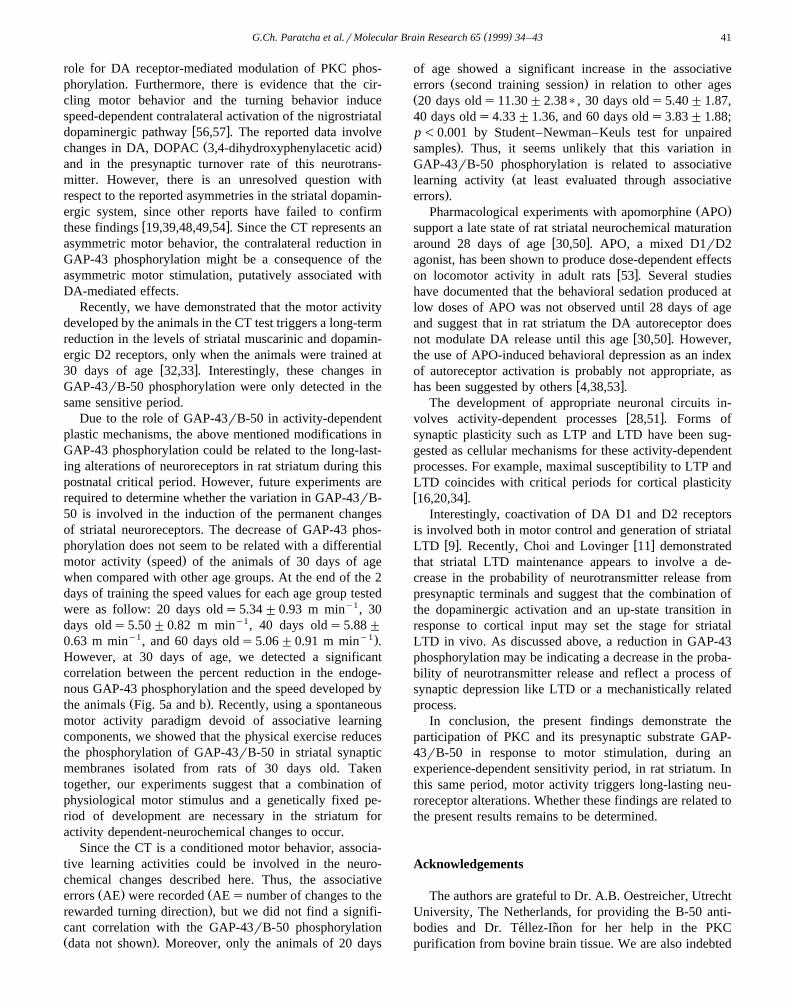
role for DA receptor-mediated modulation of PKC phos-phorylation. Furthermore, there is evidence that the cir-cling motor behavior and the turning behavior inducespeed-dependent contralateral activation of the nigrostriatal
w xdopaminergic pathway 56,57 . The reported data involveŽ .changes in DA, DOPAC 3,4-dihydroxyphenylacetic acid
and in the presynaptic turnover rate of this neurotrans-mitter. However, there is an unresolved question withrespect to the reported asymmetries in the striatal dopamin-ergic system, since other reports have failed to confirm
w xthese findings 19,39,48,49,54 . Since the CT represents anasymmetric motor behavior, the contralateral reduction inGAP-43 phosphorylation might be a consequence of theasymmetric motor stimulation, putatively associated withDA-mediated effects.
Recently, we have demonstrated that the motor activitydeveloped by the animals in the CT test triggers a long-termreduction in the levels of striatal muscarinic and dopamin-ergic D2 receptors, only when the animals were trained at
w x30 days of age 32,33 . Interestingly, these changes inGAP-43rB-50 phosphorylation were only detected in thesame sensitive period.
Due to the role of GAP-43rB-50 in activity-dependentplastic mechanisms, the above mentioned modifications inGAP-43 phosphorylation could be related to the long-last-ing alterations of neuroreceptors in rat striatum during thispostnatal critical period. However, future experiments arerequired to determine whether the variation in GAP-43rB-50 is involved in the induction of the permanent changesof striatal neuroreceptors. The decrease of GAP-43 phos-phorylation does not seem to be related with a differential
Ž .motor activity speed of the animals of 30 days of agewhen compared with other age groups. At the end of the 2days of training the speed values for each age group testedwere as follow: 20 days olds5.34"0.93 m miny1, 30days olds5.50"0.82 m miny1, 40 days olds5.88"
y1 y1.0.63 m min , and 60 days olds5.06"0.91 m min .However, at 30 days of age, we detected a significantcorrelation between the percent reduction in the endoge-nous GAP-43 phosphorylation and the speed developed by
Ž .the animals Fig. 5a and b . Recently, using a spontaneousmotor activity paradigm devoid of associative learningcomponents, we showed that the physical exercise reducesthe phosphorylation of GAP-43rB-50 in striatal synapticmembranes isolated from rats of 30 days old. Takentogether, our experiments suggest that a combination ofphysiological motor stimulus and a genetically fixed pe-riod of development are necessary in the striatum foractivity dependent-neurochemical changes to occur.
Since the CT is a conditioned motor behavior, associa-tive learning activities could be involved in the neuro-chemical changes described here. Thus, the associative
Ž . Žerrors AE were recorded AEsnumber of changes to the.rewarded turning direction , but we did not find a signifi-
cant correlation with the GAP-43rB-50 phosphorylationŽ .data not shown . Moreover, only the animals of 20 days
of age showed a significant increase in the associativeŽ .errors second training session in relation to other ages
Ž20 days olds11.30"2.38), 30 days olds5.40"1.87,40 days olds4.33"1.36, and 60 days olds3.83"1.88;p-0.001 by Student–Newman–Keuls test for unpaired
.samples . Thus, it seems unlikely that this variation inGAP-43rB-50 phosphorylation is related to associative
Žlearning activity at least evaluated through associative.errors .
Ž .Pharmacological experiments with apomorphine APOsupport a late state of rat striatal neurochemical maturation
w xaround 28 days of age 30,50 . APO, a mixed D1rD2agonist, has been shown to produce dose-dependent effects
w xon locomotor activity in adult rats 53 . Several studieshave documented that the behavioral sedation produced atlow doses of APO was not observed until 28 days of ageand suggest that in rat striatum the DA autoreceptor does
w xnot modulate DA release until this age 30,50 . However,the use of APO-induced behavioral depression as an indexof autoreceptor activation is probably not appropriate, as
w xhas been suggested by others 4,38,53 .The development of appropriate neuronal circuits in-
w xvolves activity-dependent processes 28,51 . Forms ofsynaptic plasticity such as LTP and LTD have been sug-gested as cellular mechanisms for these activity-dependentprocesses. For example, maximal susceptibility to LTP andLTD coincides with critical periods for cortical plasticityw x16,20,34 .
Interestingly, coactivation of DA D1 and D2 receptorsis involved both in motor control and generation of striatal
w x w xLTD 9 . Recently, Choi and Lovinger 11 demonstratedthat striatal LTD maintenance appears to involve a de-crease in the probability of neurotransmitter release frompresynaptic terminals and suggest that the combination ofthe dopaminergic activation and an up-state transition inresponse to cortical input may set the stage for striatalLTD in vivo. As discussed above, a reduction in GAP-43phosphorylation may be indicating a decrease in the proba-bility of neurotransmitter release and reflect a process ofsynaptic depression like LTD or a mechanistically relatedprocess.
In conclusion, the present findings demonstrate theparticipation of PKC and its presynaptic substrate GAP-43rB-50 in response to motor stimulation, during anexperience-dependent sensitivity period, in rat striatum. Inthis same period, motor activity triggers long-lasting neu-roreceptor alterations. Whether these findings are related tothe present results remains to be determined.
Acknowledgements
The authors are grateful to Dr. A.B. Oestreicher, UtrechtUniversity, The Netherlands, for providing the B-50 anti-bodies and Dr. Tellez-Inon for her help in the PKC´ ˜purification from bovine brain tissue. We are also indebted

( )G.Ch. Paratcha et al.rMolecular Brain Research 65 1999 34–4342
to Dr. Florın Christensen who kindly agreed to revise the´English version of the manuscript, Guillermo Mendizabalfor skillful technical assistance and Lic. Martın Cammarota´for densitometric measurements. This work was supportedby grants from Consejo Nacional de Investigaciones
Ž .Cientıficas y Tecnicas CONICET and University of´ ´Ž .Buenos Aires UBA , Argentina. G.Ch.P. is a recipient of
a Research Fellowship from CONICET, G.R.I. is a recipi-ent of a Research Fellowship from Fundacion Antorchas´and M.J.W. is a recipient of Research Fellowship fromUBA.
References
w x1 K.A. Alexander, B.T. Wakim, G.S. Doyle, K.A. Walsh, D.R. Storm,Identification and characterization of the calmodulin-binding domainof neuromodulin, a neurospecific calmodulin binding protein, J.
Ž .Biol. Chem. 263 1988 7544–7549.w x2 S.M. Ali, S. Bullock, S. Rose, Phosphorylation of synaptic proteins
in chick forebrain: changes with development and passive avoidanceŽ .training, J. Neurochem. 50 1988 1579–1587.
w x3 J. Aloyo, H. Zwiers, W.H. Gispen, Phosphorylation of B-50 proteinby calcium activated phospholipid-dependent protein kinase and
Ž .B-50 protein kinase, J. Neurochem. 41 1983 649–653.w x4 S.L. Andersen, R.A. Gazzara, The ontogeny of apomorphine-in-
duced alterations of neostriatal dopamine release: effects on sponta-Ž .neous release, J. Neurochem. 61 1993 2247–2255.
w x5 P. Angel, M. Imagawa, R. Chui, B. Stein, R.J. Imbra, H.J. Rahms-dorf, C. Jonart, P. Herrlich, M. Karin, Phorbolester-inducible genescontain a common cis element recognized by a TPA-modulated
Ž .trans-acting factor, Cell 49 1987 729–739.w x6 D.E. Apel, D.W. Litchfield, R.H. Clark, E.G. Krebs, D.R. Storm,
Ž .Phosphorylation of neuromodulin GAP-43 by casein kinase II, J.Ž .Biol. Chem. 266 1991 10544–10551.
w x 147 J.M. Azcurra, E. De Robertis, Binding of kimethyl- C-D-tubo-curarine-methyl-14C-hexamethonium and 3H-allopherine by isolatedsynaptic membranes of brain cortex, Int. J. Neuropharmacol. 6Ž .1967 15–26.
w x8 L.I. Benowitz, A. Routtenberg, A membrane phosphoprotein associ-ated with neural development, axonal regeneration, phospholipid
Ž .metabolism and synaptic plasticity, Trends Neurosci. 10 1987527–531.
w x9 P. Calabresi, A. Pisani, N.B. Mercuri, G. Bernardi, The corticostri-atal projection: from synaptic plasticity to dysfunctions of the basal
Ž .ganglia, Trends Neurosci. 19 1996 19–24.w x10 M. Cammarota, G.Ch. Paratcha, M. Levi de Stein, R. Bernabeu, I.
Izquierdo, J.H. Medina, B-50rGAP-43 phosphorylation and PKCactivity are increased in rat hippocampal synaptosomal membranes
Ž .after an inhibitory avoidance training, Neurochem. Res. 22 1997499–505.
w x11 S. Choi, D.M. Lovinger, Decreased probability of neurotransmitterrelease underlies striatal long-term depression and postnatal develop-ment of corticostriatal synapses, Proc. Natl. Acad. Sci. USA 94Ž .1997 2665–2670.
w x12 P.J. Coggins, W.E. Gispen, Evidence for a single PKC-mediatedphosphorylation site in rat brain protein B-50, J. Neurochem. 53Ž .1989 1895–1901.
w x13 G. Cole, K.R. Dobkin, L.A. Hansen, R.D. Terry, T. Saitoh, De-Ž .creased levels of PKC in Alzheimer brain, Brain Res. 453 1988
165–174.w x14 J.T. Coyle, P. Campochiaro, Ontogenesis of dopaminergic–
cholinergic in the rat striatum: a neurochemical study, J. Neurochem.Ž .27 1976 673–678.
w x15 J.T. Coyle, H.I. Yamamura, Neurochemical aspects of the ontogene-
Ž .sis of cholinergic neurons in the rat brain, Brain Res. 118 1976429–440.
w x16 M.C. Crair, R.C. Malenka, A critical period for long-term potentia-Ž .tion at thalamocortical synapses, Nature 375 1995 325–328.
w x17 L.V. Dekker, P.N.E. De Graan, W.H. Gispen, Transmitter release:Ž .target of regulation by protein kinase C, Prog. Brain Res. 46 1991
209–233.w x18 L.V. Dekker, P.N.E. De Graan, D.H.G. Versteeg, A.B. Oestreicher,
Ž .W.H. Gispen, Phosphorylation of B-50 GAP-43 is correlated withneurotransmitter released in rat hippocampal slices, J. Neurochem.
Ž .52 1989 24–30.w x19 M. Diana, M. Garcıa-Munoz, J. Richards, C.R. Freed, Electrophysio-´ ˜
logical analysis of dopamine cells from the substantia nigra parsŽ .compacta of circling rat, Exp. Brain Res. 74 1989 625–630.
w x20 S.M. Dudek, M.F. Bear, A biochemical correlate of the critic periodŽ .for synaptic modifications in the visual cortex, Science 240 1989
673–675.w x21 S. Elkabes, J.A. Cherry, A.A. Schoups, I.B. Black, Regulation of
protein kinase C activity by sensory deprivation in the olfactory andŽ .visual systems, J. Neurochem. 60 1993 1835–1842.
w x22 C.T. Giambalvo, R.L. Wagner, Activation of D1 and D2 dopaminereceptors inhibits protein kinase C activity in striatal synaptoneuro-
Ž .somes, J. Neurochem. 63 1994 169–176.w x23 C.T. Giambalvo, Protein kinase C and dopamine release: effects of
Ž .dopamine acting drugs in vivo, Biochem. Pharmacol. 37 19884009–4017.
w x24 C. Gianotti, M.G. Nunzi, W.H. Gispen, R. Corradetti, Phosphoryla-Ž .tion of the presynaptic protein B-50 GAP-43 is increased during
Ž .electrically induced long-term potentiation, Neuron 8 1992 843–848.
w x25 W.H. Gispen, H.B. Nielander, P.N.E. De Graan, A.B. Oestreicher,L.H. Schrama, P. Schotman, Role of the growth-associated protein
Ž .B-50rGAP-43 in neuronal plasticity, Mol. Neurobiol. 5 199161–85.
w x26 M.E. Gnegy, P. Hong, S.T. Ferrell, Phosphorylation of neuromod-ulin in rat striatum after acute and repeated, intermittent am-
Ž .phetamine, Mol. Brain Res. 20 1993 289–298.w x27 M.L. Gomez, E.E. Medrano, E.G.A. Caferatta, M.T. Tellez-Inon,´ ´ ˜
Protein kinase C is differentially regulated by thrombin, insulin andepidermal growth factor in human mammary tumor cells, Exp. Cell.
Ž .Res. 175 1988 74–80.w x28 C.S. Goodman, C.J. Shatz, Developmental mechanisms that generate
Ž .precise patterns of neuronal connectivity, Cell 72 1993 77–98.w x29 K. Haga, T. Haga, Ichiyama, Phosphorylation by protein kinase C of
Ž .the muscarinic acetylcholine receptor, J. Neurochem. 54 19901639–1644.
w x30 T. Hedner, P. Lundborg, Development of dopamine autoreceptors inŽ .the postnatal rat brain, J. Neural. Transm. 62 1985 53–63.
w x31 L. Hsu, Neurite-promoting effects of 12-O-tetradecanoyl-phorbol-Ž .13-acetate on chick embryo neurons, Neurosci. Lett. 62 1985
183–189.w x32 G.R. Ibarra, G.Ch. Paratcha, M.J. Wolansky, J.M. Azcurra, Co-alter-
ation of dopamine D2 receptor and muscarinic acetylcholine recep-tor binding in rat striatum after circling training, NeuroReport 7Ž .1996 2491–2494.
w x33 G.R. Ibarra, J.A. Rodriguez, G.Ch. Paratcha, J.M. Azcurra, Perma-nent alteration of muscarinic acetylcholine receptor binding in ratstriatum after circling training during development, Brain Res. 705Ž .1995 39–44.
w x34 A. Kirkwood, L. Hey-Kyoung, M.F. Bear, Co-regulation of long-termpotentiation and experience-dependent synaptic plasticity in visual
Ž .cortex by age and experientia, Nature 35 1995 328–331.w x35 T. Kitano, M. Go, V. Kikkawa, Y. Nishizuka, Assay and purifica-
Ž .tion of protein kinase C, Methods Enzymol. 124 1986 349–352.w x36 G.I. Kristjansson, H. Zwiers, A.B. Oestreicher, W.H. Gispen, Evi-
dence that the synaptic phosphoprotein B-50 is localized exclusivelyŽ .in nerve tissue, J. Neurochem. 39 1982 371–378.

( )G.Ch. Paratcha et al.rMolecular Brain Research 65 1999 34–43 43
w x37 O.H. Lowry, N.J. Rosebrough, A. Farr L, R.J. Randall, ProteinŽ .measurement with follin reagent, J. Biol. Chem. 193 1951 265–272.
w x38 M.R. Lynch, Dissociation of autoreceptor activation and behavioralconsequences of low-dose apomorphine treatment, Prog. Neuropsy-
Ž .chopharmacol. Biol. Psychiatry 15 1991 689–698.w x39 M.E. Morgan, B.K. Yamamoto, C.R. Freed, Unilateral activation of
caudate tyrosine hydroxylase during voluntary circling behavior, J.Ž .Neurochem. 43 1984 737–741.
w x40 H.B. Nielander, L.G. Schrama, H. Van Rosen, M. Kasperaitis, A.B.Oestreicher, W.H. Gispen, P. Schotman, Mutation of serine 41 in the
Ž .neuron specific protein B-50 GAP-43 prohibits phosphorylation byŽ .PKC, J. Neurochem. 55 1990 1442–1445.
w x41 Y. Nishizuka, Studies and perspectives of protein kinase C, ScienceŽ .223 1986 305–312.
w x42 A.B. Oestreicher, C.J. Van Dongen, H. Zwiers, W.H. Gispen,Affinity-purified anti B-50 protein antibody: interference with thefunction of the phosphoprotein B-50 in synaptic plasma membranes,
Ž .J. Neurochem. 41 1983 267–279.w x43 G.Ch. Paratcha, G.R. Ibarra, R. Cabrera, J.M. Azcurra, Decreased
phosphorylation of GAP-43rB-50 in striatal synaptic plasma mem-Ž .branes after circling motor activity, Neurochem. Res. 23 1998
1241–1249.w x44 J.V. Pardo, Y. Cresse, D.R. Burt, S.H. Snyder, Ontogenesis of
dopamine receptor binding in corpus striatum of the rat, Brain Res.Ž .125 1977 376–382.
w x45 E. Perez-Navarro, J. Alberch, J. Marsal, Postnatal development of´functional dopamine opioid and tachykinin receptors that regulateacetylcholine release from rat neostriatal slices: effect of 6-hydroxy-
Ž .dopamine lesion, Int. J. Dev. Neurosci. 11 1993 701–708.w x46 N.I. Perrone-Bizzozero, R.L. Neve, N. Irwin, S. Lewis, I. Fischer,
L.I. Benowitz, Post-transcriptional regulation of GAP-43 levels dur-ing neuronal differentiation and nerve regeneration, Mol. Cell. Neu-
Ž .rosci. 2 1991 402–409.w x47 G.M.J. Ramakers, P.N.E. De Graan, I.J.A. Urban, D. Kraay, T.
Tang, P. Pasinelli, A.B. Oestreicher, W.H. Gispen, Temporal differ-ences in the phosphorylation state of pre-and post synaptic protein
kinase C substrates B-50rGAP-43 and neurogranin during LTP, J.Ž .Biol. Chem. 270 1995 13892–13898.
w x48 K.E. Sabol, J.B. Richards, C.R. Freed, In vivo dialysis measurementof dopamine and DOPAC in rats trained to turn on a circular
Ž .treadmill, Pharmacol. Biochem. Behav. 36 1990 21–28.w x49 R. Schwarting, J.P. Huston, Dopamine and serotonin metabolism in
brain sites ipsi and contralateral to direction of conditioned turningŽ .in rats, J. Neurochem. 48 1987 1473–1479.
w x50 I.A. Shalaby, L.P. Spear, Psychopharmacological effects of low andhigh doses of apomorphine during ontogeny, Eur. J. Pharmacol. 67Ž .1980 451–459.
w x51 W. Singer, Development and plasticity of cortical processing archi-Ž .tecture, Science 270 1995 758–764.
w x52 S.P. Sivam, J.E. Krause, G.R. Brese, J.S. Hong, Dopamine-depen-dent postnatal development of enkephalin and tachykinin neurons of
Ž .rat basal ganglia, J. Neurochem. 56 1991 1499–1508.w x53 L. Stahle, Do autoreceptors mediate dopamine agonist-induced
yawning and suppression of exploration? A critical review, Psy-Ž .chopharmacology 106 1992 1–13.
w x54 L. Stahle, T.U. Ungersted, Yawning and suppression of explorationinduced by dopamine agonist: no relation to extracellular striatal
Ž .levels of dopamine, Pharmacol. Biochem. Behav. 35 1990 201–209.
w x55 H. Towbin, T. Staehelin, Gordon, Electrophoretic transfer of pro-teins from polyacrylamide gels to nitrocellulose sheets: procedure
Ž .and some applications, Proc. Natl. Acad. Sci. USA 76 19794350–4354.
w x56 B.K. Yamamoto, C.R. Freed, Asymmetric dopamine and striatummetabolism in nigrostriatal and limbic structures of the trained
Ž .circling rat, Brain Res. 297 1984 115–119.w x57 B.K. Yamamoto, C.R. Freed, The trained circling rat: a model for
Ž .inducing unilateral caudate dopamine metabolism, Nature 298 1982467–468.
w x58 M.J. Wolansky, G.Ch. Paratcha, G.R. Ibarra, J.M. Azcurra, NGFpreserves a critical motor period in rat striatum, J. Neurobiol., inpress.
Copyright © 2022 FDOKUMEN