Evaluation of FTIR Spectroscopy as a diagnostic tool for lung cancer using sputum
Upload
independentCategory
view
2download
0
Cytological and Transcript Analyses RevealFat and Lazy Persister-Like Bacilliin Tuberculous SputumNatalie J. Garton
1[, Simon J. Waddell
2[, Anna L. Sherratt
1, Su-Min Lee
1, Rebecca J. Smith
1, Claire Senner
2,
Jason Hinds2
, Kumar Rajakumar1,3
, Richard A. Adegbola4
, Gurdyal S. Besra5
, Philip D. Butcher2*
, Michael R. Barer1,3*
1 Department of Infection, Immunity and Inflammation, University of Leicester Medical School, Leicester, United Kingdom, 2 Medical Microbiology, Division of Cellular and
Molecular Medicine, St George’s University of London, London, United Kingdom, 3 Department of Clinical Microbiology, University Hospitals of Leicester National Health
Service Trust, Leicester, United Kingdom, 4 Medical Research Council Laboratories, Fajara, Banjul, The Gambia, 5 School of Biosciences, University of Birmingham, Edgbaston,
Birmingham, United Kingdom
Funding: This work was supportedby grants from the Medical ResearchCouncil (UK) to MRB and GSB, theHenry Smith Charity and the BritishLung Foundation to MRB and theWellcome Trust to NJG. The wholegenome M. tuberculosis microarraywas constructed and analysed at StGeorge’s University of London aspart of the multi-collaborativemicrobial pathogen microarrayfacility (BlG@S), for which fundingfrom The Wellcome Trust’sFunctional Genomics ResourcesInitiative is acknowledged (grantnumber 062511). These fundingagencies played no part in studydesign, data collection and analysis,decision to publish, or preparationof the manuscript.
Competing Interests: The authorshave declared that no competinginterests exist.
Academic Editor: Olivier Neyrolles,Institut de Pharmacologie et deBiologie Structurale-CNRS, France
Citation: Garton NJ, Waddell SJ,Sherratt AL, Lee S-M, Smith RJ, et al.(2008) Cytological and transcriptanalyses reveal fat and lazy persister-like bacilli in tuberculous sputum.PLoS Med 5(4) e75. doi:10.1371/journal.pmed.0050075
Received: August 24, 2007Accepted: February 14, 2008Published: April 1, 2008
Copyright: � 2008 Garton et al. Thisis an open-access article distributedunder the terms of the CreativeCommons Attribution License, whichpermits unrestricted use,distribution, and reproduction in anymedium, provided the originalauthor and source are credited.
Abbreviations: AFB, acid-fast bacilli;NRP, nonreplicating persistent
* To whom correspondence shouldbe addressed. E-mail: [email protected] (PDB); [email protected](MRB)
[ These authors contributed equallyto this work.
A B S T R A C T
Background
Tuberculous sputum provides a sample of bacilli that must be eliminated by chemotherapyand that may go on to transmit infection. A preliminary observation that Mycobacteriumtuberculosis cells contain triacylglycerol lipid bodies in sputum, but not when growing in vitro,led us to investigate the extent of this phenomenon and its physiological basis.
Methods and Findings
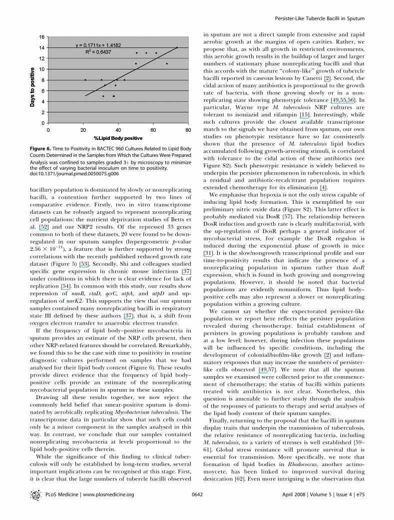
Microscopy-positive sputum samples from the UK and The Gambia were investigated fortheir content of lipid body–positive mycobacteria by combined Nile red and auramine staining.All samples contained a lipid body–positive population varying from 3% to 86% of the acid-fastbacilli present. The recent finding that triacylglycerol synthase is expressed by mycobacteriawhen they enter in vitro nonreplicating persistence led us to investigate whether this state wasalso associated with lipid body formation. We found that, when placed in laboratory conditionsinducing nonreplicating persistence, two M. tuberculosis strains had lipid body levelscomparable to those found in sputum. We investigated these physiological findings furtherby comparing the M. tuberculosis transcriptome of growing and nonreplicating persistencecultures with that obtained directly from sputum samples. Although sputum has traditionallybeen thought to contain actively growing tubercle bacilli, our transcript analyses refute thehypothesis that these cells predominate. Rather, they reinforce the results of the lipid bodyanalyses by revealing transcriptional signatures that can be clearly attributed to slowlyreplicating or nonreplicating mycobacteria. Finally, the lipid body count was highly correlated(R2¼ 0.64, p , 0.03) with time to positivity in diagnostic liquid cultures, thereby establishing adirect link between this cytological feature and the size of a potential nonreplicatingpopulation.
Conclusion
As nonreplicating tubercle bacilli are tolerant to the cidal action of antibiotics and resistant tomultiple stresses, identification of this persister-like population of tubercle bacilli in sputumpresents exciting and tractable new opportunities to investigate both responses to chemo-therapy and the transmission of tuberculosis.
The Editors’ Summary of this article follows the references.
PLoS Medicine | www.plosmedicine.org April 2008 | Volume 5 | Issue 4 | e750634
PLoSMEDICINE
Introduction
Mycobacterium tuberculosis infects one in three worldwide andkills more people each year than any other bacterialpathogen. Routine treatment of tuberculosis requires combi-nation antibiotic therapy for a minimum of six months, andplaces a substantial burden on health care systems, partic-ularly in resource-poor countries. Over eight million newcases every year testify to this obligate pathogen’s ongoingsuccess in transmission [1], yet we know little about what theorganism needs to achieve this essential step.
Expectorated tubercle bacilli have been thought tooriginate from rapid and extensive bacterial growth at themargins of liquefied lesions in the lung [2,3]. Sputum providesa tractable sample of the bacterial population that must betargeted by antibiotic therapy and a snapshot of the organismon its way to a new host. It follows that the bacilli inmicroscopy smear-positive tuberculosis sputum expressproperties required for transmission—properties that mightexplain the existence of drug-tolerant persister subpopula-tions and account for the prolonged antibiotic therapynecessary for relapse-free treatment [4]. Since transmissionis required for evolutionary survival, we may assume that M.tuberculosis experiences powerful selection pressures to main-tain and express these as-yet unidentified properties. Thus,any bacillary phenotype recognised preferentially in sputumcould provide clues to these properties.
We have previously shown that nonpathogenic mycobac-teria readily accumulate intracellular triacylglycerol lipidbodies in vitro [5]; these bodies could not be demonstratedunder similar conditions with M. tuberculosis, yet anecdotallyhave been seen in acid-fast bacilli (AFB) in tuberculoussputum [5]. The recent discovery of a novel class ofdiacylglycerol acyl transferase enzymes in Acinetobacter [6]and the subsequent characterisation of 15 members of thisclass as triacylglycerol synthase-encoding genes (tgs1–tgs15) inM. tuberculosis [7] provide a biochemical basis for the presenceof lipid bodies in this organism. Intriguingly, Tgs1, the mostactive of these enzymes, is a member of the DosR regulon [8],a set of genes responsive to hypoxia and linked to long-termsurvival ofM. tuberculosis in animal hosts [9–13]. It has recentlybeen shown that triacylglycerol is accumulated by M. tuber-culosis following hypoxic and other stresses [7,14] and maycontribute to long-term mycobacterial survival. These obser-vations raise the possibility that lipid body–positive cells insputum may be in a nonreplicating persistent (NRP) state,which, given that NRP bacilli display antibiotic tolerance[11,13,15], would have implications for chemotherapy.
Defining the phenotypes of bacterial pathogens in theirnatural environments remains a key challenge. Accurateknowledge of the properties expressed at different stages ofinfection enables precise targeting of therapeutic andpreventive measures. While much has been learnt aboutbacterial pathogens from in vitro and in vivo (animal model)transcriptome studies [16,17] as well as from human lungtissue [18], there have, to the best of our knowledge, been nopublished studies of transcript profiles in sputum samples—aclinically tractable sample. Such methods as rapidly stabilisedRNA, differential cell lysis, and RNA amplification haveenabled us to report here the transcriptome of M. tuberculosisin the sputum of patients prior to treatment.
Methods
PatientsPatients attending the public clinic at the MRC Laborato-
ries, Fajara, The Gambia and identified as sputum smear–positive by routine microscopy were invited to provide early-morning samples for transcriptome analysis. Patients whoagreed to participate gave informed oral consent (study nos.L2002.52 and L2006.60, ethical committee, MRC Laborato-ries, Fajara, The Gambia). Sputum from nine patients yieldedsufficient mycobacterial RNA for analysis by microarray orPCR; these were designated sputum samples 1–9.
Mycobacterial Strains and Growth ConditionsM. tuberculosis complex for direct microarray transcriptome
analysis was isolated from an aliquot of sputum 1 usingstandard methods [19]. M. tuberculosis complex was grown on7H10 agar with oleic acid-albumin-dextrose-catalase [20]supplement or in 7H9 broth with albumin-dextrose-catalasesupplement [20], 0.2% glycerol and 0.05% Tween-80. Forhypoxic (nonreplicating persistence) cultures M. tuberculosisstrains H37Rv and CH [21] were grown in Dubos Tween-albumin broth.
Routine Culturing of Smear-Positive Sputum SamplesDiagnostic sputum specimens were stained with auramine-
phenol [19], and positive smears confirmed and scored byZiehl-Neelsen staining after initial examination by fluores-cence microscopy. Smears were scored as either 1þ (1–10 AFBin 100 fields of view), 2þ (1–10 AFB in ten fields of view), or 3þ(1–10 AFB in one field of view). Decontamination of speci-mens was performed by the NaOH-NALC method [19]. Eachdecontaminated specimen was inoculated into one vial ofBACTEC 9000 MB medium for isolation of M. tuberculosis. Thetime to positivity of the BACTEC culture was recorded indays. All mycobacterial cultures were identified and con-firmed as M. tuberculosis complex using standard procedures.
Auramine-Nile Red Labelling of Sputum SamplesWhole sputum (;1–4 ml) was digested for 15 min with an
equal volume of 0.5% w/v N-acetyl L-cysteine in 50 mMsodium citrate [19]. Phosphate buffer (67 mM [pH 6.8]) wasadded to a final volume of 20 ml, and bacteria wereconcentrated (1,398g, 20 min). The pellet was resuspendedin 0.5 ml of phosphate-buffered saline and a smear preparedwith ;10 ll of the suspension. Heat-fixed smears werelabelled with auramine-Nile red as previously described [5].Preparations were observed by epifluorescence microscopyusing a Nikon Diphot 300 inverted microscope with a 100 Wmercury light source. Images were recorded using a 12/10bit,high speed Peltier-cooled CCD camera (FDI, PhotonicScience) using Image-Pro Plus (Media Cybernetics) software.The 11001V2 Blue (excitation 470 6 40 nm; emission . 515nm; Chroma Technology) and the G-2A (excitation 510–560nm; emission: 590 6 10 nm, Nikon) filter sets were used forepifluorescence microscopy.
Nile Red Labelling of Nonreplicating Persistence M.tuberculosis CulturesM. tuberculosis H37Rv and strain CH [21] were grown as
agitated, aerated cultures (370 rpm) to mid-log phase inDubos liquid medium, supplemented with Dubos mediumalbumin. M. tuberculosis NRP1/2 (nonreplicating persistence
PLoS Medicine | www.plosmedicine.org April 2008 | Volume 5 | Issue 4 | e750635
Persister-Like Tubercle Bacilli in Sputum
stage 1 and 2) cultures were incubated with continuousstirring at 37 8C for 168, 288, and 504 h, respectively,according to Wayne and Hayes [15]. At these time points onetube of each culture was destructively sampled for micro-scopic analysis. 10 ll of each sample was spread on a slide,heat fixed, and labelled with Nile red as previously described[5].
RNA Extraction from Tuberculous SputaWith the exception of sputum sample 1, which was frozen
in liquid nitrogen within 10 min of expectoration, approx-imately four volumes of GTC solution (5 M guanidiniumthiocyanate, 0.5% w/v sodium N-lauryl sarcosine, 25 mMtrisodium citrate, 0.1 M 2-mercaptoethanol, 0.5% w/v Tween80 [pH 7.0]) [22] were added to sputum within 5 min ofcollection. Mycobacteria were harvested by centrifugation(1398g, 30 min), resuspended in 400 ll of sterile deionizedwater, and added to 1 ml of Trizol LS (Invitrogen). RNA wasextracted using a method modified from that of DesJardin etal. [23] with chloroform replacing chloroform:isoamyl alcoholwashes and the Cleanascite step omitted. The mixture wastransferred to a glass matrix tube for cell lysis (Lysing matrixB; Q-Biogene) and processed in a spin/rotation instrumentfor cell lysis (Ribolyser; Hybaid), with a speed setting of 6.5and a time setting of 45 s. After processing, 200 ll ofchloroform was added to the mixture and it was vortex-mixedfor 2 min. The aqueous and organic layers were separated bymicrocentrifugation for 15 min at room temperature at16,000g. The aqueous phase containing the RNA was washedonce with an equal volume of chloroform. The aqueous phasewas removed to a fresh tube and 1 ll of glycoblue (Ambion),0.1 volume of 5 M ammonium acetate, and an equal volume ofisopropanol were added. The RNA was precipitated over-night at �20 8C. The resulting RNA pellet was washed oncewith 70% v/v and once with 95% v/v ethanol, dried andresuspended in 100 ll of RNase-free H2O (Sigma). Thecognate M. tuberculosis complex isolate from sputum sample 1was cultured for 6 d in 100ml 7H9 broth at 37 8C, 200 rpm atwhich time the absorbance was 0.22 at 580 nm. MycobacterialRNA was stabilised with GTC solution and extracted aspreviously described. Total RNA from sputum samples (5, 7,8, and 9) for amplification was quantified using the Nano-Drop ND-1000 Spectrophotometer (NanoDrop Technologies)and Agilent 2100 Bioanalyser (Agilent Technologies).
Microarray Analysis of RNA from Sputum 1RNA from sputum 1 and the in vitro-grown cognate isolate
was cleaned using the RNeasy kit (Qiagen). A M. tuberculosiswhole genome microarray, generated by the Bacterial Micro-array Group at St. George’s (University of London) andconsisting of 3,924 gene-specific PCR products (designed withminimal cross-homology) to the M. tuberculosis H37Rv [24],was utilised (ArrayExpress accession number A-BUGS-1;http://bugs.sgul.ac.uk/A-BUGS-1). Hybridisations were con-ducted as previously described [25] with 15 lg of Cy5-labelledcDNA derived from M. tuberculosis RNA against 1 lg Cy3-labelled M. tuberculosis H37Rv genomic DNA. The hybridisedslides were scanned sequentially at 532 nm and 635 nmcorresponding to Cy3 and Cy5 excitation maxima using theAffymetrix 428 Array Scanner (MWG). Comparative spotintensities from the images were calculated using Imagene 5.5(BioDiscovery), and imported into GeneSpring GX 7.2
(Agilent Technologies) for further analysis. After local back-ground subtraction the measured intensity in the cDNAchannel for each gene was divided by its intensity in thegenomic DNA control channel. The array data werenormalised to the 50th percentile of all genes detected tobe present on the array and filtered to remove unreliable lowintensity data (below a value of 500 in either channel).Geneswere identified as differentially expressed in sputum with acut-off of .3-fold relative to in vitro growth.
Growth Conditions and RNA Extraction for MicroarrayAnalysisM. tuberculosis H37Rv was grown as agitated, aerated
cultures (370 rpm) to mid-log phase at 37 8C in Dubos liquidmedium, supplemented with Dubos medium albumin. M.tuberculosis NRP1/2 cultures were set up and cultured in astirred model for 72 h and 240 h, respectively, according toWayne and Hayes [15]. Mycobacterial RNA was extractedfrom in vitro models (collected straight into GTC solution)using the GTC/Trizol method as developed by Mangan et al.[26]; RNA was DNase-treated and purified using RNeasycolumns (Qiagen). Total RNA was quantified using theNanoDrop ND-1000 Spectrophotometer (NanoDrop Tech-nologies) and Agilent 2100 Bioanalyser (Agilent Technolo-gies).
RNA AmplificationAn aliquot of 5 ng of total M. tuberculosis RNA was amplified
using an Eberwine T7-oligo-dT based system after an initialpolyadenylation step (MessageAmp II Bacteria, Ambion).Using this method, bacterial RNA was polyadenylated beforepriming the first-strand cDNA synthesis reaction with T7-linked oligo-dT. Amplified RNA was generated after second-strand cDNA synthesis and cDNA purification by in vitro run-off transcription (IVT) using T7 polymerase. Single rounds ofamplification were performed, with an in vitro transcriptionreaction of 16 h at 37 8C. This amplification method has beenpreviously demonstrated to be reproducible and capable ofidentifying representative changes in gene expression [27,28].The yield and size distribution of amplified products wasassessed spectrophotometrically at OD260 and using theAgilent 2100 Bioanalyser (Agilent Technologies).
Microarray Analyses of Samples 5, 7, 8, and 9An M. tuberculosis whole-genome microarray, generated by
the Bacterial Microarray Group at St. George’s (ArrayExpressaccession number A-BUGS-23; http://bugs.sgul.ac.uk/A-BUGS-23), and consisting of 4,410 gene-specific PCRproducts (designed with minimal cross-homology) to the M.tuberculosis H37Rv [24], CDC1551 [29], and M. bovis AF2122/97[30] genomes was utilised. Hybridisations were conducted aspreviously described [25] except for the use of M. tuberculosisgenomic DNA as a common reference [31]. Using genomicDNA reduced technical variation between replicate hybrid-isations and allowed RNA profiles to be used in multiplecomparisons. 5 lg of Cy5-labelled cDNA derived fromamplified M. tuberculosis RNA was hybridised with 2 lg ofCy3-labelled M. tuberculosis H37Rv genomic DNA. A lowerratio of test cDNA to comparator gDNA was used than withsample 1, as we were able to confirm the purity of ourpreparations with the Bioanalyser at the same time andperform technical replicates. The M. tuberculosis H37Rv
PLoS Medicine | www.plosmedicine.org April 2008 | Volume 5 | Issue 4 | e750636
Persister-Like Tubercle Bacilli in Sputum
reference DNA was kindly provided by Colorado StateUniversity (http://www.cvmbs.colostate.edu/microbiology/tb/top.htm). Two biological replicates of each in vitro growthcondition and four sputum samples (5, 7, 8, and 9) werehybridised in triplicate. The microarrays were scanned andspot intensities calculated as described above. The array datawere normalised to the 50th percentile of all genes detectedto be present on the array. The dataset was filtered to includeonly cDNA elements flagged to be present on 80% of thearrays. Significantly differentially expressed genes wereidentified using ANOVA (p , 0.05 with Benjamini andHochberg multiple testing correction) and a fold change of .
2.5. The significantly differentially expressed genes werehierarchically clustered using Cluster and the results dis-played using Treeview software [32]. The hypergeometricdistribution was used to determine if functional categories ofgenes were significantly enriched in the sputum profile [33].Fully annotated microarray data are deposited in BlG@Sbase and ArrayExpress.
Quantitative Real-Time RT-PCRMycobacterial RNA (0.5 lg) from sputum sample 1 and its
cognate isolate and eight further sputum samples (samples 2–9) were reverse transcribed in a total volume of 30 ll usingrandom primers and Superscript II (Invitrogen Technologies)according to manufacturer’s instructions. To estimate DNAcontamination of samples, all were subjected to a no reversetranscriptase control, which was then subtracted from theRNA result. A no-reverse transcriptase threshold of 10% ofthe test value was taken, with the exception of four reactionsin which values which were ,20% were used for correction.PCR reactions for tgs1 were set up using Absolute QPCRSYBR green mix (ABgene), 0.4 lM primers [7] and 2 ll ofcDNA. PCR was performed using the Rotor-Gene RG-3000system (Corbett Research) heating to 56 8C for 2 min, then 958C for 15 min, before 40 cycles of 95 8C for 30s, 60 8C for 30s,and 72 8C for 30s, acquiring fluorescence at 85 8C. No-reversetranscriptase controls for both the sputum and isolate RNAwere included, and these showed no PCR product. PCR foricl1 was set up with the primers of Dubnau et al. [34] using thesame cycling conditions and acquiring fluorescence at 86 8C.An hspX PCR was performed using primers of Wilkinson et al.[35] with 40 cycles of 95 8C for 30s, 59 8C for 30s, 72 8C for 30s,acquisition of fluorescence at 85 8C. For normalisation, PCRof sigA was performed using the primers of Manganelli et al.[36], and cycling conditions used for tgs1 with fluorescenceacquisition at 86 8C. PCRs for nuoB, qcrC, and ctaD wereperformed using primers of Shi et al. [37] with conditions aspreviously described with annealing steps performed at 61 8C,56 8C, and 56 8C, and acquisition of fluorescence at 84 8C, 828C, and 83 8C, respectively. The quantity of target DNA ineach cDNA sample was determined by the threshold cycle(CT) with reference to a standard curve generated by theamplification of known amounts of M. tuberculosis H37Rvgenomic DNA.
StatisticsThe proportion of lipid body–positive AFB and time to
positivity of routine cultures were analysed by linearregression to provide R2 correlation coefficients. As the lipidbody content of these samples was not normally distributed,Pearson correlations were also performed.
Results
Lipid Body–Positive Acid-Fast Bacilli Are a UniversalFeature of Smear-Positive Tuberculous SputumIf lipid body–positive cells are a transmission-adapted
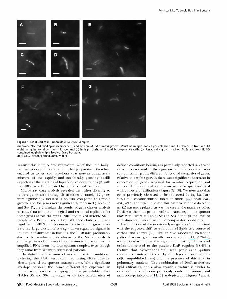
phenotype for M. tuberculosis, then such cells should bepresent in most smear-positive sputum samples. We confirmthis hypothesis in 82 smear-positive samples from patientsfrom The Gambia and the UK (69 and 13, respectively). Insamples with .100 assessable bacilli, the frequency of lipidbody–positive cells varied from 3% to 86% (mean 45%,standard deviation 20%), and these contained between twoand eight lipid bodies per cell (Figure 1). Thus, lipid body–positive tubercle bacilli are readily demonstrable in smear-positive samples from tuberculosis patients in two well-separated geographic locations and are present in asubpopulation of mycobacterial cells.
Lipid Bodies Are Readily Observed in M. tuberculosis Cellsin Nonreplicating PersistenceThe discovery and characterisation of tgs1 [6,7] raised the
possibility that lipid bodies might be formed in response to thehypoxic growth shift-down conditions that have been de-scribed by Wayne and Hayes [15], conditions known to causeup-regulation of the DosR regulon [9,10]. When M. tuberculosisH37Rv, a laboratory strain, and CH, a recent clinical isolateresponsible for a large outbreak [21], were exposed in vitro tothese conditions, abundant Nile red-staining lipid bodies wereobserved in both strains; respectively, 29% and 42% in NRP1(168 h), 50% and 65% in NRP2 (288 h), and 41% and 56% inlate NRP2 (504 h). An average of two lipid bodies per cell (rangeone to five) was observed in all samples except for the H37RvNRP1 sample in which only one (range one to three) was seenin positive cells. Thus, NRPM. tuberculosis cultures contain lipidbodies at levels comparable to those seen in sputum.
Expression Profiling of M. tuberculosis Recovered fromSputumIf lipid bodies are a biomarker for cells in an NRP state,
then the M. tuberculosis transcripts present in sputum shouldbe compatible with those observed in NRP in vitro studies[9,10]. We therefore compared the transcriptome of M.tuberculosis recovered from human sputum to that obtainedfrom in vitro aerobic cultures and NRP-inducing conditions[15]. Twenty sputa were collected from known microscopy-positive Gambian patients before they started antibiotictreatment, and the samples were rapidly stabilized againstRNA degradation. Five samples (designated 1, 5, 7, 8, and 9)were analysed by microarray hybridisation, four with and onewithout prior polyadenylation/oligo-dT based amplification.Although the results from sputum 1, the single direct(nonamplified) array, are not discussed further, they confirmthe essential details of the amplified analyses (Tables S1 andS2). This high-volume sample (;30 ml) had an exceptionallyhigh bacterial load, and we estimate that .1010 bacilli werepresent. The data from the four amplified samples wereanalysed with array hybridisations of amplified RNA ex-tracted fromM. tuberculosisH37Rv under different conditions:log-phase aerobic growth, the two stages of NRP (NRP1 t¼ 72h; NRP2 t¼ 240 h) [15], and a mixed preparation containingRNA from aerobic and NRP2 cells mixed in the proportion70:30 (w/w total RNA). This latter preparation was included
PLoS Medicine | www.plosmedicine.org April 2008 | Volume 5 | Issue 4 | e750637
Persister-Like Tubercle Bacilli in Sputum
because this mixture was representative of the lipid body–positive population in sputum. This preparation thereforeenabled us to test the hypothesis that sputum comprises amixture of the rapidly and aerobically growing bacilliexpected at the margins of liquefying caseous lesions [2] withthe NRP-like cells indicated by our lipid body studies.
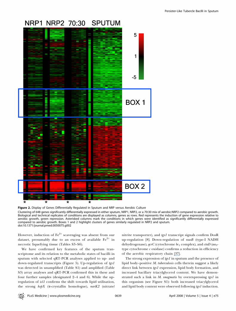
Microarray data analysis revealed that, after filtering toremove genes with low signals in either channel, 182 geneswere significantly induced in sputum compared to aerobicgrowth, and 334 genes were significantly repressed (Tables S3and S4). Figure 2 displays the results of gene cluster analysisof array data from the biological and technical replicates forthese genes across the sputa, NRP and mixed aerobic:NRP2sample sets. Boxes 1 and 2 highlight gene clusters similarlyregulated in NRP2 and sputum relative to aerobic growth. Wenote the large cluster of strongly down-regulated signals insputum, a feature lost in box 1 in the 70:30 mix, presumablydue to the aerobic signals obscuring the NRP2 signals. Asimilar pattern of differential expression is apparent for theamplified RNA from the four sputum samples, even thoughthey came from separate, untreated patients.
The data show that none of our comparator conditions,including the 70:30 aerobically replicating:NRP2 mixture,closely parallel the sputum transcriptome. While significantoverlaps between the genes differentially expressed insputum were revealed by hypergeometric probability values(Tables S5 and S6), no single or obvious combination of
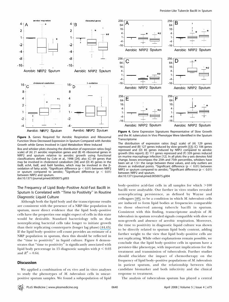
defined conditions herein, nor previously reported in vitro orin vivo, correspond to the signature we have obtained fromsputum. Amongst the different functional categories of genes,relative to aerobic growth there were significant decreases inexpression of genes required for aerobic respiration andribosomal function and an increase in transcripts associatedwith cholesterol utilisation (Figure 3) [38]. We note also thatgenes previously observed to be repressed during bacillarystasis in a chronic murine infection model [37], nuoB, ctaD,qcrC, atpA, and atpD, followed this pattern in our data whilenarK2 was up-regulated, as was the case in the murine studies.DosR was the most prominently activated regulon in sputum(box 2 in Figure 2; Tables S2 and S5), although the level ofactivation was lower than in the comparator conditions.The induction of the isocitrate lyase gene, icl1, is consistent
with the expected shift to utilisation of lipids as a source ofcarbon and energy [39]. This in vivo–associated metabolicpattern has emerged from other in vivo studies [11,12,39–42];we particularly note the signals indicating cholesterolutilisation related to the putative KstR regulon [38,43], afeature that corresponds well with prominent sputumcholesterol content detected by thin layer chromatography(NJG, unpublished data) and the presence of this lipid inpulmonary exudates. The combination of DosR activation,lipid utilisation, and a slow growth signature is similar toexperimental conditions previously studied in animal andmacrophage infections [11,12], as depicted in Figures 3 and 4.
Figure 1. Lipid Bodies in Tuberculous Sputum Samples
Auramine/Nile red-fixed sputum smears [5] and aerobic M. tuberculosis growth. Variation in lipid bodies per cell: (A) none, (B) three, (C) five, and (D)eight. Samples are shown with (E) low and (F) high proportions of lipid body–positive cells. (G) Aerobically grown mid-log M. tuberculosis H37Rvcontained negligible lipid bodies. Scale bar 2lm.doi:10.1371/journal.pmed.0050075.g001
PLoS Medicine | www.plosmedicine.org April 2008 | Volume 5 | Issue 4 | e750638
Persister-Like Tubercle Bacilli in Sputum
However, induction of Fe2þ scavenging was absent from ourdataset, presumably due to an excess of available Fe2þ innecrotic liquefying tissue (Tables S3–S6).
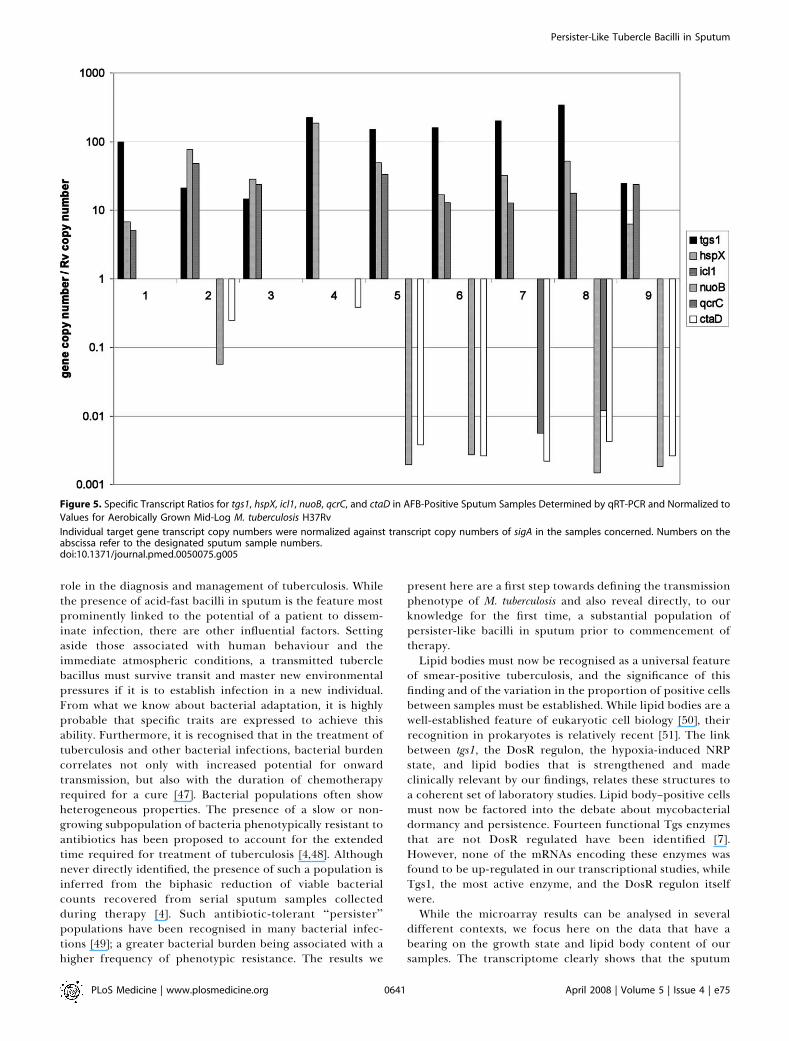
We have confirmed key features of the sputum tran-scriptome and its relation to the metabolic states of bacilli insputum with selected qRT-PCR analyses applied to up- anddown-regulated transcripts (Figure 5). Up-regulation of tgs1was detected in unamplified (Table S1) and amplified (TableS3) array analyses and qRT-PCR confirmed this in these andfour further samples (designated 2–4 and 6). While the up-regulation of icl1 confirms the shift towards lipid utilisation,the strong hspX (a-crystallin homologue), narK2 (nitrate/
nitrite transporter), and tgs1 transcript signals confirm DosRup-regulation [8]. Down-regulation of nuoB (type-I NADHdehydrogenase), qcrC (cytochrome bc1 complex), and ctaD (aa3-type cytochrome c oxidase) confirms a reduction in efficiencyof the aerobic respiratory chain [37].The strong expression of tgs1 in sputum and the presence of
lipid body–positive M. tuberculosis cells therein suggest a likelydirect link between tgs1 expression, lipid body formation, andincreased bacillary triacylglycerol content. We have demon-strated such a link in M. smegmatis by overexpressing tgs1 inthis organism (see Figure S1): both increased triacylglyceroland lipid body content were observed following tgs1 induction.
Figure 2. Display of Genes Differentially Regulated in Sputum and NRP versus Aerobic Culture
Clustering of 648 genes significantly differentially expressed in either sputum, NRP1, NRP2, or a 70:30 mix of aerobic:NRP2 compared to aerobic growth.Biological and technical replicates of conditions are displayed as columns, genes as rows. Red represents the induction of gene expression relative toaerobic growth, green repression. Asterisked columns mark the conditions in which genes were identified as significantly differentially expressedcompared to aerobic growth. Boxes 1 and 2 highlight clusters of genes similarly regulated in NRP2 and sputum.doi:10.1371/journal.pmed.0050075.g002
PLoS Medicine | www.plosmedicine.org April 2008 | Volume 5 | Issue 4 | e750639
Persister-Like Tubercle Bacilli in Sputum
The Frequency of Lipid Body–Positive Acid-Fast Bacilli inSputum Is Correlated with ‘‘Time to Positivity’’ in RoutineDiagnostic Liquid Culture
Although both the lipid body and the transcriptome resultsare consistent with the presence of a NRP-like population insputum, more direct evidence that the lipid body–positivecells have the properties one might expect of cells in this statewould be desirable. Standard bacteriology tells us thatnonreplicating bacterial cells take longer to initiate growththan their replicating counterparts (longer lag phase) [44,45].If the lipid body–positive cell count provides an estimate of aNRP population in sputum, then this should be reflected inthe ‘‘time to positivity’’ in liquid culture. Figure 6 demon-strates that ‘‘time to positivity’’ is significantly associated withlipid body percentage in 15 diagnostic samples with p , 0.03and R2 ¼ 0.64.
Discussion
We applied a combination of ex vivo and in vitro analysesto study the phenotypes of M. tuberculosis cells in smear-positive sputum samples. We found a subpopulation of lipid
body–positive acid-fast cells in all samples for which .100bacilli were analysable. Our further in vitro studies revealednonreplicating persistence, as defined by Wayne andcolleagues [46], to be a condition in which M. tuberculosis cellsare induced to form lipid bodies at frequencies comparableto those observed among tubercle bacilli in sputum.Consistent with this finding, transcriptome analysis of M.tuberculosis in sputum revealed signals compatible with slow ornon-growth and absence of aerobic respiration. Moreover,the time to positivity in diagnostic liquid culture was shownto be directly related to sputum lipid body content, addingfurther weight to the view that lipid body–positive cells arenot replicating. While other explanations remain possible, weconclude that the lipid body–positive cells in sputum have apersister-like phenotype, with important implications for thetreatment and transmission of tuberculosis. Further studiesshould elucidate the impact of chemotherapy on thefrequency of lipid body–positive populations of M. tuberculosisin patient sputum, and the relationship between thiscandidate biomarker and both infectivity and the clinicalresponse to treatment.The analysis of tuberculous sputum has played a central
Figure 3. Genes Required for Aerobic Respiration and Ribosomal
Function Show Decreased Expression in Sputum Compared with Aerobic
Growth while Genes Involved in Lipid Metabolism Were Induced
Box and whisker plots showing the distribution of expression ratios (log2scale) of (A) 21 aerobic respiration genes and (B) 45 ribosomal genes inNRP2 and sputum relative to aerobic growth using functionalclassifications defined by Cole et al., 1998 [24]; also (C) 64 genes thatmay be involved in cholesterol catabolism [38] and (D) 45 genes in thefadB, echA, fadE, and fadA families, which may be involved in the b-oxidation of fatty acids. *Significant difference (p , 0.01) between NRP2or sputum compared to aerobic; #Significant difference (p , 0.01)between NRP2 and sputum.doi:10.1371/journal.pmed.0050075.g003
Figure 4. Gene Expression Signatures Representative of Slow Growth
and the M. tuberculosis In Vivo Phenotype Were Identified in the Sputum
Transcriptome
The distribution of expression ratios (log2 scale) of (A) 129 genesrepressed and (B) 127 genes induced by slow growth [53]; (C) 106 genesrepressed and (D) 85 genes induced by NRP2 compared to aerobicgrowth (this report); (E) 111 genes repressed and (F) 339 genes inducedon murine macrophage infection [12]. In all plots the y-axis denotes foldchange, boxes encompass the 25th and 75th percentiles, whiskers havebeen set at 1.53 the range between these values, and only outliers areshown as individual points. *Significant difference (p , 0.01) betweenNRP2 or sputum compared to aerobic; #Significant difference (p , 0.01)between NRP2 and sputum.doi:10.1371/journal.pmed.0050075.g004
PLoS Medicine | www.plosmedicine.org April 2008 | Volume 5 | Issue 4 | e750640
Persister-Like Tubercle Bacilli in Sputum
role in the diagnosis and management of tuberculosis. Whilethe presence of acid-fast bacilli in sputum is the feature mostprominently linked to the potential of a patient to dissem-inate infection, there are other influential factors. Settingaside those associated with human behaviour and theimmediate atmospheric conditions, a transmitted tuberclebacillus must survive transit and master new environmentalpressures if it is to establish infection in a new individual.From what we know about bacterial adaptation, it is highlyprobable that specific traits are expressed to achieve thisability. Furthermore, it is recognised that in the treatment oftuberculosis and other bacterial infections, bacterial burdencorrelates not only with increased potential for onwardtransmission, but also with the duration of chemotherapyrequired for a cure [47]. Bacterial populations often showheterogeneous properties. The presence of a slow or non-growing subpopulation of bacteria phenotypically resistant toantibiotics has been proposed to account for the extendedtime required for treatment of tuberculosis [4,48]. Althoughnever directly identified, the presence of such a population isinferred from the biphasic reduction of viable bacterialcounts recovered from serial sputum samples collectedduring therapy [4]. Such antibiotic-tolerant ‘‘persister’’populations have been recognised in many bacterial infec-tions [49]; a greater bacterial burden being associated with ahigher frequency of phenotypic resistance. The results we
present here are a first step towards defining the transmissionphenotype of M. tuberculosis and also reveal directly, to ourknowledge for the first time, a substantial population ofpersister-like bacilli in sputum prior to commencement oftherapy.Lipid bodies must now be recognised as a universal feature
of smear-positive tuberculosis, and the significance of thisfinding and of the variation in the proportion of positive cellsbetween samples must be established. While lipid bodies are awell-established feature of eukaryotic cell biology [50], theirrecognition in prokaryotes is relatively recent [51]. The linkbetween tgs1, the DosR regulon, the hypoxia-induced NRPstate, and lipid bodies that is strengthened and madeclinically relevant by our findings, relates these structures toa coherent set of laboratory studies. Lipid body–positive cellsmust now be factored into the debate about mycobacterialdormancy and persistence. Fourteen functional Tgs enzymesthat are not DosR regulated have been identified [7].However, none of the mRNAs encoding these enzymes wasfound to be up-regulated in our transcriptional studies, whileTgs1, the most active enzyme, and the DosR regulon itselfwere.While the microarray results can be analysed in several
different contexts, we focus here on the data that have abearing on the growth state and lipid body content of oursamples. The transcriptome clearly shows that the sputum
Figure 5. Specific Transcript Ratios for tgs1, hspX, icl1, nuoB, qcrC, and ctaD in AFB-Positive Sputum Samples Determined by qRT-PCR and Normalized to
Values for Aerobically Grown Mid-Log M. tuberculosis H37Rv
Individual target gene transcript copy numbers were normalized against transcript copy numbers of sigA in the samples concerned. Numbers on theabscissa refer to the designated sputum sample numbers.doi:10.1371/journal.pmed.0050075.g005
PLoS Medicine | www.plosmedicine.org April 2008 | Volume 5 | Issue 4 | e750641
Persister-Like Tubercle Bacilli in Sputum
bacillary population is dominated by slowly or nonreplicatingbacilli, a contention further supported by two lines ofcomparative evidence. Firstly, two in vitro transcriptomedatasets can be robustly argued to represent nonreplicatingcell populations: the nutrient deprivation studies of Betts etal. [52] and our NRP2 results. Of the repressed 33 genescommon to both of these datasets, 20 were found to be down-regulated in our sputum samples (hypergeometric p-value2.56 3 10�11), a feature that is further supported by strongcorrelations with the recently published reduced growth ratedataset (Figure 5) [53]. Secondly, Shi and colleagues studiedspecific gene expression in chronic mouse infections [37]under conditions in which there is clear evidence for lack ofreplication [54]. In common with this study, our results showrepression of nuoB, ctaD, qcrC, atpA, and atpD and up-regulation of narK2. This supports the view that our sputumsamples contained many nonreplicating bacilli in respiratorystate III defined by these authors [37], that is, a shift fromoxygen electron transfer to anaerobic electron transfer.
If the frequency of lipid body–positive mycobacteria insputum provides an estimate of the NRP cells present, thenother NRP-related features should be correlated. Remarkably,we found this to be the case with time to positivity in routinediagnostic cultures performed on samples that we hadanalysed for their lipid body content (Figure 6). These resultsprovide direct evidence that the frequency of lipid body–positive cells provide an estimate of the nonreplicatingmycobacterial population in sputum in these samples.
Drawing all these results together, we now reject thecommonly held belief that smear-positive sputum is domi-nated by aerobically replicating Mycobacterium tuberculosis. Thetranscriptome data in particular show that such cells couldonly be a minor component in the samples analysed in thisway. In contrast, we conclude that our samples containednonreplicating mycobacteria at levels proportional to thelipid body-positive cells therein.
While the significance of this finding to clinical tuber-culosis will only be established by long-term studies, severalimportant implications can be recognised at this stage. First,it is clear that the large numbers of tubercle bacilli observed
in sputum are not a direct sample from extensive and rapidaerobic growth at the margins of open cavities. Rather, wepropose that, as with all growth in restricted environments,this aerobic growth results in the buildup of larger and largernumbers of stationary phase nonreplicating bacilli and thatthis accords with the mature ‘‘colony-like’’ growth of tuberclebacilli reported in caseous lesions by Canetti [2]. Second, thecidal action of many antibiotics is proportional to the growthrate of bacteria, with those growing slowly or in a non-replicating state showing phenotypic tolerance [49,55,56]. Inparticular, Wayne type M. tuberculosis NRP cultures aretolerant to isoniazid and rifampin [15]. Interestingly, whilesuch cultures provide the closest available transcriptomematch to the signals we have obtained from sputum, our ownstudies on phenotypic resistance have so far consistentlyshown that the presence of M. tuberculosis lipid bodiesaccumulated following growth-arresting stimuli, is correlatedwith tolerance to the cidal action of these antibiotics (seeFigure S2). Such phenotypic resistance is widely believed tounderpin the persister phenomenon in tuberculosis, in whicha residual and antibiotic-recalcitrant population requiresextended chemotherapy for its elimination [4].We emphasise that hypoxia is not the only stress capable of
inducing lipid body formation. This is exemplified by ourpreliminary nitric oxide data (Figure S2). This latter effect isprobably mediated via DosR [57]. The relationship betweenDosR induction and growth rate is clearly multifactorial, withthe up-regulation of DosR perhaps a general indicator ofmycobacterial stress, for example the DosR regulon isinduced during the exponential phase of growth in mice[31]. It is the slow/nongrowth transcriptional profile and ourtime-to-positivity results that indicate the presence of anonreplicating population in sputum rather than dosRexpression, which is found in both growing and nongrowingpopulations. However, it should be noted that bacterialpopulations are evidently nonuniform. Thus lipid body–positive cells may also represent a slower or nonreplicatingpopulation within a growing culture.We cannot say whether the expectorated persister-like
population we report here reflects the persister populationrevealed during chemotherapy. Initial establishment ofpersisters in growing populations is probably random andat a low level; however, during infection these populationswill be influenced by specific conditions, including thedevelopment of colonial/biofilm-like growth [2] and inflam-matory responses that may increase the numbers of persister-like cells observed [49,57]. We note that all the sputumsamples we examined were collected prior to the commence-ment of chemotherapy; the status of bacilli within patientstreated with antibiotics is not clear. Nonetheless, thisquestion is amenable to further study through the analysisof the responses of patients to therapy and serial analyses ofthe lipid body content of their sputum samples.Finally, returning to the proposal that the bacilli in sputum
display traits that underpin the transmission of tuberculosis,the relative resistance of nonreplicating bacteria, includingM. tuberculosis, to a variety of stresses is well established [59–61]. Global stress resistance will promote survival that isessential for transmission. More specifically, we note thatformation of lipid bodies in Rhodococcus, another actino-moycete, has been linked to improved survival duringdesiccation [62]. Even more intriguing is the observation that
Figure 6. Time to Positivity in BACTEC 960 Cultures Related to Lipid Body
Counts Determined in the Samples from Which the Cultures Were Prepared
Analysis was confined to samples graded 3þ by microscopy to minimizethe effect of varying bacterial inoculum on time to positivity.doi:10.1371/journal.pmed.0050075.g006
PLoS Medicine | www.plosmedicine.org April 2008 | Volume 5 | Issue 4 | e750642
Persister-Like Tubercle Bacilli in Sputum
hypoxically grown M. tuberculosis cultures, in which we havedemonstrated ;34% lipid body–positive cells (unpublisheddata), are 10-fold more infectious for guinea pigs by theaerosol route than their aerobically grown counterparts [63].Moreover, recent investigation of Beijing strains of M.tuberculosis revealed that they accumulate more triacylglyceroland express tgs1 at levels 10-fold higher than laboratorystrains, during aerobic log-phase growth [64]. The enhancedtransmissibility, evidenced by the rapid global spread of thesestrains, may reflect a greater propensity for lipid bodyformation in vivo.
We propose that lipid body positive (fat) acid fast bacilli area biomarker for nonreplicating (lazy) M. tuberculosis cells insputum; their further study offers exciting and tractableavenues for research into the treatment and prevention oftuberculosis.
Supporting Information
Figure S1. Overexpression of tgs1 in M. smegmatis Leads to EnhancedAccumulation of Triacylglycerol and Lipid Bodies
We cloned tgs1 under the control of the acetamide-induciblepromoter pSD26 [65] and expressed it in Mycobacterium smegmatis,which readily forms tiacylglycerol (TAG) lipid bodies in vitro. Testand control cells were exposed to radiolabelled oleic acid for 10 minand incorporation into TAG determined [66].(A) Triacylglycerol synthase (TGS) activity of induced MspSD26-tgs1and MspSD26 vector control, p , 0.001.(B) Pseudo-coloured fluorescence images of induced MspSD26 vectorcontrol (i) and MspSD26-tgs1 (ii) incubated with 630 lM oleic acid for10 min and labelled with Nile red [5]. Pseudo-colour (blue/min to red/max) was applied to grey levels 101–255 to demonstrate enhancedlipid body formation in the tgs1-overexpressing cells. Scale bar ¼ 2lm.
Found at doi:10.1371/journal.pmed.0050075.sg001 (6.7 MB TIF).
Figure S2. Nitric Oxide Exposure Stimulates Lipid Body Formationand Tolerance to the Cidal Action of Rifampin
Exponential phase M. tuberculosis H37Rv cultures in Sauton’s medium(;105 cfu/ml) were treated with 100 lM spermine.NO (NO donor) for4 or 24 h. These test cultures respectively contained 65% and 22%lipid body–positive cells, while the control cultures, exposed to 100lM spermine.HCl for the same times, contained , 1% . Subsequentexposure of test and control cultures to rifampin (1 lg/ml) for 7 drevealed diminished killing in the NO exposed cultures. Error bars¼þ 1 standard deviation (n ¼ 3).
Found at doi:10.1371/journal.pmed.0050075.sg002 (194 KB TIF).
Table S1. Genes Determined to Be Greater than 3-fold Up-regulatedin Sputum 1 Compared with In Vitro Growth of the Cognate Isolateby Direct Microarray
Found at doi:10.1371/journal.pmed.0050075.st001 (52 KB DOC).
Table S2. Hypergeometric Probability Analysis of Genes Determinedto be . 3-Fold Up-regulated in Sputum 1 Compared to In VitroCognate Isolate Growth by Direct Microarray
Found at doi:10.1371/journal.pmed.0050075.st002 (85 KB DOC).
Table S3. Genes Determined to be Greater than 2.5-Fold Up-regulated in Sputum Compared with In Vitro Growth by AmplifiedMicroarray
Found at doi:10.1371/journal.pmed.0050075.st003 (403 KB DOC).
Table S4. Genes Determined to be Greater than 2.5-fold Down-regulated in Sputum Compared with In Vitro Growth by AmplifiedMicroarray
Found at doi:10.1371/journal.pmed.0050075.st004 (664 KB DOC).
Table S5. Hypergeometric Probability Analysis of Genes Determinedto be .2.5-Fold Up-regulated in Sputum Compared to In VitroH37Rv Growth by Amplified Microarray
Found at doi:10.1371/journal.pmed.0050075.st005 (104 KB DOC).
Table S6. Hypergeometric Probability Analysis of Genes Determinedto be .2.5-Fold Down-regulated in Sputum Compared to In VitroH37Rv Growth by Amplified Microarray
Found at doi:10.1371/journal.pmed.0050075.st006 (86 KB DOC).
Accession NumbersThe fully annotated microarray data from this study are deposited inBlG@Sbase (accession number: E-BUGS-52; http://bugs.sgul.ac.uk/E-BUGS-52) and ArrayExpress (accession number: E-BUGS-52; http://www.ebi.ac.uk/arrayexpress/experiments/E-BUGS-52).
Acknowledgments
We thank Sarah Fandrich and Helen Smith for technical assistance;Jenny Bryan for helpful comments on the manuscript; and KeithMcAdam, Tumani Corah, Bouke de Jong, Omar Ceesay, Babou Faye,and Jacob Otu for their assistance in The Gambia. M. tuberculosisH37Rv reference DNA was kindly provided by Colorado StateUniversity (Contract No. HHSN266200400091C; NIH, NIAID N01-AI-40091; ‘‘Tuberculosis Vaccine Testing and Research MaterialsContract’’; http://www.cvmbs.colostate.edu/microbiology/tb/top.htm).Joanna Bacon, Health Protection Agency, Porton Down, UK, kindlysupplied a sample of her hypoxically grown bacilli for lipid bodyanalysis.
Author contributions. NJG, SJW, RAA, PDB, and MRB designed thestudy, analysed results and contributed to the writing of the paper.KR and GSB contributed to the analysis of results and writing of thepaper. NJG, SJW, ALS, SML, RJS, CS, and JH were involved inperforming the experiments and analysing the data.
References1. WHO (2007) Global tuberculosis control: surveillance, planning, financing.
Geneva: WHO. WHO/HTM/TB/2007.376. Available: http://www.who.int/tb/publications/global_report/en/. Accessed 20 February 2008.
2. Canetti G (1955) The tubercle bacillus in the pulmonary lesion of man.Histobacteriology and its bearing on therapy of pulmonary tuberculosis.The tubercle bacillus in the pulmonary tuberculous lesion. New York:Springer Publishing Company. pp. 29–85.
3. Young DB, Duncan K (1995) Prospects for new interventions in thetreatment and prevention of mycobacterial disease. Annu Rev Microbiol49: 641–673.
4. Mitchison DA (2004) The search for new sterilizing anti-tuberculosis drugs.Front Biosci 9: 1059–1072.
5. Garton NJ, Christensen H, Minnikin DE, Adegbola RA, Barer MR (2002)Intracellular lipophilic inclusions of mycobacteria in vitro and in sputum.Microbiology 148: 2951–2958.
6. Kalscheuer R, Steinbuchel A (2003) A novel bifunctional wax ester synthase/acyl-CoA:diacylglycerol acyltransferase mediates wax ester and triacylgly-cerol biosynthesis in Acinetobacter calcoaceticus ADP1. J Biol Chem 278: 8075–8082.
7. Daniel J, Deb C, Dubey VS, Sirakova TD, Abomoelak B, et al. (2004)Induction of a novel class of diacylglycerol acyltransferases and triacylgly-cerol accumulation in Mycobacterium tuberculosis as it goes into a dormancy-like state in culture. J Bacteriol 186: 5017–5030.
8. Park HD, Guinn KM, Harrell MI, Liao R, Voskuil MI, et al. (2003) Rv3133c/dosR is a transcription factor that mediates the hypoxic response ofMycobacterium tuberculosis. Mol Microbiol 48: 833–843.
9. Voskuil MI, Visconti KC, Schoolnik GK (2004) Mycobacterium tuberculosisgene expression during adaptation to stationary phase and low-oxygendormancy. Tuberculosis (Edinb) 84: 218–227.
10. Muttucumaru DG, Roberts G, Hinds J, Stabler RA, Parish T (2004) Geneexpression profile of Mycobacterium tuberculosis in a nonreplicating state.Tuberculosis (Edinb) 84: 239–246.
11. Karakousis PC, Yoshimatsu T, Lamichhane G, Woolwine SC, NuermbergerEL, et al. (2004) Dormancy phenotype displayed by extracellular Mycobacte-rium tuberculosis within artificial granulomas in mice. J Exp Med 200: 647–657.
12. Schnappinger D, Ehrt S, Voskuil MI, Liu Y, Mangan JA, et al. (2003)Transcriptional adaptation of Mycobacterium tuberculosis within macro-phages: Insights into the phagosomal environment. J Exp Med 198: 693–704.
13. Neyrolles O, Hernandez-Pando R, Pietri-Rouxel F, Fornes P, Tailleux L, etal. (2006) Is adipose tissue a place for Mycobacterium tuberculosis persistence?PLoS ONE 1: e43. doi:10.1371/journal.pone.0000043
14. Sirakova TD, Dubey VS, Deb C, Daniel J, Korotkova TA, et al. (2006)Identification of a diacylglycerol acyltransferase gene involved in accumu-lation of triacylglycerol in Mycobacterium tuberculosis under stress. Micro-biology 152: 2717–2725.
15. Wayne LG, Hayes LG (1996) An in vitro model for sequential study ofshiftdown of Mycobacterium tuberculosis through two stages of nonreplicatingpersistence. Infect Immun 64: 2062–2069.
PLoS Medicine | www.plosmedicine.org April 2008 | Volume 5 | Issue 4 | e750643
Persister-Like Tubercle Bacilli in Sputum
16. Conway T, Schoolnik GK (2003) Microarray expression profiling: capturinga genome-wide portrait of the transcriptome. Mol Microbiol 47: 879–889.
17. Waddell SJ, Butcher PD (2007) Microarray analysis of whole genomeexpression of intracellular Mycobacterium tuberculosis. Curr Mol Med 7: 287–296.
18. Rachman H, Strong M, Ulrichs T, Grode L, Schuchhardt J, et al. (2006)Unique transcriptome signature of Mycobacterium tuberculosis in pulmonarytuberculosis. Infect Immun 74: 1233–1242.
19. Sommers HM, Good RC (1985) Mycobacterium. In: Lennette EH, Balows A,Hausler Jr WJ, Shadomy HJ, editors. Manual of clinical microbiology.Washington (D.C.): ASM. pp. 216–248.
20. Larsen MH (2000) Some common methods in mycobacterial genetics. In:Hatfull GF, Jacobs J W. R., editors. Molecular genetics of mycobacteria.Washington (D.C.): ASM. pp. 216–248.
21. Rajakumar K, Shafi J, Smith RJ, Stabler RA, Andrew PW, et al. (2004) Use ofgenome level-informed PCR as a new investigational approach for analysisof outbreak-associated Mycobacterium tuberculosis isolates. J Clin Microbiol42: 1890–1896.
22. Monahan IM, Mangan JA, Butcher PD (2001) Extraction of RNA fromintracellular Mycobacterium tuberculosis: Methods, considerations and appli-cations. In: Stoker TPaNG, editor. Mycobacterium tuberculosis Protocols.Totowa (New Jersey): Humana Press. pp. 31–42.
23. Desjardin LE, Perkins MD, Wolski K, Haun S, Teixeira L, et al. (1999)Measurement of sputum Mycobacterium tuberculosis messenger RNA as asurrogate for response to chemotherapy. Am J Respir Crit Care Med 160:203–210.
24. Cole ST, Brosch R, Parkhill J, Garnier T, Churcher C, et al. (1998)Deciphering the biology of Mycobacterium tuberculosis from the completegenome sequence. Nature 393: 537–544.
25. Stewart GR, Wernisch L, Stabler R, Mangan JA, Hinds J, et al. (2002)Dissection of the heat-shock response in Mycobacterium tuberculosis usingmutants and microarrays. Microbiology 148: 3129–3138.
26. Mangan JA, Monahan IM, Butcher PD (2002) Gene expression during host-pathogen interactions: approaches to bacterial mRNA extraction andlabelling for microarray analysis. In: Wren Dorrell, editors. Methods inmicrobiology. London: Academic Press. pp. 137–151.
27. Waddell SJ, Laing K, Senner C, Butcher PD (2008) Microarray analysis ofdefined Mycobacterium tuberculosis populations using RNA amplificationstrategies BMC Genomics 2008, 9: 94 (25 February 2008)
28. Rohde KH, Abramovitch RB, Russell DG (2007) Mycobacterium tuberculosisinvasion of macrophages: linking bacterial gene expression to environ-mental cues. Cell Host Microbe 2: 352–364.
29. Fleischmann RD, Alland D, Eisen JA, Carpenter L, White O, et al. (2002)Whole-genome comparison of Mycobacterium tuberculosis clinical andlaboratory strains. J Bacteriol 184: 5479–5490.
30. Garnier T, Eiglmeier K, Camus JC, Medina N, Mansoor H, et al. (2003) Thecomplete genome sequence of Mycobacterium bovis. Proc Natl Acad Sci U S A100: 7877–7882.
31. Talaat AM, Lyons R, Howard ST, Johnston SA (2004) The temporalexpression profile of Mycobacterium tuberculosis infection in mice. Proc NatlAcad Sci U S A 101: 4602–4607.
32. Eisen MB, Spellman PT, Brown PO, Botstein D (1998) Cluster analysis anddisplay of genome-wide expression patterns. Proc Natl Acad Sci U S A 95:14863–14868.
33. Waddell SJ, Stabler RA, Laing K, Kremer L, Reynolds RC, et al. (2004) Theuse of microarray analysis to determine the gene expression profiles ofMycobacterium tuberculosis in response to anti-bacterial compounds. Tuber-culosis (Edinb) 84: 263–274.
34. Dubnau E, Fontan P, Manganelli R, Soares-Appel S, Smith I (2002)Mycobacterium tuberculosis genes induced during infection of human macro-phages. Infect Immun 70: 2787–2795.
35. Wilkinson RJ, DesJardin LE, Islam N, Gibson BM, Kanost RA, et al. (2001)An increase in expression of a Mycobacterium tuberculosis mycolyl transferasegene (fbpB) occurs early after infection of human monocytes. Mol Microbiol39: 813–821.
36. Manganelli R, Dubnau E, Tyagi S, Kramer FR, Smith I (1999) Differentialexpression of 10 sigma factor genes in Mycobacterium tuberculosis. MolMicrobiol 31: 715–724.
37. Shi L, Sohaskey CD, Kana BD, Dawes S, North RJ, et al. (2005) Changes inenergy metabolism of Mycobacterium tuberculosis in mouse lung and under invitro conditions affecting aerobic respiration. Proc Natl Acad Sci U S A102: 15629–15634.
38. Van der Geize R, Yam K, Heuser T, Wilbrink MH, Hara H, et al. (2007) Agene cluster encoding cholesterol catabolism in a soil actinomyceteprovides insight into Mycobacterium tuberculosis survival in macrophages.Proc Natl Acad Sci U S A 104: 1947–1952.
39. McKinney JD, Honer zu Bentrup K, Munoz-Elias EJ, Miczak A, Chen B, et al.
(2000) Persistence of Mycobacterium tuberculosis in macrophages and micerequires the glyoxylate shunt enzyme isocitrate lyase. Nature 406: 735–738.
40. Timm J, Post FA, Bekker LG, Walther GB, Wainwright HC, et al. (2003)Differential expression of iron-, carbon-, and oxygen-responsive mycobac-terial genes in the lungs of chronically infected mice and tuberculosispatients. Proc Natl Acad Sci U S A 100: 14321–14326.
41. Talaat AM, Ward SK, Wu CW, Rondon E, Tavano C, et al. (2007)Mycobacterial bacilli are metabolically active during chronic tuberculosisin murine lungs: Insights from genome-wide transcriptional profiling. JBacteriol 189: 4265–4274.
42. Fenhalls G, Stevens L, Moses L, Bezuidenhout J, Betts JC, et al. (2002) In situdetection of Mycobacterium tuberculosis transcripts in human lung granulo-mas reveals differential gene expression in necrotic lesions. Infect Immun70: 6330–6338.
43. Kendall SL, Withers M, Soffair CN, Moreland NJ, Gurcha S, et al. (2007) Ahighly conserved transcriptional repressor controls a large reguloninvolved in lipid degradation in Mycobacterium smegmatis and Mycobacteriumtuberculosis. Mol Microbiol 65: 684–699.
44. Weichart DH, Kell DB (2001) Characterization of an autostimulatorysubstance produced by Escherichia coli. Microbiology 147: 1875–1885.
45. Mukamolova GV, Turapov OA, Young DI, Kaprelyants AS, Kell DB, et al.(2002) A family of autocrine growth factors in Mycobacterium tuberculosis. MolMicrobiol 46: 623–635.
46. Wayne LG, Sohaskey CD (2001) Nonreplicating persistence ofMycobacteriumtuberculosis. Annu Rev Microbiol 55: 139–163.
47. Connolly LE, Edelstein PH, Ramakrishnan L (2007) Why is long-termtherapy required to cure tuberculosis? PLoS Med 4: e120. doi:10.1371/journal.pmed.0040120
48. Mitchison DA (1979) Basic mechanisms of chemotherapy. Chest 76: 771–781.
49. Dhar N, McKinney JD (2007) Microbial phenotypic heterogeneity andantibiotic tolerance. Curr Opin Microbiol 10: 30–38.
50. Murphy DJ (2001) The biogenesis and functions of lipid bodies in animals,plants and microorganisms. Prog Lipid Res 40: 325–438.
51. Waltermann M, Steinbuchel A (2005) Neutral lipid bodies in prokaryotes:recent insights into structure, formation, and relationship to eukaryoticlipid depots. J Bacteriol 187: 3607–3619.
52. Betts JC, Lukey PT, Robb LC, McAdam RA, Duncan K (2002) Evaluation of anutrient starvation model of Mycobacterium tuberculosis persistence by geneand protein expression profiling. Mol Microbiol 43: 717–731.
53. Beste DJ, Laing E, Bonde B, Avignone-Rossa C, Bushell ME, et al. (2007)Transcriptomic analysis identifies growth rate modulation as a componentof the adaptation of mycobacteria to survival inside the macrophage. JBacteriol 189: 3969–3976.
54. Munoz-Elias EJ, Timm J, Botha T, Chan WT, Gomez JE, et al. (2005)Replication dynamics of Mycobacterium tuberculosis in chronically infectedmice. Infect Immun 73: 546–551.
55. Gomez JE, McKinney JD (2004) M. tuberculosis persistence, latency, anddrug tolerance. Tuberculosis (Edinb) 84: 29–44.
56. Paramasivan CN, Sulochana S, Kubendiran G, Venkatesan P, Mitchison DA(2005) Bactericidal action of gatifloxacin, rifampin, and isoniazid onlogarithmic- and stationary-phase cultures of Mycobacterium tuberculosis.Antimicrob Agents Chemother 49: 627–631.
57. Kumar A, Toledo JC, Patel RP, Lancaster JR Jr., Steyn AJ (2007)Mycobacterium tuberculosis DosS is a redox sensor and DosT is a hypoxiasensor. Proc Natl Acad Sci U S A 104: 11568–11573.
58. Levin BR, Rozen DE (2006) Non-inherited antibiotic resistance. Nat RevMicrobiol 4: 556–562.
59. Siegele DA, Kolter R (1992) Life after log. J Bacteriol 174: 345–348.60. Kolter R, Siegele DA, Tormo A (1993) The stationary phase of the bacterial
life cycle. Annu Rev Microbiol 47: 855–874.61. Smeulders MJ, Keer J, Speight RA, Williams HD (1999) Adaptation of
Mycobacterium smegmatis to stationary phase. J Bacteriol 181: 270–283.62. Alvarez HM, Silva RA, Cesari AC, Zamit AL, Peressutti SR, et al. (2004)
Physiological and morphological responses of the soil bacterium Rhodo-coccus opacus strain PD630 to water stress. FEMS Microbiol Ecol 50: 75–86.
63. Bacon J, James BW, Wernisch L, Williams A, Morley KA, et al. (2004) Theinfluence of reduced oxygen availability on pathogenicity and geneexpression in Mycobacterium tuberculosis. Tuberculosis (Edinb) 84: 205–217.
64. Reed MB, Gagneux S, Deriemer K, Small PM, Barry CE 3rd (2007) The W-Beijing lineage of Mycobacterium tuberculosis overproduces triglycerides andhas the DosR dormancy regulon constitutively upregulated. J Bacteriol 189:2583–2589.
65. Daugelat S, Kowall J, Mattow J, Bumann D, Winter R, et al. (2003) The RD1proteins of Mycobacterium tuberculosis: expression in Mycobacterium smegmatisand biochemical characterization. Microbes Infect 5: 1082–95.
66. Nakagawa H, Kashiwabara Y, Matsuki G (1976) Metabolism of triacylgly-cerol in Mycobacterium smegmatis. J Biochem (Tokyo) 80: 923–928.
PLoS Medicine | www.plosmedicine.org April 2008 | Volume 5 | Issue 4 | e750644
Persister-Like Tubercle Bacilli in Sputum
Editors’ Summary
Background. Every year, nearly nine million people develop tuber-culosis—a contagious infection usually of the lungs—and about twomillion people die from the disease. Tuberculosis is caused byMycobacterium tuberculosis, bacteria that are spread in airborne dropletswhen people with the disease cough or sneeze. The symptoms oftuberculosis include a persistent cough, weight loss, and night sweats.Diagnostic tests include chest X-rays, the tuberculin skin test, andsputum analysis. For the last of these tests, a sample of sputum (mucusand other matter brought up from the lungs by coughing) is collectedand then taken to a laboratory where bacteriologists look for M.tuberculosis using special stains—tuberculosis-positive sputum contains‘‘acid-fast bacilli’’—and also try to grow bacteria from the sample.Tuberculosis can be cured by taking several powerful antibiotics forseveral months. It is very important that this treatment is completed toensure that all the M. tuberculosis bacteria in the body are killed and toprevent the emergence of drug-resistant bacteria.
Why Was This Study Done? Strenuous efforts are being made to reducethe global burden of tuberculosis but with limited success so far formany reasons. One barrier to success is the efficiency with which M.tuberculosis spreads from one person to another. Very little is knownabout this part of the bacteria’s life cycle. If scientists could understandmore about the transmission of M. tuberculosis between people, theymight identify new therapeutic and preventative targets. In the study,therefore, the researchers examine the acid-fast bacilli in tuberculosis-positive sputum samples to get a snapshot of M. tuberculosis at the pointof its transmission to a new person and ask how the characteristics ofthese bacilli compare with those of M. tuberculosis growing in thelaboratory.
What Did the Researchers Do and Find? The researchers collectedsputum samples from patients with tuberculosis in the UK and TheGambia before they received any treatment, and looked for the presenceof acid-fast bacilli containing ‘‘lipid bodies.’’ These small structurescontain a fat called triacylglycerol. M. tuberculosis accumulates triacyl-glycerol when it is exposed to several stresses present during infection(for example, reduced oxygen or hypoxia) and the researchers suggestthat the presence of this fat may help the bacteria survive duringtransmission and establish a new infection. They found that all thesamples contained some lipid body–positive acid-fast bacilli. Next, theresearchers showed that M. tuberculosis grown in the laboratory underhypoxic conditions, which induce the bacteria to enter an antibiotic-tolerant condition called a ‘‘nonreplicating persistent’’ (NRP) state, alsoaccumulated lipid bodies. This result suggests that the lipid body–
positive acid-fast bacilli in sputum might be in an NRP state. To test thisidea, the researchers compared the pattern of mRNAs (the templatesfrom which proteins are produced; the pattern of mRNAs is called thetranscriptome and gives an idea of which proteins a cell is making undergiven conditions) made by growing cultures of M. tuberculosis, by M.tuberculosis maintained in the NRP state, and by the acid-fast bacilli inseveral sputum samples. The transcriptome of the sputum samplerevealed production of many proteins made in the NRP state. Finally, theresearchers showed that the time needed to grow M. tuberculosis fromsputum samples increased as the proportion of lipid body–positive acid-fast bacilli in the sputum increased, just as one would suspect if thepresence of lipid bodies signifies nongrowing cells.
What Do These Findings Mean? It has been generally assumed that theacid-fast bacilli in sputum collected from patients with tuberculosis arerapidly replicating M. tuberculosis released from infected areas of thelungs. By identifying a population of bacteria that contain lipid bodiesand that are in an NRP-like state in all the samples of sputum examinedfrom two geographical sites, this study strongly challenges thisassumption. The characteristics of this population of bacteria, theresearchers suggest, might help them survive the adverse conditionsthat M. tuberculosis encounters during transmission between people andmight partly explain why complete clearance of M. tuberculosis requiresextended treatment with antibiotics. To establish the clinical significanceof these findings, future studies will need to examine whether antibiotictreatment affects the frequency of lipid body–positive M. tuberculosisbacteria in patients’ sputum and whether there is any relationshipbetween this measurement and infectiousness, or clinical response totreatment.
Additional Information. Please access these Web sites via the onlineversion of this summary at http://dx.doi.org/10.1371/journal.pmed.0050075.
� The MedlinePlus encyclopedia contains pages on tuberculosis and onsputum culture (in English and Spanish)� The US National Institute of Allergy and Infectious Diseases provides
information on all aspects of tuberculosis� The US Centers for Disease Control and Prevention Division of
Tuberculosis Elimination provides several fact sheets and otherinformation resources about tuberculosis� The World Health Organization provides information on efforts to
reduce the global burden of tuberculosis
PLoS Medicine | www.plosmedicine.org April 2008 | Volume 5 | Issue 4 | e750645
Persister-Like Tubercle Bacilli in Sputum
Copyright © 2022 FDOKUMEN