Molecular mechanisms of enterotoxigenic Escherichia coli infection

Crystal structure of the CusBA heavy-metal efflux complex ofEscherichia coli
Chih-Chia Su1,ψ, Feng Long1,ψ, Michael T. Zimmermann2, Kanagalaghatta R.Rajashankar3, Robert L. Jernigan2,4, and Edward W. Yu1,2,4,5,*1Department of Chemistry, Iowa State University, Ames, IA 50011, USA2Bioinformatics and Computational Biology Interdepartmental Graduate Program, Iowa StateUniversity, Ames, IA 50011, USA3NE-CAT and Department of Chemistry and Chemical Biology, Cornell University, Bldg. 436E,Argonne National Laboratory, 9700 S. Cass Avenue, Argonne. IL 60439, USA4Department of Biochemistry, Biophysics and Molecular Biology, Iowa State University, Ames, IA50011, USA5Department of Physics and Astronomy, Iowa State University, Ames, IA 50011, USA
AbstractGram-negative bacteria, such as Escherichia coli, expel toxic chemicals via tripartite efflux pumpsspanning both the inner and outer membranes. The three parts are: 1) a membrane fusion proteinconnecting 2) a substrate-binding inner membrane transporter to 3) an outer membrane-anchoredchannel in the periplasmic space. A crystallographic model of this tripartite efflux complex hasbeen unavailable simply because co-crystallization of different components of the system hasproven to be extremely difficult. We previously described the crystal structures of both the innermembrane transporter CusA1 and membrane fusion protein CusB2 of the CusCBA effluxsystem3,4 from E. coli. We here report the co-crystal structure of the CusBA efflux complex,revealing the trimeric CusA efflux pump interacts with six CusB protomers at the upper half of theperiplasmic domain. These six CusB molecules form a channel extending contiguously from thetop of the pump. The affinity of the CusA and CusB interaction was found to be in the micromolarrange. Finally, we predicted a three-dimensional structure of the trimeric CusC outer membranechannel, and develop a model of the tripartite efflux assemblage. This CusC3-CusB6-CusA3 modelpresents a 750 kDa efflux complex spanning the entire bacterial cell envelope to export Cu(I)/Ag(I) ions.
In Gram-negative bacteria, efflux systems of the resistance-nodulation-division (RND)family play major roles in the intrinsic and acquired tolerance of antibiotics and toxiccompounds.5,6 They represent key components for Gram-negative pathogens to use inovercoming toxic environments unfavorable for their survival. An RND efflux pump7-14
works in conjunction with a periplasmic membrane fusion protein,15-18 and an outermembrane channel to form a functional protein complex.19,20 In Escherichia coli, one such
* To whom correspondence should be addressed. [email protected].ψC.S. and F.L. contributed equally to this work.Author ContributionsC.-C.S., F.L., and E.W.Y. designed research; C.-C.S., and F.L. performed experiments; M.T.Z. and R.L.J. performed docking; C.-C.S., F.L., K.R.R., and E.W.Y. performed model building and refinement; and C.-C.S., F.L., R.L.J., and E.W.Y. wrote the paper.Author InformationAtomic coordinates and structure factors have been deposited with the Protein Data Bank under the code 3NE5.
NIH Public AccessAuthor ManuscriptNature. Author manuscript; available in PMC 2012 February 24.
Published in final edited form as:Nature. 2011 February 24; 470(7335): 558–562. doi:10.1038/nature09743.
NIH
-PA Author Manuscript
NIH
-PA Author Manuscript
NIH
-PA Author Manuscript

tripartite efflux system CusCBA is responsible for extruding biocidal Cu(I) and Ag(I) ions.3,4 CusA is a large proton-motive-force-dependent inner membrane RND efflux pumpcomprising 1,047 amino acids.3,4 CusC is a 457 amino acid protein that forms an outermembrane channel.3,4 The membrane fusion protein CusB, containing 379 amino acids,bridges between CusA and CusC to form a tripartite efflux complex CusCBA.3,4 This three-component system spans the entire cell envelope of E. coli to export Ag(I)/Cu(I).
Between the cusC and cusB genes, there is a small chromosomal gene that encodes aperiplasmic protein CusF.4 CusF functions as a chaperone carrying Cu(I)/Ag(I) to theCusCBA efflux pump.21,22
We previously reported the crystal structure of the full-length CusB membrane fusionprotein at a resolution of 3.40 Å, revealing four linearly-arranged domains (Domains 1-4)comprising approximately 80% of the protein.2 Overall, CusB is folded into an elongatedstructure, ~120 Å long and ~40 Å wide. The first three domains (Domains 1-3) of theprotein are mostly β-strands. However, the fourth domain (Domain 4) is all α-helices and isfolded into a three-helix bundle structure.
We also determined the crystal structure of the full-length CusA RND pump (at the 3.52-Åresolution) that includes approximately 98% of the amino acids.1 The structure suggests thatCusA exists as a homotrimer. Each subunit of CusA consists of 12 transmembrane helices(TM1-TM12) and a large periplasmic domain formed by two periplasmic loops betweenTM1 and TM2, and TM7 and TM8, respectively. The periplasmic domain of CusA can bedivided into a pore domain (comprising sub-domains PN1, PN2, PC1, PC2) and a CusCdocking domain (containing sub-domains DN and DC). Through the use of lysine-lysinecross-linking and mass spectrometry, it was determined that Domain 1 of CusB directlycontacts the upper portion of sub-domain PN1 of the CusA efflux pump.2
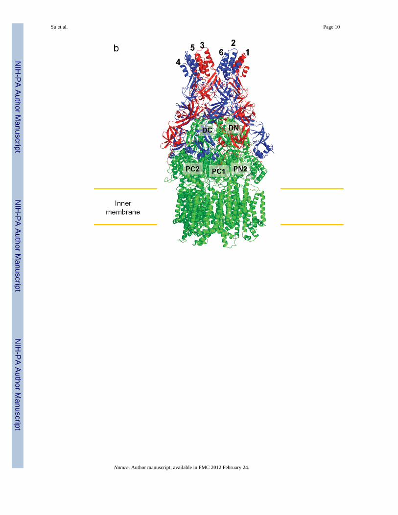
Here, we describe the co-crystal structure of the CusBA heavy-metal efflux complex. Weused molecular replacement with single-wavelength anomalous dispersion (MRSAD) todetermine the structure (Table S1 and Fig. S1), revealing each protomer of CusA interactsspecifically with two elongated molecules of CusB (molecules 1 and 2) at the upper halfportion of the periplasmic domain (Fig. 1). The two CusB adaptors are tilted at an angle of~50° with respect to the membrane surface, and establish a close fit with the transporter atthe concave surface formed by Domains 1 and 2 of the adaptor. Molecule 1 of CusBcontacts mainly the upper regions of PN2 and PC1, and the DN sub-domain of CusA.Molecule 2 of CusB, however, predominantly bridges to the upper regions of PC1 and PC2,and also the sub-domain DC of the pump. These two adaptor molecules are also seen tospecifically contact one another, primarily through Domains 1, 2 and 3 of these twoelongated molecules. The trimeric CusA pump therefore directly contacts six CusB adaptormolecules, which form a channel at the top of the CusA trimer (Figs. 1 and 2).
Intriguingly, molecule 1 of CusB interacts predominantly with CusA through charge-chargeinteractions. Residues K95, D386, E388 and R397 of this CusB molecule form four saltbridges with D155, R771, R777 and E584 of CusA, respectively. In addition, T89, thebackbone oxygen of N91, and R292 of molecule 1 of CusB form hydrogen bonds withK594, R147, and the backbone oxygen of Q198 of CusA to secure the interaction (Fig. 3a).However, the interaction between molecule 2 of CusB and CusA appears to be governedprincipally by charge-dipole and dipole-dipole interactions. Specifically, Q108, S109, S253and N312 of CusB (molecule 2) form hydrogen bonds with Q785, Q194, D800 and Q198 ofCusA, respectively. The backbone oxygens of L92 and T335 of this CusB molecule alsocontribute two additional hydrogen bonds with the side chains of K591 and T808 of theCusA pump to anchor the proteins (Fig. 3b).
Su et al. Page 2
Nature. Author manuscript; available in PMC 2012 February 24.
NIH
-PA Author Manuscript
NIH
-PA Author Manuscript
NIH
-PA Author Manuscript

For CusB-CusB interactions, molecule 1 of CusB makes a close contact with molecule 2 ofCusB. Domains 1-3 of these two molecules are involved in the binding. E118, Y119, R186,E252 and E292 of molecule 1 of CusB participate to form hydrogen bonds with T139, D142,T206, N312 and N113 of molecule 2 of CusB, respectively (Fig. 4a). Further, molecule 1 ofCusB also contributes to contact molecule 6 of CusB, which is anchored to the next subunitof CusA. The majority of the interactions come from Domains 2 and 3 of these twomolecules. Particularly, N113, N228, and N312 of molecule 1 of CusB pair up with R292,the backbone oxygen of A126, and E252 of molecule 6 of CusB to form three hydrogenbonds. In addition, D142 of molecule 1 of CusB participates to form two hydrogen bondswith Y119 and R297 of molecule 6 of CusB to secure the binding (Fig. 4b).
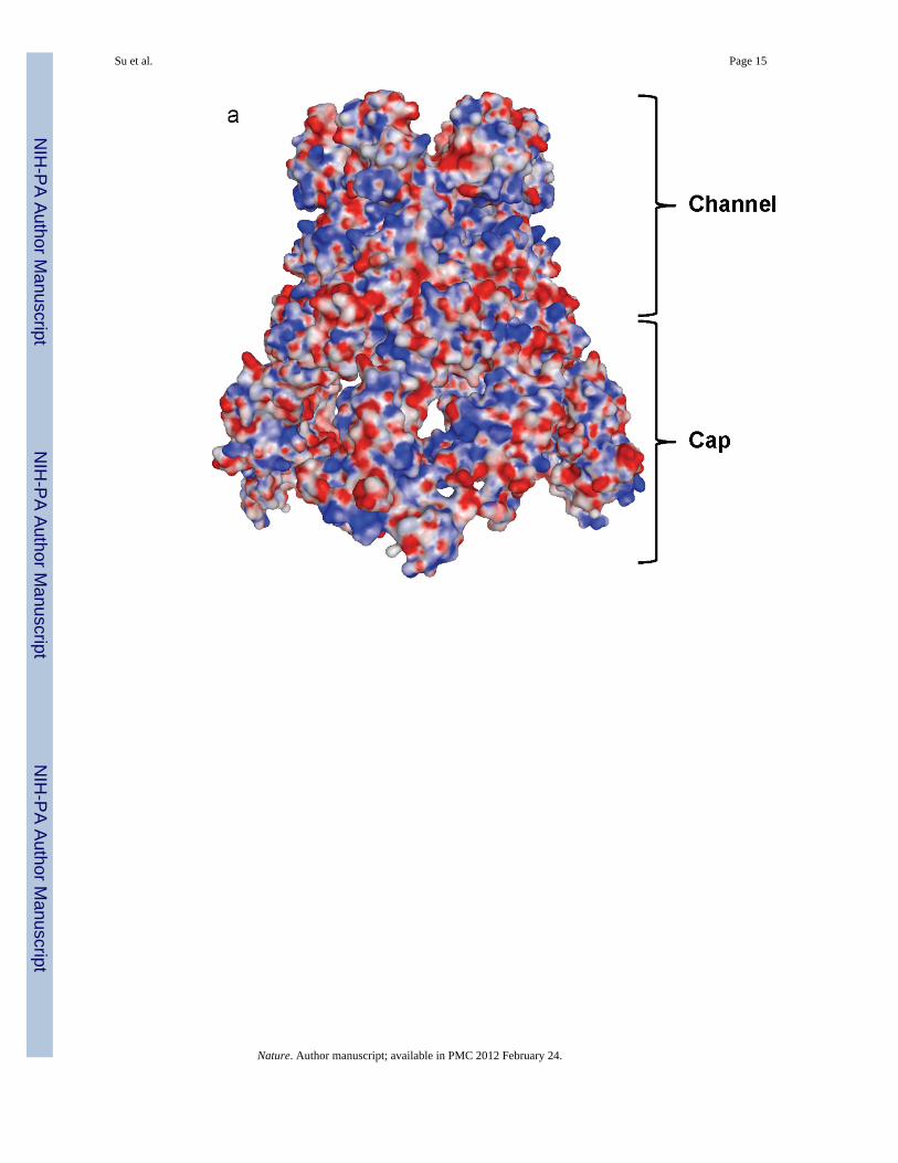
The hexameric arrangement of CusB allows this protein to create a funnel-like structure withthe central channel formed along the crystallographic threefold symmetry axis. Theseprotomers utilize a side-by-side packing arrangement to form this funnel (Fig. 2). Domain 1and the lower half of Domain 2 of CusB primarily create a cap-like structure whereas theupper half of Domain 2, Domain 3 and Domain 4 of the adaptor contribute to the centralchannel of the funnel. The inner surface of the cap fits closely with the outer surface of theupper portion of the periplasmic domain of the CusA trimer. The channel formed above thecap of the adaptor is ~62 Å long with an average internal diameter of ~37 Å. Thus, theinterior of the channel gives rise to a large elongated cavity with a volume of ~65,000 Å3.The lower half of the channel is primarily created by β-barrels whereas the upper half is anall α-helical tunnel. The diameter of the channel is gradually constricted and then dilated asit approaches the outer membrane. Thus, the α-helices of Domain 4 create an invertedconical structure. The narrowest section of the central channel is located at residue D232,which is close to the hinge region between Domains 3 and 4 of the membrane fusion protein.The widest section of the channel appears to form at the top edge with an inner diameter of~56 Å. The inner surface of the channel is predominantly negatively charged, as indicatedby the electrostatic surface diagram (Fig. 5), suggesting that the interior surface of thechannel may have the capacity to bind positively charged metal ions.
We used isothermal titration calorimetry (ITC) to determine the binding affinity of CusB tothe CusA pump. The ITC data indicates an equilibrium dissociation constant of 5.1 ± 0.3 μM(Fig. S2).
Previously, it was found that the N and C-terminal ends (residue 29-88 and 386-407) of theCusB adaptor are intrinsically disordered and cannot be identified in the electron densitymaps of the CusB crystals.2 Here, the co-crystal structure suggests that this region formsseveral short α-helices. In molecule 1 of CusB, residues 392-399 at the C-terminal end forma short α-helix. However, residues 79-95 of the N-terminus feature a long random coil andthese amino acids are located immediately outside the cleft formed between sub-domainsPC1 and PC2 of the CusA pump. For molecule 2 of CusB, the N-terminal residues 79-85and 86-92 participate to form a random coil and a short α-helix, respectively. However, theC-terminal residues 382-392 and 394-400 appear to create two short α-helices. Likemolecule 1 of CusB, the N-terminus of molecule 2 of CusB is near the periplasmic cleft ofthe pump (Fig. 1).
It has been proposed that the N-terminal residues M49, M64 and M66 of CusB form a three-methionine metal-binding site.23 Although these three methionine residues cannot beidentified in the electron density maps of our co-crystal, the co-crystal structure does showthat the N-terminal tails of both molecule 1 and 2 of CusB are located outside the cleftformed between PC1 and PC2 of the CusA pump. Thus, it is possible that CusB might helpto transfer the metal ions via the N-terminal three-methionine binding site into theperiplasmic cleft of CusA. Indeed, a similar suggestion has been made with the adaptor
Su et al. Page 3
Nature. Author manuscript; available in PMC 2012 February 24.
NIH
-PA Author Manuscript
NIH
-PA Author Manuscript
NIH
-PA Author Manuscript

protein AcrA whereas this protein might assist in transporting a drug from the periplasm intothe pump.24
The co-crystal structure presented here highlights the structural importance of theperiplasmic membrane fusion protein. Given the fact that six CusB molecules assemble toform a channel at the CusA funnel top, this suggests that the adaptor is likely to be involvedin the active extrusion of metal ions.
We then constructed a CusCBA model based on the CusBA crystal complex structure andthe predicted CusC model. The final CusC3-CusB6-CusA3 structural model represents a 750kDa tripartite efflux complex spanning both the inner and outer membranes of E. coli toextrude Cu(I)/Ag(I) (Fig. S3).
We believe that CusA can take up metal ions from both the periplasm and cytoplasmutilizing the methionine-residue ion relay network.1 Metal ions could enter the three-methionine binding-site, formed by M573, M623 and M672, inside the cleft between sub-domains PN2 and PC1 on the periplasmic portion of CusA or via the methionine pairswithin the transmembrane domain of the pump. It has been demonstrated that the chaperoneCusF can directly transfer its bound Cu(I) to the CusB membrane fusion protein.25 Thus, itis likely that CusF is responsible for delivering metal ions to the CusCBA tripartite effluxsystem in the periplasm. There is a chance that the initial step of metal transport through theperiplasmic cleft of the CusA pump may involve a direct transfer from CusF to thepreviously proposed three-methionine metal binding site23 (M49, M64 and M66) at the longN-terminal tail of CusB, situating near the PN2/PC1 cleft of the CusA pump. Thus, thesecond step could well be the delivery of the bound metal ion from CusB to the three-methionine cluster (M573, M623 and M672) inside the periplasmic cleft of CusA.1 Thebound metal ion could then be released to the nearest methionine pair (M271-M755) locateddirectly above the three-methionine metal binding site,1 from which the ion could bereleased into the central funnel of CusA and eventually the CusC channel for final extrusion.It is not yet known whether the interior of the hexameric CusB channel forms part of theextrusion pathway for Cu(I) and Ag(I). Exactly what this tripartite efflux system'smechanism is must await confirmation by elucidation of additional crystal structures of theCusCBA tripartite complex.
Methods SummaryCrystallization of CusA
The procedures for cloning, expression and purification of the CusA and CusB proteins havebeen described previously.1,2 Co-crystals of the CusBA complex were obtained usingsitting-drop vapor diffusion. A 2 μl protein solution containing 0.1 mM CusA and 0.1 mMCusB in buffer solution containing 20 mM Na-HEPES (pH 7.5) and 0.05% (w/v) CYMAL-6was mixed with 2 μl of reservoir solution containing 10% PEG 6000, 0.1 M Na-HEPES (pH7.5), 0.1 M ammonium acetate and 20% glycerol. The resultant mixture was equilibratedagainst 500 μl of the reservoir solution. The co-crystallization conditions for CusA (native)-CusB (SeMet) were the same as those for the native CusBA complex. Co-crystals of CusBAgrew to full size in the drops within two months. Cryoprotection was achieved by raising theglycerol concentration stepwise to 30% in 5% increments.
Structural determination and refinementData of the CusA (native)-CusB (SeMet) co-crystal were collected (Table S1). SAD phasingusing the program PHASER26 was employed to obtain experimental phases in addition tothe phases from the structural model of apo-CusA. Phases were subjected to densitymodification and phase extension to 2.90 Å-resolution using the program RESOLVE.27 The
Su et al. Page 4
Nature. Author manuscript; available in PMC 2012 February 24.
NIH
-PA Author Manuscript
NIH
-PA Author Manuscript
NIH
-PA Author Manuscript

full-length CusB protein contains nine methionine residues, and six selenium sites per CusBmolecule (12 selenium sites per asymmetric unit) were identified. After tracing the initialmodel manually using the program Coot,28 the model was refined against the native data at2.90 Å-resolution using TLS refinement adopting a single TLS body as implemented inPHENIX29 leaving 5% of the reflections in the Free-R set. Iterations of refinement usingPHENIX29 and CNS30 and model building in Coot28 lead to the current model, whichconsists of 1,686 residues (residues 4-1043 of CusA; residues 79-400 of molecule 1 ofCusB; and residues 79-402 of molecule 2 of CusB).
Supplementary MaterialRefer to Web version on PubMed Central for supplementary material.
AcknowledgmentsThis work is supported by NIH Grants R01GM074027 (E.W.Y.), R01GM086431 (E.W.Y.), R01GM081680(R.L.J.) and R01GM072014 (R.L.J.). This work is based upon research conducted at the Northeastern CollaborativeAccess Team beamlines of the Advanced Photon Source, supported by award RR-15301 from the National Centerfor Research Resources at the National Institutes of Health. Use of the Advanced Photon Source is supported by theU.S. Department of Energy, Office of Basic Energy Sciences, under Contract No. DE-AC02-06CH11357.
Methods
Isothermal Titration CalorimetryWe used isothermal titration calorimetry to examine the binding of the purified CusBadaptor to the purified CusA pump. Measurements were performed on a VP-Microcalorimeter (MicroCal, Northampton, MA) at 25 °C. Before titration, the CusA andCusB proteins were thoroughly dialyzed against buffer containing 20 mM Na-HEPES pH7.5 and 0.05% CYMAL-6, respectively. The protein concentrations were determined usingthe Bradford assay. The CusA protein sample was then adjusted to a final monomericconcentration of 14 μM. The CusB protein solution consisting of 350 μM monomeric CusBin 20 mM Na-HEPES pH 7.5 and 0.05% CYMAL-6 was prepared as the titrant. Thesamples were degassed before they were loaded into the cell and syringe. Bindingexperiments were carried out with the CusA protein solution (1.5 ml) in the cell and theCusB protein solution as the injectant. Ten microliter injections of the CusB solution wereused for data collection.
Injections occurred at intervals of 300 s, and the duration time of each injection was 10 s.Heat transfer (μcal/s) was measured as a function of elapsed time (s). The mean enthalpiesmeasured from injection of the ligand in the buffer were subtracted from raw titration databefore data analysis with ORIGIN software (MicroCal). Titration curves were fitted by anonlinear least squares method to a function for the binding of a ligand to a macromolecule.Nonlinear regression fitting to the binding isotherm provided us with the equilibriumbinding constant (KA = 1/KD) and enthalpy of binding (ΔH). Based on the values of KA, thechange in free energy (ΔG) and entropy (ΔS) were calculated with the equation: ΔG = - RTlnKA = ΔH - TΔS, where T is 273 K and R is 1.9872 cal/K per mol. Calorimetry trials werealso carried out in the absence of CusA in the same experimental conditions. No change inheat was observed in the injections throughout the experiment.
Structural prediction and docking of CusC onto CusBAA homology model of CusC was generated using the crystal structure of OprM (PDB ID:1wp1)20 as a template through the I-TASSER server.31 Alignment of protein sequences
Su et al. Page 5
Nature. Author manuscript; available in PMC 2012 February 24.
NIH
-PA Author Manuscript
NIH
-PA Author Manuscript
NIH
-PA Author Manuscript

suggests that these two channel proteins share 42% identity. A steered molecular dynamicssimulation was then used to rigidly dock the trimeric CusC channel to the crystal form of thehexameric CusB channel bound to the trimeric CusA pump. During the rigid-body dockingprocess, the molecules were aligned with the z-axis. CusB was then held rigidly, allowingCusC to pull along the z-axis toward CusB until no further change in the center of mass wasmeasured.
We next investigated the possible interaction of CusC with Domain 2 of CusB, which alsobrings it into close proximity to CusA. First, the trimeric CusA pump and Domains 1 and 2of the hexameric CusB channel were held rigidly, while Domains 3 and 4 of each CusBmonomer were pulled outward using customized harmonic forces to open the annulus of thehexameric channel. Next, we manually inserted CusC into the opened CusB ring. Finally,simulated annealing followed by energy minimization was performed to relax CusB andCusC from these positions and allow them to assume a lower energy conformation. Allsimulations were performed using NAMD32 and the CHARMM27+CMAP force field.33
References1. Long F, Su C-C, Zimmermann MT, Boyken SE, Rajashankar KR, Jernigan RL, Yu EW. Crystal
structures of the CusA heavy-metal efflux pump suggest methionine-mediated metal transportmechanism. Nature. 2010; 467:484–488. [PubMed: 20865003]
2. Su C-C, Yang F, Long F, Reyon D, Routh MD, Kuo DW, Mokhtari AK, Van Ornam JD, Rabe KL,Hoy JA, Lee YJ, Rajashankar KR, Yu EW. Crystal structure of the membrane fusion protein CusBfrom Escherichia coli. J. Mol. Biol. 2009; 393:342–355. [PubMed: 19695261]
3. Franke S, Grass G, Nies DH. The product of the ybdE gene of the Escherichia coli chromosome isinvolved in detoxification of silver ions. Microbiol. 2001; 147:965–972.
4. Franke S, Grass G, Rensing C, Nies DH. Molecular analysis of the copper-transporting effluxsystem CusCFBA of Escherichia coli. J. Bacteriol. 2003; 185:3804–3812. [PubMed: 12813074]
5. Tseng TT, Gratwick KS, Kollman J, Park D, Nies DH, Goffeau A, Saier MH Jr. The RND permeasesuperfamily: an ancient, ubiquitous and diverse family that includes human disease anddevelopment protein. J. Mol. Microbiol. Biotechnol. 1999; 1:107–125. [PubMed: 10941792]
6. Nies DH. Efflux-mediated heavy metal resistance in prokaryotes. FEMS Microbiol. Rev. 2003;27:313–339. [PubMed: 12829273]
7. Murakami S, Nakashima R, Yamashita E, Yamaguchi A. Crystal structure of bacterial multidrugefflux transporter AcrB. Nature. 2002; 419:587–593. [PubMed: 12374972]
8. Yu EW, McDermott G, Zgruskaya HI, Nikaido H, Koshland DE Jr. Structural basis of multiple drugbinding capacity of the AcrB multidrug efflux pump. Science. 2003; 300:976–980. [PubMed:12738864]
9. Murakami S, Nakashima R, Yamashita E, Matsumoto T, Yamaguchi A. Crystal structures of amultidrug transporter reveal a functionally rotating mechanism. Nature. 2006; 443:173–179.[PubMed: 16915237]
10. Seeger MA, Schiefner A, Eicher T, Verrey F, Dietrichs K, Pos KM. Structural asymmetry of AcrBtrimer suggests a peristaltic pump mechanism. Science. 2006; 313:1295–1298. [PubMed:16946072]
11. Sennhauser G, Amstutz P, Briand C, Storchengegger O, Grütter MG. Drug export pathway ofmultidrug exporter AcrB revealed by DARPin inhibitors. PLoS Biol. 2007; 5:e7. [PubMed:17194213]
12. Yu EW, Aires JR, McDermott G, Nikaido H. A periplasmic-drug binding site of the AcrBmultidrug efflux pump: a crystallographic and site-directed mutagenesis study. J. Bacteriol. 2005;187:6804–6815. [PubMed: 16166543]
13. Sennhauser G, Bukowska MA, Briand C, Grütter MG. Crystal structure of the multidrug exporterMexB from Pseudomonas aeruginosa. J. Mol. Biol. 2009; 389:134–145. [PubMed: 19361527]
Su et al. Page 6
Nature. Author manuscript; available in PMC 2012 February 24.
NIH
-PA Author Manuscript
NIH
-PA Author Manuscript
NIH
-PA Author Manuscript

14. Su C-C, Li M, Gu R, Takatsuka Y, McDermott G, Nikaido H, Yu EW. Conformation of the AcrBmultidrug efflux pump in mutants of the putative proton relay pathway. J. Bacteriol. 2006;188:7290–7296. [PubMed: 17015668]
15. Higgins MK, Bokma E, Koronakis E, Hughes C, Koronakis V. Structure of the periplasmiccomponent of a bacterial drug efflux pump. Proc. Natl. Acad. Sci. USA. 2004; 101:9994–9999.[PubMed: 15226509]
16. Akama H, Matsuura T, Kashiwag S, Yoneyama H, Narita S, Tsukihara T, Nakagawa A, Nakae T.Crystal structure of the membrane fusion protein, MexA, of the multidrug transporter inPseudomonas aeruginosa. J. Biol. Chem. 2004; 279:25939–25942. [PubMed: 15117957]
17. Mikolosko J, Bobyk K, Zgurskaya HI, Ghosh P. Conformational flexibility in the multidrug effluxsystem protein AcrA. Structure. 2006; 14:577–587. [PubMed: 16531241]
18. Symmons M, Bokma E, Koronakis E, Hughes C, Koronakis V. The assembled structure of acomplete tripartite bacterial multidrug efflux pump. Proc. Natl. Acad. Sci. USA. 2009; 106:7173–7178. [PubMed: 19342493]
19. Koronakis V, Sharff A, Koronakis E, Luisi B, Hughes C. Crystal structure of the bacterialmembrane protein TolC central to multidrug efflux and protein export. Nature. 2000; 405:914–919. [PubMed: 10879525]
20. Akama H, Kanemaki M, Yoshimura M, Tsukihara T, Kashiwag T, Yoneyama H, Narita S,Nakagawa A, Nakae T. Crystal structure of the drug discharge outer membrane protein, OprM, ofPseudomonas aeruginosa. J. Biol. Chem. 2004; 279:52816–52819. [PubMed: 15507433]
21. Xue Y, Davis AV, Balakrishnan G, Stasser JP, Staehlin BM, Focia P, Spiro TG, Penner-Hahn JE,O'Halloran TV. Cu(I) recognition via cation-π and methionine interactions in CusF. Nat. Chem.Biol. 2008; 4:107–109. [PubMed: 18157124]
22. Loftin IR, Franke S, Blackburn NJ, McEvoy MM. Unusual Cu(I)/Ag(I) coordination ofEscherichia coli CusF as revealed by atomic resolution crystallography and X-ray absorptionspectroscopy. Prot. Sci. 2007; 16:2287–2293.
23. Bagai I, Liu W, Rensing C, Blackburn N, McEvoy MM. Substrate-linked conformational change inthe periplasmic component of Cu(I)/Ag(I) efflux system. J. Biol. Chem. 2007; 282:35695–35702.[PubMed: 17893146]
24. Gerken H, Misra R. Genetic evidence for functional interactions between TolC and AcrA proteinsof a major antibiotic efflux pump of Escherichia coli. Mol. Microbiol. 2004; 54:620–631.[PubMed: 15491355]
25. Bagai I, Rensing C, Blackburn NJ, McEvoy MM. Direct metal transfer between periplasmicproteins identifies a bacterial copper chaperone. Biochemistry. 2008; 47:11408–11414. [PubMed:18847219]
26. McCoy AJ, Grosse-Kunstleve RW, Adams PD, Winn MD, Storoni LC, Read RJ. Phasercrystallographic software. J. Appl. Crystallogr. 2007; 40:658–674. [PubMed: 19461840]
27. Terwilliger TC. Maximum-likelihood density modification using pattern recognition of structuralmotifs. Acta Cryst. 2001; D57:1755–1762.
28. Emsley P, Cowtan K. Coot: model-building tools for molecular graphics. Acta Crystallogr. 2004;D60:2126–2132.
29. Adams PD, Grosse-Kunstleve RW, Hung LW, Ioerger TR, McCroy AJ, Moriarty NW, et al.PHENIX: building new software for automated crystallographic structure determination. ActaCrystallogr. 2002; 58:1948–1954.
30. Brünger AT, Adams PD, Clore GM, DeLano WL, Gros P, Grosse-Kunstleve RW, Jiang JS,Kuszewski J, Nilges M, Pannu NS, Read RJ, Rice LM, Simonson T, Warren GL. Crystallography& NMR system: A new software suite for macromolecular structure determination. Acta Cryst.1998; D54:905–921.
31. Roy A, Kucukural A, Zhang Y. I-TASSER: a unified platform for automated protein structure andfunction prediction. Nature Protocols. 2010; 5:725–738.
32. Phillips JC, Braun R, Wang W, Gumbart J, Tajkhorshid E, Villa E, Chipot C, Skeel RD, Kale L,Schulten K. Scalable molecular dynamics with NAMD. J. Comp. Chem. 2005; 26:1781–1802.[PubMed: 16222654]
Su et al. Page 7
Nature. Author manuscript; available in PMC 2012 February 24.
NIH
-PA Author Manuscript
NIH
-PA Author Manuscript
NIH
-PA Author Manuscript

33. Feller SE, MacKerell AD Jr. An improved empirical potential energy for molecular simulations ofphospholipids. J. Phys. Chem. B. 2000; 104:7510–7515.
Su et al. Page 8
Nature. Author manuscript; available in PMC 2012 February 24.
NIH
-PA Author Manuscript
NIH
-PA Author Manuscript
NIH
-PA Author Manuscript

Su et al. Page 9
Nature. Author manuscript; available in PMC 2012 February 24.
NIH
-PA Author Manuscript
NIH
-PA Author Manuscript
NIH
-PA Author Manuscript
Su et al. Page 10
Nature. Author manuscript; available in PMC 2012 February 24.
NIH
-PA Author Manuscript
NIH
-PA Author Manuscript
NIH
-PA Author Manuscript

Fig. 1.Structure of the CusBA efflux complex. (a) Ribbon diagram of the structures of one CusAprotomer (green) and two CusB protomers (red and blue) in the asymmetric unit of thecrystal lattice. (b) Side view of the CusBA efflux complex. Each subunit of CusA is coloredgreen. Molecules 1, 3 and 5 of CusB are colored red. Molecule 2, 4 and 6 of CusB are inblue. (c) Top view of the CusBA efflux complex. Each subunit of CusA is in green.Molecules 1, 3 and 5 of CusB are in red. Molecules 2, 4 and 6 of CusB are in blue.
Su et al. Page 11
Nature. Author manuscript; available in PMC 2012 February 24.
NIH
-PA Author Manuscript
NIH
-PA Author Manuscript
NIH
-PA Author Manuscript

Fig. 2.Structure of the hexameric CusB channel. (a) Side view of the hexameric CusB channel. Thesix molecules CusB are shown in ribbons (cyan, molecule 1; magenta, molecule 2; slate,molecule 3; green, molecule 4; pink, molecule 5; orange, molecule 6). (b) Top view of thehexameric CusB channel. The six molecules CusB are shown in ribbons (cyan, molecule 1;magenta, molecule 2; slate, molecule 3; green, molecule 4; pink, molecule 5; orange,molecule 6). Residue D232, which forms the narrowest region of the central channel, fromeach subunit of CusB is in sticks. The internal diameter of this narrowest region is ~18 Å.
Su et al. Page 12
Nature. Author manuscript; available in PMC 2012 February 24.
NIH
-PA Author Manuscript
NIH
-PA Author Manuscript
NIH
-PA Author Manuscript

Fig. 3.CusA-CusB interactions. (a) The interactions between molecule 1 of CusB and CusA.Residues K95, D386, E388 and R397 of this CusB molecule form four salt bridges withD155, R771, R777 and E584 of CusA, respectively. In addition, T89, the backbone oxygenof N91, and R292 of molecule 1 of CusB form hydrogen bonds with K594, R147, and thebackbone oxygen of Q198 of CusA. (b) The interactions between molecule 2 of CusB andCusA. Residues Q108, S109, S253 and N312 of molecule 2 of CusB form hydrogen bondswith Q785, Q194, D800 and Q198 of CusA, respectively. The backbone oxygens of L92 andT335 of this CusB molecule also contribute two additional hydrogen bonds with the sidechains of K591 and T808 of the CusA pump to anchor the proteins.
Su et al. Page 13
Nature. Author manuscript; available in PMC 2012 February 24.
NIH
-PA Author Manuscript
NIH
-PA Author Manuscript
NIH
-PA Author Manuscript

Fig. 4.CusB-CusB interactions. (a) The interactions between molecules 1 and 2 of CusB. ResiduesE118, Y119, R186, E252 and E292 of molecule 1 of CusB participate to form hydrogenbonds with residues T139, D142, T206, N312 and N113 of molecule 2 of CusB,respectively. These hydrogen-bonded distances are 2.7, 2.7, 3.0, 3.0 and 3.0 Å, respectively.(b) The interactions between molecules 1 and 6 of CusB. Residues N113, N228, and N312of molecule 1 of CusB pair up with R292, the backbone oxygen of A126, and E252 ofmolecule 6 of CusB to form three hydrogen bonds. D142 of molecule 1 of CusB alsoparticipates to form two hydrogen bonds with Y119 and R297 of molecule 6 of CusB. Thesehydrogen-bonded distances are 2.7, 2.8, 3.1, 2.7 and 2.8 Å, respectively.
Su et al. Page 14
Nature. Author manuscript; available in PMC 2012 February 24.
NIH
-PA Author Manuscript
NIH
-PA Author Manuscript
NIH
-PA Author Manuscript
Su et al. Page 15
Nature. Author manuscript; available in PMC 2012 February 24.
NIH
-PA Author Manuscript
NIH
-PA Author Manuscript
NIH
-PA Author Manuscript

Fig. 5.Electrostatic surface potential of CusB. (a) Side view of the electrostatic surface potential ofthe hexameric CusB channel. This view shows the cap and channel regions formed by theCusB hexamer. Blue (+15 kBT) and red (-15 kBT) indicate the positively and negativelycharged areas, respectively, of the protein. (b) Top view of the electrostatic surface potentialof the hexameric CusB channel. The widest section of the hexameric channel appears toform at the top edge with its internal diameter of ~56 Å. Blue (+15 kBT) and red (-15 kBT)indicate the positively and negatively charged areas, respectively, of the protein.
Su et al. Page 16
Nature. Author manuscript; available in PMC 2012 February 24.
NIH
-PA Author Manuscript
NIH
-PA Author Manuscript
NIH
-PA Author Manuscript
Copyright © 2022 FDOKUMEN