Thermally induced single crystal to single crystal transformation leading to polymorphism
Upload
independentCategory
view
2download
0
Structure
Article
Crystal Structure of HumanNicotinamide Riboside KinaseJaved A. Khan,1,2,3 Song Xiang,1,3 and Liang Tong1,*1 Department of Biological Sciences, Columbia University, New York, NY 10027, USA2 Present address: Research and Development, Bristol-Myers Squibb, Princeton, NJ 08540, USA.3 These authors contributed equally to this work.*Correspondence: [email protected]
DOI 10.1016/j.str.2007.06.017
SUMMARY
Nicotinamide riboside kinase (NRK) has an im-portant role in the biosynthesis of NAD+ as wellas the activation of tiazofurin and other NR ana-logs for anticancer therapy. NRK belongs to thedeoxynucleoside kinase and nucleoside mono-phosphate (NMP) kinase superfamily, althoughthe degree of sequence conservation is verylow. We report here the crystal structures ofhuman NRK1 in a binary complex with the reac-tion product nicotinamide mononucleotide(NMN) at 1.5 A resolution and in a ternarycomplex with ADP and tiazofurin at 2.7 A resolu-tion. The active site is located in a groovebetween the central parallel b sheet core andthe LID and NMP-binding domains. The hy-droxyl groups on the ribose of NR are recog-nized by Asp56 and Arg129, and Asp36 is thegeneral base of the enzyme. Mutation of resi-dues in the active site can abolish the catalyticactivity of the enzyme, confirming the structuralobservations.
INTRODUCTION
Nicotinamide adenine dinucleotide (NAD+) is well known
as a coenzyme in oxidation/reduction reactions. NAD+
also serves as a substrate in several biochemical reac-
tions, including ADP ribosylation, protein deacetylation,
and ADP-ribose cyclization, which have important effects
on genome stability, aging, calcium signaling, and other
cellular processes (Berger et al., 2004; Blander and Guar-
ente, 2004; Denu, 2005; Guse, 2005; Khan et al., 2007;
Lee, 2004; Li et al., 2006; Magni et al., 1999, 2004a;
Revollo et al., 2007; Schreiber et al., 2006; Ying, 2006).
NAD+ donates its ADP-ribosyl group in these reactions.
As a result, the glycosidic bond between the nicotinamide
group and the ribose is broken, and the NAD+ molecule is
destroyed. NAD+ biosynthesis is therefore crucial in cells
that undergo rapid turnover of this molecule. For example,
inhibition of NAD+ biosynthesis in cancer cells can lead to
a decrease in cellular NAD+ levels, ultimately causing
Structure 15, 1005–1
apoptosis (Hasmann and Schemainda, 2003; Khan
et al., 2006).
Several different pathways are known for NAD+ bio-
synthesis (Khan et al., 2007; Magni et al., 1999, 2004a;
Rongvaux et al., 2003). The de novo pathway produces
NAD+ from tryptophan in most eukaryotes, whereas the
salvage pathways produce NAD+ from nicotinic acid (NA)
or nicotinamide. NA is generally acquired from the diet or
from the hydrolysis of nicotinamide, whereas nicotinamide
is the breakdown product of NAD+. The compounds NA
and nicotinamide are first converted to their mononucleo-
tide forms (NAMN or NMN), and then the enzyme NA/nico-
tinamide adenylyltransferase (NMNAT) produces the dinu-
cleotides (NAAD+ or NAD+) (Magni et al., 2004b). NAAD+ is
converted to NAD+ by the enzyme NAD+ synthetase (Jauch
et al., 2005; Rizzi et al., 1996; Wojcik et al., 2006).
Recently, a fourth pathway of NAD+ biosynthesis that
uses nicotinamide riboside (NR) as the starting point was
characterized (Bieganowski and Brenner, 2004). The en-
zyme nicotinamide riboside kinase (NRK) catalyzes the
phosphorylation of NR to produce NMN (Figure 1A), which
can then be converted to NAD+ by NMNAT. NRK is also the
enzyme responsible for the activation of tiazofurin
(Figure 1B) and several other anticancer agents (Biega-
nowski and Brenner, 2004). These compounds are ulti-
mately converted to NAD+ analogs, and their clinical
effects are derived from inhibition of inosine mononucleo-
tide dehydrogenase, the rate-limiting enzyme in guanine
nucleotide biosynthesis (Grifantini, 2000; Jager et al.,
2002; Pankiwicz et al., 2004). Therefore, besides its role
in NAD+ metabolism, NRK is also an important enzyme in
anticancer therapy.
NRK is found in most eukaryotes, from yeast to humans.
There are two isoforms of this enzyme in humans, NRK1
and NRK2. The amino acid sequences of these enzymes
are highly conserved (Figure 1C). They belong to the deox-
ynucleoside kinase (dNK) superfamily (Eriksson et al.,
2002), although the sequence conservation is very low
(less than 20% identity). Weak sequence homology is
also recognized with the nucleoside monophosphate
kinase (NMP kinase) superfamily (Yan and Tsai, 1999).
Currently, no structural information is available on any of
the NRKs. We report here the crystal structures of human
NRK1 in a binary complex with the product NMN at 1.5 A
resolution and in a ternary complex with tiazofurin and
ADP at 2.7 A resolution.
013, August 2007 ª2007 Elsevier Ltd All rights reserved 1005

Structure
Crystal Structure of Human NRK1
Figure 1. Sequence Alignment of Nicotinamide Riboside Kinases(A) The chemical reaction catalyzed by nicotinamide riboside kinases (NRKs).
(B) The chemical structure of tiazofurin.
(C) Sequence alignment of human NRK1 (SwissProt entry Q9NWW6), NRK2 (Q9NPI5), and yeast (S. cerevisiae) NRK1 (P53915). The secondary-struc-
ture elements are labeled. Residues shown in red are involved in substrate binding and/or catalysis. Residues in italics are disordered in the current
structure. A dot represents a deletion.
RESULTS AND DISCUSSION
Overall Structure of Human NRK1
The crystal structure of human NRK1 in complex with the
reaction product NMN has been determined at 1.5 A res-
olution by the selenomethionyl single-wavelength anoma-
lous diffraction (SAD) method (Hendrickson, 1991). The
crystal structure of NRK1 in a ternary complex with ADP
and the anticancer drug tiazofurin has been determined
at 2.7 A resolution. The refined structures have excellent
agreement with the crystallographic data and the ex-
pected bond lengths, bond angles, and other geometric
parameters (Table 1). The majority of the residues (90%)
are in the most favored region of the Ramachandran
plot, and none of the residues are in the disallowed
region.
The current refined model for the NMN complex con-
tains residues 2–82 and 92–193 of NRK1 (Figure 2A),
and that for the ADP:tiazofurin complex contains residues
2–79 and 92–192 (Figure 2B). No electron density was ob-
served for residues 83–91 and those at the C terminus of
the recombinant protein (including the hexahistidine tag).
These residues are probably disordered in the crystals.
They correspond to regions of poor sequence conserva-
tion among these proteins (Figure 1C). In fact, yeast
NRK1 has an insertion of more than 20 residues in the re-
gion between residues 83 and 91 of human NRK1.
1006 Structure 15, 1005–1013, August 2007 ª2007 Elsevier Ltd
The structure of NRK1 contains a central, five-stranded
parallel b sheet (strands b1–b5), with five a helices on both
faces (Figure 2A). There are two inserts to the central a/b/
a core of the structure. One is in the connection between
strand b2 and helix aB and contains a small b hairpin
(strands b20 and b30). The other insert is in the connection
between b4 and aE and contains a helix (aD). Both inserts
are located at the C-terminal end (referred to as the top,
Figure 2A) of the parallel b sheet in the core and help
form the active site of the enzyme (see below).
The missing segment (residues 83–91) corresponds to
the loop connecting helix aB and strand b3 and is located
at the bottom of the b sheet, far away from the active site
(Figure 2A).
Structural Conservation with Other Kinases
Comparisons with structures in the Protein Data Bank, by
using the program Dali (Holm and Sander, 1993), show that
NRK1 shares significant structural similarity (Z score of
9-15) with a large number of kinases, including members
of the dNK superfamily (Figure 3A) (Eriksson et al., 2002;
Welin et al., 2007), the NMP kinase superfamily (adenylate
kinase, guanylate kinase, UMP kinase) (Figure 3B) (Abele
and Schulz, 1995; Appleby et al., 2005; Stehle and Schulz,
1992; Yan and Tsai, 1999), gluconate kinase (Kraft et al.,
2002), shikimate kinase (Hartmann et al., 2006), thymidy-
late kinase (Lavie et al., 1998a, 1998b), panthothenate
All rights reserved
Structure
Crystal Structure of Human NRK1
kinase (Yun et al., 2000), and chloramphenicol phospho-
transferase (Izard and Ellis, 2000). However, the sequence
conservation between NRK1 and these other enzymes is
very low, in the 13%–20% range for structurally equivalent
residues.
The overall structures of these enzymes contain a cen-
tral, five-stranded parallel b sheet, and the active site is
located at the top of this sheet. The two inserts in the
structures provide crucial residues for substrate recogni-
tion and catalysis. Specifically, ATP is bound between
the second insert (known as the LID) and the core of the
enzyme (Figure 3B), whereas the phospho-acceptor sub-
strate is bound between the LID and the first insert
(Figure 3B), also known as the NMP-binding domain.
In most of these structures, the LID contains a helix fol-
lowed by a loop. Two Arg residues, corresponding to
Arg128 and Arg132 in NRK1, are highly conserved in this
region(Figure 1C). The former is important for binding the
adenine base of ATP (Figure 3B), whereas the latter is
important for binding the phosphate groups.
In comparison, the structure of the NMP-binding do-
main is highly divergent among these structures (Figures
2A, 3A, and 3B), reflecting the fact that these enzymes
phosphorylate a diverse set of (small-molecule) sub-
strates. In fact, the small b hairpin structure appears to
be unique to NRK1.
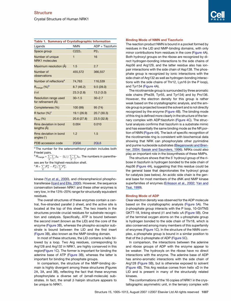
Table 1. Summary of Crystallographic Information
Ligands NMN ADP + Tiazofurin
Space group C2221 P21
Number of unique
NRK1 molecules
1 16
Maximum resolution (A) 1.5 2.7
Number of
observations
455,572 366,557
Number of reflectionsa 74,763 116,539
Rmerge (%)b 8.7 (46.2) 9.5 (28.0)
I/sI 23.3 (2.8) 13.2 (3.0)
Resolution range used
for refinement (A)
30–1.5 30–2.7
Completeness (%) 100 (99) 95 (74)
R factor (%)c 19.1 (26.9) 20.7 (30.3)
Rfree (%) 20.6 (27.8) 23.5 (32.8)
Rms deviation in bond
lengths (A)
0.004 0.010
Rms deviation in bondangles (�)
1.2 1.5
PDB accession code 2QG6 2QL6
a The number for the selenomethionyl protein includes both
Friedel pairs.b Rmerge =
P
h
P
i
jIhi � hIhij=P
h
P
i
Ihi. The numbers in parenthe-
ses are for the highest-resolution shell.c R =
P
h
jFoh � Fc
h j=P
h
Foh .
Structure 15, 1005–1
Binding Mode of NMN and Tiazofurin
The reaction product NMN is bound in a pocket formed by
residues in the LID and NMP-binding domains, with only
minor contributions from residues in the core (Figure 4A).
Both hydroxyl groups on the ribose are recognized by di-
rect hydrogen-bonding interactions to the side chains of
Asp56 and Arg129, and the latter residue also has ion-
pair interactions with the side chain of Asp138. The phos-
phate group is recognized by ionic interactions with the
side chain of Arg132 as well as hydrogen-bonding interac-
tions with the side chains of Thr12, Lys16 (in the P loop),
and Tyr134 (Figure 4A).
The nicotinamide group is surrounded by three aromatic
side chains (Phe39, Tyr55, and Tyr134) and by Pro136.
However, the electron density for this group is rather
weak based on the crystallographic analysis, and the am-
ide group is projected toward the solvent and is not directly
recognized by the enzyme (Figure 4B). The binding mode
of this ring is defined more clearly in the structure of the ter-
nary complex with ADP:tiazofurin (Figure 4C). The struc-
tural analysis confirms that tiazofurin is a substrate mimic
and has essentially the same binding mode as the NR por-
tion of NMN (Figure 4A). The lack of specific recognition of
the nicotinamide ring is consistent with biochemical data
showing that NRK can phosphorylate other pyrimidine
and purine nucleoside substrates (Bieganowski and Bren-
ner, 2004; Sasiak and Saunders, 1996). NRKs could also
play an important role in the biosynthesis of these NMPs.
The structure shows that the 50 hydroxyl group of the ri-
bose in tiazofurin is hydrogen bonded to the side chain of
Asp36 (Figure 4A), suggesting that this residue could be
the general base that deprotonates the hydroxyl group
for catalysis (see below). An acidic side chain is the gen-
eral base for most members of the dNK and NMP kinase
superfamilies of enzymes (Eriksson et al., 2002; Yan and
Tsai, 1999).
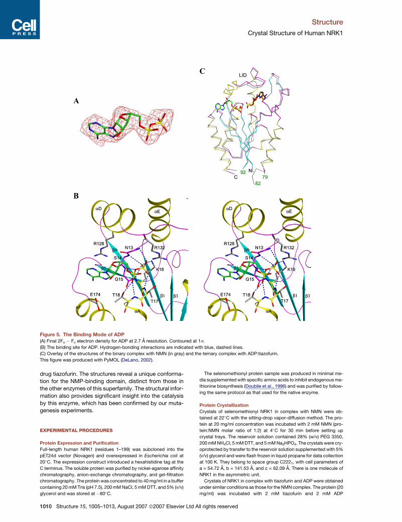
Binding Mode of ADP
Clear electron density was observed for the ADP molecule
based on the crystallographic analysis (Figure 5A). The
b-phosphate group interacts with the P loop, 10-GVTNS
GKTT-18, linking strand b1 and helix aA (Figure 5B). One
of the terminal oxygen atoms on the a-phosphate group
is hydrogen bonded to the side chain of Thr18, which is
also conserved among many members of this superfamily
of enzymes (Figure 1C). In the structure of the NMN com-
plex, a phosphate group is bound in a similar position to
that of the b-phosphate of ADP (Figure 5C).
In comparison, the interactions between the adenine
and ribose groups of ADP with the enzyme appear to
be weaker. The hydroxyls on the ribose have no direct
interactions with the enzyme. The adenine base of ADP
has amino-aromatic interactions with the side chain of
Arg128 (Figure 5B), but is otherwise exposed to solvent
(Figure 4B). This Arg residue comes from helix aD in the
LID and is present in many of the structurally related
kinases.
The conformations of the 16 copies of NRK1 in the crys-
tallographic asymmetric unit, in the ternary complex with
013, August 2007 ª2007 Elsevier Ltd All rights reserved 1007

Structure
Crystal Structure of Human NRK1
Figure 2. Structure of Human NRK1
(A) Schematic representation of the structure of human NRK1 in a binary complex with reaction product NMN (in orange for carbon atoms).
(B) Structure of human NRK1 in a ternary complex with ADP (green) and tiazofurin (black).
This figure was produced with PyMOL (DeLano, 2002).
ADP and tiazofurin, are essentially identical; there is an
rms distance of 0.1 A among their equivalent Ca atoms.
Human NRK1 is monomeric in solution (unpublished
data and Sasiak and Saunders, 1996). In comparison,
the dNKs are dimeric in solution (Eriksson et al., 2002;
Welin et al., 2007). Noncrystallographic symmetry (NCS)
restraint was applied in the refinement, but reducing the
weight on the NCS restraint did not significantly affect
the structures. On the other hand, there are clear struc-
tural differences between this ternary complex and the
1008 Structure 15, 1005–1013, August 2007 ª2007 Elsevier L
binary complex with NMN. This is especially true for the
LID and the aA helix (Figure 5C), which are involved in
binding ADP. In comparison, the central b sheet has few
structural differences in the two complexes. The rms dis-
tance among 176 equivalent Ca atoms of the 2 structures
is 0.67 A.
Implications for Catalysis by NRK
Our structures of the binary complex with NMN and the ter-
nary complex with ADP and tiazofurin provide significant
Figure 3. Structural Homologs of NRK1
(A) Schematic representation of the structure of M. mycoides deoxyadenosine kinase in complex with dCTP (Welin et al., 2007).
(B) Schematic representation of the structure of yeast adenylate kinase in complex with Ap5A (Abele and Schulz, 1995).
This figure was produced with PyMOL (DeLano, 2002).
td All rights reserved

Structure
Crystal Structure of Human NRK1
Figure 4. The Binding Modes of NMN and Tiazofurin
(A) The binding site for NMN. Hydrogen-bonding interactions are indicated with blue, dashed lines. The binding mode of tiazofurin is also shown
(in black).
(B) Molecular surface of the active-site region of NRK1, colored based on electrostatic potential.
(C) Final 2Fo � Fc electron density for tiazofurin at 2.7 A resolution. Contoured at 1s.
(A) and (C) were produced with PyMOL (DeLano, 2002); (B) was produced with Grasp (Nicholls et al., 1991).
insights into the catalysis by NRK. The structural informa-
tion allowed us to produce a model for the enzyme in
complex with its substrates ATP and NR (Figure 6A), as
well as with its products ADP and NMN (Figure 6B). The
structural analysis suggests that Asp36 is the general
base of the enzyme, extracting the proton from the 50
hydroxyl of the NR substrate. In the model, this hydroxyl
is only 3.4 A from the g-phosphorus atom in ATP and is
in the correct position to initiate nucleophilic attack on
this phosphate. The distance between the g-phosphate
group of ATP and the phosphate group in NMN is 1.7 A,
suggesting that only a relatively small movement is needed
for the phospho-transfer reaction. The structures show
that the nicotinamide group is not recognized specifically
by the enzyme, suggesting that NRK could be active with
other phospho-acceptors, as has been observed with
tiazofurin and other nucleosides (Bieganowski and Bren-
ner, 2004; Sasiak and Saunders, 1996).
Mutagenesis Studies Confirm the Structural
Observations
To assess the information obtained from the structures,
we selected several residues that appear to play important
Structure 15, 1005–
roles in substrate binding and/or catalysis and character-
ized their functional importance by mutagenesis experi-
ments. The kinetic assay converts the NMN product to
NADH, through the coupling enzymes NMNAT and alco-
hol dehydrogenase (Khan et al., 2006; Revollo et al.,
2004). The substrate NR was produced by dephosphory-
lation of NMN (Bieganowski and Brenner, 2004). The
assays showed that human NRK1 has robust catalytic
activity toward the NR substrate, which has a KM of about
3.7 mM (Figure 7A) (Sasiak and Saunders, 1996).
Mutation of the catalytic base, Asp36, essentially abol-
ished the activity of the enzyme (Figure 7B). Similarly,
mutations of Asp56, which has an important role in recog-
nizing the ribose hydroxyls of NR, and Lys16 in the P loop,
which is crucial for ATP binding, also inactivated the
enzyme. In comparison, mutation of Asp138, ion paired
to Arg129 that is hydrogen bonded to the ribose hy-
droxyls, has only minor effects on catalysis by the enzyme
(Figure 7B). Overall, our structural observations are con-
firmed by the mutagenesis and kinetic studies.
In summary, we have determined the crystal structures
of human NRK1 in a binary complex with its product NMN
and in a ternary complex with ADP and the anticancer
1013, August 2007 ª2007 Elsevier Ltd All rights reserved 1009
Structure
Crystal Structure of Human NRK1
Figure 5. The Binding Mode of ADP
(A) Final 2Fo � Fc electron density for ADP at 2.7 A resolution. Contoured at 1s.
(B) The binding site for ADP. Hydrogen-bonding interactions are indicated with blue, dashed lines.
(C) Overlay of the structures of the binary complex with NMN (in gray) and the ternary complex with ADP:tiazofurin.
This figure was produced with PyMOL (DeLano, 2002).
drug tiazofurin. The structures reveal a unique conforma-
tion for the NMP-binding domain, distinct from those in
the other enzymes of this superfamily. The structural infor-
mation also provides significant insight into the catalysis
by this enzyme, which has been confirmed by our muta-
genesis experiments.
EXPERIMENTAL PROCEDURES
Protein Expression and Purification
Full-length human NRK1 (residues 1–199) was subcloned into the
pET24d vector (Novagen) and overexpressed in Escherichia coli at
20�C. The expression construct introduced a hexahistidine tag at the
C terminus. The soluble protein was purified by nickel-agarose affinity
chromatography, anion-exchange chromatography, and gel-filtration
chromatography. The protein was concentrated to 40 mg/ml in a buffer
containing 20 mM Tris (pH 7.5), 200 mM NaCl, 5 mM DTT, and 5% (v/v)
glycerol and was stored at �80�C.
1010 Structure 15, 1005–1013, August 2007 ª2007 Elsevier
The selenomethionyl protein sample was produced in minimal me-
dia supplemented with specific amino acids to inhibit endogenous me-
thionine biosynthesis (Doublie et al., 1996) and was purified by follow-
ing the same protocol as that used for the native enzyme.
Protein Crystallization
Crystals of selenomethionyl NRK1 in complex with NMN were ob-
tained at 22�C with the sitting-drop vapor-diffusion method. The pro-
tein at 20 mg/ml concentration was incubated with 2 mM NMN (pro-
tein:NMN molar ratio of 1:2) at 4�C for 30 min before setting up
crystal trays. The reservoir solution contained 28% (w/v) PEG 3350,
200 mM NH4Cl, 5 mM DTT, and 5 mM Na2HPO4. The crystals were cry-
oprotected by transfer to the reservoir solution supplemented with 5%
(v/v) glycerol and were flash frozen in liquid propane for data collection
at 100 K. They belong to space group C2221, with cell parameters of
a = 54.72 A, b = 141.53 A, and c = 62.09 A. There is one molecule of
NRK1 in the asymmetric unit.
Crystals of NRK1 in complex with tiazofurin and ADP were obtained
under similar conditions as those for the NMN complex. The protein (20
mg/ml) was incubated with 2 mM tiazofurin and 2 mM ADP
Ltd All rights reserved

Structure
Crystal Structure of Human NRK1
Figure 6. Models for the Substrate and Product Complexes of NRK1
(A) Model of NRK1 in a ternary complex with the substrates ATP and NR. Asp36 is the general base that extracts the proton from NR.
(B) Model of NRK1 in a ternary complex with the products ADP and NMN.
This figure was produced with PyMOL (DeLano, 2002).
(protein:ligand molar ratio of 1:2) at 4�C for 30 min before setting up
trays. The pH of the ADP solution was adjusted to 7.8. The crystals
were washed once with the reservoir solution supplemented with 2
mM tiazofurin and 2 mM ADP and were flash cooled in liquid nitrogen.
They belong to space group P21, with cell parameters of a = 145.71 A,
b = 99.96 A, c = 145.59 A and b = 91.57�. There are 16 NRK1 mono-
mers in the asymmetric unit.
Data Collection and Processing
A selenomethionyl SAD data set at 1.5 A resolution was collected at 100
K on the NMN complex at the X4A beamline of Brookhaven National
Laboratory. A native reflection data set was collected for the ADP:tiazo-
furin complex at the X29 beamline. The diffraction images were pro-
cessed and scaled with the HKL package (Otwinowski and Minor,
1997). The data processing statistics are summarized in Table 1.
Structure Determination and Refinement
For the NMN complex, the locations of the selenomethionyl atoms
were determined with the program BnP (Weeks and Miller, 1999).
Reflection phases to 1.5 A resolution were calculated based on the
SAD data and were improved with the program SOLVE/RESOLVE
Structure 15, 1005–10
(Terwilliger, 2003), which also automatically located most of the resi-
dues. The complete atomic model was fit into the electron density
with the program O (Jones et al., 1991). The structure refinement
was carried out with the program CNS (Brunger et al., 1998). The re-
finement statistics are summarized in Table 1.
Crystals of the ADP:tiazofurin complex were microscopically
twinned, possibly due to the fact that the a and c axes of the unit cell
are almost identical, and that the b angle is close to 90�. A twinning
fraction of 0.37 was estimated by CNS, by simulating the hemihedral
twinning case of P4. The structure was solved by molecular replace-
ment with the program Phaser (Storoni et al., 2004). Twinned refine-
ment was carried out with CNS, with the twinning operator l�k h. Non-
crystallographic symmetry restraints were applied to the 16 NRK1
molecules in the asymmetric unit.
Mutagenesis
Mutants of NRK1 were designed based on the structural information,
created with the QuikChange kit (Stratagene), and sequenced to con-
firm the incorporation of the correct mutation. The mutant proteins
were purified by following the same protocol as that used for the
wild-type enzyme.
Figure 7. Mutagenesis Studies Confirm the Structural Observations
(A) Purified NRK1 has robust activity toward the NR substrate. The experimental data (black squares) were fitted to the Michaelis-Menten equation
(red curve) by nonlinear regression, defining the KM and Vmax values. The assay followed the production of NADH, through the coupling enzymes
NMNAT and alcohol dehydrogenase (Bieganowski and Brenner, 2004; Khan et al., 2006; Revollo et al., 2004). Data from one representative exper-
iment of several different repeats are shown.
(B) Mutations in the active-site region can abolish the catalytic activity. The mutants were designed based on the structural information, and their
effects on catalysis were determined by the kinetic assay.
13, August 2007 ª2007 Elsevier Ltd All rights reserved 1011

Structure
Crystal Structure of Human NRK1
NRK Assays
The substrate nicotinamide riboside (NR) was generated by following
a published protocol (Bieganowski and Brenner, 2004), by treating
NMN with calf intestinal alkaline phosphatase (CIP) at 37�C overnight.
A typical reaction contained 20 mM Tris (pH 7.9), 100 mM NaCl, 5 mM
MgCl2, 100 mM NMN (Sigma), and 1500 U CIP (New England Biolabs)
in a volume of 0.5 ml. The NR was separated from CIP by centrifugation
through a 10 kDa filter (Millipore). The quality of this substrate was
analyzed by the NRK1 assay described below, except that the NRK1
enzyme was omitted from the reaction mixture. Typically, no visible
contamination by NMN was detected even when the substrate was
used at high concentrations (�20 mM).
All of the assays were repeated several times to ensure the repro-
ducibility of the experiments. The catalytic activity of NRK1 was deter-
mined by using a coupled enzyme spectrometric assay. Briefly, the
NMN product of NRK was converted to NAD+ with the enzyme NMN/
NAMN adenylyltransferase (NMNAT), and NAD+ was then reduced to
NADH by alcohol dehydrogenase (Sigma) by using ethanol as the sub-
strate (Khan et al., 2006; Revollo et al., 2004). By monitoring the ap-
pearance of NADH at 340 nm, the activity of NRK could be determined.
Human NMNAT was overexpressed in E. coli and was purified by
following a published protocol (Zhou et al., 2002). The reaction buffer
contained 50 mM Tris (pH 7.5), 100 mM NaCl, various concentrations
of NR, 2.5 mM ATP, 12 mM MgCl2, 1.5% (v/v) ethanol, 10 mM semicar-
bazide (to remove the acetaldehyde product of ethanol oxidation),
0.02% (w/v) BSA, 10 mg/ml NMNAT, 30 mg/ml alcohol dehydrogenase,
and 0.2 mM NRK. The reactions are carried out at room temperature.
For the mutants, the reactions were monitored at 25 mM substrate
concentration.
ACKNOWLEDGMENTS
We thank Victor Marquez at the National Cancer Institute for the gift of
tiazofurin, Todd Yeates for helpful discussions about twinning, Randy
Abramowitz and John Schwanof for setting up the X4A beamline, and
Howard Robinson and Wuxian Shi for setting up the X29 beamline at
the National Synchrotron Light Source. This research is supported in
part by a grant from the National Institutes of Health to L.T.
Received: May 5, 2007
Revised: June 13, 2007
Accepted: June 27, 2007
Published: August 14, 2007
REFERENCES
Abele, U., and Schulz, G.E. (1995). High-resolution structures of
adenylate kinase from yeast ligated with inhibitor Ap5A, showing the
pathway of phosphoryl transfer. Protein Sci. 4, 1262–1271.
Appleby, T.C., Larson, G., Cheney, I.W., Walker, H., Wu, J.Z., Zhong,
W., Hong, Z., and Yao, N. (2005). Structure of human uridine-cytidine
kinase 2 determined by SIRAS using a rotating-anode X-ray generator
and a single samarium derivative. Acta Crystallogr. D Biol. Crystallogr.
61, 278–284.
Berger, F., Ramirez-Hernandez, M.H., and Ziegler, M. (2004). The new
life of a centenarian: signaling functions of NAD(P). Trends Biochem.
Sci. 29, 111–118.
Bieganowski, P., and Brenner, C. (2004). Discoveries of nicotinamide
riboside as a nutrient and conserved NRK genes establish a Preiss-
Handler independent route to NAD+ in fungi and humans. Cell 117,
495–502.
Blander, G., and Guarente, L. (2004). The Sir2 family of protein deace-
tylases. Annu. Rev. Biochem. 73, 417–435.
Brunger, A.T., Adams, P.D., Clore, G.M., DeLano, W.L., Gros, P.,
Grosse-Kunstleve, R.W., Jiang, J.-S., Kuszewski, J., Nilges, M.,
Pannu, N.S., et al. (1998). Crystallography & NMR System: a new
1012 Structure 15, 1005–1013, August 2007 ª2007 Elsevier Ltd A
software suite for macromolecular structure determination. Acta
Crystallogr. D54, 905–921.
DeLano, W.L. (2002). The PyMOL Molecular Graphic System (San
Carlos, CA: DeLano Scientific).
Denu, J.M. (2005). The Sir2 family of protein deacetylases. Curr. Opin.
Chem. Biol. 9, 431–440.
Doublie, S., Kapp, U., Aberg, A., Brown, K., Strub, K., and Cusack, S.
(1996). Crystallization and preliminary X-ray analysis of the 9 kDa
protein of the mouse signal recognition particle and the selenome-
thionyl-SRP9. FEBS Lett. 384, 219–221.
Eriksson, S., Munch-Petersen, B., Johansson, K., and Eklund, H.
(2002). Structure and function of cellular deoxyribonucleoside kinases.
Cell. Mol. Life Sci. 59, 1327–1346.
Grifantini, M. (2000). Tiazofurine ICN pharmaceuticals. Curr. Opin.
Investig. Drugs 1, 257–262.
Guse, A.H. (2005). Second messenger function and the structure-
activity relationship of cyclic adenosine diphosphoribose (cADPR).
FEBS J. 272, 4590–4597.
Hartmann, M.D., Bourenkov, G.P., Oberschall, A., Strizhov, N., and
Bartunik, H.D. (2006). Mechanism of phosphoryl transfer catalyzed
by shikimate kinase from Mycobacterium tuberculosis. J. Mol. Biol.
364, 411–423.
Hasmann, M., and Schemainda, I. (2003). FK866, a highly specific non-
competitive inhibitor of nicotinamide phosphoribosyltransferase,
represents a novel mechanism for induction of tumor cell apoptosis.
Cancer Res. 63, 7436–7442.
Hendrickson, W.A. (1991). Determination of macromolecular struc-
tures from anomalous diffraction of synchrotron radiation. Science
254, 51–58.
Holm, L., and Sander, C. (1993). Protein structure comparison by align-
ment of distance matrices. J. Mol. Biol. 233, 123–138.
Izard, T., and Ellis, J. (2000). The crystal structures of chloramphenicol
phosphotransferase reveal a novel inactivation mechanism. EMBO J.
19, 2690–2700.
Jager, W., Salamon, A., and Szekeres, T. (2002). Metabolism of the
novel IMP dehydrogenase inhibitor benzamide riboside. Curr. Med.
Chem. 9, 781–786.
Jauch, R., Humm, A., Huber, R., and Wahl, M.C. (2005). Structures of
Escherichia coli NAD synthetase with substrates and products reveal
mechanistic rearrangements. J. Biol. Chem. 280, 15131–15140.
Jones, T.A., Zou, J.Y., Cowan, S.W., and Kjeldgaard, M. (1991).
Improved methods for building protein models in electron density
maps and the location of errors in these models. Acta Crystallogr. A
47, 110–119.
Khan, J.A., Tao, X., and Tong, L. (2006). Molecular basis for the inhibi-
tion of human NMPRTase, a novel target for anticancer agents. Nat.
Struct. Mol. Biol. 13, 582–588.
Khan, J.A., Forouhar, F., Tao, X., and Tong, L. (2007). Nicotinamide ad-
enine dinucleotide metabolism as attractive target for drug discovery.
Expert Opin. Ther. Targets 11, 695–705.
Kraft, L., Sprenger, G.A., and Lindqvist, Y. (2002). Conformational
changes during the catalytic cycle of gluconate kinase as revealed
by X-ray crystallography. J. Mol. Biol. 318, 1057–1069.
Lavie, A., Konrad, M., Brundiers, R., Goody, R.S., Schlichting, I., and
Reinstein, J. (1998a). Crystal structure of yeast thymidylate kinase
complexed with the bisubstrate inhibitor P1-(50-adenosyl) P5-(50-
thymidyl) pentaphosphate (TP5A) at 2.0 A resolution: implications for
catalysis and AZT activation. Biochemistry 37, 3677–3686.
Lavie, A., Ostermann, N., Brundiers, R., Goody, R.S., Reinstein, J.,
Konrad, D., and Schlichting, I. (1998b). Structural basis for efficient
phosphorylation of 30-azidothymidine monophosphate by Escherichia
coli thymidylate kinase. Proc. Natl. Acad. Sci. USA 95, 14045–14050.
ll rights reserved
Structure
Crystal Structure of Human NRK1
Lee, H.C. (2004). Multiplicity of Ca2+ messengers and Ca2+ stores:
a perspective from cyclic ADP-ribose and NAADP. Curr. Mol. Med.
4, 227–237.
Li, F., Chong, Z.Z., and Maiese, K. (2006). Cell life versus cell longevity:
the mysteries surrounding the NAD+ precursor nicotinamide. Curr.
Med. Chem. 13, 883–895.
Magni, G., Amici, A., Emanuelli, M., Raffaelli, N., and Ruggieri, S.
(1999). Enzymology of NAD+ synthesis. Adv. Enzymol. Relat. Areas
Mol. Biol. 73, 135–182.
Magni, G., Amici, A., Emanuelli, M., Orsomando, G., Raffaelli, N., and
Ruggieri, S. (2004a). Enzymology of NAD+ homeostasis in man. Cell.
Mol. Life Sci. 61, 19–34.
Magni, G., Amici, A., Emanuelli, M., Orsomando, G., Raffaelli, N., and
Ruggieri, S. (2004b). Structure and function of nicotinamide mononu-
cleotide adenylyltransferase. Curr. Med. Chem. 11, 873–885.
Nicholls, A., Sharp, K.A., and Honig, B. (1991). Protein folding and as-
sociation: insights from the interfacial and thermodynamic properties
of hydrocarbons. Proteins 11, 281–296.
Otwinowski, Z., and Minor, W. (1997). Processing of X-ray diffraction
data collected in oscillation mode. Methods Enzymol. 276, 307–326.
Pankiwicz, K.W., Patterson, S.E., Black, P.L., Jayaram, H.N., Risal, D.,
Goldstein, B.M., Stuyver, L.J., and Schinazi, R.F. (2004). Cofactor
mimics as selective inhibitors of NAD-dependent inosine monophos-
phate dehydrogenase (IMPDH)-the major therapeutic target. Curr.
Med. Chem. 11, 887–900.
Revollo, J.R., Grimm, A.A., and Imai, S.-I. (2004). The NAD biosynthesis
pathway mediated by nicotinamide phosphoribosyltransferase
regulates Sir2 activity in mammalian cells. J. Biol. Chem. 279, 50754–
50763.
Revollo, J.R., Grimm, A.A., and Imai, S. (2007). The regulation of nico-
tinamide dinucleotide biosynthesis by Nampt/PBEF/visfatin in mam-
mals. Curr. Opin. Gastroenterol. 23, 164–170.
Rizzi, M., Nessi, C., Mattevi, A., Coda, A., Bolognesi, M., and Galizzi, A.
(1996). Crystal structure of NH3-dependent NAD+ synthetase from
Bacillus subtilis. EMBO J. 15, 5125–5134.
Rongvaux, A., Andris, F., van Gool, F., and Leo, O. (2003). Recon-
structing eukaryotic NAD metabolism. Bioessays 25, 683–690.
Sasiak, K., and Saunders, P.P. (1996). Purification and properties of
a human nicotinamide ribonucleotide kinase. Arch. Biochem. Biophys.
333, 414–418.
Structure 15, 1005–1
Schreiber, V., Dantzer, F., Ame, J.C., and de Murcia, G. (2006). Poly-
(ADP-ribose): novel functions for an old molecule. Nat. Rev. Mol. Cell
Biol. 7, 517–528.
Stehle, T., and Schulz, G.E. (1992). Refined structure of the complex
between guanylate kinase and its substrate GMP at 2.0 A resolution.
J. Mol. Biol. 224, 1127–1141.
Storoni, L.C., McCoy, A.J., and Read, R.J. (2004). Likelihood-
enhanced fast rotation functions. Acta Crystallogr. D Biol. Crystallogr.
60, 432–438.
Terwilliger, T.C. (2003). SOLVE and RESOLVE: automated structure
solution and density modification. Methods Enzymol. 374, 22–37.
Weeks, C.M., and Miller, R. (1999). The design and implementation of
SnB v2.0. J. Appl. Cryst. 32, 120–124.
Welin, M., Wang, L., Eriksson, S., and Eklund, H. (2007). Structure-
function analysis of a bacterial deoxyadenosine kinase reveals the
basis for substrate specificity. J. Mol. Biol. 366, 1615–1623.
Wojcik, M., Seidle, H.F., Bieganowski, P., and Brenner, C. (2006).
Glutamine-dependent NAD+ synthetase. How a two-domain, three-
substrate enzyme avoids waste. J. Biol. Chem. 281, 33395–33402.
Yan, H., and Tsai, M.D. (1999). Nucleoside monophosphate kinases:
structure, mechanism, and substrate specificity. Adv. Enzymol. Relat.
Areas Mol. Biol. 73, 103–134.
Ying, W. (2006). NAD+ and NADH in cellular functions and cell death.
Front. Biosci. 11, 3129–3148.
Yun, M., Park, C.-G., Kim, J.-Y., Rock, C.O., Jackowski, S., and Park,
H.-W. (2000). Structural basis for the feedback regulation of Escheri-
chia coli pantothenate kinase by coenzyme A. J. Biol. Chem. 275,
28093–28099.
Zhou, T., Kurnasov, O., Tomchick, D.R., Binns, D.D., Grishin, N.V.,
Marquez, V.E., Osterman, A.L., and Zhang, H. (2002). Structure of hu-
man nicotinamide/nicotinic acid mononucleotide adenylyltransferase.
Basis for the dual substrate specificity and activation of the oncolytic
agent tiazofurin. J. Biol. Chem. 277, 13148–13154.
Accession Numbers
Coordinates have been deposited in the Protein Data Bank with acces-
sion codes 2QG6 and 2QL6.
013, August 2007 ª2007 Elsevier Ltd All rights reserved 1013
Copyright © 2022 FDOKUMEN