Functional Analysis of Two PLA2G2A Variants Associated with Secretory Phospholipase A2-IIA Levels

Critical Role of TLR2 and MyD88 for Functional Responseof Macrophages to a Group IIA-Secreted PhospholipaseA2 from Snake VenomElbio Leiguez1, Karina Cristina Giannotti1, Vanessa Moreira1, Marcio Hideki Matsubara1, Jose
Marıa Gutierrez2, Bruno Lomonte2, Juan Pablo Rodrıguez3,4, Jesus Balsinde4, Catarina Teixeira1*
1 Laboratorio de Farmacologia, Instituto Butantan, Sao Paulo, Sao Paulo State, Brazil, 2 Instituto Clodomiro Picado, Facultad de Microbiologıa, Universidad de Costa Rica,
San Jose, San Jose, Costa Rica, 3 Laboratorio de Investigacion en Proteınas – FACENA-Universidad Nacional del Nordeste, Corrientes, Corrientes, Argentina, 4 The
eicosanoid research division, Instituto de Biologıa y Genetica Molecular, Valladolid, Valladolid, Spain
Abstract
The snake venom MT-III is a group IIA secreted phospholipase A2 (sPLA2) enzyme with functional and structural similaritieswith mammalian pro-inflammatory sPLA2s of the same group. Previously, we demonstrated that MT-III directly activates theinnate inflammatory response of macrophages, including release of inflammatory mediators and formation of lipid droplets(LDs). However, the mechanisms coordinating these processes remain unclear. In the present study, by using TLR22/2 orMyD882/2 or C57BL/6 (WT) male mice, we report that TLR2 and MyD88 signaling have a critical role in MT-III-inducedinflammatory response in macrophages. MT-III caused a marked release of PGE2, PGD2, PGJ2, IL-1b and IL-10 and increasedthe number of LDs in WT macrophages. In MT-III-stimulated TLR22/2 macrophages, formation of LDs and release ofeicosanoids and cytokines were abrogated. In MyD882/2 macrophages, MT-III-induced release of PGE2, IL-1b and IL-10 wasabrogated, but release of PGD2 and PGJ2 was maintained. In addition, COX-2 protein expression seen in MT-III-stimulatedWT macrophages was abolished in both TLR22/2 and MyD882/2 cells, while perilipin 2 expression was abolished only inMyD882/2 cells. We further demonstrated a reduction of saturated, monounsaturated and polyunsaturated fatty acids and arelease of the TLR2 agonists palmitic and oleic acid from MT-III-stimulated WT macrophages compared with WT controlcells, thus suggesting these fatty acids as major messengers for MT-III-induced engagement of TLR2/MyD88 signaling.Collectively, our findings identify for the first time a TLR2 and MyD88-dependent mechanism that underlies group IIA sPLA2-induced inflammatory response in macrophages.
Citation: Leiguez E, Giannotti KC, Moreira V, Matsubara MH, Gutierrez JM, et al. (2014) Critical Role of TLR2 and MyD88 for Functional Response of Macrophagesto a Group IIA-Secreted Phospholipase A2 from Snake Venom. PLoS ONE 9(4): e93741. doi:10.1371/journal.pone.0093741
Editor: Patricia T. Bozza, Fundacao Oswaldo Cruz, Brazil
Received November 21, 2013; Accepted March 6, 2014; Published April 9, 2014
Copyright: � 2014 Leiguez et al. This is an open-access article distributed under the terms of the Creative Commons Attribution License, which permitsunrestricted use, distribution, and reproduction in any medium, provided the original author and source are credited.
Funding: This investigation was supported by research grants from FAPESP, Sao Paulo, Brazil (www.fapesp.br), grants 11/21341-5 and 10/06345-1, INCTTOX, SaoPaulo, Brazil (www.incttox.com.br), grant 573790/2008-6, CNPq PQ, Brazil (www.cnpq.br), grant 306920/2011-5, Brazil, Spanish Ministery of Science andInnovation, Spain (http://web.micinn.es/), grant BFU2010-18826. The funders had no role in study design, data collection and analysis, decision to publish, orpreparation of the manuscript.
Competing Interests: The authors have declared that no competing interests exist.
* E-mail: [email protected]
Introduction
Secreted phospholipases A2 (sPLA2s) are enzymes that hydrolyse
glycerophospholipids at the sn-2 position of the glycerol backbone
in a calcium-dependent manner, releasing fatty acids and lysopho-
spholipids. These enzymes are members of the PLA2 superfamily,
which is widely distributed in nature, and 15 groups and
subgroups have been described, including five categories termed
secreted PLA2, cytosolic PLA2, Ca2+ independent PLA2, the
platelet-activating factor acetylhydrolase, and lysosomal PLA2 [1].
Among them, group IIA sPLA2 includes mammalian inflamma-
tory-type and Viperid snake venom sPLA2s [2,3]. Besides their
relevant role in cell membrane physiology, mammalian group IIA
sPLA2s are recognized as important paracrine and autocrine
players in inflammatory processes by their ability to release fatty
acids from cell membranes, leading to subsequent production of
pro-inflammatory mediators such as prostaglandins, leukotrienes
[1]. Accordingly, sPLA2 enzymes, particularly group IIA sPLA2s,
have been implicated in the pathophysiology of several inflam-
matory diseases, such as atherosclerosis, inflammatory bowel
disease, asthma and arthritis [4,5,6].
Besides structural homology, snake venom group IIA sPLA2s
share functional features with mammalian group IIA sPLA2s,
owing to their ability to induce inflammatory events in both in vivo
and in vitro experimental models [7]. sPLA2s are abundant
components in the proteomes of snake venoms from Bothrops
genus [8] and have been associated with the prominent local
inflammatory effects induced by these venoms. An Asp49 sPLA2,
named myotoxin-III (MT- III), has been purified from Bothrops
asper snake venom [9]. This group IIA sPLA2 has been reported to
induce inflammatory events in in vivo experimental models [10,11].
Furthermore, this venom sPLA2 exhibits the ability to directly
activate inflammatory functions in isolated macrophages, such as
increased phagocytic capacity, release of cytokines [11,12], up-
regulation of lipid mediator cascade, with release of prostaglandins
[13], and increased formation of lipid droplets (LDs) [14], which
are organelles implicated in the differentiation of macrophages
PLOS ONE | www.plosone.org 1 April 2014 | Volume 9 | Issue 4 | e93741

into foam cells. However, the initial steps leading to this sPLA2-
induced activation of macrophages are still unknown.
Macrophages are pivotal cells in innate immune responses.
Activation of these cells initiates cascades of events that lead to
acute inflammatory processes and the switch from the acute to the
resolutive phase of inflammation, by releasing resolutive media-
tors, such as PGJ2 and IL-10 [15,16]. In addition, macrophages
are relevant cells in the development of many chronic inflamma-
tory diseases, such as atherosclerosis. Macrophages loaded with
LDs, termed foam cells, constitute an early hallmark of
atherosclerotic lesion formation and a major cell source of
inflammatory mediators [17,18].
The macrophage innate response is initiated by activation of
diverse membrane recognition receptors, such as the Toll-like
receptors (TLRs). Nowadays, at least 13 TLRs have been
described, each of them with a degree of specificity for various
endogenous and exogenous ligands, eliciting different, but
sometimes overlapping, immune responses [19,20]. In inflamma-
tory processes, recognition of pathogen-associated molecular
patterns (PAMPs) by TLRs except TLR3 activates cell-signaling
pathways mediated by the MyD88 [20]. Other kinds of stimuli,
named endogenous damage-associated molecular patterns
(DAMPs), are also capable of activating MyD88-dependent innate
immune signaling by TLRs [21]. Some of the well- characterized
DAMPs include: fibronectins [22], heat-shock proteins [23], pro-
inflammatory cytokines from the monocyte-macrophage system
[24], and free fatty acids (FFA) [25]. Among the TLRs, TLR2
constitutes a molecular link between elevated circulating FFA and
inflammation [26–28]. TLR2 binds lipid-based structures via their
long-chain saturated FFA moieties [25,29], triggering inflamma-
tory responses through MyD88 signaling [30]. Although TLRs are
critical receptors directing specific inflammatory programs in
macrophages, the involvement of TLR2 and MyD88 adaptor
molecule signaling pathways in the activation of macrophages by
group IIA sPLA2 remains unknown. The understanding of the
mechanism(s) involved in sPLA2s-induced activation of in a pivotal
immune innate cell, such as the macrophage, shall provide
valuable information on new targets to confront the inflammatory
diseases where group IIA sPLA2s enzymes are involved.
In the present study the involvement of TLR2 and MyD88
adaptor molecule in the signaling cascade (inflammatory signaling)
elicited by MT-III in macrophages was investigated. To test this
possibility, the effects of MT-III in macrophages from wild-type
(WT) C57BL/6 mice, TLR2 gene knockout (TLR22/2) mice and
MyD88 gene knockout (MyD882/2) were evaluated in terms of
PGE2, PGD2, PGJ2, IL-1b and IL-10 biosynthesis, LDs formation,
COX-2 and perilipin-2 expression and release of free fatty acids.
Herein, we present the first evidence that TLR2 and MyD88
adaptor molecule are implicated in the inflammatory response
triggered by a group IIA sPLA2 enzyme in macrophages.
Engagement of TLR2/MyD88 signaling pathway by the venom
sPLA2 MT-III coordinates events related to both initiation and
resolution of the inflammatory response in macrophages. Our
observations also suggest that saturated free fatty acids are major
agonists for engagement of TLR2/MyD88 pathway induced by
this group IIA sPLA2.
Materials and Methods
Ethics StatementThis study includes experiments with macrophages collected
from pathogen-free 5-week-old wild type (WT), MyD882/2 and
TLR22/2 mice (all on a C57BL/6 genetic background). These
animals were bred in animal facilities at the University of Sao
Paulo and were housed in a temperature-controlled room (22–
24uC) with a 12 h light-dark cycle and fresh water and food ad
libitum until used. All experimental procedures involving these
animals were performed in accordance with the procedures laid
down by the Universities Federation for Animal Welfare and were
approved by the Animal Experimentation Ethics Committee of
University of Sao Paulo (Sao Paulo, Brazil; Reference No 94/
2010).
Chemicals and ReagentsHema-3 stain from Biochemical Sciences (Swedesboro, NJ,
USA). MTT and L-glutamine were obtained from USB (Cleve-
land, OH, USA). E-toxate LAL kit and mouse mAb anti-b-actinwas purchased from Sigma-Aldrich (St. Louis, MO, USA) and
guinea pig polyclonal antibody anti- mouse perilipin-2 (PLIN2)
from Research Diagnostics (Flanders, NJ, USA). PGE2 and PGD2
enzyme immunoassay kits and polyclonal antibodies against
COX-2 were purchased from Cayman Company, Ann Arbor,
MI, USA. 15-deoxy-D12,14– prostaglandin J2 (PGJ2) and IL-10 and
IL-1b enzyme immunoassay kits were purchased from Enzo Life
Sciences (Ann Arbor, MI, USA) and eBioscience, Inc. (San Diego,
CA, USA), respectively. Secondary antibodies, anti- mouse and
anti-guinea pig, conjugated to HRP and nitrocellulose membrane,
were obtained from GE Healthcare (Buckinghamshire, UK).
Gentamicin was purchased from Schering-Plough (Whitehouse
Station, NJ, USA) and DMSO from Amresco (Solon, OH, USA).
RPMI 1640, thiocarbohydrazide, and Osmium tetroxide (OsO4)
were purchased from Sigma-Aldrich and RPMI from Life
Technologies (Sao Paulo, SP, Brazil). Chloroform and methanol
(HPLC grade) were purchased from Fisher Scientific (Hampton,
NH), and [1–14C] palmitic acid and oleic acid from Perkin Elmer
(Waltham, MA). Silicagel thin layer chromatography plates were
purchased from Macherey-Nagel (Duren, Germany). Thioglyco-
late was obtained from Merck (Darmstadt, Germany) and all salts
used were purchased from Sigma-Aldrich.
Phospholipase A2 (PLA2)Aspartate-49 sPLA2 named MT-III (Uniprot accession no.:
P20474) from B. asper venom was purified by ion-exchange
chromatography on CM-Sephadex C-25, using a KCl gradient
from 0 to 0.75 M at pH 7.0 as described [2], followed by RP-
HPLC on a semipreparative C8 column (Vydac; 106250 mm,
5 mm particle size) eluted at a flow rate of 2.5 ml/min with a
gradient of acetonitrile (0–70%, containing 0.1% trifluoroacetic
acid) over 30 min. Homogeneity was verified by SDS-PAGE, run
under reducing conditions, in which a single band of ,14 kDa
was observed. The complete amino acid sequence of this enzyme
has been described previously [31,32]. The absence of endotoxin
contamination in the MT-III batches used was demonstrated by
performing the quantitative LAL test [33], which revealed
undetectable levels of endotoxin (,0.125 EU/mL).
Macrophages Harvesting and StimulationAnimals were killed under CO2 atmosphere and cells were then
harvested by washing peritoneal cavities with 3 mL apyrogenic
phosphate buffered saline (PBS), pH 7.2. Aliquots of the washes
were used for total cell counts in a Neubauer chamber, after
dilution (1:20, v/v) in Turk solution (0.2% crystal violet dye in
30% acetic acid). Differential cell counts were performed on
smears stained with Hema3. More than 95% of the cell population
consisted of macrophages, as determined by conventional mor-
phological criteria. The remaining wash volumes were centrifuged
at 1296g for 6 min (4uC), supernatants were discarded and cell
pellets were appropriately diluted and maintained in serum-free
TLR2 and MyD88 Signal for sPLA2-Induced Effects
PLOS ONE | www.plosone.org 2 April 2014 | Volume 9 | Issue 4 | e93741

RPMI 1640 medium, supplemented with 2 mM L-glutamine and
40 mg/mL gentamicin sulfate at 37uC with 5% CO2, and
processed according to the experimental protocols used, as follows:
1) 16106 macrophages/well were seeded in 12 wells culture plates
and maintained in culture medium for 24 h before stimulation,
and then used for Western blotting analysis of COX-2 and
perilipin-2 protein expression; 2) 26105 macrophages, harvested
4 days after i.p. injection of 1 mL 3% thioglycolate, were seeded
on glass coverslips and incubated in culture medium for 30 min for
adhesion, followed by washing with PBS, and then used for LDs
evaluation; 3) 16107 macrophages were seeded in cell culture dish,
maintained in culture medium for 18 h, and used for gas
chromatography/mass spectrometry analysis of fatty acid methyl
esters; 4) 26105 macrophages/well were seeded in 12 well culture
plates, maintained with culture medium for 30 min for adhesion
and then incubated with exogenous [1–14C] Oleic or [1–14C]
Palmitic acid (1 mM; 0.25 mCi/ml) for 18 h at 37uC. Cells werethen scraped with 0.1% Triton X-100 in PBS and supernatants
were removed and total 14C-radioactivity levels were determined
by scintillation counting. After each of the above described
procedures, cells were stimulated with MT-III diluted in RPMI
1640 medium in a final concentration of 0.4 mM or RPMI 1640
medium alone (control) for selected periods of time and
maintained at 37uC in a humidified atmosphere (5% CO2).
Quantification of Eicosanoids and CytokinesProstaglandins (PGE2, PGD2 and PGJ2) and cytokines (IL-1b
and IL-10) were quantified in supernatants from macrophages
cultures by using commercially available enzyme immune assay
kits, performed according to instructions of the manufacturer.
Lipid Droplet (LD) Staining and QuantificationAnalysis of LD numbers was performed in osmium-stained cells.
In brief, macrophages adhered to glass coverslips were fixed in 4%
paraformaldehyde in 0.1 M phosphate buffer (pH 7.2) for 15 min
and stained with OsO4. The coverslips were then rinsed in 0.1 M
phosphate buffer, stained with 1% OsO4 (30 min), rinsed in
deionized H2O, immersed in 1.0% thiocarbohydrazide (5 min),
rinsed again in 0.1 M phosphate buffer, re-stained with 1% OsO4
(3 min), rinsed with H2O, and then dried and mounted [42]. The
morphology of the fixed cells was observed, and round intracel-
lular osmiophilic structures were identified as LDs, which were
then counted under phase-contrast microscopy using the 1006ob-
jective lens in 50 consecutively scanned leukocytes in each
coverslip.
Western Blotting for COX-2 and Perilipin-2COX-2 and perilipin-2 protein levels in cultured macrophages
were detected by Western blotting. Aliquots of MT-III-stimulated
and non-stimulated cells (1.56106 cells) were lysed with 100 mLsample buffer (0.5 M Tris-HCl, pH 6.8, 20% SDS, 1% glycerol,
1 M b-mercapto ethanol, 0.1% bromophenol blue) and boiled for
10 min. Samples were resolved by SDS-PAGE on 10% bis-
acrylamide gels overlaid with a 5% stacking gel. Proteins were
then transferred to nitrocellulose membranes using a Mini Trans-
Blot (Bio-Rad Laboratories, Richmond, CA, USA). The mem-
branes were blocked for 1 h with 5% nonfat dry milk in Tris-
buffered saline (TBS) (20 mM Tris, 100 mM NaCl, and 0.5%
Tween 20, pH 7.2), and incubated with primary antibodies
against COX-2 (1:1000 dilution), perilipin-2 (1:2000) and b-actin(1:3000) for 1 h. They were then washed and incubated with the
appropriate secondary antibody conjugated to horseradish perox-
idase. The immunonereactive bands were detected by the entry-
level peroxidase substrate for enhanced chemiluminescence (ECL),
according to the instructions of the manufacturer (GE Healthcare).
Band densities were quantified with a GS 700 densitometer (Bio-
Rad Laboratories) using the image analysis software Molecular
Analyst (Bio-Rad Laboratories).
Gas Chromatography/mass Spectrometry Analysis ofFatty Acid Methyl EstersA cell extract was used and, before extraction, 10 nmol of 1,2,3-
triheptadecanoylglycerol was added as internal standard. Then,
total lipids were extracted according to the Bligh and Dyer method
[34], and glycerolipids were transmethylated with 500 ml of 0.5 M
KOH in methanol for 60 min at 37uC. For neutralization, 500 mlof 0.5 M HCl were added and the extraction of fatty acid methyl
esters was performed with 1 ml of n-hexane twice. Analysis of fatty
acid methyl esters was carried out in a Agilent 7890A gas
chromatograph coupled to an Agilent 5975C mass---selective
detector operated in electron impact mode (EI, 70 eV), equipped
with an Agilent 7693 autosampler and an Agilent DB23 column
(60 m length6250 mm internal diameter60.15 mm film thickness)
under the following conditions: oven temperature was held at
50uC for 1 min, then increased to 175uC at a rate of 25uC/min,
then increased to 215uC at a rate of 1.5uC/min, and the final
ramp being reached at 235uC at a rate of 10uC/min. The final
temperature was maintained for 5 min, and the run time was
39.67 min. Data analysis was carried out with the Agilent
G1701EA MSD Productivity Chemstation software, revision
E.02.00, and the quantification of fatty acid methyl esters was
achieved by integration of chromatographic peaks comparing with
authentic analytical standards [35,36]. The fatty acids evaluated
are indicated below in table 1.
Quantification of Fatty Acids (FA) ReleasedThe cells were placed in serum-free medium and exposed to
exogenous [14C] Oleic or [14C] Palmitic acid (1 mM; 0.25 mCi/ml)
for 18 h at 37uC. Afterward, the cells were washed four times with
PBS containing 0.5% albumin to remove the fatty acids that had
not been incorporated, and next stimulated with MT-III or RPMI
medium only (control) for 6 h. Cells were scraped with 0.1%
Triton X-100 in PBS, supernatants were removed, and total 14C-
radioactivity levels were determined by scintillation counting.
Statistical AnalysisData are expressed as the mean 6 standard error of mean
(S.E.M) of at least three animals. Multiple comparisons among
groups were performed using one-way ANOVA and Tukey test as
a post-test. Differences between experimental groups were
considered significant for P-values ,0.05. All statistic tests were
performed using Prism version 5 software (GraphPad, San Diego,
CA).
Results
Prostaglandin Production Induced by MT-III RequiresDistinct TLR Signaling PathwaysProstaglandins are produced in abundance at sites of inflam-
mation and regulate many aspects of the inflammatory response.
Prostaglandins such as PGE2 and PGD2 have well-defined roles in
acute inflammation, such as the regulation of blood flow and
edema formation, cytokine generation, and leukocyte migration
[37,38]. In contrast to these lipid mediators, PGJ2, which is a
PGD2-derived prostanoid, has been described as a resolutive
mediator, which is involved in the switch from the acute into the
regenerative phases of inflammatory processes [16]. We have
TLR2 and MyD88 Signal for sPLA2-Induced Effects
PLOS ONE | www.plosone.org 3 April 2014 | Volume 9 | Issue 4 | e93741

previously reported that the sPLA2 MT-III elicits the biosynthesis
of prostaglandins in isolated peritoneal macrophages [13]. These
relevant cells of the innate immune system sense danger signals via
recognition receptors such as TLRs, and then initiate a
coordinated inflammatory response. However, whether TLR
pathways, particularly TLR2 and MyD88 adaptor molecule
activation, are required for this and other MT-III-induced effects
in macrophages is unknown, and deserve further investigation.
Therefore, we initially evaluated the involvement of TLR2 and
MyD88 signaling in MT-III-induced PGE2, PGD2 and PGJ2production in TLR2- and MyD88- deficient macrophages. Results
presented in Figure 1 (A–C) show that PGE2 levels were
significantly increased in MT-III-stimulated macrophages collect-
ed from WT mice. This MT-III- induced PGE2 release was
impaired in macrophages obtained either from TLR22/2 or
MyD882/2mice. Notably, sPLA2 MT-III failed to induce PGD2
and PGJ2 production in TLR22/2, but did induce production of
these mediators in MyD882/2 macrophages when compared with
MT-III- stimulated WT cells. These observations prompted us to
consider that TLR2 signaling is engaged for the biosynthesis of
PGE2, PGD2 and PGJ2 in macrophages under stimuli by MT-III.
However, the TLR2 via MyD88 adaptor molecule pathway
operates only for biosynthesis of PGE2, whereas production of
PGD2 and PGJ2 occurs through an adaptor molecule other than
MyD88. Of note, these findings further suggest that upon
activation by the sPLA2 MT-III, TLR2 and MyD88 signaling
pathways can coordinate macrophage responses related to both
the development and resolution steps of inflammation.
Up-regulation of COX-2 Protein Expression Induced byMT-III is Dependent on TLR2 via MyD88 SignalingPathwayActivation of TLR2 via MyD88-mediated signaling leads to
activation of transcription factors such as NF-kB, AP-1 and IRF,
which in turn play an important role in gene transcription of
several molecules involved in the inflammatory response, such as
cytokines and cyclooxygenase-2 (COX-2) [39–42]. Prostaglandins
are produced by cyclooxygenases, which occur in constitutive
(COX-1) and inducible (COX-2) forms [6]. We have previously
demonstrated that MT-III up-regulates COX-2 protein expression
in peritoneal macrophages and that this isoform is responsible for
the increased levels of prostaglandins induced by this sPLA2 [13].
However, the involvement of TLR2 and MyD88 signaling
pathway in this process is unknown. The potential role of TLR2
and MyD88 signaling in MT-III-induced COX-2 protein expres-
sion was then analyzed. Figure 1 (D) shows that TLR22/2 and
MyD882/2 macrophages stimulated by MT-III did not present
the increased COX-2 protein expression seen in MT-III-
stimulated WT macrophages. These data clearly indicate that
TLR2 and MyD88 are essential to this effect induced by the
sPLA2 MT-III, constituting a key step in the onset of MT-III-
induced activation of macrophages. Participation of MyD88
signaling in this MT-III-induced effect is consistentwith previous
reports by others that activation of this adaptor molecule signals
for gene expression of COX-2 [19,43].
TLR2 via MyD88 Signaling Pathway is Required for MT-III-induced Production of IL-1b and IL-10 in MacrophagesIL-1b and IL-10 are relevant mediators involved in triggering
and down-regulation of inflammatory processes, respectively
[44,45]. Despite the distinct roles played by these two cytokines
in inflammation, the production of these mediators has been
shown to be regulated by TLR2 and MyD88 signaling [46]. The
ability of MT-III to induce the release of inflammatory cytokines,
such as IL-1b, has been previously demonstrated [12]. However,
whether this MT-III- induced event is coordinated by TLR2 and
MyD88 pathways is unknown. Moreover, neither the ability of
MT-III to induce release of IL-10 by macrophages nor the
participation of TLR2 and MyD88 pathways in this effect has
been investigated. We therefore explored the potential role of
TLR2 and MyD88 signaling in MT-III-induced synthesis of these
cytokines. As demonstrated in Figure 2 (A–B), stimulation of WT
macrophages with MT-III induced a significant release of IL- 1band IL-10 from these cells. However, stimulation of TLR22/2
and MyD882/2 macrophages by MT-III did not result in an
increased release of these two cytokines as compared with MT-III-
stimulated WT macrophages. These data confirm previous
observations on the ability of MT-III to induce the release of
IL-1b and additionally evidenced the capacity of this sPLA2 to
induce the release of IL-10. Furthermore, these data implicate the
TLR2 via MyD88 signaling pathway in the generation of IL-1band IL-10 induced by MT-III in macrophages.
Table 1. Fatty acid nomenclature.
Abbreviation Systematic Name Commom name
16_0 Hexadecanoic Acid Palmitic acid
18_0 Octadecanoic Acid stearic acid
18_1 n-9 9-Octadecenoic Acid oleic acid
18_1 n-7 11-Octadecemoic Acid cis-vaccenic acid
18_2 n-6 9,12-Octadecadienoic Acid Linoleic acid
20_1 n-9 11-Eicosenoic Acid Gondoic Acid
20_2 n-6 11,14-Eicosadienoic Acid none
20_3 n-6 8,11,14-Eicosatrienoic Acid Dihomo-gamma-linolenic acid
20_4 n-6 5,8,11,14-Eicosatetraenoic Acid Arachidonic acid
22_5 n-6 4,7,10,13,16-Docosapentaenoic Acid Osbond Acid
22_5 n-3 7,10,13,16,19-Docosapentaenoic Acid Clupanodonic Acid
22_6 n-3 4,7,10.13.16,19-Docosahexaenoic Acid Cervonic Acid
doi:10.1371/journal.pone.0093741.t001
TLR2 and MyD88 Signal for sPLA2-Induced Effects
PLOS ONE | www.plosone.org 4 April 2014 | Volume 9 | Issue 4 | e93741

Figure 1. TLR2 and Myd88 signaling pathways are involved in MT-III-induced prostanoid biosynthesis and COX-2 proteinexpression. Wild type (WT) or TLR22/2 or MyD882/2 peritoneal macrophages were incubated with MT-III (0.4 mM) or RPMI (control) for 6 h. PGE2
(A), PGD2 (B) and PGJ2 (C) concentrations were quantified in culture supernatants by EIA commercial kits; (D) Western blotting immunoreactive bandsof COX-2 and b-actin (loading control) representative of at least three samples/experimental group; (E) Densitometric analysis of immunoreactiveCOX-2 band intensities. Densities (in arbitrary units) were normalized with those of b-actin. Results are expressed as mean 6 SEM from 3–6 animals.*p,0.05 as compared with control cells; #p,0.05 as compared with MT-III-stimulated cells.doi:10.1371/journal.pone.0093741.g001
Figure 2. TLR2 and MyD88 signaling pathways are relevant for MT-III-induced IL-1b and IL-10 release. Wild type (WT) or TLR22/2 orMyD882/2 peritoneal macrophages were incubated with MT-III (0.4 mM) or RPMI (control) for 6 h. IL-1b (A) and IL-10 (B) concentrations werequantified in culture supernatants by EIA commercial kits. Results are expressed as mean 6 SEM from 3–6 animals. *p,0.05 as compared with controlgroup; #p,0.05 as compared with MT-III- stimulated WT cells.doi:10.1371/journal.pone.0093741.g002
TLR2 and MyD88 Signal for sPLA2-Induced Effects
PLOS ONE | www.plosone.org 5 April 2014 | Volume 9 | Issue 4 | e93741

TLR2 via MyD88 is a Critical Pathway for MT-III-inducedLD FormationIt has been demonstrated that LDs formation in leukocytes is a
highly regulated event that depends on the interaction of cellular
receptors with their ligands. These organelles were shown to be
involved in the synthesis of inflammatory mediators and are
markers of leukocyte activation [47,48]. Moreover, the role of
TLRs-mediated pathogen recognition and activation in the
mechanism of LDs formation has been demonstrated by others
[48,49]. We have recently shown that MT-III induces the
formation of LDs in macrophages [14]. However, the mechanisms
underlying this MT-III-induced effect remain unclear. To further
understand this issue we investigated the involvement of TLR2
and MyD88 signaling. As demonstrated in Figure 3A, the sPLA2
MT-III failed to induce LDs formation in macrophages from both
TLR22/2 and MyD882/2 mice when compared with macro-
phages from WT mice, at both time intervals evaluated. These
findings identify TLR2 via MyD88 adaptor molecule as an
essential signaling pathway regulating LD biogenesis induced by
the venom sPLA2 MT-III in macrophages. Considering that LD-
laden macrophages constitute the foam cells seen at the sites of
atherosclerotic lesion, in which levels of the sPLA2s are increased,
our present findings suggest that activation of TLR2/MyD88-
mediated signaling pathway is likely to be an important
mechanism that accounts for foam cell formation stimulated by
group IIA sPLA2s.
Requirement of TLR2 and/or MyD88 Signaling for MT-III-induced Perilipin-2 up- Regulation and MobilizationPerilipin-2 has long been reported as a pivotal protein for
neutral lipid accumulation that serves as scaffolding during LD
formation in diverse cell types, including macrophages [50,51].
This protein is constitutively and ubiquitously expressed in
macrophages as a major component of intracellular LDs [50,52–
54]. Moreover, the expression of perilin-2 can be up-regulated
during inflammatory processes [51,55,56]. Consistent with these
properties, perilipin-2 has been used as a marker of lipid loading
and LD assembly in inflammatory cells [57]. Accumulating
evidence indicates that activation of the Toll like receptors triggers
foam cell formation and increase of perilipin-2 protein expression
[49,58,59]. In macrophages, MT-III has been shown to up-
regulate perilipin-2 protein expression and to trigger mobilization
of constitutive perilipin-2 to form new LDs in macrophages [14].
On this basis, we assessed the involvement of TLR2 and MyD88
in perilipin-2 protein expression induced by MT-III in macro-
phages for 6 h. As demonstrated in Figure 3B, following MT-III
stimulation of WT macrophages, there is a significant increase of
perilipin-2 protein expression in comparison with RPMI-stimu-
lated WT macrophages. This increased protein expression was
observed in TLR22/2 macrophages stimulated by MT-III when
compared with WT macrophages. Conversely, MyD882/2
macrophages stimulated by this sPLA2 were unable to up-regulate
perilipin-2 protein expression as compared to WT macrophages.
These data indicate that perilipin-2 protein expression induced by
MT-III is dependent on MyD88 pathway, but not on TLR2
activation. Next we evaluated the potential role of TLR2 and
MyD88 signaling in subcellular mobilization of perilipin-2 induced
by MT-III. As illustrated in Figure 4, WT macrophages stimulated
Figure 3. TLR2 and MyD88 are required for lipid droplet formation, but only MyD88 is relevant to perilipin-2 protein expressioninduced by MT-III. Wild type (WT) or TLR22/2 or MyD882/2 peritoneal macrophages were incubated either with MT-III (0.4 mM) or RPMI (control)for 6 or 12 h. (A) Lipid droplets numbers; (B) Western blotting immunoreactive bands of perilipin-2 (PLIN2) and b-actin (loading control); (C)Densitometric analysis of immunoreactive PLIN2 band intensities. Densities (in arbitrary units) were normalized with those of b-actin. Results areexpressed as mean 6 SEM from 3–6 animals. *p,0.05 as compared with control group; #p,0.05 as compared with MT- III- stimulated WT cells.doi:10.1371/journal.pone.0093741.g003
TLR2 and MyD88 Signal for sPLA2-Induced Effects
PLOS ONE | www.plosone.org 6 April 2014 | Volume 9 | Issue 4 | e93741

by MT-III exhibited strong fluorescent staining (green) for
perilipin-2, with a punctate cytoplasmic pattern, which was absent
in RPMI-stimulated control cells. Fluorescent Nile Red-labeled
LDs were also visualized after MT-III-induced stimulation and
were virtually absent in non-stimulated WT macrophages. After
stimulation with MT-III, cytoplasmic-stained perilipin-2 matched
perfectly with Nile Red- marked neutral lipid inclusions. As
expected, no significant staining was detected in WT control
macrophages. However, this phenomenon was not observed in
macrophages from TLR22/2 or MyD882/2 mice, demonstrating
that perilipin-2 subcellular distribution induced by MT-III is
dependent on TLR2 and MyD88 signaling pathway in a tight
correlation with the regulation of MT-III-induced LD formation
by TLR2/MyD88 pathway.
MT-III Releases Free Fatty Acids from MacrophagesTo better understand the mechanisms underlying activation of
TLR2 and MyD88 signaling by MT-III in macrophages, we
analyzed the ability of this sPLA2 to generate free fatty acids,
which are recognized as efficient agonists of TLRs, mainly TLR2
[25,28,29,60]. The total fatty acid profile was then analyzed in
MT-III-stimulated macrophages by measuring fatty acid methyl
esters using gas chromatography mass spectrometry coupled.
Figure 5A shows that stimulation of WT macrophages with the
sPLA2 MT-III resulted in a remarkable reduction of fatty acids
presumably due to the action of this enzyme on membrane
phospholipids. Large decreases were observed in palmitic, stearic,
oleic, and particularly in arachidonic acid, which clearly generated
a pool of intracellular free fatty acids, that must be reorganized in
order to keep intact the cellular homeostasis. To accomplish this,
two of the possible underlying mechanisms are: 1) lipid droplet
formation and 2) excretion of fatty acids into the extracellular
environment. In order to test this latter hypothesis, we previously
added labeled free fatty acids (palmitic and oleic acid) to the
peritoneal macrophage culture and let them to incorporate; then
we determined the blink per minute in a radioactivity counter.
Figure 5B shows that higher amounts of palmitic and oleic acids
were found in culture supernatants of MT-III-stimulated WT
macrophages than in controls, suggesting that these FFA may
consequently stimulate the Toll like receptors by an autocrine
mechanism.
Discussion
Group IIA secreted PLA2 enzymes are considered important
players in inflammatory process owing to their ability to induce the
biosynthesis of second messengers that stimulate innate responses
of inflammatory cells, including macrophages. As TLRs are
recognized to trigger a pro-inflammatory program in macrophages
and respond to exogenous molecules and host-derived endogenous
ligands [20,61], in the present study we investigated the
participation of TLR2 and MyD88 adaptor molecule in the
stimulatory effects of the venom sPLA2 MT-III in macrophages.
The data presented herein indicate a major role for TLR2 in
MT-III-induced production of inflammatory mediators by mac-
rophages, since the production of PGE2, PGD2 and PGJ2 induced
by MT-III was abrogated in TLR22/2 macrophages, indicating
the engagement of TLR2 upon stimulus by this sPLA2. To our
knowledge, this is the first demonstration that TLR2 pathway is
activated by a secreted PLA2 enzyme leading to biosynthesis of
lipid mediators. Reports from literature have shown that
endogenous Group V and Group X sPLA2s are capable to
amplify signaling through TLR2 and TLR4/MyD88 in cells
stimulated by the pathogen- activated molecular patterns
Pam3CSK4 and LPS, leading to biosynthesis of eicosanoids in
mast cells and macrophages, respectively [31,69]. Furthermore,
we found that the adaptor molecule MyD88 plays a role in MT-
III-induced production of PGE2, since the release of this mediator
was abrogated in MyD882/2 macrophages stimulated by MT-III.
However, the increment of PGD2 and PGJ2 induced by MT-III
was not altered in MyD882/2 macrophages, suggesting that their
synthesis is triggered through an adaptor molecule other than
MyD88, in the present experimental condition. This is the first
demonstration that a secreted group IIA PLA2 directly activates
macrophages to release PGJ2. Noteworthy, the present findings
also evidence, for the first time, that the production of a resolutive
mediator is regulated by the TLR2 pathway.
A previous report of our group demonstrated that, similarly to
mammalian GIIA sPLA2s [70], MT-III up-regulates COX-2
protein expression, a critical enzyme for PGs production in
macrophages [13]. We herein extended the understanding of this
mechanism by showing that MT-III up-regulates COX-2 expres-
sion via TLR2 and MyD88 signaling pathways in macrophages,
based on the inability of TLR22/2 and MyD882/2 macrophages
to express COX-2 in response to MT-III stimulus. Taking these
data into account, and considering results showing that MyD88
adaptor molecule is involved in MT-III-induced production of
PGE2, but not PGD2 or PGJ2, it is suggested that COX-2 is the
major PGE2 biosynthetic pathway, whereas production of PGD2
and PGJ2 may be associated to both COX-2 and COX-1
biosynthetic pathways. Consistent with this hypothesis, a previous
report has shown the ability of MT-III to induce an increment in
the activity of COX-1 along with expression of COX-2 in
macrophages [13].
The ability of sPLA2s, including MT-III, to induce the release of
cytokines has been previously demonstrated [12,71]. The data
presented herein demonstrate that this sPLA2 induces the release
of IL-10 and IL-1b in a process mediated by TLR2/MyD88
signaling pathway. These findings also bring additional evidence
that engagement of TLR2/MyD88 signaling pathway coordinates
both the acute and resolutive responses in macrophages upon
stimulation by the sPLA2 MT-III. Furthermore, increased IL-10
biosynthesis, observed in MT-III-stimulated WT macrophages,
suggests that in our experimental conditions this cytokine regulates
the synthesis of PGE2 via TLR2/MyD88 pathway activation, as a
tight regulation of PGE2 biosynthesis by IL-10 has been previously
demonstrated in macrophages [62–64].
LDs are cytoplasmic inclusions found in numerous immune cells
associated with inflammation [51,54,65], such as foam macro-
phages, which are major cells of atherosclerotic lesions [66]. We
recently demonstrated that MT-III induces LDs formation,
recruitment and increased expression of the LD scaffold protein
perilipin-2 in macrophages [14]. The results obtained herein
demonstrate, for the first time, that TLR2 and MyD88 are
essential elements for LD formation induced by MT-III, since
TLR22/2 and MyD882/2 macrophages failed to respond with
increased numbers of LDs. These results are in agreement with
previous reports by others showing that TLR2/MyD88 pathway is
involved in LDs formation in leukocytes [47–49]. On the other
hand, our data further demonstrate that TLR2 deletion does not
affect the ability of MT-III to induce a significant increase of
perilipin-2 protein expression, but inhibits its capacity to mobilize
constitutive perilipin-2 in the cytosol, evidencing that although not
signaling for up regulation of perilipin-2 expression, this receptor is
engaged for its recruitment, triggering an important mechanism
for LD formation. In contrast, MyD88 deleted cells blocked MT-
III- induced expression and mobilization of perilipin-2, implying
the participation of this adaptor molecule in this effect. Therefore,
TLR2 and MyD88 Signal for sPLA2-Induced Effects
PLOS ONE | www.plosone.org 7 April 2014 | Volume 9 | Issue 4 | e93741

activation of a TLR other than TLR2 is involved in MT-III-
induced expression of perilipin-2. To our knowledge, this is the
first demonstration of participation of TLR2 and MyD88
pathways in the mechanisms leading to LDs formation induced
by group IIA sPLA2, and that MyD88 is a crucial adaptor
molecule mediating perilipin-2 expression and mobilization
induced by this class of enzymes. Experimental studies have
previously demonstrated that TLRs, particularly TLR2 via
MyD88-dependent pathway, have relevant roles in the differen-
tiation of recruited monocytes into lipid-laden macrophages (foam
cells), and ultimately to the development of atherosclerotic lesions
[67,68]. Considering the reported increased expression of group
IIA sPLA2s at the site of atherosclerotic lesions [9], our present
findings may shed light on the early mechanisms involved in the
ability of sPLA2s to induce formation of foam macrophages.
The mechanisms by which MT-III activates TLR2 were not
presently investigated. It has been shown that a free fatty acid
(FFA)-rich microenvironment leads to the activation of TLR2
[28,60]. Moreover, there are data demonstrating that products of
cleavage of membrane phospholipids by sPLA2s, including
saturated fatty acids, lysophosphatidylcholine and lysophosphati-
dylserine, are able to activate this receptor [60,72,73,74]. In this
Figure 4. TLR2 and MyD88 are essential for MT-III-induced perilipin-2 (PLIN2) subcellular distribution. Wild Type (WT), TLR22/2 orMyD882/2 peritoneal macrophages were incubated with RPMI (control) or MT-III (0.4 mM) for 6 h and labeled for both lipid droplets (fluorescent Nilered) and anti-PLIN2 (FITC-conjugated immunocomplex). Merged image shows localization of PLIN2 to lipid droplets in WT macrophages. Cell nucleiwere stained with propidium iodide. IgG control was included and showed negative stain. The micrographs are representative of at least threesamples/experimental group.doi:10.1371/journal.pone.0093741.g004
TLR2 and MyD88 Signal for sPLA2-Induced Effects
PLOS ONE | www.plosone.org 8 April 2014 | Volume 9 | Issue 4 | e93741

context, we herein demonstrate that MT-III reduced the levels of
saturated FFA, such as palmitic and stearic acid, as well as
monounsaturated and polyunsaturated fatty acids in macrophages.
Decreased levels of both palmitic and oleic acids correlated with
increased amounts of these fatty acids in the extracellular medium,
indicating that they are released from macrophages upon the
action of MT-III, thus allowing their interaction with surface
membrane receptors. On this basis, we suggest that, in the present
experimental conditions, saturated FFAs resulting from the
catalytic action of MT-III on membrane phospholipids are
autocrine ligands for TLR2, which then triggers an inflammatory
program in macrophages, via pathways dependent on MyD88.
However, participation of lysophospholipids in this mechanism
cannot be ruled out. In support of our hypothesis, when catalytic
activity of MT-III is inhibited, increments in LD formation are
abrogated [14]. In addition, the involvement of TLR2 in the
release of the cytokines IL-1b and IL-10 induced by minimally
modified LDL in macrophages has been previously described [46].
In conclusion, we provide evidence of a critical role for TLR2 and
MyD88 adaptor molecule in mediating sPLA2-induced inflamma-
tory responses in macrophages. We have shown for the first time
that the TLR2 and Myd88 adaptor molecule are involved in
cellular and molecular inflammatory processes triggered by the
venom sPLA2 MT-III in macrophages, that include the produc-
tion of eicosanoids and cytokines and the formation of LDs, as well
as an increased expression of COX-2 and perilipin-2 mobilization
and expression. Interestingly, these findings indicate that TLR2,
via MyD88 adaptor molecule, links sPLA2s with LD and foam cell
formation. Moreover, we speculate that saturated free fatty acids
released by MT-III enzyme activity from phospholipids in the
macrophage membrane, are autocrinally recognized by TLR2,
thereby initiating an inflammatory signaling cascade in macro-
phages. Altogether, these findings amplify the current knowledge
on the mechanisms involved in the stimulatory effects of Group
IIA sPLA2s in macrophages, specially those that trigger inflam-
matory events, including foam cell formation, which is a hallmark
of atherosclerosis. Such a mechanism may contribute to the
identification of useful new targets for the development of novel
therapeutic strategies for inflammatory diseases related to lipid
imbalance where group IIA sPLA2s enzymes are involved.
Figure 5. MT-III affects macrophage fatty acid content causing the release of palmitic and oleic acids. (A) The profile of major fatty acidsin peritoneal macrophages from WT mice stimulated with MT-III (0.4 mM) (gray bars) or RPMI (control) (open bars) for 6 h was determined by GC- MSafter converting the glyceryl fatty acid into fatty acid methyl esters. (B) Peritoneal macrophages were incubated with 1 mM [14C]palmitic acid or (C)[14C] oleic acid for 18 h. Afterward, [14C]palmitic acid or [14C] oleic acid incorporation was measured in macrophages and their supernatants 6 h afterstimulation with either MT-III or RPMI (control) as described in Materials and Methods. The 14C radioactivity incorporated is expressed as a percentageof the radioactivity originally added to the cells. Results are expressed as means 6 SEM from 3–6 animals. *p,0.05 as compared with thecorresponding control.doi:10.1371/journal.pone.0093741.g005
TLR2 and MyD88 Signal for sPLA2-Induced Effects
PLOS ONE | www.plosone.org 9 April 2014 | Volume 9 | Issue 4 | e93741
Acknowledgments
The authors acknowledge Renata Hage do Amaral for technical assistance
and Dr. Henrique Krambeck Rofatto (Butantan Institute), for supporting
confocal laser scanning microscopy analysis.
Author Contributions
Conceived and designed the experiments: CT EL JB JMG BL. Performed
the experiments: EL KCG VM MHM JPR. Analyzed the data: CT EL
KCG VM MHM JPR. Contributed reagents/materials/analysis tools: CT
JB JMG BL. Wrote the paper: EL CT JB JMG BL.
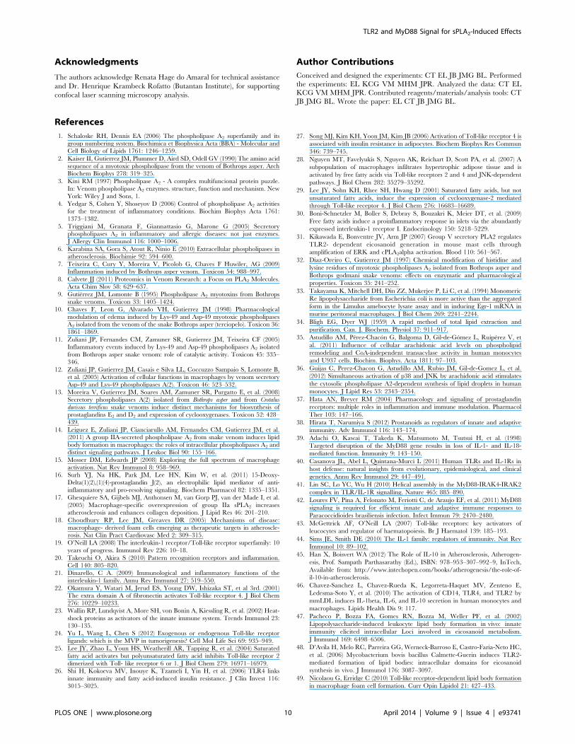
References
1. Schaloske RH, Dennis EA (2006) The phospholipase A2 superfamily and its
group numbering system. Biochimica et Biophysica Acta (BBA) - Molecular andCell Biology of Lipids 1761: 1246–1259.
2. Kaiser II, Gutierrez JM, Plummer D, Aird SD, Odell GV (1990) The amino acid
sequence of a myotoxic phospholipase from the venom of Bothrops asper. ArchBiochem Biophys 278: 319–325.
3. Kini RM (1997) Phospholipase A2 - A complex multifuncional protein puzzle.In: Venom phospholipase A2 enzymes. structure, function and mechanism. New
York: Wiley J and Sons, 1.
4. Yedgar S, Cohen Y, Shoseyov D (2006) Control of phospholipase A2 activitiesfor the treatment of inflammatory conditions. Biochim Biophys Acta 1761:
1373–1382.
5. Triggiani M, Granata F, Giannattasio G, Marone G (2005) Secretoryphospholipases A2 in inflammatory and allergic diseases: not just enzymes.
J Allergy Clin Immunol 116: 1000–1006.
6. Karabina SA, Gora S, Atout R, Ninio E (2010) Extracellular phospholipases in
atherosclerosis. Biochimie 92: 594–600.
7. Teixeira C, Cury Y, Moreira V, Picolob G, Chaves F Huwiler, AG (2009)Inflammation induced by Bothrops asper venom. Toxicon 54: 988–997.
8. Calvete JJ (2011) Proteomics in Venom Research: a Focus on PLA2 Molecules.Acta Chim Slov 58: 629–637.
9. Gutierrez JM, Lomonte B (1995) Phospholipase A2 myotoxins from Bothrops
snake venoms. Toxicon 33: 1405–1424.
10. Chaves F, Leon G, Alvarado VH, Gutierrez JM (1998) Pharmacological
modulation of edema induced by Lys-49 and Asp-49 myotoxic phospholipasesA2 isolated from the venom of the snake Bothrops asper (terciopelo). Toxicon 36:
1861–1869.
11. Zuliani JP, Fernandes CM, Zamuner SR, Gutierrez JM, Teixeira CF (2005)Inflammatory events induced by Lys-49 and Asp-49 phospholipases A2 isolated
from Bothrops asper snake venom: role of catalytic activity. Toxicon 45: 335–346.
12. Zuliani JP, Gutierrez JM, Casais e Silva LL, Coccuzzo Sampaio S, Lomonte B,
et al. (2005) Activation of cellular functions in macrophages by venom secretoryAsp-49 and Lys-49 phospholipases A(2). Toxicon 46: 523–532.
13. Moreira V, Gutierrez JM, Soares AM, Zamuner SR, Purgatto E, et al. (2008)Secretory phospholipases A(2) isolated from Bothrops asper and from Crotalus
durissus terrificus snake venoms induce distinct mechanisms for biosynthesis of
prostaglandins E2 and D2 and expression of cyclooxygenases. Toxicon 52: 428–439.
14. Leiguez E, Zuliani JP, Cianciarullo AM, Fernandes CM, Gutierrez JM, et al.(2011) A group IIA-secreted phospholipase A2 from snake venom induces lipid
body formation in macrophages: the roles of intracellular phospholipases A2 and
distinct signaling pathways. J Leukoc Biol 90: 155–166.
15. Mosser DM, Edwards JP (2008) Exploring the full spectrum of macrophage
activation. Nat Rev Immunol 8: 958–969.
16. Surh YJ, Na HK, Park JM, Lee HN, Kim W, et al. (2011) 15-Deoxy-
Delta(1)(2),(1)(4)-prostaglandin J(2), an electrophilic lipid mediator of anti-
inflammatory and pro-resolving signaling. Biochem Pharmacol 82: 1335–1351.
17. Ghesquiere SA, Gijbels MJ, Anthonsen M, van Gorp PJ, van der Made I, et al.
(2005) Macrophage-specific overexpression of group IIa sPLA2 increasesatherosclerosis and enhances collagen deposition. J Lipid Res 46: 201–210.
18. Choudhury RP, Lee JM, Greaves DR (2005) Mechanisms of disease:
macrophage- derived foam cells emerging as therapeutic targets in atheroscle-rosis. Nat Clin Pract Cardiovasc Med 2: 309–315.
19. O’Neill LA (2008) The interleukin-1 receptor/Toll-like receptor superfamily: 10
years of progress. Immunol Rev 226: 10–18.
20. Takeuchi O, Akira S (2010) Pattern recognition receptors and inflammation.
Cell 140: 805–820.
21. Dinarello, C A. (2009) Immunological and inflammatory functions of the
interleukin-1 family. Annu Rev Immunol 27: 519–550.
22. Okamura Y, Watari M, Jerud ES, Young DW, Ishizaka ST, et al 3rd. (2001)The extra domain A of fibronectin activates Toll-like receptor 4. J Biol Chem
276: 10229–10233.
23. Wallin RP, Lundqvist A, More SH, von Bonin A, Kiessling R, et al. (2002) Heat-
shock proteins as activators of the innate immune system. Trends Immunol 23:
130–135.
24. Yu L, Wang L, Chen S (2012) Exogenous or endogenous Toll-like receptor
ligands: which is the MVP in tumorigenesis? Cell Mol Life Sci 69: 935–949.
25. Lee JY, Zhao L, Youn HS, Weatherill AR, Tapping R, et al. (2004) Saturated
fatty acid activates but polyunsaturated fatty acid inhibits Toll-like receptor 2
dimerized with Toll- like receptor 6 or 1. J Biol Chem 279: 16971–16979.
26. Shi H, Kokoeva MV, Inouye K, Tzameli I, Yin H, et al. (2006) TLR4 links
innate immunity and fatty acid-induced insulin resistance. J Clin Invest 116:3015–3025.
27. Song MJ, Kim KH, Yoon JM, Kim JB (2006) Activation of Toll-like receptor 4 is
associated with insulin resistance in adipocytes. Biochem Biophys Res Commun
346: 739–745.
28. Nguyen MT, Favelyukis S, Nguyen AK, Reichart D, Scott PA, et al. (2007) A
subpopulation of macrophages infiltrates hypertrophic adipose tissue and is
activated by free fatty acids via Toll-like receptors 2 and 4 and JNK-dependent
pathways. J Biol Chem 282: 35279–35292.
29. Lee JY, Sohn KH, Rhee SH, Hwang D (2001) Saturated fatty acids, but not
unsaturated fatty acids, induce the expression of cyclooxygenase-2 mediated
through Toll-like receptor 4. J Biol Chem 276: 16683–16689.
30. Boni-Schnetzler M, Boller S, Debray S, Bouzakri K, Meier DT, et al. (2009)
Free fatty acids induce a proinflammatory response in islets via the abundantly
expressed interleukin-1 receptor I. Endocrinology 150: 5218–5229.
31. Kikawada E, Bonventre JV, Arm JP (2007) Group V secretory PLA2 regulates
TLR2- dependent eicosanoid generation in mouse mast cells through
amplification of ERK and cPLA2alpha activation. Blood 110: 561–567.
32. Diaz-Oreiro C, Gutierrez JM (1997) Chemical modification of histidine and
lysine residues of myotoxic phospholipases A2 isolated from Bothrops asper and
Bothrops godmani snake venoms: effects on enzymatic and pharmacological
properties. Toxicon 35: 241–252.
33. Takayama K, Mitchell DH, Din ZZ, Mukerjee P, Li C, et al. (1994) Monomeric
Re lipopolysaccharide from Escherichia coli is more active than the aggregated
form in the Limulus amebocyte lysate assay and in inducing Egr-1 mRNA in
murine peritoneal macrophages. J Biol Chem 269: 2241–2244.
34. Bligh EG, Dyer WJ (1959) A rapid method of total lipid extraction and
purification. Can. J. Biochem. Physiol 37: 911–917.
35. Astudillo AM, Perez-Chacon G, Balgoma D, Gil-de-Gomez L, Ruiperez V, et
al. (2011) Influence of cellular arachidonic acid levels on phospholipid
remodeling and CoA-independent transacylase activity in human monocytes
and U937 cells. Biochim. Biophys. Acta 1811: 97–103.
36. Guijas C, Perez-Chacon G, Astudillo AM, Rubio JM, Gil-de-Gomez L, et al.
(2012) Simultaneous activation of p38 and JNK by arachidonic acid stimulates
the cytosolic phospholipase A2-dependent synthesis of lipid droplets in human
monocytes. J Lipid Res 53: 2343–2354.
37. Hata AN, Breyer RM (2004) Pharmacology and signaling of prostaglandin
receptors: multiple roles in inflammation and immune modulation. Pharmacol
Ther 103: 147–166.
38. Hirata T, Narumiya S (2012) Prostanoids as regulators of innate and adaptive
immunity. Adv Immunol 116: 143–174.
39. Adachi O, Kawai T, Takeda K, Matsumoto M, Tsutsui H, et al. (1998)
Targeted disruption of the MyD88 gene results in loss of IL-1- and IL-18-
mediated function. Immunity 9: 143–150.
40. Casanova JL, Abel L, Quintana-Murci L (2011) Human TLRs and IL-1Rs in
host defense: natural insights from evolutionary, epidemiological, and clinical
genetics. Annu Rev Immunol 29: 447–491.
41. Lin SC, Lo YC, Wu H (2010) Helical assembly in the MyD88-IRAK4-IRAK2
complex in TLR/IL-1R signalling. Nature 465: 885–890.
42. Loures FV, Pina A, Felonato M, Feriotti C, de Araujo EF, et al. (2011) MyD88
signaling is required for efficient innate and adaptive immune responses to
Paracoccidioides brasiliensis infection. Infect Immun 79: 2470–2480.
43. McGettrick AF, O’Neill LA (2007) Toll-like receptors: key activators of
leucocytes and regulator of haematopoiesis. Br J Haematol 139: 185–193.
44. Sims JE, Smith DE (2010) The IL-1 family: regulators of immunity. Nat Rev
Immunol 10: 89–102.
45. Han X, Boisvert WA (2012) The Role of IL-10 in Atherosclerosis, Atherogen-
esis, Prof. Sampath Parthasarathy (Ed.), ISBN: 978–953–307–992–9, InTech,
Available from: http://www.intechopen.com/books/atherogenesis/the-role-of-
il-10-in-atherosclerosis.
46. Chavez-Sanchez L, Chavez-Rueda K, Legorreta-Haquet MV, Zenteno E,
Ledesma-Soto Y, et al. (2010) The activation of CD14, TLR4, and TLR2 by
mmLDL induces IL-1beta, IL-6, and IL-10 secretion in human monocytes and
macrophages. Lipids Health Dis 9: 117.
47. Pacheco P, Bozza FA, Gomes RN, Bozza M, Weller PF, et al. (2002)
Lipopolysaccharide-induced leukocyte lipid body formation in vivo: innate
immunity elicited intracellular Loci involved in eicosanoid metabolism.
J Immunol 169: 6498–6506.
48. D’Avila H, Melo RC, Parreira GG, Werneck-Barroso E, Castro-Faria-Neto HC,
et al. (2006) Mycobacterium bovis bacillus Calmette-Guerin induces TLR2-
mediated formation of lipid bodies: intracellular domains for eicosanoid
synthesis in vivo. J Immunol 176: 3087–3097.
49. Nicolaou G, Erridge C (2010) Toll-like receptor-dependent lipid body formation
in macrophage foam cell formation. Curr Opin Lipidol 21: 427–433.
TLR2 and MyD88 Signal for sPLA2-Induced Effects
PLOS ONE | www.plosone.org 10 April 2014 | Volume 9 | Issue 4 | e93741

50. Brasaemle DL, Barber T, Wolins NE, Serrero G, Blanchette-Mackie EJ, et al.
(1997) Adipose differentiation-related protein is an ubiquitously expressed lipidstorage droplet- associated protein. J Lipid Res 38: 2249–2263.
51. Larigauderie G, Furman C, Jaye M, Lasselin C, Copin C, et al. (2004)
Adipophilin enhances lipid accumulation and prevents lipid efflux from THP-1macrophages: potential role in atherogenesis. Arterioscler Thromb Vasc Biol 24:
504–510.52. Heid HW, Moll R, Schwetlick I, Rackwitz HR, Keenan TW (1998) Adipophilin
is a specific marker of lipid accumulation in diverse cell types and diseases. Cell
Tissue Res 294: 309–321.53. Persson J, Degerman E, Nilsson J, Lindholm MW (2007) Perilipin and
adipophilin expression in lipid loaded macrophages. Biochem Biophys ResCommun 363: 1020–1026.
54. Robenek H, Robenek MJ, Troyer D (2005) PAT family proteins pervade lipiddroplet cores. J Lipid Res 46: 1331–1338.
55. Corsini E, Sheasgreen J, Marinovich M, Galli CL (2002) Use of differential
display- polymerase chain reaction to identify genes selectively modulated bychemical allergens in reconstituted human epidermis. Toxicol In Vitro 16: 427–
431.56. Sarov-Blat L, Kiss RS, Haidar B, Kavaslar N, Jaye M, et al. (2007)
Predominance of a proinflammatory phenotype in monocyte-derived macro-
phages from subjects with low plasma HDL-cholesterol. Arterioscler ThrombVasc Biol 27: 1115–1122.
57. Brasaemle DL, Dolios G, Shapiro L, Wang R (2004) Proteomic analysis ofproteins associated with lipid droplets of basal and lipolytically stimulated 3T3-
L1 adipocytes. J Biol Chem 279: 46835–46842.58. Feingold KR, Kazemi MR, Magra AL, McDonald CM, Chui LG, et al. (2010)
ADRP/ADFP and Mal1 expression are increased in macrophages treated with
TLR agonists. Atherosclerosis 209: 81–88.59. Wang J, Si Y, Wu C, Sun L, Ma Y, et al. (2012) Lipopolysaccharide promotes
lipid accumulation in human adventitial fibroblasts via TLR4-NF-kappaBpathway. Lipids Health Dis 11: 139.
60. Huang S, Rutkowsky JM, Snodgrass RG, Ono-Moore KD, Schneider DA, et al.
(2012) Saturated fatty acids activate TLR-mediated proinflammatory signalingpathways. J Lipid Res 53: 2002–2013.
61. Iwata H, Nagai R (2012) Novel immune signals and atherosclerosis. CurrAtheroscler Rep 14: 484–490.
62. MacKenzie KF, Clark K, Naqvi S, McGuire VA, Noehren G, et al. (2013)PGE(2) induces macrophage IL-10 production and a regulatory-like phenotype
via a protein kinase A-SIK-CRTC3 pathway. J Immunol 190: 565–577.
63. Nemeth K, Leelahavanichkul A, Yuen PS, Mayer B, Parmelee A, et al. (2009)
Bone marrow stromal cells attenuate sepsis via prostaglandin E(2)-dependent
reprogramming of host macrophages to increase their interleukin-10 production.
Nat Med 15: 42–49.
64. Heusinkveld M, de Vos van Steenwijk PJ, Goedemans R, Ramwadhdoebe TH,
Gorter A, et al. (2011) M2 macrophages induced by prostaglandin E2 and IL-
6 from cervical carcinoma are switched to activated M1 macrophages by CD4+Th1 cells. J Immunol 187: 1157–1165.
65. Krahmer N, Farese Jr RV, Walther TC (2013) Balancing the fat: lipid droplets
and human disease. EMBO Mol Med 5: 905–915.
66. Ley K, Miller YI, Hedrick CC (2011) Monocyte and macrophage dynamics
during atherogenesis. Arterioscler Thromb Vasc Biol 31: 1506–1516.
67. Cole JE, Mitra AT, Monaco C (2010) Treating atherosclerosis: the potential of
Toll-like receptors as therapeutic targets. Expert Rev Cardiovasc Ther 8: 1619–
1635.
68. Liu Y, Yu H, Zhang Y, Zhao Y (2008) TLRs are important inflammatory factors
in atherosclerosis and may be a therapeutic target. Med Hypotheses 70: 314–
316.
69. Shridas P, Bailey WM, Talbott KR, Oslund RC, Gelb MH, et al. (2011) Group
X secretory phospholipase A2 enhances TLR4 signaling in macrophages.
J Immunol 187: 482–9.
70. Bryant KJ, Bidgood MJ, Lei PW, Taberner M, Salom C, et al. (2011) A
bifunctional role for group IIA secreted phospholipase A2 in human rheumatoid
fibroblast-like synoviocyte arachidonic acid metabolism. J Biol Chem 286: 2492–
503.
71. Granata F, Petraroli A, Boilard E, Bezzine S, Bollinger J, et al. (2005) Activation
of cytokine production by secreted phospholipase A2 in human lung
macrophages expressing the M-type receptor. J Immunol 174: 464–474.
72. Magalhaes K, Almeida PE, Atella G, Maya-Monteiro CM, Castro-Faria-Neto
H, et al. (2010) Schistosomal-derived lysophosphatidylcholine are involved in
eosinophil activation and recruitment through Toll-like receptor-2-dependent
mechanisms. J Infect Dis., 202: 1369–79.
73. Frasch SC, Bratton DL (2012) Emerging roles for lysophosphatidylserine in
resolution of inflammation. Prog Lipid Res., 51: 199–207.
74. Carneiro AB, Iaciura BM, Nohara LL, Lopes CD, Veas EM, et al. (2013)
Lysophosphatidylcholine triggers TLR2- and TLR4-mediated signaling path-
ways but counteracts LPS-induced NO synthesis in peritoneal macrophages by
inhibiting NF-kB translocation and MAPK/ERK phosphorylation. PLoS One
8:e76233.
TLR2 and MyD88 Signal for sPLA2-Induced Effects
PLOS ONE | www.plosone.org 11 April 2014 | Volume 9 | Issue 4 | e93741
Copyright © 2022 FDOKUMEN