Partial Neuroprotective Effect of Pretreatment with Tanshinone IIA on Neonatal Hypoxia-Ischemia...
7
Partial Neuroprotective Effect of Pretreatment with Tanshinone IIA on Neonatal Hypoxia-Ischemia Brain Damage WEN JIE XIA, MO YANG, TAI FAI FOK, KAREN LI, WOOD YEE CHAN, PAK-CHEUNG NG, HO KEUNG NG, KI WAI CHIK, CHI CHIU WANG, GOLDIE JIA SHI GU, KAM SANG WOO, AND KWOK PUI FUNG The Institute of Chinese Medicine [W.J.X., K.P.F.], Departments of Paediatrics [M.Y., T.F.F., K.L., P.-C.N., K.W.C., G.J.S.G.], Anatomy [W.Y.C.], Anatomical and Cellular Pathology [H.K.N.], Obstetrics and Gynaecology [C.C.W.], and Medicine and Therapeutics [K.S.W.], The Chinese University of Hong Kong, Shatin, N.T., Hong Kong, People’s Republic of China Tanshinone IIA is a compound purified from the Chinese herb Danshen (Radix Salviae Miltiorrhiza Bge). The neuroprotective effect of tanshinone IIA was investigated in a neonatal rat model of hypoxia-ischemia brain damage. Hypoxia-ischemia encepha- lopathy was induced in rats at day 7 of postnatal age by ligation of the right common carotid artery, followed by 2 h of hypoxia. Tanshinone IIA (10 mg/kg, i.p.) was injected daily from day 2 before surgery for 9 or 16 d. Our results demonstrated significant and sustained brain damage in the hypoxia-ischemia– and vehi- cle-treated groups at 1 and 3 wk after surgery. Treatment with tanshinone IIA significantly reduced the severity of brain injury, as indicated by the increase in ipsilateral brain weight and neuron density, compared with those of sham-operated animals. The recovery of sensorimotor function and histology was observed in animals that received tanshinone IIA. The plasma of tanshinone IIA–treated rats exhibited higher antioxidant activities, as re- flected by the oxygen radical absorbance capacity assay, com- pared with the vehicle-treated rats. In the neural progenitor cell line C17.2 that was subjected to 2,2'-azobis (2-amidino propane hydrochloride)–induced oxidative stress, tanshinone IIA in- creased cell viability and protected against mitochondrial dam- age (JC-1 assay). Our results suggest that tanshinone IIA has antioxidative activities and that treatment that is started before a hypoxic-ischemic insult is partially neuroprotective. Further studies are required to elucidate whether rescue treatment with tanshinone IIA is effective and to determine whether its protec- tive effect is also associated with secondary cooling of the brain. (Pediatr Res 58: 784–790, 2005) Abbreviations AAPH, 2,2'-azobis (2-amidino propane hydrochloride) AchE, acetylcholinesterase HIE, hypoxia-ischemia encephalopathy MTT, 3-(4,5-dimethylthiazol-2-yl)-2,5-diphenyl tetrazolium bromide NSE, neuron-specific enolase ORAC, oxygen radical absorbance capacity Hypoxia-ischemia encephalopathy (HIE) in the newborn infant is a major cause of acute mortality and chronic neuro- logic disability in survivors. During hypoxic-ischemic brain injury, mitochondrial functions are compromised and the lack of electron acceptors leads to free radical leakage from the mitochondria into the cytoplasm, causing lipid peroxidation of polyunsaturated fatty acid–rich neuronal membranes (1,2). Danshen is derived from the dried root or rhizome of Salviae Miltiorrhizae Bge. In recent decades, Danshen has been com- monly used in China for the treatment of angina pectoris and cerebrovascular disorders such as ischemic stroke (3). Tanshi- none IIA, a derivative of phenanthrenequinone, is one of the key components of Danshen (Fig. 1). It has antioxidant prop- erties and has been demonstrated to protect against lipid per- oxidation in in vitro and in vivo studies (4,5). Tanshinone IIA can prevent the neuroblastoma cell line PC12 from apoptosis induced by serum-free culture conditions (6). It has the poten- tial to penetrate the blood– brain barrier and acts as an inhibitor of acetylcholinesterase (AchE) (7). These observations prompted us to speculate that tanshinone IIA may protect against HIE in neonates. In the present study, we investigated the neuroprotective effect of tanshinone IIA on a validated Received September 16, 2004; accepted February 16, 2005. Correspondence: Kwok Pui Fung, Ph.D., Department of Biochemistry and The Institute of Chinese Medicine, The Chinese University of Hong Kong, Shatin, N.T., Hong Kong; e-mail: [email protected]. This study was supported by an Area of Excellence grant from the University Grants Committee of the Hong Kong SAR for the project “Chinese Medicine Research and Further Development” (Ref no. AoE/B-10/01). DOI: 10.1203/01.PDR.0000180550.99162.BC 0031-3998/05/5804-0784 PEDIATRIC RESEARCH Vol. 58, No. 4, 2005 Copyright © 2005 International Pediatric Research Foundation, Inc. Printed in U.S.A. ABSTRACT 784
-
Upload
independent -
Category
Documents
-
view
1 -
download
0
Transcript of Partial Neuroprotective Effect of Pretreatment with Tanshinone IIA on Neonatal Hypoxia-Ischemia...
Partial Neuroprotective Effect of Pretreatmentwith Tanshinone IIA on NeonatalHypoxia-Ischemia Brain Damage
WEN JIE XIA, MO YANG, TAI FAI FOK, KAREN LI, WOOD YEE CHAN, PAK-CHEUNG NG,HO KEUNG NG, KI WAI CHIK, CHI CHIU WANG, GOLDIE JIA SHI GU, KAM SANG WOO, AND
KWOK PUI FUNG
The Institute of Chinese Medicine [W.J.X., K.P.F.], Departments of Paediatrics [M.Y., T.F.F., K.L.,P.-C.N., K.W.C., G.J.S.G.], Anatomy [W.Y.C.], Anatomical and Cellular Pathology [H.K.N.], Obstetricsand Gynaecology [C.C.W.], and Medicine and Therapeutics [K.S.W.], The Chinese University of Hong
Kong, Shatin, N.T., Hong Kong, People’s Republic of China
Tanshinone IIA is a compound purified from the Chinese herbDanshen (Radix Salviae Miltiorrhiza Bge). The neuroprotectiveeffect of tanshinone IIA was investigated in a neonatal rat modelof hypoxia-ischemia brain damage. Hypoxia-ischemia encepha-lopathy was induced in rats at day 7 of postnatal age by ligationof the right common carotid artery, followed by 2 h of hypoxia.Tanshinone IIA (10 mg/kg, i.p.) was injected daily from day 2before surgery for 9 or 16 d. Our results demonstrated significantand sustained brain damage in the hypoxia-ischemia– and vehi-cle-treated groups at 1 and 3 wk after surgery. Treatment withtanshinone IIA significantly reduced the severity of brain injury,as indicated by the increase in ipsilateral brain weight and neurondensity, compared with those of sham-operated animals. Therecovery of sensorimotor function and histology was observed inanimals that received tanshinone IIA. The plasma of tanshinoneIIA–treated rats exhibited higher antioxidant activities, as re-flected by the oxygen radical absorbance capacity assay, com-pared with the vehicle-treated rats. In the neural progenitor cellline C17.2 that was subjected to 2,2'-azobis (2-amidino propane
hydrochloride)–induced oxidative stress, tanshinone IIA in-creased cell viability and protected against mitochondrial dam-age (JC-1 assay). Our results suggest that tanshinone IIA hasantioxidative activities and that treatment that is started before ahypoxic-ischemic insult is partially neuroprotective. Furtherstudies are required to elucidate whether rescue treatment withtanshinone IIA is effective and to determine whether its protec-tive effect is also associated with secondary cooling of the brain.(Pediatr Res 58: 784–790, 2005)
AbbreviationsAAPH, 2,2'-azobis (2-amidino propane hydrochloride)AchE, acetylcholinesteraseHIE, hypoxia-ischemia encephalopathyMTT, 3-(4,5-dimethylthiazol-2-yl)-2,5-diphenyl tetrazoliumbromideNSE, neuron-specific enolaseORAC, oxygen radical absorbance capacity
Hypoxia-ischemia encephalopathy (HIE) in the newborninfant is a major cause of acute mortality and chronic neuro-logic disability in survivors. During hypoxic-ischemic braininjury, mitochondrial functions are compromised and the lackof electron acceptors leads to free radical leakage from themitochondria into the cytoplasm, causing lipid peroxidation ofpolyunsaturated fatty acid–rich neuronal membranes (1,2).
Danshen is derived from the dried root or rhizome of SalviaeMiltiorrhizae Bge. In recent decades, Danshen has been com-monly used in China for the treatment of angina pectoris andcerebrovascular disorders such as ischemic stroke (3). Tanshi-none IIA, a derivative of phenanthrenequinone, is one of thekey components of Danshen (Fig. 1). It has antioxidant prop-erties and has been demonstrated to protect against lipid per-oxidation in in vitro and in vivo studies (4,5). Tanshinone IIAcan prevent the neuroblastoma cell line PC12 from apoptosisinduced by serum-free culture conditions (6). It has the poten-tial to penetrate the blood–brain barrier and acts as an inhibitorof acetylcholinesterase (AchE) (7). These observationsprompted us to speculate that tanshinone IIA may protectagainst HIE in neonates. In the present study, we investigatedthe neuroprotective effect of tanshinone IIA on a validated
Received September 16, 2004; accepted February 16, 2005.Correspondence: Kwok Pui Fung, Ph.D., Department of Biochemistry and The Institute
of Chinese Medicine, The Chinese University of Hong Kong, Shatin, N.T., Hong Kong;e-mail: [email protected].
This study was supported by an Area of Excellence grant from the University GrantsCommittee of the Hong Kong SAR for the project “Chinese Medicine Research andFurther Development” (Ref no. AoE/B-10/01).
DOI: 10.1203/01.PDR.0000180550.99162.BC
0031-3998/05/5804-0784PEDIATRIC RESEARCH Vol. 58, No. 4, 2005Copyright © 2005 International Pediatric Research Foundation, Inc. Printed in U.S.A.
ABSTRACT
784

neonatal rat model of hypoxia-ischemia brain injury. We ex-amined changes in neuropathology and sensorimotor functionsin response to tanshinone IIA in this model. We also studiedthe antioxidant capacity in the plasma of these animals and incultures of a mouse neural progenitor cell line C17.2. Ourresults suggest that tanshinone IIA has antioxidative activitiesand provides neuroprotection against hypoxia-ischemia–induced brain injury in neonatal rats.
METHODS
Animal protocols. All procedures were carried out in accordance withguidelines approved by the Animal Ethics Committee of The Chinese Univer-sity of Hong Kong. Sprague-Dawley rat pups were kept with their dams in theLaboratory Animal Services Center with a light:dark cycle of 12:12 h andallowed food and water ad libitum.
Induction of hypoxia-ischemia in neonatal rats. Hypoxic-ischemic braininjury was induced in rat pups (12–15 g) on day 7 of postnatal age. They wereanesthetized by ether. The right common carotid artery was exposed andligated with size 4-0 surgical sutures to stop the blood flow. The entireprocedure was completed in �10 min. After carotid ligation, the pups werereturned to their dams and allowed to recover for 2 h. Hypoxia then wasinduced by exposing the animals to a humidified gas mixture that contained 8%oxygen in nitrogen at 37°C for 2 h. The daily injection of tanshinone IIA wasadministered to the pups before they were returned to their dams after hypoxicexposure. Sham-operated pups underwent the same surgical procedure but didnot receive carotid ligation and exposure to hypoxia.
Administration of tanshinone IIA. Tanshinone IIA was ethanol extractedand purified from Radix Salviae Miltiorrhiza Bge (Sichuan, China) with apurity of 99.9% determined by HPLC (System Gold 508/125/168; BeckmanCoulter Inc., Fullerton, CA). The extract was dissolved in 0.1% DMSO (SigmaChemical Co., St. Louis, MO) and made up to a concentration of 1 mg/mL inPBS (pH 7.4). The rat pups were randomly divided into three groups: sham-operated group (n � 50), vehicle-treated group (PBS that contained 0.1%DMSO; n � 59), and tanshinone IIA–treated group (n � 59). Tanshinone IIAwas administered by i.p. injection at a dose of 10 mg · kg�1 · d�1 from 2 dbefore surgery (postnatal day 5) for 9 or 16 d. The injection of tanshinone IIAor vehicle on the day of the surgery was administered to the pups immediatelyafter hypoxia exposure, and they then were returned to their dams. The animalswere killed at 1, 2, or 3 wk after surgery by using ketamine (0.05 mL/kg) andxylazine (0.01 mg/kg; Alfasan, Woerden, Holland).
Brain weight. The cerebral hemispheres, brain stem, and cerebellum wereremoved from the skull. The hemispheres were separated by a longitudinalmidline incision, and each hemisphere was weighed on a high-precision,digital balance (sensitivity �0.001 g). The difference in weights between theipsilateral (right) and contralateral (left) brain was calculated using the formula% Damage � (C � I)/C � 100, where C and I denote weights of thecontralateral and ipsilateral hemispheres, respectively.
Histology. Rat brains at 3 wk after surgery were fixed in 10% neutralformaldehyde and kept at 4°C. Two-millimeter-thick coronal blocks were cutfrom the brain, with the most frontal cut being 2 mm from the frontal pole ofthe intact hemisphere. The tissue blocks were dehydrated in 70% ethanol,embedded in paraffin wax, and sectioned. Five-micrometer sections werestained with hematoxylin and eosin and examined under light microscopy
Immunostaining with neuron-specific enolase. At 3 wk after surgery, threerats from each group were anesthetized with ketamine (0.05 mL/kg) and
xylazine (0.01 mg/kg) and then transcardially infused with 0.9% saline fol-lowed by 10% ice-cold neutral formaldehyde (Sigma Chemical Co.). Theirbrains were removed, placed in 10% neutral formaldehyde overnight at roomtemperature, and processed for paraffin histology. Five-micrometer sectionswere cut and deparaffinized in xylene and graded alcohol before they wereimmersed in citrate buffer (pH 7.6) for antigen retrieval in a microwave oven.The sections were placed in 3% hydrogen peroxide for 20 min to blockendogenous peroxidase and then incubated in 5% rabbit serum (Dako,Glostrup, Demark) for 10 min. The primary MAb against neuron-specificenolase (NSE; 1:400 dilution; Calbiochem, Darmstadt, Germany) (8) wasadded onto the sections, which then were incubated overnight at room tem-perature. The sections were treated further with a biotinylated rabbit anti-mouse antibody (1:1000 dilution; Dako) for 40 min before incubation withhorseradish peroxidase (HRP; Zymed, San Francisco, CA) for 45 min. Colordevelopment was performed in 3,3'-diaminobenzidine tetrahydrochloride(Sigma Chemical Co.) solution for 10 min. After staining, the sections werewashed and coverslipped with Permount (Fisher Scientific, Loughborough,UK).
Counting of cortical neurons in the sensorimotor area of the forelimb.The number of cortical neurons in the sensorimotor area of the forelimb (9)were counted in five randomly selected frontal sections by an investigator whowas blinded to the allocation of treatment groups. These neurons were iden-tified by their location, size, and NSE staining. The neuron density of eachgroup (three rats) was expressed as the mean number of neurons per 10,000�m2.
Functional test. A standard postural reflex test was performed to evaluatethe extent of neural recovery in rat pups 3 wk after surgery (10). Theinvestigator had no previous information on the treatment group of the rats.The pup (sham-operated group n � 23; vehicle-treated group n � 26;tanshinone IIA–treated group n � 27) was held by the tail 50 cm above a table.Normal rat pups extended both forelimbs toward the table (score 0). Pups withbrain damage flexed the forelimb contralateral to the damaged hemisphere(score 1). The pup then was put onto the table, and a lateral pressure wasapplied behind the shoulder until the forelimbs slid. A reduced resistance tothis lateral force toward the left side (contralateral to the brain damage) wasconsidered abnormal (score 2). Results are presented as the percentage of ratsin each of the functional groups.
Body temperature measurement. In an independent experiment, dailyrectal temperature was measured in the three treatment groups of neonatal rats,using a Bioprober temperature sensor (0.7 mm diameter) connected directly tothe Powerlab/16S system (AD Instruments Pty Ltd, Victoria, Australia), anddata were analyzed by the Powerlab Chart V4.2 software. The unrestrainedpups were acclimatized inside an infant incubator (Air-shields-C100; NarcoScientific, Hatboro) at 32°C for 30 min before their rectal temperatures wererecorded (11).
Oxygen radical absorbance capacity assay. With the use of the sameperoxyl radical generator for both lipophilic and hydrophilic antioxidants,ORAC levels were measured in the plasma of rat pups 2 wk after surgery (12).Ten pups were randomly selected from each treatment group. Peripheral bloodsamples were collected from the left heart ventricle. They were centrifuged at10,000 � g for 10 min at 4°C. Total ORAC in plasma was measured asdescribed by Wang et al. Ten �L 2,2'-azobis (2-amidinopropane) hydrochlo-ride (160 mM; AAPH; Sigma Chemical Co.) was added to a reaction mixtureof 300 �L of �-phycoerythrin (3.8 mg/L; Sigma Chemical Co.) and 20 �L ofTrolox (range 0–100 �M; Sigma Chemical Co.) or whole plasma (1:200dilution) and incubated in a dual-scanning spectrofluorometer (Gemini Spec-traMAX; Molecular Devices, Tokyo, Japan) at 37°C. The phycoerythrinfluorescence decay was monitored at 2-min intervals for 70 min at 546 nmexcitation and 565 nm emission. ORAC values were calculated according tothe area under the curve using the SOFTmax PRO software (MolecularDevices, Tokyo, Japan) and expressed as the equivalence of 1 �M Trolox.
Effects of tanshinone IIA on C17.2 cell viability. The mouse neuralprogenitor cell line C17.2 was cultured in IMDM (GIBCO, CA) supplementedwith 10% (vol/vol) FCS (GIBCO) in an atmosphere of 5% CO2/95% humid-ified air at 37°C. AAPH was prepared as a stock solution (100 mg/mL) inIMDM and diluted to a 4-mg/mL working solution immediately before use.C17.2 cells (5 � 103/well) were seeded in a 96-well plate. After being culturedin IMDM with 10% FCS overnight, the cells were washed twice with PBS andthen incubated with AAPH at 4 mg/mL in serum-free IMDM. In the vehicle-treated and tanshinone IIA–treated cultures, the vehicle and tanshinone IIA atdifferent doses (0, 0.02, 0.05, 0.1, and 0.2 �g/mL) were added. After 8 h ofincubation, 3-(4,5-dimethylthiazol-2-yl)-2,5-diphenyl tetrazolium bromide(MTT) solution (5 mg/mL, 20 �L per well) was added. After 4 h, thesupernatant was discarded and DMSO (200 �L) was added to each well. Thesuspension was placed on a micro-vibrator for 5 min, and the absorbance (A)was measured at 570 nm with a microplate spectrophotometer (�-QuantMicroplate Spectrophotometer; Bio-tek, Instruments Inc., Winooski, VT). Cell
Figure 1. Structural formula of tanshinone IIA.
785TANSHINONE IIA NEUROPROTECTIVE EFFECT
viability relative to the control culture (without AAPH) was calculated by theformula (absorbance of treated sample/absorbance of control sample) � 100%.
Effects of tanshinone IIA on mitochondrial integrity. Mitochondrial in-tegrity was assessed by monitoring the mitochondrial membrane potentialchanges using the ApoAlert Mitochondrial Membrane Sensor Kit (BD Clon-tech, Palo Alto, CA) according to the manufacturer’s instructions. JC-1 is amembrane-permeable lipophilic cation fluorescent indicator that is specificallyaccumulated in the mitochondria as a result of a negative interior potential. Theloss of this membrane potential causes shifts in fluorescence emission from red(aggregates) to green (monomers). C17.2 cells in IMDM cultures that con-tained 10% FCS were subjected to oxidative stress (AAPH, 4 mg/mL) andtreated with 0 or 0.1 �g/mL tanshinone IIA. After 8 h, 1 � 106 cells wereincubated with 1 mL of diluted JC-1 for 15 min at 37°C in a 5% CO2 incubator.The cells then were washed with 1 mL of incubation buffer and resuspendedin 1 mL of buffer. The monomers and aggregates of JC-1 were detected at theFL1 and FL2 channels, respectively.
Statistical analysis. The data are expressed as means � SD. Results fromin vivo studies were compared by one-way ANOVA. The �2 test was per-formed for the mortality assay and functional test. The functional scoringswere also compared by the Mann Whitney U test. The paired t test was usedto analyze in vitro results. A p � 0.05 was considered to be statisticallysignificant.
RESULTS
Mortality. The mortality rates of pups in the vehicle-treatedand tanshinone IIA–treated groups were 11.0 and 12%, respec-tively. These pups died during either surgery or hypoxia. Allsurviving pups in the three treatment groups did not displaydifferences in their total body weights, with mean range of23.3–24.4 g at 1 wk and 96.4–100 g at 3 wk after surgery. Nodiscernible physiologic and behavioral changes as a result ofintoxication were observed.
Brain weight. At both assessment time points of 1 and 3 wkafter hypoxia-ischemia treatment, pups in the vehicle grouphad decreased weights of the ipsilateral hemisphere (hypoxia-ischemia side), compared with those in the sham-operatedgroup (p � 0.05; Table 1). Pups that were treated with tanshi-none IIA for 9 or 16 d had significantly heavier right brainweights and less brain damage (p � 0.05) in comparison withthose of the vehicle group. Similar effects were observed in thetotal brain weight at 3 wk after surgery (p � 0.05). Thecontralateral brain weights of all groups were similar at bothtime points. The recovery, however, remained incomplete asdemonstrated by the 13.2 and 11.5% reduction of the relativebrain weights at 1 and 3 wk, respectively (p � 0.05, tanshinoneIIA versus sham-operated).
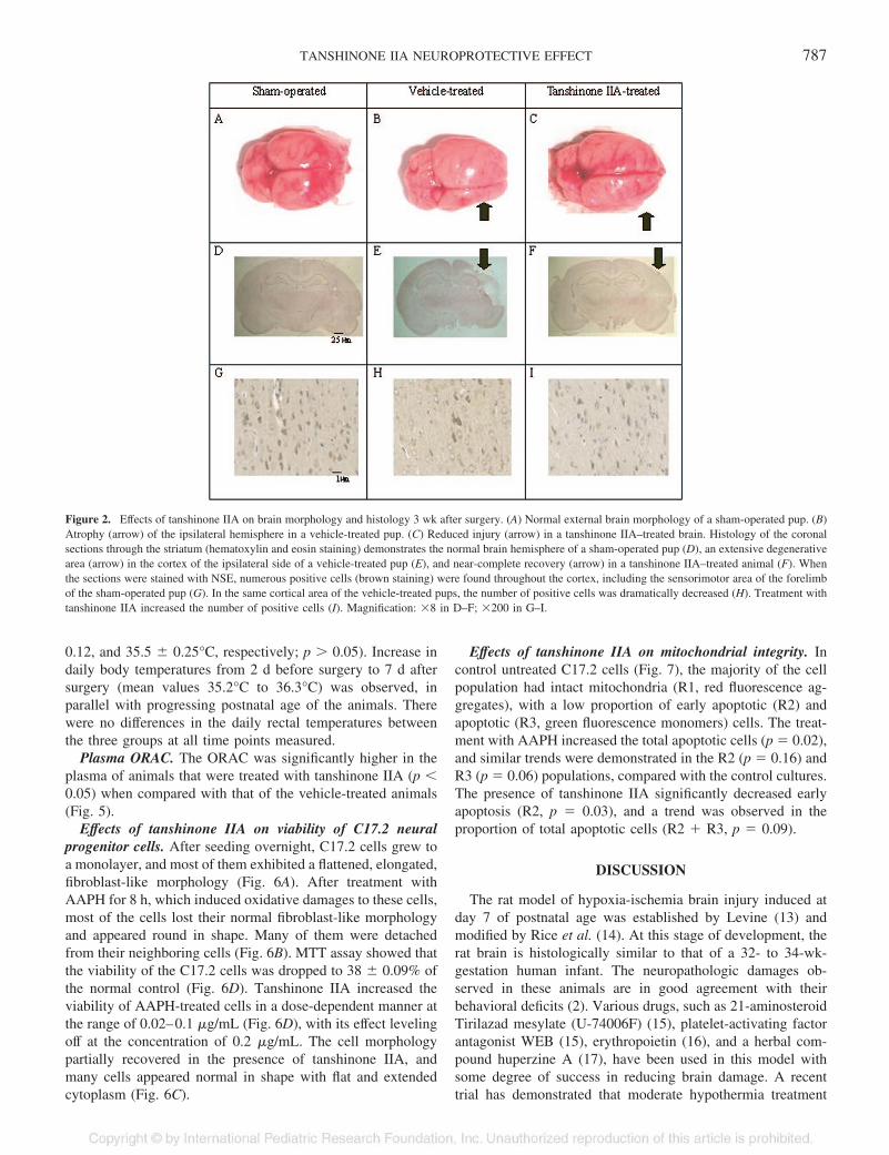
Cerebral cortical atrophy. Examination of the external mor-phology of the brain at 3 wk after the hypoxia-ischemiatreatment demonstrated that whereas the sham-operated pupshad normal brain size and hemispheric morphology (Fig. 2A),
severe atrophy of the right brain occurred in the vehicle-treatedanimals (Fig. 2B). Tanshinone IIA treatment reduced the extentof atrophy, and the right hemisphere almost returned to itsnormal size and morphology (Fig. 2C). Coronal sectionsthrough the striatum showed that sham-operated pups hadnormal brain histology (Fig. 2D), whereas vehicle-treated pupsthat were subjected to hypoxia-ischemia had brain damage atvarying extents, ranging from slight neuronal loss to a markeddegeneration of the ipsilateral hemisphere (Fig. 2E). Cell deathand necrotic nuclei were found scattered in the cerebral cortex,and the degenerative change was especially obvious in theouter layer of the cerebral cortex. Treatment with tanshinoneIIA resulted in a remarkable reduction in the severity of injury(Fig. 2F). The neuronal damage was moderate, compared withthose of the vehicle group. The hemispheric structure seemedintact, with the only abnormality being the slight reduction insize.
Cortical neurons by NSE staining. At 3 wk after surgery,the number of cortical neurons in the sensorimotor area of theforelimb in the ipsilateral hemisphere of the vehicle-treatedanimals was significantly decreased in comparison with that ofthe sham-operated group (p � 0.05; Figs. 2G and H and Fig.3). Treatment with tanshinone IIA significantly increased thenumber of neurons (p � 0.05; Figs. 2H and I and Fig. 3),although it remained significantly less than that of the sham-operated pups (p � 0.05; Fig. 3). The numbers of corticalneurons in the contralateral hemispheres were similar in thethree treatment groups.
Postural reflex test. All sham-operated animals obtainedscore 0, and none of them had abnormal postural responses ofscore 1 and score 2 (Fig. 4). The vehicle-treated pups demon-strated abnormal postural responses to the reflex test, andsignificant numbers of them were classified in scores 1 and 2 (p� 0.05). Some animals in this group displayed normal posturalresponses. Rat pups in the tanshinone IIA group had improvedpostural reflex as demonstrated by the increased proportion ofthose that got a score 0 and reduced numbers with scores 1 and2 (p � 0.05). Despite the improved functions, the performanceof tanshinone IIA–treated pups remained inferior to that ofsham-operated pups (p � 0.05). Similar results were observedwhen these scores were compared using the Mann Whitney Utest.
Body temperature. At 4 h after hypoxia, the rectal temper-atures of the sham-operated, vehicle-treated, and tanshinoneIIA–treated animals were not different (35.7 � 0.30, 35.6 �
Table 1. Brain weights of treatment groups
GroupTotal brain weight
(g)Contralateral (left)brain weight (g)
Ipsilateral (right)brain weight (g)
% Damage � 100 �(C � I)/C
1 Week Sham-operated (n � 10) 1.043 � 0.030§ 0.422 � 0.009 0.446 � 0.028§ 0.1 � 4.3%§Vehicle (n � 16) 0.930 � 0.109* 0.383 � 0.029 0.302 � 0.046* 21.0 � 11.6%*Tanshinone IIA (n � 15) 0.956 � 0.075 0.403 � 0.034 0.403 � 0.036** 13.2 � 7.1%**
3 Weeks Sham-operated (n � 23) 1.374 � 0.023§ 0.557 � 0.024 0.543 � 0.014§ 0.5 � 2.8%§Vehicle (n � 26) 1.244 � 0.053* 0.552 � 0.018 0.418 � 0.050* 24.3 � 1.8%*Tanshinone IIA (n � 27) 1.327 � 0.034** 0.561 � 0.020 0.496 � 0.023** 11.5 � 2.0%**
* Vehicle vs Sham-operated group p � 0.05** Vehicle vs Tanshinone IIA group p � 0.05§ Sham-operated vs Tanshinone IIA group
786 XIA ET AL.
0.12, and 35.5 � 0.25°C, respectively; p � 0.05). Increase indaily body temperatures from 2 d before surgery to 7 d aftersurgery (mean values 35.2°C to 36.3°C) was observed, inparallel with progressing postnatal age of the animals. Therewere no differences in the daily rectal temperatures betweenthe three groups at all time points measured.
Plasma ORAC. The ORAC was significantly higher in theplasma of animals that were treated with tanshinone IIA (p �0.05) when compared with that of the vehicle-treated animals(Fig. 5).
Effects of tanshinone IIA on viability of C17.2 neuralprogenitor cells. After seeding overnight, C17.2 cells grew toa monolayer, and most of them exhibited a flattened, elongated,fibroblast-like morphology (Fig. 6A). After treatment withAAPH for 8 h, which induced oxidative damages to these cells,most of the cells lost their normal fibroblast-like morphologyand appeared round in shape. Many of them were detachedfrom their neighboring cells (Fig. 6B). MTT assay showed thatthe viability of the C17.2 cells was dropped to 38 � 0.09% ofthe normal control (Fig. 6D). Tanshinone IIA increased theviability of AAPH-treated cells in a dose-dependent manner atthe range of 0.02–0.1 �g/mL (Fig. 6D), with its effect levelingoff at the concentration of 0.2 �g/mL. The cell morphologypartially recovered in the presence of tanshinone IIA, andmany cells appeared normal in shape with flat and extendedcytoplasm (Fig. 6C).
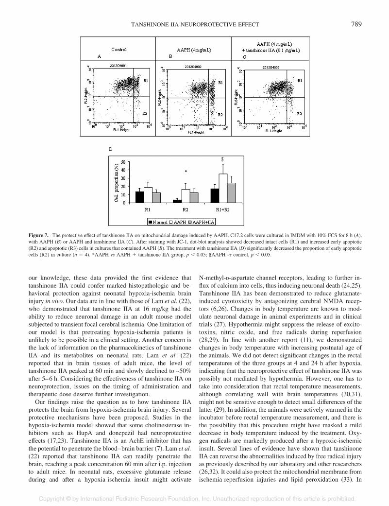
Effects of tanshinone IIA on mitochondrial integrity. Incontrol untreated C17.2 cells (Fig. 7), the majority of the cellpopulation had intact mitochondria (R1, red fluorescence ag-gregates), with a low proportion of early apoptotic (R2) andapoptotic (R3, green fluorescence monomers) cells. The treat-ment with AAPH increased the total apoptotic cells (p � 0.02),and similar trends were demonstrated in the R2 (p � 0.16) andR3 (p � 0.06) populations, compared with the control cultures.The presence of tanshinone IIA significantly decreased earlyapoptosis (R2, p � 0.03), and a trend was observed in theproportion of total apoptotic cells (R2 � R3, p � 0.09).
DISCUSSION
The rat model of hypoxia-ischemia brain injury induced atday 7 of postnatal age was established by Levine (13) andmodified by Rice et al. (14). At this stage of development, therat brain is histologically similar to that of a 32- to 34-wk-gestation human infant. The neuropathologic damages ob-served in these animals are in good agreement with theirbehavioral deficits (2). Various drugs, such as 21-aminosteroidTirilazad mesylate (U-74006F) (15), platelet-activating factorantagonist WEB (15), erythropoietin (16), and a herbal com-pound huperzine A (17), have been used in this model withsome degree of success in reducing brain damage. A recenttrial has demonstrated that moderate hypothermia treatment
Figure 2. Effects of tanshinone IIA on brain morphology and histology 3 wk after surgery. (A) Normal external brain morphology of a sham-operated pup. (B)Atrophy (arrow) of the ipsilateral hemisphere in a vehicle-treated pup. (C) Reduced injury (arrow) in a tanshinone IIA–treated brain. Histology of the coronalsections through the striatum (hematoxylin and eosin staining) demonstrates the normal brain hemisphere of a sham-operated pup (D), an extensive degenerativearea (arrow) in the cortex of the ipsilateral side of a vehicle-treated pup (E), and near-complete recovery (arrow) in a tanshinone IIA–treated animal (F). Whenthe sections were stained with NSE, numerous positive cells (brown staining) were found throughout the cortex, including the sensorimotor area of the forelimbof the sham-operated pup (G). In the same cortical area of the vehicle-treated pups, the number of positive cells was dramatically decreased (H). Treatment withtanshinone IIA increased the number of positive cells (I). Magnification: �8 in D–F; �200 in G–I.
787TANSHINONE IIA NEUROPROTECTIVE EFFECT

improved the most serious outcomes (death and severe motorscores) in human neonatal encephalopathy (18).
In the present study, rat pups were subjected to right com-mon artery occlusion combined with systemic hypoxia. Weobserved a reproducible pattern of brain injury, in terms ofbrain weight, histopathology, and number of sensorimotorneurons, in the ipsilateral but not the contralateral hemispheres.The neural damage was persistent at 3 wk after surgery and
was translated to a compromised response to the postural reflexchallenge. To investigate the protective effects of tanshinoneIIA on this animal model, we adopted a pretreatment protocolas reported by several research teams (19–21). Our datashowed that rat pups that were treated with tanshinone IIA 2 dbefore the induction of ischemia hypoxia for 16 d had markedrecoveries in the ipsilateral hemispheres in all studied param-eters. More significant, these animals scored better in thefunctional test, compared with the vehicle-treated group. To
Figure 3. NSE-positive cortical neurons in the sensorimotor area of theforelimb at 3 wk after surgery. The number of the cortical neurons wassignificantly decreased in the vehicle-treated pups compared with the sham-operated control (*p � 0.05). Tanshinone IIA treatment significantly increasedthe neuron number, compared with the vehicle-treated group (§p � 0.05; n �3 for each group).
Figure 4. Scores of the postural reflex test at 3 wk after surgery. Sham-operated ( �; n � 23) pups exhibited normal response. Vehicle-treatedanimals (□; n � 26) had defective response as shown by the low distributionin score 0 and high distribution in scores 1 and 2 (*p � 0.05). Tanshinone-treated pups (□; n � 27) performed better than the vehicle-treated animals inall three scores (§p � 0.05), but their reflex functions remained inferior to thesham-operated animals (**p � 0.05).
Figure 5. The ORAC in rat plasma. Tanshinone IIA increased the antioxidantcapacity of rat plasma when compared with the vehicle-treated group (*p �0.05; n � 10 for each group).
Figure 6. In vitro effect of tanshinone IIA on neural progenitor cell lineC17.2. C17.2 cells were cultured in the presence of AAPH (4 mg/mL) for 8 hwith or without tanshinone IIA (0.1 �g/mL). (A) Without AAPH and tanshi-none IIA, cells had normal morphology. (B) After treatment with AAPH, manycells rounded up and were detached from their neighboring cells. (C) Co-incubation with tanshinone IIA effectively protected the cells from AAPH-induced damages with improved morphology. (D) In the MTT assay, tanshi-none IIA significantly increased cell viability in a dose-dependent manner(0.02–0.2 �g/mL; * p � 0.05, eight replicated wells in three independentexperiments, n � 3). Magnification: �20 in A–C.
788 XIA ET AL.
our knowledge, these data provided the first evidence thattanshinone IIA could confer marked histopathologic and be-havioral protection against neonatal hypoxia-ischemia braininjury in vivo. Our data are in line with those of Lam et al. (22),who demonstrated that tanshinone IIA at 16 mg/kg had theability to reduce neuronal damage in an adult mouse modelsubjected to transient focal cerebral ischemia. One limitation ofour model is that pretreating hypoxia-ischemia patients isunlikely to be possible in a clinical setting. Another concern isthe lack of information on the pharmacokinetics of tanshinoneIIA and its metabolites on neonatal rats. Lam et al. (22)reported that in brain tissues of adult mice, the level oftanshinone IIA peaked at 60 min and slowly declined to ~50%after 5–6 h. Considering the effectiveness of tanshinone IIA onneuroprotection, issues on the timing of administration andtherapeutic dose deserve further investigation.
Our findings raise the question as to how tanshinone IIAprotects the brain from hypoxia-ischemia brain injury. Severalprotective mechanisms have been proposed. Studies in thehypoxia-ischemia model showed that some cholinesterase in-hibitors such as HupA and donepezil had neuroprotectiveeffects (17,23). Tanshinone IIA is an AchE inhibitor that hasthe potential to penetrate the blood–brain barrier (7). Lam et al.(22) reported that tanshinone IIA can readily penetrate thebrain, reaching a peak concentration 60 min after i.p. injectionto adult mice. In neonatal rats, excessive glutamate releaseduring and after a hypoxia-ischemia insult might activate
N-methyl-D-aspartate channel receptors, leading to further in-flux of calcium into cells, thus inducing neuronal death (24,25).Tanshinone IIA has been demonstrated to reduce glutamate-induced cytotoxicity by antagonizing cerebral NMDA recep-tors (6,26). Changes in body temperature are known to mod-ulate neuronal damage in animal experiments and in clinicaltrials (27). Hypothermia might suppress the release of excito-toxins, nitric oxide, and free radicals during reperfusion(28,29). In line with another report (11), we demonstratedchanges in body temperature with increasing postnatal age ofthe animals. We did not detect significant changes in the rectaltemperatures of the three groups at 4 and 24 h after hypoxia,indicating that the neuroprotective effect of tanshinone IIA waspossibly not mediated by hypothermia. However, one has totake into consideration that rectal temperature measurements,although correlating well with brain temperatures (30,31),might not be sensitive enough to detect small differences of thelatter (29). In addition, the animals were actively warmed in theincubator before rectal temperature measurement, and there isthe possibility that this procedure might have masked a milddecrease in body temperature induced by the treatment. Oxy-gen radicals are markedly produced after a hypoxic-ischemicinsult. Several lines of evidence have shown that tanshinoneIIA can reverse the abnormalities induced by free radical injuryas previously described by our laboratory and other researchers(26,32). It could also protect the mitochondrial membrane fromischemia-reperfusion injuries and lipid peroxidation (33). In
Figure 7. The protective effect of tanshinone IIA on mitochondrial damage induced by AAPH. C17.2 cells were cultured in IMDM with 10% FCS for 8 h (A),with AAPH (B) or AAPH and tanshinone IIA (C). After staining with JC-1, dot-blot analysis showed decreased intact cells (R1) and increased early apoptotic(R2) and apoptotic (R3) cells in cultures that contained AAPH (B). The treatment with tanshinone IIA (D) significantly decreased the proportion of early apoptoticcells (R2) in culture (n � 4). *AAPH vs AAPH � tanshinone IIA group, p � 0.05; §AAPH vs control, p � 0.05.
789TANSHINONE IIA NEUROPROTECTIVE EFFECT
this study, we demonstrated that tanshinone IIA significantlyimproved the antioxidant capacity in rat plasma and protectedthe C17.2 progenitors against AAPH-induced cell death. Wefurther confirmed that tanshinone IIA exerted a protectiveeffect on mitochondrial membrane potential and decreased celldeath secondary to oxidative stress.
The contribution of tanshinone IIA–induced antioxidant ca-pacity to the protection against brain damage in our modelremains to be investigated further. Our data and other evidencehave shown an effective neuroprotective action by tanshinoneIIA, possibly mediated by multiple pathways in hypoxic-ischemic neonatal animals. To date, the applications of neuro-protective agents, including tanshinone IIA, have not achievedtotal recovery of the damaged brain in various animal models.It thus is important to investigate the mechanism of each agentand the possibility of an integrated treatment strategy in max-imizing the outcome. The present study suggests that pretreat-ment with tanshinone IIA is partially protective for hypoxia-ischemia in the immature rat. As the treatment protocolspanned a very wide period before and after the hypoxic-ischemic insult, it is uncertain which treatment times wereoptimal for protection or whether rescue treatment is equallyeffective. We have confirmed that tanshinone IIA has antioxi-dant effects that may be beneficial, although other potentiallyrelevant pathways might be involved. This study, however, didnot purposefully evaluate whether brain cooling secondary tothe treatment may have contributed to neuroprotection. Furtherstudies are required to address these issues to verify the clinicalpotential of tanshinone IIA in helping to alleviate braindamage.
Acknowledgments. We thank David Lo, Department ofAnatomical and Cellular Pathology, The Chinese University ofHong Kong, for providing practical advice and support. Wealso thank Dr. David Walsh, Department of Anatomy, TheUniversity of New South Wales, Australia, for providing theC17.2 cell line.
REFERENCES
1. Buonocore G, Perrone S, Bracci R 2001 Free radicals and brain damage in thenewborn. Biol Neonate 79:180–186
2. Feng Y, Shi W, Huang M, LeBlanc MH 2003 Oxypurinol administration fails toprevent hypoxic-ischemic brain injury in neonatal rats. Brain Res Bull 59:453–457
3. Zhu XP 1998 Chinese Material Medica: Chemistry, Pharmacology and Applications.Harwood Academic Publisher, Amsterdam, pp 459–463
4. Niu XL, Ichimori K, Yang X, Hirota Y, Hoshiai K, Li M, Nakazawa H 2000Tanshinone II-A inhibits low density lipoprotein oxidation in vitro. Free Radic Res33:305–312
5. Zhou GY, Zhao BL, Hou JW, Ma GE, Xin WJ 1999 Protective effects of sodiumtanshinone IIA sulphonate against adriamycin-induced lipid peroxidation in micehearts in vivo and in vitro. Pharmacol Res 40:487–491
6. Ji ZN, Liu GQ 2001 Inhibition of serum deprivation-induced PC12 cell apoptosis bytanshinone II A. Acta Pharmacol Sin 22:459–462
7. Ren Y, Houghton PJ, Hider RC, Howes MJ 2004 Novel diterpenoid acetylcholines-terase inhibitors from Salvia miltiorrhiza. Planta Med 70:201–204
8. Riederer BM, Berbel P, Innocenti GM 2004 Neurons in the corpus callosum of the catduring postnatal development. Eur J Neurosci 19:2039–2046
9. Paxinox GWC 1986 The Rat Brain in Stereotaxic Coordination. Academic Press Inc.,San Diego, pp 15–23
10. Calvert JW, Yin W, Patel M, Badr A, Mychaskiw G, Parent AD, Zhang JH 2002Hyperbaric oxygenation prevented brain injury induced by hypoxia-ischemia in aneonatal rat model. Brain Res 951:1–8
11. Colman AS, Miller JH 2002 Lack of involvement of mu(1) opioid receptors indermorphin-induced inhibition of hypoxic and hypercapnic ventilation in rat pups.Respir Physiol Neurobiol 131:199–212
12. Wang CC, Chu CY, Chu KO, Choy KW, Khaw KS, Rogers MS, Pang CP 2004Trolox-equivalent antioxidant capacity assay versus oxygen radical absorbance ca-pacity assay in plasma. Clin Chem 50:952–954
13. Levine S 1960 Anoxic-ischemic encephalopathy in rats. Am J Pathol 36:1–1714. Rice JE 3rd, Vannucci RC, Brierley JB 1981 The influence of immaturity on
hypoxic-ischemic brain damage in the rat. Ann Neurol 9:131–14115. Viswanath M, Palmer C, Roberts RL 2000 Reduction of hypoxic-ischemic brain
swelling in the neonatal rat with PAF antagonist WEB 2170: lack of long-termprotection. Pediatr Res 48:109–113
16. Kumral A, Ozer E, Yilmaz O, Akhisaroglu M, Gokmen N, Duman N, Ulukus C, GencS, Ozkan H 2003 Neuroprotective effect of erythropoietin on hypoxic-ischemic braininjury in neonatal rats. Biol Neonate 83:224–228
17. Wang LS, Zhou J, Shao XM, Tang XC 2002 Huperzine A attenuates cognitive deficitsand brain injury in neonatal rats after hypoxia-ischemia. Brain Res 949:162–170
18. Eicher DJ, Wagner CL, Katikaneni LP, Hulsey TC, Bass WT, Kaufman DA, HorganMJ, Languani S, Bhatia JJ, Givelichian LM, Sankaran K, Yager JY 2005 Moderatehypothermia in neonatal encephalopathy: efficacy outcomes. Pediatr Neurol 32:11–17
19. Ekert P, MacLusky N, Luo XP, Lehotay DC, Smith B, Post M, Tanswell AK 1997Dexamethasone prevents apoptosis in a neonatal rat model of hypoxic-ischemicencephalopathy (HIE) by a reactive oxygen species-independent mechanism. BrainRes 747:9–17
20. Galvin KA, Oorschot DE 2003 Continuous low-dose treatment with brain-derivedneurotrophic factor or neurotrophin-3 protects striatal medium spiny neurons frommild neonatal hypoxia/ischemia: a stereological study. Neuroscience 118:1023–1032
21. Wang X, Zhu C, Wang X, Gerwien JG, Schrattenholz A, Sandberg M, Leist M,Blomgren K 2004 The nonerythropoietic asialoerythropoietin protects against neo-natal hypoxia-ischemia as potently as erythropoietin. J Neurochem 91:900–910
22. Lam BY, Lo AC, Sun X, Luo HW, Chung SK, Sucher NJ 2003 Neuroprotectiveeffects of tanshinones in transient focal cerebral ischemia in mice. Phytomedicine10:286–291
23. Zhang HY, Tang XC 2000 Huperzine B, a novel acetylcholinesterase inhibitor,attenuates hydrogen peroxide induced injury in PC12 cells. Neurosci Lett 292:41–44
24. Abdel-Hamid KM, Tymianski M 1997 Mechanisms and effects of intracellularcalcium buffering on neuronal survival in organotypic hippocampal cultures exposedto anoxia/aglycemia or to excitotoxins. J Neurosci 17:3538–3553
25. Egawa-Tsuzuki T, Ohno M, Tanaka N, Takeuchi Y, Uramoto H, Faigle R, Funa K,Ishii Y, Sasahara M 2004 The PDGF B-chain is involved in the ontogenic suscepti-bility of the developing rat brain to NMDA toxicity. Exp Neurol 186:89–98
26. Wang AM, Sha SH, Lesniak W, Schacht J 2003 Tanshinone (Salviae miltiorrhizaeextract) preparations attenuate aminoglycoside-induced free radical formation in vitroand ototoxicity in vivo. Antimicrob Agents Chemother 47:1836–1841
27. Asai S, Zhao H, Kohno T, Takahashi Y, Nagata T, Ishikawa K 2000 Quantitativeevaluation of extracellular glutamate concentration in postischemic glutamate re-uptake, dependent on brain temperature, in the rat following severe global brainischemia. Brain Res 864:60–68
28. Battin MR, Dezoete JA, Gunn TR, Gluckman PD, Gunn AJ 2001 Neurodevelopmen-tal outcome of infants treated with head cooling and mild hypothermia after perinatalasphyxia. Pediatrics 107:480–484
29. DeBow SB, Clark DL, MacLellan CL, Colbourne F 2003 Incomplete assessment ofexperimental cytoprotectants in rodent ischemia studies. Can J Neurol Sci 30:368–374
30. Bona E, Hagberg H, Loberg EM, Bagenholm R, Thoresen M 1998 Protective effectsof moderate hypothermia after neonatal hypoxia-ischemia: short- and long-termoutcome. Pediatr Res 43:738–745
31. Thoresen M, Bagenholm R, Loberg EM, Apricena F, Kjellmer I 1996 Posthypoxiccooling of neonatal rats provides protection against brain injury. Arch Dis Child FetalNeonatal Ed 74:F3–F9
32. Wu TW, Zeng LH, Fung KP, Wu J, Pang H, Grey AA, Weisel RD, Wang JY 1993Effect of sodium tanshinone IIA sulfonate in the rabbit myocardium and on humancardiomyocytes and vascular endothelial cells. Biochem Pharmacol 46:2327–2332
33. Zhao BL, Jiang W, Zhao Y, Hou JW, Xin WJ 1996 Scavenging effects of salviamiltiorrhiza on free radicals and its protection for myocardial mitochondrial mem-branes from ischemia-reperfusion injury. Biochem Mol Biol Int 38:1171–1182
790 XIA ET AL.