Nuclear factor I-A represses expression of the cell adhesion molecule L1
Upload
oregonstateCategory
view
7download
0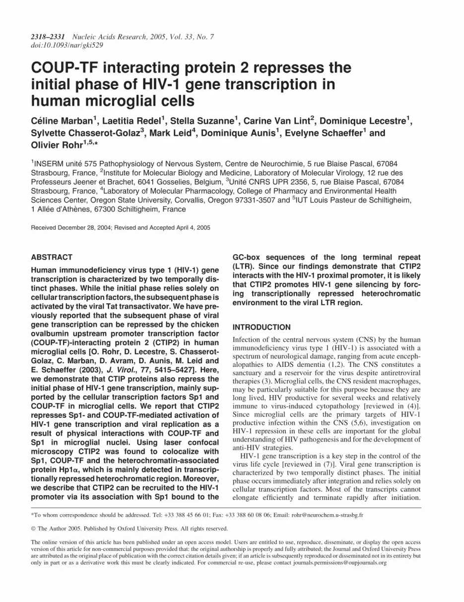
COUP-TF interacting protein 2 represses theinitial phase of HIV-1 gene transcription inhuman microglial cellsCeline Marban1, Laetitia Redel1, Stella Suzanne1, Carine Van Lint2, Dominique Lecestre1,
Sylvette Chasserot-Golaz3, Mark Leid4, Dominique Aunis1, Evelyne Schaeffer1 and
Olivier Rohr1,5,*
1INSERM unite 575 Pathophysiology of Nervous System, Centre de Neurochimie, 5 rue Blaise Pascal, 67084Strasbourg, France, 2Institute for Molecular Biology and Medicine, Laboratory of Molecular Virology, 12 rue desProfesseurs Jeener et Brachet, 6041 Gosselies, Belgium, 3Unite CNRS UPR 2356, 5, rue Blaise Pascal, 67084Strasbourg, France, 4Laboratory of Molecular Pharmacology, College of Pharmacy and Environmental HealthSciences Center, Oregon State University, Corvallis, Oregon 97331-3507 and 5IUT Louis Pasteur de Schiltigheim,1 Allee d’Athenes, 67300 Schiltigheim, France
Received December 28, 2004; Revised and Accepted April 4, 2005
ABSTRACT
Human immunodeficiency virus type 1 (HIV-1) genetranscription is characterized by two temporally dis-tinct phases. While the initial phase relies solely oncellular transcription factors, thesubsequentphase isactivated by the viral Tat transactivator. We have pre-viously reported that the subsequent phase of viralgene transcription can be repressed by the chickenovalbumin upstream promoter transcription factor(COUP-TF)-interacting protein 2 (CTIP2) in humanmicroglial cells [O. Rohr, D. Lecestre, S. Chasserot-Golaz, C. Marban, D. Avram, D. Aunis, M. Leid andE. Schaeffer (2003), J. Virol., 77, 5415–5427]. Here,we demonstrate that CTIP proteins also repress theinitial phase of HIV-1 gene transcription, mainly sup-ported by the cellular transcription factors Sp1 andCOUP-TF in microglial cells. We report that CTIP2represses Sp1- and COUP-TF-mediated activation ofHIV-1 gene transcription and viral replication as aresult of physical interactions with COUP-TF andSp1 in microglial nuclei. Using laser confocalmicroscopy CTIP2 was found to colocalize withSp1, COUP-TF and the heterochromatin-associatedprotein Hp1a, which is mainly detected in transcrip-tionally repressed heterochromatic region. Moreover,we describe that CTIP2 can be recruited to the HIV-1promoter via its association with Sp1 bound to the
GC-box sequences of the long terminal repeat(LTR). Since our findings demonstrate that CTIP2interacts with the HIV-1 proximal promoter, it is likelythat CTIP2 promotes HIV-1 gene silencing by forc-ing transcriptionally repressed heterochromaticenvironment to the viral LTR region.
INTRODUCTION
Infection of the central nervous system (CNS) by the humanimmunodeficiency virus type 1 (HIV-1) is associated with aspectrum of neurological damage, ranging from acute enceph-alopathies to AIDS dementia (1,2). The CNS constitutes asanctuary and a reservoir for the virus despite antiretroviraltherapies (3). Microglial cells, the CNS resident macrophages,may be particularly suitable for this purpose because they arelong lived, HIV productive for several weeks and relativelyimmune to virus-induced cytopathology [reviewed in (4)].Since microglial cells are the primary targets of HIV-1productive infection within the CNS (5,6), investigation onHIV-1 repression in these cells are important for the globalunderstanding of HIV pathogenesis and for the development ofanti-HIV strategies.
HIV-1 gene transcription is a key step in the control of thevirus life cycle [reviewed in (7)]. Viral gene transcription ischaracterized by two temporally distinct phases. The initialphase occurs immediately after integration and relies solely oncellular transcription factors. Most of the transcripts cannotelongate efficiently and terminate rapidly after initiation.
*To whom correspondence should be addressed. Tel: +33 388 45 66 01; Fax: +33 388 60 08 06; Email: [email protected]
ª The Author 2005. Published by Oxford University Press. All rights reserved.
The online version of this article has been published under an open access model. Users are entitled to use, reproduce, disseminate, or display the open accessversion of this article for non-commercial purposes provided that: the original authorship is properly and fully attributed; the Journal and Oxford University Pressare attributed as the original place of publication with the correct citation details given; if an article is subsequently reproduced or disseminated not in its entirety butonly in part or as a derivative work this must be clearly indicated. For commercial re-use, please contact [email protected]
2318–2331 Nucleic Acids Research, 2005, Vol. 33, No. 7doi:10.1093/nar/gki529

However, a few transcripts elongate throughout the genome,resulting in transcription and expression of the viral transactiv-ator Tat. The subsequent phase of transcription occurs whenenough Tat protein has accumulated. Tat binds to TAR,recruits pTEFb complex and dramatically stimulates HIV-1gene transcription [reviewed in (8,9)].
We have previously studied some of the cellular transcrip-tion factors that impact HIV-1 gene transcription inmicroglial cells. Chicken ovalbumin upstream promoter tran-scription factor I (COUP-TFI)/Ear3, Arp1/COUP-TFII andEar2/COUP-TFIII proteins are the three members of theCOUP-TF orphan nuclear receptors superfamily. We havedescribed that the orphan nuclear receptor COUP-TFI/Ear3(10,11) is expressed in numerous human CNS cell lineages,including microglial cells. COUP-TFI is an activator of HIV-1 gene transcription in oligodendrocytes (12), microglia (13)and T lymphocytes (14). Moreover, we have revealed theimportance of functional interactions between the nuclearfactor for interleukin 6 (NF-Il6), Sp1 and COUP-TFI inthe regulation of the initial phase of HIV-1 gene transcriptionin brain cells (15). The transcription factor Sp1 is one of thecrucial cellular proteins for efficient HIV-1 gene transcription(16–18). A number of studies have reported that Sp1 canserve as an anchor for indirect binding of other transcriptionfactors to the HIV-1 long terminal repeat (LTR) region. In allcell types, an interaction between Sp1 and Tat is required foroptimal HIV-enhancer activation (19). Moreover, Sp1 is ableto recruit the Cyclin T1 subunit of P-TEFb to the LTR, whichhelps to bypass a requirement for TAR/Tat and promotesprocessive transcription without Tat (20). In microglialcells, the main HIV-1 target cells in the brain (6), Sp1anchors COUP-TF (13) and NF-IL6 (15) to the three GC-boxes adjacent to the viral CATA box. The CATA box isused instead of the TATAA box for optimal HIV-1 genetranscription and replication (21). We have shown thatSp1 and COUP-TF interact and cooperate in the transcrip-tional activation of HIV-1 gene transcription (13). Moreover,COUP-TF can substitute to Sp1 association with the viral Tatprotein to restore Tat function and HIV-1 replication inmicroglial cells (22). These results highlight the key rolesof Sp1 and COUP-TF proteins in HIV-1 expression inmicroglial cells.
Members of the COUP-TF family have been shown to bindrelated zinc finger proteins named COUP-TF-interactingprotein 1 (CTIP1) and 2 (CTIP2) that are highly expressedin brain and immune systems (23,24). Recent results supportthe selective contribution of these proteins in the developmentand function of the nervous and immune systems (24). Werecently described that the nuclear cofactor CTIP2 inhibits thesubsequent phase of HIV-1 gene transcription and viral rep-lication by relocating the viral transactivator Tat protein totranscriptionally inactive regions of chromatin via Hp1a (25).In the present work, CTIP2 overexpression leads to the repres-sion of HIV-1 replication. In contrast, CTIP1 was unable toaffect Tat function. Using confocal microscopy to visualizeTat subcellular distribution in the presence of each CTIPproteins, we found that CTIP2, but not CTIP1, leads to thedisruption of Tat nuclear localization and to its recruitmentwithin CTIP2-induced nuclear ball-like structures. In addition,we showed that CTIP2 and Hp1a associate with Tat to forma three-protein complex. These findings suggest that the
inhibition of HIV-1 expression by CTIP2 correlates withthe recruitment of Tat within CTIP2-induced structures andits relocalization within inactive regions of chromatin.
On line with this study, we noticed that CTIP proteins alsoaffect HIV-1 gene transcription in the absence of Tat, whichmeans that CTIP proteins may also impair endogenous cellulartranscription factor functions of importance for the initialphase of HIV-1 gene transcription.
Here, we report that CTIP proteins repress HIV-1 genetranscription via the proximal LTR region which bindsSp1 and COUP-TF transactivators. Since COUP-TF andSp1 cellular transcription factors are two of the major con-tributors to HIV-1 gene transcription in microglial cells, wehave postulated that Sp1- and COUP-TF-mediated LTR-driven activation may be impaired by CTIP1 and CTIP2.We report here that both CTIP1 and CTIP2 repress Sp1-and COUP-TF-mediated stimulation of HIV-1 gene expres-sion. Since the CTIP2 repressive activity is much strongerthan the CTIP1-mediated inhibition of HIV-1 replication, wefocused our study on CTIP2. CTIP2-mediated repression ofSp1 and COUP-TF functions results from direct physicalinteractions of these cellular transcription factors withCTIP2. Association with CTIP2 forces Sp1 and COUP-TFrelocation to CTIP2-induced subnuclear structures containingthe heterochromatin-associated protein Hp1a. Moreover,CTIP2 can be recruited to the HIV-1 promoter by a physicalassociation with Sp1 bound to the GC-box regions of theLTR. Since Sp1 or COUP-TF transcription factors are neces-sary for the Tat function in microglial cells (22), CTIP2-mediated repression of COUP-TF and Sp1 functions maylargely contribute to the previously described CTIP2-mediated impairment of Tat activity and viral replicationin microglial cells (25).
MATERIALS AND METHODS
Plasmids
Most of the constructs used in our assays were describedpreviously: HIV-1 (LAI) 50 LTR chloramphenicol acetyltrans-ferase (pLTR-CAT) (22), pGC-WAP-CAT (26), pLTR-LUC287–535 (13), pLTR-CAT mutGC and pLTR-CAT DGC (22),pcDNA3, full-length constructs HA-CTIP1, Flag-CTIP2 andGST-CTIP2 (23), deletions constructs Flag-CTIP2 (25), RSV-COUP-TF, CMV-Sp1, GST-COUP-TF1, GST-COUP-TF2and GST-COUP-TF3 (13).
Several plasmids were kindly provided by the followinginvestigators: GST-Sp1 constructs were provided byH. Rotheneder (Vienna, Austria) (27) and pLTR-LUC byC. Van Lint (Gosselies, Belgium). pNL4-3 (28) was obtainedthrough the AIDS Research and Reference Reagent Program,division of AIDS, National Institute of Allergy and InfectiousDiseases (NIAID), NIH.
To construct RFP-CTIP2, the expression vector Flag-CTIP2was digested with EcoRI and the CTIP2 insert was subclonedinto the EcoRI site of the pDSRed vector (Clontech). TheGFP-Sp1 was constructed by isolating the XhoI–HindIIISp1 insert from the GST-Sp1 expression vector and subclonedinto the XhoI–HindIII sites of the pEGFP-C1 vector(Clontech).
Nucleic Acids Research, 2005, Vol. 33, No. 7 2319

Cell culture
The human microglial (provided by M. Tardieu, Paris, France)(29), the TZM-bl (30–32) and the HEK 293T cell lines weremaintained in DMEM containing 10% fetal calf serum and100 U/ml penicillin–streptomycin.
Viral replication
Microglial cells cultured in 12-well plates were transfectedusing the calcium phosphate coprecipitation method withHIV-1 pNL4-3 (1.5 mg), CMV-Sp1 (1 mg) or RSV-COUP-TF (1 mg) plasmids and the indicated HA-CTIP1 or Flag-CTIP2 expression vectors (0.1, 0.5 or 1 mg). Total amountsof DNA were identical in each experiment. Compensationswere made by adding the corresponding empty vectors. Eachtransfection was carried out in duplicate and repeated aminimum of three separate times with two different plasmidpreparations. HIV-1 replication was monitored as describedpreviously (25).
CAT assays
Microglial cells cultured in 12-well plates were transfectedusing the calcium phosphate coprecipitation method withpLTR-CAT (3 mg), pGC-WAP-CAT (3 mg) or pLTR-CATDGC (3 mg), CMV-Sp1 (1 mg) or RSV-COUP-TF (1 mg)plasmids and the indicated HA-CTIP1 or Flag-CTIP2 (0.1,0.5 or 1 mg) expression vectors. Total amounts of DNAwere identical in each experiments. Compensations weremade by adding the corresponding empty vectors. Each trans-fection was carried out in duplicate and repeated a minimum ofthree separate times with two different plasmid preparations.CAT assays were carried out using standard techniques.
Co-immunoprecipitation assays
Microglial cells cultured in 100 mm diameter dishes weretransfected using LipofectamineTM 2000 Reagent (Invitrogen)with the indicated Flag-CTIP2 (30 mg). Twenty four hourspost-transfection, the cells were washed twice with coldphosphate-buffered saline, harvested and prepared for nuclearextracts (12). Nuclear proteins were first diluted in TNE buffer[50 mM Tris, pH 8, 1% Nonidet, 2 mM EDTA, proteaseinhibitors cocktail (Roche)], cleared with Protein A/G Plus-Agarose (Santa Cruz Biotechnology) and finally incubatedovernight with primary antibodies: anti-Sp1 (Sigma), anti-COUP-TF (kindly provided by J. E. Mertz, Madison, WI)or anti-Hp1a (kindly provided by R. Losson, IGBMC,Strasbourg, France). The extracts were then immunoprecipit-ated by the addition of Protein A/G Plus-Agarose (Santa CruzBiotechnology). After extensive washing with TNE buffer, theimmunoprecipitates were processed for SDS–PAGE andwestern blot analysis.
Glutathione S-transferase (GST) pull-down assays
Production of GST fusion proteins was described previously(13) and visualized by Coomassie blue staining. The 35S-labeled proteins were prepared by in vitro transcription andtranslation using the TNT1 T7 Coupled Wheat Germ ExtractSystem (Promega). GST pull-down assays were performed asdescribed previously (25).
SDS–PAGE and western blot analysis
SDS–PAGE was performed using standard techniques.Proteins were detected using antibodies directed against theFlag epitope (M2 mouse monoclonal; Sigma), against theCOUP-TF protein (kindly provided by J. E. Mertz) and againstthe Sp1 protein (Sigma). Proteins were visualized by chemi-luminescence using the Super Signal ChemiluminescenceDetection System (Pierce).
Electrophoretic mobility shift assays (EMSA)
The probe used in our experiments has been described previ-ously (13) and corresponds to the three Sp1 binding sites of theHIV-1 proximal LTR region. Once produced, GST fusionproteins were eluted in glutathione buffer (50 mM Tris,pH 8, 20 mM reduced glutathione). Purified Sp1 (Promega)and GST fusion proteins were then incubated with the 32P-labeled probe in binding buffer (20 mM HEPES, pH 7.9, 1 mMMgCl2, 60 mM KCl, 0.5 mM EDTA, 1 mM DTT and 10%glycerol) at 4�C for 15 min. For supershift experiments, GSTfusion proteins were incubated with the primary antibodies:anti-COUP-TF (kindly provided by J. E. Mertz), anti-CTIP2and anti-Sp1 (Santa Cruz Biotechnology) for �16 h beforeadding the probe. EMSA assays were performed as describedpreviously (25).
Indirect immunofluorescence and confocal microscopy
Microglial cells cultured in 48-well plates were transfected ornot using LipofectamineTM 2000 Reagent (Invitrogen) withFlag-CTIP2, RFP-CTIP2 and/or GFP-Sp1 expression vectors.Cells were fixed and permeabilized as described previously(25). The coverslips were then incubated for 1 h at roomtemperature with primary antibodies directed againstCOUP-TF (Santa Cruz Biotechnology or kindly providedby J. E. Mertz), Sp1 (sigma) and Hp1a proteins and/or againstthe Flag epitope (M2 mouse monoclonal; Sigma). The primaryimmunocomplexes were revealed by CY2- or CY3-labeledsecondary anti-species antibodies. The stained cells were ana-lyzed by confocal microscopy using a Zeiss laser scanningmicroscope (model 510 invert) equipped with a Planapo oil(63·) immersion lens (numerical aperture = 1.4).
Chromatin immunoprecipitation (ChIP) assays
TZM-bl and HEK 293T cells cultured in 100 mm diameterdishes were transfected using the calcium phosphate coprecip-itation method with the indicated pLTR-LUC, pLTR-CATmutGC and Flag-CTIP2 (30 mg) expression vector. ChIPassays were performed using the ChIP Assay Kit (Upstate)48 h post-transfection. The primary antibodies used for theChIP were anti-Sp1 (Upstate), anti-Hp1a (Upstate), anti-COUP-TF (Santa Cruz Biotechnology) and anti-Flag M2mouse monoclonal (Sigma). DNA was subjected to PCRamplification using a 50 primer (50-GATAAGGTAGAA-GAGGCC-30) corresponding to the LTR sequence located293 nt downstream of the transcriptional start site and a 30
primer (50-CTAACCAGAGAGACCCAGTAC-30) corres-ponding to a region just upstream of the transcriptional startsite. The resulting PCR product (307 bp) was analyzed byagarose gel electrophoresis and ethidium bromide staining.Three separate experiments were performed.
2320 Nucleic Acids Research, 2005, Vol. 33, No. 7

RESULTS
CTIP1 and CTIP2 proteins repress HIV-1 genetranscription via the LTR proximal region
As previously shown, CTIP1 and CTIP2 proteins inhibited theLTR-driven transcription in transient transfection assays(Figure 1, lanes 2 and 3) (25). To delineate the LTR regionresponsible for CTIP1- and CTIP2-mediated HIV-1 gene tran-scriptional repression, microglial cells were transfected with a50 deleted pLTR-CAT reporter plasmid in the presence orabsence of CTIP1 and CTIP2 expression vectors. Deletionof the 50 region downstream of the two proximal GC-boxsequences did not affect CTIP1 and CTIP2 ability to repressLTR-driven CAT activity (Figure 1, lanes 5 and 6), indicatingthat CTIP proteins repressive function can be mediated by theproximal region of the LTR encompassing two GC-boxsequences, the CATA sequence (21) and the TAR region.We have previously observed that the cellular transcriptionfactors, Sp1 and Sp3, are directly bound to the LTR GC-boxsequences in microglial cells (13). Moreover, the orphannuclear receptor COUP-TF is indirectly anchored to thisregion via its association with Sp1 (13). We have largelydescribed that Sp1 and COUP-TF transcription factors aretwo of the major contributors to the initial phase of HIV-1gene transcription in microglial cells (9,13,22). Takentogether, these findings strongly suggest that CTIP repressiveactivity may result from impairment of endogenous Sp1 andCOUP-TF protein functions.
CTIP1 and CTIP2 cofactors inhibit Sp1- andCOUP-TF-mediated activation of HIV-1 genetranscription and related viral replication
To decipher the mechanism whereby CTIP proteins affect theinitial phase of HIV-1 gene transcription, we investigated theimpacts of CTIP proteins on Sp1 and COUP-TF functions.Microglial cells were transfected with pLTR-CAT plasmidtogether with the vectors expressing COUP-TF (Figure 2A)or Sp1 (Figure 2B) proteins and with increasing amounts ofHA-CTIP1 or Flag-CTIP2 expression vectors. The two pro-teins were able to repress COUP-TF- and Sp1-mediatedstimulations of CAT activity. As a control, we performedwestern blot experiments, indicating that COUP-TF and Sp1
expressions were not downregulated but slightly upregulatedby the cofactors. Since overexpression of COUP-TF and Sp1stimulates HIV-1 gene expression, this modest upregulationcould not be responsible for the CTIPs repressive function. Tocorrelate CTIP1 and CTIP2 repressive activity on LTR-driventranscription to the level of HIV-1 replication, we studiedtheir impacts on Sp1- and COUP-TF-mediated stimulationsof viral replication. Cells were transfected with HIV-1 pNL4-3 and COUP-TF (Figure 2C) or Sp1 (Figure 2D) expressionvectors in the presence of increasing amounts of CTIP1 orCTIP2 expression vectors. The level of viral replication wasinvestigated by quantifying p24 Gag expression in the culturesupernatants 2 days after transfection. While both CTIP1 andCTIP2 inhibited COUP-TF (Figure 2C) and Sp1 (Figure 2D)activation of HIV-1 NL4-3 replication, the strongest repres-sion was observed with CTIP2 (Figure 2C and D, lanes 6–8),indicating that the repressing activity of CTIP2 on HIV-1replication cannot be explained only by its previouslydescribed capacity to repress the viral Tat protein (25).These results strongly suggest that CTIP1 and CTIP2 repressthe initial phase of HIV-1 gene transcription through direct orindirect interactions with endogenous Sp1 and COUP-TFproteins.
The HIV-1 Tat function and the subsequent phase of theHIV-1 gene transcription also depend on Tat association withSp1 or COUP-TF in microglial cells (22). Since CTIP2-mediated repressive activities are much stronger than thoseobserved for CTIP1 and since CTIP2, but not CTIP1, inhibitsthe subsequent phase of the HIV-1 gene transcription, wefocused our mechanistic investigation on CTIP2.
CTIP2 interacts with COUP-TF and Sp1 in vitroby two interfaces
To decipher the mechanism whereby CTIP2 represses COUP-TF and Sp1 stimulatory activities, we first examined whetherthese proteins were able to interact in vitro. GST pull-downassays were performed with in vitro translated 35S-labeledCTIP2 and equal amounts of full-length or truncated GST-COUP-TF and GST-Sp1 fusion proteins (Figure 3A). Resultsshow that CTIP2 bound specifically to GST-COUP-TF andGST-Sp1 (Figure 3A, lanes 3 and 8, respectively) but not to thecontrol GST protein (Figure 3A, lanes 2 and 7, respectively).
Figure 1. CTIP1 and CTIP2 proteins repress HIV-1 gene transcription via the proximal LTR region. Microglial cells were transfected with 3mg of pLTR-CAT or 3mgof pLTR-CAT (287–535) in the presence or absence of 1 mg of HA-CTIP1 or Flag-CTIP2. Two days post-transfection, CAT activities were measured and expressedrelative to the CAT activity obtained with pLTR-CAT alone with the standard deviations indicated (values correspond to an average of at least three independentexperiments performed in duplicate).
Nucleic Acids Research, 2005, Vol. 33, No. 7 2321
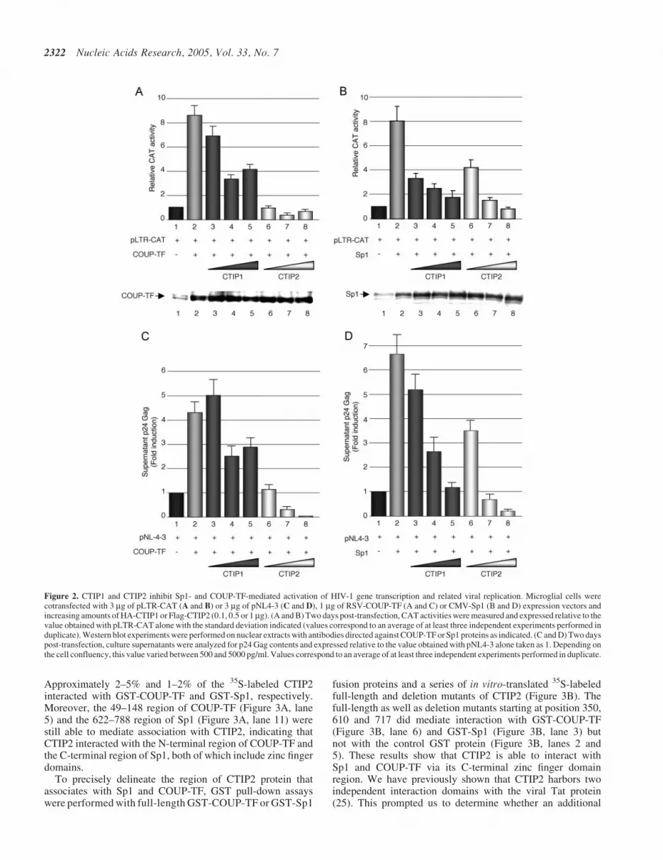
Approximately 2–5% and 1–2% of the 35S-labeled CTIP2interacted with GST-COUP-TF and GST-Sp1, respectively.Moreover, the 49–148 region of COUP-TF (Figure 3A, lane5) and the 622–788 region of Sp1 (Figure 3A, lane 11) werestill able to mediate association with CTIP2, indicating thatCTIP2 interacted with the N-terminal region of COUP-TF andthe C-terminal region of Sp1, both of which include zinc fingerdomains.
To precisely delineate the region of CTIP2 protein thatassociates with Sp1 and COUP-TF, GST pull-down assayswere performed with full-length GST-COUP-TF or GST-Sp1
fusion proteins and a series of in vitro-translated 35S-labeledfull-length and deletion mutants of CTIP2 (Figure 3B). Thefull-length as well as deletion mutants starting at position 350,610 and 717 did mediate interaction with GST-COUP-TF(Figure 3B, lane 6) and GST-Sp1 (Figure 3B, lane 3) butnot with the control GST protein (Figure 3B, lanes 2 and5). These results show that CTIP2 is able to interact withSp1 and COUP-TF via its C-terminal zinc finger domainregion. We have previously shown that CTIP2 harbors twoindependent interaction domains with the viral Tat protein(25). This prompted us to determine whether an additional
Figure 2. CTIP1 and CTIP2 inhibit Sp1- and COUP-TF-mediated activation of HIV-1 gene transcription and related viral replication. Microglial cells werecotransfected with 3 mg of pLTR-CAT (A and B) or 3 mg of pNL4-3 (C and D), 1 mg of RSV-COUP-TF (A and C) or CMV-Sp1 (B and D) expression vectors andincreasing amounts of HA-CTIP1 or Flag-CTIP2 (0.1, 0.5 or 1mg). (A and B) Two days post-transfection, CAT activities were measured and expressed relative to thevalue obtained with pLTR-CAT alone with the standard deviation indicated (values correspond to an average of at least three independent experiments performed induplicate). Western blot experiments were performed on nuclear extracts with antibodies directed against COUP-TF or Sp1 proteins as indicated. (C and D) Two dayspost-transfection, culture supernatants were analyzed for p24 Gag contents and expressed relative to the value obtained with pNL4-3 alone taken as 1. Depending onthe cell confluency, this value varied between 500 and 5000 pg/ml. Values correspond to an average of at least three independent experiments performed in duplicate.
2322 Nucleic Acids Research, 2005, Vol. 33, No. 7

interaction interface was located in the N-terminal or thecentral region of CTIP2. To this end, GST pull-down experi-ments were performed with additional deletion constructs. Asexpected, the deletion mutant 350–716 was not able to interactwith COUP-TF and Sp1, confirming that the C-terminal regionof CTIP2 containing residues 717–813 mediates binding tothese transcription factors. However, the 1–354 and the 145–434 constructs appeared to be able to restore interactions,suggesting that the central region of CTIP2 located betweenthe residues 145 and 354 might be sufficient for COUP-TF andSp1 association.
Thus, these findings indicate that CTIP2 interacts in vitrowith the zinc finger domains of COUP-TF and Sp1 by twointerfaces located in the central region (residues 145–354) andthe C-terminal region (residues 717–813).
CTIP2 colocalizes with Sp1 and COUP-TF withinHp1a-associated structures
To visualize the association of CTIP2 with Sp1 and COUP-TFwithin the nucleus, Flag-CTIP2 transfected microglialcells were observed by immunofluorescence confocal laser
A
B
Figure 3. CTIP2 interacts in vitro with COUP-TF and Sp1 by two interfaces. (A) Upper panels: schematic representation of the COUP-TF and Sp1 proteins; lowerpanels: GST pull-down assays were performed with 35S-labeled CTIP2 incubated with GST (lanes 2 and 7) or GST fusion proteins of the indicated COUP-TF domains(lanes 3–5) or Sp1 domains (lanes 8–11). Approximately 1% of the total 35S-labeled CTIP2 obtained was loaded as input control (lanes 1 and 6). (B) GST pull-downassays were performed with 35S-labeled full-length or truncated CTIP2 proteins incubated with GST (lanes 2 and 5), GST-COUP-TF (lane 6) or GST-Sp1 (lane 3)fusion proteins. Approximately 1% of the total 35S-labeled proteins used were loaded as input control (lanes 1 and 4). Representative Coomassie stainings of GST,GST-Sp1 and GST-COUP-TF proteins were presented (B lower panels).
Nucleic Acids Research, 2005, Vol. 33, No. 7 2323
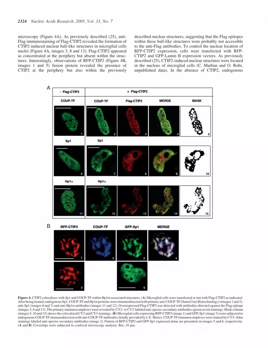
microscopy (Figure 4A). As previously described (25), anti-Flag immunostaining of Flag-CTIP2 revealed the formation ofCTIP2-induced nuclear ball-like structures in microglial cellsnuclei (Figure 4A, images 3, 8 and 13). Flag-CTIP2 appearedas concentrated at the periphery but absent within the struc-tures. Interestingly, observations of RFP-CTIP2 (Figure 4B,images 1 and 5) fusion protein revealed the presence ofCTIP2 at the periphery but also within the previously
described nuclear structures, suggesting that the Flag epitopeswithin these ball-like structures were probably not accessibleto the anti-Flag antibodies. To control the nuclear location ofRFP-CTIP2 expression, cells were transfected with RFP-CTIP2 and GFP-Lamin B expression vectors. As previouslydescribed (25), CTIP2-induced nuclear structures were locatedin the nucleus of microglial cells (C. Marban and O. Rohr,unpublished data). In the absence of CTIP2, endogenous
A
B
Figure 4. CTIP2 colocalizes with Sp1 and COUP-TF within Hp1a-associated structures. (A) Microglial cells were transfected or not with Flag-CTIP2 as indicated.After being treated, endogenous Sp1, COUP-TF and Hp1aproteins were immunodetected with primary anti-COUP-TF (Santa Cruz Biotechnology) (images 1 and 2),anti-Sp1 (images 6 and 7) and anti-Hp1a antibodies (images 11 and 12). Overexpressed Flag-CTIP2 was detected with antibodies directed against the Flag epitope(images 3, 8 and 13). The primary immunocomplexes were revealed by CY2- or CY3-labeled anti-species secondary antibodies (green or red staining). Mask column(images 5, 10 and 15) shows the colocalized CY2 and CY3 stainings. (B) Microglial cells expressing RFP-CTIP2 (image 1) and GFP-Sp1 (image 3) were subjected toendogenous COUP-TF immunodetection with anti-COUP-TF antibodies (kindly provided by J. E. Mertz). COUP-TF immunocomplexes were stained by CY5- (bluestaining) labeled anti-species secondary antibodies (image 2). Pattern of RFP-CTIP2 and GFP-Sp1 expressed alone are presented on images 5 and 6, respectively.(A and B) Coverslips were subjected to confocal microscopy analysis. Bar, 10 mm.
2324 Nucleic Acids Research, 2005, Vol. 33, No. 7
COUP-TF and Sp1 proteins were expressed in the nucleo-plasm with a speckled and diffused staining pattern (images1 and 6). Moreover, in the presence of Flag-CTIP2, thesetranscription factors were at least partially located toCTIP2-induced nuclear structures (Figure 4A, images 2 and7). Endogenous Sp1 (Figure 4A, images 9 and 10) and COUP-TF (Figure 4A, images 4 and 5) proteins colocalized withCTIP2 at the periphery of the immunostained structures. Upto 87 and 70% of endogenous COUP-TF and Sp1 proteinscolocalized with CTIP2, respectively. No staining wasobserved inside the ball-like structures. We have reportedthat CTIP2 relocates the viral Tat transactivator to theheterochromatin-associated protein Hp1a (25). EndogenousHp1a exhibited a diffused and speckled nuclear distributionin the absence of CTIP2 (Figure 4A, image 11). To visualizethe presence of Hp1a in CTIP2-induced structures in theabsence of Tat, we performed immunodetection of endogen-ous Hp1a in microglial cells expressing Flag-CTIP2. Asshown in Figure 4A (images 14 and 15), endogenous Hp1acolocalized with CTIP2, suggesting that endogenous COUP-TF, Sp1 and Hp1a proteins are relocated to the same CTIP2-induced nuclear structures. To address whether COUP-TF andSp1 also colocalized with CTIP2 inside the ball-like structures,we performed immunodetection of the endogenous COUP-TFprotein with other different polyclonal antibodies (kindlyprovided by J. E. Mertz) in the presence of RFP-CTIP2 andGFP-Sp1 fusion proteins. As shown in Figure 4B (image 2),endogenous COUP-TF was observed inside the structures,confirming that the epitope accessibility to the antibodies isdeterminant to COUP-TF immunodetection. Since RFP-CTIP2 was expressed in ball-like structures (Figure 4B,images 1 and 5), GFP-Sp1 expressed alone was localized inthe nucleoplasm with a diffused staining pattern (Figure 4B,image 6). Moreover, in the presence of RFP-CTIP2, GFP-Sp1is relocated to CTIP2-induced structures (Figure 4B, image 3).COUP-TF and GFP-Sp1 relocation was not restricted to theperiphery of the CTIP2-induced nuclear structures, since theywere also detected inside the structures as shown by the pres-ence of white staining resulting from colocalized red, blue andgreen staining (Figure 4B, image 4). Thus, endogenous Sp1proteins were probably relocated within the heterochromaticstructures too. However, they remained unaccessible to thecurrently used antibodies (Figure 4A, images 7 and 9).These results strongly suggest that the cellular transcriptionfactors, Sp1 and COUP-TF, associate with CTIP2 inmicroglial cells.
CTIP2 interacts with Sp1, COUP-TF and Hp1a inmicroglial cells
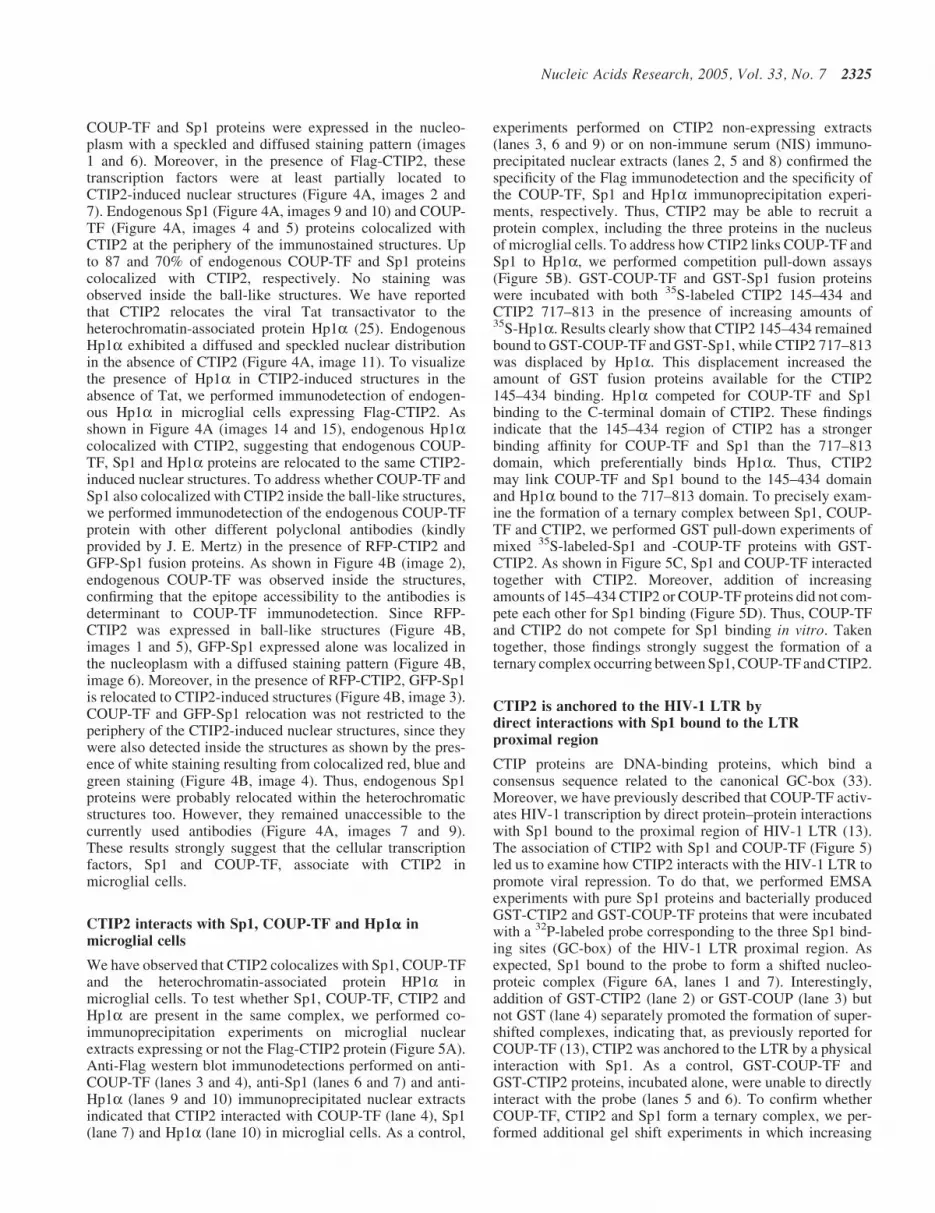
We have observed that CTIP2 colocalizes with Sp1, COUP-TFand the heterochromatin-associated protein HP1a inmicroglial cells. To test whether Sp1, COUP-TF, CTIP2 andHp1a are present in the same complex, we performed co-immunoprecipitation experiments on microglial nuclearextracts expressing or not the Flag-CTIP2 protein (Figure 5A).Anti-Flag western blot immunodetections performed on anti-COUP-TF (lanes 3 and 4), anti-Sp1 (lanes 6 and 7) and anti-Hp1a (lanes 9 and 10) immunoprecipitated nuclear extractsindicated that CTIP2 interacted with COUP-TF (lane 4), Sp1(lane 7) and Hp1a (lane 10) in microglial cells. As a control,
experiments performed on CTIP2 non-expressing extracts(lanes 3, 6 and 9) or on non-immune serum (NIS) immuno-precipitated nuclear extracts (lanes 2, 5 and 8) confirmed thespecificity of the Flag immunodetection and the specificity ofthe COUP-TF, Sp1 and Hp1a immunoprecipitation experi-ments, respectively. Thus, CTIP2 may be able to recruit aprotein complex, including the three proteins in the nucleusof microglial cells. To address how CTIP2 links COUP-TF andSp1 to Hp1a, we performed competition pull-down assays(Figure 5B). GST-COUP-TF and GST-Sp1 fusion proteinswere incubated with both 35S-labeled CTIP2 145–434 andCTIP2 717–813 in the presence of increasing amounts of35S-Hp1a. Results clearly show that CTIP2 145–434 remainedbound to GST-COUP-TF and GST-Sp1, while CTIP2 717–813was displaced by Hp1a. This displacement increased theamount of GST fusion proteins available for the CTIP2145–434 binding. Hp1a competed for COUP-TF and Sp1binding to the C-terminal domain of CTIP2. These findingsindicate that the 145–434 region of CTIP2 has a strongerbinding affinity for COUP-TF and Sp1 than the 717–813domain, which preferentially binds Hp1a. Thus, CTIP2may link COUP-TF and Sp1 bound to the 145–434 domainand Hp1a bound to the 717–813 domain. To precisely exam-ine the formation of a ternary complex between Sp1, COUP-TF and CTIP2, we performed GST pull-down experiments ofmixed 35S-labeled-Sp1 and -COUP-TF proteins with GST-CTIP2. As shown in Figure 5C, Sp1 and COUP-TF interactedtogether with CTIP2. Moreover, addition of increasingamounts of 145–434 CTIP2 or COUP-TF proteins did not com-pete each other for Sp1 binding (Figure 5D). Thus, COUP-TFand CTIP2 do not compete for Sp1 binding in vitro. Takentogether, those findings strongly suggest the formation of aternary complex occurring between Sp1, COUP-TF and CTIP2.
CTIP2 is anchored to the HIV-1 LTR bydirect interactions with Sp1 bound to the LTRproximal region
CTIP proteins are DNA-binding proteins, which bind aconsensus sequence related to the canonical GC-box (33).Moreover, we have previously described that COUP-TF activ-ates HIV-1 transcription by direct protein–protein interactionswith Sp1 bound to the proximal region of HIV-1 LTR (13).The association of CTIP2 with Sp1 and COUP-TF (Figure 5)led us to examine how CTIP2 interacts with the HIV-1 LTR topromote viral repression. To do that, we performed EMSAexperiments with pure Sp1 proteins and bacterially producedGST-CTIP2 and GST-COUP-TF proteins that were incubatedwith a 32P-labeled probe corresponding to the three Sp1 bind-ing sites (GC-box) of the HIV-1 LTR proximal region. Asexpected, Sp1 bound to the probe to form a shifted nucleo-proteic complex (Figure 6A, lanes 1 and 7). Interestingly,addition of GST-CTIP2 (lane 2) or GST-COUP (lane 3) butnot GST (lane 4) separately promoted the formation of super-shifted complexes, indicating that, as previously reported forCOUP-TF (13), CTIP2 was anchored to the LTR by a physicalinteraction with Sp1. As a control, GST-COUP-TF andGST-CTIP2 proteins, incubated alone, were unable to directlyinteract with the probe (lanes 5 and 6). To confirm whetherCOUP-TF, CTIP2 and Sp1 form a ternary complex, we per-formed additional gel shift experiments in which increasing
Nucleic Acids Research, 2005, Vol. 33, No. 7 2325

amounts of CTIP2 proteins were added to the binarySp1/COUP-TF complex (lanes 9 and 10). As shown, the shif-ted complex formed by COUP-TF and Sp1 bound to the tem-plate (lane 8) was supershifted by increasing the amount ofCTIP2 in a dose-dependent manner. Moreover, the intensity ofthe supershifted complexes also increased with CTIP2 quant-ity. Those findings are in favor of the formation of a ternarycomplex anchored to the HIV-1 Sp1 binding sites. To preciselyexamine this complex, the mixed three proteins were incub-ated with their respective antibody. EMSA experimentsdemonstrated that the complex observed in the presence ofthe three proteins (Figure 6A, lane 10 and Figure 6B, lane 1)was supershifted by the antibodies directed against CTIP2(Figure 6B, lane 2) and COUP-TF (Figure 6B, lane 3) butnot by non-immune serum (Figure 6B, lane 5) confirmingthe presence of the two proteins in the complex. Moreover,
incubation of the mixed three proteins with the anti-Sp1 anti-bodies (Figure 6B, lane 4) inhibited the formation of the nuc-leoprotein complex. Thus, impairing Sp1 binding to theproxixmal region of the HIV-1 LTR results in the total aboli-tion of the CTIP2 and COUP-TF ability to bind this region ofthe viral promoter. These results confirm that both CTIP2 andCOUP-TF interact with Sp1 bound to the LTR in vitro.Previously published EMSA experiments performed with nuc-lear extracts of microglial cells have revealed that this regionof the viral promoter binds the cellular transcription factors,Sp1 and Sp3, in microglial cells (13). EMSA experimentsperformed with nuclear extracts of microglial cells expressingCTIP2 could not allow us to observe CTIP2 indirect bindingto the LTR (C. Marban and O. Rohr, unpublished data). Tobypass this technical issue and to address whether CTIP2interacts with the viral promoter in vivo, ChIP assays were
A
B
C D
Figure 5. CTIP2 interacts with COUP-TF, Sp1 and Hp1a in microglial cells. (A) Nuclear extracts of microglial cells expressing (lanes 2, 4, 5, 7, 8 and 10) or not (lanes3, 6 and 9) Flag-CTIP2 were immunoprecipitated with antibodies directed against COUP-TF (lanes 3 and 4), Sp1 (lanes 6 and 7) or Hp1a (lanes 9 and 10) proteins.Proteins were separated by SDS–PAGE and western blot analysis with anti-Flag antibodies were performed. (B) GST pull-down competition assays were performedwith equal amounts of 35S-labeled 145–434 and 717–813 CTIP2 proteins and increasing amounts of 35S-labeled Hp1a. Proteins were incubated with GST-COUP-TFor GST-Sp1 fusion proteins as indicated. (C) GST pull-down assays were performed with equal amounts of 35S-labeled Sp1 and COUP-TF proteins and GST or GST-CTIP2 fusion proteins as indicated. (D) GST pull-down competition assays were performed with the indicated amounts of 35S-labeled 145–434 CTIP2 and COUP-TFproteins and GST-Sp1. (A–D) Approximately 1% of the total 35S-labeled proteins used were loaded as input control.
2326 Nucleic Acids Research, 2005, Vol. 33, No. 7
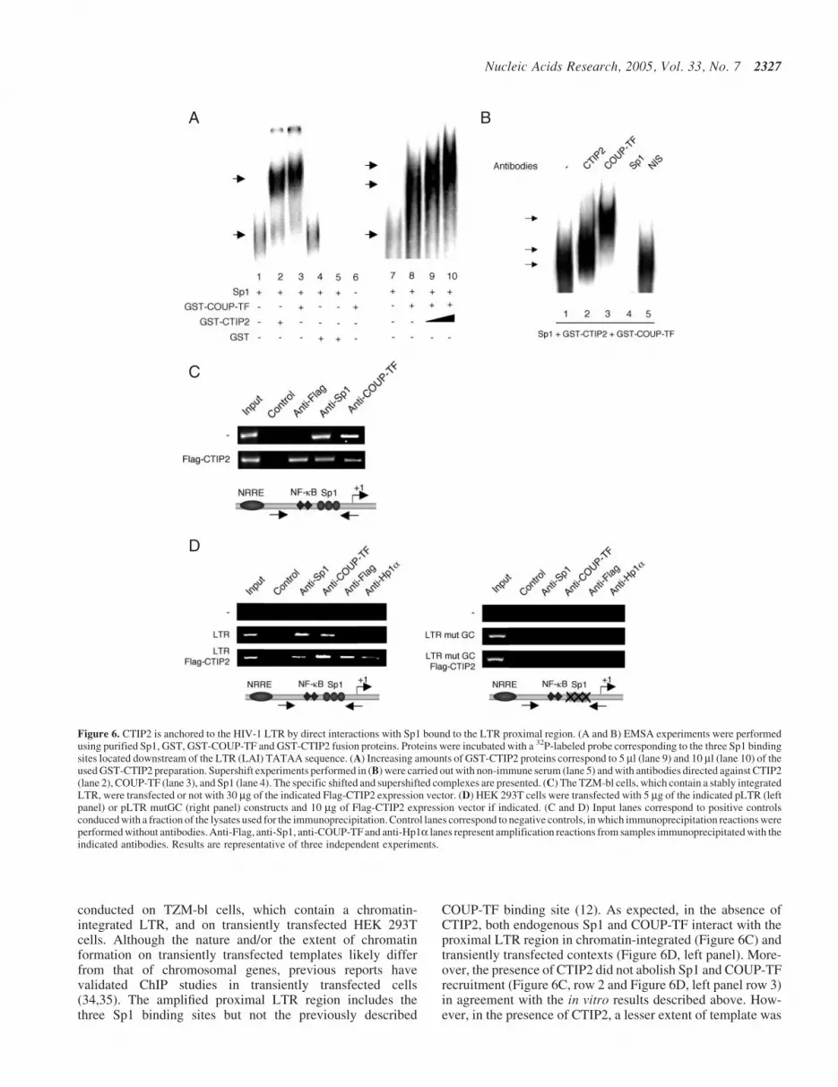
conducted on TZM-bl cells, which contain a chromatin-integrated LTR, and on transiently transfected HEK 293Tcells. Although the nature and/or the extent of chromatinformation on transiently transfected templates likely differfrom that of chromosomal genes, previous reports havevalidated ChIP studies in transiently transfected cells(34,35). The amplified proximal LTR region includes thethree Sp1 binding sites but not the previously described
COUP-TF binding site (12). As expected, in the absence ofCTIP2, both endogenous Sp1 and COUP-TF interact with theproximal LTR region in chromatin-integrated (Figure 6C) andtransiently transfected contexts (Figure 6D, left panel). More-over, the presence of CTIP2 did not abolish Sp1 and COUP-TFrecruitment (Figure 6C, row 2 and Figure 6D, left panel row 3)in agreement with the in vitro results described above. How-ever, in the presence of CTIP2, a lesser extent of template was
A
C
D
B
Figure 6. CTIP2 is anchored to the HIV-1 LTR by direct interactions with Sp1 bound to the LTR proximal region. (A and B) EMSA experiments were performedusing purified Sp1, GST, GST-COUP-TF and GST-CTIP2 fusion proteins. Proteins were incubated with a 32P-labeled probe corresponding to the three Sp1 bindingsites located downstream of the LTR (LAI) TATAA sequence. (A) Increasing amounts of GST-CTIP2 proteins correspond to 5 ml (lane 9) and 10 ml (lane 10) of theused GST-CTIP2 preparation. Supershift experiments performed in (B) were carried out with non-immune serum (lane 5) and with antibodies directed against CTIP2(lane 2), COUP-TF (lane 3), and Sp1 (lane 4). The specific shifted and supershifted complexes are presented. (C) The TZM-bl cells, which contain a stably integratedLTR, were transfected or not with 30 mg of the indicated Flag-CTIP2 expression vector. (D) HEK 293T cells were transfected with 5 mg of the indicated pLTR (leftpanel) or pLTR mutGC (right panel) constructs and 10 mg of Flag-CTIP2 expression vector if indicated. (C and D) Input lanes correspond to positive controlsconduced with a fraction of the lysates used for the immunoprecipitation. Control lanes correspond to negative controls, in which immunoprecipitation reactions wereperformed without antibodies. Anti-Flag, anti-Sp1, anti-COUP-TF and anti-Hp1a lanes represent amplification reactions from samples immunoprecipitated with theindicated antibodies. Results are representative of three independent experiments.
Nucleic Acids Research, 2005, Vol. 33, No. 7 2327
immunoprecipitated with the antibodies directed against Sp1and COUP-TF in the context of the integrated LTR (Figure 6C,row 2). The same observations were made with the antibodiesdirected against Sp1 in the transiently transfected LTR context(Figure 6D, left panel). As shown in Figure 6C and D (leftpanel), CTIP2 interacted with the proximal region of the viralpromoter. In addition, mutation of the Sp1 binding sites abol-ished the interaction of Sp1, COUP-TF and CTIP2 with thisregion of the viral LTR (Figure 6D, right panel). Takentogether, our in vitro and in vivo findings suggest that theSp1-mediated recruitment of CTIP2 to the LTR occurs in acellular context. We could not detect Hp1a interactions withthe proximal region of the LTR in the absence of CTIP2,suggesting that CTIP2 significantly increase the recruitmentof Hp1a to the HIV-1 proximal promoter (Figure 6D, leftpanel). In addition, mutation of the Sp1 binding sites alsoabolished CTIP2-mediated recruitment of Hp1a (Figure 6D,right panel). Since Hp1a associates with heterochromaticregions, it is likely that CTIP2 promotes transcriptionallyrepressed heterochromatic environment to the viral LTRregion.
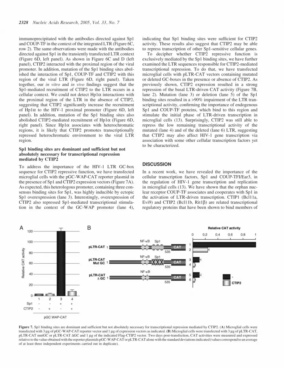
Sp1 binding sites are dominant and sufficient but notabsolutely necessary for transcriptional repressionmediated by CTIP2
To address the importance of the HIV-1 LTR GC-boxsequence for CTIP2 repressive function, we have transfectedmicroglial cells with the pGC-WAP-CAT reporter plasmid inthe presence of Sp1 and CTIP2 expression vectors (Figure 7A).As expected, this heterologous promoter, containing three con-sensus binding sites for Sp1, was highly inducible by ectopicSp1 overexpression (lane 3). Interestingly, overexpression ofCTIP2 also repressed Sp1-mediated transcriptional stimula-tion in the context of the GC-WAP promoter (lane 4),
indicating that Sp1 binding sites were sufficient for CTIP2activity. These results also suggest that CTIP2 may be ableto repress transcription of other Sp1-sensitive cellular genes.
To decipher whether CTIP2 repressive function isexclusively mediated by the Sp1 binding sites, we have furtherexamined the LTR sequences responsible for CTIP2-mediatedtranscriptional repression. To do that, we have transfectedmicroglial cells with pLTR-CAT vectors containing mutatedor deleted GC-boxes in the presence or absence of CTIP2. Aspreviously shown, CTIP2 expression resulted in a strongrepression of the basal LTR-driven CAT activity (Figure 7B,lane 2). Mutation (lane 3) or deletion (lane 5) of the Sp1binding sites resulted in a >90% impairment of the LTR tran-scriptional activity, confirming the importance of endogenousSp1 and COUP-TF proteins, which bind to this region andstimulate the initial phase of LTR-driven transcription inmicroglial cells (13). Surprisingly, CTIP2 was still able torepress the low remaining transcriptional activity of themutated (lane 4) and of the deleted (lane 6) LTR, suggestingthat CTIP2 may also affect HIV-1 gene transcription viaassociation with some other cellular transcription factors yetto be characterized.
DISCUSSION
In a recent work, we have revealed the importance of thecellular transcription factors, Sp1 and COUP-TFI/Ear3, inthe regulation of HIV-1 gene transcription and replicationin microglial cells (13). We have shown that the orphan nuc-lear receptor COUP-TF associates and cooperates with Sp1 inthe activation of LTR-driven transcription. CTIP1 (Bcl11a,Evi9) and CTIP2 (Bcl11b, Rit1b) are related transcriptionalregulatory proteins that have been shown to bind members of
A B
Figure 7. Sp1 binding sites are dominant and sufficient but not absolutely necessary for transcriptional repression mediated by CTIP2. (A) Microglial cells weretransfected with 3 mg of pGC-WAP-CAT reporter vector and 1 mg of expression vectors as indicated. (B) Microglial cells were transfected with 3 mg of pLTR-CAT,pLTR-CAT mutGC or pLTR-CAT DGC and 1 mg of the indicated Flag-CTIP2 vector. Two days post-transfection, CAT activities were measured and expressedrelative to the value obtained with the reporter plasmids pGC-WAP-CAT or pLTR-CAT alone with the standard deviations indicated (values correspond to an averageof at least three independent experiments carried out in duplicate).
2328 Nucleic Acids Research, 2005, Vol. 33, No. 7

the COUP-TF family (23). In a more recent work, we havedescribed that the nuclear cofactor CTIP2 inhibits thesubsequent phase of HIV-1 gene transcription by relocatingthe viral Tat protein to transcriptionally inactive regions ofchromatin via Hp1a (25).
Here, we addressed the mechanism whereby CTIP2 impairsthe initial phase (prior to Tat expression) of HIV-1 genetranscription in microglial cells.
We report that CTIP proteins inhibit Sp1- and COUP-TF-mediated activation of HIV-1 gene transcription and replica-tion in microglial cells. To decipher the mechanism wherebyCTIP2 alters COUP-TF and Sp1 functions, we examinedwhether these proteins interact in vitro and in cells. GSTpull-down experiments revealed that CTIP2 associates withthe N-terminal region of COUP-TF and with the C-terminalregion of Sp1 in vitro. In this way, CTIP2 presents two inter-faces for Sp1 and COUP-TF interactions. COUP-TF and Sp1proteins bind the central and the C-terminal region of CTIP2 invitro. These results are consistent with the previously reportedinterface domains of Arp1 (COUP-TFII) with CTIP1 (23).Confocal microscopy observations of COUP-TF and Sp1 loca-tion in the presence or absence of CTIP2 reveal that CTIP2colocalizes with COUP-TF and Sp1 within nuclear ball-likestructures. Moreover, the heterochromatin-associated proteinHp1a is also present in these structures. There are three Hp1protein family members in mammals, Hp1a, Hp1b and Hp1g .The Hp1a isoform is mainly detected in transcriptionallyrepressed heterochromatic region (36). Thus, the colocaliza-tion of CTIP2 with endogenous Sp1, COUP-TF and Hp1aproteins as reported here strongly suggests that these proteinsare relocated to transcriptionally inactive regions ofheterochromatin in microglial cells. Those CTIP2-inducedstructures may represent a new class of nuclear structures(25). Moreover, our observations may favor the hypothesis,suggesting that the formation of nuclear bodies inhibits HIV-1gene transcription by sequestering a variety of factors requiredfor transcriptional activation [reviewed in (37)].
Co-immunoprecipitation data indicate that the describedsubnuclear colocalization results from physical interactionsbetween Sp1, COUP-TF, Hp1a and CTIP2 in the nucleusof microglial cells. Since Hp1a, Sp1 and COUP-TF proteinsassociate with CTIP2 in microglial cells, we address theirrespective interaction interfaces in vitro. Our findings showthat the central zinc finger domain of CTIP2 may preferen-tially bind Sp1 and COUP-TF proteins, while the C-terminalregion may bind to Hp1a. In the presence of the viral Tattransactivator, CTIP2 also associates with Hp1a via itsC-terminal region. Taken together, these findings suggest apermanent association of CTIP2 with Hp1a via its C-terminalregion. The central domain of CTIP2 may be reserved for theinteractions with the functionally repressed transcriptionfactors. Our observations indicate that COUP-TF and Sp1could bind together to CTIP2 and that COUP-TF andCTIP2 do not compete for Sp1 binding. Those findings suggestthe formation of a ternary complex, including COUP-TF, Sp1and CTIP2 proteins. Thereby, CTIP2 links these transcrip-tional activators to transcriptionally repressed heterochromaticregion. CTIP1 and CTIP2 proteins have been described assequence-specific DNA-binding proteins (33). The core-binding site identified in this study is highly related to thecanonical GC-box sequence. Since Sp1 anchors COUP-TF
to the GC-box sequence of the viral LTR region (12), weaddressed how CTIP2 interacts with the HIV-1 promoter.Our findings demonstrate that CTIP2 is not able to directlybind the three viral GC-box sequences in vitro. CTIP2 isrecruited to the HIV-1 LTR via its association with Sp1bound to this region. Moreover, CTIP2 is not in competitionwith COUP-TF for Sp1-mediated anchorage to the viral pro-moter, indicating that the three proteins are linked together tothe LTR in vitro. The ChIP experiments conducted in a cellularcontext confirm that CTIP2, Sp1 and COUP-TF proteins dobind the proximal region of the HIV-1 promoter in chromatin-integrated and non-integrated contexts.
The surprising reduction of Sp1 or COUP-TF binding to theviral promoter in the presence of CTIP2 apparently does notagree with the in vitro observations. We show that Sp1 anchorsCTIP2 and COUP-TF to the HIV-1 LTR and that Sp1 bindingto the GC-box sequence is necessary for CTIP2 recruitment tothis region. Nevertheless, since Sp1 and COUP-TF proteinsare not accessible to their related antibodies in the CTIP2-induced nuclear structures, one wonders whether the observa-tion of reduced binding to the LTR may result from proteinsequestration and/or chromatin environment. ChIP experi-ments also indicate that CTIP2 promotes Hp1a recruitmentto the LTR encompassing the Sp1 binding sequences. Muta-tion of the Sp1 binding sites abolished this recruitment. Takentogether, these results suggest that the Sp1-mediated anchor-age of CTIP2 to the HIV-1 LTR region may recruit a largecomplex that promotes Hp1a association, heterochromatinenvironment and viral gene transcriptional silencing. In Jurkatcells, CTIP2 associates with the histone deacetylase SirT1 in alarge complex. CTIP2-mediated recruitment of SirT1 to pro-moter template results in a deacetylation of the bound histonesH3 and H4 and in a transcriptional repression (35). The chro-matin structure close to the HIV-1 gene promoter is involvedin viral post-integration latency phenomenon (38). Furtherstudies will be necessary to examine the CTIP2-recruitedcomplex that promotes HIV-1 gene silencing.
To control whether the CTIP2 repressive function is medi-ated by Sp1 binding sites, we have examined its ability torepress a heterologous promoter containing three consensusGC-boxes. Our findings reveal that GC-box sequences aresufficient for CTIP2-mediated repression of Sp1 stimulation,suggesting that CTIP2 may impact the transcriptional activityof other GC-box containing promoters. In agreement withthese observations, CTIP2 is still able to repress LTR-driven transcriptional activity of a LTR deleted downstreamof the GC-box sequences. Recruitment of CTIP1 to the tem-plate by a COUP-TF family member has been found to resultin a transcriptional repression of a reporter gene harboring aCOUP-TF binding site (23). In the context of the HIV-1promoter, deletion of the previously described COUP-TFbinding site did not impact CTIP2 repressive function. More-over, EMSA and ChIP experiments demonstrate that CTIP2interacts with the proximal region of the LTR excluding theCOUP-TF binding sequence. Taken together, these findingsindicate that the COUP-TF binding sequence, located down-stream to the GC-box region, might not be implicated in theCTIP2 repressive functions. As widely reported, deletion ofthe LTR GC-box region results in a drastic (>90%) reductionof the LTR-driven transcriptional activity (13–15,22). Theseobservations confirm the crucial contribution of Sp1 and
Nucleic Acids Research, 2005, Vol. 33, No. 7 2329
COUP-TF transcription factors to support viral gene transcrip-tion in microglial cells. Moreover, they highlight the import-ance of CTIP2-mediated repression of Sp1 and COUP-TFprotein functions. Most of the 80% CTIP2-mediated reductionof the basal transcriptional level may be charged on to thisrepressive activity. Surprisingly, CTIP2 is still able to repressthe 10% remaining transcriptional activity driven by a GC-boxdeleted LTR, suggesting that CTIP2 may also be able torepress the expression of mutated strains of virus. This remain-ing activity represents <10% of the global CTIP2-mediatedrepressive activity.
Nevertheless, we have previously suggested that COUP-TFrestores the viral Tat function in the context of a GC-boxmutated LTR by anchoring Tat to the basal transcriptionalmachinery (22). Interestingly, we observed that CTIP2 isalso able to repress this functional cooperation (C. Marbanand O. Rohr, unpublished data). These results suggest that, inthis context, COUP-TF may bind CTIP2 via its N-terminalregion and the general transcription factor TFIIB via its C-terminal part as described previously (39). Thus, in theabsence of Sp1-mediated linkage to the GC-box region ofthe HIV-1 LTR, CTIP2 may bypass Sp1 association by aCOUP-TF-mediated linkage to the basal transcriptionalmachinery. Moreover, this proposed indirect linkage mightbe weak since it appeared undetectable by ChIP experiments.Along this line, CTIP2 might also associate to some othercellular transcription factors yet to be characterized.
These studies fully complete those reporting CTIP2-mediated repression of the viral Tat function (25). SinceSp1 or COUP-TF association is necessary for Tat functionin microglial cells (22), it can be postulated that CTIP2-mediated impairment of Sp1 and COUP-TF function contrib-utes to the previously observed repression of Tat function.
The chromatin structure at the site of provirus integrationis reminiscent to post-integration latency phenomenon (38).In vitro, latent T-cell clones frequently contain HIV-1 genomeintegrated in heterochromatin structures. This is in contrast toa productive infection where integration in or near heterochro-matin is disfavored (40). No antiretroviral drugs that are cur-rently available can inhibit transcription of HIV RNA from theintegrated HIV proviral DNA in infected cells. Nevertheless,HDAC inhibitors are able to induce quiescent provirus, sug-gesting that derepression of heterochromatin structures mayforce viral expression from latently infected reservoirs (41,42).Thus, investigation on factors that may promote heterochro-matin structures to the viral gene promoter, such as CTIP2,appears crucial for understanding these phenomena and for thedevelopment of new anti-HIV strategies. At this stage, furtherstudies will be necessary to precisely examine the CTIP2-associated enzymatic activities that promote transcriptionalsilencing. In addition, since our investigations were restrictedto the transcriptional step of the viral life cycle, it cannot beexcluded that CTIP2 also counteracts other critical stepsnecessary to optimal viral replication. This possibility needsfurther clarification.
ACKNOWLEDGEMENTS
The authors thank R. Losson for providing Hp1a vectors andanti-Hp1a antibodies, H. Rotheneder for providing GST-SP1
vectors, J. E. Mertz for providing anti-COUP-TF antibodiesand Ann Dekoninck for technical support. The followingreagents were obtained through the NIH AIDS Research andReference Reagent Program, Division of AIDS, NIAID, NIH:TZM-bl from Dr John C. Kappes, Dr Xiaoyun Wu andTranzyme Inc. and pNL4-3 from Dr Malcolm Martin. Thiswork was supported by the Institut National de la Sante etde la Recherche Medicale (INSERM), by the AgenceNationale de Recherches sur le SIDA (ANRS), by grantsfrom the French Ministry of Research (‘ACI JC 5364’ toO.R. and grant to S.S.) and by a grant to C.M. from theConseil Regional d’Alsace. C.V.L. is a ‘Maıtre deRecherches’ at the ‘Fonds National de la RechercheScientifique’ (FNRS, Belgium) and O.R. is a ‘Maıtre deConferences’ at the ‘IUT Louis Pasteur de Schiltigheim’(University of Strasbourg I, France). Work in the LeidLaboratory was supported by a grant from NIH (GM60852)and in part by grant number P30 ES00210 from the NationalInstitute of Environmental Health Sciences, NIH. Funding topay the Open Access publication charges for this article wasprovided by INSERM.
Conflict of interest statement. None declared.
REFERENCES
1. Fauci,A.S. (1988) The human immunodeficiency virus: infectivity andmechanisms of pathogenesis. Science, 239, 617–622.
2. Portegies,P. and Brew,B.J. (1991) Update on HIV-related neurologicalillness. AIDS, 5 (Suppl. 2), S211–S217.
3. Pialoux,G., Fournier,S., Moulignier,A., Poveda,J.D., Clavel,F. andDupont,B. (1997) Central nervous system as a sanctuary for HIV-1infection despite treatment with zidovudine, lamivudine and indinavir.AIDS, 11, 1302–1303.
4. Kolson,D.L., Lavi,E. and Gonzalez-Scarano,F. (1998) The effects ofhuman immunodeficiency virus in the central nervous system. Adv.Virus Res., 50, 1–47.
5. Peudenier,S., Hery,C., Montagnier,L. and Tardieu,M. (1991) Humanmicroglial cells: characterization in cerebral tissue and in primary culture,and study of their susceptibility to HIV-1 infection. Ann. Neurol., 29,152–161.
6. Perry,V.H., Lawson,L.J. and Reid,D.M. (1994) Biology of themononuclear phagocyte system of the central nervous system and HIVinfection. J. Leukoc. Biol., 56, 399–406.
7. Greene,W.C. and Peterlin,B.M. (2002) Charting HIV’s remarkablevoyage through the cell: Basic science as a passport to future therapy.Nature Med., 8, 673–680.
8. Pereira,L.A., Bentley,K., Peeters,A., Churchill,M.J. and Deacon,N.J.(2000) A compilation of cellular transcription factor interactions with theHIV-1 LTR promoter. Nucleic Acids Res., 28, 663–668.
9. Rohr,O., Marban,C., Aunis,D. and Schaeffer,E. (2003) Regulation ofHIV-1 gene transcription: from lymphocytes to microglial cells.J. Leukoc. Biol., 74, 736–749.
10. Wang,L.H., Tsai,S.Y., Cook,R.G., Beattie,W.G., Tsai,M.J. andO’Malley,B.W. (1989) COUP transcription factor is a member of thesteroid receptor superfamily. Nature, 340, 163–166.
11. Wang,L.H., Tsai,S.Y., Sagami,I., Tsai,M.J. and O’Malley,B.W. (1987)Purification and characterization of chicken ovalbumin upstreampromoter transcription factor from HeLa cells. J. Biol. Chem., 262,16080–16086.
12. Sawaya,B.E., Rohr,O., Aunis,D. and Schaeffer,E. (1996) Chickenovalbumin upstream promoter transcription factor, a transcriptionalactivator of HIV-1 gene expression in human brain cells. J. Biol. Chem.,271, 23572–23576.
13. Rohr,O., Aunis,D. and Schaeffer,E. (1997) COUP-TF and Sp1 interactand cooperate in the transcriptional activation of the humanimmunodeficiency virus type 1 long terminal repeat in human microglialcells. J. Biol. Chem., 272, 31149–31155.
2330 Nucleic Acids Research, 2005, Vol. 33, No. 7

14. Rohr,O., Schwartz,C., Aunis,D. and Schaeffer,E. (1999) CREB andCOUP-TF mediate transcriptional activation of the human immunode-ficiency virus type 1 genome in Jurkat T cells in response to cyclic AMPand dopamine. J. Cell. Biochem., 75, 404–413.
15. Schwartz,C., Catez,P., Rohr,O., Lecestre,D., Aunis,D. and Schaeffer,E.(2000) Functional interactions between C/EBP, Sp1, and COUP-TFregulate human immunodeficiency virus type 1 gene transcription inhuman brain cells. J. Virol., 74, 65–73.
16. Jones,K.A., Kadonaga,J.T., Luciw,P.A. andTjian,R. (1986)Activationofthe AIDS retrovirus promoter by the cellular transcription factor, Sp1.Science, 232, 755–759.
17. Harrich,D., Garcia,J., Wu,F., Mitsuyasu,R., Gonazalez,J. and Gaynor,R.(1989) Role of SP1-binding domains in in vivo transcriptional regulationof the human immunodeficiency virus type 1 long terminal repeat.J. Virol., 63, 2585–2591.
18. Sune,C. and Garcia-Blanco,M.A. (1995) Transcriptional trans activationby human immunodeficiency virus type 1 Tat requires specificcoactivators that are not basal factors. J. Virol., 69, 3098–3107.
19. Perkins,N.D., Edwards,N.L., Duckett,C.S., Agranoff,A.B., Schmid,R.M.and Nabel,G.J. (1993) A cooperative interaction between NF-kappa Band Sp1 is required for HIV-1 enhancer activation. EMBO J., 12,3551–3558.
20. Yedavalli,V.S., Benkirane,M. and Jeang,K.T. (2003) Tat and trans-activation-responsive (TAR) RNA-independent induction of HIV-1 longterminal repeat by human and murine cyclin T1 requires Sp1. J. Biol.Chem., 278, 6404–6410.
21. van Opijnen,T., Kamoschinski,J., Jeeninga,R.E. and Berkhout,B. (2004)The human immunodeficiency virus type 1 promoter contains a CATAbox instead of a TATA box for optimal transcription and replication.J. Virol., 78, 6883–6890.
22. Rohr,O., Schwartz,C., Hery,C., Aunis,D., Tardieu,M. and Schaeffer,E.(2000) The nuclear receptor chicken ovalbumin upstream promotertranscription factor interacts with HIV-1 Tat and stimulates viralreplication in human microglial cells. J. Biol. Chem., 275, 2654–2660.
23. Avram,D., Fields,A., Pretty On Top,K., Nevrivy,D.J., Ishmael,J.E. andLeid,M. (2000) Isolation of a novel family of C(2)H(2) zinc fingerproteins implicated in transcriptional repression mediated bychicken ovalbumin upstream promoter transcription factor(COUP-TF) orphan nuclear receptors. J. Biol. Chem., 275, 10315–10322.
24. Leid,M., Ishmael,J.E., Avram,D., Shepherd,D., Fraulob,V. and Dolle,P.(2004) CTIP1 and CTIP2 are differentially expressed during mouseembryogenesis. Gene Expr. Patterns, 4, 733–739.
25. Rohr,O., Lecestre,D., Chasserot-Golaz,S., Marban,C., Avram,D.,Aunis,D., Leid,M. and Schaeffer,E. (2003) Recruitment of Tat toheterochromatin protein HP1 via interaction with CTIP2 inhibits humanimmunodeficiency virus type 1 replication in microglial cells. J. Virol.,77, 5415–5427.
26. Rohr,O., Sawaya,B.E., Lecestre,D., Aunis,D. and Schaeffer,E. (1999)Dopamine stimulates expression of the human immunodeficiency virustype 1 via NF-kappaB in cells of the immune system. Nucleic Acids Res.,27, 3291–3299.
27. Karlseder,J., Rotheneder,H. and Wintersberger,E. (1996) Interaction ofSp1 with the growth- and cell cycle-regulated transcription factor E2F.Mol. Cell. Biol., 16, 1659–1667.
28. Adachi,A., Koenig,S., Gendelman,H.E., Daugherty,D., Gattoni-Celli,S.,Fauci,A.S. and Martin,M.A. (1987) Productive, persistent infection of
humancolorectal cell lineswithhumanimmunodeficiencyvirus.J.Virol.,61, 209–213.
29. Janabi,N., Peudenier,S., Heron,B., Ng,K.H. and Tardieu,M. (1995)Establishment of human microglial cell lines after transfection of primarycultures of embryonic microglial cells with the SV40 large T antigen.Neurosci. Lett., 195, 105–108.
30. Derdeyn,C.A., Decker,J.M., Sfakianos,J.N., Wu,X., O’Brien,W.A.,Ratner,L., Kappes,J.C., Shaw,G.M. and Hunter,E. (2000) Sensitivity ofhuman immunodeficiency virus type 1 to the fusion inhibitor T-20 ismodulated by coreceptor specificity defined by the V3 loop of gp120.J. Virol., 74, 8358–8367.
31. Platt,E.J., Wehrly,K., Kuhmann,S.E., Chesebro,B. and Kabat,D. (1998)Effects of CCR5 and CD4 cell surface concentrations on infections bymacrophagetropic isolates of human immunodeficiency virus type 1.J. Virol., 72, 2855–2864.
32. Wei,X., Decker,J.M., Liu,H., Zhang,Z., Arani,R.B., Kilby,J.M.,Saag,M.S., Wu,X., Shaw,G.M. and Kappes,J.C. (2002) Emergence ofresistant human immunodeficiency virus type 1 in patients receivingfusion inhibitor (T-20) monotherapy. Antimicrob. Agents Chemother.,46, 1896–1905.
33. Avram,D., Fields,A., Senawong,T., Topark-Ngarm,A. and Leid,M.(2002) COUP-TF (chicken ovalbumin upstream promoter transcriptionfactor)-interacting protein 1 (CTIP1) is a sequence-specific DNA bindingprotein. Biochem. J., 368, 555–563.
34. Luo,R.X., Postigo,A.A. and Dean,D.C. (1998) Rb interacts with histonedeacetylase to repress transcription. Cell, 92, 463–473.
35. Senawong,T., Peterson,V.J., Avram,D., Shepherd,D.M., Frye,R.A.,Minucci,S. and Leid,M. (2003) Involvement of the histone deacetylaseSIRT1 in chicken ovalbumin upstream promoter transcription factor(COUP-TF)-interacting protein 2-mediated transcriptional repression.J. Biol. Chem., 278, 43041–43050.
36. Nielsen,A.L., Oulad-Abdelghani,M., Ortiz,J.A., Remboutsika,E.,Chambon,P. and Losson,R. (2001) Heterochromatin formation inmammalian cells: interaction between histones and HP1 proteins. Mol.Cell, 7, 729–739.
37. Marcello,A., Lusic,M., Pegoraro,G., Pellegrini,V., Beltram,F. andGiacca,M. (2004) Nuclear organization and the control of HIV-1transcription. Gene, 326, 1–11.
38. Jordan,A., Defechereux,P. and Verdin,E. (2001) The site of HIV-1integration in the human genome determines basal transcriptional activityand response to Tat transactivation. EMBO J., 20, 1726–1738.
39. Ing,N.H., Beekman,J.M., Tsai,S.Y., Tsai,M.J. and O’Malley,B.W.(1992) Members of the steroid hormone receptor superfamily interactwith TFIIB (S300-II). J. Biol. Chem., 267, 17617–17623.
40. Jordan,A., Bisgrove,D. and Verdin,E. (2003) HIV reproduciblyestablishes a latent infection after acute infection of T cells in vitro.EMBO J., 22, 1868–1877.
41. Ylisastigui,L., Archin,N.M., Lehrman,G., Bosch,R.J. and Margolis,D.M.(2004) Coaxing HIV-1 from resting CD4 T cells: histone deacetylaseinhibition allows latent viral expression. AIDS, 18, 1101–1108.
42. Quivy,V., Adam,E., Collette,Y., Demonte,D., Chariot,A., Vanhulle,C.,Berkhout,B., Castellano,R., de Launoit,Y., Burny,A. et al. (2002)Synergistic activation of human immunodeficiency virus type 1 promoteractivity by NF-kappaB and inhibitors of deacetylases: potential per-spectives for the development of therapeutic strategies. J. Virol., 76,11091–11103.
Nucleic Acids Research, 2005, Vol. 33, No. 7 2331
Copyright © 2022 FDOKUMEN