Findings of magnetic resonance imaging of intra-articular ...
Upload
independentCategory
view
3download
0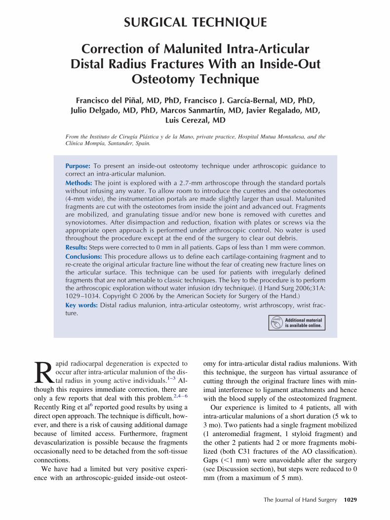
SURGICAL TECHNIQUE
RtoRdebdoc
e
Correction of Malunited Intra-ArticularDistal Radius Fractures With an Inside-Out
Osteotomy Technique
Francisco del Piñal, MD, PhD, Francisco J. García-Bernal, MD, PhD,Julio Delgado, MD, PhD, Marcos Sanmartín, MD, Javier Regalado, MD,
Luis Cerezal, MD
From the Instituto de Cirugía Plástica y de la Mano, private practice, Hospital Mutua Montañesa, and theClínica Mompía, Santander, Spain.
Purpose: To present an inside-out osteotomy technique under arthroscopic guidance tocorrect an intra-articular malunion.Methods: The joint is explored with a 2.7-mm arthroscope through the standard portalswithout infusing any water. To allow room to introduce the curettes and the osteotomes(4-mm wide), the instrumentation portals are made slightly larger than usual. Malunitedfragments are cut with the osteotomes from inside the joint and advanced out. Fragmentsare mobilized, and granulating tissue and/or new bone is removed with curettes andsynoviotomes. After disimpaction and reduction, fixation with plates or screws via theappropriate open approach is performed under arthroscopic control. No water is usedthroughout the procedure except at the end of the surgery to clear out debris.Results: Steps were corrected to 0 mm in all patients. Gaps of less than 1 mm were common.Conclusions: This procedure allows us to define each cartilage-containing fragment and tore-create the original articular fracture line without the fear of creating new fracture lines onthe articular surface. This technique can be used for patients with irregularly definedfragments that are not amenable to classic techniques. The key to the procedure is to performthe arthroscopic exploration without water infusion (dry technique). (J Hand Surg 2006;31A:1029–1034. Copyright © 2006 by the American Society for Surgery of the Hand.)Key words: Distal radius malunion, intra-articular osteotomy, wrist arthroscopy, wrist frac-ture.
otciw
i3(tlG(
apid radiocarpal degeneration is expected tooccur after intra-articular malunion of the dis-tal radius in young active individuals.1–3 Al-
hough this requires immediate correction, there arenly a few reports that deal with this problem.2,4–6
ecently Ring et al6 reported good results by using airect open approach. The technique is difficult, how-ver, and there is a risk of causing additional damageecause of limited access. Furthermore, fragmentevascularization is possible because the fragmentsccasionally need to be detached from the soft-tissueonnections.
We have had a limited but very positive experi-
nce with an arthroscopic-guided inside-out osteot- mmy for intra-articular distal radius malunions. Withhis technique, the surgeon has virtual assurance ofutting through the original fracture lines with min-mal interference to ligament attachments and henceith the blood supply of the osteotomized fragment.Our experience is limited to 4 patients, all with
ntra-articular malunions of a short duration (5 wk tomo). Two patients had a single fragment mobilized
1 anteromedial fragment, 1 styloid fragment) andhe other 2 patients had 2 or more fragments mobi-ized (both C31 fractures of the AO classification).aps (�1 mm) were unavoidable after the surgery
see Discussion section), but steps were reduced to 0
m (from a maximum of 5 mm).The Journal of Hand Surgery 1029
STapplit
ps
wsttmdAsmvirf
pstasbsfiA
Frp
FfaCp
1030 The Journal of Hand Surgery / Vol. 31A No. 6 July–August 2006
urgical Techniquehe arm is exsanguinated with an Esmarch bandage,nd the tourniquet is inflated. Before suspending theatient’s hand, the proposed site of fixation is pre-ared with the patient’s arm on the hand table. Aimited volar–ulnar incision is used for volar shear-ng malunion. A limited Henry approach is used ifhere is a radial styloid or multipiece malunion. A
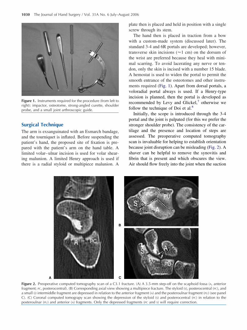
igure 1. Instruments required for the procedure (from left toight): impactor, osteotome, strong-angled curette, shoulderrobe, and a small joint arthroscopic guide.
igure 2. Preoperative computed tomography scan of a C3.1ragment; PC, posterocentral). (B) Corresponding axial view shsmall (I) intermiddle fragment are depressed in relation to the). (C) Coronal computed tomograpy scan showing the dep
osteroulnar (PU) and anterior (A) fragments. Only the depressed frlate then is placed and held in position with a singlecrew through its stem.
The hand then is placed in traction from a bowith a custom-made system (discussed later). The
tandard 3-4 and 6R portals are developed; however,ransverse skin incisions (�1 cm) on the dorsum ofhe wrist are preferred because they heal with mini-al scarring. To avoid lacerating any nerve or ten-
on, only the skin is incised with a number 15 blade.hemostat is used to widen the portal to permit the
mooth entrance of the osteotomes and other instru-ents required (Fig. 1). Apart from dorsal portals, a
oloradial portal always is used. If a Henry-typencision is planned, then the portal is developed asecommended by Levy and Glickel,7 otherwise weollow the technique of Doi et al.8
Initially, the scope is introduced through the 3-4ortal and the joint is palpated (for this we prefer thetronger shoulder probe). The consistency of the car-ilage and the presence and location of steps aressessed. The preoperative computed tomographycan is invaluable for helping to establish orientationecause joint disruption can be misleading (Fig. 2). Ahaver can be helpful to remove the synovitis andbrin that is present and which obscures the view.ir should flow freely into the joint when the suction
re. (A) A 3.5-mm step-off on the scaphoid fossa (A, anteriora multipiece fracture. The styloid (S), posterocentral (PC), andior fragment (A) and the posteroulnar fragment (PU) (see paneln of the styloid (S) and posterocentral (PC) in relation to the
fractuowing
anterressio
agments (PC and S) will require correction.

osttge
om6fwgtttTitpdtmcl
cmvdhtvaN
tsb
tsopu2taHitrs(t(
sIsp
dtt
BStRwrtoadtu
F6or
del Piñal et al / Inside-Out Osteotomy Technique 1031
f the synoviotome or burr is working, otherwise theuction will suck the capsule in, which will obscurehe view. A sufficient amount of air enters throughhe side tube of the arthroscope’s sheath. The sur-eon should keep the valve open throughout thentire procedure.
Once major cartilage destruction has been ruledut (see Discussion section) and the fragments to beobilized are defined, then the scope is moved to the
R portal and the 3-4 and voloradial portals are usedor instrumentation. We use periosteal elevators,hich are available commercially for shoulder sur-ery (Artrex AR-1342-30°, Artrex AR-1342-15°; Ar-rex, Naples, FL) to cut the fragments. These eleva-ors have a 4-mm width and are strong enough to cuthe fragments by gently tapping them with a hammer.o avoid the risk of lacerating extensor tendons when
ntroducing the osteotomes through the 3-4 portal,he blade of the osteotome should be introducedarallel to the tendon. The articular surface first isefined fully with the osteotome and then the os-eotome is advanced to cut the external callus. Gentleaneuvers are necessary because there is a risk of
utting the tendons if the osteotome is plunged vo-arly or dorsally.
The displaced fragments are mobilized fully byarefully prying them out with the osteotome. Inost cases, the fragments are disimpacted and ele-
ated easily by hooking them with the strong shoul-er probe and pulling upward. On one occasion, wead to resort to pushing up the fragment from outsidehe joint by using a Steinmann pin, which was ad-anced into the exact spot with the help of a smallrthroscopic guide (Ref 4291; Dyonics, Smith &ephew Inc., Andover, MA).Many times scarring and new bone formation be-
ween the fragments impedes full reduction. Thishould be resected with the help of small curettes or
igure 3. Correction of a step-off in the same patient as shoR portal viewing radially. The radial styloid is at the verysteotome (entering the joint through the voloradial portal)eduction.
urrs introduced through the portals. Once the reduc- p
ion is thought to be the best possible (Fig. 3), then 1urgeon maintains the bones in position while thether inserts the distal screws of the plate. In 1atient, we used a single screw to secure a volar–lnar fragment and, in another patient, we used a.7-buttress plate to stabilize a styloid fragment. Inhe most common 4-part or more fragments situation,
plate with locking distal pegs is preferred (DVR;and Innovations, Miami, FL). The joint then is
rrigated with 10 to 30 mL of saline that is introducedhrough the scope’s side tube, and a shaver is used toemove all of the debris. The rest of the plate isecured in the standard manner with the hand flatFig. 4). Fluoroscopy controls can be performedhroughout the procedure as many times as requiredsee later).
The portals are closed with paper tape or a singletitch, and the wrist is placed in a removable splint.n all of our patients, the fixation was sufficientlytable as to start protected range of motion on the firstreoperative visit (�48 h).The surgeon may be inconvenienced by 2 factors
uring the procedure: blurred vision and hand posi-ioning. The following technical tips have been foundo be of great help.
lurred Visionplashes of blood or soft-tissue debris may obscure
he surgeon’s vision by sticking to the scope tip.emoving the scope and wiping off the lens with aet sponge is efficacious but time consuming. We
ecommend other alternatives. If debris is obscuringhe surgeon’s vision, then the optic can be cleanedff easily by gently wiping its tip on the capsule orny neighboring soft tissue. Ideally, blood should beried from the surgical field with a sponge. Becausehere is no such thing as an arthrosponge, we havesed small (13 mm � 13 mm) surgical patties for this
Figure 2. The right wrist is shown; the arthroscope is in theper left corner. (A) Step-off on the scaphoid fossa. (B) Thetes the malunited fragments. (C) Corresponding view after
wn infar upsepara
urpose (Ref: 80-04000, size: 0.5 in � 0.5 in;
Nrttidwtiqdmia
CCDscpmmpsfurg
met
DTerfbhtcmflmf
agadedith
F(
1032 The Journal of Hand Surgery / Vol. 31A No. 6 July–August 2006
euray, Xomed, Jacksonville, FL). The patty isolled and introduced into the joint by a grasper. Atimes it can become entangled, and pulling from theail (or with the grasper) can help to remove it. Thiss the method we used most often (including for acuteistal radius fractures). Alternatively the joint can beashed out with 5 mL to 10 mL of water infused
hrough the sheath of the arthroscope while aspirat-ng with a synoviotome. Although effective, this isuite time consuming because the joint has to beried out with a sponge to obtain a clear view. (As inicrosurgery, the field can be either absolutely dry or
mmersed in water; however, working with a smallmount of water tends to be quite annoying.)
hanging Hand Positioning Under Sterileonditionsuring surgery most surgeons9 will place the hand
uspended with the fingers pointing upward. Fluoros-opy is extremely cumbersome with the hand in thisosition; ideally the hand should lay flat. Further-ore during some parts of the surgery the surgeonay want the hand on the operating table, not sus-
ended. Changing the position of the hand from auspended position to a flat position can be per-ormed easily when using a traction tower. If one issing a classic bow, however, the hand cannot beeleased from the bow except by preparing the fin-
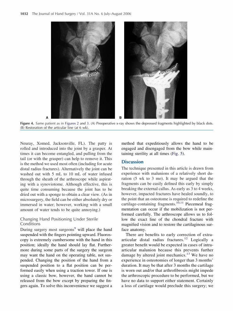
igure 4. Same patient as in Figures 2 and 3. (A) PreoperativB) Restoration of the articular line (at 6 wk).
ers again. To solve this inconvenience we suggest a a
ethod that expeditiously allows the hand to bengaged and disengaged from the bow while main-aining sterility at all times (Fig. 5).
iscussionhe technique presented in this article is drawn fromxperience with malunions of a relatively short du-ation (5 wk to 3 mo). It may be argued that theragments can be easily defined this early by simplyreaking the external callus. As early as 3 to 4 weeks,owever, impacted fractures have healed soundly, tohe point that an osteotome is required to redefine theartilage-containing fragments.10,11 Piecemeal frag-entation can occur if the mobilization is not per-
ormed carefully. The arthroscope allows us to fol-ow the exact line of the chondral fracture withagnified vision and to restore the cartilaginous sur-
ace anatomy.There are benefits to early correction of extra-
rticular distal radius fractures.12 Logically areater benefit would be expected in cases of intra-rticular malunion because this prevents furtheramage by altered joint mechanics.13 We have noxperience in osteotomies of longer than 3 months’uration. It may be that after 3 months the cartilages worn out and/or that arthrofibrosis might impedehe arthroscopic procedure to be performed, but weave no data to support either statement. Certainly
y shows the depressed fragments highlighted by black dots.
e x-raloss of cartilage would preclude this surgery; we

tdFp(
wctnttiWanglge
tmdawa
mcodcwvo
Tdtsiit(shoJgi
aa
1p
Fseppia ill ma
del Piñal et al / Inside-Out Osteotomy Technique 1033
hen would switch to a reconstructive osteochon-ral graft procedure14 or a partial arthrodesis.15,16
or this reason, an exploratory arthroscopy beforelating would be our choice in older malunions�3 mo).
The whole arthroscopic procedure is performedithout water (what we call the dry technique) ex-
ept at the very end to flush out the debris or to clearhe field of blood as explained earlier. The dry tech-ique avoids the cumbersome run-off of waterhrough the (large) portals, the dangerous extravasa-ion to surrounding soft tissues, but more importantlyt is much easier than the standard (wet) technique.
e in fact have moved away from the wet (classic)rthroscopic technique to perform the procedureearly always without water (ulnar head recession oranglion removal can be performed easily). Water isikely to be needed if a laser or electrocautery isoing to be used, but we do not have experience withither of these techniques.
The inside-out technique is a minimally invasiveechnique that allows full assessment of the defor-
ity, accurate osteotomy, and mobilization of theisplaced fragments. Even irregular fragments thatre not amenable to other techniques can be dealtith by this procedure. Correction of step-offs was
igure 5. Method for maintaining sterility when changing fromurgical position, with the hand flat on the table. (A) The hand,ight descender (F8), “carabiner 2” (C #2), and the “bow’s rope curchased at any climbing shop.) The surgeon, under sterile coersonnel. (C) During arthroscopy, only the lower hole of F8 is cndirect contact with C#R. (D) To transfer the hand to a horizongain, the surgeon needs only to fasten C#1 to F8-LOW, which w
chieved in every patient with an accuracy of 0
m; however, residual gaps of about 1 mm wereommon as a result of cartilage loss, interpositionf newly formed bone, and presumably cartilageestruction from the original injury. The procedurean be incorporated easily by any surgeon familiarith arthroscopy (video 1; this video may beiewed at the Journal’s Web site, www.jhandsurg.rg).
he authors would like to thank Mr. Robert Jenkins for his helpuring the English translation of this article. The arthroscopic dryechnique was developed as an input from other specialties andpecialists. The mechanics of a laparoscopy procedure was a flash ofnspiration; no water is used except for diluting blood clots; the waters, however, aspirated as the optic cavity is maintained by air at allimes. The authors would like to recognize Drs. Atzei and Luchettifrom Italy), who shared with us that they were not infusing water inome of the steps of the arthroscopy exploration. Similarly the authorseard Dr. C. Zaidenberg (from Argentina) say at the Annual Meetingf the American Association for Hand Surgery (Cancún, Mexico,anuary 9 –14, 2002) that during arthroscopic resection of wrist gan-lions that no water was used until after the ganglion stem wasdentified.
No benefits in any form have been received or will be received fromcommercial party related directly or indirectly to the subject of this
rticle.Corresponding author: Francisco del Piñal, MD, Calderón de la Barca
6-entlo, E-39002, Santander, Spain; e-mail: [email protected];[email protected].
rtical traction position, suitable for arthroscopy, to a standardith Chinese traps, hangs from “carabiner 1” (C #1), a figure ofer” (C#R). (B) (Carabiners and figure of eight descenders can bens, connects C#2 to C#R, which is held by the operating roomered sterile (F8-LOW) because the other hole (F8-TOP) comes intoition, the surgeon unfastens C#1. To put the hand into tractionintain sterility at all times. All but C#R are sterile.
a veheld warabinnditioonsid
tal pos
Copyright © 2006 by the American Society for Surgery of the Hand

R
1
1
1
1
1
1
1
Ifiao
1034 The Journal of Hand Surgery / Vol. 31A No. 6 July–August 2006
0363-5023/000006/31A06-0025$32.00/0doi:10.1016/j.jhsa.2006.04.005
eferences1. Knirk JL, Jupiter JB. Intra-articular fractures of the distal
end of the radius in young adults. J Bone Joint Surg 1986;68A:647–659.
2. Fernandez DL. Reconstructive procedures for malunion andtraumatic arthritis. Orthop Clin North Am 1993;24:341–363.
3. Trumble TE, Schmitt SR, Vedder NB. Factors affectingfunctional outcome of displaced intra-articular distal radiusfractures. J Hand Surg 1994;19A:325–340.
4. González del Pino J, Nagy L, González Hernandez E, Bar-tolome del Valle E. Osteotomías intraarticulares complejasdel radio por fractura. Indicaciones y técnica quirúrgica. RevOrtop Traumatol 2000;44:406–417.
5. Thivaios GC, McKee MD. Sliding osteotomy for deformitycorrection following malunion of volarly displaced distalradial fractures. J Orthop Trauma 2003;17:326–333.
6. Ring D, Prommersberger KJ, Gonzalez del Pino J, Capo-massi M, Slullitel M, Jupiter JB. Corrective osteotomy forintra-articular malunion of the distal part of the radius.J Bone Joint Surg 2005;87A:1503–1509.
7. Levy HJ, Glickel SZ. Arthroscopic assisted internal fixationof volar intraarticular wrist fractures. Arthroscopy 1993;9:122–124.
8. Doi K, Hattori Y, Otsuka K, Abe Y, Yamamoto H. Intra-articular
fractures of the distal aspect of the radius: arthroscopically assistedreduction compared with open reduction and internal fixation.J Bone Joint Surg 1999;81A:1093–1110.
9. Huracek J, Troeger H. Wrist arthroscopy without distrac-tion. A technique to visualise instability of the wrist aftera ligamentous tear. J Bone Joint Surg 2000;82B:1011–1012.
0. Marx RG, Axelrod TS. Intraarticular osteotomy of distalradial malunions. Clin Orthop 1996;327:152–157.
1. del Piñal F, Garcia-Bernal FJ, Delgado J, Sanmartin M,Regalado J. Results of osteotomy, open reduction, and in-ternal fixation for late-presenting malunited intra-articularfractures of the base of the middle phalanx. J Hand Surg2005;30A:1200–1210.
2. Jupiter JB, Ring D. A comparison of early and late recon-struction of malunited fractures of the distal end of theradius. J Bone Joint Surg 1996;78A:739–748.
3. Wagner WF Jr, Tencer AF, Kiser P, Trumble TE. Effects ofintra-articular distal radius depression on wrist joint contactcharacteristics. J Hand Surg 1996;21A:554–660.
4. del Piñal F, García-Bernal JF, Delgado J, Sanmartin M,Regalado J. Reconstruction of the distal radius facet by afree vascularized osteochondral autograft: anatomic studyand report of a case. J Hand Surg 2005;30A:1200 –1210.
5. Saffar P. Radio-lunate arthrodesis for distal radial intraartic-ular malunion. J Hand Surg 1996;21B:14–20.
6. Garcia-Elias M, Lluch A, Ferreres A, Papini-Zorli I, Rahim-toola ZO. Treatment of radiocarpal degenerative osteoarthri-tis by radioscapholunate arthrodesis and distal scaphoidec-
tomy. J Hand Surg 2005;30A:8–15.Erratumn the article by Dr. Yoshitaka Hamada, et al, that appeared in the April 2006 issue of the Journal (“Effects of monofilament nylon coated with basicbroblast growth factor on endogenous intrasynovial flexor tendon healing.” Vol 31A, No 4, pp 530–540), the authors were listed incorrectly. The correctuthor listing is as follows: Yoshitaka Hamada, Shinsuke Katoh, Naohito Hibino, Hirohumi Kosaka, Daisuke Hamada, and Natsuo Yasui from Universityf Tokushima; and Makoto Ozeki, Yu Kimura, and Yasuhiko Tabata from Kyoto University.
Copyright © 2022 FDOKUMEN