Findings of magnetic resonance imaging of intra-articular ...
56
FINDINGS OF MAGNETIC RESONANCE IMAGING OF INTRA-ARTICULAR KNEE PATHOLOGY: RADIOARTHROSCOPIC CORRELATION i < ( A RETROSPECTIVE AND PROSPECTIVE STUDY OF KNEE MRI FINDINGS AT FIVE RADIOLOGICAL IMAGING CENTRES IN NAIROBI CORRELATED WITH ARTHROSCOPY BY: Dr.^Thiga L.M, University of Nairobi. 2008 DISSERTATION SUBMITTED IN PART-FULFILMENT OF THE DEGREE OF MASTER OF MEDICINE IN DIAGNOSTIC RADIOLOGY, UNIVERSITY OF NAIROBI SUPERVISORS: Dr (Mrs). Wambugu M.N, M.B.Ch.B, M.Med (Radiology), Consultant Radiologist & Senior Lecturer, Department of Medical Imaging and Radiation Medicine, School of Medicine, University of Nairobi. 1 Dr. Owinga S.O, M.B.Ch.B, M.Med (Surgery), Consultant Surgeon, UN1VFR?,TV Qr NAIROBI (Orthopaedic, Trauma & Arthroscopy), MEDICAL LIBRARY Aga Khan University - Nairobi. ISJ ) • - ^ *—-"‘'‘>1.1' O.ML) /
-
Upload
khangminh22 -
Category
Documents
-
view
1 -
download
0
Transcript of Findings of magnetic resonance imaging of intra-articular ...

FINDINGS OF MAGNETIC RESONANCE IMAGING OF INTRA-ARTICULAR KNEE PATHOLOGY: RADIOARTHROSCOPIC CORRELATION i
< (
A RETROSPECTIVE AND PROSPECTIVE STUDY OF KNEE MRI FINDINGS AT FIVE RADIOLOGICAL IMAGING CENTRES IN NAIROBI CORRELATED WITH
ARTHROSCOPY
BY:
Dr.^Thiga L.M, University of Nairobi.
2008
DISSERTATION SUBMITTED IN PART-FULFILMENT OF THE DEGREE OF MASTER OF MEDICINE IN DIAGNOSTIC RADIOLOGY, UNIVERSITY OF
NAIROBI
SUPERVISORS:
Dr (Mrs). Wambugu M.N,M.B.Ch.B, M.Med (Radiology),
Consultant Radiologist & Senior Lecturer, Department of Medical Imaging and Radiation Medicine,
School of Medicine,University of Nairobi.
1
Dr. Owinga S.O,M.B.Ch.B, M.Med (Surgery),
Consultant Surgeon, UN 1V FR ?,TV Qr NAIROBI(Orthopaedic, Trauma & Arthroscopy), MEDICAL LIBRARY
Aga Khan University - Nairobi.
ISJ ) • - ^*—-"‘'‘>1.1' O.ML ) /
DECLARATION
Candidate:
I declare that this dissertation has not been submitted for another degree in this or any other
University or Institution o f Higher learning and that the views expressed herein are mine
unless otherwise stated, and where such is the case acknowledgement or reference has been
quoted.
Signed:......................
Dr. Thiga L.M,
M.B.Ch.B (Nairobi),
Email: [email protected]
Date l5>-
SUPERVISORS:
This dissertation has been submitted for examination with my approval as a University supervisor.
Signed: ..................................................................................D ate.. .Q - Q . / .......
Dr (Mrs). Wambugu M.N,M.B.Ch.B, M.Med (Radiology),Consultant Radiologist & Senior Lecturer,Department of Medical Imaging and Radiation Medicine,School of Medicine,University of Nairobi.
Date % o-J.of
M.B.Ch.B, M.Med (Surgery), Consultant Surgeon,(Orthopaedic, Trauma & Arthroscopy), Aga Khan University Hospital Nairobi.

DEDICATION
7 his w o rs ts dedicated to myfamify: my parents Mr. an d Mrs. Ihiga Machira, my -wife
<Edna andson (DanieC, my sister <Eva, my 6rotfiers 4(am, Jo6 andhisfam iC yfor their Cove,
patience an d encouragement.
Ihey have Seen an d continue to he my inspiration.
iii
ACKNOWLEDGEMENT
This study has been made possible with the assistance of a number of persons to whom I would
like to express my sincere gratitude.
I am indebted to my supervisors, Dr (Mrs). Wambugu M.N and Dr. Owinga S.O for their
continued guidance, suggestions, comments and encouragement throughout the period and
completion of this study.
I would also like to express my gratitude to Dr. Byakika and his staff for his interest in the study
and his assistance in part of the arthroscopic work and Ms A Lakati for her assistance with the
statistical analysis of the data collected during this study.
I also wish to appreciate the contribution of the staffs of The Department of Diagnostic Imaging
and Radiation Medicine of the University of Nairobi, Department of Radiology Kenyatta
National Hospital, Medical Imaging and Therapeutic Center-Nairobi, The Aga Khan University
Hospital-Nairobi, MRI center-Nairobi and The Nairobi Hospital for the assistance they afforded
me during the study.
Finally, I owe special thanks to my family for their encouragement and moral support. Thank you
all for your prayers.
MAY GOD BLESS YOU ALL
IV

TABLE OF CONTENTS
D E C L A R A T IO N ......................................................................................................................................................................................... ii
D E D IC A T IO N ............................................................................................................................................................................................iii
A C K N O W L E D G E M E N T .................................................................................................................................................................... iv
T A B L E O F C O N T E N T S ____________________________________________________________________________________ v
L IST O F A B B R E V IA T IO N S ..............................................................................................................................................................vi
L IST O F F IG U R E S ................................................................................................................................................................................v ii
L IST O F T A B L E S ...................................................................................................................................................................................v ii
L IST O F G R A P H S ________________________________________________________________________________________ v ii
L IST O F PIE C H A R T S ........................................................................................................................................................................ v ii
L IST O F A P P E N D IC E S .......................................................................................................................................................................v ii
S U M M A R Y ................................................................................................................................................................................................v iii
C H A P T E R O N E : IN T R O D U C T IO N A N D L IT E R A T U R E R E V IE W _______________________ 11.1 INTRODUCTION................................................................................................................................................................................ 1
1.2 IMAGING MODALITIES OF THE KNEE JOINT.....................................................................................................................2
1.3 KNEE ANATOMY AND PATHOLOGY AS SEEN ON M R I................................................................................................ 7
1.4 KNEE ARTHROSCOPY................................................................................................................................................................ 20
1.5 DIAGNOSTIC ARTHROSCOPY VERSUS MRI KNEE........................................................................................................21
C H A P T E R T W O : JU S T IF IC A T IO N A N D O B J E C T IV E S .............................................................................................. 222.1 JUSTIFICATION...............................................................................................................................................................................22
2.2 RESEARCH QUESTION......................... 22
2.3 OBJECTIVES.....................................................................................................................................................................................22
C H A P T E R T H R E E : S T U D Y D E SIG N A N D M E T H O D O L O G Y ................................................................................. 233.1 STUDY DESIGN...............................................................................................................................................................................23
3.2 STUDY A REA .................................................................................................................................................................................. 23
3.3 STUDY POPULATION...................................................................................................................................................................23
3.4 SAMPLING........................................................................................................................................................................................ 23
3.5 MATERIALS AND PROCEDURES............................................................................................................................................24
3.6 STUDY LIMITATIONS................................................................................................................................................................. 25
C H A P T E R F O U R : E T H IC A L C O N S ID E R A T IO N .............................................................................................................. 26
C H A P T E R F IV E : R E S U L T S .............................................................................................................................................................275.1 SOCIO-DEMOGRAPHIC CHARACTERISTICS.....................................................................................................................27
5.2 INTRA-ARTICULAR KNEE PATHOLOGY............................................................................................................................ 28
C H A P T E R SIX : D IS C U S S IO N .........................................................................................................................................................37
C H A P T E R SE V E N : C O N C L U S IO N A N D R E C O M M E N D A T IO N S ____________________________________ 417.1 CONCLUSION.................................................................................................................................................................................. 41
7.2 RECOMMENDATIONS................................................................................................................................................................. 41
A P P E N D IC E S ............................................................................................................................................................................................ 43
R E F E R E N C E S _____________________________________________________________________________________________45
v

l ist o f a b b r e v ia t io n s
ACL Anterior Cruciate Ligamenta k u h Aga Khan University Hospital - NairobiCECT Contrast Enhanced CTCM Contrast MediaCT Computerized TomographyDDR Department of Diagnostic RadiologyETL Echo Train LengthFN False NegativeFOV Field O f ViewFP False PositiveFSE Fast Spin EchoGd GadoliniumGE Gradient EchoGRASS Gradient Recalled Accusation in the Steady State SequencesHU Hounsfield UnitsIV Intra VenousKNH Kenyatta National HospitalLCL Lateral collateral LigamentMCL Medial Collateral LigamentMITC Medical Imaging, and Therapeutic CentreMR Magnetic ResonanceMRI Magnetic Resonance ImagingMTC Magnetization Transfer ContrastNECT Non-Enhanced Computerized TomographyNPV Negative Predictive ValuePCL Posterior Cruciate LigamentPD Proton DensityPD-fs Proton Density — fat suppressedPPV Positive Predictive ValueRNI Radionuclide ImagingSE Spin EchoSNR Signal to Noise RatioSPSS Statistical Package for Social ScientistsSTIR Short Tau Inversion RecoveryT1WI T l Weighted ImagesT2WI T2 Weighted imagesTE Time EchoTHK ThicknessTI Time IntervalTN True NegativeTP True PositiveTR Repetition TimeTSE Turbo Spin Echo*UoN University of NarirobiWHO World Health Organization2D Two Dimensionaal3D Three Dimensional
VI

Figure 1: Schematic showing the gross anatomy o f the Knee joint........................................................................................................ 1
Figure 2: Schematic showing menisci and attachments o f the cruciate ligaments on an axial section............................................1
Figure 3: Schematic showing a sagittal section o f the Knee joint.......................................................................................................... 2
Figure 4: Schematic showing a normal meniscus giving ‘bow-tie’ appearance.................................................................................. 8
Figure 5: Sagittal T1W MR image seen through body o f a normal meniscus giving ‘bow-tie’ appearance................................. 8
Figure 6: Sagittal T2W MR image showing a Complex (degenerative) tear...................................................................................... 9
Figure 7: Sagittal T2W MR image showing a Bucket-handle tear with dislocated fragment intercondylar...............................10
Figure 8: Sagittal T2W MR image showing a Flipped lateral meniscus............................................................................................ 11
Figure 9: Sagittal T2W MR image showing a total tear o f the ACL...................................................................................................13
Figure 10: Sagittal PD-fs MR image showing Bone bruise.................................................................................................................... 14
Figure 11: Axial T2W MR image showing chondromalacia patellae...................................................................................................16
Figure 12: Coronal FSE T2W & Axial T2W MR images showing Patellar dislocation..................................................................17
Figure 13: Schematic showing location o f common bursae on the medial side o f the knee............................................................ 19
LIST OF TABLES
Table 1: R outine K nee protoco l.............................................................................................................................................................. 6
Table 2: M edial M eniscus: M RI findings Correlated w ith A rthroscopy...............................................................................29
Table 3: Lateral M eniscus: M RI findings Correlated w ith Arthroscopy................................................................................30
Table 4: Anterior Cruciate Ligament: M RI findings Correlated w ith Arthroscopy......................................................... 31
Table 5: Posterior Cruciate Ligament: MRI findings Correlated w ith Arthroscopy......................................................... 31
Table 6: M edial Collateral Ligament: M RI findings Correlated w ith Arthroscopy...........................................................32
Table 7: Lateral Collateral Ligament: MRI findings Correlated w ith Arthroscopy......................................................... 32
Table 8: Patella: M RI findings Correlated w ith Arthroscopy.....................................................................................................33
Table 9: Femoral articular cartilage: MRI findings Correlated w ith Arthroscopy............................................................. 33
Table 10: Tibial articular cartilage: M RI find ings Correlated w ith Arthroscopy............................................................... 34
Table 11: Joint effusion: MRI findings Correlated w ith Arthroscopy.....................................................................................35
Table 12: Other jo in t pathology: M RI findings Correlated w ith Arthroscopy.....................................................................36
LIST OF GRAPHS
Bar Graph 1: A ge group and Sex distribution o f the Patients w ith intra-articular knee jo in t pathology................. 27
LIST OF PIE CHARTS
Pie chart 1: Duration o f clin ical history before K nee M R im aging and Arthroscopy..................................................... 28
LIST OF APPENDICES
A PPE N D IX 1: D A T A COLLECTION F O R M ............................................................................................................................. 43
l ist o f f ig u r e s
vii

SUMMARY
MRI and arthroscopic findings of 46 knees with varied pathologies were reviewed in a
retrospective - prospective study that covered the period between January 2006 and August 2007.
The Knee magnetic Resonance Imaging was carried out at five Radiological centers providing
MRI services in Nairobi. These centers are. 1) Kenyatta National Hospital (KNH); 2) Medical
Imaging and Therapeutic Centre-Nairobi (MITC); 3) Aga Khan University Hospital-Nairobi
(AKUH); 4) MRI centre-Nairobi and 5) The Nairobi Hospital. The study was carried out in
collaboration with two Orthopaedic and Trauma surgeons carrying out Knee arthroscopic surgery
in Nairobi. There were 30 retrospective and 16 prospective cases collected during the duration of
the study. All the 46 knees included in the study had both MRI findings and corresponding knee
arthroscopic data acquired following diagnostic or therapeutic knee arthroscopic surgery. The
Knee MRI images / reports done before the surgery and the arthroscopic findings of the
corresponding knee formed the basis of this study.
The aim of this study was to determine the pattern of knee joint pathology as seen at five
radiological imaging centres in Nairobi by MRI, the age and sex distribution and correlate these
findings to the arthroscopic findings.
Males (58.7%) were more affected than females (41.3%). Majority of the patients were in the 26-
50 years age group, with the right knee (67.4%) being affected more than the left (32.6%). The
commonest findings encountered were meniscal tears and joint effusions constituting 78.3% and
65.2% of the cases seen respectively. Both showed a male predilection. Other pathologies seen
included chondromalacia patellae, cruciate ligament tears, collateral ligament disruptions, bone
bruises, patella tendinosis, synovitis and intraarticular loose bodies.
MRI showed a high sensitivity in the medial meniscus, where it was sensitive in detecting a tear
in 88.9% of cases. Sensitivity for tears in the lateral meniscus was higher at 93.3%. However,
specificity for a meniscal tear was lower in the medial meniscus than in the lateral meniscus
(36.1% and 63.3% respectively). The sensitivity and specificity of the MRI for anterior cruciate
ligament rupture (whether complete or partial) was 83.3% and 90.9% respectively. For the
articular cartilage, sensitivity was 46.2% and 16.7% for femoral and tibial articular cartilage
abnormalities with a specificity of 81.8% and 75.0% respectively. The sensitivity and specificity
of the MRI for chondromalacia patellae was 26.7% and 81.8% respectively.
The results of this study support the use of MRI in the diagnosis of internal derangements of the
knee. However, it should always be used in conjunction with a full history and clinical
examination.
viii

CHAPTER ONE
1.1 INTRODUCTION
The knee is the largest and most complex joint in the body. It is a synovial hinge joint made up of
three individual joints: two condylotibial and a patellofemoral. It is composed of the articular
margins of the condyles and trochlea surface of the femur, the condyles of the tibia, the patella
(Figure 1), and several bursal structures encased by numerous external and internal ligaments,
joined by an articular capsule (1). The Cruciate ligaments and the menisci are the main internal
structures of the knee joint (Figures 2 and 3). The knee joint is prone to trauma and degenerative
changes and is one of the joints associated with high morbidity especially in sports and in the
elderly.
F amuf
Lateral Kf'OC Ligament
Fibula
Side View of Knee
Figure 1: Schematic showing the gross anatomy o f the Knee joint.
Figure 2: Schematic showing menisci and attachments o f the cruciate ligaments on an axial section o f Upper end of the Tibia: A=Lateral meniscus, B=Popliteus tendon, C=Anterior and Posterior meniscofemoral ligaments, D=Anterior cruciate
ligament, E=Transverse ligament, F=Medial meniscus, G=Posterior cruciate ligament.
1

Figure 3: Schematic showing a sagittal section o f the Knee joint: A=Femoral articular cartilage, B=Posterior cruciate ligament, C=Fibrous capsule, D=Synovium, E=Anterior cruciate ligament, F=Tibial articular cartilage, G=Quadriceps
tendon, H=Suprapatellar bursa, I=Patella, J=Prepatellar bursa, K=Patellar tendon, L=Fat pad, M=Deep infrapatellar bursa.
1.2 IMAGING MODALITIES OF THE KNEE JOINT
MRI has established itself as the investigation of choice for the assessment of the knee and the
identification of internal knee derangements. A variety of imaging modalities are however
available for imaging of the knee. Therefore, both radiologists and the clinicians must be familiar
with the possibilities of each imaging method and its indications as well as contraindications. In
some instances a single modality is not sufficient to provide all the information that is needed,
therefore all methods should be considered complementary. Below is a brief description of the
various imaging modalities available for the assessment of the knee with a special emphasis on
MRI.
1.2.1 RADIOGRAPHY
In most conditions, radiographic examination of the knee joint should be the first step in imaging.
Radiographs of the knee in two planes are mostly sufficient to assess bony lesions, anatomical
reconstruction, bone healing and callus formation and implant position. Conventional
radiography is available in most clinical institutions and it is a low cost and time-sparing
examination. The technique and radiographic projections of the knee depend on the clinical
indications. A large variety of positioning and special projections have been published (2,3). The
standard radiographic positions are anteroposterior (AP) and lateral views in supine position of
the patient with the knee joint in full extension. Other views include the tunnel view which is
2
taken in 40-50° flexion of the knee with the X-ray beam angulated to the same degree in order to
visualize the intercondylar notch. The patella is investigated using an axial view of the patella.
For evaluation of the joint space, radiographs in a posteroanterior (PA) view with the patient
weight bearing in 45° knee flexion may be taken.
1.2.2 CONVENTIONAL ARTHROGRAPHY
The investigation and elucidation of suspected meniscal or cruciate ligament injury provides the
main indication for double contrast knee arthrography (4). However, these are less well
demonstrated than with MRI. Presence or rupture of a popliteal cyst or a loose body can also be
readily confirmed by arthrography. A lateral injection site is the most convenient and is
commonly used, however, a medial injection site can also be used. The patient lies supine and a
19-Gor 21-G needle is introduced 1 cm lateral or medial to the mid-point of the patellar border.
Aspiration of any synovial effusion present is done as completely as possible prior to injection of
contrast medium in order to avoid dilution or bubble formation. A test injection of a small volume
of contrast medium is made under fluoroscopic control to ensure the needle is correctly
positioned in the joint cavity. If a satisfactory position is demonstrated, 3-5mls of positive
contrast medium is injected. This is followed by 40-80.mls of air or carbon dioxide depending on
the capacity of the joint. The needle is then removed and the knee manipulated to ensure even
distribution of contrast medium within the joint. The patient is then placed in the prone position
with a pad under the thigh of the side to be examined. For the purpose of the examination, the
knee is divided into an anterior and a posterior quadrant for each of the medial and lateral
compartments with the X-ray beam collimated to the compartment being examined. For better
visualisation of the compartments, traction with simultaneous valgus or varus strain is applied to
the knee during the examination. A variable degree of flexion may also be required to bring the
tibial plateau and meniscus into profile. Four views of each quadrant are taken, rotating the leg
approximately 20° between each spot view. This results into eight views per meniscus. Small
meniscal tears and meniscal cysts may be better seen on delayed films as contrast may take time
to adhere.
1.2.3 COMPUTED TOMOGRAPHY
Computed tomography (CT) of the knee is particularly suited to demonstrate the bony
morphology, patellofemoral tracking and intraarticular fractures (5). The accurate three-
dimensional localization of the bony anatomy with CT can be used to calculate the mechanical
axis of long bones, rotational deformities of the leg or malrotation of a prosthesis causing
complications after total knee arthroplasty (6). Evaluation of the location and bony remodelling
of the tunnel after anterior cruciate ligament (ACL) reconstruction, which is important in revision3

cases, is possible by CT (7). With newer models of CT scanners, the single transverse sections
can be reformatted to coronal and surface-rendered 3D-reconstruction producing graphic images,
which improves spatial assessment of pathologies. Soft tissue pathologies, however, are less well
demonstrated than with MRI.
CT arthrography may be used as an alternative to MRI in many instances. CT arthrography is a
valuable alternative to MR imaging for the assessment of internal derangements of the knee. In
comparison with MR imaging, it is more invasive because of the intra-articular injection of
contrast material and the use of ionizing radiation. Its ability for detecting meniscal, ligamentous
and cartilaginous lesions is derived from its superior spatial resolution, multiplanar capabilities,
and the high-contrast resolution inherent to the intra-articular injection of contrast material (8,9).
Advantages of CT arthrography include short examination time, high spatial resolution, and
multiplanar capability and reduced imaging artefacts because of the presence of metallic
hardware and debris, which may hinder MR imaging studies (10,11).
In CT arthrography, intra-articular injection of 15-20 mL of iodinated contrast material is
performed under fluoroscopic guidance. A small volume of epinephrine (0.1 mL of a 0.1%
solution) is also added to the injection to delay synovial absorption of contrast material from the
joint (12,13). This is followed by active knee flexion and extension. This raises intra-articular
hydrostatic pressure and helps to force the contrast material into all the recesses of the knee,
including meniscal tears (14).
The presence of contrast material around the menisci enables assessment of meniscal integrity.
Contrast material is normally present between the anterior horn of the lateral meniscus and the
transverse meniscal ligament, or between the posterior horn of the lateral meniscus and the
meniscofemoral ligaments. There should not be any contrast material between the capsule and the
periphery of the medial meniscus (8). Meniscal pathology is diagnosed when contrast material is
noted within the meniscal substance or when the meniscal contour is deformed.
1.2.4 ULTRASONOGRAPHY
Ultrasound in conjunction with the history and clinical examination can prove to be a simple,
noninvasive, cost effective, real time, dynamic and effective modality to assess the tendons,
ligaments and meniscus around the knee joint. A high frequency broadband linear transducer of at
least 7.5 MHz is used. The greatest advantage of ultrasound however is for the detailed evaluation
of the soft tissues within and surrounding the knee. These include ligamentous, tendinous,
fibrous, fatty, synovial, and neurovascular structures. These tissues can be assessed for size,
continuity, anatomic orientation, and echogenicity. Furthermore, they can be readily compared
contralaterally. Evaluation of intra-articular elements such as the menisci, cruciate ligaments, and
4

articular cartilage is limited mainly by inaccessibility, which results from the small acoustic
windows obtainable in most patients.
1.2.5 RADIONUCLIDE IMAGING
Radionuclide imaging in the form of single photon emission computed tomographic (SPECT)
bone scintigram also has a role in imaging knee disorders and seems to compare favourably with
MRI in diagnosis of internal derangement of the knee (15,16).
Bone scintigraphy allows investigation of contralateral knee and other joints at the same time. It
also provides physiological detail unlike other imaging modalities. The conventional planar
scintigraphy uses ionizing radiation and gives a very high resolution but poor anatomical detail of
the knee lesions. With advent of the SPECT scintigraphy and its ability to image in coronal,
sagittal and transaxial planes, localization of knee lesions in overlapping structures is possible.
Technically, " mTc methyl diphosphonate is injected intravenously and a two-phase bone scan is
acquired followed by SPECT scintigram. Anterior and posterior blood pool images and four static
views in anterior, posterior, medial and lateral planes are obtained. SPECT scintigram is
performed after securing the knees with a band keeping the legs straight. In general 360° elliptical
orbit is used. Based on the type of equipment, individual departments use filters and protocols
best suited to obtain optimal studies.
MRI involves no use of ionizing radiation and gives relatively good anatomical details. It has thus
established itself as the investigation of choice in identifying internal knee derangements.
However, bone scintigraphy and magnetic resonance imaging (MRI) are complementary in
diagnosing knee lesions and where MRI is contra-indicated, bone scintigram has established itself
as an alternative.
1.2.6 MAGNETIC RESONANCE IMAGING
In the developed world, MRI has become an established imaging technique of the knee joint. In
order to examine soft tissue, cartilage and ligamentous structures, a large number of MRI
scanning methods have been described. Images are obtained on the sagittal, coronal and axial
planes. The optimal data acquisition obtains a maximum amount of information in the shortest
period of time without compromising image quality. Metal implants or prostheses do not present
an absolute contraindication for MRI, however, they may heat up in the magnetic field and thus
constitute a high risk for patients. The following image contrast and pulse sequences are often
applied:
• T1-weighted spin echo sequences,
• Proton density-weighted spin echo images,
5

. T2-weighted spin echo sequences,
. Gradient echo sequences,
• Fast spin echo sequences,
• Fat suppression techniques (fat saturation and inversion recovery).
There is no standard protocol and most institutions establish a routine protocol, which provides
adequate visualization and answers the majority of questions. However, the choice of slice
thickness, field of view, coil, and imaging matrix play an important role in determining spatial
resolution. Improved spatial resolution provides the radiologist with data to present interpretation
with confidence.
A typical Knee MRI protocol will have a three plane localizer, Axial PD, Coronal PD/T2,
Sagittal PD/T2, Sagittal PD-fs and a Coronal STIR. A protocol, such as the one outlined below
(Table 1), seems to address most common requests. Most MR system manufacturers now offer a
dedicated knee coil, which is often a transmit-receive coil as standard. These coils use a
cylindrical configuration, similar to the head coil, to provide a homogenous imaging volume, and
a quadrature design that provide improved signal to noise ratio. Flexible surface coils are used as
an alternative when the knee joint is too large to fit in the rigid knee coil or when the patient is
unable to extend the knee, usually following trauma. Spatial resolution is necessary to image the
small structures that are found in the knee and the Field of View (FOV) should be kept small
ideally not exceed the length of the coil. The range of FOV in the supero-inferior length is of 160-
200mm. Using a matrix of 256, allows an in-plane resolution of less than 1mm. Thin slices in the
range of 3-4mm and gap of not more than 0.5mm ensure high spatial resolution. 3D acquisition
using isotropic matrix is useful to provide high-resolution visualization of anatomy in any plane.
TRmsec
TEmsec
TI Matrix NEX ETL FOV THK SAT Time
Axial PD 2775 20 384x224 2 16 16 3/0.4 S,1 2:46
Coronal PD/T2 2500 25/102 384x256 2 8 16 4/0.4 S,I 5:25
Sagittal PD/T2 2500 25/100 384x256 2 8 16 4/0.4 S,I 5:47
Sagittal PD-fs 3275 20 384x224 3 8 16 3/0.4 S,I Fat 4:41
Coronal STIR 4775 SO 150 256x192 2 10 16 4/1 S,I 5:05
Table 1: Routine Knee protocol.
6

Intravenous (IV) contrast may be indicated in knee joint MR imaging especially in the delineation
of bone tumours, scar tissue in repeat surgery and repeat trauma. Magnetic resonance
arthrography is practiced in many imaging centres for the diagnosis of meniscal tears and
chondral defects. This is done by introducing a dilute solution of gadolinium in saline (1.1000)
into the joint capsule. The knee is then imaged in three planes using T1W or PD weighted both
fat suppressed. Indirect arthrography could be used where the synovial fluid within the joint
enhances 15 minutes post IV administration of gadolinium. This occurs due to slow spread of the
contrast from the synovial vascular network to the mtraarticular surface of the membrane and
thus into the intraarticular cavity where its concentration remains high for about 1 hour. This
increases conspicuity of meniscal tears.
1.3 KNEE ANATOMY AND PATHOLOGY AS SEEN ON MRI
1.3.1 MENISCI
The Menisci are embryological remnants of the discs commonly found at condylar joints. They
are fibrocartilaginous structures that allow motion in all directions, and increase stability by
deepening the socket in which the femur moves. Each knee has two menisci (a lateral and a
medial) that primarily follow the movement of the femoral condyles. Each of these menisci is C-
shaped and has its largest dimensions in the axial plane. Each meniscus covers about two-thirds
of its corresponding tibial articular surface. The lateral meniscus is smaller than the medial
meniscus and has an increased curvature and thus a shorter radius (Figure 2 on page 1). For ease
of diagnostic interpretation, the meniscus is divided into three parts (anterior, middle and
posterior thirds). The meniscus is further subdivided radially into the inner edge, middle and
penpheral regions.
The collagen bundles within each meniscus form two distinct zones. Circumferential fibres are
found in the peripheral third of the meniscus, whereas the transverse collagen fibres connect the
circumferential zone to the meniscal free edge. In the adult, the peripheral 10-20% of the
meniscus receives a blood supply from the capillary plexus that surrounds it, but the central
portion of each meniscus is relatively avascular. Because of this vascularization difference, tears
may heal spontaneously, especially in young patients, and suturing of meniscal tears in this
vascularized zone is an option instead of partial resection.
Menisci are triangular shaped when imaged in a plane perpendicular to the axial plane. On MR
images normal menisci have low signal intensity, or even signal void. Signal intensity may be
somewhat increased in the peripheral vascular zone, especially in children. The signal intensity is
higher on gradient echo (GE) images than on spin echo (SE) images. In MRI the menisci are best
viewed in the sagittal and coronal planes. In the sagittal plane, the normal menisci appear as7

homogenous low signal structure on all sequences, and have a triangular structure with an outer
convex border (Figures 4 and 5).
Figure 4: Schematic drawing of a normal meniscus on the left. Sagittal cuts through A and B show how consecutive sagittal MR images (A and B) should be seen through body o f meniscus giving ‘bow-tie’ appearance.
Figure 5: Sagittal T1-weighted MR image seen through body o f a normal meniscus (arrows) giving ‘bow-tie’ appearance.
DISCOID MENISCI
There exist a large variety of abnormal meniscal shapes. Menisci may be completely or partially
discoid. Typically these are found in children or adolescents, more commonly on the lateral, but
also on the medial side. They have a higher incidence of tears. Discoid menisci can be diagnosed
on sagittal images. More than two connecting bow ties on 3-5 mm-thick slices indicate the
presence of a discoid meniscus. It is, however, easier to make this diagnosis on coronal images.
8

M EN ISC A L t e a r s
Meniscal tears are best seen on short echo time (TE) sequences, such as conventional SE T l-
weighted or proton-density weighted images or GE sequences. When a conventional SE sequence
is used, a TR of 2000 ms or more and a TE of 20 ms is a good set of parameters. Slice thickness
can be 5 mm or preferably less. Tears can also be seen on T2 STIR-weighted, or T1-weighted fat-
suppressed GE images, but the risk of false positives is higher than with SE or fast SE (FSE)
sequences. There has been a lot of debate about the accuracy of turbo SE (TSE) relative to SE
sequences (17,18). Although conventional SE is probably superior to TSE, the latter will suffice
for routine clinical imaging if TE and echo train length (ETL) are short, and if advanced sequence
optimisation such as tailored radio frequency pulses (reduced blurring) is used. This means an
ETL of 5 or less and a TE of maximal 20 ms.
A grading system is used to describe meniscal abnormalities (19). Grade 1 represents histological
mucoid degeneration and is seen as a globular shape of abnormal signal intensity not abutting the
surface (Figure 6). Grade 2 represents histologically more severe degeneration and collagen
fragmentation. It is seen as a linear configuration of abnormal signal intensity not abutting an
articular surface. Grade 3, histologically corresponding to a real tear, is a linear (Grade 3A) or
irregularly (Grade 3B) shaped abnormal signal intensity abutting an articular margin. Some prefer
to grade a severely fragmented meniscus as a grade 4.
Figure 6: Sagittal T2W MRI showing a Complex (degenerative) tear (arrow) in posterior horn o f medial meniscus.
9
.

jhere are many descriptive terms for the various types of tears. The most common and most
important ones are:
a) The bucket-handle tear: This is a complete, in the axial plane-oriented vertical tear with
displacement of one or two meniscal fragments. The inner segment is the handle of the bucket,
which may dislocate into the intercondylar fossa (Figure 7). When the inner part of the meniscus
is dislocated or when it has been removed at arthroscopy, the inner margin of the meniscus lacks
the pointed shape on sagittal and coronal images. Tom meniscal fragments may rotate and
dislocate anteriorly; this is the so-called flipped meniscus sign (Figure 8).
b) Radial tears: are located on the inner margin, and are difficult to detect. A subtle high-signal
intensity line may be observed on sagittal images, or an irregular inner margin is seen on coronal
images.
Figure 7: Sagittal T2W MRI showing a Bucket-handle tear with dislocated fragment intercondylar (arrow). The ACL isintact.
10

Figure 8: Sagittal T2W MRI showing a Flipped lateral meniscus: the posterior horn o f the lateral meniscus is partiallydislocated (arrow).
The sensitivity of MRI in the diagnosis of medial meniscal tears is reported to be 87-97%,
sensitivity for lateral meniscal tears is 69-92%; specificities are 82-91% and 91-98%
respectively (20-32). Thus, the performance of MRI is clinically excellent especially if one takes
into account that small peripheral tears missed by MRI are not clinically relevant, and that
arthroscopy is not an ideal gold standard. When the anterior cruciate ligament (ACL) is tom,
accuracy for meniscal tears decreases and especially tears in the posterior lateral meniscus may
be missed.
MENISCAL CYSTS
These are frequently seen in association with horizontal meniscal tears on the lateral side of the
knee. The dissecting meniscal tear is contiguous with the fluid collection. The signal intensity
may be lower than typical for fluid because of dehydration. MR arthrography using gadolinium
improves their delineation. They are usually small, but may be large, multiloculated, and may
erode bone
1.3.2 LIGAMENTS
a) COLLATERAL LIGAMENTS - these are on each side of the joint and strengthen the
capsule. The medial collateral ligament extends from the medial condylar region and attaches 4-
5cm inferior to the tibial plateau and posterior to the pes anserinus insertion. Deep to this, the

medial capsular ligament is composed of the meniscofemoral and meniscotibial attachments to
the meniscus. The ligament represents the main restraint against valgus strain. The lateral
collateral ligament (LCL) strengthens the capsule laterally, and lies somewhat posterior. LCL is
made up of two layers and lies deep to the insertions of the distal iliotibial tract and biceps
femoris posteriorly, and to the quadriceps retinaculum anteriorly. Deep to the LCL are the
meniscofemoral and meniscotibial attachments. The intracapsular popliteus tendon passes medial
t0 the LCL, and posterior fibres of the LCL blend with the deep capsule, which contributes to the
arcuate popliteal ligament. The collateral ligaments consist of multiple layers: the deep lying
meniscofemoral and meniscotibial ligaments are capsule reinforcements. Superficial to this are
the tibial collateral and fibular collateral ligaments. Between the deep and more superficial layers,
fat and bursa may be visualized.
Injury to the medial ligament is much more common than that to the lateral ligament. Tears and
meniscocapsular separations are best evaluated in the coronal plane on T2-weighted SE, T2-
weighted TSE (with fat-selective saturation) or T2 STIR-weighted GE images. Coronal images
are more accurate than axial images for grading injuries of the medial and lateral collateral
ligaments. MRI may show high signal intensity within and or surrounding the traumatized
ligament. The ligament may be thickened in chronic injuries, or interrupted and or buckled in
acute complete tears. Distraction bone bruises, located at the level of ligamentous insertions, are
frequently seen in severe injury (33).
b) CRUCIATE LIGAMENTS - are intracapsular but extrasynovial. The anterior cruciate
ligament (ACL) connects the posterior medial aspect of the lateral femoral condyle to the anterior
tibial intercondylar region. It is composed of two functional fibre bundles: the longer
anteromedial bundle tightens with knee flexion, and the shorter but bulkier posterolateral bundle
which tightens with knee extension.
The posterior cruciate ligament (PCL) connects the lateral aspect of the medial femoral condyle
to the posterior intercondylar fossa of the tibia, and passes medial to the ACL. In relation to the
PCL two other ligaments may pass anterior (ligament of Humphrey) or more commonly posterior
(Ligament of Wrisberg). In some knees, both ligaments are present.
The cruciate ligaments are, as central supporting structures, major contributors to stability.
Complete ACL tears are easily diagnosed clinically by use of clinical tests like the anterior
drawer test at 30° and 90°, the Lackman’s test and the pivot shift test in the elderly. On MRI,
some indirect signs may be used to diagnose an ACL tear. When the posterior horn of the lateral
Meniscus is at the level of, or even posterior to the posterior margin of the lateral tibia plateau,
this indicates that the tibia is not restricted by the ACL and is allowed to move anterior relative to
the meniscus and femur. The meniscus moves with the femur as one unit. The Cruciate ligaments
12
are best visualized in the sagittal (0-10°external rotation) plane. The coronal plane offers an
important second look at the cruciate ligaments.
On sagittal images the ACL is normally parallel to the intercondylar roof. On MRI there is often
high signal intensity interspersed within the fibres of the ACL. The ACL consists of multiple
collagen bundles that are twisted around each other like the fascicles of a rope; this organization
of collagen explains the intermediate signal intensity on MR images. A second contributor to the
signal is the magic-angle phenomenon. Collagen bundles that make an angle of 55° with the main
field have increased signal intensity when TE is short.
The posterior cruciate ligament (PCL), a thicker and more homogeneous structure, has as a
consequence lower signal intensity. Because knee MRI is performed in extension the PCL
normally has a sharp bend known as the genu. The PCL is typically more uniformly hypointense
than the ACL, although there may be slight increased signal intensity on short TE secondary to
magic angle effect.
Acute tears are seen as loss of continuity, as an ill-defined mass, retraction of torn ends, concavity
of the anterior margin or increased signal intensity within the ligament. Most tears are mid
substance tears. In proximal tears an empty notch sign may be seen. This sign describes the
presence of fluid, instead of ligament, seen on coronal images at the side of origin at the inner
aspect of the femoral condyle. Because haematoma and oedema resolve in time, a sub-acute or
chronic tear is better defined than an acute one (34). The chronically torn ACL may be absent or
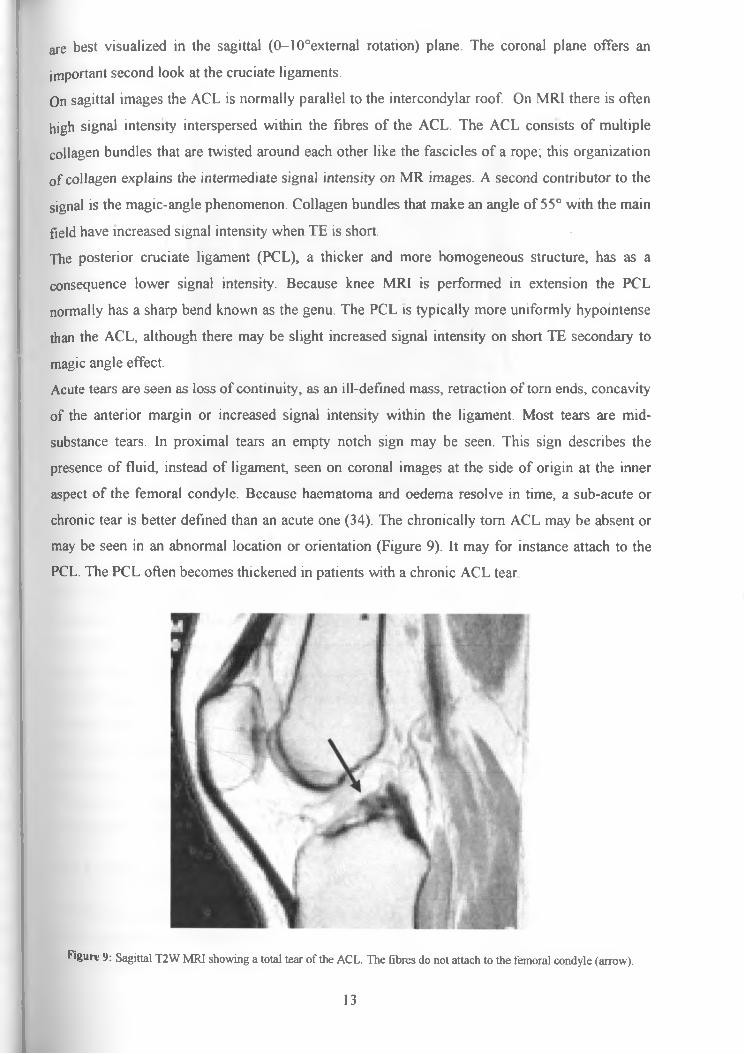
may be seen in an abnormal location or orientation (Figure 9). It may for instance attach to the
PCL. The PCL often becomes thickened in patients with a chronic ACL tear
Figure 9: Sagittal T2W MR] showing a total tear o f the ACL. The fibres do not attach to the femoral condyle (arrow).
13
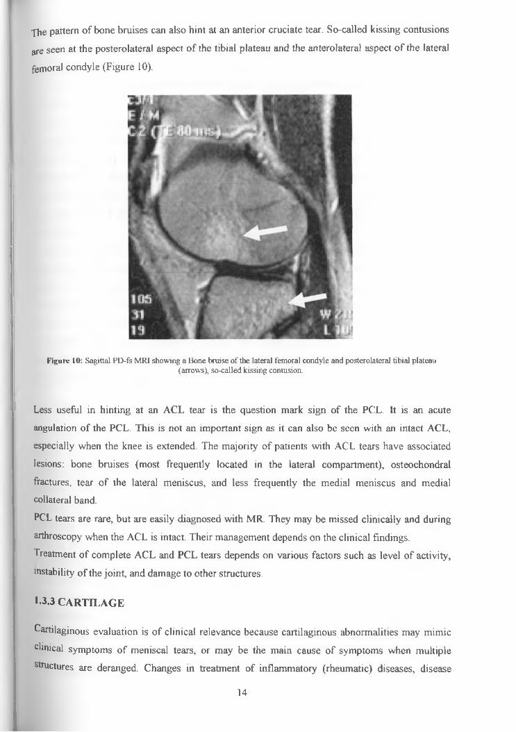
The pattern of bone bruises can also hint at an anterior cruciate tear. So-called kissing contusions
are seen at the posterolateral aspect of the tibial plateau and the anterolateral aspect of the lateral
femoral condyle (Figure 10).
Figure 10: Sagittal PD-fs MRI showing a Bone bruise o f the lateral femoral condyle and posterolateral tibial plateau(arrows), so-called kissing contusion.
Less useful in hinting at an ACL tear is the question mark sign of the PCL. It is an acute
angulation of the PCL. This is not an important sign as it can also be seen with an intact ACL,
especially when the knee is extended. The majority of patients with ACL tears have associated
lesions: bone bruises (most frequently located in the lateral compartment), osteochondral
fractures, tear of the lateral meniscus, and less frequently the medial meniscus and medial
collateral band.
PCL tears are rare, but are easily diagnosed with MR. They may be missed clinically and during
arthroscopy when the ACL is intact. Their management depends on the clinical findings.
Treatment of complete ACL and PCL tears depends on various factors such as level of activity,
instability of the joint, and damage to other structures.
1-3.3 CARTILAGE
Cartilaginous evaluation is of clinical relevance because cartilaginous abnormalities may mimic
clinical symptoms of meniscal tears, or may be the main cause of symptoms when multiple
structures are deranged. Changes in treatment of inflammatory (rheumatic) diseases, disease
14

modifying drugs in osteoarthritis and renewed interest in surgical repair such as debridement with
abrasion of subchondral bone, osteotomy to modify loading characteristics, placement of artificial
matrix, perichondreal grafts, chondrocyte transplantation, and osteochondral transplantation have
also made diagnosis of cartilaginous disorders rapidly gain significance. In inflammatory disease
MR mainly focuses on synovial disorders characterized by enhancement features, while cartilage
itself is the main target in case of trauma or osteoarthritis.
Traumatic chondral defects are typically well-defined, large and (near) full thickness with
subchondral osseous injury, which facilitates detection. Assessment of cartilage is important in
differentiating between stable osteochondritis dissecans with intact overlying cartilage, and
unstable osteochondritis dissecans with disrupted overlying cartilage. Patients with intact
cartilage are preferably treated conservatively, whereas surgery is used when overlying cartilage
is disrupted.
In osteoarthritis, the collagen network becomes disrupted and proteoglycan content decreases
resulting in swelling representing increased water content. There is no spontaneous healing of the
hyaline cartilage and ensuing repair response results in thickened fibro-cartilage or may lead to
cartilage loss like fibrillation, erosions, or even cracks. These later stages of this irreversible
process can be visualized under certain conditions using MRI with variable accuracy. The range
of accuracies reported in literature is large and ranges from 50% to 80%. Both the joint
component (cartilage loading) and the subchondral bone component play an important role in the
development of osteoarthritis.
1.3.4 BONE
Bone bruise was introduced as a new radiologic diagnosis with the advent of MRI (35, 36). Bone
bruises may be isolated, but are also often encountered in association with fracture of the
cartilage (osteochondral fracture, osteochondritis dissecans) and or with meniscal and ligament
sprains or tears. Bone bruises or radiographically occult fractures are significant MR findings
because they may be career-ending injuries especially in professional athletes when not
diagnosed. Patients with isolated bone bruises have decreased function and more symptoms
relative to those without this pathology. Symptoms disappear and function returns with
conservative management within 6 months (37). Cartilage and bone marrow is best assessed in all
three planes using fat suppressed sequences and T1 -Weighted images. Muscle signal is similar to
other parts of the musculoskeletal system.
When properly diagnosed with MR, arthroscopy can be avoided. Contusion of the posterolateral
t'bial plateau is one of the commonest bruises encountered (Figure 10 on page 14) and is
characteristically associated with an ACL tear. Another location associated with ACL tear is
compression fracture including bone bruise at the level of the femoral notch. Bone bruises can
15

on|y be excluded on short tau inversion recovery (STIR) or fat selectively suppressed T2-
vveighted TSE sequences, as these sequences have superior sensitivity over other sequences.
1.3.5 PATELLA
MRI is useful in the diagnosis of dislocation of the patella that may be associated with patella
chondral defects. It is also useful in demonstrating degenerative changes of patella articular
cartilage (Figure 11). Patella dislocation often rapidly reduces on its own and only half of the
patients with patella dislocation are aware of them. Findings on MR are easily interpreted as a
patella dislocation (38). A contusion characteristically occurs on the anterolateral femoral
condyle (Figure 12). It results from the impaction of the patella as it either dislocates or reduces.
There may be a kissing contusion on the medial side of the patella and on the anterolateral
femoral condyle. In patella dislocation, the medial retinaculum (Medial patello-femoral ligament)
is always injured, although it may be difficult to appreciate a frank tear. A key finding is the
patella cartilage. If a piece of the cartilage is missing, an arthroscopy procedure is necessary
whereas if normal, the patient is treated conservatively. The main role of the radiologist is to
carefully examine the patella cartilage and to evaluate the trochlear notch, which is often
hypoplastic in patients with dislocating patellae and is a predisposing factor to subsequent
dislocations.
Figure 11: Axial T2W MR image showing a large chondral defect (arrows) on the apex o f the patella (chondromalaciapatellae).
16

Figure 12: Patellar dislocation, a) Fast spin-echo T2W coronal MRI with fat suppression reveals large contusion in lateral femoral condyle, b) Axial T2W image shows large joint effusion with large chondral defect (arrow) on apex o f patella.
1.3.6 PATELLA TENDON
Pain in the inferior patellar region in athletes may be attributed to patella tendinosis, which is
often seen in MRI as thickening of the proximal patella tendon with high signal in and around it
on T2-weighted images (39). Patella tendinosis is an important diagnosis that may be missed on
the MRI if not actively looked for. It can be debilitating to an athlete and can require surgery to
remove the focus of myxoid degeneration in the tendon. In inflammatory disease, MR
arthrography mainly focuses on disorders characterized by enhancement features. There is a role
for the use of contrast media (gadolinium) in the investigation for Patella tendinosis.
1.3.7 SYNOVIAL PLICAE
Embryologically, the knee is divided into compartments by superior, inferior and medial patellar
plicae. The plicae are fibrous remnants of the embryological development of the knee derived
from the synovial membrane (40). Half of all knees show one or more of the plicae on MRI. The
range of reported medial plicae in the literature is large and ranges from 18.5% to 55% with only
less than 3% being symptomatic. It may be thickened, stiff and trapped between the patella and
the femur and cause pain, clicking and locking like a tom meniscus (so called Plica syndrome).
Axial T2-weighted images showing joint fluid are required to visualize the medial plica. Plica
syndrome is an uncommon diagnosis but an inflamed plica is easily removed at arthroscopy. The
SuPrapatellar and infrapatellar plicae are also commonly seen on MRI but do not cause plica
syndrome. However, at arthroscopy, the ligamentum mucosum (plica infrapatellaris) may be
17

confused with the anterior cruciate ligament, which runs almost parallel with it. Occasionally, the
ligamentum mucosum makes it difficult to change from one compartment to another.
Ligamentum mucosum is of no importance and can be resected if necessary.
1.3.8 SYNOVIAL SPACE AND BURSAE
A synovial membrane, articular cartilage and menisci surround this space. It is variable and
depends on the position of the knee. The synovial membrane of the knee bulges upward from the
patella to form the suprapatellar bursa situated beneath the quadriceps tendon. Inflamed bursae
may cause symptoms that mimic intraarticular abnormality and result to inappropriate therapy
including surgery. Bursae around the knee will not be seen on arthroscopy and therefore MR1 or
physical examination must be used to identify them. Four of the important bursae are described
below;
a) Popliteal bursa or Baker’s cyst: An inconstant posterior pouch extends into the popliteal
space in about 13% of knees. When this synovial membrane pouch is obstructed or the bursa
becomes inflamed, the bursa is called a ‘Baker’s cyst’. It extends from the knee joint posteriorly
between the tendons of the medial head of the gastrocnemius and the semimembrinosus (Figure
13 on page 19). It can contain some small amounts of fluid but greater than 5-10ml of fluid
should be reported because it could be a source of symptoms. The bursae may become quite large
and cause compartment syndrome, or may extend far down the leg and may rupture and cause
inflammation of the surrounding musculature (mimics deep venous thrombosis clinically).
b) Prepatellar bursitis: It occurs anterior to the patella (Figure 3 on page 2) and is one of the
common causes of anterior knee pain. Its cause is repetitive trauma and kneeling. On MRI, it is
shown as a fluid collection superficial to the patella.
c) Pes anserinus bursa: It occurs on the anteromedial tibia just below the joint line (Figure 13 on
page 19). The pes tendons are the gracilis, sartorius and the semimembrinosus. The bursa lies
beneath the tendons and when inflamed, it extends proximally towards the joint (41).
d) Semimembrinosus tibial-collateral bursa: It may mimic an internal knee derangement (42).
It is commonly inflamed and has a characteristic appearance hence easily picked on MRI. It
occurs on the joint line and drapes over the semimembrinosus tendon like a horseshoe (Figure 13
on page 19). On coronal and sagittal images, it appears to arise at the meniscus and extending
■nferiorly (DDX: meniscal cyst but this has a connection to a meniscal tear).
e) Tibial-collateral ligament bursa: This bursa is uncommonly seen. It lies deep in relation to
the MCL and extends vertically above and below the joint line. The bursa can be confused with a
18

nieniscocapsular separation, but unlike a traumatic separation, the fluid is well contained and cyst
like rather than diffusely distributed.
Figure 13: Schematic showing location o f common bursae on the medial side o f the knee (Anterior is to the left): A=Tibial collateral ligament, B=Pes Anserinus bursa, C=Baker’s cyst, D=Semimembrinosus tendon, E=Semimembrinosus tibial-
collateral-ligament bursa.
1.3.9 EXTENSOR MECHANISM
This is a structure that consists of the quadriceps tendon, the patella, the patellar ligament,
Hoffa’s fat pad, and the medial and lateral patellar retinacula. The quadriceps tendon is made up
from the rectus femoris, vastus lateralis, vastus medialis and vastus intermedius.
19

1.4 KNEE ARTHROSCOPY
Knee arthroscopy allows an orthopaedic surgeon to diagnose and treat knee disorders by
providing a clear view of the inside of the knee with small incisions, utilizing a pencil-sized
instrument (Usually 4-5mm in diameter) called an arthroscope Modem or contemporary
arthroscopy of the knee was first performed in the late 1960s. With improvements on
arthroscopes and higher-resolution cameras, the procedure has become highly effective for both
the accurate diagnosis and proper treatment of knee problems.
Today, the arthroscope in common use consists of a Telescope with laterally attached light cable.
Arthroscopes with the universally accepted angulation (30° for the standard scopes and 70° for
special indications), in combination with the option to advance the tip of the scope into more
distant areas of the joint, allows inspection of almost all intraarticular structures. A sheath with a
sharp and a blunt trocar is used to introduce the instrument into the joint to protect the optical
system from bending and breaking. The sheath also contains the irrigation port for gas or liquids.
Usually the arthroscope is attached to a video monitor that the surgeon uses to see the inside of
the knee. The video monitor also aids in maintaining sterility during the arthroscopic surgery.
Knee arthroscopy is one of the most common orthopaedic procedures especially in the developed
countries used to diagnose and or treat many of the following problems:
a) Indications for diagnostic arthroscopy;
• Diagnostic uncertainty or uncertainty about the exact location of meniscal tears and or
persistent knee pain.
• Follow-up of treatment results.
b) Indications for surgical arthroscopy grouped by structure;
• Synovium - biopsy, synovectomy, lysis of adhesions, lateral release and division of plica.
• Synovial space - lavage and removal of loose bodies.
• Intra-articular structures - menisectomy, suture of meniscus and repair of ligaments (ACL
and PCL).
• Cartilage - shaving of the patella, refixing an osteochondritis dissecans fragment and
chondroplasty.
• Bone - debridement and chondroplasty (mesenchymal stimulation techniques).
Portals are the key to success in arthroscopy. They define the access a surgeon will have to both
view the joint and to intervene in the joint. Proper portal placement is essential to successful knee
arthroscopy, as it defines the surgeon's access to the joint. Numerous approaches have been
described in the literature, from the so-called "universal" antero-lateral portal to a posterior trans-
septal portal. Although the various named portals are described as fixed locations, they are
adjustable to address the pathology at hand while minimizing any impact on articular cartilage.20
planning for portal selection is based on the purpose of the arthroscopy. Whether diagnostic or
therapeutic, the portals are planned to optimize the surgeon's access to the relevant area(s) of the
knee.
Most orthopedic surgeons use a standard 3-portal technique (the antero-lateral, antero-medial, and
supero-medial portals), although 2-portal technique is gaining popularity. Additional portals may
be used based on the needs of the arthroscopist. These additional portals include postero-medial,
supero-lateral, postero-lateral, midpatellar, posterior trans-septal and central portals. The
indications for specific portals are based on the access a surgeon expects to need in a particular
arthroscopy.
1.5 DIAGNOSTIC ARTHROSCOPY VERSUS MRI KNEE
MRI of the knee is increasingly becoming available as an alternative to diagnostic arthroscopy.
Where MRI use in medical practice is well established, MRI has now tremendously reduced the
number of diagnostic arthroscopies being performed, and it is more appropriately being used as a
screening tool for therapeutic arthroscopy. Fast three-dimensional MRI allows identification of all
relevant intra-articular pathologies of the knee joint within a few minutes, with high accuracy
comparable to arthroscopy (43).
Where studies have been done, MRI has proven to be accurate for the diagnosis of intra- and peri
articular pathology, especially for meniscal pathology and ligamentous injuries. It is good
enough, especially when using the concept of composite injury (sensitivity of 87.3% and
specificity of 88.4%), to appropriately identify patients, who require arthroscopic therapy (44).
Composite injury diagnosis refers to the overall MRI assessment of the entire joint and is more
important than accurate diagnosis of all specific lesions of the various anatomic structures. In
playing this role MRI has diagnostic and therapeutic impact. MRI, when used in all patients with
high clinical suspicion of intra-articular knee pathology instead of direct arthroscopy, can avoid
35% of arthroscopies (44).
By influencing the therapy received by a patient, MRI also has the ability to influence patient
outcome and societal costs. In patients with sub-acute and chronic knee complaints and with high
clinical suspicion of intra-articular knee pathology MRI of the knee can be used to select patients
for arthroscopy without additional societal costs and without disadvantageous effect on patient
outcome.
21

CHAPTER TWO
2.1 JUSTIFICATION
Locally, access to MRI services has been limited by cost and availability. KNH, the biggest
teaching and referral hospital in Kenya has acquired an MRI unit in the recent past. With this,
magnetic resonance imaging is bound to increasingly become more available and affordable.
Where studies have been done, Comprehensive MRI examination of the knee has been shown to
give surgeons information that they cannot obtain clinically and noninvasively, and with this it
has increasingly become available as an alternative to diagnostic arthrography and or arthroscopy.
In addition, MRI has proven to be accurate for the diagnosis of intra- and peri-articular pathology,
with sensitivity and specificity for meniscal pathology in the 80-95% range and for the cruciate
ligaments injuries close to 100%.
To the best of my knowledge, no local studies had been done by anyone before this study to show
the reliability of MRI in the diagnosis of intra-articular knee pathology in our setup.
2.2 RESEARCH QUESTION
What is the reliability of the MRI in predicting the possible diagnosis of intra-articular knee
lesions in our set up?
2.3 OBJECTIVES
2.3.1 BROAD OBJECTIVE
The broad objective for this study was to determine the pattern of presentation of knee joint
pathology as well as to find out the correlation between the radiological and arthroscopic findings
of the knee in patients seen at five radiological imaging centers in Nairobi.
2.3.2 SPECIFIC OBJECTIVES - there were three specific objectives in this study:
1. To determine the age and sex distribution of the patients with these lesions.
2. To determine the spectrum of knee pathology seen by MRI scan and the anatomical
distribution and frequency of these lesions in the knee.
3. To determine the correlation between the radiological diagnoses and the arthroscopic
diagnoses of these lesions.
22

CHAPTER THREE
3.1 STUDY DESIGN
This was a descriptive retrospective-prospective study.
3.2 STUDY AREA
This descriptive retrospective-prospective study was carried out at five radiological imaging
centers providing Magnetic Resonance Imaging services in collaboration with two Orthopaedic
and Trauma surgeons carrying out Knee arthroscopic surgery in Nairobi. The MRI centers
included in the study that carried the knee MRI examinations are: 1) Kenyatta National Hospital
(KNH); 2) Medical Imaging and Diagnostic Center (MITC)-Nairobi; 3) Aga Khan University
Hospital (AKUH)-Nairobi; 4) MRI center-Nairobi and 5) The Nairobi Hospital. The arthroscopic
examinations / surgeries were done in The Aga Khan University Hospital (AKUH)-Nairobi, The
Nairobi Hospital, The Mater Hospital and the Upper Hill Medical Center.
3.3 STUDY POPULATION
The study only included those patients who had a clinical diagnosis of intra-articular knee joint
lesion(s) and had reports for both MRI and diagnostic or curative arthroscopy of the imaged knee.
The patients included in this study were seen at the five study centers over a period of 20 Months
(between January 2006 and August 2007). Patients who had both reports but had been seen in
centers that opened during the study period were excluded from the study.
3.4 SAMPLING
3.4.1 SAMPLE SIZE DETERMINATION
Fourty six knees that met the inclusion criteria outlined below were evaluated in this study.
Prior to the start of the study, a sample size had been calculated using the following formulae:
dWhere;
n = sample sizey _c = standard error from the mean
P = assumed accuracy of MRI=80%
d = absolute precision (5%)
Calculated sample size = 52corresponding to 95%
23

3.4.2 SAMPLING METHOD
Fourty six consecutive knees in Fourty six patients managed for intra-articular knee joint lesions
within the specified period of time (January 2006 and August 2007) and meeting the inclusion
criteria outlined below were included in the study.
3.4.3 INCLUSION CRITERIA
■ Patients of all age groups were considered.
■ All these patients had a clinical diagnosis of intra-articular knee joint lesion(s) and had
been sent for a Knee MRI by their primary physician. All these patients only underwent
the MRI that the primary physician had requested.
■ All these patients underwent knee diagnostic or curative arthroscopy after the MRI
examination and the recorded arthroscopy finding(s) were available. Decision for the
patients to undergo the arthroscopy was made by the primary physician and was based on
the findings of the MRI.
3.4.4 EXCLUSION CRITERIA
■ Patient with no knee MRI report and or films and who had corresponding knee
arthroscopy report.
■ Patients with MRI report and or films but with no corresponding knee arthroscopy report.
3.5 MATERIALS AND PROCEDURES
3.5.1 DATA COLLECTION
■ The principal researcher, with assistance from radiologists and medical records staff from
the five MR imaging centers and the two knee arthroscopic surgeons, obtained the
relevant records (MRI films and or MRI reports and arthroscopy reports) retrospectively
as well as prospectively to cover the study period.
■ Prospectively, patients who underwent knee MRI during the study period were followed
up with the referring clinicians and were only included in the study after they underwent
diagnostic or therapeutic arthroscopy of the imaged knee.
■ Retrospectively, Information on the names and patient numbers of the study subjects was
obtained from both the theatre lists and the daybooks of the institutions / Surgeons
involved in the study.
■ The patient’s particulars including hospital number, X-ray number, age, sex, relevant
clinical history and date of the MRI examination were obtained from the patients file or
24

MRI request form and entered into the data collection sheet (Appendix I) by the principal
investigator. The MRI findings were recorded using the format outlined in the data
collection sheet.
■ The arthroscopy findings were similarly obtained from the patient’s file by the principal
investigator using the format outlined in the data collection sheet (Appendix I), and noted
down only after ascertaining that the MRI report and the arthroscopy findings referred to
the same knee in the same patient.
3.5.2 DATA ENTRY AND ANALYSIS
■ Only the researcher filled the data collection sheets, as well as transferred the data into the
computer. This ensured uniformity of data entry.
■ The data was analyzed with the help of a statistician using the statistical package for
social scientists (SPSS) computer software.
■ The analyzed data is summarized using tables, pie charts and graphs as outlined in
chapter five with reference to the specific objectives.
The accuracy, sensitivity, specificity, negative predictive value (NPV), and positive predictive
value (PPV) were calculated using the following equations:
. ppv = TP/(TP + FP)
■ NPV = TN/(TN + FN)
■ Sensitivity = TP/(TP + FN)
■ Specificity = TN/(FP + TN)
■ Accuracy = (TP + TN)/(TP + TN + FP + FN).
3.6 STUDY LIMITATIONS
1. Some patients’ records were incomplete, inadequate, lost or illegible hence they were
excluded from the study.
2. Some patients who underwent MRI were lost to follow up.
3. Some patients who had undergone MRI delayed to undergo scheduled arthroscopy
beyond the study period due to financial difficulties.
4. Patients who underwent the MRI and arthroscopy examinations were only those referred
by the clinicians. This brought about selection bias.
5. Awareness of the use of MRI in management of knee pathology is not widespread among
clinicians thus the desired sample size was not achieved due to a lower rate of referrals
for knee MRI during the period of study.
25

CHAPTER FOUR
4.0 ETHICAL CONSIDERATION:
1. Before commencement of the study, requests to carry out the study were sought from the
Ethical and Research Committee of KNH and the arthroscopic surgeons whose patient’s
data was included in this study.
2. The study did not interfere with the management of the patients in any way. The primary
physician(s) decided on the MRI and the subsequent arthroscopy examinations these
patients underwent. Data collection using the format outlined in the attached appendix I
was only done after the patients had undergone the examinations.
3. Information obtained during the study has been treated with total confidentiality. Only
patients’ hospital numbers, MRI and arthroscopy findings as outlined in the data
collection sheet were recorded for referral purposes and data analysis and this helped to
maintain confidentiality.
4. No other examination or procedure was done on the patient apart from the one requested
by the primary physician and hence, no extra financial demands were incurred by these
patients.
The results of this study will be for the purposes of M.Med dissertation and thereafter the
respective centers and surgeons involved in the study will receive copies of the study results for
future reference and to facilitate any possible improvements in patient management.
26

CHAPTER FIVE
5.0 STUDY RESULTS
5.1 SOCIO-DEMOGRAPHIC CHARACTERISTICS
The study included 27 males (58.7%) and 19 females (41.3%). Overall, the mean and median
ages were 35.64 and 35 respectively. The standard deviation was 13.75 with majority of the
patients studied being in the 26-50 years age group as shown in Bar Graph 1.
53.8%
<25 years 26-50 years >50 years
Age group
□ Males ■ Female
Bar Graph 1: Age group and Sex distribution o f the Patients with intra-articular knee joint pathology
Majority of the patients (71.7%) presented for Knee MRI and subsequent arthroscopy with two or
more weeks of clinical history as shown in pie chart 1. 15.2% of the patients included in the study
had clinical history of less than two weeks while in 13.1%, the duration of the clinical history was
not indicated by the referring clinician.
27

13.1% 15.2%
71.7%
□ < 2w eeks ■ > 2 w eeks □ unknow n
Pie chart 1: Duration o f clinical history before Knee MR imaging and Arthroscopy
5.2 INTRA-ARTICULAR KNEE PATHOLOGY
The right knee was affected in 31 of the Knees studied (constituting 67.4%) while the left was
affected in 15 Knees (constituting 32.6%). All the 46 knees had complete MRI examination and
all underwent arthroscopy. Both MRI and arthroscopy findings were recorded as per attached
Data Collection Sheet (Appendix 1). Arthroscopy in one of the knees was however incomplete as
it was abandoned during the surgery due to technical failure of the arthroscope.
5.2.1 THE MENISCI
Overall, the MRI had a higher sensitivity (88.9% for medial meniscal tear and 93.3% for lateral
meniscal tear) than specificity (36.1% for medial meniscal tear and 63.3% for lateral meniscal
tear).
5.2.1.1 The Medial Menisci
On MRI, 5 of the medial menisci were not seen, 13 were normal and pathology was reported in
28. Meniscal tears were reported in 16 of the menisci with pathology, degenerative meniscal
change in 9, while 3 menisci were described as compressed, irregular, small and deformed. On
arthroscopy, 1 meniscus was not seen, 36 were normal and pathology was reported in 9.
As shown in Table 2 below, of the 5 medial menisci not seen on MRI, 1 was confirmed absent, 1
had a longitudinal tear and 3 were normal on arthroscopy respectively. All the 13 medial menisci
2 8

reported normal on MR1 were confirmed to be normal on arthroscopy. The 1 Radial tear reported
on MRI was confirmed a radial tear on arthroscopy. Of the 8 longitudinal tears reported on MRI,
1 was reported as a flap tear, 1 as degenerative meniscal change, 2 as radial tears and 4 as normal
menisci on arthroscopy respectively. Of the 3-bucket handle tears reported on MRI, 2 were
confirmed bucket handle tears while the other was a normal meniscus on arthroscopy. Medial
menisci reported to have horizontal and flap tears and degenerative meniscal changes were
normal on arthroscopy. 2 of the 3 medial menisci described as compressed, irregular, small and
deformed on MRI were normal while 1 was a bucket handle tear on arthroscopy. Overall, the
sensitivity and specificity of the MRI for the medial meniscal tears was 88.9% and 36.1%
respectively, with a Positive Predictive Value (PPV) of 28.6% and a Negative Predictive Value
(NPV) of 100%.
IviEDIAL MENISCUS ARTHROSCOPY FINDINGS
MRI FINDINGS Total Not seen NormalRadialtear
Longitudinaltear
Buckettear
Flaptear
DegenerativeChange
Not seen 5 1 3 0 1 0 0 0Normal 13 0 13 0 0 0 0 0Radial tear 1 0 0 1 0 0 0 0Horizontal tear 3 0 3 0 0 0 0 0Longitudinal tear 8 0 4 2 0 0 1 1Bucket tear 3 0 1 0 0 2 0 0Flap tear 1 0 1 0 0 0 0 0Degenerative change 9 0 9 0 0 0 0 0Other lesion(s) 3 0 2 0 0 1 0 0Total 46 1 36 3 1 3 1 1
Table 2: Medial Meniscus: MRI findings Correlated with Arthroscopy
5.2.1.2 The Lateral Menisci
On MRI, 20 Lateral menisci were normal while pathology was reported in 26. Meniscal tears
were reported in 18 of the menisci with pathology, degenerative meniscal change in 5 while 3
lateral menisci were described as compressed, irregular, small and deformed. On arthroscopy, 1
lateral meniscus was not seen, 30 were normal and pathology was reported in 15.
As shown in Table 3 below, of the 20 Lateral menisci reported normal on MRI, 19 were
confirmed normal while the remaining one meniscus had a longitudinal tear seen on arthroscopy.
The 2 Radial tears reported on MRI were confirmed on arthroscopy. Of the 7 Lateral menisci
reported on MRI to have horizontal tears, four were normal, one had a flap tear and one was
reported to have a complex tear while the remaining one was reported to be small and fluffy on
29

arthroscopy respectively. Of the 6 longitudinal tears reported on MRI, 2 were normal, 1 was
reported as a complex tear, 1 as an old tear, 1 frayed and 1 to have contusion on arthroscopy
respectively. Of the 3 Lateral menisci reported to have bucket handle tears on MRI, 2 were
confirmed indeed to have bucket handle tears, while one was not seen on arthroscopy. Of the 5
Lateral meniscus reported to show degenerative meniscal change on MRI, only one was reported
to show the degenerative meniscal change while the other 4 were reported normal on arthroscopy.
A tear reported in a discoid Lateral meniscus on MRI was confirmed on arthroscopy. One lateral
meniscus reported on MRI to have a reticulated tear was normal on arthroscopy. The lateral
menisci described as deformed and irregular on MRI was reported to have a longitudinal tear on
arthroscopy. Overall, the sensitivity and specificity of the MRI for the lateral meniscal tears was
93.3% and 63.3% respectively, with a Positive predictive Value (PPV) of 53.8% and a Negative
Predictive Value (NPV) of 95%.
ARTHROSCOPY FINDINGSUTERAL MENISCUS
MRI FINDINGS Total Not seen NormalRadialtear
Longitudinaltear
Buckettear
Flaptear
DegenerativeChange
OtherLesion
Not seen 0 0 0 0 0 0 0 0 0Normal 20 0 19 0 1 0 0 0 0Radial tear 2 0 0 2 0 0 0 0 0Horizontal tear 7 0 4 0 0 0 1 0 2Longitudinal tear 6 0 2 0 0 0 0 0 4Bucket tear 3 1 0 0 0 2 0 0 0Flap tear 0 0 0 0 0 0 0 0 0Degenerative change 5 0 4 0 0 0 0 1 0Other lesion(s) 3 0 1 0 1 0 0 0 1Total 46 1 30 2 2 2 1 1 7
Table 3: Lateral Meniscus: MRI findings Correlated with Arthroscopy.
5.2.2 THE CRUCIATE LIGAMENTS
5.2.2.1 The Anterior Cruciate Ligament
On MRI, 33 (71.7%) of the 46 anterior cruciate ligaments studied were reported normal, 10
(21.7%) had complete rupture, 2 (4.3%) had partial rupture and 1 (2.2%) was reported as thinning
of the Ligament (Table 4). Of the 33-reported normal on MRI, 30 were confirmed normal, 1 had
a complete tear, 1 had a partial tear and 1 was not seen on arthroscopy (arthroscopy was
abandoned due to technical failure of the scope during surgery before it was examined). The
sensitivity and specificity of the MRI for anterior cruciate ligament rupture (whether complete or
Partial) was 83.3% and 90.9% respectively.
30

ARTHROSCOPY FINDINGSAnterior Cruciate Ligament
MRI FINDINGS Total Not seen NormalPartialrupture
Completerupture Others
Not seen 0 0 0 0 0 0Normal 33 1 30 1 1 0
Tartial rapture 2 0 2 0 0 0Complete rapture 10 0 0 0 9 1Others 1 0 1 0 0 0Total 46 1 33 1 10 1
Table 4: Anterior Cruciate Ligament: MRI findings Correlated with Arthroscopy.
5.2.2.2 The Posterior Cruciate Ligament
All the 46 (100%) Posterior Cruciate ligaments reviewed were reported normal on MRI as shown
in Table 5. On arthroscopy, 44 were confirmed normal, 1 was reported bruised while 1 was not
seen (arthroscopy was abandoned due to technical failure of the scope during surgery before it
was examined).
Posterior Cruciate Ligament ARTHROSCOPY FINDINGSMRI FINDINGS Total Not seen Normal OthersNormal 1 44 1 46Total 1 44 1 46
Table 5: Posterior Cruciate Ligament: MRI findings Correlated with Arthroscopy.
5.2.3 THE COLLATERAL LIGAMENTS
5.2.3.1 The Medial Collateral Ligament
The medial collateral ligament was reported normal on MRI in 44 (95.7%) of the 46 knees
reviewed in this study (Table 6). In 2 (4.3%) knees, damage to the meniscal femoral part of the
ligament was seen. Of the 44-reported normal on MRI, 43 were confirmed normal while 1 was
not seen on arthroscopy, a specificity of 95.5%.
31
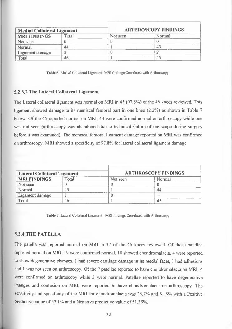
ARTHROSCOPY FINDINGSMedial Collateral LigamentMRI FINDINGS Total Not seen NormalNot seen 0 0 0Normal 44 1 43Ligament damage 2 0 2Total 46 1 45
Table 6: Medial Collateral Ligament: MR1 findings Correlated with Arthroscopy.
5.2.3.2 The Lateral Collateral Ligament
The Lateral collateral ligament was normal on MRI in 45 (97.8%) of the 46 knees reviewed. This
ligament showed damage to its meniscal femoral part in one knee (2.2%) as shown in Table 7
below. Of the 45-reported normal on MRI, 44 were confirmed normal on arthroscopy while one
was not seen (arthroscopy was abandoned due to technical failure of the scope during surgery
before it was examined). The meniscal femoral ligament damage reported on MRI was confirmed
on arthroscopy. MRI showed a specificity of 97.8% for lateral collateral ligament damage.
ARTHROSCOPY FINDINGSLateral Collateral LigamentMRI FINDINGS Total Not seen NormalNot seen 0 0 0Normal 45 1 44Ligament damage 1 0 1Total 46 1 45
Table 7: Lateral Collateral Ligament: MRI findings Correlated with Arthroscopy.
5.2.4 THE PATELLA
The patella was reported normal on MRI in 37 of the 46 knees reviewed. Of those patellae
reported normal on MRI, 19 were confirmed normal, 10 showed chondromalacia, 4 were reported
to show degenerative changes, 1 had severe cartilage damage in its medial facet, 1 had adhesions
and 1 was not seen on arthroscopy. Of the 7 patellae reported to have chondromalacia on MRI, 4
were confirmed on arthroscopy while 3 were normal. Patellae reported to have degenerative
changes and contusion on MRI, were reported to have chondromalacia on arthroscopy. The
sensitivity and specificity of the MRI for chondromalacia was 26.7% and 81.8% with a Positive
predictive value of 57.1% and a Negative predictive value of 51.35%.
32

Patella ARTHROSCOPY FINDINGSMRI FINDINGS Total Not seen Normal Chondromalacia OtherNot seen 0 0 0 0 0Normal 37 1 19 10 7Chondromalacia 7 0 3 4 0Others 2 0 0 1 1Total 46 1 22 15 8
Table 8: Patella: MRI findings Correlated with Arthroscopy.
5.2.5 THE FEMORAL ARTICULAR CARTILAGE
The femoral articular surface was reported normal in 32 knees on MRI (Table 9). Of these, it was
confirmed normal in 24, irregular in 4 and 3 had ulcer extending to the bone. It was not fully
examined in one knee as arthroscopy was abandoned during surgery due to technical failure of
the scope. Of the 11 femoral articular surfaces reported to be irregular on MRI, 7 were normal, 2
irregular and 2 had chondral flaps on arthroscopy. Femoral articular surface ulcer reported on
MRI was reported as irregular surface on arthroscopy. An osteochondral defect/flap seen on MRI
was confirmed on arthroscopy. Femoral articular cartilage described to have degenerative
changes on MRI was reported normal on arthroscopy. The sensitivity and specificity of the MRI
for femoral articular cartilage pathology was 46.2% and 75.0% with a positive predictive value of
42.3% and a negative predictive value of 75%.
ARTHROSCOPY FINDINGSFemoral Articular Cartilage
MRI FINDINGS Total Not seen NormalIrregularsurface
Ulcer to the bone Other
Not seen 0 0 0 0 0 0Normal 32 1 24 4 3 0Irregular surface 11 0 7 2 0 2Ulcer to bone 1 0 0 I 0 0Other Lesion(s) 2 0 1 0 0 1Total 46 1 32 7 3 3
Table 9: Femoral articular cartilage: MRI findings Correlated with Arthroscopy.
5.2.6 THE TIBIAL ARTICULAR CARTILAGE
The tibial articular surface was reported normal in 42 knees on MRI (Table 10). It was confirmed
normal in 31 knees, irregular in 8, ulcerated in one, fibrillated in one and was not fully examined
in one knee as arthroscopy was abandoned during surgery due to technical failure of the scope. Of
33

the 3 tibial articular surfaces reported to have irregular surface on MRI, in 2 it was confirmed
irregular but was normal in one on arthroscopy. Tibial articular cartilage described to show
degenerative changes on MRI was reported normal on arthroscopy. The sensitivity and specificity
of the MRI for tibial articular cartilage pathology was 16.7% and 93.9% with a positive predictive
value of 50% and a negative predictive value of 73.8%.
ARTHROSCOPY FINDINGSTibial Articular Cartilage
MRI FINDINGS Total Not seen NormalIrregularsurface
Ulcer to the bone Other
Not seen 0 0 0 0 0 0Normal 42 1 31 8 1 1Irregular surface 3 0 1 2 0 0Ulcer to bone 0 0 0 0 0 0Other Lesion(s) 1 0 1 0 0 0Total 46 1 33 10 1 1
Table 10: Tibial articular cartilage: MRI findings Correlated with Arthroscopy.
5.2.7 BONE LESIONS
Varied bone lesions were described on MRI in 13 (28.3%) of the 46 knees included in this study.
The commonest bone lesion reported on MRI was bone bruise/edema. Bone bruise/edema was
reported in the lateral femoral condyle in 5 knees, in the lateral tibial condyle in 5 knees, in the
medial femoral condyle in 2 knees and in the medial tibial condyle in one knee. Other bone
lesions seen on MRI included bony osteonechrosis reported in the medial tibial condyle in 2
knees and in the lateral femoral condyle in one knee. A Subchondral cyst was reported in one
lateral femoral condyle. All these lesions were not seen on arthroscopy (the bones remain hidden
to the arthroscope).
5.2.8 THE PATELLA TENDON
The patella tendon was normal on MRI in 43 (93.5%) of the 46 knees reviewed. Signal change
signifying edema (tendinosis) was reported in two knees while partial rupture of the tendon was
described in one. On arthroscopy, one of the two patella tendons reported on MRI to have edema
was normal while the other was confirmed a case of patella tendinosis with a taut ligamentum
pericase. The patella tendon reported to have partial rupture on MRI was normal on arthroscopy
and of the 43 patella tendons reported normal on MRI, 40 were confirmed normal, one was
reported hypertrophied and two were not seen (one due to generalized synovitis in the joint while
34
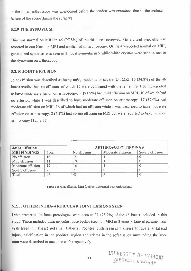
in the other, arthroscopy was abandoned before the tendon was examined due to the technical
failure of the scope during the surgery).
5.2.9 THE SYNOVIUM
This was normal on MRI in 45 (97.8%) of the 46 knees reviewed. Generalized synovitis was
reported in one Knee on MRI and confirmed on arthroscopy. Of the 45-reported normal on MRI,
generalized synovitis was seen in 3, local synovitis in 5 while white crystals were seen in one in
the Synovium on arthroscopy
5.2.10 JOINT EFFUSION
Joint effusion was described as being mild, moderate or severe. On MRI, 16 (34.8%) of the 46
knees studied had no effusion, of which 15 were confirmed with the remaining 1 being reported
to have moderate effusion on arthroscopy. 11(23.9%) had mild effusion on MRI, 10 of which had
no effusion while 1 was described to have moderate effusion on arthroscopy. 17 (37.0%) had
moderate effusion on MRI, 16 of which had no effusion while 1 was described to have moderate
effusion on arthroscopy. 2 (4.3%) had severe effusion on MRI but were reported to have none on
arthroscopy (Table 11).
Joint Effusion ARTHROSCOPY FINDINGSMRI FINDINGS Total No effusion Moderate effusion Severe effusionNo effusion 16 15 1 0Mild effusion 11 10 1 0Moderate effusion 17 16 1 0Severe effusion 2 2 0 0Total 46 43 3 0
Table 11: Joint effusion: MRI findings Correlated with Arthroscopy.
5.2.11 OTHER INTRA-ARTICULAR JOINT LESIONS SEEN
Other intraarticular knee pathologies were seen in 11 (23.9%) of the 46 knees included in this
study. These included intra-articular loose bodies (seen on MRI in 2 knees), Lateral parameniscal
cysts (seen in 3 knees) and small Baker’s / Popliteal cysts (seen in 3 knees). Infrapatellar fat pad
injury, calcification in the popliteal region and edema in the soft tissues surrounding the knee
joint were described in one knee each respectively.
35IM'VE.TOTY 0r „ a
USRARy ^

O th e r Joint Pathology ARTHROSCOPY FINDINGS
jVfRI FINDINGS Total Normal/notseen
Loosebody
LigamentumMucosae
LateralParameniscal cyst
ErosiveChondrallesion
Intra-articularcystals
MedialPlicae
jjormal 35 29 2 2 0 1 0 1
[ l o o s e body 2 0 2 0 0 0 0 0
■pjteral Parameniscalcgt____________________
3 1 0 0 2 0 0 0
popliteal (Baker’s cyst) 3 3 0 0 0 0 0 0
&ift tissue calcification 1 0 0 0 0 0 1 0
jjjfrapatellar fat padiniun
1 1 0 0 0 0 0 0
"Soft tissue edema 1 1 0 0 0 0 0 0
'T o t a l 46 35 4 2 2 1 1 1
Table 12: Other joint pathology: MRI findings Correlated with Arthroscopy.
Of the 35 knees reported normal on MRI, Loose bodies were seen on arthroscopy in 2 while
Ligamentum mucosae, erosive chondral lesion and Medial plicae were described in 2 knees, 1
knee and 1 knee respectively. Loose bodies reported in 2 knees on MRI were confirmed on
arthroscopy. The lateral parameniscal cysts reported in 3 knees on MRI were associated with
lateral meniscal tears and in 2 of the knees, the cysts were confirmed on arthroscopy while the
lateral compartment of the third knee was not fully examined as arthroscopy was abandoned
during surgery due to technical failure of the scope. Soft tissue calcification seen in 1 knee on
MRI was confirmed to be intra-articular crystals on arthroscopy. Knees reported to have popliteal
cyst (Baker’s cyst), infrapatellar fat pad injury and soft tissue edema on MRI were all reported
normal on arthroscopy.
36

CHAPTER SIX
6.0 DISCUSSION
The objective of this study was to determine the pattern of presentation of knee joint pathology as
well as to find out the correlation between the radiological and arthroscopic findings in the knee
in patients seen at five radiological imaging centers in Nairobi.
Pattern ofpresentation o f knee joint pathology
Males (58.7%) were more affected than females (41.3%). Majority of the patients were in the 26-
50 years age group, with the right knee (67.4%) being affected more than the left (32.6%).
The commonest findings encountered were meniscal tears and joint effusions constituting 78.3%
and 65.2% of the cases seen respectively. Both showed a male predilection.
Other pathologies seen included chondromalacia patellae, cruciate ligament tears, collateral
ligament disruptions, bone bruises, patella tendinosis, synovitis and intraarticular loose bodies.
Correlation between the radiological and arthroscopic findings
Overall, this study has highlighted a positive correlation between accuracy of MRI results and
arthroscopic findings for intra-articular knee pathology (especially ACL and meniscal lesions),
using arthroscopy results as a gold standard.
MRI showed a high sensitivity in the medial meniscus, where it was sensitive in detecting a tear
in 88.9% of cases. Sensitivity for tears in the lateral meniscus was higher at 93.3%. However,
specificity for a meniscal tear was lower in the medial meniscus than in the lateral meniscus
(36.1% and 63.3% respectively). The sensitivity and specificity of the MRI for anterior cruciate
ligament rupture (whether complete or partial) was 83.3% and 90.9% respectively. For the
articular cartilage, sensitivity was 46.2% and 16.7% for femoral and tibial articular cartilage
abnormalities with a specificity of 81.8% and 75.0% respectively. The sensitivity and specificity
of the MRI for chondromalacia patellae was 26.7% and 81.8% respectively.
Use of MRI in diagnosis of intra-articular knee pathology is relatively new in our setup.
Musculoskeletal MRI has a long learning curve and this may partly explain the lower specificities
in diagnosis of intra-articular knee lesions seen in this study. From this, it is important to note that
if MRI was to be used as the only form of pre-operative screening for these conditions, then there
may well have been unnecessary arthroscopies performed.
37

M R! in the diagnosis o f m eniscal and cruciate ligam ent tears
The menisci are the most often damaged structures in the knee joint. The most common site for
meniscal tear is the posterior horn of the medial meniscus. The a fore mentioned differences
between medial and lateral meniscus (see page 7), and the predominantly medial loading in the
lower extremity render the lateral meniscus less susceptible to tears. In the literature, the
sensitivity of MRI in the diagnosis of medial meniscal tears is reported to be 87-97%, sensitivity
for lateral meniscal tears is 69-92%; specificities are 82-91% and 91-98% respectively (20-32).
Thus, the performance of MRI is clinically excellent especially if one takes into account that
small peripheral tears missed by MRI are not clinically relevant, and that arthroscopy is not an
ideal gold standard. When the anterior cruciate ligament (ACL) is tom, accuracy for meniscal
tears decreases and especially tears in the posterior lateral meniscus may be missed.
The accuracy and sensitivity of MRI for complete cruciate tears are reported to be between 92%
and 100% (21,22,23,29,45,46). It is likely that MRI is more accurate in the diagnosis of partial
cruciate ligament tears than arthroscopy. On the other hand, partial tears are commonly over
diagnosed. The term partial tear of the ACL remains controversial, as partial ruptures are difficult
to define in equal terms by the radiologist and the orthopaedic surgeon. Therefore, a scoring
system proposed by Rubin et al (47) that distinguishes between high- and low-grade injuries and
thereby discards the diagnosis of partial rupture, will probably facilitate communication with the
orthopaedic surgeon.
MRI in the diagnosis o f other knee pathology
The extent of cartilage abnormalities can be concealed when MRI is used as the only diagnostic
tool. Arthroscopic evaluation is more useful than radiographs or MRI to grade osteoarthritis and
assess surface cartilage abnormalities. MRI may miss superficial cartilage abnormalities but is
more sensitive at revealing deep chondral lesions, and also helps locate subchondral lesions
undetectable by arthroscopy (48). FSE sequences and especially 3D fat-suppressed GE sequence
are the most commonly used cartilage sequences (49-53). This is further facilitated by the fact
that magnetization transfer contrast (MTC) effects in TSE sequences result in excellent contrast
between synovial fluid and cartilage. MTC decreases when collagen is disrupted leading to
increased signal intensity. TSE is fast, has good signal-to-noise ratio and probably detects larger
cartilaginous defects with acceptable accuracy. Small defects are not well visualized but tend to
be of little clinical concern. In addition to 3D fat-suppressed GE and FSE sequences, the steady-
state water excitated sequences are useful in the examination of the cartilage. The main issue in
this very effective sequence is the necessity of homogeneous magnetic field.
MRI is useful to diagnose bone injuries in patients with acute knee effusions who have no
ligament laxity on examination and normal findings on plain radiograph. Bone injury is the most38

common cause of acute effusion in this group of patients (54). An anterior-posterior
measurement of 10 mm of fluid or less in the lateral aspect of the suprapatellar pouch is a
reasonable threshold value for distinguishing a physiologic from pathologic amount of fluid in the
knee joint (55).
Lesions in extra-articular spaces, which remain hidden at arthroscopy, are more likely to be
detected with MRJ. In our study, Knees reported to have popliteal cyst (Baker’s cyst),
infrapatellar fat pad injury and soft tissue edema on MRI were all reported normal on
arthroscopy. All these lesions remain hidden to the arthroscope.
False positives and false negatives
MRI studies have higher false positive than false negative results (56). We also found this to be
true when examining the combined results from meniscal lesions and ACL tears.
Radial meniscal tears are difficult to visualize on MRI; hence, they account for a large number of
tears missed by MRI. However, MRI is a useful diagnostic tool in detecting radial tears of the
posterior horn of the medial meniscus, which are common in elderly patients who also often have
osteoarthritis that masks their symptoms. In this study, out of 6 radial tears reported on
arthroscopy, only 2 (33.3%) had been described on MRI. Radial tears involve the free edge of the
meniscus. Thus, the key to interpretation of this injury is the recognition of absence or blunting of
the inner point of the meniscal triangle (57).
False positive MRI scans seen in the posterior horn of the medial meniscus may reflect an
inability to completely visualize the area at arthroscopy, and tears that extend to the inferior
surface of the meniscus may be difficult to see (58). Some false positive findings on MRI can be
attributed to inadequate visualization of the meniscus at surgery and to the fact that the diagnosis
of a tear can be subjective (28).
The high NPV and high specificity confirm the use of MRI as a screening tool highly predictive
in avoiding unnecessary arthroscopies. The MRI examination techniques recommended in the
literature at present are not able to replace arthroscopy for diagnosis of cartilage damage in the
knee (59). Although MRI is being used with increasing frequency, it is unlikely to replace clinical
diagnosis. It should be used in connection with clinical findings and history to provide a more
complete picture. Clinical examination when combined with MRI, as earlier pointed out, provides
the most accurate non-invasive source of information currently available for pathological findings
in the menisci and the ACL (60).
MRI films need to be carefully examined because a meniscal tear is unlikely when MRI scans
show a focus of high signal in a meniscus that does not unequivocally extend to involve the
surface of the meniscus. In this study, all the ten medial and lateral menisci reported on MRI to
have degenerative changes were normal on arthroscopy. Grade 1 and 2 signals (degenerative

changes) are focal or linear areas of high signal confined to the substance of the meniscus with
intact outer contour lines: these are not visible at arthroscopy and would be classified as a false
positive result as in this study leading to unnecessary arthroscopy. Grade 1 and 2 signals
(degenerative changes) are not indications for arthroscopy.
MRI is non-invasive but arthroscopy has surgical risks, with a complication rate of 2.5% in
arthroscopic meniscal surgery (61). These complications include operation on wrong limb,
saphenous and peroneal nerve injures, deep infections, superficial infections, vascular injuries and
pulmonary embolism. Sometimes, arthroscopy reveals no abnormality or possibly minor non-
pathological lesions such as plicae or chondromalacia patellae. This means that a patient could be
exposed to surgical risk with no symptom benefit.
Technical aspects o f MRI that may affect accuracy o f diagnosis o f knee pathology
It is important to note that there is no standard Knee MRI protocol and most institutions establish
their own routine protocol, which provides adequate visualization and answers the majority of
questions. However, the choice of slice thickness, field of view, coil, and imaging matrix play an
important role in determining spatial resolution. Improved spatial resolution provides the
radiologist with data to present interpretation with confidence. The bandwidth is another
important factor that may affect imaging especially at high field strengths. The signal-to-noise
ratio can also be improved by using a narrow receive bandwidth. One disadvantage of narrow
bandwidth is the increase in chemical shift artefact and spatial misregistration, which is increased
at the interfaces between cortical bone, hyaline cartilage and fibrocartilage. This effect may
adversely affect diagnostic accuracy as these artefacts can simulate or obscure thinning of the
cortex or overlying hyaline cartilage. Fat-suppression techniques eliminate chemical shift
misregistration and so a narrow bandwidth can be used with these latter techniques.
Magnetic field strength is another important factor, which is not under the practitioner’s control.
Diagnostic quality in knee imaging has been researched and reported with field strengths ranging
from 0.2 to 1.5T. The levels of confidence in reporting was better in high field strength than low
field strength and in general, lower field strengths units must use more signal averages to provide
equivalent quality as that obtained with high field strengths. This results in increased scan times.
All these technical issues together with the expertise of the radiologist may contribute to
differences in the reliability of the different centers. This study did not take into account some of
these technical considerations as they were considered beyond the scope of the study.
40

CHAPTER SEVEN
7.0 CONCLUSION AND RECOMMENDATIONS
7.1 CONCLUSION
MRI is able to detect most internal derangements of the knee efficiently, for example in our study
it was accurate in detecting a medial meniscal tear in 88.9% of the cases. Generally, MRI has a
higher specificity (i.e. correctly identifies the absence of an internal derangement of the knee)
than sensitivity (i.e. accurately identifying an internal derangement of the knee). As was shown in
this study, MRI also has a higher NPV (reliability of a negative MRI result) than PPV (reliability
of a positive MRI result). Thus, if a patient is given a result of a negative MRI scan, the high
specificity and NPV of the scan mean that this is likely to be a true negative result.
MRI is the most appropriate screening tool prior to performing a therapeutic arthroscopy. It is
preferable to diagnostic arthroscopy in most patients because it is faster and avoids surgical risks.
However, MRI is not 100% accurate: if an MRI is reported as negative but the patient is still
complaining of ongoing symptoms, then arthroscopy should be considered.
The results of this study support the use of MRI in the diagnosis of internal derangements of the
knee. However, it should always be used in conjunction with a full history and clinical
examination.
7.2 RECOMMENDATIONS
■ Establishment of an imaging protocol would standardize examinations and reduce
variability in results. This might involve:
o A standard MRI protocol for knee imaging and modification where necessary.
o Recommendations on Contrast media use in knee MR imaging for example in
delineation of bone tumours, scar tissue in repeat surgery, repeat trauma and in
inflammatory (non traumatic) lesions like patella tendinosis and meniscal cysts.
o Standardization of print settings for hard copies would ease review of
retrospective cases. A standardized way of interpreting the Knee MR images
would also increase the accuracy of the reports.
■ Availability and affordability- During the time of this study, two more centers offering
MRI services opened up to the public, and with these and more opening up, MR imaging
is bound to be more available but there is still serious need to make it more affordable.
41

■ Sensitization of clinicians on the benefits of MR imaging of the knee- MRI is able to
detect most internal derangements of the knee efficiently and with improvement in
technology and its use in musculoskeletal imaging, there is need to keep our colleagues
abreast with these developments for improved management of patients.
■ Records - great difficulty was encountered in acquiring clinical data both retrospectively
and prospectively. Clinicians need to be sensitized on the need to provide accurate and
exhaustive information on the patients they send for imaging. This aids in interpretation
of the imaging findings.
■ Possible areas of future research include:
o A study to determine the local etiological factors of specific lesions for example
role of trauma as an etiological factor of the commoner pathologies would prove
useful in aiding in their prevention.
o Use of specific MRI sequences to identify pathology in each of the various tissues
in and around the knee (ligaments, menisci, tendons, articular surface and bone),
while keeping the investigation within acceptable times and costs.
O Patella and articular cartilage pathologies seen proved to be diagnostic challenges
radiologically. A more extensive radiologic-arthroscopic correlative study to
assess pathologies of the articular cartilage would prove useful.
42

APPENDICESAPPENDIX 1: DATA COLLECTION FORM
Patients study N o............................................
PATIENTS DEMOCRAPHIC DATA
A g e ...................yearsS ex ....................01: M 02: F
CLINICAL HISTORY AND DIAGNOSIS
K N E E :........................................................... 01: Right Knee 02: Left kneeCLINICAL HISTORY:
CLINICAL IM PRESSION:.........................................................................
RADIOLOGICAL EXAMINATIONS AND DIAGNOSIS
a) PLAIN RADIOGRAPHY FINDINGS / DIAGNOSIS (If available):
b) ARTHROGRAPHY FINDINGS / DIAGNOSIS (If available):
c) MRI FINDINGS AND DIAGNOSIS:MRI N o :....................................................... Date MRI done:.............................................MRI Centre o f study:.................................. 01: KNH 03: MITC
02: AKUH 04: MRI Centre- NairobiAGE OF IN IU R Y :..................................01: Fresh Injury (<2weeks) 02: Old Injury (>2weeks)MRI FINDINGS AND DIAGNOSIS:1) MENISCUS: MEDIAL01 Not seen02 Normal03 Radial tear
..................... LATERAL.04 Horizontal tear05 Longitudinal tear06 Bucket handle tear
07 Flap tear08 Degenerative09 Others (specify)
2) COLLATERAL LIGAMENTS: M ED IA L.......... LATERAL.........................................01 Not seen 04 Meniscotibial ligament damaged02 Normal 05 Others (specify)03 Meniscofemoral ligament damaged3) CRUCIATE LIGAMENTS: ANTERIOR.......... POSTERIOR.....................................01 Not seen 03 Partial rupture02 Normal 04 Complete rupture4) PATELLA..................................................................................01 Not seen 03 Chondromalacia02 Normal 04 Subluxation5) PATELLA TENDON..............................................................01 Not seen 03 Partial rupture02 Normal 04 Complete rupture6) POPLITEAL TENDON...........................................................01 Not seen 03 Partial rupture02 Normal 04 Complete rupture7) CARTILAGE............................................................................01 Not seen 03 Irregular surface02 Normal 04 Ulcer to the bone
05 Others (specify)
05 Others(specify)
05 Others(specify)
05 Others(specify)
05 Others(specify)
43

8) Synovia......................................................................................01 Not seen 03 Local synovitis 05 Others(specify)02 Normal 04 General synovitis9) MR1 DIAGNOSIS:...............................................................................................................
ARTHROSCOPIC EXAMINATION AND DIAGNOSIS
Hospital N o : ................................................. Date Arthroscopy done..............................................Hospital where arthroscopy done:.........................................................AGE OF IN JU RY :..................................01: Fresh Injury (<2weeks) 02: Old Injury (>2weeks)ARTHROSCOPY FINDINGS AND DIAGNOSIS:1) MENISCUS: MEDIAL............................... LATERAL..................................................01 Not seen 04 Horizontal tear 07 Flap tear02 Normal 05 Longitudinal tear 08 Degenerative03 Radial tear 06 Bucket handle tear 09 Others (specify)2) COLLATERAL LIGAMENTS: M ED IA L.........LATERAL....................................................01 Not seen 04 Meniscotibial ligament damaged02 Normal 05 Others (specify)03 Meniscofemoral ligament damaged3) CRUCIATE LIGAMENTS: ANTERIOR.......... POSTERIOR01 Not seen 03 Partial rupture02 Normal 04 Complete rupture4) PATELLA..................................................................................01 Not seen 03 Chondromalacia02 Normal 04 Subluxation5) POPLITEAL TENDON...........................................................01 Not seen 03 Partial rupture02 Normal 04 Complete rupture6) CARTILAGE.....................................................................................01 Not seen 03 Local synovitis02 Normal 04 General synovitis7) Synovia......................................................................................01 Not seen 03 Local synovitis 05Others(specify)02 Normal 04 General synovitis
05 Others (specify)
05 Others(specify)
05 Others(specify)
05 Others(specify)
8) Arthroscopy diagnosis
CONCLUSIONMRI diagnosis confirmed correct on Arthroscopy MRI diagnosis confirmed incorrect on Arthroscopy
COMM ENTS:
44

REFERENCES1. Isadore Meschan. An Atlas of Normal Radiographic Anatomy. W.B Saunders,
2nd Edition, Pg 168-170.2. Pavlov H, Burke M, Giesa M et al. Orthopaedic's Guide to Plain Film Imaging.
Thieme, New York, 1999.3. Bontraeger K. Textbook o f Radiographic Positioning and Related Anatomy.
Mosby, St Louis, MO, 1999.4. Whitehouse G, Worthington B. Techniques in Diagnostic Radiology. Blackwell
Scientific publications, 1983, Pg 306-308.5. Gray SD, Kaplan OA, Dussault RG. Imaging of the Knee. Current Status. Orthop
Clin North Am. 1997; 28:643-658.6. Berger RA, Crossett LS, Jacobs JJ. Malrotation causing patellofemoral
complications after total knee arthroplasty. Clin Orthop 1998; 356: 144-153.7. Me Guire DA, Hendricks SD, Sanders HM. The relationship between anterior
cruciate ligament reconstruction tibial tunnel location and the anterior aspect of the posterior cruciate ligament insertion. Arthroscopy 1997; 13: 465-473.
8. Vande Berg BC, Lecouvet FE, Poilvache P, Maldague B, Malghem J. Spiral CT arthrography of the knee: technique and value in the assessment of internal derangement of the knee. Eur Radiol 2002; 12: 1800-1810.
9. Vande Berg BC, Lecouvet FE, Malghem J. Frequency and topography of lesions of the femoro-tibial cartilage at spiral CT arthrography of the knee: a study in patients with normal knee radiographs and without history of trauma. Skeletal Radiol 2002; 31: 643-649.
10. Vande Berg BC, Lecouvet FE, Poilvache P et al. Dual-detector spiral CT arthrography of the knee: accuracy for detection of meniscal abnormalities and unstable meniscal tears. Radiology 2000; 216: 851-857
11. Vande Berg BC, Lecouvet FE, Poilvache P, Maldague B, Malghem J. Spiral CT arthrography of the postoperative knee. Semin Musculoskelet Radiol 2002; 6: 47-55.
12. Rand T, Brossmann J, Pedowitz R, Ahn JM, Haghigi P, Resnick D. Analysis of patellar cartilage. Comparison of conventional MR imaging and MR and CT arthrography in cadavers. Acta Radiol 2000; 41: 492-497.
13. Toms AP, White LM, Marshall TJ, Donell ST. Imaging the post-operative meniscus. Eur J Radiol 2005; 54: 189-198.
14. Mutschler C, Vande Berg BC, Lecouvet FE et al. Postoperative meniscus: assessment at dual-detector row spiral CT arthrography of the knee. Radiology 2003; 228: 635-641.
15. Ryan PJ, Reddy K, Fleetcroft J. A prospective comparison of clinical examination, MRI, bone SPECT and arthroscopy to detect meniscal tears. Clin NuclMed 1998; 23: 803-806.
16. Vellala RP, Manjure S, Ryan PJ. Single photon emission computed tomography scanning in the diagnosis of knee pathology. J Orthop Surg (Hong Kong) 2004; 12: 87-90.
17. Eustace S, Hentzen P, Adams J, Harper K, Jara H. Comparison of conventional and turbo spin-echo T1-weighted MR imaging in acute knee trauma. AJR Am JRoentgenol 1999; 172: 1393-1395.
45

18 Escobedo EM, Hunter JC, Zink-Brody GC, Wilson AJ, Harrison SD, Fisher DJ. Usefulness of turbo spin-echo MR imaging in the evaluation of meniscal tears: comparison with a conventional spin-echo sequence. AJR Am J Roentgenol 1996; 167: 1223-1227.
19. Lotysch M, Mink J, Crues JV, Schwartz A. Magnetic resonance imaging in the detection of meniscal injuries. Magn Reson Imaging 1986; 4: 185.
20. Crues JVd, Mink J, Levy TL, Lotysch M, Stoller DW. Meniscal tears of the knee: accuracy of MR imaging. Radiology 1987; 164: 445-448.
21. Fischer SP, Fox JM, Del Pizzo W, Friedman MJ, Snyder SJ, Ferkel RD. Accuracy of diagnoses from magnetic resonance imaging of the knee. A multi-center analysis of one thousand and fourteen patients. J Bone Joint Surg (Am) 1991; 73: 2- 10.
22. Bui-mansfield LT, Youngberg RA, Warme W, Pitcher JD, Nguyen PLL. Potential cost savings of MR imaging obtained before arthroscopy of the knee: evaluation of 50 consecutive patients. AJR Am J Roentgenol 1997; 168: 913-918.
23. Boeree NR, Watkinson AF, Ackroyd CE, Johnson C. Magnetic resonance imaging of meniscal and cruciate injuries of the knee. J Bone Joint Surg (Br) 1991; 73: 452^157.
24. Cheung LP, Li KC, Hollett MD, Bergman AG, Herfkens RJ. Meniscal tears of the knee: accuracy of detection with fast spin-echo MR imaging and arthroscopic correlation in 293 patients. Radiology 1997; 203: 508-512.
25. De Smet AA, Norris MA, Yandow DR, Quintana FA, Graf BK, Keene JS. MR diagnosis of meniscal tears of the knee: importance of high signal in the meniscus that extends to the surface. AJR Am J Roentgenol 1993; 161: 101-107.
26. Justice WW, Quinn SF. Error patterns in the MR imaging evaluation of menisci of the knee. Radiology 1995; 196:617-621.
27. Munk B, Madsen F, Lundorf E, etal. Clinical magnetic resonance imaging and arthroscopic findings in knees: a comparative prospective study of meniscus anterior cruciate ligament and cartilage lesions. Arthroscopy 1998; 14:171-175.
28. Quinn SF, Brown TF. Meniscal tears diagnosed with MR imaging versus arthroscopy: how reliable a standard is arthroscopy? Radiology 1991; 181: 843- 847.
29. Rappeport ED, Wieslander SB, Stephensen S, Lausten GS, Thomsen HS. MRI preferable to diagnostic arthroscopy in knee joint injuries: a double-blind comparison of 47 patients. Acta Orthop Scand 1997; 68: 277-281.
30. Reicher MA, Hartzman S, Duckwiler GR, Bassett LW, Anderson LJ, Gold RH. Meniscal injuries: detection using MR imaging. Radiology 1986; 159: 753-757.
31. Stoller DW, Martin C, Crues JV HI, Kaplan L, Mink JH. Meniscal tears: pathologic correlation with MR imaging. Radiology 1987; 163: 731-735.
32. Weinstabl R, Muellner T, Vecsei V, Kainberger F, Kramer M. Economic considerations for the diagnosis and therapy of meniscal lesions: can magnetic resonance imaging help reduce the expense? World J Surg 1997; 21: 363-368.
33. Schweitzer ME, Tran D, Deely DM, Hume EL. Medial collateral ligament injuries: evaluation of multiple signs, prevalence and location of associated bone bruises, and assessment with MR imaging. Radiology 1995; 194: 825-829.
34. Recht M, Bobic V, Burstein D, etal. Magnetic resonance imaging of articular cartilage. Clin Orthop 2001; 391(Suppl.): S379-S396.
46

35. Yao L, Lee JK. Occult intraosseous fracture: detection with MR imaging. Radiology 1988; 167: 749-751.
36. Mink JH, Deutsch AL. Occult cartilage and bone injuries of the knee: detection, classification, and assessment with MR imaging. Radiology 1989; 170: 823-829.
37. Vincken PW, Ter Braak BP, van Erkel AR, Coerkamp EG, Mallens WM, Bloem JL. Clinical consequences of bone bruise around the knee. Eur Radiol 2005; 16: 97-107.
38. Virolainen H, Visuri T, Kuusela T. Acute dislocation of the patella: MR findings. Radiology 1993; 189: 243-246.
39. Yu JS, Popp JE, Kaeding CC. Lucas J. Correlation of MR imaging and pathologic findings in atheletes undergoing surgery for chronic patellar tendonitis. AJR 1995; 165:115-118.
40. Deutsch AL, Resnick D, Dalinka MK, etal. Synovial plicae of the knee. Radiology. 1981; 141: 627-634.
41. Forbes JR, Helms CA, Janzen DL. Acute pes anserine bursitis: MR imaging. Radiology. 1995; 194: 525-527.
42. Rothstein CP, Laorr A, Helms CA, Tirman P. Semimembrinosus-tibial collateral ligament bursitis: MR Imaging findings. AJR. 1996; 166:875-877.
43. Gluckert K, Klandy B, Blank-Schol A, Hofmann G. MRI of knee joint with a 3D gradient echo sequence. Arch Orthop Trauma Surg (1992) 112:5-14.
44. Vincken PW, ter Braak BP, van Erkel AR et al. Effectiveness of MR imaging in selection of patients for arthroscopy of the knee. Radiology 2002; 223: 739-746.
45. Cotten A, Delfaut E, Demondion X, etal. MR imaging of the knee at 0.2 and 1.5 T: correlation with surgery. AJR Am J Roentgenol 2000; 174: 1093-1097.
46. Ha TP, Li KC, Beaulieu CF, etal. Anterior cruciate ligament injury: fast spin- echo MR imaging with arthroscopic correlation in 217 examinations. AJR Am J Roentgenol 1998; 170: 1215-1219.
47. Rubin DA, Kettering JM, Towers JD, Britton CA. MR imaging of knees having isolated and combined ligament injuries. AJR Am J Roentgenol 1998; 170: 1207- 1213.
48. Irie K, Yamada T, Inoue K. A comparison of magnetic resonance imaging and arthroscopic evaluation of chondral lesions of the knee. Orthopedics (2000) 23:561-564.
49. Peterfy CG. Scratching the surface: articular cartilage disorders in the knee. Magn Reson Imaging Clin N Am 2000; 8: 409-430.
50. Recht M, Bobic V, Burstein D, etal. Magnetic resonance imaging of articular cartilage. Clin Orthop 2001; 391(Suppl.): S379-S396.
51. McCauley TR, Recht MP, Disler DG. Clinical imaging of articular cartilage in the knee. Semin Musculoskelet Radiol 2001; 5: 293-304.
52. Gold GE, McCauley TR, Gray ML, Disler DG. What's new in cartilage? Radiographics 2003; 23: 1227-1242.
53. Komaat PR, Doombos J, van der Molen AJ et al Magnetic resonance imaging of knee cartilage using a water selective balanced steady-state free precession sequence. J Magn Reson Imaging 2004; 20: 850-856.
54. Duncan JB, Hunter R, Purnell M, Freeman J. Injured stable knee with acute effusion MRI evaluation. J South Orthop Ass (1996) 15:13-19.
47

55. Kolman BH, Daffner RH, Sciulli RL, Soehnlen MW. Correlation of joint fluid and internal derangement on knee MRI. Skelet Radiol (2004) 33:91-95.
56. Heron CW, Calvert PT. 3D gradient echo MRI of the knee: comparison with arthroscopy in 100 patients. Radiology (1992) 183:839-844.
57. Tuckman GA, Miller WJ, Remo JW, Fritts HM, Rozansky MI. Radial tears of the menisci: MR findings. Am J Roentgenol (1994) 163:395-400.
58. Chissell HR, Keightley A, Allum RL MRI of the knee: its cost effective use in a DGH. Ann R Coll Surg Engl (1994) 76:26-29.
59. Friemert B, Oblerlander Y, Schwarz W, et al. Diagnosis of chondral lesions of the knee joint: can MRI replace arthroscopy? Knee Surg Sports Traumatol Arthrosc(2004) 12:58-64.
60. Glashow JL, Katz R, Schneider M, Scott WN. Double-blind assessment of the value of MRI in the diagnosis of ACL and meniscal lesions. J Bone Joint Surg Am (1989) 71:113-119.
61. Small NC. Complications in arthroscopic meniscal surgery. Clin Sports Med (1990)9:609-617.
U N h rMfc u n .
n - A'ROBf
48