Trigeminal ganglion innervation of the cochlea—a retrograde transport study
Upload
independentCategory
view
2download
0
Inducible Ablation of Melanopsin-Expressing RetinalGanglion Cells Reveals Their Central Role in Non-ImageForming Visual ResponsesMegumi Hatori1, Hiep Le1, Christopher Vollmers1, Sheena Racheal Keding1, Nobushige Tanaka1¤,
Christian Schmedt2, Timothy Jegla3, Satchidananda Panda1*
1 The Salk Institute for Biological Studies, La Jolla, California, United States of America, 2 Genomics Institute of Novartis Research Foundation, San Diego, California, United
States of America, 3 Department of Cell Biology and Institute for Childhood and Neglected Diseases, The Scripps Research Institute, La Jolla, California, United States of
America
Abstract
Rod/cone photoreceptors of the outer retina and the melanopsin-expressing retinal ganglion cells (mRGCs) of the innerretina mediate non-image forming visual responses including entrainment of the circadian clock to the ambient light, thepupillary light reflex (PLR), and light modulation of activity. Targeted deletion of the melanopsin gene attenuates theseadaptive responses with no apparent change in the development and morphology of the mRGCs. Comprehensiveidentification of mRGCs and knowledge of their specific roles in image-forming and non-image forming photoresponses arecurrently lacking. We used a Cre-dependent GFP expression strategy in mice to genetically label the mRGCs. This revealedthat only a subset of mRGCs express enough immunocytochemically detectable levels of melanopsin. We also used a Cre-inducible diphtheria toxin receptor (iDTR) expression approach to express the DTR in mRGCs. mRGCs develop normally, butcan be acutely ablated upon diphtheria toxin administration. The mRGC-ablated mice exhibited normal outer retinalfunction. However, they completely lacked non-image forming visual responses such as circadian photoentrainment, lightmodulation of activity, and PLR. These results point to the mRGCs as the site of functional integration of the rod/cone andmelanopsin phototransduction pathways and as the primary anatomical site for the divergence of image-forming and non-image forming photoresponses in mammals.
Citation: Hatori M, Le H, Vollmers C, Keding SR, Tanaka N, et al. (2008) Inducible Ablation of Melanopsin-Expressing Retinal Ganglion Cells Reveals Their CentralRole in Non-Image Forming Visual Responses. PLoS ONE 3(6): e2451. doi:10.1371/journal.pone.0002451
Editor: Michael Hendricks, Temasek Life Sciences Laboratory, Singapore
Received April 25, 2008; Accepted May 14, 2008; Published June 11, 2008
Copyright: � 2008 Hatori et al. This is an open-access article distributed under the terms of the Creative Commons Attribution License, which permitsunrestricted use, distribution, and reproduction in any medium, provided the original author and source are credited.
Funding: MH, HL, CV, SK, and SP were supported by Pew Scholars Program in Biomedical Sciences, Dana Foundation, Whitehall Foundation and NIH grantEY016807. CS and TJ were supported by funding from GNF. The funding agencies had no role in study design, data collection and analysis, decision to publish, orpreparation of the manuscript.
Competing Interests: The authors have declared that no competing interests exist.
* E-mail: [email protected]
¤ Current address: Kyorin University, Tokyo, Japan
Introduction
The rod/cone photoreceptors of the outer retina signal via
multisynaptic pathways to the retinal ganglion cells (RGCs) of the
inner retina. The RGCs, in turn, transmit the visual information to
the brain via their axonal projections. A small subset of RGCs
exclusively expresses the functional photopigment melanopsin
(OPN4) and is intrinsically photosensitive, but also receives rod/
cone inputs (Figure 1A) [1-5]. These mRGCs, along with the rod/
cone photoreceptors, mediate several non-image forming, or
adaptive ocular photoresponses (AOPs), which help organisms
optimize their physiological performance in variable ambient light
conditions. These AOPs include rapid adjustment of pupil size,
modulation of general activity and endocrine function, and tuning
of the phase and period length of the circadian clock to adapt to
the light environment (reviewed in [6]).
Mouse genetics has established the complementary roles of both
rod/cone and melanopsin in these AOPs. Mice with outer retinal
degeneration (rd/rd) retain functional mRGCs and exhibit almost
intact AOPs [7–11]. However, these adaptive responses are
completely abolished in mice that lack both melanopsin and
functional outer retina photoreceptors, thus establishing a
dominant role of melanopsin photopigment in these processes
[12,13]. Yet, the AOPs are attenuated, not eliminated, in
melanopsin deficient (Opn42/2) mice, an observation that suggests
a role for rod/cone photoreceptors in AOPs [14–16]. The
mRGCs develop normally in Opn42/2 mice and make normal
monosynaptic projections to the suprachiasmatic nucleus (SCN)
and olivary pretectal nucleus (OPN) which regulate the circadian
behavior and pupillary constriction, respectively [14]. Both the
SCN and OPN also receive direct and indirect projections from
RGCs that do not express melanopsin [17]. Therefore, it is
unclear whether rod/cone-initiated light signal is transmitted
primarily via the mRGCs or via other RGCs to brain regions that
regulate AOPs. Additionally, the mRGCs have also been
suggested to play a role in modulating rod/cone initiated image-
forming functions [18,19].
To understand the specific role of mRGCs in both image-
forming and adaptive ocular photoresponses, we generated mice
expressing Cre-inducible diphtheria toxin receptor exclusively in
PLoS ONE | www.plosone.org 1 June 2008 | Volume 3 | Issue 6 | e2451
the mRGC lineage. Diphtheria toxin (DT) crosses the blood-brain
barrier after systemic injection and has been shown to trigger cell
death in neurons expressing a primate DTR without triggering a
significant immune response [20,21]. This strategy allows normal
embryonic and postnatal differentiation and development of the
target cell type which can be verified in adult mice. Subsequent acute
cell ablation with local or systemic DT administration circumvents
any potential developmental compensation. Systemic DT injection
in the adult mice with DTR expressing mRGCs triggers a profound
loss of mRGCs. The mRGC-ablated mice lose non-image forming
visual responses while maintaining largely intact image-forming
functions. This demonstrates a central role of the mRGCs as the site
of integration of melanopsin and rod/cone initiated photoresponses
for generating adaptive light responses in mammals.
Results
Fluorescent tagging of mRGCsTo comprehensively tag mRGCs with transcriptionally active
melanopsin locus, we generated a mouse line which carries a Cre
recombinase and a Cre-dependent bTau-Yellow Fluorescent
Protein (YFP) expression cassette knocked-in to the native
melanopsin locus (Figure 1). However, no detectable YFP expression
was found in the retina of Opn4Cre/+ mouse, which may be due to
weak transcriptional activity from the native melanopsin promoter
and/or low expression of the second transcript downstream of an
internal ribosomal entry site (IRES) cassette. Next, we bred the
Opn4Cre/+ mouse with Z/EG mouse (Figure 1B) [22]. This strategy
allows Cre-dependent expression of green fluorescent protein
(GFP) from a strong b-actin promoter, such that GFP is uniformly
expressed in all mRGCs irrespective of the heterogeneity in the
level of transcription from the native melanopsin locus or in the level
of immunologically detectable melanopsin protein. In the retina of
adult Opn4Cre/+;Z/EG mice, GFP expressing cells were mostly
found in the RGC sub-layer, and these cells had extensive
dendritic arborization characteristic of the mRGCs. An average of
131 GFP expressing cells/mm2 (625.4, SD, n = 3) were found in
these retina, 42.6% of which also expressed immunologically
detectable levels of melanopsin (Figures 2A–2C). These cells with
detectable levels of melanopsin protein likely represent the M1
type mRGCs [23]. The second group of GFP positive mRGCs
may express very low level of melanopsin representing the M2 type
of mRGCs [23], or may represent cells where melanopsin
promoter is almost silent in adulthood. A small number of RGCs
stained positive for melanopsin, but showed no detectable level of
GFP fluorescence. This may represent cells with insufficient Cre
expression, Cre activity, or GFP level. In summary, GFP
expression pattern in Opn4Cre/+;Z/EG mice established (a)
restricted expression of melanopsin in RGC layer and (b) sufficient
CRE activity in vast majority of both M1 and M2 type mRGCs.
Specific ablation of mRGCs in adult miceTo achieve inducible ablation of mRGC lineage, we bred the
Opn4Cre/+ mouse with a mouse strain in which the inducible
diphtheria toxin receptor (simian Hbegf) (iDTR) is knocked-in to
the ROSA26 locus (R26iDTR). The iDTR can only be expressed
after Cre-mediated excision of a transcriptional STOP cassette
[20] (Figures 1C and 1D). As melanopsin function is haplosuffi-
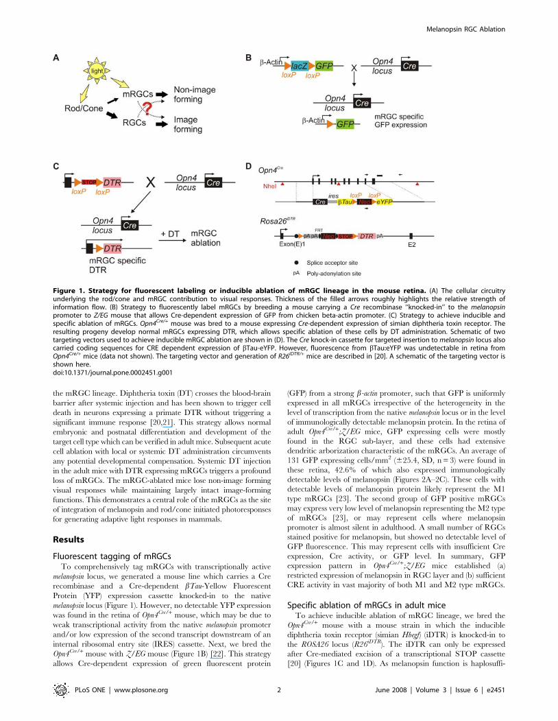
Figure 1. Strategy for fluorescent labeling or inducible ablation of mRGC lineage in the mouse retina. (A) The cellular circuitryunderlying the rod/cone and mRGC contribution to visual responses. Thickness of the filled arrows roughly highlights the relative strength ofinformation flow. (B) Strategy to fluorescently label mRGCs by breeding a mouse carrying a Cre recombinase ‘‘knocked-in’’ to the melanopsinpromoter to Z/EG mouse that allows Cre-dependent expression of GFP from chicken beta-actin promoter. (C) Strategy to achieve inducible andspecific ablation of mRGCs. Opn4Cre/+ mouse was bred to a mouse expressing Cre-dependent expression of simian diphtheria toxin receptor. Theresulting progeny develop normal mRGCs expressing DTR, which allows specific ablation of these cells by DT administration. Schematic of twotargeting vectors used to achieve inducible mRGC ablation are shown in (D). The Cre knock-in cassette for targeted insertion to melanopsin locus alsocarried coding sequences for CRE dependent expression of bTau-eYFP. However, fluorescence from bTau:eYFP was undetectable in retina fromOpn4Cre/+ mice (data not shown). The targeting vector and generation of R26iDTR/+ mice are described in [20]. A schematic of the targeting vector isshown here.doi:10.1371/journal.pone.0002451.g001
Melanopsin RGC Ablation
PLoS ONE | www.plosone.org 2 June 2008 | Volume 3 | Issue 6 | e2451
cient in mice [14], this approach allows normal differentiation and
function of the melanopsin cell lineage in the double heterozygote
mice (Opn4Cre/+;R26iDTR/+) and their specific ablation upon DT
administration.
The retina of 8 week old adult Opn4Cre/+;R26iDTR/+ mice
showed normal stratification and density of melanopsin-immuno-
staining (Figures 2D and 3A). Two weeks after DT administration
(intraperitoneal injections of DT at 50 mg/kg body weight, 2–3
times at 3 d apart) there was a dramatic reduction of mRGCs by
over 90% in Opn4Cre/+;R26iDTR/+(Figure 2E), but not in wild type
(WT) mice carrying only one or no copy of either of the two
transgenes (Figure 2F). The DT administration did not cause any
widespread cell death in the retina as the normal stratification of
the major cell layers remained intact (Figure 3A). This
demonstrated the accessibility of DT across the blood-retina
barrier and the success of the iDTR system in specific ablation of
differentiated RGC subtypes in live mice.
Normal image-forming visual responses in mRGC ablatedmice
Image-forming vision requires normal retina architecture, light-
induced electrical activities in the retina and RGC-mediated
transmission of the light information to the image processing brain
centers. As shown in Figure 3A, DT-induced mRGC ablation had
minimal impact on the overall retina architecture. These mice also
exhibited normal electroretinogram (ERG) (Figure 3B). Rod-
mediated responses to scotopic illumination, cone-mediated
responses to photopic illumination after photobleaching of rods
and maximal rod/cone combined responses were comparable in
WT and Opn4Cre/+;R26iDTR/+ mice injected with DT. Finally, to
evaluate pattern forming visual responses, we tested the perfor-
mance of mice in a visual cliff test (Figure 3C). The test evaluates
the ability of the mice to visually discriminate between a ‘‘safe’’
and an ‘‘unsafe’’ landing space and accordingly step down from a
slightly raised platform to the safe area. Typically, mice with intact
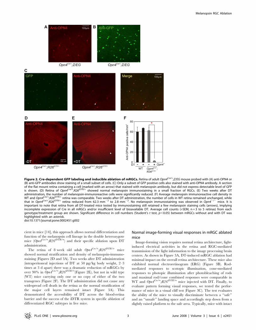
Figure 2. Cre-dependent GFP labeling and inducible ablation of mRGCs. Retina of adult Opn4Cre/+;Z/EG mouse probed with (A) anti-OPN4 or(B) anti-GFP antibodies show staining of a small subset of cells. (C) Only a subset of GFP positive cells also stained with anti-OPN4 antibody. A sectionof the flat mount retina containing a cell (marked with an arrow) that stained with melanopsin antibody, but did not express detectable level of GFPis shown. (D) Retina of Opn4Cre/+;R26iDTR/+ showed normal melanopsin immunostaining in a small fraction of RGCs. (E) Two weeks after DTadministration, the number of melanopsin-immunoreactive cells were significantly reduced. (F) Average melanopsin immunoreactive cell density inWT and Opn4Cre/+;R26iDTR/+ retina was comparable. Two weeks after DT administration, the number of cells in WT retina remained unchanged, whilethat in Opn4Cre/+;R26iDTR/+ retina reduced from 62.3 mm22 to 2.8 mm22. No melanopsin immunostaining was observed in Opn42/2 mice. It isimportant to note that retina from all DT-treated mice tested by immunostaining still retained a few melanopsin staining cells (arrows), implyingincomplete expression of Cre in all mRGCs and/or insufficient level of bioavailable DT. Average cell counts (+SEM, n = 3 to 5 retinas) from eachgenotype/treatment group are shown. Significant difference in cell numbers (Student’s t test, p,0.05) between mRGCs without and with DT washighlighted with an asterisk.doi:10.1371/journal.pone.0002451.g002
Melanopsin RGC Ablation
PLoS ONE | www.plosone.org 3 June 2008 | Volume 3 | Issue 6 | e2451
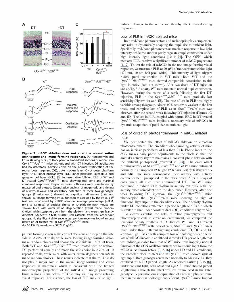
pattern forming vision make correct decisions and step on the safe
side in .70% of trials, while mice lacking image-forming vision
make random choices and choose the safe side in ,50% of trials.
Both WT and Opn4Cre/+;R26iDTR/+ mice treated with or without
DT performed equally and made the safe choice in .70% of 10
repeated trials, while mice with outer retina degeneration (rd/rd)
made random choices. These results indicate that the mRGCs do
not play a major role in the overall image-forming and visual
responses in mammals, which is consistent with the limited
monosynaptic projections of the mRGCs to image processing
brain regions. Nonetheless, mRGCs may still play some roles in
visual responses. For instance, the loss of PLR may cause light-
induced damage to the retina and thereby affect image-forming
responses.
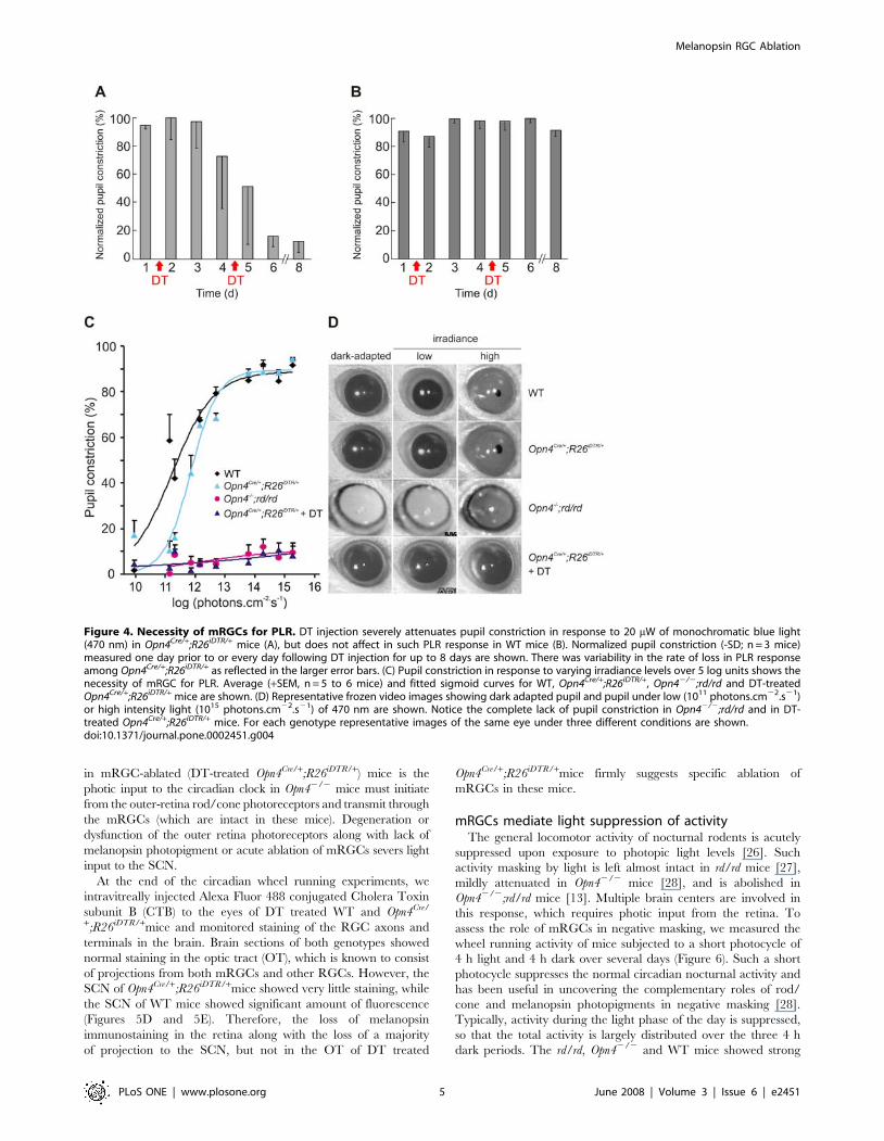
Loss of PLR in mRGC ablated miceBoth rod/cone photoreceptors and melanopsin play complemen-
tary roles in dynamically adapting the pupil size to ambient light.
Specifically, rod/cone photoreceptors mediate response to low light
intensity, while melanopsin partly regulates pupil constriction under
high intensity light conditions [12–14,24]. The OPN, which
mediates PLR, receives a significant number of mRGC projections
[4,17]. To test the role of mRGCs in the non-image forming visual
responses, we measured PLR at 20 mW of monochromatic blue light
(470 nm, 10 nm half-peak width). This intensity of light triggers
,80% pupil constriction in WT mice. Both WT and the
Opn4Cre/+;R26iDTR/+ mice showed comparable constriction to this
light intensity (data not shown). After two doses of DT injection
(50 mg/kg, 3 d apart), WT mice maintain normal pupil constriction.
However, during the course of a week following the first DT
injection, PLR in the Opn4Cre/+;R26iDTR/+ mice gradually lost
sensitivity (Figures 4A and 4B). The rate of loss in PLR was highly
variable among this group. Almost 90% sensitivity was lost in the first
week, and complete loss of PLR as in Opn42/2;rd/rd mice was
observed after the second week following DT injection (Figures 4C
and 4D). The loss in PLR, coupled with normal ERG in DT-treated
Opn4Cre/+;R26iDTR/+ mice implies a necessary role of mRGCs in
dynamic adaptation of pupil size to ambient light.
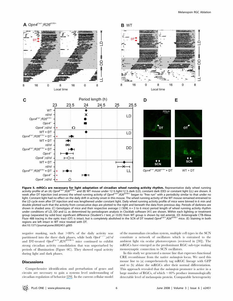
Loss of circadian photoentrainment in mRGC ablatedmice
We next tested the effect of mRGC ablation on circadian
photoentrainment. The circadian wheel running activity of mice
has an intrinsic periodicity of less than 24 h. Photic input to the
SCN makes daily phase adjustments to the clock so that the
animal’s activity rhythm maintains a constant phase relation with
the ambient photoperiod (reviewed in [25]). The daily wheel
running activity of Opn4Cre/+;R26iDTR/+ and of WT mice entrained
normally to an imposed 12 h light:12 h dark (LD) cycle (Figures 5A
and 5B). The mice consolidated their activity with activity
commencement juxtaposed to the dark onset. After 10 days of
entrainment all mice were administered DT. The WT mice
continued to exhibit 24 h rhythm in activity-rest cycle with the
activity onset coincident with the dark onset. However, after one
week following DT injection, the Opn4Cre/+;R26iDTR/+ mice
phenocopied the Opn42/2;rd/rd mice showing no sign of
functional light input to the circadian clock. Their activity rhythm
under LD conditions exhibited a period length of ,23.5 h which
is similar to that under constant dark (DD) conditions (Figure 5C).
To clearly establish the roles of retina photopigments and
photoreceptor cells in circadian entrainment, we compared the
temporal activity rhythms of DT-treated WT and DT-treated
Opn4Cre/+;R26iDTR/+ with those of rd/rd, Opn42/2 and Opn42/2;rd/rd
mice under three different lighting conditions: LD, DD and LL
(constant light). Mice with complete loss of photopigments or acute
loss of mRGC lineage in adulthood showed a DD period length that
was indistinguishable from that of WT mice, thus implying normal
function of the SCN oscillator sustains without tonic input from the
mRGCs. As shown before [8,15,16] under LD and LL conditions,
the circadian clock in rd/rd and in Opn42/2 mice showed signs of
light input. Both genotypes entrained normally to LD cycle; i.e. they
exhibited 24 h LD period length. As reported earlier [13,15,16],
under constant light, both rd/rd and Opn42/2 mice showed period
lengthening–although the effect was less pronounced in the latter
genotype. A parsimonious interpretation of circadian photoentrain-
ment in melanopsin photopigment deficient (Opn42/2) mice, but not
Figure 3. mRGC ablation does not alter the normal retinaarchitecture and image-forming responses. (A) Hematoxylin andEosin staining of 5 mm thick paraffin embedded sections of retina fromOpn4Cre/+;R26iDTR/+ mice without and with DT injection. DT applicationhad no detectable adverse effect on the normal stratification of theretina (outer segment (OS), outer nuclear layer (ONL), outer plexiformlayer (OPL), inner nuclear layer (INL), inner plexiform layer (IPL), andganglion cell layer (GCL)). (B) Representative full-field ERG of WT andDT-treated Opn4Cre/+;R26iDTR/+ mice showing rod, cone and maximalcombined responses. Responses from both eyes were simultaneouslymeasured and plotted. Quantitative analysis of magnitude and timingof a-wave, b-wave and oscillatory potentials of these two genotypegroups (3 mice each) showed no significant difference (data notshown). (C) Image forming visual function as assessed by the visual clifftest was unaffected by mRGC ablation. Average percentage (+SEM,n = 5 to 13 mice) of positive choice in 10 trials for each mouse areshown. Mice with outer retina degeneration (rd/rd) made randomchoices while stepping down from the platform and were significantlydifferent (Student’s t test, p,0.05; red asterisk) from the other fourgroups. No significant difference in test performance was found amongnative or DT-treated WT or Opn4Cre/+;R26iDTR/+ mice.doi:10.1371/journal.pone.0002451.g003
Melanopsin RGC Ablation
PLoS ONE | www.plosone.org 4 June 2008 | Volume 3 | Issue 6 | e2451
in mRGC-ablated (DT-treated Opn4Cre/+;R26iDTR/+) mice is the
photic input to the circadian clock in Opn42/2 mice must initiate
from the outer-retina rod/cone photoreceptors and transmit through
the mRGCs (which are intact in these mice). Degeneration or
dysfunction of the outer retina photoreceptors along with lack of
melanopsin photopigment or acute ablation of mRGCs severs light
input to the SCN.
At the end of the circadian wheel running experiments, we
intravitreally injected Alexa Fluor 488 conjugated Cholera Toxin
subunit B (CTB) to the eyes of DT treated WT and Opn4Cre/
+;R26iDTR/+mice and monitored staining of the RGC axons and
terminals in the brain. Brain sections of both genotypes showed
normal staining in the optic tract (OT), which is known to consist
of projections from both mRGCs and other RGCs. However, the
SCN of Opn4Cre/+;R26iDTR/+mice showed very little staining, while
the SCN of WT mice showed significant amount of fluorescence
(Figures 5D and 5E). Therefore, the loss of melanopsin
immunostaining in the retina along with the loss of a majority
of projection to the SCN, but not in the OT of DT treated
Opn4Cre/+;R26iDTR/+mice firmly suggests specific ablation of
mRGCs in these mice.
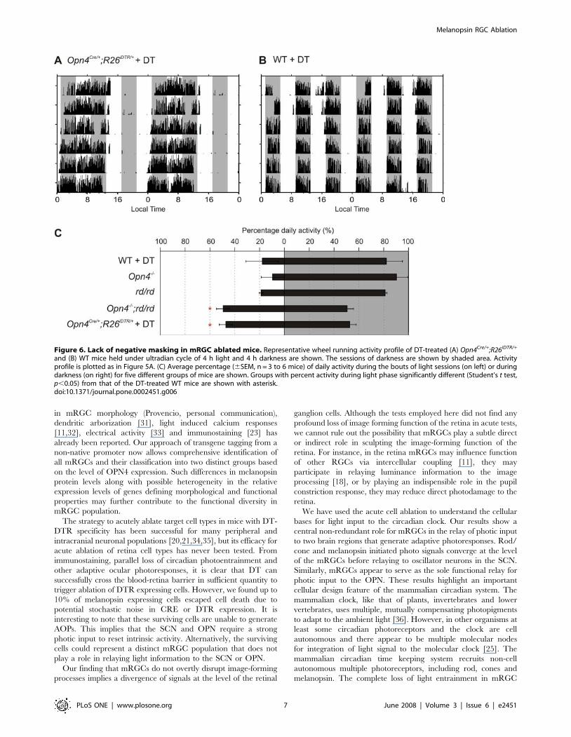
mRGCs mediate light suppression of activityThe general locomotor activity of nocturnal rodents is acutely
suppressed upon exposure to photopic light levels [26]. Such
activity masking by light is left almost intact in rd/rd mice [27],
mildly attenuated in Opn42/2 mice [28], and is abolished in
Opn42/2;rd/rd mice [13]. Multiple brain centers are involved in
this response, which requires photic input from the retina. To
assess the role of mRGCs in negative masking, we measured the
wheel running activity of mice subjected to a short photocycle of
4 h light and 4 h dark over several days (Figure 6). Such a short
photocycle suppresses the normal circadian nocturnal activity and
has been useful in uncovering the complementary roles of rod/
cone and melanopsin photopigments in negative masking [28].
Typically, activity during the light phase of the day is suppressed,
so that the total activity is largely distributed over the three 4 h
dark periods. The rd/rd, Opn42/2 and WT mice showed strong
Figure 4. Necessity of mRGCs for PLR. DT injection severely attenuates pupil constriction in response to 20 mW of monochromatic blue light(470 nm) in Opn4Cre/+;R26iDTR/+ mice (A), but does not affect in such PLR response in WT mice (B). Normalized pupil constriction (-SD; n = 3 mice)measured one day prior to or every day following DT injection for up to 8 days are shown. There was variability in the rate of loss in PLR responseamong Opn4Cre/+;R26iDTR/+ as reflected in the larger error bars. (C) Pupil constriction in response to varying irradiance levels over 5 log units shows thenecessity of mRGC for PLR. Average (+SEM, n = 5 to 6 mice) and fitted sigmoid curves for WT, Opn4Cre/+;R26iDTR/+, Opn42/2;rd/rd and DT-treatedOpn4Cre/+;R26iDTR/+ mice are shown. (D) Representative frozen video images showing dark adapted pupil and pupil under low (1011 photons.cm22.s21)or high intensity light (1015 photons.cm22.s21) of 470 nm are shown. Notice the complete lack of pupil constriction in Opn42/2;rd/rd and in DT-treated Opn4Cre/+;R26iDTR/+ mice. For each genotype representative images of the same eye under three different conditions are shown.doi:10.1371/journal.pone.0002451.g004
Melanopsin RGC Ablation
PLoS ONE | www.plosone.org 5 June 2008 | Volume 3 | Issue 6 | e2451
negative masking, such that .80% of the daily activity was
partitioned into the three dark phases, while both Opn42/2;rd/rd
and DT-treated Opn4Cre/+;R26iDTR/+ mice continued to exhibit
strong circadian activity consolidation that was unperturbed by
periods of illumination (Figure 6C). They showed equal activity
during light and dark phases.
Discussions
Comprehensive identification and perturbation of genes and
circuits are necessary to gain a systems level understanding of
circadian regulation of behavior [29]. In the current cellular model
of the mammalian circadian system, multiple cell types in the SCN
constitute a network of oscillators which is entrained to the
ambient light via ocular photoreceptors (reviewed in [30]). The
mRGCs have emerged as the predominant RGC sub-type making
monosynaptic connection to SCN oscillators.
In this study we generated a mouse line that expresses functional
CRE recombinase from the native melanopsin locus. We used this
mouse line to (a) comprehensively tag mRGC lineage with GFP
and to (b) ablate the mRGCs after their normal differentiation.
This approach revealed that the melanopsin promoter is active in a
large number of RGCs, of which ,40% produce immunologically
detectable level of melanopsin protein. Comparable heterogeneity
Figure 5. mRGCs are necessary for light adaptation of circadian wheel running activity rhythm. Representative daily wheel runningactivity profile of an (A) Opn4Cre/+;R26iDTR/+ and (B) WT mouse under 12 h light:12 h dark (LD), constant dark (DD) or constant light (LL) are shown. Aweek after DT injection (red arrows) the wheel running activity of Opn4Cre/+;R26iDTR/+ began to ‘‘free run’’ with a periodicity similar to that under nolight. Constant light had no effect on the daily drift in activity onset in this mouse. The wheel running activity of the WT mouse remained entrained tothe LD cycle even after DT injection and was lengthened under constant light. Daily wheel running activity profile of mice were binned in 6 min anddouble plotted such that the activity from consecutive days are plotted to the right and beneath the data from previous day. Periods of darkness areshown in shaded area. (C) Genotypes of mice and their respective average (6SEM, n = 3 to 6 mice) period length of wheel running activity rhythmunder conditions of LD, DD and LL as determined by periodogram analysis in Clocklab software [41] are shown. Within each lighting or treatmentgroup (separated by solid box) significant difference (Student’s t test, p,0.05) from WT group is shown by red asterisk. (D) Anterograde CTB-AlexaFluor 488 tracing in the optic tract (OT) is intact, but is completely abolished in the SCN of DT treated Opn4Cre/+;R26iDTR/+ mice. (E) Staining in bothregions are left intact in WT mice treated with DT.doi:10.1371/journal.pone.0002451.g005
Melanopsin RGC Ablation
PLoS ONE | www.plosone.org 6 June 2008 | Volume 3 | Issue 6 | e2451
in mRGC morphology (Provencio, personal communication),
dendritic arborization [31], light induced calcium responses
[11,32], electrical activity [33] and immunostaining [23] has
already been reported. Our approach of transgene tagging from a
non-native promoter now allows comprehensive identification of
all mRGCs and their classification into two distinct groups based
on the level of OPN4 expression. Such differences in melanopsin
protein levels along with possible heterogeneity in the relative
expression levels of genes defining morphological and functional
properties may further contribute to the functional diversity in
mRGC population.
The strategy to acutely ablate target cell types in mice with DT-
DTR specificity has been successful for many peripheral and
intracranial neuronal populations [20,21,34,35], but its efficacy for
acute ablation of retina cell types has never been tested. From
immunostaining, parallel loss of circadian photoentrainment and
other adaptive ocular photoresponses, it is clear that DT can
successfully cross the blood-retina barrier in sufficient quantity to
trigger ablation of DTR expressing cells. However, we found up to
10% of melanopsin expressing cells escaped cell death due to
potential stochastic noise in CRE or DTR expression. It is
interesting to note that these surviving cells are unable to generate
AOPs. This implies that the SCN and OPN require a strong
photic input to reset intrinsic activity. Alternatively, the surviving
cells could represent a distinct mRGC population that does not
play a role in relaying light information to the SCN or OPN.
Our finding that mRGCs do not overtly disrupt image-forming
processes implies a divergence of signals at the level of the retinal
ganglion cells. Although the tests employed here did not find any
profound loss of image forming function of the retina in acute tests,
we cannot rule out the possibility that mRGCs play a subtle direct
or indirect role in sculpting the image-forming function of the
retina. For instance, in the retina mRGCs may influence function
of other RGCs via intercellular coupling [11], they may
participate in relaying luminance information to the image
processing [18], or by playing an indispensible role in the pupil
constriction response, they may reduce direct photodamage to the
retina.
We have used the acute cell ablation to understand the cellular
bases for light input to the circadian clock. Our results show a
central non-redundant role for mRGCs in the relay of photic input
to two brain regions that generate adaptive photoresponses. Rod/
cone and melanopsin initiated photo signals converge at the level
of the mRGCs before relaying to oscillator neurons in the SCN.
Similarly, mRGCs appear to serve as the sole functional relay for
photic input to the OPN. These results highlight an important
cellular design feature of the mammalian circadian system. The
mammalian clock, like that of plants, invertebrates and lower
vertebrates, uses multiple, mutually compensating photopigments
to adapt to the ambient light [36]. However, in other organisms at
least some circadian photoreceptors and the clock are cell
autonomous and there appear to be multiple molecular nodes
for integration of light signal to the molecular clock [25]. The
mammalian circadian time keeping system recruits non-cell
autonomous multiple photoreceptors, including rod, cones and
melanopsin. The complete loss of light entrainment in mRGC
Figure 6. Lack of negative masking in mRGC ablated mice. Representative wheel running activity profile of DT-treated (A) Opn4Cre/+;R26iDTR/+
and (B) WT mice held under ultradian cycle of 4 h light and 4 h darkness are shown. The sessions of darkness are shown by shaded area. Activityprofile is plotted as in Figure 5A. (C) Average percentage (6SEM, n = 3 to 6 mice) of daily activity during the bouts of light sessions (on left) or duringdarkness (on right) for five different groups of mice are shown. Groups with percent activity during light phase significantly different (Student’s t test,p,0.05) from that of the DT-treated WT mice are shown with asterisk.doi:10.1371/journal.pone.0002451.g006
Melanopsin RGC Ablation
PLoS ONE | www.plosone.org 7 June 2008 | Volume 3 | Issue 6 | e2451
ablated mice firmly establishes a unique mammalian-specific
cellular network design to integrate light information from these
photopigments to the circadian clock. The mRGCs in this design
serve as the principal site of signal integration and therefore, have
now emerged as a unique cellular target for therapeutic
intervention in circadian clock related disorders. The complete
loss of non-image forming photoresponses also highlights the role
of mRGCs as the principal cellular node where photic information
for image-forming and non-image forming responses diverge.
During the course of this study, another manuscript [37]
describing specific ablation of mRGCs was published. That study
employed direct expression of an attenuated diphtheria toxin from
the melanopsin locus, which caused slow degeneration of the
mRGCs over several months, as assessed by the loss of a Tau:LacZ
transgene expression or melanopsin immunostaining. Both these
techniques detect only a subset of mRGCs ([23] and Figure 2).
Therefore, the slow degeneration of non-image forming photo-
sensitivity in the study by Guler et al. [37] may have resulted from
likely heterogeneity in toxin production from the native promoter,
homeostatic synaptic strengthening and/or early developmental
compensation. Despite the differences in the pace of cell death,
and the approach, both manuscripts arrived at similar conclusion
that the mRGCs are the primary node of convergence of
melanopsin and rod/cone initiated photoresponses.
Materials and Methods
Generation of miceAll animal care and procedures were approved by the
Institutional Animal Care and Use Committee of the Genomics
Institute of the Novartis Research Foundation and The Salk
Institute for Biological Studies. R26iDTR/+ mouse strain was a kind
gift of Dr. Ari Waisman [20] and Z/EG mouse strain [22] was
purchased from Jackson Laboratory. A schematic diagram of the
targeting construct for generating the R26iDTR/+ mouse is shown in
Figure 1D.
The Opn4Cre/+ mouse was generated by replacing the first seven
exons of the mouse melanopsin gene with a gene cassette containing
Cre (Figure 1D). For generation of the targeting construct,
genomic DNA of 129S1SvImJ inbred mouse strain was used as a
template and 3.5 kb of genomic sequence immediately upstream of
the translation start site and 3.4 kb of sequence 39 distal to the 7th
exon of the of Opn4 gene were PCR amplified and cloned into two
multicloning sites flanking the Cre:IRES:bTau:loxP:Neo:lox-
P:eYFP cassette. The left arm of the targeting vector also carried
a HSV-TK resistance gene. The construct was linearized by NotI
digestion and microinjected into an embryonic stem (ES) cell line
from 129S mice. ES cell clones with integration of the targeting
construct were selected on G418, and 288 antibiotic resistant
clones were screened by PCR for homologous recombination of
the targeting construct to the melanopsin locus. Genomic DNA from
seven PCR positive clones was digested with NheI and subjected to
Southern blot hybridization with a probe that lies within the right
arm. Sequences of the genotyping PCR primers are; wild type
(Primer a = CACTTCAGAGACAGCCAGAAGCAGG, Primer
b = GACTGACACTGAAGCCTGGCAAACG) and mutant
(Primer a and Primer c = CCATTTCCGGTTATTCAACTTG-
CACC). One clone with appropriately recombined DNA was
injected into C57BL/6J blastocysts and introduced into C57BL/6J
pseudopregnant females. Chimeric male progeny were bred to
C57BL/6J females and the resulting heterozygous agouti coat-
colored progeny were mated with C57BL/6J. Heterozygous mice
were bred among each other and with Z/EG or Rosa26iDTR/+ mice
which were also in 129S;C57BL/6 mixed background. Targeted
integration of Cre leading to loss of melanopsin protein in Opn4Cre/
Cre mice was verified by lack of anti-OPN4 immunostaining (data
not shown). Opn4Cre/+;Z/EG mice were used for assessment of
CRE function and fluorescent tagging of mRGCs in the retina.
Littermate Opn4Cre/+;R26iDTR/+ and WT mice were used in all
subsequent experiments for acute ablation of mRGCs.
The Opn42/2 mice used in this study were generated in 129S
background and characterized earlier [15]. These mice were bred
to a line of C57BL/6 carrying the Pde6brd/rd mutation. Progeny
from this breeding, which were also in a 129S;C57BL/6
background were genotyped and mice of Opn42/2, rd/rd and
Opn42/2;rd/rd genotypes were used for behavioral studies.
DT injectionDT (D0564, Sigma-Aldrich, St. Louis, MO) was dissolved in
sterile PBS (1 mg/ml) and stored at 280uC till use. Freshly thawed
DT stock solution was diluted in sterile PBS and injected
intraperitoneally (50 mg/kg body weight) [21] to 8–12 weeks old
Opn4Cre/+;R26iDTR/+ and WT littermate mice. The dose was
repeated once or twice at 3 d interval.
Retina stainingFor flat mount, adult mice were sacrificed, the eyes quickly
removed and placed into aerated Ames medium (Sigma-Aldrich,
St. Louis, MO). After removal of the corneas and lenses, eyecups
were fixed in 4% paraformaldehyde for 15 min. Following three
washes in PBS, retinas were dissected from eyecups, stretched onto
filter paper, and processed in 24-well plates. The retinas were
incubated in a blocking solution (0.3% Triton X-100, 5% normal
donkey serum, and 0.5% glycine in PBS) for 1 h at room
temperature. After three washes in PBS, the retinas were
incubated in a 1:5,000 dilution of rabbit anti-OPN4 antiserum
(against a peptide consisting of the 15 N-terminal amino acids of
mouse melanopsin [38]) or in 1:500 dilution of rabbit anti-GFP
antibody (Cat# A11122, Invitrogen, Carlsbad, CA) in the
blocking solution for overnight at 4uC and rinsed with PBS.
Melanopsin immunoreactivity was visualized with Cy3-conjugated
donkey anti-rabbit IgG (1:500, Cat# 711-165-152, Jackson
ImmunoResearch Laboratories, West Grove, PA) or FITC-
conjugated anti-rabbit IgG (1:20, Cat# 401314, Calbiochem,
San Diego, CA) in blocking solution. Finally, retinas were washed
with PBS and mounted with PermaFluor (Cat# IM0752,
Beckman Coulter, Fullerton, CA). Fluorescent images were
captured using an Olympus Fluoview500 confocal microscope or
Leica TCS SP2 AOBS confocal microscope. For Hematoxylin and
Eosin (H&E) staining, eyes were removed and eyecups were fixed
in 4% paraformaldehyde for 2 h, washed three times with PBS,
and embedded in paraffin. Five micron thick paraffin sections
were used for H&E staining. Stained slides were visualized under a
Leica microscope.
Anterograde tracing with cholera toxinMice were anesthetized with ketamine (70 mg/kg) and xylazine
(10 mg/kg) and one drop each of 1% tropicamide and 0.5%
proparacaine (Bausch & Lomb, Tampa, FL) were applied to their
eyes. An incision was made with a 30 gauge needle below the
limbus region and 1 ml of 1% Cholera Toxin-B subunit (CTB)
conjugated to Alexa Fluor 488 (Cat# C34775, Invitrogen, CA)
was injected into the vitreous. After 48 h, the mice were
anesthetized with ketamine (70 mg/kg) and xylazine (10 mg/kg),
perfused with 4% paraformaldehyde in 16PBS. The brain was
extracted, further fixed at 4uC overnight, embedded in OCT and
40 mm thick sections cut in a microtome were imaged using an
Olympus Fluoview500 confocal microscope.
Melanopsin RGC Ablation
PLoS ONE | www.plosone.org 8 June 2008 | Volume 3 | Issue 6 | e2451
ERGMice were dark adapted for 1 h, anesthetized with ketamine
(70 mg/kg) and xylazine (10 mg/kg). The eyes were applied with
1% tropicamide, 0.5% proparacaine and Genteal lubricant eye gel
(CIBA vision, Duluth, GA, USA). Electrodes were connected
under dim red light. ERGs were recorded from both eyes with an
ERG system (Model LE-2000, Tomey, Nagoya, Japan). White
LED light embedded in contact lens recording electrodes were
used as light sources. ERG recordings were done following the
standards of the International Society for Clinical Electrophysiol-
ogy of Vision (ISCEV; http://www.iscev.org/standards/erg1999.
html). After an additional 5 min of dark adaptation, rod response
was measured by averaging 4 dim light flashes of 80 cd.m2260.12
msec at .2 sec interval. Then mice were further dark adapted for
3 min and maximal combined (rod and cone) response and
oscillatory potential were determined from the average of
responses to four flashes of 6,000 cd.m2260.5 msec at .10 sec
inter-stimulus interval. Finally, after 10min light adaptation (25
cd.m22), average single-flash cone response was determined from
4 flashes of 6,000 cd.m2260.5 msec at .15 sec interval.
Visual cliff testThe visual cliff test was assessed as described in [39]. Half of a
31 cm length645 cm width650 cm tall clear acrylic box had a black
and white checkerboard pattern on the top (the ‘‘safe’’ side) and the
other side had a clear top (the ‘‘unsafe’’ side/cliff side). The unsafe
side had a checkerboard pattern on the bottom to give an illusion of
added depth. Black paper was placed on the inside of the walls to
reduce reflections. A block of 31 cm length63.5 cm width62.5 cm
height was placed at the center of the box to separate these two sides.
The mouse was placed on the block and allowed to make a
spontaneous step down between the safe side and the unsafe side.
Data was measured as positive when the mouse chose to step down
onto the horizontal checkered surface (the safe side) and as negative
onto the clear side (the unsafe side). Each mouse was tested for 10
consecutive trials. Fifty percent positive indicated that the mouse
moved off the ridge onto the two sides with random chance.
PLRPLR was assessed as described in [40] with some modification.
Mice were dark adapted for at least an hour prior to PLR
assessment. Unanaesthetized mice were hand restrained with the
left eye apposed to one of the outlets of an integrating sphere. The
pupil of right eye was video monitored under infrared light with a
consumer grade digital video camera. An additional +2 diaptor
lens and an infrared filter were fitted to the light path of the
camera to improve the image quality. After collecting images of
dark adapted pupils, the contralateral eye was exposed to light
from the integrating sphere. Light from a 300 Watt Xenon Arc
lamp light source (Sutter Instrument, Novato, CA, USA) was
filtered, collimated and delivered to the integrating sphere through
a liquid light guide. An inline 470 nm filter, a filter wheel with a
series of neutral density filters, and a Lambda 10-3 optical filter
changer with SmartShutterTM were used to control the spectral
quality, intensity and duration of light. Light intensity was
measured with a Melles Griot power meter. Video images of
pupil constriction were analyzed by ViewPoint EyeTrackerTM
software (Arrington Research Inc., Scottsdale, AZ). Dark adapted
pupil area prior to light pulse and pupil area after 30 sec of light
were measured and pupil constriction was defined as 1-(dark
adapted pupil width)̂2/(pupil width after 30 sec of light)̂2. The
squares come in to account for the calculation of pupil area. Pupil
area constriction was plotted against the log of irradiance. Data
was fitted to a sigmoidal dose response curve with the lower
asymptote fixed to y = 0 using Origin lab 8 software.
For tracking the gradual changes in PLR response following DT
injection, pupil area constrictions over 8 days were normalized to
maximum constriction taken as 100%.
Locomotor activity measurementDaily locomotor activity of mice individually housed in wheel
running cages was measured as described in [41]. Typically, 6–
10 week old mice were individually housed in wheel running cages
placed inside light tight boxes with independent illumination.
During the light phase, the mice received ,150 lux of white light
from fluorescent light source. Wheel running activity in 1 min bins
was continuously collected and later analyzed by Clocklab
software (Actimetrics, Evanston, IL, USA). All routine animal
husbandry practices during the dark phase were performed under
dim red light.
Acknowledgments
We would like to thank Dr. Ari Waisman for providing R26iDTR/+ mouse
strain, Van M. Lee, Victoria Piamonte, Kathryn Spencer and Dusko
Trajkovic for technical assistance, and members of the Panda lab for
critical discussion of the results.
Author Contributions
Conceived and designed the experiments: SP. Performed the experiments:
CS SP TJ MH HL NT SK. Analyzed the data: SP MH VC. Contributed
reagents/materials/analysis tools: CS TJ. Wrote the paper: SP MH.
References
1. Provencio I, Rodriguez IR, Jiang G, Hayes WP, Moreira EF, et al. (2000) A
novel human opsin in the inner retina. J Neurosci 20: 600–605.
2. Gooley JJ, Lu J, Chou TC, Scammell TE, Saper CB (2001) Melanopsin in cells
of origin of the retinohypothalamic tract. Nat Neurosci 4: 1165.
3. Berson DM, Dunn FA, Takao M (2002) Phototransduction by retinal ganglion
cells that set the circadian clock. Science 295: 1070–1073.
4. Hattar S, Liao HW, Takao M, Berson DM, Yau KW (2002) Melanopsin-
containing retinal ganglion cells: architecture, projections, and intrinsic
photosensitivity. Science 295: 1065–1070.
5. Belenky MA, Smeraski CA, Provencio I, Sollars PJ, Pickard GE (2003)
Melanopsin retinal ganglion cells receive bipolar and amacrine cell synapses.
J Comp Neurol 460: 380–393.
6. Nayak SK, Jegla T, Panda S (2006) Role of a novel photopigment, melanopsin,
in behavioral adaptation to light. Cell Mol Life Sci 64: 144–154.
7. Keeler CE (1927) Iris movements in blind mice. American Journal of Physiology
81: 107–112.
8. Foster RG, Provencio I, Hudson D, Fiske S, De Grip W, et al. (1991) Circadian
photoreception in the retinally degenerate mouse (rd/rd). J Comp Physiol [A]
169: 39–50.
9. Lucas RJ, Freedman MS, Munoz M, Garcia-Fernandez JM, Foster RG (1999)
Regulation of the mammalian pineal by non-rod, non-cone, ocular photore-
ceptors. Science 284: 505–507.
10. Mrosovsky N, Lucas RJ, Foster RG (2001) Persistence of masking responses to
light in mice lacking rods and cones. J Biol Rhythms 16: 585–588.
11. Sekaran S, Foster RG, Lucas RJ, Hankins MW (2003) Calcium imaging reveals a
network of intrinsically light-sensitive inner-retinal neurons. Curr Biol 13: 1290–1298.
12. Hattar S, Lucas RJ, Mrosovsky N, Thompson S, Douglas RH, et al. (2003)
Melanopsin and rod-cone photoreceptive systems account for all major
accessory visual functions in mice. Nature 424: 76–81.
13. Panda S, Provencio I, Tu DC, Pires SS, Rollag MD, et al. (2003) Melanopsin is
required for non-image-forming photic responses in blind mice. Science 301:
525–527.
14. Lucas RJ, Hattar S, Takao M, Berson DM, Foster RG, et al. (2003) Diminished
pupillary light reflex at high irradiances in melanopsin-knockout mice. Science
299: 245–247.
15. Panda S, Sato TK, Castrucci AM, Rollag MD, DeGrip WJ, et al. (2002)
Melanopsin (Opn4) requirement for normal light-induced circadian phase
shifting. Science 298: 2213–2216.
Melanopsin RGC Ablation
PLoS ONE | www.plosone.org 9 June 2008 | Volume 3 | Issue 6 | e2451
16. Ruby NF, Brennan TJ, Xie X, Cao V, Franken P, et al. (2002) Role of
melanopsin in circadian responses to light. Science 298: 2211–2213.17. Hattar S, Kumar M, Park A, Tong P, Tung J, et al. (2006) Central projections of
melanopsin-expressing retinal ganglion cells in the mouse. J Comp Neurol 497:
326–349.18. Dacey DM, Liao HW, Peterson BB, Robinson FR, Smith VC, et al. (2005)
Melanopsin-expressing ganglion cells in primate retina signal colour andirradiance and project to the LGN. Nature 433: 749–754.
19. Barnard AR, Hattar S, Hankins MW, Lucas RJ (2006) Melanopsin regulates
visual processing in the mouse retina. Curr Biol 16: 389–395.20. Buch T, Heppner FL, Tertilt C, Heinen TJ, Kremer M, et al. (2005) A Cre-
inducible diphtheria toxin receptor mediates cell lineage ablation after toxinadministration. Nat Methods 2: 419–426.
21. Luquet S, Perez FA, Hnasko TS, Palmiter RD (2005) NPY/AgRP neurons areessential for feeding in adult mice but can be ablated in neonates. Science 310:
683–685.
22. Novak A, Guo C, Yang W, Nagy A, Lobe CG (2000) Z/EG, a double reportermouse line that expresses enhanced green fluorescent protein upon Cre-
mediated excision. Genesis 28: 147–155.23. Baver SB, Pickard GE, Sollars PJ (2008) Two types of melanopsin retinal
ganglion cell differentially innervate the hypothalamic suprachiasmatic nucleus
and the olivary pretectal nucleus. Eur J Neurosci 27: 1763–1770.24. Lucas RJ, Douglas RH, Foster RG (2001) Characterization of an ocular
photopigment capable of driving pupillary constriction in mice. Nat Neurosci 4:621–626.
25. Panda S, Hogenesch JB, Kay SA (2003) Circadian light input in plants, flies andmammals. Novartis Found Symp 253: 73–82. discussion 82–78, 102–109, 281–
104..
26. Mrosovsky N (1999) Masking: history, definitions, and measurement. Chron-obiol Int 16: 415–429.
27. Mrosovsky N, Foster RG, Salmon PA (1999) Thresholds for masking responsesto light in three strains of retinally degenerate mice. J Comp Physiol [A] 184:
423–428.
28. Mrosovsky N, Hattar S (2003) Impaired masking responses to light inmelanopsin-knockout mice. Chronobiol Int 20: 989–999.
29. De Haro L, Panda S (2006) Systems biology of circadian rhythms: an outlook.
J Biol Rhythms 21: 507–518.
30. Herzog ED (2007) Neurons and networks in daily rhythms. Nat Rev Neurosci 8:
790–802.
31. Provencio I, Rollag MD, Castrucci AM (2002) Photoreceptive net in the
mammalian retina. This mesh of cells may explain how some blind mice can still
tell day from night. Nature 415: 493.
32. Sekaran S, Lupi D, Jones SL, Sheely CJ, Hattar S, et al. (2005) Melanopsin-
dependent photoreception provides earliest light detection in the mammalian
retina. Curr Biol 15: 1099–1107.
33. Tu DC, Zhang D, Demas J, Slutsky EB, Provencio I, et al. (2005) Physiologic
diversity and development of intrinsically photosensitive retinal ganglion cells.
Neuron 48: 987–999.
34. Tatsumi S, Ishii K, Amizuka N, Li M, Kobayashi T, et al. (2007) Targeted
ablation of osteocytes induces osteoporosis with defective mechanotransduction.
Cell Metab 5: 464–475.
35. Bennett CL, Clausen BE (2007) DC ablation in mice: promises, pitfalls, and
challenges. Trends Immunol 28: 525–531.
36. Panda S (2007) Multiple photopigments entrain the Mammalian circadian
oscillator. Neuron 53: 619–621.
37. Guler AD, Ecker JL, Lall GS, Haq S, Altimus CM, et al. (2008) Melanopsin cells
are the principal conduits for rod-cone input to non-image-forming vision.
Nature 453: 102–105.
38. Pulivarthy SR, Tanaka N, Welsh DK, De Haro L, Verma IM, et al. (2007)
Reciprocity between phase shifts and amplitude changes in the mammalian
circadian clock. Proc Natl Acad Sci U S A 104: 20356–20361.
39. Crawley JN (2000) Behavioral phenotyping of transgenic and knockout mice. In:
Crawley JN, ed (2000) What is wrong with my mouse? 1 ed. New York: Wiley-
Liss. pp 65–81.
40. Van Gelder RN (2005) Nonvisual ocular photoreception in the mammal.
Methods Enzymol 393: 746–755.
41. Siepka SM, Takahashi JS (2005) Methods to record circadian rhythm wheel
running activity in mice. Methods Enzymol 393: 230–239.
Melanopsin RGC Ablation
PLoS ONE | www.plosone.org 10 June 2008 | Volume 3 | Issue 6 | e2451
Copyright © 2022 FDOKUMEN