Corpus Callosum Structure is Topographically Correlated with the Early Course of Cognition and...
12
Uncorrected Author Proof Journal of Alzheimer’s Disease xx (20xx) x–xx DOI 10.3233/JAD-142895 IOS Press 1 Corpus Callosum Structure is Topographically Correlated with the Early Course of Cognition and Depression in Alzheimer’s Disease 1 2 3 4 Margherita Di Paola a,∗ , Owen Phillips a , Maria Donata Orfei a , Fabrizio Piras a , Claudia Cacciari a , Carlo Caltagirone a,b and Gianfranco Spalletta a,∗ 5 6 a Department of Clinical and Behavioural Neurology,IRCCS Santa Lucia Foundation, Rome, Italy 7 b Department of Neuroscience, Tor Vergata University, Rome, Italy 8 Accepted 15 January 2015 Abstract. Corpus callosum (CC) abnormalities may cause cognitive and neuropsychiatric complications due to reduced hemi- spheric integration. Over a one-year period, we investigated whether the CC structure of 20 patients with mild Alzheimer’s disease (AD) was linked to the evolution of cognitive and neuropsychiatric symptoms. We also investigated whether this anatomical- clinical relationship was localized topographically on the CC by combining voxel-based morphometry and diffusion tensor imaging approaches. We assessed patients’ global cognitive deterioration and neuropsychiatric symptoms with the Mini-Mental State Examination and the Neuropsychiatric Inventory. Increased global cognitive deterioration during the early course of AD was significantly related to reduced white matter density (p = 0.004) and fractional anisotropy (FA) (p = 0.012) and increased mean diffusivity (MD) (p =0.017) at the level of the CC isthmus/splenium. Further, increased depression severity was signif- icantly related to reduced FA (p = 0.008) and increased MD (p = 0.018) at the level of the CC rostrum. These results indicate that changes in early myelinated CC fibers, which subserve the lateral temporal and parietal cortices and are less vulnerable to damage, may be related to cognitive impairment. Furthermore, changes in late myelinated CC fibers, which connect the orbitofrontal cortices and are more vulnerable to damage, may be related to the earliest neuropsychiatric symptoms of AD, such as depression. 9 10 11 12 13 14 15 16 17 18 19 20 21 Keywords: Cognitive domain, corpus callosum, depression, diffusion tensor imaging , diffusivity, drug-free, first diagnosed AD, fractional anisotropy, mean diffusivity, mild Alzheimer’s disease, Mini-Mental State Examination, neuropsychiatric symptoms, structural MRI, volume, voxel-based morphometry 22 23 24 INTRODUCTION 25 Alzheimer’s disease (AD) is a neurodegenerative 26 disorder with clinical manifestations of cognitive 27 ∗ Correspondence to: Margherita Di Paola, Psy. S., PhD, IRCCS Santa Lucia Foundation, Via Ardeatina 306, 00179 Rome, Italy. Tel.: +39 06 51501121; Fax: +39 06 51501213; E-mail: [email protected]. and Gianfranco Spalletta, MD, PhD, IRCCS Santa Lucia Foundation, Via Ardeatina 306, 00179 Rome, Italy. Tel.: +39 06 51501575; Fax: +39 06 51501575; E-mail: [email protected]. impairment that are quite linear during the course 28 of the illness [1]. By contrast, the course of the 29 neuropsychiatric manifestations, including affective, 30 psychotic, and behavioral symptoms is unpredictable. 31 Neuropsychiatric symptoms have a specific pattern 32 of temporal onset. Apathy, anxiety, and depression 33 are already present in the early stages of the illness 34 [2–5], and delusions, hallucinations, and aggression 35 are more common later in the disease course [6]. More- 36 over, with the exception of apathy, which increases 37 continuously with the progression of disease severity 38 ISSN 1387-2877/15/$35.00 © 2015 – IOS Press and the authors. All rights reserved
Transcript of Corpus Callosum Structure is Topographically Correlated with the Early Course of Cognition and...

Unc
orre
cted
Aut
hor P
roof
Journal of Alzheimer’s Disease xx (20xx) x–xxDOI 10.3233/JAD-142895IOS Press
1
Corpus Callosum Structure isTopographically Correlated with theEarly Course of Cognition and Depressionin Alzheimer’s Disease
1
2
3
4
Margherita Di Paolaa,∗, Owen Phillipsa, Maria Donata Orfeia, Fabrizio Pirasa, Claudia Cacciaria,Carlo Caltagironea,b and Gianfranco Spallettaa,∗
5
6
aDepartment of Clinical and Behavioural Neurology, IRCCS Santa Lucia Foundation, Rome, Italy7
bDepartment of Neuroscience, Tor Vergata University, Rome, Italy8
Accepted 15 January 2015
Abstract. Corpus callosum (CC) abnormalities may cause cognitive and neuropsychiatric complications due to reduced hemi-spheric integration. Over a one-year period, we investigated whether the CC structure of 20 patients with mild Alzheimer’s disease(AD) was linked to the evolution of cognitive and neuropsychiatric symptoms. We also investigated whether this anatomical-clinical relationship was localized topographically on the CC by combining voxel-based morphometry and diffusion tensorimaging approaches. We assessed patients’ global cognitive deterioration and neuropsychiatric symptoms with the Mini-MentalState Examination and the Neuropsychiatric Inventory. Increased global cognitive deterioration during the early course of ADwas significantly related to reduced white matter density (p = 0.004) and fractional anisotropy (FA) (p = 0.012) and increasedmean diffusivity (MD) (p = 0.017) at the level of the CC isthmus/splenium. Further, increased depression severity was signif-icantly related to reduced FA (p = 0.008) and increased MD (p = 0.018) at the level of the CC rostrum. These results indicatethat changes in early myelinated CC fibers, which subserve the lateral temporal and parietal cortices and are less vulnerableto damage, may be related to cognitive impairment. Furthermore, changes in late myelinated CC fibers, which connect theorbitofrontal cortices and are more vulnerable to damage, may be related to the earliest neuropsychiatric symptoms of AD, suchas depression.
9
10
11
12
13
14
15
16
17
18
19
20
21
Keywords: Cognitive domain, corpus callosum, depression, diffusion tensor imaging , diffusivity, drug-free, first diagnosed AD,fractional anisotropy, mean diffusivity, mild Alzheimer’s disease, Mini-Mental State Examination, neuropsychiatric symptoms,structural MRI, volume, voxel-based morphometry
22
23
24
INTRODUCTION25
Alzheimer’s disease (AD) is a neurodegenerative26
disorder with clinical manifestations of cognitive27
∗Correspondence to: Margherita Di Paola, Psy. S., PhD, IRCCSSanta Lucia Foundation, Via Ardeatina 306, 00179 Rome, Italy.Tel.: +39 06 51501121; Fax: +39 06 51501213; E-mail:[email protected]. and Gianfranco Spalletta, MD, PhD,IRCCS Santa Lucia Foundation, Via Ardeatina 306, 00179 Rome,Italy. Tel.: +39 06 51501575; Fax: +39 06 51501575; E-mail:[email protected].
impairment that are quite linear during the course 28
of the illness [1]. By contrast, the course of the 29
neuropsychiatric manifestations, including affective, 30
psychotic, and behavioral symptoms is unpredictable. 31
Neuropsychiatric symptoms have a specific pattern 32
of temporal onset. Apathy, anxiety, and depression 33
are already present in the early stages of the illness 34
[2–5], and delusions, hallucinations, and aggression 35
are more common later in the disease course [6]. More- 36
over, with the exception of apathy, which increases 37
continuously with the progression of disease severity 38
ISSN 1387-2877/15/$35.00 © 2015 – IOS Press and the authors. All rights reserved

Unc
orre
cted
Aut
hor P
roof
2 M.D. Paola et al. / Corpus Callosum and AD Symptom Course
[2], cognitive and neuropsychiatric alterations seem39
to be independent [3], thus making AD even more40
heterogeneous.41
Many studies have investigated the brain correlates42
of cognitive deficits and neuropsychiatric symptoms43
in the early and late stages of AD pathology. They44
show that these clinical manifestations are associated45
with both regional grey matter damage ([7, 8] for a46
review) and abnormal white matter [9–12]. However,47
most studies are cross-sectional and report the brain48
changes resulting from AD in patients who are at dif-49
ferent phases of the illness.50
We previously studied white matter changes in AD51
by selectively investigating the largest commissure in52
the brain, i.e., the corpus callosum (CC), and found that53
it was already affected in the early stages of the disease54
[13–15]. Although many other studies have reported55
changes in the white matter of the CC, the precise cal-56
losal pattern modification varies from small regions to57
the entire commissure (reviewed in [15]).58
The CC is the most important structure involved59
in transmitting interhemispheric information. As the60
organization in the callosal commissure is roughly61
topographical, different patterns of interhemispheric62
disconnection may lead to different forms of callosal63
atrophy, resulting in heterogeneous functional/clinical64
disabilities. Nevertheless, the potential relationship65
between the CC structure and changes in clinical per-66
formance in AD over time is not well known. Thus, the67
present study was designed to investigate whether the68
changes in cognitive and neuropsychiatric symptoms69
in a group of mild AD patients after one year already70
correlated with the callosal structure at baseline.71
To achieve this goal, we investigated a unique patient72
population using multimodal imaging methods. We73
studied newly diagnosed mild AD patients who were74
not taking psychotropic drugs or acetylcholinesterase75
inhibitors (AChEIs). They were drug-free at base-76
line. Here, we combined macro- and microstructural77
magnetic resonance imaging (MRI) methods, such as78
voxel-based morphometry (VBM) and diffusion tensor79
imaging (DTI), to investigate whether specific cal-80
losal subregions and specific neuroimaging parameters81
were related to the clinical changes. In fact, VBM82
allows assessing macrostructural brain changes (i.e.,83
atrophy) and DTI allows measuring the microstruc-84
tural integrity of transcallosal fibers. Both have been85
shown to be impaired in AD (see [15] for a review).86
Imaging acquisition was performed only at baseline,87
whereas clinical, cognitive, and neuropsychiatric mea-88
sures were evaluated at baseline and re-evaluated89
a year later.90
MATERIALS AND METHODS 91
Subjects 92
We included patients with a diagnosis of mild AD 93
who were recruited consecutively in our memory clinic 94
in Rome, Italy. The diagnosis of probable AD was 95
made by a trained clinical neuropsychiatrist, accord- 96
ing to the revised criteria for AD published by the 97
Alzheimer’s Association Research Roundtable [16]. 98
We included only drug-free patients with a new diag- 99
nosis of AD who were not undergoing treatment with 100
AChEI and had not been treated with psychotropic 101
drugs (i.e., antidepressants, antipsychotics, anxiolyt- 102
ics, or mood stabilizers) in the previous two years. 103
Inclusion criteria were: 1) mild global cognitive 104
impairment, defined as a Mini-Mental State Exami- 105
nation (MMSE) score higher than 18 and a Clinical 106
Dementia Rating (CDR) Scale score of 1; 2) laboratory 107
values within the appropriate reference intervals; 3) 108
vision and hearing sufficient for compliance with test- 109
ing procedures (eyeglasses and/or hearing aids were 110
permissible). Exclusion criteria were: 1) major med- 111
ical illnesses (e.g., unstabilized diabetes, obstructive 112
pulmonary disease or asthma; hematologic/oncologic 113
disorders; vitamin B12 or folate deficiency, as evi- 114
denced by blood concentrations below the lower limits 115
of the reference intervals; pernicious anemia; clini- 116
cally significant and unstable active gastrointestinal, 117
renal, hepatic, endocrine or cardiovascular system dis- 118
ease; newly treated hypothyroidism); 2) comorbidity 119
of primary psychiatric or neurological disorders (e.g., 120
schizophrenia, mood depression, stroke, Parkinson’s 121
disease, seizure disorder and head injury with loss 122
of consciousness) or any other significant mental or 123
neurological disorder; 3) known or suspected his- 124
tory of drug/alcohol dependence and abuse during 125
the lifetime; 4) any potential brain abnormalities or 126
microvascular lesions appearing on conventional T2- 127
and FLAIR-scans; in particular, the presence, sever- 128
ity, and location of vascular lesions was rated by two 129
expert radiologists who used a protocol designed for 130
the Rotterdam Scan Study [17]. Generally, they were 131
considered present in cases of hyperintense lesions 132
on both proton-density and T2-weighted (see image 133
acquisition) images and rated semiquantitatively as 0 134
(none), 1 (pencil-thin lining), 2 (smooth halo), or 3 135
(large confluent) for three separate regions, i.e., adja- 136
cent to the frontal horns (frontal caps), adjacent to 137
the wall of the lateral ventricles (bands), and adja- 138
cent to the occipital horns (occipital caps). The total 139
vascular lesion load was calculated by summing the 140

Unc
orre
cted
Aut
hor P
roof
M.D. Paola et al. / Corpus Callosum and AD Symptom Course 3
Table 1Socio-demographic and clinical characteristics of 20 patients with mild Alzheimer’s disease (AD) at the first diagnostic visit and the one-year
follow-up
Subject Group Statistic
Time 0 One-year follow-up Paired t-test df p value
Gender (F/M) 11/9Age, Mean (SD) 72.75 (6.77)Range 59–85Educational level, Mean (SD) 6.9 (4.05)Range 1–18MMSE scores, Mean (SD) 23.40 (2.62) 22.50 (3.98) 1.119 19 0.277a
Range 19–28 10–28CDR value 1 (0.00) 1.15 (0.37) –2.042 19 0.55a
ADL 7.15 (1.46) 9.00 (2.71) –3.231 19 0.004a
IADL 12.4 (4.42) 15.30 (5.29) –2.964 19 0.008a
MMSE, Mini-Mental State Examination; CDR, Clinical Dementia Rating; ADL, Activities of Daily Living; IADL, Instrumental Activities ofDaily Living. a mild AD time zero < mild AD after 12 months: paired T test (when referred to a cognitive or clinical scale comparison, lowerpunctuations mean lesser impairment).
region-specific scores (range, 0–9). In the present141
study, only participants who obtained a score of 0-1142
were included; 5) lack of a “reliable” caregiver who143
was able to report to the clinic, fill in the scales, ensure144
compliance with treatment and clinical visits (MRI145
acquisition, neuropsychological and psychiatric eval-146
uation at baseline and at the 12-month follow-up), and147
contact the patient at least twice a week (one contact148
had to be a personal visit).149
After the first screening, 15 out of 40 patients were150
excluded from the initial sample because they did151
not fulfill the inclusion criteria: six had major medi-152
cal illnesses, two lacked reliable caregivers, three had153
comorbidity of primary psychiatric or neurological dis-154
orders, and four had MRI evidence of parenchymal155
abnormalities or neoplasms. Thus, 25 patients were156
included at baseline.157
After one year, the 25 AD patients underwent158
follow-up examinations to investigate the progression159
of global cognitive deterioration, performance of basic160
(ADL) and instrumental (IADL) activities of daily liv-161
ing and severity of neuropsychiatric disorders. Five162
out of the 25 patients did not complete the follow-up163
procedure: three withdrew from the study for different164
reasons (unwillingness to continue, lack of caregiver165
reliability, death) and two developed major medical166
illnesses (e.g., ictus, renal system disease). In sum-167
mary, only 20 out of the initial 25 patients carried out168
both the baseline and the follow-up procedure satis-169
factorily and were included in the study group. The170
sociodemographic, clinical, cognitive, and functional171
characteristics of the final clinical sample are summa-172
rized in Table 1.173
After the baseline diagnostic evaluation, the174
patients’ medical care was handled by a clinical175
neuropsychiatrist, who was blind to the aims of the 176
study. The clinician was able to prescribe AChEI or 177
psychotropic agents at any dosage, according to inter- 178
national and Italian Neurological Society guidelines 179
for the treatment of AD. Table 2 reports the drug treat- 180
ment of individual subjects. 181
Clinical, cognitive, and neuropsychiatric 182
assessment 183
Clinical assessment 184
To make the clinical assessment we administered 185
the MMSE [18], which provides a global index of cog- 186
nitive deterioration and is commonly used to make 187
the first dementia diagnosis and to assess its progres- 188
sion and severity. MMSE scores range from 30 (no 189
impairment) to 0 (maximum impairment). We also 190
rated the effect of cognitive impairment on functional 191
daily activities using the CDR Scale [19]. Partici- 192
pants with a CDR = 1 were classified as mild AD. 193
Finally, to assess the patients’ ability to perform the 194
activities of daily living independently, we adminis- 195
tered the ADL [20] and IADL [21]. The ADL scores 196
range from 0 (severe functional impairment) to 6 (fully 197
functioning). The IADL scale scores range from 0 198
(low functioning), dependent) to 8 (high functioning), 199
independent). 200
Cognitive assessment 201
To examine cognitive status, performance in specific 202
cognitive domains was measured using an extensive 203
neuropsychological test battery: logical reasoning was 204
assessed with Raven’s Progressive Matrices 47 [22]; 205
short-term visual memory with the Immediate Visual 206
Memory test [23]; long-term verbal and visuo-spatial 207

Unc
orre
cted
Aut
hor P
roof
4 M.D. Paola et al. / Corpus Callosum and AD Symptom Course
Table 2Pharmacological treatment of individual AD patients after their inclusion in the study
Drug Category Acetylcholinesterase Inhibitors Antidepressants Antipsychotics Benzodiazepines
Active ingredient Donepezil Rivastigmine Galantamine Citalopram Quetiapine Alprazolam
Patients under medication (number) 7/20 9/20 3/20 8/20 1/20 3/20Dosage (mg/day equivalent values) Dosage Dosage Dosage Dosage Dosage DosagePatient 01 5Patient 02 6Patient 03 5 13Patient 04 3Patient 05 3 26 2Patient 06 5Patient 07 6 20Patient 08 6Patient 09Patient 10 16 16Patient 11 10 40Patient 12 6Patient 13 5 0.8Patient 14 6 40Patient 15 16 13 50 1Patient 16 5Patient 17 8Patient 18 3 26Patient 19 3Patient 20 10
memory with Rey’s 15-word Immediate and Delayed208
Recall and Rey’s Figure Delayed Recall [24, 25]; exec-209
utive functions with the Stroop test [26]; language210
performance with Phonological Verbal Fluency and the211
Semantic Verbal Fluency tests [27, 28]; constructional 212
praxis abilities with Rey’s Figure Copy [29]; atten- 213
tion with the Double Barrage test [30] (for details see 214
Table 3). 215
Table 3Neuropsychological scores of 20 patients with mild Alzheimer’s disease (AD) at the first diagnostic visit and at the one-year follow-up
Time 0 Mean (SD) One-year follow-up Mean (SD) Paired T test df p
General intelligence Raven’s Colored Matrices 19.05 (5.5) 17.2 (6.54) 1.603 19 0.125Short-term memory Immediate Visual Memory 16.1 (3.81) 16.5 (3.28) −0.545 19 0.592Declarative memory 15-word learning task
Immediate recall 18.75 (5.28) 19.3 (6.51) −0.516 19 0.61115 min delayed recall 1.25 (1.77) 1.20 (1.88) 0.195 19 0.847Rey’s figure15 min delayed recall 3.4 (3.77) 2.82 (4.81) 0.822 19 0.422
Executive functions Stroop test*Reading time in s 26.68 (12.2) 27.60 (10.7) −0.395 18 0.697Reading errors 0.16 (0.50) 0.15 (0.49) 0.809 18 0.429Color time in s 33 (10.92) 35.80 (15.38) −0.195 18 0.848Color errors 0.53 (1.43) 0.95 (1.47) −0.721 18 0.48Interference time in s. 73.74 (27.77) 72.60 (22.70) 0.246 18 0.809Interference errors 2.37 (3.35) 5.65 (7.36) −1.606 18 0.126
Language Phonological Verbal Fluency 18.90 (6.05) 15.75 (6.52) 2.181 19 0.042a
(cut-off 17.35)Semantic Verbal Fluency 10.40 (3.08) 10.15 (4.11) 0.27 19 0.79(cut-off 12)
Praxic abilities Copy of Rey’s figure 21.82 (8.27) 20.90 (8.98) 0.489 19 0.63Attention Double Barrage test
Time in s 124.00 (56.42) 107.80 (56.16) 1.124 19 0.275Recognitions 9.85 (3.56) 9.35 (3.26) 0.845 19 0.409Intrusions 3.65 (6.18) 2.3 (3.26) 0.884 19 0.388
amild AD time zero > mild AD after 12 months: paired T test, (when referred to a cognitive scale comparison, higher punctuations indicate lessimpairment). p = 0.003 after Bonferroni correction. *Stroop test: missing data for 1 mild AD time zero.

Unc
orre
cted
Aut
hor P
roof
M.D. Paola et al. / Corpus Callosum and AD Symptom Course 5
Neuropsychiatric assessment216
To evaluate the severity of participants’ neu-217
ropsychiatric symptoms, we administered the Neu-218
ropsychiatric Inventory (NPI) [31]. This instrument219
measures 12 neuropsychiatric symptoms that are220
common in dementia: delusions, hallucinations, agita-221
tion/aggression, dysphoria/depression, anxiety, eupho-222
ria/elation, apathy, disinhibition, irritability/lability,223
aberrant motor behaviors, nighttime behavioral dis-224
turbances, and appetite/eating disturbances (for details225
see Table 4).226
The clinical, cognitive, and neuropsychiatric assess-227
ments were performed both at baseline and at the228
one-year follow-up. Consent was obtained from all229
participants, in agreement with the Declaration of230
Helsinki, and the Santa Lucia Foundation Research231
Ethics Committee approved the study.232
MRI data acquisition233
All MRI data were acquired with a 3T Allegra234
MRI system (Siemens, Erlangen, Germany) using235
a birdcage head coil. Scans were collected in a236
single session with the following pulse sequences:237
1) proton density and T2-weighted double turbo238
spin-echo (SE) sequences were acquired in trans-239
verse planes (time repetition [TR]: 4500 ms, time240
echo [TE]: 12 ms, time to inversion [TI]: 112 ms,241
field of view [FOV]: 230 × 3 × 172 mm, matrix:242
320 × 3 × 240, slice thickness: 5 mm, number of243
slices: 24); 2) fluid-attenuated inversion recov-244
ery was acquired in the same planes as the245
SE sequence (TR/TE/TI: 8500/109/2000 ms; FOV:246
230 × 3 × 168 mm, matrix: 256 × 3 × 256, slice thick-247
ness: 5 mm, number of slices: 24); 3) T1-weighted 3D248
images, with partitions acquired in the sagittal plane249
using a modified driven equilibrium Fourier transform 250
sequence (TE/TR/TI: 2.4/7.92/910 ms, flip angle: 15◦, 251
1 mm3 isotropic voxels); and 4) diffusion-weighted 252
images were acquired using echo-planar imaging 253
(SE-EPI, TE/TR = 89/8500 ms, 52 axial slices, band- 254
width = 1860 Hz/vx, voxel size 1.5×1.5 × 2.0 mm3) 255
with 12 isotropically distributed orientations for 256
the diffusion-sensitizing gradients at a b-value of 257
1000s·mm– 2 and 2 b = 0 images. 258
Imaging data were collected only at baseline. 259
Callosal white matter density (VBM approach) 260
Images were processed and analyzed using VBM 261
[32] in the statistical parametric mapping framework 262
(SPM8, Wellcome Department of Imaging Neuro- 263
science, University College, London, UK). To improve 264
image registration, images were first manually reori- 265
ented to approximate the orientation of the ICBM-152 266
default SPM8 template. 267
According to the procedure (see http://www. 268
fil.ion.ucl.ac.uk/spm/doc/spm8 manual.pdf), we first 269
segmented (Unified segmentation) each volume in 270
native space into grey and white matter (we did not 271
change the custom default settings, thus we did not 272
obtain the cerebrospinal fluid partition). 273
Then, the Diffeomorphic Anatomical Registration 274
Through Exponentiated Lie Algebra (DARTEL, [33]) 275
Toolbox was applied to both grey and white matter 276
partitions. DARTEL is a high dimensional warping 277
process that increases the registration between indi- 278
viduals, thus leading to improved localization and 279
increased sensitivity in analyses. 280
Taking the parameter files produced by the initial 281
segmentation step, we generated rigidly aligned grey 282
and white matter images of each subject, so that they 283
Table 4Neuropsychiatric scores of 20 patients with mild Alzheimer’s disease at the first diagnostic visit and at the one-year follow-up
Neuropsychiatric Inventory Baseline (mean, SD) Follow-up (mean, SD) Paired T test df p (one-tailed)
Delusions 0.05, 0.22 0.10, 0.45 −1.000 19 0.165Hallucinations 0.00, 0.00 0.00, 0.00 – – –Agitation 3.40, 3.90 2.50, 3.19 0.846 19 0.204Depression 3.45, 3.05 3.25, 3.86 0.215 19 0.416Anxiety 3.25, 3.73 3.40, 4.27 −0.115 19 0.455Euphoria 1.10, 2.94 0.70, 1.45 0.818 19 0.211Apathy 3.95, 3.32 4.55, 4.03 −0.763 19 0.227Disinhibition 1.70, 3.44 1.05, 3.14 1.047 19 0.154Irritability 2.40, 2.96 3.05, 3.03 −0.861 19 0.200Aberrant motor behavior 2.80, 3.82 3.15, 4.63 −0.374 19 0.356Night time behavioral disturbances 2.05, 4.06 1.50, 2.76 0.470 19 0.322Appetite/eating disturbances 1.55, 3.59 2.95, 4.07 −1.812 19 0.043Total score 27.7, 20.23 26.00, 16.95 0.397 19 0.083
p = 0.003 after Bonferroni correction.

Unc
orre
cted
Aut
hor P
roof
6 M.D. Paola et al. / Corpus Callosum and AD Symptom Course
were in as close alignment as possible with the tissue284
probability maps.285
The next step was non-linear registration. The aim286
of this step is to estimate the nonlinear deformation287
that best aligns all of the tissue class images. This is288
achieved by alternating between building a template289
and registering the tissue class images with the tem-290
plate. The procedure begins by creating a mean of all291
the images that is used as an initial template. Defor-292
mations from this template to each of the individual293
images are computed and the template is then regen-294
erated by applying the inverses of the deformations to295
the images and averaging. This procedure is repeated296
a number of times. Indeed, the end result is a series of297
templates and a series of flow field files that parame-298
terize the deformations. The first template, as already299
mentioned, is based on the average of the original300
rigidly aligned gray and white matter partitions, and301
the last is the average of the Dartel registered data.302
Then we registered the last DARTEL templates and303
the individual Dartel registered images to MNI space304
with Donald McLaren’s script, which applies an affine305
transformation without resampling the output files.306
Finally, after the DARTEL processing, white matter307
partitions (unmodulated data) were smoothed using an308
8 mm full-width Gaussian kernel at half maximum and309
were entered into subsequent statistical analyses.310
The outcome of each step of the imaging prepro-311
cessing was visually inspected to verify the accuracy312
of the result.313
Callosal fractional anisotropy and mean314
diffusivity analysis (DTI processing)315
Diffusion-weighted images were processed with316
the FMRIB Software Library (FSL 4.1 http://317
www.fmrib.ox.ac.uk/fsl/). Images were corrected for318
movement and eddy current distortion. The non-319
diffusion-weighted images were skull stripped320
with FSL’s Brain Extraction Tool (BET) (http://321
fsl.fmrib.ox.ac.uk/fsl/fsl-4.1.9/bet2/index.html) and322
used to mask all diffusion-weighted images [34]. A323
diffusion tensor model was fitted at each voxel to gen-324
erate fractional anisotropy (FA) and mean diffusivity325
(MD) maps. FA measures the directionality of water326
diffusion; it is sensitive to intra-voxel directional327
coherence and is often used as a measure of tissue328
integrity. MD is a measure of the average motion of329
water diffusion, independent of tissue directionality; it330
is sensitive to the microstructural architecture of cell331
membranes [35]. The native DTI data (FA and MD332
maps) we obtained were then nonlinearly registered to333
the MNI template using SPM8. Thus, DTI and VBM 334
data were in the same space. 335
Before running the statistical analysis, FA and MD 336
maps were smoothed with a Gaussian kernel of 8 mm 337
full-width at half maximum and entered into subse- 338
quent statistical analyses. 339
The outcome of each step of the imaging prepro- 340
cessing was visually inspected to verify the accuracy 341
of the result. 342
Statistical analysis 343
Differences among the clinical, cognitive, and neu- 344
ropsychiatric variables at baseline and at follow-up 345
were assessed by running paired sample t-tests. The 346
statistical analyses were performed using SPSS 21.0. 347
A p-value of <0.003 after Bonferroni correction 348
was considered as significant (Bonferroni correction 349
p = 0.05/17 number of paired t-tests carried out). 350
Correlation analyses of the clinical, cognitive, neu- 351
ropsychiatric, and neuroimaging data were performed 352
using the statistical parametric mapping framework 353
(SPM8, Wellcome Department of Imaging Neuro- 354
science, University College, London, UK). 355
First, we calculated the progression of clinical, cog- 356
nitive, and neuropsychiatric symptoms over one year 357
of the disease using the difference (we called it Delta, 358
� values) between the two time points (baseline and 359
follow-up). We calculated the Delta value so that a 360
high one always corresponded to a worsening of symp- 361
toms over the one-year course of the illness, regardless 362
of the original meaning of the clinical, cognitive, and 363
neuropsychiatric measure. For example, in the MMSE, 364
where a higher score indicates a better global cognitive 365
condition, we applied the following formula: differ- 366
ence between MMSE score at follow-up and MMSE 367
score at baseline. For the NPI, where a higher score 368
indicates a worse symptom condition, we applied the 369
following formula: difference between NPI score at 370
baseline and NPI score at follow-up. We continued in 371
the same manner for all the other scales and tests used 372
and listed in the previous session. 373
Then, we performed correlation analyses on the 374
clinical, cognitive, and neuropsychiatric Delta values 375
calculated and white matter integrity parameters (white 376
matter intensity, FA and MD maps) using the multiple 377
regression statistical model in SPM8. In each SPM8 378
matrix we entered individual clinical, cognitive, or 379
neuropsychiatric Delta values and all individual MRI 380
parameters (white matter density, FA and MD maps). 381
To control for the effect of clinical, cognitive, and neu- 382
ropsychiatric values at baseline on the main effect, we 383

Unc
orre
cted
Aut
hor P
roof
M.D. Paola et al. / Corpus Callosum and AD Symptom Course 7
entered these as covariates. Analysis of the images384
(white matter partition, FA and MD maps) was then385
restricted to the CC, choosing the option “inclusive386
mask” in the SPM window’s box, which limits the sta-387
tistical analysis to the brain region provided as a mask.388
To do this, we used a predefined 3-dimensional vol-389
ume of interest as the mask. It was drawn by one of390
the authors (M.D.P.) on three sequential sagittal slices391
of the mean white matter image (i.e., based on the392
averaged, normalized white matter partitions of all sub-393
jects included). Since VBM and DTI images were in394
the same MNI space, the same CC mask was applied395
to all of the data. The resulting statistical maps were396
thresholded at p < 0.05 FWE-corrected for multiple397
comparisons.398
RESULTS399
Demographic and clinical characteristics400
Table 1 shows patients’ sociodemographic, clinical,401
and functional characteristics.402
Tables 3 and 4 report cognitive and NPI scores at the 403
first diagnostic visit and at the one-year follow-up. 404
Statistically significant changes were found in the 405
ADL and the IADL (see Table 1), in Phonological 406
Verbal Fluency (see Table 3), and in appetite/eating 407
disturbances (see Table 4). However, these changes did 408
not survive Bonferroni correction (p = 0.003). 409
Correlation between CC parameters and delta 410
value of clinical, cognitive, and neuropsychiatric 411
scores 412
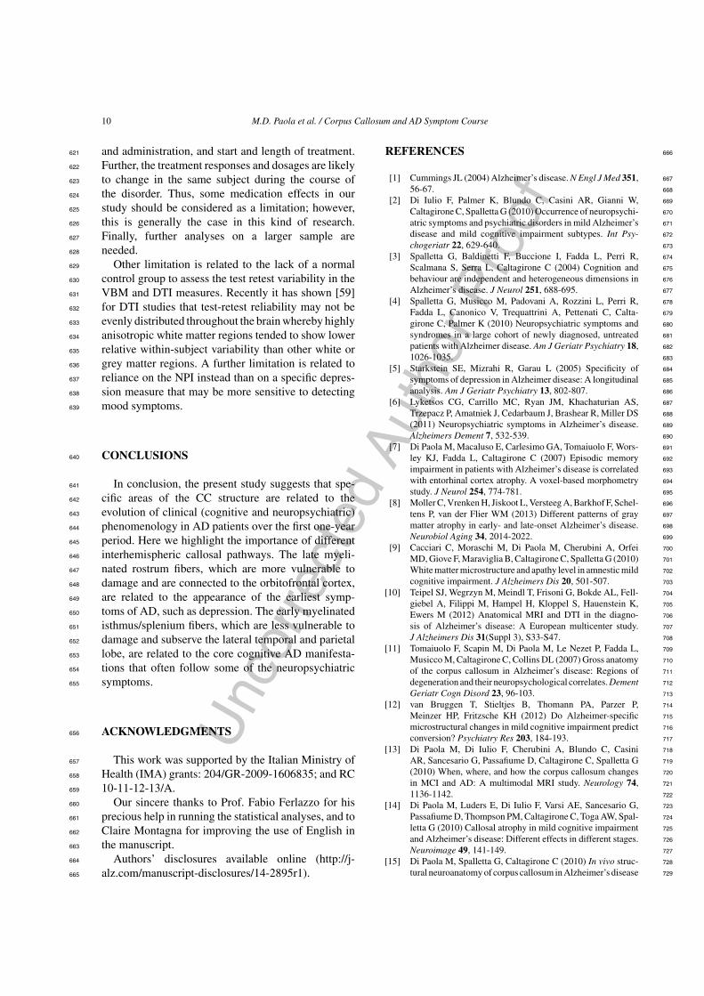
We found significant correlations in the evolution 413
of global cognitive deterioration (measured by the 414
MMSE �-values) and white matter density, FA, and 415
MD of the CC. For all three MRI parameters, the 416
significant correlation with the MMSE �-values 417
was localized at the level of the isthmus/splenium 418
(see Fig. 1, upper panel): white matter density and 419
FA correlated positively with the MMSE �-values 420
(white matter density isthmus/splenium Z = 2.69, 421
p FWE corrected = 0.004; FA isthmus/splenium 422
Fig. 1. Relationships between corpus callosum (CC) micro- and macrostructural parameters and changes in global cognitive performance anddepression severity over one year in 20 patients with Alzheimer’s disease at the first diagnostic visit. The upper panel describes the relationshipbetween MMSE �-values (axis x) and mean callosal white matter (WM) density, fractional anisotropy (FA), and mean diffusivity (MD) value(axis y), computed after applying the CC region of interest (ROI). The lower panel shows the relationship between Depression �-values (axisx) and mean callosal FA and MD value (axis y), computed after applying the CC ROI. On the extreme right panel, the CC areas of alterations(shown in the left panels) are overlaid on the CC of the white matter template in the default SPM8 on the midsagittal slice (X = 0). The datashown in the scatterplot were extracted from the statistical significance clusters. MMSE, Mini-Mental State Examination.

Unc
orre
cted
Aut
hor P
roof
8 M.D. Paola et al. / Corpus Callosum and AD Symptom Course
Z = 2.27, p FWE corrected = 0.012); MD correlated423
negatively with the MMSE �-values (MD isth-424
mus/splenium Z = 2.12, p FWE corrected = 0.017).425
No significant findings emerged when we ran cor-426
relations in the opposite direction (i.e., negative427
correlation between white matter density, FA, and428
MMSE �-values; positive correlation between MD429
and MMSE �-values).430
We also found statistically significant correlations431
between FA and MD values and changes in depres-432
sion symptoms measured by the NPI Depression433
�-values. For both DTI parameters, the correlation434
with the NPI Depression �-values was localized at435
the level of the callosal rostrum (see Fig. 1, lower436
panel). In particular, FA correlated positively with437
the NPI Depression �-values (FA rostrum Z = 2.42,438
p FWE corrected = 0.008), and MD correlated nega-439
tively with the NPI Depression �-values (MD rostrum440
Z = 2.10, p FWE corrected = 0.018). No significant441
findings emerged when we ran correlations in the oppo-442
site direction (i.e., negative correlation between FA443
and Depression �-values; positive correlation between444
MD and Depression �-values).445
No significant correlations were found between CC446
parameters and the other clinical, cognitive, and neu-447
ropsychiatric Delta values.448
DISCUSSION449
This is a longitudinal study, using multimodal imag-450
ing methods to investigate the relationship between451
CC micro- and macrostructure and the progression452
of global cognitive deterioration and neuropsychiatric453
symptom severity in early stage AD patients drug-free454
at baseline.455
Corpus callosum and cognitive aspects456
Our first finding was that a negative evolution457
of global cognitive performance over the one-year458
AD course was related to impaired isthmus/splenium459
CC macrostructure and microstructure at the first460
diagnostic visit. This means that the greater the isth-461
mus/splenium damage at the first diagnostic visit462
(lower white matter density, lower FA, and higher MD),463
the greater the evolution of global cognitive function-464
ing (i.e., the greater the MMSE score decrease over the465
one-year period).466
The macrostructural/volumetric data are in agree-467
ment with data obtained using a cross-sectional468
methodology, which revealed a correlation between469
global cognitive impairment, as measured by the470
MMSE, and CC volume [36]. In our previous article on 471
volumetric callosal changes in AD [11], we reported 472
a significant correlation between the CC isthmus and 473
global cognitive impairment. Since callosal axons arise 474
from cerebral cortex neurons, we hypothesized that the 475
correlation between global cognitive performance and 476
the structure of the CC isthmus might be a useful in vivo 477
indicator of the progress of neocortical disintegration 478
in AD. In support of this hypothesis, Ryberg and col- 479
leagues [37] carried out a cross-sectional study in the 480
LADIS population and found that the structure of the 481
CC isthmus correlated significantly with global cogni- 482
tive impairment, as assessed by the MMSE score. More 483
recently, the same authors [38] carried out a three-year 484
prospective follow-up study in a mixed elderly popu- 485
lation and found that CC atrophy predicted cognitive 486
impairment. 487
Thus, although previous studies have already ana- 488
lyzed this issue using macrostructural/volumetric data 489
from the CC [11, 37, 38], the present study is the 490
first to describe a relationship between microstruc- 491
tural/diffusivity changes of the posterior portion of the 492
CC and global cognitive performance, as measured 493
with the MMSE. The posterior callosal subregions 494
(i.e., isthmus and splenium) subserve two-thirds of 495
the higher-order processing areas of the lateral tem- 496
poral and parietal lobes [39]. Note that most cognitive 497
domains measured by the MMSE, such as orientation, 498
language, attention, and visuo-spatial function, rely on 499
these brain regions as anatomical substrates [40]. This 500
is in accordance with our findings which indicate that 501
the degree of integrity of the isthmus/splenium may 502
be correlated with the evolution of global cognitive 503
impairment in the early stages of AD. 504
The CC is not related to the evolution of single cog- 505
nitive domains. Perhaps this is due to the nature of the 506
CC, which has a comprehensive function in the brain. 507
Thus, a global cognitive value, such as the MMSE 508
score, more than single domain scores, is the elective 509
measure to be linked to the CC. 510
Corpus callosum and neuropsychiatric symptoms 511
We also found that increased severity of depressive 512
symptoms over the first year of illness was related to 513
impaired microstructural organization (decreased FA 514
and increased MD) at the level of the CC rostrum. This 515
means that the greater the impairment of the rostrum 516
at the first diagnostic visit (lower FA, higher MD), the 517
greater the progression of depression symptom severity 518
(increased NPI depression score severity over the one- 519
year period). FA measures the directionality of water 520

Unc
orre
cted
Aut
hor P
roof
M.D. Paola et al. / Corpus Callosum and AD Symptom Course 9
diffusion. Decreased FA indicates the loss of water521
directionality likely due to damage of the structural522
organization of the tissue [41]. MD is a measure of the523
average motion of water diffusion, independent of tis-524
sue directionality. It is sensitive to the microstructural525
architecture of cell membranes. A higher MD value526
implies greater diffusion of water molecules [42].527
Thus, the DTI parameters suggest that in our group528
microstructural damage was present at the level of the529
rostrum. This CC rostrum finding is intriguing because530
the rostrum contains interconnecting fibers that orig-531
inate in the orbito-frontal cortices [43]. According532
to previous studies, dysregulated orbitofrontal cortex533
connectivity is strongly related to mood regulation534
abnormalities [44, 45]. In particular, previous cross-535
sectional studies found the same results in depressed536
patients in both the pre-dementia [46] and the mild537
stage [47] of AD. Furthermore, our data are consis-538
tent with the results of neuroimaging studies on the539
anatomical correlates of depression in other neurode-540
generative disorders. Indeed, orbitofrontal and callosal541
changes have been reported in depressed subjects with542
Parkinson’s disease [48] and also in older depressed543
patients with no neurodegenerative disorders (see [49]544
for a review). Therefore, although late-life depression545
in subjects with and without dementia may have het-546
erogeneous mechanisms, it appears to have a similar547
regional profile within the CC. This is in accor-548
dance with the hypothesis of a fronto-limbic circuit549
of depression [50]. This finding is also in line with550
the assumption that white matter pathology is the key551
component of neuropsychiatric symptoms [51].552
It is also important to note that of all the neuropsy-553
chiatric dimensions investigated here only increased554
depression severity in the early stages of AD was cor-555
related with callosal changes. Indeed no significant556
correlations were found between CC parameters and557
the other neuropsychiatric Delta values. This is in line558
with previous reports [6, 52, 53] that depression is one559
of the most severe symptoms of AD already present in560
the early stage of the illness.561
The possible mechanism562
In general, the association between macro- and563
microstructural CC changes, cognitive impairment,564
and neuropsychiatric symptoms of depression is most565
likely a logical consequence of the interhemispheric566
disconnection. Most axons in the CC arise from cere-567
bral cortex cells and connect homologous areas of568
the two hemispheres. Indeed, previous research in AD569
patients has suggested that the pattern of CC atrophy570
reflects corresponding regional cortical neuronal loss, 571
possibly resulting from the degeneration of axons of 572
pyramidal neurons in cortical layer III [54]. These cal- 573
losal structural changes may lead to altered functional 574
connectivity between cerebral cortices and, finally, to 575
cognitive and neuropsychiatric dysfunction [55]. 576
Although the results of a correlational analysis can- 577
not be explained in mechanistic terms, we consider it 578
important to introduce a hypothesis about the possi- 579
ble underlying mechanism, a hypothesis that of course 580
needs to be validated by targeted studies. 581
In this regard, it is interesting to note that the cal- 582
losal fibers implicated in our results had a different time 583
of myelination. The rostrum contains late-myelinating 584
fibers and the isthmus/splenium early-myelinating 585
fibers [56]. The late mylenating fibers are comprised 586
of smaller axons and the myelin sheaths have fewer 587
myelin lamellae [57]. Therefore, these regions tend 588
to be more vulnerable to breakdown by a variety of 589
brain insults than the early myelinated fibers. Myelin 590
breakdown is an important component of the illness 591
process in AD [58]. Specifically, the fibers that myeli- 592
nate first in development are the last to be affected 593
by AD and those that myelinate much later in nor- 594
mal development are the first to be affected by the AD 595
degenerative process [58]. This matches perfectly with 596
our results, which on one hand link depression (a neu- 597
ropsychiatric symptom that sometimes precedes the 598
earliest manifestations of cognitive impairment) with 599
the late-myelinating and more vulnerable fibers in the 600
rostrum, and on the other hand relate the cognitive 601
dimension (i.e., the core clinical manifestation of AD, 602
which very often temporally follows manifestations of 603
depression) with the early-myelinating and less vulner- 604
able fibers of the isthmus/splenium. In other words, the 605
correlation we found between clinical, neuropsychi- 606
atric symptoms, and commissural tissue proprieties can 607
be explained by the myelination mechanism of Bart- 608
zokis’s hypothesis. Furthermore, it has been shown that 609
depression is not a cognitively related symptom in AD 610
[2, 4]. This supports the association among depres- 611
sion and cognition and different CC subregions and 612
pathways. 613
Limitations 614
On a related note, the analysis of imaging data 615
obtained from patients who are undergoing drug treat- 616
ment is challenging and can potentially affect study 617
results. Unfortunately, it is difficult to control for the 618
effect of this variable because there is high inter- 619
individual variability in drug selection, drug dosage 620
Unc
orre
cted
Aut
hor P
roof
10 M.D. Paola et al. / Corpus Callosum and AD Symptom Course
and administration, and start and length of treatment.621
Further, the treatment responses and dosages are likely622
to change in the same subject during the course of623
the disorder. Thus, some medication effects in our624
study should be considered as a limitation; however,625
this is generally the case in this kind of research.626
Finally, further analyses on a larger sample are627
needed.628
Other limitation is related to the lack of a normal629
control group to assess the test retest variability in the630
VBM and DTI measures. Recently it has shown [59]631
for DTI studies that test-retest reliability may not be632
evenly distributed throughout the brain whereby highly633
anisotropic white matter regions tended to show lower634
relative within-subject variability than other white or635
grey matter regions. A further limitation is related to636
reliance on the NPI instead than on a specific depres-637
sion measure that may be more sensitive to detecting638
mood symptoms.639
CONCLUSIONS640
In conclusion, the present study suggests that spe-641
cific areas of the CC structure are related to the642
evolution of clinical (cognitive and neuropsychiatric)643
phenomenology in AD patients over the first one-year644
period. Here we highlight the importance of different645
interhemispheric callosal pathways. The late myeli-646
nated rostrum fibers, which are more vulnerable to647
damage and are connected to the orbitofrontal cortex,648
are related to the appearance of the earliest symp-649
toms of AD, such as depression. The early myelinated650
isthmus/splenium fibers, which are less vulnerable to651
damage and subserve the lateral temporal and parietal652
lobe, are related to the core cognitive AD manifesta-653
tions that often follow some of the neuropsychiatric654
symptoms.655
ACKNOWLEDGMENTS656
This work was supported by the Italian Ministry of657
Health (IMA) grants: 204/GR-2009-1606835; and RC658
10-11-12-13/A.659
Our sincere thanks to Prof. Fabio Ferlazzo for his660
precious help in running the statistical analyses, and to661
Claire Montagna for improving the use of English in662
the manuscript.663
Authors’ disclosures available online (http://j-664
alz.com/manuscript-disclosures/14-2895r1).665
REFERENCES 666
[1] Cummings JL (2004) Alzheimer’s disease. N Engl J Med 351, 667
56-67. 668
[2] Di Iulio F, Palmer K, Blundo C, Casini AR, Gianni W, 669
Caltagirone C, Spalletta G (2010) Occurrence of neuropsychi- 670
atric symptoms and psychiatric disorders in mild Alzheimer’s 671
disease and mild cognitive impairment subtypes. Int Psy- 672
chogeriatr 22, 629-640. 673
[3] Spalletta G, Baldinetti F, Buccione I, Fadda L, Perri R, 674
Scalmana S, Serra L, Caltagirone C (2004) Cognition and 675
behaviour are independent and heterogeneous dimensions in 676
Alzheimer’s disease. J Neurol 251, 688-695. 677
[4] Spalletta G, Musicco M, Padovani A, Rozzini L, Perri R, 678
Fadda L, Canonico V, Trequattrini A, Pettenati C, Calta- 679
girone C, Palmer K (2010) Neuropsychiatric symptoms and 680
syndromes in a large cohort of newly diagnosed, untreated 681
patients with Alzheimer disease. Am J Geriatr Psychiatry 18, 682
1026-1035. 683
[5] Starkstein SE, Mizrahi R, Garau L (2005) Specificity of 684
symptoms of depression in Alzheimer disease: A longitudinal 685
analysis. Am J Geriatr Psychiatry 13, 802-807. 686
[6] Lyketsos CG, Carrillo MC, Ryan JM, Khachaturian AS, 687
Trzepacz P, Amatniek J, Cedarbaum J, Brashear R, Miller DS 688
(2011) Neuropsychiatric symptoms in Alzheimer’s disease. 689
Alzheimers Dement 7, 532-539. 690
[7] Di Paola M, Macaluso E, Carlesimo GA, Tomaiuolo F, Wors- 691
ley KJ, Fadda L, Caltagirone C (2007) Episodic memory 692
impairment in patients with Alzheimer’s disease is correlated 693
with entorhinal cortex atrophy. A voxel-based morphometry 694
study. J Neurol 254, 774-781. 695
[8] Moller C, Vrenken H, Jiskoot L, Versteeg A, Barkhof F, Schel- 696
tens P, van der Flier WM (2013) Different patterns of gray 697
matter atrophy in early- and late-onset Alzheimer’s disease. 698
Neurobiol Aging 34, 2014-2022. 699
[9] Cacciari C, Moraschi M, Di Paola M, Cherubini A, Orfei 700
MD, Giove F, Maraviglia B, Caltagirone C, Spalletta G (2010) 701
White matter microstructure and apathy level in amnestic mild 702
cognitive impairment. J Alzheimers Dis 20, 501-507. 703
[10] Teipel SJ, Wegrzyn M, Meindl T, Frisoni G, Bokde AL, Fell- 704
giebel A, Filippi M, Hampel H, Kloppel S, Hauenstein K, 705
Ewers M (2012) Anatomical MRI and DTI in the diagno- 706
sis of Alzheimer’s disease: A European multicenter study. 707
J Alzheimers Dis 31(Suppl 3), S33-S47. 708
[11] Tomaiuolo F, Scapin M, Di Paola M, Le Nezet P, Fadda L, 709
Musicco M, Caltagirone C, Collins DL (2007) Gross anatomy 710
of the corpus callosum in Alzheimer’s disease: Regions of 711
degeneration and their neuropsychological correlates. Dement 712
Geriatr Cogn Disord 23, 96-103. 713
[12] van Bruggen T, Stieltjes B, Thomann PA, Parzer P, 714
Meinzer HP, Fritzsche KH (2012) Do Alzheimer-specific 715
microstructural changes in mild cognitive impairment predict 716
conversion? Psychiatry Res 203, 184-193. 717
[13] Di Paola M, Di Iulio F, Cherubini A, Blundo C, Casini 718
AR, Sancesario G, Passafiume D, Caltagirone C, Spalletta G 719
(2010) When, where, and how the corpus callosum changes 720
in MCI and AD: A multimodal MRI study. Neurology 74, 721
1136-1142. 722
[14] Di Paola M, Luders E, Di Iulio F, Varsi AE, Sancesario G, 723
Passafiume D, Thompson PM, Caltagirone C, Toga AW, Spal- 724
letta G (2010) Callosal atrophy in mild cognitive impairment 725
and Alzheimer’s disease: Different effects in different stages. 726
Neuroimage 49, 141-149. 727
[15] Di Paola M, Spalletta G, Caltagirone C (2010) In vivo struc- 728
tural neuroanatomy of corpus callosum in Alzheimer’s disease 729
Unc
orre
cted
Aut
hor P
roof
M.D. Paola et al. / Corpus Callosum and AD Symptom Course 11
and mild cognitive impairment using different MRI tech-730
niques: A review. J Alzheimers Dis 20, 67-95.731
[16] DeKosky ST, Carrillo MC, Phelps C, Knopman D, Petersen732
RC, Frank R, Schenk D, Masterman D, Siemers ER, Cedar-733
baum JM, Gold M, Miller DS, Morimoto BH, Khachaturian734
AS, Mohs RC (2011) Revision of the criteria for Alzheimer’s735
disease: A symposium. Alzheimers Dement 7, e1-12.736
[17] Ikram MA, van der Lugt A, Niessen WJ, Krestin GP, Koud-737
staal PJ, Hofman A, Breteler MM, Vernooij MW (2011) The738
Rotterdam Scan Study: Design and update up to 2012. Eur J739
Epidemiol 26, 811-824.740
[18] Folstein MF, Folstein SE, McHugh PR (1975) "Mini-mental741
state". A practical method for grading the cognitive state of742
patients for the clinician. J Psychiatr Res 12, 189-198.743
[19] Hughes CP, Berg L, Danziger WL, Coben LA, Martin RL744
(1982) A new clinical scale for the staging of dementia. Br J745
Psychiatry 140, 566-572.746
[20] Katz S (1983) Assessing self-maintenance: Activities of daily747
living, mobility, and instrumental activities of daily living.748
J Am Geriatr Soc 31, 721-727.749
[21] Lawton MP, Brody EM (1969) Assessment of older people:750
Self-maintaining and instrumental activities of daily living.751
Gerontologist 9, 179-186.752
[22] Raven JC. (1947) Progressive Matrices. Sets A, Ab, B: Bords753
and Book forms., Lewis, London.754
[23] Caltagirone C, Gainotti G, Masullo C, Miceli G (1979) Valid-755
ity of some neuropsychological tests in the assessment of756
mental deterioration. Acta Psychiatr Scand 60, 50-56.757
[24] Rey A (1941) L’examen psychologie dan les cas758
d’encephalopathie traumatique (Les problemes). Arch759
Psychol 28, 286-340.760
[25] Rey A (1958) Memorisation d’une serie de 15 mots en 5 repe-761
titions. In In Rey A. L’examen clinique en psycologie, Presses762
Universitaires des France, Paris.763
[26] Stroop JR (1935) Studies of interference in serial verbal reac-764
tions. J Exp Psychol 12, 643-662.765
[27] Borkowsky JG, Benton AL, Spreen O (1967) Word fluency766
and brain damage. Neuropsychologia 5, 135-140.767
[28] Lucas JA, Ivnik RJ, Smith GE, Bohac DL, Tangalos EG,768
Graff-Radford NR, Petersen RC (1998) Mayo’s older Amer-769
icans normative studies: Category fluency norms. J Clin Exp770
Neuropsychol 20, 194-200.771
[29] Osterrieth PA (1944) Le test de copie d’une figure complexe.772
Arch Psychol 30, 206-356.773
[30] Gainotti G, Marra C, Villa G (2001) A double dissociation774
between accuracy and time of execution on attentional tasks775
in Alzheimer’s disease and multi-infarct dementia. Brain 124,776
731-738.777
[31] Cummings JL (1997) The Neuropsychiatric Inventory:778
Assessing psychopathology in dementia patients. Neurology779
48, S10-S16.780
[32] Good CD, Johnsrude IS, Ashburner J, Henson RN, Friston KJ,781
Frackowiak RS (2001) A voxel-based morphometric study of782
ageing in 465 normal adult human brains. Neuroimage 14,783
21-36.784
[33] Ashburner J (2007) A fast diffeomorphic image registration785
algorithm. Neuroimage 38, 95-113.786
[34] Smith SM (2002) Fast robust automated brain extraction. Hum787
Brain Mapp 17, 143-155.788
[35] Pierpaoli C, Basser PJ (1996) Toward a quantitative789
assessment of diffusion anisotropy. Magn Reson Med 36,790
893-906.791
[36] Teipel SJ, Hampel H, Alexander GE, Schapiro MB, Horwitz792
B, Teichberg D, Daley E, Hippius H, Moller HJ, Rapoport793
SI (1998) Dissociation between corpus callosum atrophy and794
white matter pathology in Alzheimer’s disease. Neurology 51, 795
1381-1385. 796
[37] Ryberg C, Rostrup E, Stegmann MB, Barkhof F, Scheltens P, 797
van Straaten EC, Fazekas F, Schmidt R, Ferro JM, Baezner 798
H, Erkinjuntti T, Jokinen H, Wahlund LO, O’Brien J, Basile 799
AM, Pantoni L, Inzitari D, Waldemar G (2007) Clinical 800
significance of corpus callosum atrophy in a mixed elderly 801
population. Neurobiol Aging 28, 955-963. 802
[38] Ryberg C, Rostrup E, Paulson OB, Barkhof F, Scheltens P, 803
van Straaten EC, van der Flier WM, Fazekas F, Schmidt R, 804
Ferro JM, Baezner H, Erkinjuntti T, Jokinen H, Wahlund LO, 805
Poggesi A, Pantoni L, Inzitari D, Waldemar G (2011) Corpus 806
callosum atrophy as a predictor of age-related cognitive and 807
motor impairment: A 3-year follow-up of the LADIS study 808
cohort. J Neurol Sci 307, 100-105. 809
[39] Schmahmann J, Pandya D. (2006) Fiber pathways of the brain, 810
Oxford Universiy Press, New York. 811
[40] Denes G, Pizzamiglio L. (1999) Manuale di neuropsicologia. 812
Normalita e patologia dei processi cognitivi., Zanichelli, II 813
edizione, Bologna. 814
[41] Schulte T, Sullivan EV, Muller-Oehring EM, Adalsteinsson 815
E, Pfefferbaum A (2005) Corpus callosal microstructural 816
integrity influences interhemispheric processing: A diffusion 817
tensor imaging study. Cereb Cortex 15, 1384-1392. 818
[42] Basser PJ, Jones DK (2002) Diffusion-tensor MRI: Theory, 819
experimental design and data analysis - a technical review. 820
NMR Biomed 15, 456-467. 821
[43] Abe O, Masutani Y, Aoki S, Yamasue H, Yamada H, 822
Kasai K, Mori H, Hayashi N, Masumoto T, Ohtomo K 823
(2004) Topography of the human corpus callosum using 824
diffusion tensor tractography. J Comput Assist Tomogr 28, 825
533-539. 826
[44] Jackowski AP, Araujo Filho GM, Almeida AG, Araujo CM, 827
Reis M, Nery F, Batista IR, Silva I, Lacerda AL (2012) The 828
involvement of the orbitofrontal cortex in psychiatric disor- 829
ders: An update of neuroimaging findings. Rev Bras Psiquiatr 830
34, 207-212. 831
[45] Konarski JZ, McIntyre RS, Kennedy SH, Rafi-Tari S, 832
Soczynska JK, Ketter TA (2008) Volumetric neuroimaging 833
investigations in mood disorders: Bipolar disorder versus 834
major depressive disorder. Bipolar Disord 10, 1-37. 835
[46] Lee GJ, Lu PH, Hua X, Lee S, Wu S, Nguyen K, Teng E, 836
Leow AD, Jack CR Jr, Toga AW, Weiner MW, Bartzokis G, 837
Thompson PM (2012) Depressive symptoms in mild cognitive 838
impairment predict greater atrophy in Alzheimer’s disease- 839
related regions. Biol Psychiatry 71, 814-821. 840
[47] Lebedev AV, Beyer MK, Fritze F, Westman E, Ballard C, Aars- 841
land D. (2014) Cortical changes associated with depression 842
and antidepressant use in Alzheimer and Lewy body demen- 843
tia: An MRI surface-based morphometric study. Am J Geriatr 844
Psychiatry 22, 4-13 e11. 845
[48] Kostic VS, Agosta F, Petrovic I, Galantucci S, Spica V, 846
Jecmenica-Lukic M, Filippi M (2010) Regional patterns of 847
brain tissue loss associated with depression in Parkinson dis- 848
ease. Neurology 75, 857-863. 849
[49] Jellinger KA (2013) Organic bases of late-life depression: A 850
critical update. J Neural Transm 120, 1109-1125. 851
[50] Tekin S, Cummings JL (2002) Frontal-subcortical neuronal 852
circuits and clinical neuropsychiatry: An update. J Psychosom 853
Res 53, 647-654. 854
[51] Tighe SK, Oishi K, Mori S, Smith GS, Albert M, Lyketsos 855
CG, Mielke MM (2012) Diffusion tensor imaging of neu- 856
ropsychiatric symptoms in mild cognitive impairment and 857
Alzheimer’s dementia. J Neuropsychiatry Clin Neurosci 24, 858
484-488. 859
Unc
orre
cted
Aut
hor P
roof
12 M.D. Paola et al. / Corpus Callosum and AD Symptom Course
[52] Pellegrino LD, Peters ME, Lyketsos CG, Marano CM (2013)860
Depression in cognitive impairment. Curr Psychiatry Rep 15,861
384.862
[53] Jost BC, Grossberg GT (1996) The evolution of psychiatric863
symptoms in Alzheimer’s disease: A natural history study.864
J Am Geriatr Soc 44, 1078-1081.865
[54] Hampel H, Teipel SJ, Alexander GE, Pogarell O, Rapoport SI,866
Moller HJ (2002) In vivo imaging of region and cell type spe-867
cific neocortical neurodegeneration in Alzheimer’s disease.868
Perspectives of MRI derived corpus callosum measurement869
for mapping disease progression and effects of therapy. Evi-870
dence from studies with MRI, EEG and PET. J Neural Transm871
109, 837-855.872
[55] Hinkley LB, Marco EJ, Findlay AM, Honma S, Jeremy RJ,873
Strominger Z, Bukshpun P, Wakahiro M, Brown WS, Paul874
LK, Barkovich AJ, Mukherjee P, Nagarajan SS, Sherr EH875
(2012) The role of corpus callosum development in functional876
connectivity and cognitive processing. PLoS One 7, e39804.877
[56] Aboitiz F, Scheibel AB, Fisher RS, Zaidel E (1992) Fiber 878
composition of the human corpus callosum. Brain Res 598, 879
143-153. 880
[57] Chia LS, Thompson JE, Moscarello MA (1983) Changes in 881
lipid phase behaviour in human myelin during maturation and 882
aging. Involvement of lipid peroxidation. FEBS Lett 157, 155- 883
158. 884
[58] Bartzokis G (2004a) Age-related myelin breakdown: A devel- 885
opmental model of cognitive decline and Alzheimer’s disease. 886
Neurobiol Aging 25, 5-18; author reply 49-62. 887
[59] Cole JH, Farmer RE, Rees EM, Johnson HJ, Frost C, Scahill 888
RI, Hobbs NZ. (2014) Test-retest reliability of diffusion tensor 889
imaging in Huntington’s disease. PLoS Curr 6, pii: ecur- 890
rents.hd.f19ef63fff962f5cd9c0e88f4844f43b. 891