Corpus callosum and prefrontal functions in adolescents with history of very preterm birth
6
Please cite this article in press as: Narberhaus, A., et al., Corpus callosum and prefrontal functions in adolescents with history of very preterm birth, Neuropsychologia (2007), doi:10.1016/j.neuropsychologia.2007.08.004 ARTICLE IN PRESS +Model NSY-2694; No. of Pages 6 Neuropsychologia xxx (2007) xxx–xxx Corpus callosum and prefrontal functions in adolescents with history of very preterm birth Ana Narberhaus a , Dolors Segarra a,∗ , Xavier Cald´ u a,b , Monica Gim´ enez a,b , Roser Pueyo a , Francesc Botet c , Carme Junqu´ e b a Department of Psychiatry and Clinical Psychobiology, University of Barcelona, Spain b Institut d’Investigacions Biom` ediques August Pi i Sunyer (IDIBAPS), Hospital Cl´ ınic, Faculty of Medicine, University of Barcelona, Spain c Paediatrics Section, Department of Obstetrics & Gynaecology, Paediatrics, Radiology & Medical Physics, Hospital Cl´ ınic, Barcelona, Spain Received 24 April 2007; received in revised form 27 July 2007; accepted 6 August 2007 Abstract Very preterm (VPT) birth can account for thinning of the corpus callosum and poorer cognitive performance. Research findings about preterm and VPT adolescents usually describe a small posterior corpus callosum, although our research group has also found reductions of the anterior part, specifically the genu. The aim of the present study was to investigate the functional implications of this concrete reduction. Fifty-two VPT adolescents were compared with 52 adolescents born at term; there were no significant differences in age and gender, and socioeconomic status was similar between the groups. All participants underwent a magnetic resonance imaging (MRI) study and assessment of prefrontal functioning and vocabulary. The VPT group showed significant reductions of the genu, isthmus and splenium, as well as a significantly worse performance on category verbal fluency, executive functions, everyday memory and vocabulary. Although several parts of the corpus callosum correlated with some prefrontal functions, the genu was the part which principally explained these correlations. The subtest Vocabulary only correlated with the splenium. The relationship between genu and prefrontal functions and between splenium and vocabulary may be due to the fact that these parts of the corpus callosum connect prefrontal and posterior parietal cortex, respectively. The work presented here provides evidence of specific associations between reductions in the anterior corpus callosum (genu) and lower prefrontal functioning in VPT adolescents. © 2007 Published by Elsevier Ltd. Keywords: Genu; Prefrontal skills; Low performance; Adolescence; Correlations 1. Introduction Very preterm (VPT) individuals have an increased risk of brain abnormalities, especially prominent in white matter, which are evident in both childhood (H¨ uppi et al., 1998; Inder et al., 1999; Nagy et al., 2003) and adolescence (Gim´ enez, Junqu´ e, Narberhaus, Bargall´ o et al., 2006; Stewart et al., 1999). The corpus callosum is the main interhemispheric commissure of the brain (Tomasch, 1954). In humans it begins to develop 9 weeks after conception and continues to grow post-natally, showing morphological changes related to the ongoing myelination of interhemispheric fibers (Volpe, 2001). ∗ Corresponding author at: Department of Psychiatry and Clinical Psychobiol- ogy, Faculty of Psychology, University of Barcelona, Passeig de la Vall d’Hebron 171, E-08035 Barcelona, Spain. Tel.: +34 933125057; fax: +34 934021584. E-mail address: [email protected] (D. Segarra). Magnetic resonance imaging (MRI) studies have demon- strated thinning of the corpus callosum in VPT or preterm children and adolescents (Narberhaus, Segarra, Cald´ u, et al., 2007; Nosarti et al., 2004; Peterson et al., 2000; Stewart et al., 1999). Such injury may be partly explained by the vulnerability of the developing corpus callosum to hypoxic-ischemic damage, possibly due to intrinsic vulnerability of immature oligodendro- cytes (Back et al., 2001). Peterson et al. (2000) reported that 8-year-old VPT subjects showed a generalized corpus callosum reduction, including the anterior and posterior regions. In a qualitative MRI study of 14–15-year-old VPT individuals, Stewart et al. (1999) reported a reduction predominantly in the posterior corpus callosum. In the same sample, Nosarti et al. (2004) confirmed this result through a quantitative MRI technique able to detect even subtle cerebral abnormalities. More recently, and also using this technique, our research group has consistently found reductions of both the 0028-3932/$ – see front matter © 2007 Published by Elsevier Ltd. doi:10.1016/j.neuropsychologia.2007.08.004
-
Upload
independent -
Category
Documents
-
view
6 -
download
0
Transcript of Corpus callosum and prefrontal functions in adolescents with history of very preterm birth

N
A
apawaostpa©
K
1
ba1Ncbami
o1
0d
ARTICLE IN PRESS+ModelSY-2694; No. of Pages 6
Neuropsychologia xxx (2007) xxx–xxx
Corpus callosum and prefrontal functions in adolescentswith history of very preterm birth
Ana Narberhaus a, Dolors Segarra a,∗, Xavier Caldu a,b, Monica Gimenez a,b,Roser Pueyo a, Francesc Botet c, Carme Junque b
a Department of Psychiatry and Clinical Psychobiology, University of Barcelona, Spainb Institut d’Investigacions Biomediques August Pi i Sunyer (IDIBAPS), Hospital Clınic, Faculty of Medicine, University of Barcelona, Spain
c Paediatrics Section, Department of Obstetrics & Gynaecology, Paediatrics, Radiology & Medical Physics, Hospital Clınic, Barcelona, Spain
Received 24 April 2007; received in revised form 27 July 2007; accepted 6 August 2007
bstract
Very preterm (VPT) birth can account for thinning of the corpus callosum and poorer cognitive performance. Research findings about pretermnd VPT adolescents usually describe a small posterior corpus callosum, although our research group has also found reductions of the anteriorart, specifically the genu. The aim of the present study was to investigate the functional implications of this concrete reduction. Fifty-two VPTdolescents were compared with 52 adolescents born at term; there were no significant differences in age and gender, and socioeconomic statusas similar between the groups. All participants underwent a magnetic resonance imaging (MRI) study and assessment of prefrontal functioning
nd vocabulary. The VPT group showed significant reductions of the genu, isthmus and splenium, as well as a significantly worse performancen category verbal fluency, executive functions, everyday memory and vocabulary. Although several parts of the corpus callosum correlated withome prefrontal functions, the genu was the part which principally explained these correlations. The subtest Vocabulary only correlated with
he splenium. The relationship between genu and prefrontal functions and between splenium and vocabulary may be due to the fact that thesearts of the corpus callosum connect prefrontal and posterior parietal cortex, respectively. The work presented here provides evidence of specificssociations between reductions in the anterior corpus callosum (genu) and lower prefrontal functioning in VPT adolescents.2007 Published by Elsevier Ltd.
ns
sc21opc
eywords: Genu; Prefrontal skills; Low performance; Adolescence; Correlatio
. Introduction
Very preterm (VPT) individuals have an increased risk ofrain abnormalities, especially prominent in white matter, whichre evident in both childhood (Huppi et al., 1998; Inder et al.,999; Nagy et al., 2003) and adolescence (Gimenez, Junque,arberhaus, Bargallo et al., 2006; Stewart et al., 1999). The
orpus callosum is the main interhemispheric commissure of therain (Tomasch, 1954). In humans it begins to develop 9 weeks
Please cite this article in press as: Narberhaus, A., et al., Corpus callosumbirth, Neuropsychologia (2007), doi:10.1016/j.neuropsychologia.2007.08
fter conception and continues to grow post-natally, showingorphological changes related to the ongoing myelination of
nterhemispheric fibers (Volpe, 2001).
∗ Corresponding author at: Department of Psychiatry and Clinical Psychobiol-gy, Faculty of Psychology, University of Barcelona, Passeig de la Vall d’Hebron71, E-08035 Barcelona, Spain. Tel.: +34 933125057; fax: +34 934021584.
E-mail address: [email protected] (D. Segarra).
sa1rsaar
028-3932/$ – see front matter © 2007 Published by Elsevier Ltd.oi:10.1016/j.neuropsychologia.2007.08.004
Magnetic resonance imaging (MRI) studies have demon-trated thinning of the corpus callosum in VPT or pretermhildren and adolescents (Narberhaus, Segarra, Caldu, et al.,007; Nosarti et al., 2004; Peterson et al., 2000; Stewart et al.,999). Such injury may be partly explained by the vulnerabilityf the developing corpus callosum to hypoxic-ischemic damage,ossibly due to intrinsic vulnerability of immature oligodendro-ytes (Back et al., 2001).
Peterson et al. (2000) reported that 8-year-old VPT subjectshowed a generalized corpus callosum reduction, including thenterior and posterior regions. In a qualitative MRI study of4–15-year-old VPT individuals, Stewart et al. (1999) reported aeduction predominantly in the posterior corpus callosum. In the
and prefrontal functions in adolescents with history of very preterm.004
ame sample, Nosarti et al. (2004) confirmed this result throughquantitative MRI technique able to detect even subtle cerebralbnormalities. More recently, and also using this technique, ouresearch group has consistently found reductions of both the

IN+ModelN
2 psych
ppa
rmecc
tJF2oFdTtTbTbB2ebtniw
feM2tfiLN
e2hccsoitcsapa
f
cgV
2
2
b1atwc
dp(7dahtw
Smvr
wwts(
as7
Bp
2
(Tpthem in the same position. A mid-sagittal slice in which the anterior and pos-terior commissures, as well as the fornix, were clearly identifiable was selectedin order to draw the corpus callosum, which was semi-automatically segmentedinto seven parts following the approach described by Witelson (1989) and usingAnalyze 6.0 (Biomedical Imaging Resource, Mayo Clinic). This computer pro-
Table 1Sample characteristics
Mean (S.D.), range
VPT (n = 52) Controls (n = 52)
Gestational age 29.69 (2.02), 25–32 39.58 (1.49), 37–43
ARTICLESY-2694; No. of Pages 6
A. Narberhaus et al. / Neuro
osterior and anterior corpus callosum, specifically the genu, inreterm and VPT adolescents (Narberhaus, Segarra, Caldu, etl., 2007).
In the corpus callosum, the region which extends from theostrum to the posterior part of the midbody is predominantlyade up of fibers interconnecting the prefrontal cortex (Park
t al., in press). This fact enhances the importance of studyingognitive functions that are highly dependent on the prefrontalortex, namely prefrontal functions.
Preterm adolescents seem to perform significantly worsehan term born subjects in phonetic verbal fluency (Gimenez,unque, Narberhaus, Botet et al., 2006; Taylor, Minich, Bangert,ilipek, & Hack, 2004) and category verbal fluency (Allin et al.,001; Gimenez, Junque, Narberhaus, Botet et al., 2006). Forther prefrontal functions, studies report contradictory results.or instance, while Tideman (2000) and Rushe et al. (2001)escribed no significant differences in attention/concentration,aylor, Klein, Minich, and Hack (2000) found specific difficul-
ies on this cognitive function. As regards executive functioning,aylor et al. (2000, 2004) reported significant differencesetween preterms and controls on several different measures.ideman (2000) also showed a significantly worse performance,ut only on one specific task the Trail Making Test (TMT), mainly measuring planning and cognitive flexibility (Baron,004). In contrast, Rushe et al. (2001) and, recently, Saavalainent al. (2006) observed no differences in executive functionsetween groups assessed through the TMT B and the Stroopest, respectively. Related to the above mentioned studies, it isoteworthy that Taylor et al. (2000, 2004) differ from the othersn that they explore specifically extremely and very low birtheight subjects.Additionally some studies report a significantly worse per-
ormance in preterm adolescents compared to term controls inveryday memory, as assessed by the Rivermead Behaviouralemory Test (RBMT) (Narberhaus, Segarra, Gimenez, et al.,
007). This test mainly evaluates episodic memory, prospec-ive memory and orientation, so the medial temporal lobe andnally the prefrontal cortex are involved (Kahn et al., 2005;ezak, Howieson, & Loring, 2004; Rami et al., 2003; Umeda,agumo, & Kato, 2006).Corpus callosum reduction has been related to a worse gen-
ral cognitive performance in VPT children (Peterson et al.,000) and adolescents (Caldu et al., 2006). Only two studiesave analyzed the relationship between prefrontal functions andorpus callosum in VPT adolescents. Results showed an asso-iation between phonetic verbal fluency and the mid-posteriorurface area (Nosarti et al., 2004), and between everyday mem-ry and total corpus callosum area (Caldu et al., 2006). However,n this study the authors did not analyze the possible correla-ions between prefrontal functions and each part of the corpusallosum, while Nosarti et al. (2004) only found this relation-hip for VPT boys (n = 33). To our knowledge there is no studyddressing the issue about the relationship between the different
Please cite this article in press as: Narberhaus, A., et al., Corpus callosumbirth, Neuropsychologia (2007), doi:10.1016/j.neuropsychologia.2007.08
arts of the corpus callosum and prefrontal functioning in VPTdolescents.
The specific aim of the present study was to examine theunctional implications of the reductions in the anterior corpus
BA
PRESSologia xxx (2007) xxx–xxx
allosum, specifically the genu. We hypothesised that a smallerenu would be associated with worse prefrontal functioning inPT adolescents compared to the term control group.
. Method
.1. Participants
Subjects with a history of prematurity were selected from the populationorn between 1982 and 1994 at the Hospital Clınic i Provincial, and between988 and 1989 at the Hospital Vall d’Hebron, both in Barcelona (Spain). In therchives of these hospitals 488 cases of prematurity were currently available atheir databases. Inclusion criteria for the present study were: gestational age <33eeks (VPT) and age at assessment between 11 and 18 years; remaining 278
ases, from which the clinical histories were inaccessible in 44 cases.This resultant cohort was then reduced for several reasons: missing clinical
ata or exitus (72), no updated address or telephone number (75), refusal toarticipate in the study (5) or presence of one or more of the exclusion criteria30). These exclusion criteria were: (a) full intelligence quotient (FIQ) below0; (b) history of traumatic brain injury, cerebral palsy or other neurologicaliagnosis; (c) motor or sensory impairment that precluded neuropsychologicalssessment; (d) metal orthodontic prosthesis; (e) claustrophobia or anxiety levelsigh enough to require sedation. The available clinical data allow us to confirmhat these subjects lost for investigation did not differ from those studied in birtheight, gestational age, gender ratio, mode of delivery nor in condition at birth.
The final sample thus comprised 52 VPT adolescents (27 girls/25 boys).eventy-five percent presented hypoxic risk with apneas and/or hyalineembrane disease, requiring oxygen therapy. Only seven cases presented intra-
entricular haemorrhage grade III and/or IV. None of the other cases presentedelevant complications in the peri/neonatal period.
Fifty-two controls (26 girls/26 boys) born at a gestational age ≥37 weeks andith normal pre-, peri- and post-natal data were recruited. This control groupas mainly composed of relatives or acquaintances of the study sample, and
heir educational level and socio-economic status were similar. There were noignificant differences between VPT subjects and controls in age at assessmentt = −0.20; p = 0.84) or gender (χ2 = 0.08; p = 0.81) (Table 1).
All participants underwent a magnetic resonance imaging (MRI) study andneuropsychological assessment. According to the neuroradiologist (NB) no
ubject had parenchymal lesions. FIQ of the VPT individuals (mean: 101, range:5–135) and controls (mean: 114, range: 92–141) were normal.
The study was approved by the Ethics Committee of the University ofarcelona and all the subjects or their family gave written informed consentrior to participating in the study.
.1.1. MRI acquisition and analysesT1-weighted morphological images were acquired on a 1.5 T Signa GE
Milwaukee, WI) using a 3D FSPGR-IR sequence (TR = 12 ms; TE = 5.2 ms;I = 300 ms; 1 NEX; FOV = 24 cm × 24 cm; matrix size = 256 × 256), yieldingartitions of 1.5 mm in thickness. All the images were reoriented in order to place
and prefrontal functions in adolescents with history of very preterm.004
(in weeks)irth weight (g) 1273 (337.7), 690–2160 3421 (428), 2340–4300ge at assessment(in years)
14.21 (1.74), 11–18 14.29 (2.22), 10–19
IN+ModelN
psych
gwa
p(s(f(
oNv
2
dafta&
ttwaoAalSmoc
2
Sntp
cra
3
3
yntsv
3
owan(
3
tevSb
TC
C
C
I
ARTICLESY-2694; No. of Pages 6
A. Narberhaus et al. / Neuro
ram automatically calculated the area of each part. The total callosum areaas obtained by summing the different parts. Additionally each sub-region was
djusted for the mid-sagittal area.Inter- and intra-rater reliability, performed on 11 randomly selected inde-
endent ratings of total corpus callosum area, were 0.98 (p < 0.0001) and 0.95p < 0.0001), respectively. Intra-rater reliability performed on all corpus callo-um subregions for the same 11 ratings was: 0.84 (p < 0.001) for the rostrum, 0.94p < 0.0001) for the genu, 0.92 (p < 0.0001) for the rostral body, 0.91 (p < 0.0001)or the anterior midbody, 0.95 (p < 0.0001) for the posterior midbody, 0.95p < 0.0001) for the isthmus and 0.96 (p < 0.0001) for the splenium.
In addition, images were segmented using SPM2 (Wellcome Departmentf Cognitive Neuroscience, London, UK) running on Matlab 6.0 (Mathworks,atick, MA) in order to obtain gray matter, white matter and cerebrospinal fluidolumes.
.1.2. Neuropsychological assessmentThe same psychologist (AN) evaluated all the participants in the study in two
ifferent sessions lasting 90 min each. Measures of several types of cognitivebilities were taken. For the present study we analyzed a number of prefrontalunctions, specifically, verbal fluency, attention and motor speed, executive func-ioning and everyday memory. Additionally, and as a comparison measure, wenalyzed vocabulary, which is more related to posterior temporal cortex (Kolb
Whishaw, 2003).Phonetic verbal fluency was assessed with a modified version of the Con-
rolled Oral Word Association Test (COWAT), in which participants were askedo produce as many words as they could, beginning with the letters P, M and Rithin 1 min. For the category verbal fluency, subjects were required to produce
s many names of animals as possible in the same time frame. The total numberf words produced in each task was recorded. The Trail Making Test (TMT)
was used to evaluate attention and motor speed. Executive functioning wasssessed through the TMT B and the subtest Digit Symbol of the Wechsler Intel-igence Scale for Children-Revised (WISC-R) or Wechsler Adult Intelligencecale-Third Edition (WAIS-III). Everyday memory was assessed with the River-ead Behavioural Memory Test (RBMT). Additionally the subtest Vocabulary
f the WISC-R/WAIS-III was considered. Detailed information on these testsan be found in Lezak et al. (2004).
.1.3. Statistical analyses
Please cite this article in press as: Narberhaus, A., et al., Corpus callosumbirth, Neuropsychologia (2007), doi:10.1016/j.neuropsychologia.2007.08
Statistical analyses were performed using the Statistical Package for theocial Sciences (SPSS Win., v.12.0). To test for differences in morphological andeuropsychological measures between groups, we used an independent samples-test. Relationships between corpus callosum areas and neuropsychologicalerformance in the VPT group were determined using Pearson’s product moment
c
te
able 2erebral measures of the VPT and control groups
Mean (S.D.)
VPT (n = 52)
orrected corpus callosum sub-regionsa
Rostrum 0.14 (0.06)Genu 0.90 (0.20)Rostral body 0.60 (0.12)Anterior midbody 0.49 (0.09)Posterior midbody 0.47 (0.09)Isthmus 0.49 (0.11)Splenium 0.98 (0.21)
orrected corpus callosumb
Total area 0.158 (0.03)
ntracranial volumes (expressed in mm3)White matter 361632.5 (54512.8)Gray matter 767036.3 (98010.8)Cerebrospinal fluid 314215.4 (89183.5)
a Corrected by mid-sagittal area.b Corrected by whole white matter volume.
PRESSologia xxx (2007) xxx–xxx 3
orrelations and linear regression analyses. Due to the exploratory nature of theesearch, the significance threshold was set at p < 0.05 bilaterally for all thenalyses.
. Results
.1. Brain measurements
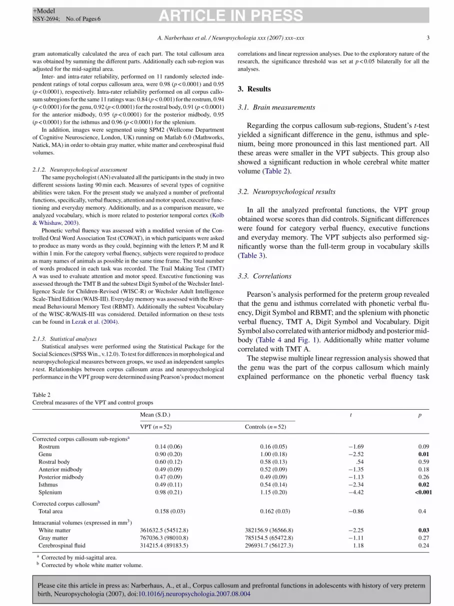
Regarding the corpus callosum sub-regions, Student’s t-testielded a significant difference in the genu, isthmus and sple-ium, being more pronounced in this last mentioned part. Allhese areas were smaller in the VPT subjects. This group alsohowed a significant reduction in whole cerebral white matterolume (Table 2).
.2. Neuropsychological results
In all the analyzed prefrontal functions, the VPT groupbtained worse scores than did controls. Significant differencesere found for category verbal fluency, executive functions
nd everyday memory. The VPT subjects also performed sig-ificantly worse than the full-term group in vocabulary skillsTable 3).
.3. Correlations
Pearson’s analysis performed for the preterm group revealedhat the genu and isthmus correlated with phonetic verbal flu-ncy, Digit Symbol and RBMT; and the splenium with phoneticerbal fluency, TMT A, Digit Symbol and Vocabulary. Digitymbol also correlated with anterior midbody and posterior mid-ody (Table 4 and Fig. 1). Additionally white matter volume
and prefrontal functions in adolescents with history of very preterm.004
orrelated with TMT A.The stepwise multiple linear regression analysis showed that
he genu was the part of the corpus callosum which mainlyxplained performance on the phonetic verbal fluency task
t p
Controls (n = 52)
0.16 (0.05) −1.69 0.091.00 (0.18) −2.52 0.010.58 (0.13) .54 0.590.52 (0.09) −1.35 0.180.49 (0.09) −1.13 0.260.54 (0.14) −2.34 0.021.15 (0.20) −4.42 <0.001
0.162 (0.03) −0.86 0.4
382156.9 (36566.8) −2.25 0.03785154.5 (65472.8) −1.11 0.27296931.7 (56127.3) 1.18 0.24

Please cite this article in press as: Narberhaus, A., et al., Corpus callosumbirth, Neuropsychologia (2007), doi:10.1016/j.neuropsychologia.2007.08
ARTICLE IN PRESS+ModelNSY-2694; No. of Pages 6
4 A. Narberhaus et al. / Neuropsychologia xxx (2007) xxx–xxx
Table 3Neuropsychological results
Mean (S.D.) t p
VPT (n = 52) Controls (n = 52)
Prefrontal functionsPhonetic verbal fluency
P, M, R (number of words) 28.0 (7.9) 33.2 (10.4) −1.91 0.06
Category verbal fluencyAnimals (number of words) 19.0 (5.1) 21.5 (4.2) −2.69 0.008
Attention and motor speedTrail Making Test A (in s) 25.2 (9.5) 21.8 (9.8) 1.83 0.07
Executive functionsTrail Making Test B (in s) 54.4 (26.7) 41.2 (21.3) 2.80 0.006Digit Symbol (scaled score) 9.5 (3.4) 11.2 (2.6) −3.37 0.001
Everyday memoryRivermead Behavioural Memory Test (total score) 10.5 (1.5) 11.3 (0.8) −3.46 0.001
VocabularyWISC-R/WAIS-III (scaled score) 11.6 (3.2) 14.2 (2.1) −4.75 <0.001
Table 4Correlations between corpus callosum and neuropsychological results in the VPT group
Phonetic fluency Category fluency Trail Making Test A Trail Making Test B Digit Symbol RBMT Vocabulary
Rostrum −0.11 −0.21 0.07 0.07 0.02 0.05 −0.23Genu 0.37** 0.24 −0.06 −0.02 0.44** 0.37** 0.16Rostral body −0.05 0.06 −0.18 −0.02 0.18 0.01 −0.03Anterior midbody −0.01 −0.04 −0.06 −0.06 0.43** 0.09 0.14Posterior midbody −0.03 0.14 −0.13 0.14 0.41** 0.20 0.05Isthmus 0.28* 0.08 0.04 −0.11 0.31* 0.32* 0.17Splenium 0.32* 0.15 −0.29* ** *
* p ≤ 0.05.** p ≤ 0.01.
Fig. 1. Relationship between Corpus Callosum Genu and Digit Symbol in theVPT group.
(R
4
rVriamc1
psrTts
fa
−0.20 0.38 0.25 0.32
r = 0.37, p = 0.007), Digit Symbol (r = 0.37, p = 0.007) andBMT (r = 0.44, p = 0.001).
. Discussion
Our findings about a more pronounced reduction in the poste-ior part of the corpus callosum agree with previous studies aboutPT adolescents (Nosarti et al., 2004; Stewart et al., 1999). This
esult could be related to the fact that in normal development,n the range 11–15 years, the posterior corpus callosum showsmore rapid growth pattern (Thompson et al., 2000). This dataay reflect a more pronounced development of interhemispheric
onnections in posterior regions during this period (Giedd et al.,999).
It is noteworthy that, in contrast to Nosarti et al. (2004), theresent study shows a reduction of the anterior corpus callo-um, specifically, the genu, this being consistent with previouseports by our group (Narberhaus, Segarra, Caldu, et al., 2007).he difference with Nosarti et al. (2004) could be partly due
o a chronological age effect: our sample comprised younger
and prefrontal functions in adolescents with history of very preterm.004
ubjects (mainly 10–13 years).The difficulties shown by our VPT sample on the pre-
rontal functions: category verbal fluency, executive functionsnd everyday memory, agree with previous reports about preterm

IN+ModelN
psych
oNTpfpHaKVtar
1btoccbpWoa(tca
asegabpgPsSttc
w(bmpic
V&mt
ag
wia
igbon
dca
syowte
A
ad0SsG
R
A
B
B
B
C
G
psychopharmacology and Biological Psychiatry, 23, 571–588.
ARTICLESY-2694; No. of Pages 6
A. Narberhaus et al. / Neuro
r VPT adolescents (Allin et al., 2001; Gimenez, Junque,arberhaus, Botet et al., 2006; Taylor et al., 2000, 2004;ideman, 2000). We did not expect a normal performance onhonetic verbal fluency, as this ability has previously beenound to be impaired in a subgroup of our total preterm sam-le (Gimenez, Junque, Narberhaus, Botet et al., 2006; n = 30).owever, our results agree with other studies which describenormal phonetic verbal fluency in preterm children (Bohm,atz-Salamon, Smedler, Lagercrantz, & Forssberg, 2002) andPT adolescents (Allin et al., 2001). It is important to mention
hat in these studies larger sample sizes have been used (n = 182nd 76, respectively), a fact which increases the validity of theiresults.
As described by Tideman (2000) in 39 preterm subjects aged9 years, we also observed significant differences in TMT But not in TMT A. These results could be due to the fact thathese tasks measure different abilities. In TMT A the subjectnly needs a simple form of attention, while TMT B is a moreomplex task which requires divided attention, detection of theharacteristics of the stimulus and response selection. The cere-ral substrates involved in these abilities are mainly the posteriorarietal cortex and the prefrontal lobe, respectively (Kolb &hishaw, 2003). In normal human development the maturation
f temporo-parietal associative areas ends around 12 years ofge, while prefrontal associative areas mature around age twentyLuria, 1966). Thus, the absence of difficulties on TMT A andhe significantly worse performance on TMT B in our adoles-ent sample may be due to a more mature parietal cortex versusless mature prefrontal cortex.
As hypothesised, the present study demonstrates thatlthough several parts of the corpus callosum correlate withome prefrontal functions, it is the genu which principallyxplains these observed correlations. The relationship betweenenu and prefrontal functions has been suggested by Mosesnd Stiles (2002), who reported a topographical relationshipetween cortical lesion sites and regional thinning of the cor-us callosum, and described a clear association between theenu and prefrontal regions. Recently, in healthy subjects,ark et al. (in press) demonstrated that the genu is exclu-ively made up of fibers arising from the prefrontal cortex.o, in our sample, a reduced genu due to the immaturity of
hese cortical areas and their fibers, may result in worse func-ioning of the cognitive abilities that depend on prefrontalortex.
It should be noted that the subtest Digit Symbol correlatesith almost all corpus callosum parts. As reported by Tideman
2000), this test has been considered very sensitive to minimalrain damage, regardless of the locus of the lesion. Further-ore, the abilities involved in this task are both diverse (i.e.
sychomotor performance, response speed, cognitive flexibil-ty and visuo-motor coordination) and supported by differentortical regions.
Unlike the above-mentioned prefrontal functions, the subtestocabulary is a measure of posterior temporal functioning (Kolb
Please cite this article in press as: Narberhaus, A., et al., Corpus callosumbirth, Neuropsychologia (2007), doi:10.1016/j.neuropsychologia.2007.08
Whishaw, 2003). The fibers arising from this cortical regionainly run through the splenium (Park et al., in press). The fact
hat vocabulary correlates with the splenium and not with the
G
PRESSologia xxx (2007) xxx–xxx 5
nterior corpus callosum thus enhances the specificity of theenu for prefrontal functioning.
Related to the sample attrition we can state that the subjectsho were lost for investigation did not differ from those studied
n terms of neonatal characteristics; but we cannot say anythingbout those subjects whose clinical histories were inaccessible.
A limitation of the present study is the sample attrition, whichs a common problem to this type of research and could restricteneralizability of our findings. However it is noteworthy thatased on the clinical histories, the subjects who were lost inur investigation did not differ from those studied in terms ofeonatal characteristics.
In summary, the work presented here provides the first evi-ence of specific associations between reductions in the anteriororpus callosum (genu) and worse prefrontal functioning in VPTdolescents.
Because evidences exist about that the prefrontal cortexometimes does not attain maximal functional maturity untiloung adulthood, it is unclear whether the prefrontal deficitsbserved in our VPT sample represent a developmental lagithin the preterm group that will resolve over time, or whether
hese adolescents will experience more profound difficulties onxecutive tasks as they age.
cknowledgments
This study was supported by grants 2005FIR 00095 (Gener-litat de Catalunya) to A. Narberhaus, AP2002-0737 (Ministerioe Educacion, Cultura y Deporte) to M. Gimenez, 2003FI0191 (Generalitat de Catalunya) to X. Caldu, and by the grantAF2005-07340 (Ministerio de Educacion y Ciencia); with theupport of the “Departament d’Educacio i Universitats de laeneralitat de Catalunya i Fons Social Europeu”.
eferences
llin, M., Matsumoto, H., Santhouse, A. M., Nosarti, C., AlAsady, M. H. S.,Stewart, A. L., et al. (2001). Cognitive and motor function and the size ofthe cerebellum in adolescents born very pre-term. Brain, 124, 60–66.
ack, S. A., Luo, N. L., Borenstein, N. S., Levine, J. M., Volpe, J. J., & Kinney,H. C. (2001). Late oligodendrocyte progenitors coincide with the develop-mental window of vulnerability for human perinatal white matter injury. TheJournal of Neuroscience, 21(4), 1302–1312.
aron, I. S. (2004). Neuropsychological evaluation of the child. Oxford: Uni-versity Press.
ohm, B., Katz-Salamon, M., Smedler, A.-C., Lagercrantz, H., & Forssberg, H.(2002). Developmental risks and protective factors for influencing cognitiveoutcome at 5.5 years of age in a very-low-birthweight children. Develop-mental Medicine and Child Neurology, 44, 508–516.
aldu, X., Narberhaus, A., Junque, C., Gimenez, M., Vendrell, P., Bargallo,N., et al. (2006). Corpus callosum size and neuropsychologic impairmentin adolescents who were born preterm. Journal of Child Neurology, 21,406–410.
iedd, J. N., Blumenthal, J., Jeffries, N. O., Rajapakse, J. C., Vaituzis, A. C.,Liu, H., et al. (1999). Development of the human corpus callosum duringchildhood and adolescence: A longitudinal MRI study. Progress in Neuro-
and prefrontal functions in adolescents with history of very preterm.004
imenez, M., Junque, C., Narberhaus, A., Bargallo, N., Botet, F., & Mercader,J. M. (2006). White matter volume and concentration reductions in adoles-cents with history of very preterm birth: A voxel-based morphometry study.Neuroimage, 32, 1485–1498.

IN+ModelN
6 psych
G
H
I
I
K
K
L
LM
N
N
N
N
P
P
R
R
S
S
T
T
T
T
T
U
ARTICLESY-2694; No. of Pages 6
A. Narberhaus et al. / Neuro
imenez, M., Junque, C., Narberhaus, A., Botet, F., Bargallo, N., & Mercader, J.M. (2006). Correlations of thalamic reductions with verbal fluency impair-ment in those born prematurely. Neuroreport, 17(5), 463–466.
uppi, P. S., Maier, S. E., Peled, S., Zientara, G. P., Barnes, P. D., Jolesz, F. A., etal. (1998). Microstructural development of human newborn cerebral whitematter assessed in vivo by diffusion tensor magnetic resonance imaging.Pediatric Research, 44(4), 584–590.
nder, T. E., Huppi, P. S., Warfield, S., Kilkinis, R., Zientara, G. P., Barnes, P.D., et al. (1999). Periventricular white matter injury in the premature infantis followed by reduced cerebral cortical gray matter volume at term. Annalsof Neurology, 46, 755–760.
saacs, E. B., Lucas, A., Chong, W. K., Wood, S. J., Johnson, C. L., Marshall,C., et al. (2000). Hippocampal volume and everyday memory in children ofvery low birth weight. Pediatric Research, 47(6), 713–720.
ahn, I., Pascual-Leone, A., Theoret, H., Fregni, F., Clark, D., & Wagner,A. D. (2005). Transient disruption of ventrolateral prefrontal cortex dur-ing verbal encoding affects subsequent memory performance. Journal ofNeurophysiology, 94, 688–698.
olb, B., & Whishaw, I. Q. (2003). Fundamentals of human neuropsychology(5th ed.). New York: Worth Publishers.
ezak, M. D., Howieson, D. B., & Loring, D. W. (2004). Neuropsychologicalassessment (4th ed.). Oxford: University Press.
uria, A. R. (1966). Higher cortical functions in man. New York: Basic Books.oses, P., & Stiles, J. (2002). The lesion methodology: Contrasting views from
adult and child studies. Developmental Psychobiology, 40, 266–277.agy, Z., Westerberg, H., Skare, S., Andersson, J. L., Lilja, A., Flodmark, O.,
et al. (2003). Preterm children have disturbances of white matter at 11 yearsof age as shown by diffusion tensor imaging. Pediatric Research, 54(5),672–679.
arberhaus, A., Segarra, D., Gimenez, M., Junque, C., Pueyo, R., & Botet, F.(2007). Memory performance in a sample of very low birth weight adoles-cents. Developmental Neuropsychology, 31(1), 129–135.
arberhaus, A., Segarra, D., Caldu, X., Gimenez, M., Junque, C., Pueyo, R., etal. (2007). Gestational age at preterm birth in relation to corpus callosumand general cognitive outcome in adolescents. Journal of Child Neurology,22(6), 761–765.
osarti, C., Rushe, T. M., Woodruff, P. W. R., Stewart, A. L., Rifkin, L., & Mur-
Please cite this article in press as: Narberhaus, A., et al., Corpus callosumbirth, Neuropsychologia (2007), doi:10.1016/j.neuropsychologia.2007.08
ray, R. M. (2004). Corpus callosum size and very preterm birth: Relationshipto neuropsychological outcome. Brain, 127, 2080–2089.
ark, H. J., Kim, J. J., Lee, S. K., Seok, J. H., Chun, J., Kim, D. I., et al. (in press).Corpus callosum connection mapping using cortical gray matter parcellationand DT-MRI. Human Brain Mapping.
V
W
PRESSologia xxx (2007) xxx–xxx
eterson, B. S., Vohr, B., Staib, S. H., Cannistraci, C. J., Dolberg, A., Schneider,D. C., et al. (2000). Regional brain volume abnormalities and long-termcognitive outcome in preterm infants. The Journal of the American MedicalAssociation, 284(15), 1939–1947.
ami, L., Gironell, A., Kulisevsky, J., Garcıa-Sanchez, C., Berthier, M., &Estevez Gonzalez, A. (2003). Effects of repetitive transcranial magneticstimulation on memory subtypes: A controlled study. Neuropsychologia,41, 1877–1883.
ushe, T. M., Rifkin, L., Stewart, A. L., Townsend, J. P., Roth, S., Wyatt, J.,et al. (2001). Neuropsychological outcome at adolescence of very pretermbirth and its relation to brain structure. Developmental Medicine and ChildNeurology, 43(4), 226–233.
aavalainen, P., Luoma, L., Bowler, D., Timonen, T., Maatta, S., Laukkanen,E., et al. (2006). Naming skills of children born preterm in comparison withtheir term peers at the ages of 9 and 16 years. Developmental Medicine andChild Neurology, 48, 28–32.
tewart, A. L., Rifkin, L., Amess, P. M., Kirkbride, V., Townsend, J. P., Miller,D. H., et al. (1999). Brain structure and neurocognitive and behaviouralfunction in adolescents who were born very preterm. The Lancet, 353, 1653–1657.
aylor, H. G., Klein, N., Minich, N. M., & Hack, M. (2000). Middle-school-ageoutcome in children with very low birthweight. Child Development, 71(6),1495–1511.
aylor, H. G., Minich, N., Bangert, B., Filipek, P. A., & Hack, M. (2004). Long-term neuropsychological outcomes of very low birth weight: Associationswith early risks for periventricular brain insults. Journal of the InternationalNeuropsychological Society, 10, 987–1004.
hompson, P. M., Giedd, J. N., Woods, R. P., MacDonald, D., Evans, A. C.,& Toga, A. W. (2000). Growth patterns in the developing brain detected byusing continuum mechanical tensor maps. Nature, 404, 190–193.
ideman, E. (2000). Longitudinal follow-up of children born preterm: Cognitivedevelopment at age 19. Early Human Development, 58, 81–90.
omasch, J. (1954). Size distribution and number of fibers in the human corpuscallosum. The Anatomical Record, 119, 119–135.
meda, S., Nagumo, Y., & Kato, M. (2006). Dissociative contributions of medialtemporal and frontal regions to prospective remembering. Reviews in theNeurosciences, 17(1/2), 267–278.
and prefrontal functions in adolescents with history of very preterm.004
olpe, J. J. (2001). Neurology of the newborn (4th ed.). Philadelphia: W.B.Saunders.
itelson, S. F. (1989). Hand and sex differences in the isthmus and genu ofthe human corpus callosum. A postmortem morphological study. Brain, 12,799–835.