Molecular underpinnings of prefrontal cortex development in ...
15
OPEN EXPERT REVIEW Molecular underpinnings of prefrontal cortex development in rodents provide insights into the etiology of neurodevelopmental disorders D Schubert 1 , GJM Martens 2 and SM Kolk 2 The prefrontal cortex (PFC), seat of the highest-order cognitive functions, constitutes a conglomerate of highly specialized brain areas and has been implicated to have a role in the onset and installation of various neurodevelopmental disorders. The development of a properly functioning PFC is directed by transcription factors, guidance cues and other regulatory molecules and requires the intricate and temporal orchestration of a number of developmental processes. Disturbance or failure of any of these processes causing neurodevelopmental abnormalities within the PFC may contribute to several of the cognitive deficits seen in patients with neurodevelopmental disorders. In this review, we elaborate on the specific processes underlying prefrontal development, such as induction and patterning of the prefrontal area, proliferation, migration and axonal guidance of medial prefrontal progenitors, and their eventual efferent and afferent connections. We furthermore integrate for the first time the available knowledge from genome-wide studies that have revealed genes linked to neurodevelopmental disorders with experimental molecular evidence in rodents. The integrated data suggest that the pathogenic variants in the neurodevelopmental disorder-associated genes induce prefrontal cytoarchitectonical impairments. This enhances our understanding of the molecular mechanisms of prefrontal (mis)development underlying the four major neurodevelopmental disorders in humans, that is, intellectual disability, autism spectrum disorders, attention deficit hyperactivity disorder and schizophrenia, and may thus provide clues for the development of novel therapies. Molecular Psychiatry (2015) 20, 795–809; doi:10.1038/mp.2014.147; published online 2 December 2014 THE PREFRONTAL CORTEX IN NEURODEVELOPMENTAL DISORDERS Neurodevelopmental disorders affect a large percentage of the population worldwide. Although the available drugs can alleviate some of the symptoms associated with these disorders, they are not curative and adverse drug reactions are often observed. In addition, many neurodevelopmental disorder-associated symp- toms, especially cognitive symptoms, still cannot be treated effectively. To improve the prognosis of a given neurodevelop- mental disorder, the effectiveness of existing therapies and the potential for finding new treatment strategies, detailed knowl- edge of the development and pathophysiology of the disorders is mandatory. 1,2 Neurodevelopmental disorders such as intellectual disability (ID), autism spectrum disorders (ASDs), attention deficit (hyperactivity) disorder (AD(H)D) and schizophrenia share parti- cular cytoarchitectonical, connectional and functional features suggesting a similar neurodevelopmental origin. Unfortunately, for the most part, detailed molecular studies of developmental events within brain areas that are involved in the etiology of these neurodevelopmental disorders are still lacking. A wealth of data indicates that the prefrontal cortex (PFC) contributes to the cognitive deficits or endophenotypes of many, if not all, neurodevelopmental disorders. 3–12 As a conglom- erate of individually unique subareas, the PFC has a key role in the execution of higher-order cognitive functions, for example, language comprehension and cognitive functions involved in decision making such as planning and reasoning. 13–16 In this respect, the different subareas within the PFC mediate various processes including response inhibition, working memory, atten- tion or autonomic control. 17–20 Furthermore, the medial regions of the PFC, the mPFC, such as the infralimbic, prelimbic and cingulated areas, have a role in the cognitive deficits of many neurodevelopmental disorders. 7,11 The main neurodevelopmental disorders—ID, ASDs, AD(H)D and schizophrenia—have a complex etiology involving a large number of genes and environmental factors that also affect prefrontal brain regions, including those of the mPFC. Although multiple genes have been found to be associated with each of these disorders, the actual function and involvement of individual genes in the developmental aspects of mPFC formation in particular are largely unknown. Abnormalities in the expression of these genes often lead to impaired or deviant functioning of several brain structures, including the mPFC, affecting behavior as previously shown in animal studies. 21,22 1 Department of Cognitive Neuroscience, Donders Institute for Brain, Cognition and Behaviour, Radboud University Nijmegen Medical Centre, Nijmegen, The Netherlands and 2 Department of Molecular Animal Physiology, Donders Institute for Brain, Cognition and Behaviour, Radboud University Nijmegen, Nijmegen, The Netherlands. Correspondence: Dr SM Kolk, Department of Molecular Animal Physiology, Donders Centre for Neuroscience (DCN), Nijmegen Centre for Molecular Life Sciences (NCMLS), Radboud University, Geert Grooteplein Zuid 28, Nijmegen 6525 GA, The Netherlands. E-mail: [email protected]; [email protected] Received 20 June 2014; revised 12 September 2014; accepted 17 September 2014; published online 2 December 2014 Molecular Psychiatry (2015) 20, 795 – 809 © 2015 Macmillan Publishers Limited All rights reserved 1359-4184/15 www.nature.com/mp
-
Upload
khangminh22 -
Category
Documents
-
view
2 -
download
0
Transcript of Molecular underpinnings of prefrontal cortex development in ...

OPEN
EXPERT REVIEW
Molecular underpinnings of prefrontal cortex developmentin rodents provide insights into the etiology ofneurodevelopmental disordersD Schubert1, GJM Martens2 and SM Kolk2
The prefrontal cortex (PFC), seat of the highest-order cognitive functions, constitutes a conglomerate of highly specialized brainareas and has been implicated to have a role in the onset and installation of various neurodevelopmental disorders. Thedevelopment of a properly functioning PFC is directed by transcription factors, guidance cues and other regulatory molecules andrequires the intricate and temporal orchestration of a number of developmental processes. Disturbance or failure of any of theseprocesses causing neurodevelopmental abnormalities within the PFC may contribute to several of the cognitive deficits seenin patients with neurodevelopmental disorders. In this review, we elaborate on the specific processes underlying prefrontaldevelopment, such as induction and patterning of the prefrontal area, proliferation, migration and axonal guidance of medialprefrontal progenitors, and their eventual efferent and afferent connections. We furthermore integrate for the first time theavailable knowledge from genome-wide studies that have revealed genes linked to neurodevelopmental disorders withexperimental molecular evidence in rodents. The integrated data suggest that the pathogenic variants in the neurodevelopmentaldisorder-associated genes induce prefrontal cytoarchitectonical impairments. This enhances our understanding of the molecularmechanisms of prefrontal (mis)development underlying the four major neurodevelopmental disorders in humans, that is,intellectual disability, autism spectrum disorders, attention deficit hyperactivity disorder and schizophrenia, and may thus provideclues for the development of novel therapies.
Molecular Psychiatry (2015) 20, 795–809; doi:10.1038/mp.2014.147; published online 2 December 2014
THE PREFRONTAL CORTEX IN NEURODEVELOPMENTALDISORDERSNeurodevelopmental disorders affect a large percentage of thepopulation worldwide. Although the available drugs can alleviatesome of the symptoms associated with these disorders, they arenot curative and adverse drug reactions are often observed. Inaddition, many neurodevelopmental disorder-associated symp-toms, especially cognitive symptoms, still cannot be treatedeffectively. To improve the prognosis of a given neurodevelop-mental disorder, the effectiveness of existing therapies and thepotential for finding new treatment strategies, detailed knowl-edge of the development and pathophysiology of the disorders ismandatory.1,2 Neurodevelopmental disorders such as intellectualdisability (ID), autism spectrum disorders (ASDs), attention deficit(hyperactivity) disorder (AD(H)D) and schizophrenia share parti-cular cytoarchitectonical, connectional and functional featuressuggesting a similar neurodevelopmental origin. Unfortunately,for the most part, detailed molecular studies of developmentalevents within brain areas that are involved in the etiology of theseneurodevelopmental disorders are still lacking.A wealth of data indicates that the prefrontal cortex (PFC)
contributes to the cognitive deficits or endophenotypes of
many, if not all, neurodevelopmental disorders.3–12 As a conglom-erate of individually unique subareas, the PFC has a key role in theexecution of higher-order cognitive functions, for example,language comprehension and cognitive functions involved indecision making such as planning and reasoning.13–16 In thisrespect, the different subareas within the PFC mediate variousprocesses including response inhibition, working memory, atten-tion or autonomic control.17–20 Furthermore, the medial regions ofthe PFC, the mPFC, such as the infralimbic, prelimbic andcingulated areas, have a role in the cognitive deficits of manyneurodevelopmental disorders.7,11
The main neurodevelopmental disorders—ID, ASDs, AD(H)Dand schizophrenia—have a complex etiology involving a largenumber of genes and environmental factors that also affectprefrontal brain regions, including those of the mPFC. Althoughmultiple genes have been found to be associated with each ofthese disorders, the actual function and involvement of individualgenes in the developmental aspects of mPFC formation inparticular are largely unknown. Abnormalities in the expressionof these genes often lead to impaired or deviant functioning ofseveral brain structures, including the mPFC, affecting behavior aspreviously shown in animal studies.21,22
1Department of Cognitive Neuroscience, Donders Institute for Brain, Cognition and Behaviour, Radboud University Nijmegen Medical Centre, Nijmegen, The Netherlands and2Department of Molecular Animal Physiology, Donders Institute for Brain, Cognition and Behaviour, Radboud University Nijmegen, Nijmegen, The Netherlands. Correspondence:Dr SM Kolk, Department of Molecular Animal Physiology, Donders Centre for Neuroscience (DCN), Nijmegen Centre for Molecular Life Sciences (NCMLS), Radboud University,Geert Grooteplein Zuid 28, Nijmegen 6525 GA, The Netherlands.E-mail: [email protected]; [email protected] 20 June 2014; revised 12 September 2014; accepted 17 September 2014; published online 2 December 2014
Molecular Psychiatry (2015) 20, 795–809© 2015 Macmillan Publishers Limited All rights reserved 1359-4184/15
www.nature.com/mp

In the following, we will give an overview of the main neuro-developmental disorders with a particular focus on the defects inthe development of the mPFC, bearing in mind that areas otherthan the mPFC may also contribute to the etiology of thedisorders.
IDThe diagnostic category mental retardation groups a number ofsyndromes with severe ID that are associated with chromosomalabnormalities such as Down Syndrome (trisomy of chromosome 21),Prader–Willi and Angelman Syndromes, Williams–Beuren Syndrome,Smith–Magenis Syndrome, DiGeorge Syndrome and monosomy ofchromosome 1p36.1.23–26 Other ID syndromes show mild-to-mode-rate phenotypes and are associated with mutations, small insertions/deletions or copy number variations affecting a single gene, forexample, fragile X syndrome, caused by a mutation in the FMR1gene27,28 and Kleefstra syndrome, caused by a functional loss of theEHMT1 gene.29 Most ID syndromes are associated with develop-mental deficits in general, including distorted development of themPFC.23,24,26,30 In this respect, during the development of the mPFCof ID patients, molecular/cellular defects have been shown to occurin (a) the proliferation of neuronal progenitor cells,31,32 (b)migration of cortical neurons33–37 and (c) synaptogenesis.32,38,39
ASDsThe ASDs include autism, Asperger’s syndrome and ‘pervasivedevelopmental disorder not otherwise specified’ Diagnostic andStatistical Manual of Mental Disorders-5th edition (DSM-V). Theyconstitute a group of wide-ranging neurodevelopmental disordersthat are characterized by variable impairments in three coresymptom domains, that is, reciprocal social interaction, (verbal andnonverbal) communication, and restricted, repetitive and stereo-typed patterns of behavior, interests and activities.40–43 Althoughmany of these behavioral impairments are driven by deficits inbasal ganglia and amygdala functioning, cognitive dysfunctionssuch as memory deficits and deficits in social interaction andperception are integrated by the mPFC.44 The neurodevelop-mental basis underlying the defects in language and speech, whichare often part of the diagnosis in ASDs relates to abnormalities infronto-striatal functioning.45–49 Regarding the development of themPFC of ASD patients, molecular/cellular defects have beenreported to occur in (a) the proliferation of neuronal progenitorcells50,51 resulting in macrocephalic and minicolumn pathology inseveral brain areas including the PFC,3,40,42,52–54 (b) migration anddifferentiation of GABA ergic parvalbumin+ (PV+) interneuronstoward the PFC,36,55,56 (c) axon guidance, as there seems to be adisconnection of long-distance axonal pathways57,58 and (d)synaptogenesis, particularly of GABAergic synapses.59–61 Deficitsin integration and early information processing can be explainedby hyperconnectivity combined with slower synapses.62 Further-more, there is evidence for amplified activation and density ofmicroglia within the PFC of ASD patients.57,63,64
AD(H)DInattention, hyperactivity/impulsivity and motivational/emotionaldysregulation are the core symptom domains in AD(H)D. In AD(H)Dpatients, the mPFC-directed cognitive functions are affected andfrequently of early onset.65–67 A delay in cortical maturationspecifically in the most prefrontal areas and its connections toother brain areas has often been observed68 and there isincreasing evidence that glutamate signaling is affected.69
During development, the PFC of patients with AD(H)D showsmolecular/cellular defects in (a) the white matter, suggesting axonguidance deficits70–72 (b) dopaminergic and noradrenergicconnectivity with the cerebellum and striatum65,67,73–76 and (c)
synaptogenesis influencing the electrophysiological propertiesand functioning of PFC neurons.77–79
SchizophreniaSchizophrenia is thought to affect mainly (social) cognition, but itusually is also associated with chronic problems of behavioral andemotional regulation.80 Schizophrenia is characterized by abreakdown of thought processes manifested as delusions andhallucinations (positive symptoms) and by poor emotionalresponsiveness, and disorganized thinking and speech (negativesymptoms). People with schizophrenia are likely to have co-morbidities such as major depression and anxiety disorders.Furthermore, working and long-term memory, attention, execu-tive functioning and speed of processing are often affected.80 Allof these symptoms can at least to some extent be linked to(impaired) PFC functioning.5,12,81–84 During development of themPFC in schizophrenia patients, molecular/cellular defects mayoccur in the (a) proliferation of neuronal progenitor cells, asreflected by the observed severely decreased gray-mattervolume,85 as well as of GABAergic PV+ interneurons,86,87 (b)postnatal pruning of dendritic trees and synapse loss,88–91 (c)general connectivity of various neurotransmitter systems such asthe glutamate, GABA and dopamine systems together with areduced connectivity with other cortical areas.92–99
RODENT MODELS OF NEURODEVELOPMENTAL DISORDERSBefore one can start to develop better and more target-specifictherapies for patients with neurodevelopmental disorders, itis necessary to first unravel elementary processes of braindevelopment in adequate animal models and to understandsubsequent developmental processes in those areas associatedwith the endophenotypes of neurodevelopmental disorders. Inthis way, fundamental hypotheses can be created and tested inrelation to the etiology of these disorders. Such parallelapproaches are crucial to eventually design optimal treatmentstrategies.As mentioned before, although the PFC is often referred to as a
single brain region, many subdivisions into distinct areas can bemade, each of which possesses its own specific cytoarchitecture,cytochemistry, connectivity and functional properties. Definingthese areas across species suffers from the fact that largeinterspecies differences exist in the layering per area, fueling thedebate on whether or not rodents possess a region equivalent tothe human PFC as they lack a granular zone in this area.100,101
However, it should be noted that the formation of the generallaminar pattern in the PFC shows a relation with phylogenesis: in‘higher’ mammalian species, such as primates and humans, PFCregions can be granular, that is, they possess a granular layer IV, aswell as an agranular layer. The ‘lower’ the species, the smaller theproportion of granular PFC regions (for reviews, see refs 100,101).Thus the concept of homologous structures with similar functionsmay apply.In this review, we will focus on the rodent mPFC and its
structure–function relationships with connected brain areas in thecontext of neurodevelopmental disorders.102,103 One example of awell-defined rodent model for neurodevelopmental disorders isthe apomorphine-susceptible and apomorphine-unsusceptibleWistar rat. The behavioral impairments seen in theapomorphine-susceptible rats resemble features of schizophre-nia.104–106 At least part of this phenotype can be attributed to thedifferences in the mesocorticolimbic projections.107
Furthermore, mouse models are ideally suited to study targetedmolecular alterations.102,108–114 In this way, genetic variantsidentified through association studies can be tested for theirbiological function and correlated with cognitive endophenotypesof human neurodevelopmental disorders. However, the traditional
PFC development and neurodevelopmental disordersD Schubert et al
796
Molecular Psychiatry (2015), 795 – 809 © 2015 Macmillan Publishers Limited

techniques of targeted mutation used in these kinds of modelsystems are systemic in nature and often result in inducingcompensation mechanisms. Cre-Lox and knock-in systems stillaffect a large part of the brain, but can offer cell-type selective andtemporally controlled strategies to achieve targeted mutationsat different pre- and postnatal ages.115 Although in uteroelectroporation-mediated gene transfer spatially restrict generepression or genetic rescues to early developmental time-points(app. E10-E17), virally mediated gene transfer can be performedpre- as well as postnatally.116 Furthermore, intersectional genetics(Flpe/Cre) to selectively mutate genes of interest in overlappingareas between a Cre and a Flpe allele (for example, Dlx5 Flpe and aregion-specific Cre to selectively target GABAergic interneurons ina region of interest) increases the spatial selectivity of suchapproaches. Using these techniques, it is possible to knock downor rescue a particular gene in a specific part of the brain (forexample, PFC) and at a specific time during brain development.By employing various behavioral tasks, it is now possible to
specifically test endophenotypes associated with mPFC function inrodent models, such as working memory, conditioned associativelearning, attentional set shifting and reversal learning.117–122
Consequently, by combining the targeted mutation with specificbehavioral tests and instead of having to study a particular diseaseas a whole, one can now molecularly unravel the individual cogni-tive endophenotypes.21,22 A further advantage of such anapproach is that a causal inference can be made between theexpression of a particular gene in a specific brain locus and one ormore cognitive (endo)phenotypes, which is not yet possible inhumans.
DEVELOPMENTAL ASPECTS OF PFC FORMATIONThe PFC represents the functionally most advanced brain areawith the longest period of maturation. This maturation includesproliferation and migration of neurons, growth of dendrites, theformation of neural micro- and macro-circuits through efferent/afferent axonal projections, and the fine-tuning of synapticcontacts and neuronal density steered by experience. Thismaturation process starts with an initial phase of cell divisionwithin an intrinsically specified PFC region, in which specifictranscription factors (TFs) have a timing-critical role (Figure 1).Developmental events such as induction, migration and axonguidance are under the control of extrinsic cues and sculpt theidentity of frontal areas. Appropriate cognitive behavior is fine-tuned over time by activity-dependent processes includingsensory stimuli and social interactions, which in turn leads topruning and cell death of unused connections.123 As a result,intricate convergence of connections with various other brainareas occurs, eventually creating the unique identity of the PFCand the subareas it encompasses (Figure 1). Here, the initial focuswill be on the early developmental events of the (fore)brain as awhole and the molecules that are relevant during this phase.Although little is known about the early developmental char-acteristics of the PFC, many early principles and main mechanismsof forebrain compartmentalization and maturation are alsoapplicable to PFC development. Important to keep in mind isthe influence of external stimuli (for example, stress, drugs andhormones) that, if excessive, can lead to an altered developmentof the PFC and its connected areas.123 Thus, the knowledge aboutthe genes that are involved in the structural and functionaldevelopment of the (fore)brain and in particular the PFC isimportant for a better understanding of the molecular mechan-isms underlying (disturbed) cognitive functions. Eventually, thisknowledge may enable us to therapeutically intervene whenthis ‘developmental balance’ is shifted toward neuropsychiatricdisorder.
Induction of (pre)frontal boundariesThe developmental progression of the forebrain starts withregional expansion through division of neuronal progenitor cellsin proliferative zones lining the embryonic ventricles of the brain.The most anterior part of the neural tube develops into threeprimary vesicles even before the posterior section of the tube hasformed: the prosencephalon (forebrain), mesencephalon (mid-brain) and rhombencephalon (hindbrain).124 After closure, theneural tube is characterized by a sequence of swellings andconstrictions along the anteroposterior axis, some of whichsubsequently develop into strict boundaries.125
Except for the specific boundary compartment, the zonalimitans intrathalamica (ZLI), no unique set of boundary markershas been identified for regions of the forebrain and most of thetelencephalon develops in an unsegmented way.125 Anterior ofthe midbrain–hindbrain border (MHB) or isthmus, the diencepha-lon consists of three neuromeres (p1–p3) according to the so-called prosomeric model.125–127 The more anterior prosomeres(p4–p6) subdivide the secondary prosencephalon (hypothalamusand telencephalon).128 The boundaries that are created functionto arrange and stabilize local signaling centers or ‘organizers’important for the early patterning of the embryonic brain (Figures
Figure 1. Bird’s eye view of developmental events required forprefrontal cortex (PFC) formation. The identity of the PFC is sculptedover time by intrinsic developmental mechanisms such as expan-sion by proliferation and regional specification by the differentialexpression of intrinsic factors (e.g., transcription factors), indicated inblue. These intrinsic factors can control genes (transcriptionalcontrol) that affect other developmental events such as theexpression and release of soluble morphogens, migration ofneurons or guidance molecules that direct axons from other brainareas towards the PFC and vice versa to establish appropriateconnectivity. These extrinsic factors are depicted in red. Pruning ofappropriate connections and neuron death are under the control ofexternal stimuli (green).
PFC development and neurodevelopmental disordersD Schubert et al
797
© 2015 Macmillan Publishers Limited Molecular Psychiatry (2015), 795 – 809

2a and b). Gradually, gradients of soluble morphogens and growthfactors (Fgfs, BMPs, SHH and Wnts)129,130 are secreted fromsignaling centers and regulate the graded expressionof certain intrinsic TFs, a process that is called induction131
(Figures 2a and b).
Fgfs, especially Fgf8, Fgf17 and Fgf18 from the rostralpatterning center (also called anterior neural ridge) provide, apartfrom their role in other areas, positional information on thepresumptive prefrontal region along the rostro-caudal axis of theforebrain.132,133 The dorsal patterning center or cortical hem
Figure 2. Molecular stages in the development of the PFC. (a) Schematic representation of the frontal view of a young (E11.5) mouse forebrainshowing inductive influences (morphogens such as Fgfs, Wnts, SHH and BMPs; stage I). (b) Sagittal schematic views. These morphogens (stageI) have an effect on regional specification through intrinsic expression of transcription factors (stage II). This combinatorial code will have itseffect on the cell-type specification of the major neurotransmitter systems (stage III). The neurotransmitter systems will connect to the PFC,shaping it and establishing the respective neural networks (stage IV). ANR, anterior neural ridge; DA, dopaminergic; DI, diencephalon; MES,mesencephalon; MET, metencephalon; MHB, mid-hindbrain border; NA, noradrenergic; PFC, prefrontal cortex; RPC, rostral patterning center;SHH, sonic hedgehog; Tel, telencephalon; VSC, ventral signaling center; ZL, zona limitans; 5-HT, serotonergic.
PFC development and neurodevelopmental disordersD Schubert et al
798
Molecular Psychiatry (2015), 795 – 809 © 2015 Macmillan Publishers Limited

secretes Bmp4/Wnt3A, which has a role in medial and dorsalpallium patterning,134–136 but in combination with SHH also steersprefrontal formation (Figures 2a and b). SHH is expressed by theventral signaling center and regulates Fgf8 expression through thetranscriptional repressor Gli3.137–140 Absence of Fgf17 leads to areduced PFC size and abnormal social behavior.141,142 Thus, Bmp,Wnt and Fgf proteins all work coordinately to pattern the mostrostral telencephalon.139,143 Interference with each of the threeFgf receptor subtypes results in reduced numbers of eitherexcitatory or inhibitory neurons, specifically in the prefrontal areaand often resulting in altered behavior.144–149
Regional identity of the PFC through intrinsic patterningThe gradients of morphogens and signaling molecules from theearly patterning centers impart positional information influencingthe expression of intrinsic TFs (Figure 2b). These have a crucial rolein the regionalization of the forebrain and correlate withmorphologic boundaries, the so-called regional specificationunderlying the spatio-temporal control of postnatal arealiza-tion.131,150–152 The regional identity that is created by theexpression of TFs includes the final cell-type specification.153 Theinductive signals provided by morphogens and signaling mole-cules regulate the combinatorial expression of TFs and otherregulatory factors, resulting in the generation of specific neuronalsubtypes154,155 (Figure 2a and b).The interaction between extrinsic growth factors and intrinsic
TFs during the early developmental events evolves through rostralpatterning by the factors Fgf8 and Fgf17 through the Fgfreceptors. This Fgf-signaling promotes the expression of the TFsFoxg1, Six3, Sp8, Pax6, Erm (etv5), Er81 (etv1), Nkx2.1 and Pea3,and represses the expression of Coup-tf1 and Emx2 morecaudally.131,133,156 Although it is most likely the expression of acombination of multiple TFs that underlies the identity of an area,there are a few individual TFs that are specifically linked to thedevelopment of the most rostral part of the cortex. The expressionof the TFs Pax6 and Emx2, for example, is known to have a role incortical identity in general.131,157,158 Yet, very few TFs arespecifically expressed in and linked to early PFC development.During the course of development, distinct neuronal cell types
will express a variety of proteins that are involved in migration,targeting (for example, axon guidance) and specific neurotrans-mitter release. This set of proteins is unique for each cell type,thereby regulating the formation of functional areas.159 Theexpression of the respective genes (extrinsic genes) is under thecontrol of a distinct combinatorial code of TFs generatingneuronal diversity160(Figure 1 and Figure 2). Other TFs such asRest4 and Nurr1 display increased expression in the PFC and areinvolved in various aspects of cognitive behavior.161,162 Althoughan abundance of genome-wide expression data shows thatspecific TFs are expressed in later stages of PFC development,their downstream targets and functional relevance are largelyunknown.163–166 In fact, the existing data are now congruent witha model in which each neuronal cell type within the PFC (but alsoother areas) most likely uses an exclusive code of intrinsic genes tocontrol the expression of extrinsic genes. This code is unique toeach particular cell type essential for the sequential steps indevelopment. The next level of complexity starts off whenextrinsic mechanisms such as migration and afferent input beginto have a role in the development of the prefrontal areas.
Proliferation and migration of PFC neuronsThe PFC, like other cortical areas, expands by generating newneurons through (a) symmetric divisions of radial glia cells in the(sub)ventricular zone lining the ventricles.167,168 During thisprocess, reduction of the extrinsic morphogen Fgf8 results in lessproliferation and more apoptosis, which ultimately changes theidentity of the cortex.132,169,170 In particular Fgf has a determining
role in the production of excitatory glutamatergic pyramidalneurons in the most anterior part of the cortex with deletion ofthe gene resulting in a reduced number of excitatory corticalneurons.171 Many TFs controlling the cell cycle, including cyclinD1,drive prefrontal expansion.39 Some newborn progenitors orintermediate progenitor cells expressing Tbr2 migrate to thesubventricular zone to generate neurons. Lack of Tbr2 expressionresults in reduced cortical surface and thickness.172–175 It isfurthermore widely accepted that classical neurotransmitters suchas dopamine and serotonin have an early role in controlling theneuron numbers within the PFC.176–178
The differential expression of TFs but also of adhesion and axonguidance molecules reflects a signage map for migrating neurons.The expression patterns are graded along the anterior–posteriorand medial–lateral axes of the embryonic brain instructingneurons to establish functionally distinct lamina. During embry-ogenesis, most brain areas deploy radial migration in multiplewaves as their major route to establish lamination within thestructure.167,179,180 Radial glia cells, with their cell body within theventricular zone, send out their glial processes toward the pialsurface where they attach to the basal membrane. Newbornneurons that become (excitatory) projection neurons use the glialscaffold to migrate to their final place in the brain by using eithersomal translocation or locomotion.167,180,181 The ventricular zonegenerates the deeper layer neurons, including the subplate, layerVI and subsequently layer V projection neurons. Additionally,Cajal–Retzius neurons are generated within the cortical hem andto a lesser extent at other sites in the subpallium and septum.These layer I neurons express Reelin, a large secreted glycoproteinintricately involved in the inside-out laminar patterning of corticalneurons.182,183 At later stages, the subventricular zone gives birthto neurons which migrate radially into the cortical plate past thedeep layer neurons and form layers IV, III and II of the PFC, creatingan inside-out pattern. Most of the projection neurons (80%) useglutamate as their neurotransmitter projecting to distant corticaland subcortical targets. The basic molecular developmentalmechanisms that have been elucidated in rodent studies are inprinciple similar to those in humans, even though the humanbrain has gone through a series of additional evolutionary steps,including size, shape and gyrification modifications.184–186
Migration of GABAergic interneurons towards the PFCA small proportion of neurons, which includes the majority ofGABAergic (GAD65/67+) interneurons originating from the gang-lionic eminences, migrate tangentially to the cortical plate,then radially to reach their target lamina.187 The subpallialinterneurons migrate via a lengthy route towards the PFC usingdirectional cues to eventually position themselves betweenpyramidal projection neurons on which they synapse.167,188
Medial ganglionic eminence-derived interneurons will generatePV and somatostatin interneurons that populate all corticalstructures (as well as hippocampus, striatum, amygdala, etc).These interneurons are specified in the medial ganglioniceminence by the expression of Nkx2.1 and Lhx6 followed bySox6 expression as they start migrating. In contrast, caudalganglionic eminence-derived interneurons encompass all 5-HT3A-expressing interneurons of various morphology and physiology.188
The homeobox TFs Dlx1 and Dlx2 mainly regulate the maturationof GABAergic (inter)neurons within the ganglionic eminences,having the TF Arx as a downstream target.133 However, thecombinatorial expression of TFs such as Olig2, Dlx5, Arx, Lhx6,Cux2, NPAS1 and MafB define the various subpopulations ofinterneurons within the subpallium that end up in the (prefrontal)cortex.188,189 As development progresses, interneurons within the(prefrontal) cortex start to express transporters (GAT-1 and -3),VGAT and components of GABAergic synapses190 making themhighly adaptive to the maturing PFC.
PFC development and neurodevelopmental disordersD Schubert et al
799
© 2015 Macmillan Publishers Limited Molecular Psychiatry (2015), 795 – 809
Axon guidance, target selection and synapse formation of PFCneuronsThe assembly of neuronal circuits during embryonic developmentrelies upon the guidance of growing axons to their synaptictargets. To help them find their synaptic partners, developingaxons are tipped with a highly motile sensory structure, thegrowth cone. Growth cones are instructed to follow predeter-mined trajectories by heterogeneously distributed guidancemolecules in the extracellular environment. Binding of axonguidance molecules to receptor complexes on the growth conesurface initiates intracellular signaling events, which modulategrowth cone morphology and directionality through localmodifications of the cytoskeleton. Axon guidance molecules canact as attractants or repellents, that is, either directing growthcones toward a specific structure or preventing them fromentering inappropriate regions. Furthermore, these cues exist asmembrane-associated molecules acting at short ranges or assoluble agents with long-distance effects.191–194 The responses ofgrowing axons to particular cues, however, may change as theygrow toward their final targets.176 For example, Semaphorin 3F issuch a bidirectional guidance cue that, through binding withNeuropilin-2, initially repels dopaminergic axons from the rostralventral tegmental area on their way to the mPFC, and later attractsand orients them within the mPFC.176 When the axonal growthcone has been guided to the proper target, synaptic contacts canbe formed that are mediated by adhesion molecules such as thecadherins.195,196 Newly formed synaptic contacts change theirfunctional properties as development progresses and contributeto the maturation and functioning of an area.197,198 Furthermore,
the immature afferent projections are refined via the sameguidance molecules in topography (pruning of branches),convergence (less efferent projections onto one cell) as well aspostsynaptic compartment (less afferent dendritic innervation)in specific brain areas.197–199 Changes occurring in pyramidalmorphology in terms of expansion of dendritic complexity arespecifically apparent in layer III.200 Furthermore, during the firstfour postnatal weeks the local inhibitory interneuron networks inthe mPFC undergo an extensive process of maturation, both at thelevel of intrinsic functional as well as network properties.201,202
Given that inhibitory network activity is thought to contribute tothe proper construction of cortical networks, the refinement ofsynaptic connectivity in inhibitory and excitatory networks leadsto developmental plasticity and fine-tuning of complex behavior.
Topographic map formation in PFC connectivity: parcellationversus laminationAs mentioned above, in rodents and other phylogenetically‘higher’ species, the PFC is not one homogeneous cortical regionbut is compartmentalized into a number of structurally andfunctionally distinct prefrontal areas, each of which is thoughtto possess characteristic input–output profiles. In general, therodent PFC can be subdivided into medial, lateral and ventralsections. Within the medial portion, the anterior cingulate (Cg),prelimbic (PL) and infralimbic (IL) cortices (Figure 3) and dorsalpeduncular cortex can be distinguished from dorsal to ventral.203
The lateral and ventral PFC consists of the orbitofrontal cortexand the agranular insular cortices.204 The different areas of thePFC are connected to various other brain regions through
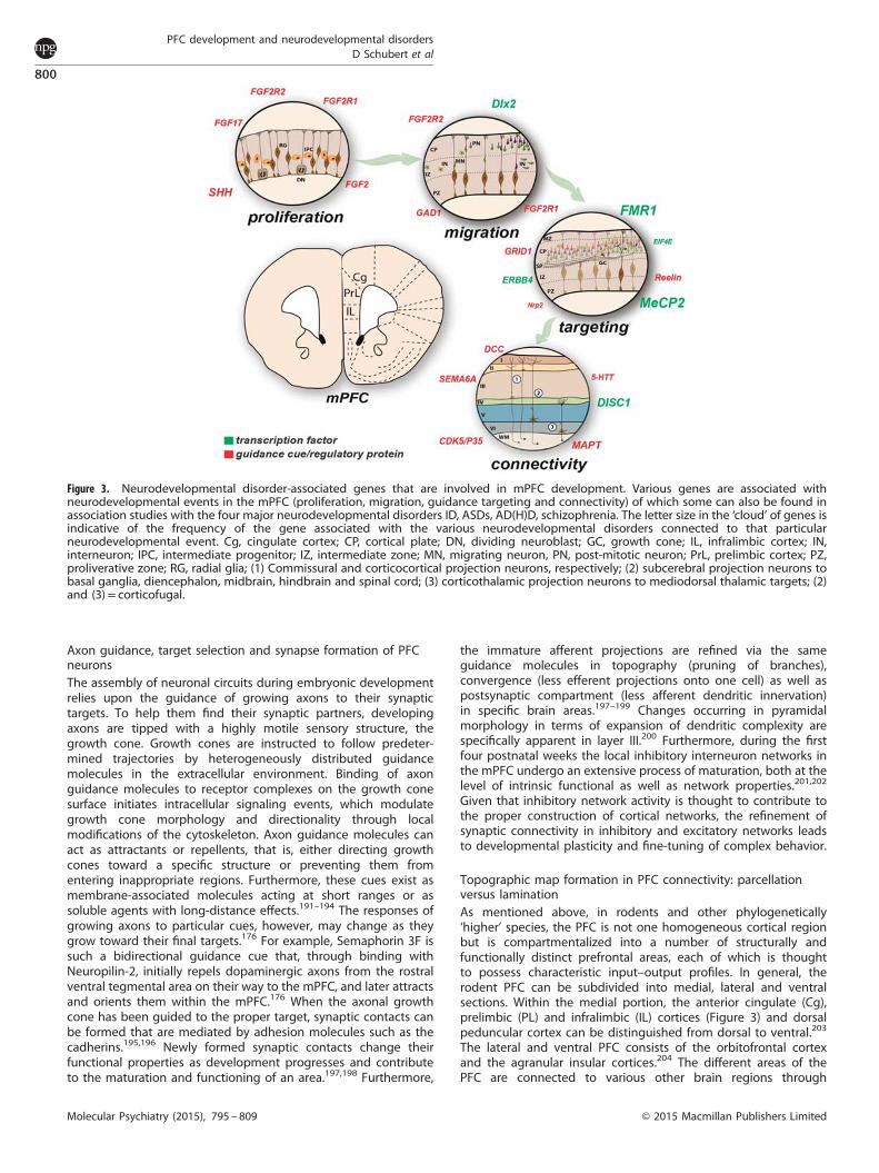
Figure 3. Neurodevelopmental disorder-associated genes that are involved in mPFC development. Various genes are associated withneurodevelopmental events in the mPFC (proliferation, migration, guidance targeting and connectivity) of which some can also be found inassociation studies with the four major neurodevelopmental disorders ID, ASDs, AD(H)D, schizophrenia. The letter size in the ‘cloud’ of genes isindicative of the frequency of the gene associated with the various neurodevelopmental disorders connected to that particularneurodevelopmental event. Cg, cingulate cortex; CP, cortical plate; DN, dividing neuroblast; GC, growth cone; IL, infralimbic cortex; IN,interneuron; IPC, intermediate progenitor; IZ, intermediate zone; MN, migrating neuron, PN, post-mitotic neuron; PrL, prelimbic cortex; PZ,proliverative zone; RG, radial glia; (1) Commissural and corticocortical projection neurons, respectively; (2) subcerebral projection neurons tobasal ganglia, diencephalon, midbrain, hindbrain and spinal cord; (3) corticothalamic projection neurons to mediodorsal thalamic targets; (2)and (3)= corticofugal.
PFC development and neurodevelopmental disordersD Schubert et al
800
Molecular Psychiatry (2015), 795 – 809 © 2015 Macmillan Publishers Limited

Table 1. Commonalities in gene association between PFC developmental events and the four major neurodevelopmental disorders
Gene Involvement in PFC development ID ASDs AD(H)D Schizophrenia
Induction of prefrontal boundariesFGF17 Fgf17 is secreted by the the rostral
patterning center (RSC) and is involvedin the induction of prefrontalboundaries.141,142,233
Fgf17 knockout mice displaydeficits in specific socialinteractions that have beenlinked to ASDs.142
SHH Shh is secreted by the VSC and regulatesthe expression of Fgf8, which is involvedin the induction of prefrontalboundaries.137–139
Mutations in SHH causeholoprosencephaly, a commonforebrain malformationassociated with craniofacialanomalies and MR.234
Significantly higher levels ofserum SHH protein were foundin children with autism.235
A mutation in SHH was foundin two boys with ADHD.236
Proliferation and migration of PFC neuronsFGF2 Fgf2 has an important role in the
production of glutamatergic pyramidalneurons in the (pre)frontal cortex.237
Fgf2 knockout mice showhyperactivity.238
Serum FGF2 levels were found tobe increased in people withschizophrenia.239
FGFR1 Fgfr1 is required for the propernumber of glutamatergic pyramidalneurons in the frontal cortex.144
Dominant or recessive FGFR1mutations are responsible forHartsfield syndrome.240
Dysfunctional Fgfr1 signallingis associated with spontaneoushyperactivity.144
FGFR1 levels are higher inschizophrenia241 and th-fgfr1(tk-)transgenic mice exhibit behaviorresembling human schizophrenia.242
FGFR2 Fgfr2 is involved in generatingexcitatory glutamatergic neuronsin the mPFC.147
Mutations in FGFR2 causeCrouzon’s or Apert syndrome,which can be associatedwith MR.243,244
Deletions of FGFR2 areassociated with ASD.245
Some Fgfr2 deficient micedisplay hyperactive behavior.246
A SNP flanking the FGFR2 geneis associated with schizophrenia.247
Migration of GABAergic interneurons into the PFCDLX2 Dlx2 controls interneurons migration
toward frontal forebrain.248Deletions of DLX2 areassociated with MR.249
DLX2 shows geneticassociation with autism.250
GAD1 Gad1 regulates the migration ofGABA-ergic interneurons tothe PFC.251,252
Gad1 is an ASD susceptibilitygene.253–256
GAD1 expression is altered inschizophreniapatients and is considered a riskgene.257–259 Review: ref 260.
Axon guidance, target selection and synapse formation of PFC neuronsERBB4 Erbb4 regulates dendritic
spine formation and density of PV+interneurons in the PFC.261–264
ERBB4 is associatedwith ID.265
Numerous studies implicate ERBB4as schizophrenia risk genes.266,267
For reviews, see refs 268,269.
EIF4E Eif4e has a role in synaptic function,dendritic spine density and synapticplasticity of PFC neurons.61
EIF4E shows genetic associationwith autism.270–272 Eif4e transgenicmice display autism-likebehaviors.61,273
FMR1 Fmr1 functions in synaptogenesisof dendritic spines of PFCneurons.62,274–277
Mutations/deletions of FMR1cause Fragile X Syndrome, mostcommon known hereditary causeof MR/ID and autism.Reviews: refs 28,30,278.
Mutations/deletions of FMR1cause Fragile X Syndrome,most common known hereditarycause of MR/ID and autism.Reviews: refs 279–281.
Human and animal modelscarrying the FMR1 mutationdisplay ADHD symptoms.282–285
Reduced levels of FMR1 andmutations of associated genesin schizophrenia patients.286–288
GRID1 Grid1 has a role in synaptogenesisof PFC neurons.289
Genetic association290 and Grid1knockout mice show autism-likebehavior.289
GRID1 shows genetic associationwith schizophrenia and gray-matterreduction in patients.291,292
NRP2 Nrp2 is involved in regulating axonguidance of PFC neurons.293
NRP2 mutations are associatedwith autism.294,295
RELN Reln is involved in regulating spinedensity and network formation.296
Disruption of RELN isassociated with MR.297
RELN shows genetic associationwith autism. Reviews: refs 298–300.
RELN shows genetic associationwith schizophrenia. Reviews:refs 301–303.
MECP2 MeCP2 plays a critical role in theregulation of GABAergic transmissionand cortical excitability ofPFC pyramidal.304
MECP2 is associated withMR/ID and especially linked toRett syndrome. Reviews: refs 305,306.
MECP2 is genetically linked toASD.307,308 Review: ref 309.
De novo mutations of MECP2found in schizophrenia patients.310,311
PFCdevelopm
entand
neurodevelopmentaldisorders
DSchubert
etal
801
©2015
Macm
illanPublishers
Limited
Molecular
Psychiatry(2015),
795–809

highly organized projections controlling decision-directedbehavior.205–207
Input connectivity of the mPFC. In terms of the afferentconnectivity of the mPFC, a comprehensive and detailedcomparison of area-specific input connectivity is still lacking. ThemPFC is known to receive long ascending projections from theventral hippocampus,208,209 from cholinergic neurons of the basalforebrain,210,211 from dopaminergic neurons of the rostral part ofthe medial ventral tegmental area176,212,213 and from serotoner-gic/cholinergic neurons of the brainstem along a highly definedtrajectory.214,215 Functionally, the connection with the ventralhippocampus is thought to be of particular importance for thefunctioning of the mPFC during cognitive tasks.216,217 Thecholinergic and dopaminergic systems are considered to mod-ulate mPFC activity and attentional performance.218,219 Interest-ingly, the dopaminergic projections from the ventral tegmentalarea show strong laminar and cell-type specificity. Theyform dense contacts exclusively with interneurons in layersV and VI,176,213,220,221 while for example projections from limbicand thalamic regions innervate both PV+ interneurons andpyramidal cells throughout layers II–VI.222–224 Furthermore, con-nections of the mPFC with both the basolateral amygdala209,225
and the striatum are implicated in motivated behavior.226,227
Interestingly, the long-range connections originating from thebasolateral amygdala have been shown to not only be layer- butalso cell-type specific. Neurons in the basolateral amygdalapreferentially target layer II pyramidal neurons in the mPFC, suchas PL, and amygdala, with which they can form reciprocalconnections.225,228
Output connectivity of the mPFC. As in other cortical areas, thelong-range efferent connections of the PFC are mediated byexcitatory projection neurons, that is, glutamatergic pyramidalcells. Depending on the PFC area, the pyramidal cells project tomany structures such as the basal forebrain, olfactory and corticalstructures, amygdala, striatum, (hypo)thalamus and the brain-stem.204,215,225,226,229 In addition, prefrontal pyramidal neuronsproject to various subcortical areas thereby modulating dopami-nergic, adrenergic, cholinergic and serotonergic projection sys-tems.101,204 The targets of the projection neurons show distinctlayer specificity. Layer III pyramidal neurons connect the mPFCmainly to other cortical areas, whereas layers V and VI pyramidalcells project primarily to subcortical targets.230,231 Furthermore,there is evidence for layer specificity of projections onto individualsubcompartments of single brain structures. In terms of thenucleus accumbens, mPFC layer II pyramidal neurons preferen-tially innervate the core region, whereas neurons of deep layers Vand VI innervate the core as well as the shell region.232
In contrast to the input connectivity, there is ample datademonstrating that the output connectivity properties of themPFC are area dependent, which supports the notion thatprefrontal areas are involved in modulating various aspects ofcognitive behavior,203,204,229 not only in rodents but also in anumber of other species.220,229,230 The dorsomedial areas of thePFC establish connections with the sensorimotor and associationcortex, which are lacking in the ventral parts of the PFC. Theventral parts, however, establish relatively strong connections withthe amygdaloid complex and limbic association cortices. Further-more, the IL has been shown to mainly project to autonomic/visceral related sites, supporting its role in visceromotor activity,204
whereas the PL primarily innervates limbic sites that are thoughtto affect cognition.
FUTURE TRANSLATIONAL AVENUES OF RESEARCHIn summary, substantial progress has been made in the pastdecades toward understanding the etiology of neurodevelop-Ta
ble1.
(Continued
)
Gene
Involvem
entin
PFCdevelopm
ent
IDASD
sAD(H)D
Schizoph
renia
PFCconn
ectivity
DCC
DCCinfluen
cestheprefrontalmaturation
andnetwork
form
ationwiththe
dopam
inergic
midbrain.312,313
Associationbetwee
nschizophrenia
andgen
etic
variationin
DCC.314
DISC1
Disc1
KD
isassociated
withden
dritic
abnorm
alitiesan
daffected
cAMP
signallin
gan
dham
persthe
mesoco
rtical
dopam
inergic
network
form
ation.21,315
DISC1showsgen
etic
associationwithau
tism
.316–319
DISC1showsgen
etic
associationwithADHD
inad
ults.320
DISC1isastrongcandidategen
efor
schizophrenia
(recen
treview
s:refs
321–
323.
CDK5/P35
Cdk5r1
knockoutmicedisplay
improper
mesolim
bic
circuitry
ofthePF
C.324
Cdk5/P35
knockoutmice
displayADHD-likebeh
avior.3
24
Lower
levelsofCD
K5/P35
inpeo
ple
withschizophrenia.325,326
MAPT
Mutationsin
MAPT
areassociated
withalteredfunctional
connectivity
inthehuman
PFC.327
MAPT
CNVsan
dmicrodeletions
inpatients
withMR.328–331
SEMA6A
Loss
ofSema6
acausesprefrontal
loss
ofco
nnectivity.332
Sema6
amutantmicedisplay
ASD
-like
beh
aviors.332
Sema6
amutantmicedisplay
schizophrenia-like
beh
aviors.332
5-HTT
5-HTTisinvo
lved
inproper
raphe-prefrontalnetwork
form
ation.215
5-HTT
isassociated
with
schizophrenia.333,334
Abbreviations:AD(H)D,atten
tiondefi
cithyp
eractivity
disorder;A
SD,autism
spectrum
disorder;G
ABA,γ-aminobutyricacid;ID,intellectual
disab
ility;P
FC,p
refrontalcortex;P
V+,p
arvalbumin
+;R
SC,rostralspinal
cord;V
SC,v
entral
signalingcenter.Synopsisofthemost
citedgen
esthat
havebee
ndirectlylin
ked—
throughroden
tstudies—
tooneormore
ofthedev
elopmen
taleven
tsofPF
Cdev
elopmen
t(in
dicated
initalics)
andthat
havebee
ndirectlygen
etically
linkedto
theetiologyofID/M
R,ASD
s,AD(H)D
and/orschizophrenia.Notes:(1)focu
swas
ononly
those
gen
esthat
wereprove
nto
beinvo
lved
inprefrontal
dev
elopmen
talev
ents
andnotjust
expressed
orinvo
lved
inco
rtical
dev
elopmen
tin
gen
eral
(e.g.,Ree
lin);(2)Aselectionofreferenceswas
mad
ewhen
more
than
threereferenceswerefound.
PFC development and neurodevelopmental disordersD Schubert et al
802
Molecular Psychiatry (2015), 795 – 809 © 2015 Macmillan Publishers Limited

mental disorders at the molecular, cellular and systems levels.Nevertheless, we have only just begun to thoroughly study thedevelopment of a conglomerate of specific brain areas that asa group define the PFC and that are involved in the etiologyof these disorders. In this context, it is remarkable that theexact molecular orchestration of the development of the PFCis still largely unknown. What are the molecular mechanismsthat create a correctly parcellated and layered PFC? How arethe extensive and highly specific interactions between varioussignaling pathways that are connecting the individual areas fine-tuned and how can we manipulate these? We are also onlybeginning to shed light on the large variety of neuronal cellsand their integration in prefrontal local and global networks,let alone that we would know all the molecules that guide theirdifferentiation and projections.To test targeted molecular variations, rodents have emerged as
an excellent model. Animal models and functional assays areinvaluable as it comes to decipher the exact functions of the largenumber of genes that are involved in the various aspects of PFCdevelopment, that is, induction of prefrontal boundaries, intrinsicpatterning of the PFC, proliferation and migration of (pyramidal)PFC neurons, migration of GABAergic interneurons toward thePFC, axon guidance, target selection and synapse formation ofPFC neurons, and PFC connectivity formation. Slowly, the view isemerging that some of these genes are identical to thesusceptibility genes of neurodevelopmental disorders (Table 1).However, up to now only a few of the genes could be directlylinked to one or more of the developmental events within the PFCas well as one or more of the four major neurodevelopmentaldisorders, that is, ID, ASDs, AD(H)D and/or schizophrenia.Especially the availability of in utero electroporation-mediated
gene transfer and other genetic approaches and hence thepossibility to locally knock down or rescue particular genes willhopefully enable us to unravel the exact orchestration of brainareas such as those within the PFC in the near future. Suchknowledge will assist in developing early intervention approachesby altering the susceptibility genes at a particular time and place,such that we deviate from the predetermined developmentalpath, even before the onset of the neurodevelopmental disorder(s) in question. Considering that individual susceptibility genes ofneurodevelopmental disorders have often been found to beassociated with multiple disorders, we can assume that severaldisorders share a common neurodevelopmental origin. It will be achallenge to dissect the individual genetic (and possibly evenepigenetic) contributions to a disorder by using functionalstudies combined with behavioral tasks. For example, gene-environment interactions are crucial to distinguish between riskand vulnerability.It is to be expected that in the coming years many more genes
regulating developmental processes in the PFC and other brainstructures will be linked to neurodevelopmental disorders and viceversa. Animal models, in which we can specifically alter geneexpression in the PFC, can be instrumental for the understandingof the aetiopathological aspects of the disorder(s), as we canmonitor the early disturbances that will eventually lead to defectsin brain maturation and behavior. In order to move toward betterand more preventive treatment of the neurodevelopmentaldisorders, bridges need to be built between disciplines such ascombining genetic analyses of patients suffering from neurode-velopmental disorders with structural and functional brainimaging and in-depth molecular in vitro and in vivo approacheswith cell and animal models. Exploring the molecular and cellularaspects during the progression of the disease process in animalmodels will clarify the pathological mechanisms, which in turnmay provide clues to develop novel treatments for thesedisorders. The earlier during life and the more personalized thetreatment strategies are applied, the better, alleviating symptomsat an early stage and reducing medical costs dramatically.
ACKNOWLEDGMENTSThis work was supported by grants from the Donders Centre for Neuroscience,Radboud University Nijmegen (DS, SMK). The authors thank Prof B. Franke, Dr W.Scheenen, Prof H. van Bokhoven and Prof A. Kriegstein for critically reviewing themanuscript and the anonymous reviewers for their comments. Also, we apologize forthose primary works not referenced here due to space limitations.
REFERENCES1 Patel V, Boyce N, Collins PY, Saxena S, Horton R. A renewed agenda for global
mental health. Lancet 2011; 378: 1441–1442.2 Belfer ML. Child and adolescent mental disorders: the magnitude of the problem
across the globe. J Child Psychol Psychiatry 2008; 49: 226–236.3 Mitchell SR, Reiss AL, Tatusko DH, Ikuta I, Kazmerski DB, Botti JA et al. Neuro-
anatomic alterations and social and communication deficits in monozygotictwins discordant for autism disorder. Am J Psychiatry 2009; 166: 917–925.
4 Shenton ME, Dickey CC, Frumin M, McCarley RW. A review of MRI findings inschizophrenia. Schizophr Res 2001; 49: 1–52.
5 Lesh TA, Niendam TA, Minzenberg MJ, Carter CS. Cognitive control deficits inschizophrenia:mechanisms and meaning. Neuropsychopharmacology 2011; 36:316–338.
6 Casanova MF. Functional and anatomical aspects of prefrontal pathology inschizophrenia. Schizophrenia bulletin 1997; 23: 517–519.
7 Arnsten AF. Stress signalling pathways that impair prefrontal cortex structureand function. Nat Rev 2009; 10: 410–422.
8 Arnsten AF. Prefrontal cortical network connections: key site of vulnerability instress and schizophrenia. Int J Dev Neurosci 29: 215–223.
9 Collins PY, Patel V, Joestl SS, March D, Insel TR, Daar AS et al. Grand challenges inglobal mental health. Nature 2011; 475: 27–30.
10 Kendler KS, Neale MC. Endophenotype: a conceptual analysis. Mol Psychiatry2010; 15: 789–797.
11 Gamo NJ, Arnsten AF. Molecular modulation of prefrontal cortex: rationaldevelopment of treatments for psychiatric disorders. Behav Neurosci 2012; 125:282–296.
12 Lewis DA, Curley AA, Glausier JR, Volk DW. Cortical parvalbumin interneuronsand cognitive dysfunction in schizophrenia. Trends Neurosci 2012; 35: 57–67.
13 Thompson-Schill SL, Bedny M, Goldberg RF. The frontal lobes and the regulationof mental activity. Curr Opin Neurobiol 2005; 15: 219–224.
14 Egner T, Hirsch J. Cognitive control mechanisms resolve conflict through corticalamplification of task-relevant information. Nat Neurosci 2005; 8: 1784–1790.
15 Etkin A, Egner T, Peraza DM, Kandel ER, Hirsch J. Resolving emotional conflict: arole for the rostral anterior cingulate cortex in modulating activity in theamygdala. Neuron 2006; 51: 871–882.
16 Miller EK. The prefrontal cortex and cognitive control. Nat Rev 2000; 1: 59–65.17 Miller EK, Cohen JD. An integrative theory of prefrontal cortex function. Ann Rev
Neurosci 2001; 24: 167–202.18 Summerfield C, Egner T, Greene M, Koechlin E, Mangels J, Hirsch J. Predictive
codes for forthcoming perception in the frontal cortex. Science 2006; 314:1311–1314.
19 Duncan J. An adaptive coding model of neural function in prefrontal cortex. NatRev 2001; 2: 820–829.
20 Mansouri FA, Tanaka K, Buckley MJ. Conflict-induced behavioural adjustment: aclue to the executive functions of the prefrontal cortex. Nat Rev 2009; 10:141–152.
21 Niwa M, Kamiya A, Murai R, Kubo K, Gruber AJ, Tomita K et al. Knockdown ofDISC1 by in utero gene transfer disturbs postnatal dopaminergic maturation inthe frontal cortex and leads to adult behavioral deficits. Neuron 2010; 65:480–489.
22 Loos M, Mueller T, Gouwenberg Y, Wijnands R, van der Loo RJ, Birchmeier C et al.Neuregulin-3 in the mouse medial prefrontal cortex regulates impulsive action.Biol Psychiatry 2014; 76: 648–655.
23 Antonarakis SE, Lyle R, Dermitzakis ET, Reymond A, Deutsch S. Chromosome 21and down syndrome: from genomics to pathophysiology. Nat Rev Genet. 2004; 5:725–738.
24 Contestabile A, Benfenati F, Gasparini L. Communication breaks-down: fromneurodevelopment defects to cognitive disabilities in Down syndrome. ProgNeurobiol 2010; 91: 1–22.
25 Gardiner K, Herault Y, Lott IT, Antonarakis SE, Reeves RH, Dierssen M. Downsyndrome: from understanding the neurobiology to therapy. J Neurosci 2010; 30:14943–14945.
26 van Bokhoven H. Genetic and epigenetic networks in intellectual disabilities.Annu Rev Genet 2011; 45: 81–104.
27 Chelly J, Mandel JL. Monogenic causes of X-linked mental retardation. Nat RevGenet. 2001; 2: 669–680.
PFC development and neurodevelopmental disordersD Schubert et al
803
© 2015 Macmillan Publishers Limited Molecular Psychiatry (2015), 795 – 809
28 Kim M, Ceman S. Fragile X mental retardation protein: past, present and future.Curr Protein Pept Sci 2012; 13: 358–371.
29 Kleefstra T, Smidt M, Banning MJ, Oudakker AR, Van Esch H, de Brouwer AP et al.Disruption of the gene euchromatin histone methyl transferase1 (Eu-HMTase1)is associated with the 9q34 subtelomeric deletion syndrome. J Med Genet 2005;42: 299–306.
30 Mercaldo V, Descalzi G, Zhuo M. Fragile X mental retardation protein in learning-related synaptic plasticity. Mol Cells 2009; 28: 501–507.
31 Cheng A, Haydar TF, Yarowsky PJ, Krueger BK. Concurrent generation of subplateand cortical plate neurons in developing trisomy 16 mouse cortex. Dev Neurosci2004; 26: 255–265.
32 Chakrabarti L, Galdzicki Z, Haydar TF. Defects in embryonic neurogenesis andinitial synapse formation in the forebrain of the Ts65Dn mouse model of Downsyndrome. J Neurosci. 2007; 27: 11483–11495.
33 Willemsen MH, Vissers LE, Willemsen MA, van Bon BW, Kroes T, de Ligt J et al.Mutations in DYNC1H1 cause severe intellectual disability with neuronalmigration defects. J Med Genet 49: 179–183.
34 Pilz DT, Matsumoto N, Minnerath S, Mills P, Gleeson JG, Allen KM et al. LIS1 andXLIS (DCX) mutations cause most classical lissencephaly, but different patterns ofmalformation. Hum Mol Genet 1998; 7: 2029–2037.
35 Rafalowska J, Dziewulska D, Podlecka A, Maslinska D. Early ontogenic dis-turbances in cell migration in mentally disabled adult. Clin Neuropathol 2001; 20:13–18.
36 Penagarikano O, Abrahams BS, Herman EI, Winden KD, Gdalyahu A, Dong H et al.Absence of CNTNAP2 leads to epilepsy, neuronal migration abnormalities, andcore autism-related deficits. Cell 2011; 147: 235–246.
37 Liu JS. Molecular genetics of neuronal migration disorders. Curr Neurol NeurosciRep 11: 171–178.
38 Alarcon JM, Malleret G, Touzani K, Vronskaya S, Ishii S, Kandel ER et al. Chromatinacetylation, memory, and LTP are impaired in CBP+/ − mice: a model for thecognitive deficit in Rubinstein-Taybi syndrome and its amelioration. Neuron2004; 42: 947–959.
39 Nobs L, Baranek C, Nestel S, Kulik A, Kapfhammer J, Nitsch C et al. Stage-specificrequirement for cyclin D1 in glial progenitor cells of the cerebral cortex. Glia2014; 62: 829–839.
40 DiCicco-Bloom E, Lord C, Zwaigenbaum L, Courchesne E, Dager SR, Schmitz Cet al. The developmental neurobiology of autism spectrum disorder. J Neurosci.2006; 26: 6897–6906.
41 Grafodatskaya D, Chung B, Szatmari P, Weksberg R. Autism spectrum disordersand epigenetics. J Am Acad Child Adolesc Psychiatry 2010; 49: 794–809.
42 Amaral DG, Schumann CM, Nordahl CW. Neuroanatomy of autism. TrendsNeurosci 2008; 31: 137–145.
43 Vorstman JA, Staal WG, van Daalen E, van Engeland H, Hochstenbach PF, FrankeL. Identification of novel autism candidate regions through analysis of reportedcytogenetic abnormalities associated with autism. Mol Psychiatry 2006; 11: 1,18–28.
44 Etkin A, Gyurak A, O'Hara R. A neurobiological approach to the cognitive deficitsof psychiatric disorders. Dialogues Clin Neurosci 2013; 15: 419–429.
45 Bishop DV. Genes, cognition, and communication: insights from neurodeve-lopmental disorders. Ann NY Acad Sci. 2009; 1156: 1–18.
46 Folia V, Udden J, Forkstam C, Ingvar M, Hagoort P, Petersson KM. Implicitlearning and dyslexia. Ann NY Acad Sci. 2008; 1145: 132–150.
47 Orban P, Lungu O, Doyon J. Motor sequence learning and developmental dys-lexia. Ann NY Acad Sci. 2008; 1145: 151–172.
48 Pennington BF, Bishop DV. Relations among speech, language, and readingdisorders. Annu Rev Psychol 2009; 60: 283–306.
49 Abrams DA, Lynch CJ, Cheng KM, Phillips J, Supekar K, Ryali S et al. Under-connectivity between voice-selective cortex and reward circuitry in childrenwith autism. Proc Natl Acad Sci USA. 2013; 110: 12060–12065.
50 Vaccarino FM, Grigorenko EL, Smith KM, Stevens HE. Regulation of cerebralcortical size and neuron number by fibroblast growth factors: implicationsfor autism. J Autism Dev Disord 2009; 39: 511–520.
51 Butler MG, Dasouki MJ, Zhou XP, Talebizadeh Z, Brown M, Takahashi TN et al.Subset of individuals with autism spectrum disorders and extreme macro-cephaly associated with germline PTEN tumour suppressor gene mutations.J Med Genet 2005; 42: 318–321.
52 Courchesne E, Campbell K, Solso S. Brain growth across the life span in autism:age-specific changes in anatomical pathology. Brain Res. 2011; 1380:138–145.
53 Courchesne E, Pierce K, Schumann CM, Redcay E, Buckwalter JA, Kennedy DPet al. Mapping early brain development in autism. Neuron 2007; 56: 399–413.
54 Schumann CM, Bloss CS, Barnes CC, Wideman GM, Carper RA, Akshoomoff Net al. Longitudinal magnetic resonance imaging study of cortical developmentthrough early childhood in autism. J Neurosci. 2010; 30: 4419–4427.
55 Fatemi SH, Halt AR, Stary JM, Kanodia R, Schulz SC, Realmuto GR. Glutamic aciddecarboxylase 65 and 67 kDa proteins are reduced in autistic parietal andcerebellar cortices. Biol Psychiatry 2002; 52: 805–810.
56 Zhang ZW, Zak JD, Liu H. MeCP2 is required for normal development ofGABAergic circuits in the thalamus. J Neurophysiol 2010; 103: 2470–2481.
57 Zikopoulos B, Barbas H. Changes in prefrontal axons may disrupt the networkin autism. J Neurosci. 2010; 30: 14595–14609.
58 Zikopoulos B, Barbas H. Altered neural connectivity in excitatory and inhibitorycortical circuits in autism. Front Hum Neurosci 2013; 7: 609.
59 Medrihan L, Tantalaki E, Aramuni G, Sargsyan V, Dudanova I, Missler M et al. Earlydefects of GABAergic synapses in the brain stem of a MeCP2 mouse model ofRett syndrome. J Neurophysiol 2008; 99: 112–121.
60 Radyushkin K, Hammerschmidt K, Boretius S, Varoqueaux F, El-Kordi A,Ronnenberg A et al. Neuroligin-3-deficient mice: model of a monogenic heritableform of autism with an olfactory deficit. Genes Brain Behav 2009; 8: 416–425.
61 Santini E, Huynh TN, MacAskill AF, Carter AG, Pierre P, Ruggero D et al. Exag-gerated translation causes synaptic and behavioural aberrations associatedwith autism. Nature 2013; 493: 411–415.
62 Testa-Silva G, Loebel A, Giugliano M, de Kock CP, Mansvelder HD, Meredith RM.Hyperconnectivity and slow synapses during early development of medialprefrontal cortex in a mouse model for mental retardation and autism. CerebCortex. 2012; 22: 1333–1342.
63 Morgan JT, Chana G, Pardo CA, Achim C, Semendeferi K, Buckwalter J et al.Microglial activation and increased microglial density observed in the dorso-lateral prefrontal cortex in autism. Biol Psychiatry 2010; 68: 368–376.
64 Morgan JT, Chana G, Abramson I, Semendeferi K, Courchesne E, Everall IP.Abnormal microglial-neuronal spatial organization in the dorsolateral prefrontalcortex in autism. Brain Res. 2012; 1456: 72–81.
65 Biederman J. Attention-deficit/hyperactivity disorder: a selective overview. BiolPsychiatry 2005; 57: 1215–1220.
66 Liston C, Malter Cohen M, Teslovich T, Levenson D, Casey BJ. Atypical prefrontalconnectivity in attention-deficit/hyperactivity disorder: pathway to disease orpathological end point? Biol Psychiatry 2011; 69: 1168–1177.
67 Brennan AR, Arnsten AF. Neuronal mechanisms underlying attention deficithyperactivity disorder: the influence of arousal on prefrontal cortical function.Ann NY Acad Sci. 2008; 1129: 236–245.
68 Shaw P, Eckstrand K, Sharp W, Blumenthal J, Lerch JP, Greenstein D et al.Attention- deficit/hyperactivity disorder is characterized by a delay in corticalmaturation. Proc Natl Acad Sci USA. 2007; 104: 19649–19654.
69 Maltezos S, Horder J, Coghlan S, Skirrow C, O'Gorman R, Lavender TJ et al.Glutamate/glutamine and neuronal integrity in adults with ADHD: a protonMRS study. Transl Psychiatry 2014; 4: e373.
70 D'Agati E, Casarelli L, Pitzianti MB, Pasini A. Overflow movements and whitematter abnormalities in ADHD. Prog Neuropsychopharmacol Biol Psychiatry 2010;34: 441–445.
71 Xia S, Li X, Kimball AE, Kelly MS, Lesser I, Branch C. Thalamic shape and con-nectivity abnormalities in children with attention-deficit/hyperactivity disorder.Psychiatry Res. 2012; 204: 161–167.
72 Hart H, Marquand AF, Smith A, Cubillo A, Simmons A, Brammer M et al.Predictive neurofunctional markers of attention-deficit/hyperactivity disorderbased on pattern classification of temporal processing. J Am Acad Child AdolescPsychiatry 2014; 53: 569–78, e1.
73 Arnsten AF. Fundamentals of attention-deficit/hyperactivity disorder: circuitsand pathways. J Clin Psychiatry 2006; 67(Suppl 8): 7–12.
74 Rommelse NN, Geurts HM, Franke B, Buitelaar JK, Hartman CA. A review oncognitive and brain endophenotypes that may be common in autism spectrumdisorder and attention- deficit/hyperactivity disorder and facilitate the search forpleiotropic genes. Neurosci Biobehav Rev 2011; 35: 1363–1396.
75 Leo D, Sorrentino E, Volpicelli F, Eyman M, Greco D, Viggiano D et al. Alteredmidbrain dopaminergic neurotransmission during development in an animalmodel of ADHD. Neurosci Biobehav Rev 2003; 27: 661–669.
76 Miller EM, Pomerleau F, Huettl P, Gerhardt GA, Glaser PE. Aberrant glutamatesignaling in the prefrontal cortex and striatum of the spontaneously hyper-tensive rat model of attention- deficit/hyperactivity disorder. Psychopharmaco-logy 2014; 231: 3019–3029.
77 Fossella JA, Sommer T, Fan J, Pfaff D, Posner MI. Synaptogenesis and heritableaspects of executive attention. Ment Retard Dev Disabil Res Rev 2003; 9: 178–183.
78 Kenar AN, Ay OI, Herken H, Erdal ME. Association of VAMP-2 and Syntaxin 1Agenes with adult attention deficit hyperactivity disorder. Psychiatry Investig 2014;11: 76–83.
79 Hawi Z, Matthews N, Wagner J, Wallace RH, Butler TJ, Vance A et al. DNAvariation in the SNAP25 gene confers risk to ADHD and is associated withreduced expression in prefrontal cortex. PloS One 2013; 8: e60274.
80 McCarthy SE, McCombie WR, Corvin A. Unlocking the treasure trove: from genesto schizophrenia biology. Schizophr Bull 2014; 40: 492–496.
PFC development and neurodevelopmental disordersD Schubert et al
804
Molecular Psychiatry (2015), 795 – 809 © 2015 Macmillan Publishers Limited
81 Lewis DA, Cruz D, Eggan S, Erickson S. Postnatal development of prefrontalinhibitory circuits and the pathophysiology of cognitive dysfunction in schizo-phrenia. Ann NY Acad Sci. 2004; 1021: 64–76.
82 Connor CM, Guo Y, Akbarian S. Cingulate white matter neurons in schizophreniaand bipolar disorder. Biol Psychiatry 2009; 66: 486–493.
83 Beneyto M, Lewis DA. Insights into the neurodevelopmental origin of schizo-phrenia from postmortem studies of prefrontal cortical circuitry. Int J DevNeurosci. 2011; 29: 295–304.
84 Arnsten AF. Prefrontal cortical network connections: key site of vulnerability instress and schizophrenia. Int J Dev Neurosci. 2011; 29: 215–223.
85 Zierhut KC, Schulte-Kemna A, Kaufmann J, Steiner J, Bogerts B, Schiltz K. Distinctstructural alterations independently contributing to working memory deficitsand symptomatology in paranoid schizophrenia. Cortex 2012; 49: 1063–1072.
86 Arnold SE, Talbot K, Hahn CG. Neurodevelopment, neuroplasticity, and newgenes for schizophrenia. Prog Brain Res 2005; 147: 319–345.
87 Bernstein HG, Smalla KH, Durrschmidt D, Keilhoff G, Dobrowolny H, Steiner Jet al. Increased density of prohibitin-immunoreactive oligodendrocytes in thedorsolateral prefrontal white matter of subjects with schizophrenia suggestsextraneuronal roles for the protein in the disease. Neuromolecular Med. 2012; 14:270–280.
88 Glantz LA, Lewis DA. Decreased dendritic spine density on prefrontal corticalpyramidal neurons in schizophrenia. Arch Gen Psychiatry 2000; 57: 65–73.
89 Bennett MR. Schizophrenia: susceptibility genes, dendritic-spine pathology andgray matter loss. Prog Neurobiol 2011; 95: 275–300.
90 Glausier JR, Fish KN, Lewis DA. Altered parvalbumin basket cell inputs in thedorsolateral prefrontal cortex of schizophrenia subjects. Mol Psychiatry 2014; 19:30–36.
91 Glausier JR, Lewis DA. Dendritic spine pathology in schizophrenia. Neuroscience2013; 251: 90–107.
92 Carlsson A. The neurochemical circuitry of schizophrenia. Pharmacopsychiatry2006; 39: S10–S14.
93 Seamans JK, Yang CR. The principal features and mechanisms of dopaminemodulation in the prefrontal cortex. Prog Neurobiol 2004; 74: 1–58.
94 Gonzalez-Burgos G, Hashimoto T, Lewis DA. Alterations of cortical GABA neuronsand network oscillations in schizophrenia. Curr Psychiatry Rep 2010; 12: 335–344.
95 Lewis DA, Gonzalez-Burgos G. Neuroplasticity of neocortical circuits in schizo-phrenia. Neuropsychopharmacology 2008; 33: 141–165.
96 Volk DW, Lewis DA. Prefrontal cortical circuits in schizophrenia. Curr Top BehavNeurosci 2010; 4: 485–508.
97 Rolls ET, Loh M, Deco G, Winterer G. Computational models of schizophrenia anddopamine modulation in the prefrontal cortex. Nat Rev 2008; 9: 696–709.
98 Chen L, Perez SM, Lodge DJ. An augmented dopamine system function is pre-sent prior to puberty in the methylazoxymethanol acetate rodent model ofschizophrenia. Dev Neurobiol. 2014; 74: 907–917.
99 Deserno L, Sterzer P, Wustenberg T, Heinz A, Schlagenhauf F. Reducedprefrontal-parietal effective connectivity and working memory deficits inschizophrenia. J Neurosci. 2012; 32: 12–20.
100 Elston GN. Cortex, cognition and the cell: new insights into the pyramidalneuron and prefrontal function. Cereb Cortex. 2003; 13: 1124–1138.
101 Ongur D, Price JL. The organization of networks within the orbital and medialprefrontal cortex of rats, monkeys and humans. Cereb Cortex. 2000; 10: 206–219.
102 Kellendonk C, Simpson EH, Kandel ER. Modeling cognitive endophenotypes ofschizophrenia in mice. Trends Neurosci 2009; 32: 347–358.
103 Jaaro-Peled H. Gene models of schizophrenia: DISC1 mouse models. Prog BrainRes 2009; 179: 75–86.
104 van Loo KM, Martens GJ. Genetic and environmental factors in complex neuro-developmental disorders. Curr Genomics 2007; 8: 429–444.
105 Coolen MW, Van Loo KM, Van Bakel NN, Pulford DJ, Serneels L, De Strooper Bet al. Gene dosage effect on gamma-secretase component Aph-1b in a ratmodel for neurodevelopmental disorders. Neuron 2005; 45: 497–503.
106 Van Schijndel JE, Van Zweeden M, Van Loo KM, Martens GJ. Gene expressionprofiling in brain regions of a rat model displaying schizophrenia-relatedfeatures. Behav Brain Res 2010; 207: 476–479.
107 van der Elst MC, Roubos EW, Ellenbroek BA, Veening JG, Cools AR. Apomorphine-susceptible rats and apomorphine-unsusceptible rats differ in the tyrosinehydroxylase-immunoreactive network in the nucleus accumbens core and shell.Exp Brain Res 2005; 160: 418–423.
108 Moron JA, Brockington A, Wise RA, Rocha BA, Hope BT. Dopamine uptakethrough the norepinephrine transporter in brain regions with low levels of thedopamine transporter: evidence from knock-out mouse lines. J Neurosci. 2002;22: 389–395.
109 Roullet FI, Crawley JN. Mouse models of autism: testing hypotheses aboutmolecular mechanisms. Curr Top Behav Neurosci 2011; 7: 187–212.
110 Moy SS, Nadler JJ. Advances in behavioral genetics: mouse models of autism.Mol Psychiatry 2008; 13: 4–26.
111 Desbonnet L, Waddington JL, Tuathaigh CM. Mice mutant for genes associatedwith schizophrenia: common phenotype or distinct endophenotypes? BehavBrain Res 2009; 204: 258–273.
112 Johnstone M, Thomson PA, Hall J, McIntosh AM, Lawrie SM, Porteous DJ.DISC1 in schizophrenia: genetic mouse models and human genomic imaging.Schizophr Bull 2011; 37: 14–20.
113 Sawa A. Genetic animal models for schizophrenia: advantages and limitations ofgenetic manipulation in drosophila, zebrafish, rodents, and primates. Prog BrainRes 2009; 179: 3–6.
114 Chen J, Lipska BK, Weinberger DR. Genetic mouse models of schizophrenia: fromhypothesis-based to susceptibility gene-based models. Biol Psychiatry 2006; 59:1180–1188.
115 Baek ST, Kerjan G, Bielas SL, Lee JE, Fenstermaker AG, Novarino G et al. Off-targeteffect of doublecortin family shRNA on neuronal migration associated withendogenous microRNA dysregulation. Neuron 2014; 82: 1255–1262.
116 Kolk SM, de Mooij-Malsen AJ, Martens GJ. Spatiotemporal molecular approach ofin utero electroporation to functionally decipher endophenotypes in neuro-developmental disorders. Front Mol Neurosci 2011; 4: 37.
117 Bissonette GB, Powell EM. Reversal learning and attentional set-shifting in mice.Neuropharmacology 2011; 62: 1168–1174.
118 Bissonette GB, Martins GJ, Franz TM, Harper ES, Schoenbaum G, Powell EM.Double dissociation of the effects of medial and orbital prefrontal cortical lesionson attentional and affective shifts in mice. J Neurosci. 2008; 28: 11124–11130.
119 Brigman JL, Rothblat LA. Stimulus specific deficit on visual reversal learning afterlesions of medial prefrontal cortex in the mouse. Behav Brain Res 2008; 187:405–410.
120 Kellendonk C, Simpson EH, Polan HJ, Malleret G, Vronskaya S, Winiger V et al.Transient and selective overexpression of dopamine D2 receptors in the striatumcauses persistent abnormalities in prefrontal cortex functioning. Neuron 2006;49: 603–615.
121 Ben Abdallah NM, Fuss J, Trusel M, Galsworthy MJ, Bobsin K, Colacicco G et al.The puzzle box as a simple and efficient behavioral test for exploring impair-ments of general cognition and executive functions in mouse models ofschizophrenia. Exp Neurol 2011; 227: 42–52.
122 Chudasama Y. Animal models of prefrontal-executive function. Behav Neurosci2011; 125: 327–343.
123 Kolb B, Mychasiuk R, Muhammad A, Li Y, Frost DO, Gibb R. Experience and thedeveloping prefrontal cortex. Proc Natl Acad Sci USA. 2012; 109: 17186–17193.
124 Vieira C, Pombero A, Garcia-Lopez R, Gimeno L, Echevarria D, Martinez S.Molecular mechanisms controlling brain development: an overview of neuro-epithelial secondary organizers. Int J Dev Biol 2010; 54: 7–20.
125 Puelles L, Rubenstein JL. Forebrain gene expression domains and the evolvingprosomeric model. Trends Neurosci 2003; 26: 469–476.
126 Andersson E, Tryggvason U, Deng Q, Friling S, Alekseenko Z, Robert B et al.Identification of intrinsic determinants of midbrain dopamine neurons. Cell 2006;124: 393–405.
127 Pombero A, Martinez S. Telencephalic morphogenesis during the process ofneurulation: an experimental study using quail-chick chimeras. J Comp Neurol2009; 512: 784–797.
128 Rubenstein JL, Martinez S, Shimamura K, Puelles L. The embryonic vertebrateforebrain: the prosomeric model. Science (New York, NY 1994; 266: 578–580.
129 Borello U, Pierani A. Patterning the cerebral cortex: traveling with morphogens.Curr Opin Genet Dev 2010; 20: 408–415.
130 Lander AD. Morpheus unbound: reimagining the morphogen gradient. Cell 2007;128: 245–256.
131 O'Leary DD, Chou SJ, Sahara S. Area patterning of the mammalian cortex. Neuron2007; 56: 252–269.
132 Fukuchi-Shimogori T, Grove EA. Neocortex patterning by the secreted signalingmolecule FGF8. Science (New York, NY 2001; 294: 1071–1074.
133 Rubenstein JL. Research Review: Development of the cerebral cortex: implica-tions for neurodevelopmental disorders. J Child Psychol Psychiatry 2010; 52:339–355.
134 Hebert JM, Hayhurst M, Marks ME, Kulessa H, Hogan BL, McConnell SK. BMPligands act redundantly to pattern the dorsal telencephalic midline. Genesis2003; 35: 214–219.
135 Fernandes M, Gutin G, Alcorn H, McConnell SK, Hebert JM. Mutations in the BMPpathway in mice support the existence of two molecular classes of holo-prosencephaly. Development (Cambridge, England) 2007; 134: 3789–3794.
136 Hebert JM, Mishina Y, McConnell SK. BMP signaling is required locally to patternthe dorsal telencephalic midline. Neuron 2002; 35: 1029–1041.
137 Rash BG, Grove EA. Patterning the dorsal telencephalon: a role for sonichedgehog? J Neurosci 2007; 27: 11595–11603.
138 Kuschel S, Ruther U, Theil T. A disrupted balance between Bmp/Wnt and Fgfsignaling underlies the ventralization of the Gli3 mutant telencephalon. Dev Biol2003; 260: 484–495.
PFC development and neurodevelopmental disordersD Schubert et al
805
© 2015 Macmillan Publishers Limited Molecular Psychiatry (2015), 795 – 809

139 Ohkubo Y, Chiang C, Rubenstein JL. Coordifnate regulation and synergistic actionsof BMP4, SHH and FGF8 in the rostral prosencephalon regulate morphogenesis ofthe telencephalic and optic vesicles. Neuroscience 2002; 111: 1–17.
140 Blaess S, Stephen D, Joyner AL. Gli3 coordinates three-dimensional patterningand growth of the tectum and cerebellum by integrating Shh and Fgf8 signaling.Development (Cambridge, England) 2008; 135: 2093–2103.
141 Cholfin JA, Rubenstein JL. Frontal cortex subdivision patterning is coordinatelyregulated by Fgf8, Fgf17, and Emx2. J Comp Neurol 2008; 509: 144–155.
142 Scearce-Levie K, Roberson ED, Gerstein H, Cholfin JA, Mandiyan VS, Shah NMet al. Abnormal social behaviors in mice lacking Fgf17. Genes Brain Behav 2008;7: 344–354.
143 Shimogori T, Banuchi V, Ng HY, Strauss JB, Grove EA. Embryonic signalingcenters expressing BMP, WNT and FGF proteins interact to pattern thecerebral cortex. Development (Cambridge, England) 2004; 131: 5639–5647.
144 Shin DM, Korada S, Raballo R, Shashikant CS, Simeone A, Taylor JR et al. Loss ofglutamatergic pyramidal neurons in frontal and temporal cortex resulting fromattenuation of FGFR1 signaling is associated with spontaneous hyperactivityin mice. J Neurosci. 2004; 24: 2247–2258.
145 Thomson RE, Kind PC, Graham NA, Etherson ML, Kennedy J, Fernandes AC et al.Fgf receptor 3 activation promotes selective growth and expansion ofoccipitotemporal cortex. Neural Dev 2009; 4: 4.
146 Thomson RE, Pellicano F, Iwata T. Fibroblast growth factor receptor 3 kinasedomain mutation increases cortical progenitor proliferation via mitogen-activated protein kinase activation. J Neurochem 2007; 100: 1565–1578.
147 Stevens HE, Smith KM, Maragnoli ME, Fagel D, Borok E, Shanabrough M et al.Fgfr2 is required for the development of the miedial prefrontal cortex and itsconnections with limbic circuits. J Neurosci. 2010; 30: 5590–5602.
148 Muller Smith K, Fagel DM, Stevens HE, Rabenstein RL, Maragnoli ME, Ohkubo Yet al. Deficiency in inhibitory cortical interneurons associates with hyperactivityin fibroblast growth factor receptor 1 mutant mice. Biol Psychiatry 2008; 63:953–962.
149 Inglis-Broadgate SL, Thomson RE, Pellicano F, Tartaglia MA, Pontikis CC, CooperJD et al. FGFR3 regulates brain size by controlling progenitor cell proliferationand apoptosis during embryonic development. Dev Biol 2005; 279: 73–85.
150 Hoch RV, Rubenstein JL, Pleasure S. Genes and signaling events that establishregional patterning of the mammalian forebrain. Semin Cell Dev Biol 2009; 20:378–386.
151 Sur M, Rubenstein JL. Patterning and plasticity of the cerebral cortex. Science(New York, NY 2005; 310: 805–810.
152 Mason I. Initiation to end point: the multiple roles of fibroblast growth factors inneural development. Nat Rev 2007; 8: 583–596.
153 Schuurmans C, Guillemot F. Molecular mechanisms underlying cell fate specifi-cation in the developing telencephalon. Curr Opin Neurobiol 2002; 12: 26–34.
154 Jessell TM. Neuronal specification in the spinal cord: inductive signals andtranscriptional codes. Nat Rev Genet 2000; 1: 20–29.
155 Goridis C, Rohrer H. Specification of catecholaminergic and serotonergicneurons. Nat Rev 2002; 3: 531–541.
156 Hasegawa H, Ashigaki S, Takamatsu M, Suzuki-Migishima R, Ohbayashi N, Itoh Net al. Laminar patterning in the developing neocortex by temporally coordinatedfibroblast growth factor signaling. J Neurosci 2004; 24: 8711–8719.
157 Chou SJ, Perez-Garcia CG, Kroll TT, O'Leary DD. Lhx2 specifies regional fatein Emx1 lineage of telencephalic progenitors generating cerebral cortex.Nat Neurosci 2009; 12: 1381–1389.
158 Mangale VS, Hirokawa KE, Satyaki PR, Gokulchandran N, Chikbire S, SubramanianL et al. Lhx2 selector activity specifies cortical identity and suppresses hippo-campal organizer fate. Science (New York, NY 2008; 319: 304–309.
159 Polleux F, Ince-Dunn G, Ghosh A. Transcriptional regulation of vertebrate axonguidance and synapse formation. Nat Rev 2007; 8: 331–340.
160 Flames N, Pla R, Gelman DM, Rubenstein JL, Puelles L, Marin O. Delineation ofmultiple subpallial progenitor domains by the combinatorial expression oftranscriptional codes. J Neurosci 2007; 27: 9682–9695.
161 Uchida S, Hara K, Kobayashi A, Funato H, Hobara T, Otsuki K et al. Early lifestress enhances behavioral vulnerability to stress through the activation ofREST4-mediated gene transcription in the medial prefrontal cortex of rodents.J Neurosci 2010; 30: 15007–15018.
162 Rojas P, Joodmardi E, Hong Y, Perlmann T, Ogren SO. Adult mice with reducedNurr1 expression: an animal model for schizophrenia. Mol Psychiatry 2007; 12:756–766.
163 Johnson MB, Kawasawa YI, Mason CE, Krsnik Z, Coppola G, Bogdanovic D et al.Functional and evolutionary insights into human brain development throughglobal transcriptome analysis. Neuron 2009; 62: 494–509.
164 Yang S, Wang K, Valladares O, Hannenhalli S, Bucan M. Genome-wide expressionprofiling and bioinformatics analysis of diurnally regulated genes in the mouseprefrontal cortex. Genome Biol 2007; 8: R247.
165 Erraji-Benchekroun L, Underwood MD, Arango V, Galfalvy H, Pavlidis P,Smyrniotopoulos P et al. Molecular aging in human prefrontal cortex is selectiveand continuous throughout adult life. Biol Psychiatry 2005; 57: 549–558.
166 Zhang YE, Landback P, Vibranovski MD, Long M. Accelerated recruitment of newbrain development genes into the human genome. PLoS Biol 2011; 9: e1001179.
167 Kriegstein AR, Noctor SC. Patterns of neuronal migration in the embryoniccortex. Trends Neurosci 2004; 27: 392–399.
168 Noctor SC, Flint AC, Weissman TA, Dammerman RS, Kriegstein AR. Neuronsderived from radial glial cells establish radial units in neocortex. Nature 2001;409: 714–720.
169 Storm EE, Garel S, Borello U, Hebert JM, Martinez S, McConnell SK et al.Dose-dependent functions of Fgf8 in regulating telencephalic patterningcenters. Development (Cambridge, England) 2006; 133: 1831–1844.
170 Garel S, Huffman KJ, Rubenstein JL. Molecular regionalization of the neocortex isdisrupted in Fgf8 hypomorphic mutants. Development (Cambridge, England)2003; 130: 1903–1914.
171 Raballo R, Rhee J, Lyn-Cook R, Leckman JF, Schwartz ML, Vaccarino FM. Basicfibroblast growth factor (Fgf2) is necessary for cell proliferation and neurogen-esis in the developing cerebral cortex. J Neurosci 2000; 20: 5012–5023.
172 Sessa A, Mao CA, Hadjantonakis AK, Klein WH, Broccoli V. Tbr2 directs conversionof radial glia into basal precursors and guides neuronal amplification by indirectneurogenesis in the developing neocortex. Neuron 2008; 60: 56–69.
173 Sessa A, Mao CA, Colasante G, Nini A, Klein WH, Broccoli V. Tbr2-positive inter-mediate (basal) neuronal progenitors safeguard cerebral cortex expansion bycontrolling amplification of pallial glutamatergic neurons and attraction ofsubpallial GABAergic interneurons. Genes Dev 2010; 24: 1816–1826.
174 Arnold SJ, Huang GJ, Cheung AF, Era T, Nishikawa S, Bikoff EK et al. The T-boxtranscription factor Eomes/Tbr2 regulates neurogenesis in the corticalsubventricular zone. Genes Dev 2008; 22: 2479–2484.
175 Sweeney NT, Tierney H, Feldheim DA. Tbr2 is required to generate a neuralcircuit mediating the pupillary light reflex. J Neurosci 2014; 34: 5447–5453.
176 Kolk SM, Gunput RA, Tran TS, van den Heuvel DM, Prasad AA, Hellemons AJ et al.Semaphorin 3F is a bifunctional guidance cue for dopaminergic axons andcontrols their fasciculation, channeling, rostral growth, and intracortical target-ing. J Neurosci 2009; 29: 12542–12557.
177 Kinast K, Peeters D, Kolk SM, Schubert D, Homberg JR. Genetic and pharmaco-logical manipulations of the serotonergic system in early life: neurodevelop-mental underpinnings of autism-related behavior. Front Cell Neurosci 2013; 7: 72.
178 Popolo M, McCarthy DM, Bhide PG. Influence of dopamine on precursor cellproliferation and differentiation in the embryonic mouse telencephalon.Dev Neurosci 2004; 26: 229–244.
179 Kriegstein A, Noctor S, Martinez-Cerdeno V. Patterns of neural stem and pro-genitor cell division may underlie evolutionary cortical expansion. Nat Rev 2006;7: 883–890.
180 Ayala R, Shu T, Tsai LH. Trekking across the brain: the journey of neuronalmigration. Cell 2007; 128: 29–43.
181 Rakic P. The radial edifice of cortical architecture: from neuronal silhouettes togenetic engineering. Brain Res Rev 2007; 55: 204–219.
182 Knuesel I. Reelin-mediated signaling in neuropsychiatric and neurodegenerativediseases. Prog Neurobiol 2010; 91: 257–274.
183 Gaiano N. Strange bedfellows: Reelin and Notch signaling interact to regulatecell migration in the developing neocortex. Neuron 2008; 60: 189–191.
184 Lui JH, Hansen DV, Kriegstein AR. Development and evolution of the humanneocortex. Cell 2011; 146: 18–36.
185 LaMonica BE, Lui JH, Wang X, Kriegstein AR. OSVZ progenitors in the humancortex: an updated perspective on neurodevelopmental disease. Curr OpinNeurobiol 2012; 22: 747–753.
186 Gertz CC, Lui JH, LaMonica BE, Wang X, Kriegstein AR. Diverse behaviors of outerradial glia in developing ferret and human cortex. J Neurosci 2014; 34:2559–2570.
187 Miyoshi G, Butt SJ, Takebayashi H, Fishell G. Physiologically distinct temporalcohorts of cortical interneurons arise from telencephalic Olig2-expressing pre-cursors. J Neurosci 2007; 27: 7786–7798.
188 Southwell DG, Nicholas CR, Basbaum AI, Stryker MP, Kriegstein AR, Rubenstein JLet al. Interneurons from embryonic development to cell-based therapy. Science(New York, NY 2014; 344: 1240622.
189 Cobos I, Long JE, Thwin MT, Rubenstein JL. Cellular patterns of transcriptionfactor expression in developing cortical interneurons. Cereb Cortex 2006; 16:i82–i88.
190 Le Magueresse C, Monyer H. GABAergic interneurons shape the functionalmaturation of the cortex. Neuron 2013; 77: 388–405.
191 Tessier-Lavigne M, Goodman CS. The molecular biology of axon guidance.Science (New York, NY 1996; 274: 1123–1133.
192 Dickson BJ. Molecular mechanisms of axon guidance. Science (New York, NY2002; 298: 1959–1964.
PFC development and neurodevelopmental disordersD Schubert et al
806
Molecular Psychiatry (2015), 795 – 809 © 2015 Macmillan Publishers Limited

193 O'Donnell M, Chance RK, Bashaw GJ. Axon growth and guidance:receptor regulation and signal transduction. Annu Rev Neurosci 2009; 32:383–412.
194 Dudanova I, Klein R. Integration of guidance cues: parallel signaling and cross-talk. Trends Neurosci 2013; 36: 295–304.
195 Kiryushko D, Berezin V, Bock E. Regulators of neurite outgrowth: role of celladhesion molecules. Ann NY Acad Sci 2004; 1014: 140–154.
196 Stoya G, Redies C, Schmid-Hertel N. Inversion of layer-specific cadherin expres-sion profiles and maintenance of cytoarchitectonic areas in the allocortex of thereeler mutant mouse. J Comp Neurol 2014.
197 Lai KO, Ip NY. Synapse development and plasticity: roles of ephrin/Eph receptorsignaling. Curr Opin Neurobiol 2009; 19: 275–283.
198 Tessier CR, Broadie K. Activity-dependent modulation of neural circuit synapticconnectivity. Front Mol Neurosci 2009; 2: 8.
199 Vanderhaeghen P, Cheng HJ. Guidance molecules in axon pruning andcell death. Cold Spring Harbor Perspect Biol 2010; 2: a001859.
200 Petanjek Z, Judas M, Kostovic I, Uylings HB. Lifespan alterations of basal dendritictrees of pyramidal neurons in the human prefrontal cortex: a layer-specificpattern. Cereb Cortex 2008; 18: 915–929.
201 de Almeida J, Jourdan I, Murer MG, Belforte JE. Refinement of neuronal syn-chronization with gamma oscillations in the medial prefrontal cortex afteradolescence. PloS One 2013; 8: e62978.
202 Yang JM, Zhang J, Yu YQ, Duan S, Li XM. Postnatal development of 2 micro-circuits involving fast-spiking interneurons in the mouse prefrontal cortex. CerebCortex 2014; 24: 98–109.
203 Hoover WB, Vertes RP. Anatomical analysis of afferent projections to the medialprefrontal cortex in the rat. Brain Struct Funct 2007; 212: 149–179.
204 George O, Koob GF. Individual differences in prefrontal cortex function and thetransition from drug use to drug dependence. Neurosci Biobehav Rev 2010; 35:232–247.
205 Lisboa SF, Stecchini MF, Correa FM, Guimaraes FS, Resstel LB. Different role ofthe ventral medial prefrontal cortex on modulation of innate and associativelearned fear. Neuroscience 2010; 171: 760–768.
206 Diamond A. Biological and social influences on cognitive control processesdependent on prefrontal cortex. Prog Brain Res 2011; 189: 319–339.
207 Thompson BM, Baratta MV, Biedenkapp JC, Rudy JW, Watkins LR, Maier SF.Activation of the infralimbic cortex in a fear context enhances extinctionlearning. Learn Mem 2010; 17: 591–599.
208 Laroche S, Davis S, Jay TM. Plasticity at hippocampal to prefrontal cortexsynapses: dual roles in working memory and consolidation. Hippocampus 2000;10: 438–446.
209 Little JP, Carter AG. Subcellular synaptic connectivity of layer 2 pyramidalneurons in the medial prefrontal cortex. J Neurosci 2012; 32: 12808–12819.
210 Golmayo L, Nunez A, Zaborszky L. Electrophysiological evidence for the exis-tence of a posterior cortical-prefrontal-basal forebrain circuitry in modulatingsensory responses in visual and somatosensory rat cortical areas. Neuroscience2003; 119: 597–609.
211 Henny P, Jones BE. Projections from basal forebrain to prefrontal cortex com-prise cholinergic, GABAergic and glutamatergic inputs to pyramidal cells orinterneurons. Eur J Neurosci 2008; 27: 654–670.
212 Hauber W. Dopamine release in the prefrontal cortex and striatum: temporal andbehavioural aspects. Pharmacopsychiatry 43: S32–S41.
213 van Schouwenburg M, Aarts E, Cools R. Dopaminergic modulation of cognitivecontrol:distinct roles for the prefrontal cortex and the basal ganglia. Curr PharmDes 2010; 16: 2026–2032.
214 Fitzgerald PJ. A neurochemical yin and yang: does serotonin activate and nor-epinephrine deactivate the prefrontal cortex? Psychopharmacology 2011; 213:171–182.
215 Witteveen JS, Middelman A, van Hulten JA, Martens GJ, Homberg JR, Kolk SM.Lack of serotonin reuptake during brain development alters rostral raphe-prefrontal network formation. Front Cell Neurosci 2013; 7: 143.
216 Goto Y, O'Donnell P. Altered prefrontal cortex-nucleus accumbens informationprocessing in a developmental animal model of schizophrenia. Ann NY Acad Sci2003; 1003: 398–401.
217 Goto Y, Grace AA. Limbic and cortical information processing in the nucleusaccumbens. Trends Neurosci 2008; 31: 552–558.
218 Sarter M, Hasselmo ME, Bruno JP, Givens B. Unraveling the attentional functionsof cortical cholinergic inputs: interactions between signal-driven and cognitivemodulation of signal detection. Brain Res 2005; 48: 98–111.
219 Del Arco A, Mora F. Neurotransmitters and prefrontal cortex-limbic systeminteractions: implications for plasticity and psychiatric disorders. J Neural Transm2009; 116: 941–952.
220 Zhang ZW, Burke MW, Calakos N, Beaulieu JM, Vaucher E. Confocal analysis ofcholinergic and dopaminergic inputs onto pyramidal cells in the prefrontalcortex of rodents. Front Neuroanat 2010; 4: 21.
221 Otani S, Daniel H, Roisin MP, Crepel F. Dopaminergic modulation of long-term synaptic plasticity in rat prefrontal neurons. Cereb Cortex 2003; 13:1251–1256.
222 Gabbott P, Headlam A, Busby S. Morphological evidence that CA1 hippocampalafferents monosynaptically innervate PV-containing neurons and NADPH-diaphorase reactive cells in the medial prefrontal cortex (Areas 25/32) ofthe rat. Brain Res 2002; 946: 314–322.
223 Gabbott PL, Warner TA, Busby SJ. Amygdala input monosynaptically innervatesparvalbumin immunoreactive local circuit neurons in rat medial prefrontalcortex. Neuroscience 2006; 139: 1039–1048.
224 Rotaru DC, Barrionuevo G, Sesack SR. Mediodorsal thalamic afferents to layer IIIof the rat prefrontal cortex: synaptic relationships to subclasses of interneurons.J Comp Neurol 2005; 490: 220–238.
225 Little JP, Carter AG. Synaptic mechanisms underlying strong reciprocal con-nectivity between the medial prefrontal cortex and basolateral amygdala.J Neurosci 2013; 33: 15333–15342.
226 Ernst M, Fudge JL. A developmental neurobiological model of motivatedbehavior: anatomy, connectivity and ontogeny of the triadic nodes. NeurosciBiobehav Reviews 2009; 33: 367–382.
227 Ernst M, Pine DS, Hardin M. Triadic model of the neurobiology of motivatedbehavior in adolescence. Psychol Med 2006; 36: 299–312.
228 Senn V, Wolff SB, Herry C, Grenier F, Ehrlich I, Grundemann J et al. Long-rangeconnectivity defines behavioral specificity of amygdala neurons. Neuron 2014;81: 428–437.
229 Heidbreder CA, Groenewegen HJ. The medial prefrontal cortex in the rat: evi-dence for a dorso-ventral distinction based upon functional and anatomicalcharacteristics. Neurosci Biobehav Rev 2003; 27: 555–579.
230 Gabbott PL, Warner TA, Jays PR, Salway P, Busby SJ. Prefrontal cortex in the rat:projections to subcortical autonomic, motor, and limbic centers. J Comp Neurol2005; 492: 145–177.
231 Dembrow NC, Chitwood RA, Johnston D. Projection-specific neuromodulation ofmedial prefrontal cortex neurons. J Neurosci 2010; 30: 16922–16937.
232 Ding DC, Gabbott PL, Totterdell S. Differences in the laminar origin of projectionsfrom the medial prefrontal cortex to the nucleus accumbens shell and coreregions in the rat. Brain Res 2001; 917: 81–89.
233 Cholfin JA, Rubenstein JL. Genetic regulation of prefrontal cortex developmentand function. Novartis Found Symp. 2007; 288: 165–173, discussion 73–7, 276–81.
234 Schell-Apacik C, Rivero M, Knepper JL, Roessler E, Muenke M, Ming JE. SONICHEDGEHOG mutations causing human holoprosencephaly impair neuralpatterning activity. Hum Genet. 2003; 113: 170–177.
235 Al-Ayadhi LY. Relationship between Sonic hedgehog protein, brain-derivedneurotrophic factor and oxidative stress in autism spectrum disorders. Neu-rochem Res. 2012; 37: 394–400.
236 Heussler HS, Suri M, Young ID, Muenke M. Extreme variability of expression of aSonic Hedgehog mutation: attention difficulties and holoprosencephaly. Arch DisChild. 2002; 86: 293–296.
237 Raballo R, Rhee J, Lyn-Cook R, Leckman JF, Schwartz ML, Vaccarino FM. Basicfibroblast growth factor (Fgf2) is necessary for cell proliferation and neurogen-esis in the developing cerebral cortex. J Neurosci. 2000; 20: 5012–5023.
238 Terwisscha van Scheltinga AF, Bakker SC, Kahn RS, Kas MJ. Fibroblast growthfactors in neurodevelopment and psychopathology. Neuroscientist 2013; 19:479–494.
239 Hashimoto K, Shimizu E, Komatsu N, Nakazato M, Okamura N, Watanabe H et al.Increased levels of serum basic fibroblast growth factor in schizophrenia.Psychiatry Res. 2003; 120: 211–218.
240 Simonis N, Migeotte I, Lambert N, Perazzolo C, de Silva DC, Dimitrov B et al.FGFR1 mutations cause Hartsfield syndrome, the unique association of holo-prosencephaly and ectrodactyly. J Med Genet 2013; 50: 585–592.
241 Gaughran F, Payne J, Sedgwick PM, Cotter D, Berry M. Hippocampal FGF-2and FGFR1 mRNA expression in major depression, schizophrenia and bipolardisorder. Brain Res Bull 2006; 70: 221–227.
242 Stachowiak MK, Kucinski A, Curl R, Syposs C, Yang Y, Narla S et al. Schizophrenia:a neurodevelopmental disorder – integrative genomic hypothesis and ther-apeutic implications from a transgenic mouse model. Schizophr Res 2013; 143:367–376.
243 Mahieu-Caputo D, Sonigo P, Amiel J, Simon I, Aubry MC, Lemerrer M et al.Prenatal diagnosis of sporadic Apert syndrome: a sequential diagnosticapproach combining three-dimensional computed tomography and molecularbiology. Fetal Diagn Ther. 2001; 16: 10–12.
244 Padmanabhan V, Hegde AM, Rai K. Crouzon's syndrome: a review of literatureand case report. Contemp Clin Dent 2011; 2: 211–214.
245 Wentz E, Vujic M, Karrstedt EL, Erlandsson A, Gillberg C. A case report of twomale siblings with autism and duplication of Xq13-q21, a region including threegenes predisposing for autism. European Child Adolesc Psychiatry 2014; 23:329–336.
PFC development and neurodevelopmental disordersD Schubert et al
807
© 2015 Macmillan Publishers Limited Molecular Psychiatry (2015), 795 – 809

246 Kaga Y, Shoemaker WJ, Furusho M, Bryant M, Rosenbluth J, Pfeiffer SE et al. Micewith conditional inactivation of fibroblast growth factor receptor-2 signaling inoligodendrocytes have normal myelin but display dramatic hyperactivity whencombined with Cnp1 inactivation. J Neurosci. 2006; 26: 12339–12350.
247 O'Donovan MC, Norton N, Williams H, Peirce T, Moskvina V, Nikolov I et al.Analysis of 10 independent samples provides evidence for association betweenschizophrenia and a SNP flanking fibroblast growth factor receptor 2. Mol Psy-chiatry 2009; 14: 30–36.
248 Stevens HE, Su T, Yanagawa Y, Vaccarino FM. Prenatal stress delays inhibitoryneuron progenitor migration in the developing neocortex. Psychoneur-oendocrinology 2013; 38: 509–521.
249 Theisen A, Rosenfeld JA, Shane K, McBride KL, Atkin JF, Gaba C et al. Refinementof the region for split hand/foot malformation 5 on 2q31.1. Mol Syndromol 2010;1: 262–271.
250 Liu X, Novosedlik N, Wang A, Hudson ML, Cohen IL, Chudley AE et al. TheDLX1and DLX2 genes and susceptibility to autism spectrum disorders. Eur J HumGenet 2009; 17: 228–235.
251 Uchida T, Furukawa T, Iwata S, Yanagawa Y, Fukuda A. Selective loss ofparvalbumin-positive GABAergic interneurons in the cerebral cortex ofmaternally stressed Gad1-heterozygous mouse offspring. Transl Psychiatry 2014;4: e371.
252 Lazarus MS, Krishnan K, Huang ZJ. GAD67 Deficiency in parvalbumin inter-neurons produces deficits in inhibitory transmission and network disinhibition inmouse prefrontal cortex. Cereb Cortex 2013 (e-pub ahead of print).
253 Bacchelli E, Blasi F, Biondolillo M, Lamb JA, Bonora E, Barnby G et al. Screening ofnine candidate genes for autism on chromosome 2q reveals rare nonsynon-ymous variants in the cAMP-GEFII gene. Mol Psychiatry 2003; 8: 916–924.
254 Rabionet R, Jaworski JM, Ashley-Koch AE, Martin ER, Sutcliffe JS, Haines JL et al.Analysis of the autism chromosome 2 linkage region: GAD1 and othercandidate genes. Neurosci Lett. 2004; 372: 209–214.
255 Buttenschon HN, Lauritsen MB, El Daoud A, Hollegaard M, Jorgensen M,Tvedegaard K et al. A population-based association study of glutamate dec-arboxylase 1 as a candidate gene for autism. J Neural Transm. 2009; 116:381–388.
256 Chang SC, Pauls DL, Lange C, Sasanfar R, Santangelo SL. Common geneticvariation in the GAD1 gene and the entire family of DLX homeobox genes andautism spectrum disorders. Am J Med Genet B Neuropsychiatr Genet 2011; 156:233–239.
257 Akbarian S, Huang HS. Molecular and cellular mechanisms of altered GAD1/GAD67 expression in schizophrenia and related disorders. Brain Res Rev 2006; 52:293–304.
258 Ayalew M, Le-Niculescu H, Levey DF, Jain N, Changala B, Patel SD et al.Convergent functional genomics of schizophrenia: from comprehensive under-standing to genetic risk prediction. Mol Psychiatry 2012; 17: 887–905.
259 Straub RE, Lipska BK, Egan MF, Goldberg TE, Callicott JH, Mayhew MB et al.Allelic variation in GAD1 (GAD67) is associated with schizophrenia andinfluences cortical function and gene expression. Mol Psychiatry 2007; 12:854–869.
260 Cherlyn SY, Woon PS, Liu JJ, Ong WY, Tsai GC, Sim K. Genetic association studiesof glutamate, GABA and related genes in schizophrenia and bipolar disorder: adecade of advance. Neurosci Biobehav Rev 2010; 34: 958–977.
261 Cooper MA, Koleske AJ. Ablation of ErbB4 from excitatory neurons leads toreduced dendritic spine density in mouse prefrontal cortex. J Comp Neurol 2014;522: 3351–3362.
262 Ting AK, Chen Y, Wen L, Yin DM, Shen C, Tao Y et al. Neuregulin 1 promotesexcitatory synapse development and function in GABAergic interneurons.J Neurosci. 2011; 31: 15–25.
263 Hou XJ, Ni KM, Yang JM, Li XM. Neuregulin 1/ErbB4 enhances synchronizedoscillations of prefrontal cortex neurons via inhibitory synapses. Neuroscience2014; 261: 107–117.
264 Yang JM, Zhang J, Chen XJ, Geng HY, Ye M, Spitzer NC et al. Development ofGABA circuitry of fast-spiking basket interneurons in the medial prefrontal cortexof erbb4-mutant mice. J Neurosci. 2013; 33: 19724–19733.
265 Kasnauskiene J, Ciuladaite Z, Preiksaitiene E, Utkus A, Peciulyte A, Kucinskas V. Anew single gene deletion on 2q34: ERBB4 is associated with intellectual dis-ability. Am J Med Genet 2013; 161A: 1487–1490.
266 Joshi D, Fullerton JM, Weickert CS. Elevated ErbB4 mRNA is related to interneurondeficit in prefrontal cortex in schizophrenia. J Psychiatr Res. 2014; 53: 125–132.
267 Del Pino I, Garcia-Frigola C, Dehorter N, Brotons-Mas JR, Alvarez-Salvado E,Martinez de Lagran M et al. Erbb4 deletion from fast-spiking interneurons causesschizophrenia-like phenotypes. Neuron 2013; 79: 1152–1168.
268 Buonanno A. The neuregulin signaling pathway and schizophrenia: from genesto synapses and neural circuits. Brain Res Bull 2010; 83: 122–131.
269 Lang UE, Puls I, Muller DJ, Strutz-Seebohm N, Gallinat J. Molecular mechanismsof schizophrenia. Cell Physiol Biochem. 2007; 20: 687–702.
270 Yonan AL, Palmer AA, Smith KC, Feldman I, Lee HK, Yonan JM et al. Bioinformaticanalysis of autism positional candidate genes using biological databases andcomputational gene network prediction. Genes Brain Behav 2003; 2: 303–320.
271 Yonan AL, Alarcon M, Cheng R, Magnusson PK, Spence SJ, Palmer AA et al.A genomewide screen of 345 families for autism-susceptibility loci. Am J HumGenet 2003; 73: 886–897.
272 Neves-Pereira M, Muller B, Massie D, Williams JH, O'Brien PC, Hughes A et al.Deregulation of EIF4E: a novel mechanism for autism. J Med Genet 2009; 46:759–765.
273 Gkogkas CG, Sonenberg N. Translational control and autism-like behaviors. CellLogist 2013; 3: e24551.
274 Krueger DD, Osterweil EK, Chen SP, Tye LD, Bear MF. Cognitive dysfunction andprefrontal synaptic abnormalities in a mouse model of fragile X syndrome. ProcNatl Acad Sci USA. 2011; 108: 2587–2592.
275 Bhakar AL, Dolen G, Bear MF. The pathophysiology of fragile X (and what itteaches us about synapses). Annu Rev Neurosci 2012; 35: 417–443.
276 Irwin SA, Galvez R, Greenough WT. Dendritic spine structural anomalies infragile-X mental retardation syndrome. Cereb Cortex. 2000; 10: 1038–1044.
277 De Rubeis S, Fernandez E, Buzzi A, Di Marino D, Bagni C. Molecular and cellularaspects of mental retardation in the Fragile X syndrome: from gene mutation/sto spine dysmorphogenesis. Adv Exp Med Biol 2012; 970: 517–551.
278 Maurin T, Zongaro S, Bardoni B. Fragile X Syndrome: from molecular pathologyto therapy. Neurosci Biobehav Rev 2014 (in press).
279 Rotschafer SE, Razak KA. Auditory processing in fragile x syndrome. Front CellNeurosci 2014; 8: 19.
280 Oddi D, Crusio WE, D'Amato FR, Pietropaolo S. Monogenic mouse models ofsocial dysfunction: implications for autism. Behav Brain Res 2013; 251: 75–84.
281 Tranfaglia MR. The psychiatric presentation of fragile x: evolution of the diag-nosis and treatment of the psychiatric comorbidities of fragile X syndrome. DevNeurosci 2011; 33: 337–348.
282 Kramvis I, Mansvelder HD, Loos M, Meredith R. Hyperactivity, perseveration andincreased responding during attentional rule acquisition in the Fragile Xmouse model. Front Behav Neurosci 2013; 7: 172.
283 Kim L, He L, Maaswinkel H, Zhu L, Sirotkin H, Weng W. Anxiety, hyperactivity andstereotypy in a zebrafish model of fragile X syndrome and autism spectrumdisorder. Prog Neuropsychopharmacol Biol Psychiatry 2014; 55: 40–49.
284 Kraan CM, Hocking DR, Georgiou-Karistianis N, Metcalfe SA, Archibald AD,Fielding J et al. Impaired response inhibition is associated with self- reportedsymptoms of depression, anxiety, and ADHD in female FMR1 premutation car-riers. Am J Med Genet B Neuropsychiatr Genet 2014; 165: 41–51.
285 Hunter JE, Epstein MP, Tinker SW, Abramowitz A, Sherman SL. The FMR1 pre-mutation and attention-deficit hyperactivity disorder (ADHD): evidence for acomplex inheritance. Behav Genet 2012; 42: 415–422.
286 Kovacs T, Kelemen O, Keri S. Decreased fragile X mental retardation protein(FMRP) is associated with lower IQ and earlier illness onset in patients withschizophrenia. Psychiatry Res. 2013; 210: 690–693.
287 Fatemi SH, Folsom TD. The role of fragile X mental retardation protein in majormental disorders. Neuropharmacology 2011; 60: 1221–1226.
288 Fromer M, Pocklington AJ, Kavanagh DH, Williams HJ, Dwyer S, Gormley P et al.De novo mutations in schizophrenia implicate synaptic networks. Nature 2014;506: 179–184.
289 Yadav R, Gupta SC, Hillman BG, Bhatt JM, Stairs DJ, Dravid SM. Deletion ofglutamate delta-1 receptor in mouse leads to aberrant emotional and socialbehaviors. PloS One 2012; 7: e32969.
290 Griswold AJ, Ma D, Cukier HN, Nations LD, Schmidt MA, Chung RH et al.Evaluation of copy number variations reveals novel candidate genes inautism spectrum disorder-associated pathways. Hum Mol Genet 2012; 21:3513–3523.
291 Guo SZ, Huang K, Shi YY, Tang W, Zhou J, Feng GY et al. A case-control asso-ciation study between the GRID1 gene and schizophrenia in the ChineseNorthern Han population. Schizophr Res 2007; 93: 385–390.
292 Treutlein J, Muhleisen TW, Frank J, Mattheisen M, Herms S, Ludwig KU et al.Dissection of phenotype reveals possible association between schizophreniaand Glutamate Receptor Delta 1 (GRID1) gene promoter. Schizophr Res 2009;111: 123–130.
293 Kolk SM, Gunput RA, Tran TS, van den Heuvel DM, Prasad AA, Hellemons AJ et al.Semaphorin 3F is a bifunctional guidance cue for dopaminergic axons andcontrols their fasciculation, channeling, rostral growth, and intracortical target-ing. J Neurosci. 2009; 29: 12542–12557.
294 Brooks-Kayal A. Molecular mechanisms of cognitive and behavioral comorbid-ities of epilepsy in children. Epilepsia 2011; 52: 13–20.
295 Wu S, Yue W, Jia M, Ruan Y, Lu T, Gong X et al. Association of the neuropilin-2(NRP2) gene polymorphisms with autism in Chinese Han population. Am J MedGenet B Neuropsychiatr Genet 2007; 144B: 492–495.
PFC development and neurodevelopmental disordersD Schubert et al
808
Molecular Psychiatry (2015), 795 – 809 © 2015 Macmillan Publishers Limited

296 Iafrati J, Orejarena MJ, Lassalle O, Bouamrane L, Gonzalez-Campo C, Chavis P.Reelin, an extracellular matrix protein linked to early onset psychiatric diseases,drives postnatal development of the prefrontal cortex via GluN2B-NMDARs andthe mTOR pathway. Mol Psychiatry 2014; 19: 417–426.
297 Zaki M, Shehab M, El-Aleem AA, Abdel-Salam G, Koeller HB, Ilkin Y et al. Iden-tification of a novel recessive RELN mutation using a homozygous balancedreciprocal translocation. Am J Med Genet 2007; 143A: 939–944.
298 Folsom TD, Fatemi SH. The involvement of Reelin in neurodevelopmental dis-orders. Neuropharmacology 2013; 68: 122–135.
299 Bartlett CW, Gharani N, Millonig JH, Brzustowicz LM. Three autism candidategenes: a synthesis of human genetic analysis with other disciplines. Int J DevNeurosci. 2005; 23: 221–234.
300 Fatemi SH. The role of Reelin in pathology of autism. Mol Psychiatry 2002; 7:919–920.
301 Fatemi SH. Reelin glycoprotein in autism and schizophrenia. Int Rev Neurobiol.2005; 71: 179–187.
302 Lakatosova S, Ostatnikova D. Reelin and its complex involvement in braindevelopment and function. Int J Biochem Cell Biol. 2012; 44: 1501–1504.
303 Frotscher M. Role for Reelin in stabilizing cortical architecture. Trends Neurosci2010; 33: 407–414.
304 Zhang W, Peterson M, Beyer B, Frankel WN, Zhang ZW. Loss of MeCP2 fromforebrain excitatory neurons leads to cortical hyperexcitation and seizures.J Neurosci. 2014; 34: 2754–2763.
305 Francke U. Mechanisms of disease: neurogenetics of MeCP2 deficiency. Nat ClinPract Neurol. 2006; 2: 212–221.
306 Samaco RC, Neul JL. Complexities of Rett syndrome and MeCP2. J Neurosci. 2011;31: 7951–7959.
307 Lanz TA, Guilmette E, Gosink MM, Fischer JE, Fitzgerald LW, Stephenson DT et al.Transcriptomic analysis of genetically defined autism candidate genes revealscommon mechanisms of action. Mol Autism 2013; 4: 45.
308 Suter B, Treadwell-Deering D, Zoghbi HY, Glaze DG, Neul JL. Brief report: MECP2mutations in people without Rett syndrome. J Autism Dev Disord 2014; 44:703–711.
309 Neul JL. The relationship of Rett syndrome and MECP2 disorders to autism.Dialogues Clin Neurosci 2012; 14: 253–262.
310 McCarthy SE, Gillis J, Kramer M, Lihm J, Yoon S, Berstein Y et al. De novo mutationsin schizophrenia implicate chromatin remodeling and support a genetic overlapwith autism and intellectual disability. Mol Psychiatry 2014; 19: 652–658.
311 Piton A, Gauthier J, Hamdan FF, Lafreniere RG, Yang Y, Henrion E et al. Sys-tematic resequencing of X-chromosome synaptic genes in autism spectrumdisorder and schizophrenia. Mol Psychiatry 2011; 16: 867–880.
312 Xu B, Goldman JS, Rymar VV, Forget C, Lo PS, Bull SJ et al. Critical roles for thenetrin receptor deleted in colorectal cancer in dopaminergic neuronal precursormigration, axon guidance, and axon arborization. Neuroscience 2010; 169:932–949.
313 Manitt C, Eng C, Pokinko M, Ryan RT, Torres-Berrio A, Lopez JP et al. Dccorchestrates the development of the prefrontal cortex during adolescence and isaltered in psychiatric patients. Transl Psychiatry 2013; 3: e338.
314 Grant A, Fathalli F, Rouleau G, Joober R, Flores C. Association between schizo-phrenia and genetic variation in DCC: a case-control study. Schizophr Res. 2012;137: 26–31.
315 El-Hassar L, Simen AA, Duque A, Patel KD, Kaczmarek LK, Arnsten AF et al. Dis-rupted in schizophrenia 1 modulates medial prefrontal cortex pyramidal neuronactivity through cAMP regulation of transient receptor potential C and small-conductance K channels. Biol Psychiatry 2014; 76: 476–485.
316 Kilpinen H, Ylisaukko-Oja T, Hennah W, Palo OM, Varilo T, Vanhala R et al.Association of DISC1 with autism and Asperger syndrome. Mol Psychiatry 2008;13: 187–196.
317 Zheng F, Wang L, Jia M, Yue W, Ruan Y, Lu T et al. Evidence for associationbetween disrupted-in-Schizophrenia 1 (DISC1) gene polymorphisms and autismin Chinese Han population: a family-based association study. Behav Brain Funct.2011; 7: 14.
318 Kenny EM, Cormican P, Furlong S, Heron E, Kenny G, Fahey C et al. Excess of rarenovel loss-of-function variants in synaptic genes in schizophrenia and autismspectrum disorders. Mol Psychiatry 2013; 19: 872–879.
319 Kalkman HO. A review of the evidence for the canonical Wnt pathway in autismspectrum disorders. Mol Autism 2012; 3: 10.
320 Jacobsen KK, Halmoy A, Sanchez-Mora C, Ramos-Quiroga JA, Cormand B, Haavik Jet al. DISC1 in adult ADHD patients: an association study in two European samples.Am J Med Genet B Neuropsychiatr Genet 2013; 162B: 227–234.
321 Narayan S, Nakajima K, Sawa A. DISC1: a key lead in studying cortical develop-ment and associated brain disorders. Neuroscientist 2013; 19: 451–464.
322 Hikida T, Gamo NJ, Sawa A. DISC1 as a therapeutic target for mental illnesses.Expert Opin Ther Targets. 2012; 16: 1151–1160.
323 Duff BJ, Macritchie KA, Moorhead TW, Lawrie SM, Blackwood DH. Human brainimaging studies of DISC1 in schizophrenia, bipolar disorder and depression: asystematic review. Schizophr Res 2013; 147: 1–13.
324 Drerup JM, Hayashi K, Cui H, Mettlach GL, Long MA, Marvin M et al. Attention-deficit/hyperactivity phenotype in mice lacking the cyclin- dependent kinase 5cofactor p35. Biol Psychiatry. 2010; 68: 1163–1171.
325 Ramos-Miguel A, Meana JJ, Garcia-Sevilla JA. Cyclin-dependent kinase-5 andp35/p25 activators in schizophrenia and major depression prefrontal cortex:basal contents and effects of psychotropic medications. Int J Neuropsycho-pharmacol. 2013; 16: 683–689.
326 Engmann O, Hortobagyi T, Pidsley R, Troakes C, Bernstein HG, Kreutz MR et al.Schizophrenia is associated with dysregulation of a Cdk5 activator thatregulates synaptic protein expression and cognition. Brain 2011; 134:2408–2421.
327 Whitwell JL, Josephs KA, Avula R, Tosakulwong N, Weigand SD, Senjem ML et al.Altered functional connectivity in asymptomatic MAPT subjects: a comparisonto bvFTD. Neurology 2011; 77: 866–874.
328 Gregor A, Krumbiegel M, Kraus C, Reis A, Zweier C. De novo triplication of theMAPT gene from the recurrent 17q21.31 microdeletion region in a patient withmoderate intellectual disability and various minor anomalies. Am J Med Genet A2012; 158A: 1765–1770.
329 Rovelet-Lecrux A, Campion D. Copy number variations involving themicrotubule-associated protein tau in human diseases. Biochem Soc Trans. 2012;40: 672–676.
330 Sapir T, Frotscher M, Levy T, Mandelkow EM, Reiner O. Tau's role in the devel-oping brain: implications for intellectual disability. Hum Mol Genet. 2012; 21:1681–1692.
331 Kitsiou-Tzeli S, Frysira H, Giannikou K, Syrmou A, Kosma K, Kakourou G et al.Microdeletion and microduplication 17q21.31 plus an additional CNV, inpatients with intellectual disability, identified by array-CGH. Gene 2012; 492:319–324.
332 Runker AE, O'Tuathaigh C, Dunleavy M, Morris DW, Little GE, Corvin AP et al.Mutation of Semaphorin-6A disrupts limbic and cortical connectivity and modelsneurodevelopmental psychopathology. PloS One 2011; 6: e26488.
333 Bosia M, Anselmetti S, Pirovano A, Ermoli E, Marino E, Bramanti P et al. HTTLPRfunctional polymorphism in schizophrenia: executive functions versus sustainedattention dissociation. Prog Neuropsychopharmacol Biol Psychiatry 2010; 34:81–85.
334 Lin C, Tang W, Hu J, Gao L, Huang K, Xu Y et al. Haplotype analysis confirmsassociation of the serotonin transporter (5-HTT) gene with schizophrenia in theHan Chinese population. Neurosci Lett. 2009; 453: 210–213.
This work is licensed under a Creative Commons Attribution 4.0International License. The images or other third party material in this
article are included in the article’s Creative Commons license, unless indicatedotherwise in the credit line; if the material is not included under the Creative Commonslicense, users will need to obtain permission from the license holder to reproduce thematerial. To view a copy of this license, visit http://creativecommons.org/licenses/by/4.0/
PFC development and neurodevelopmental disordersD Schubert et al
809
© 2015 Macmillan Publishers Limited Molecular Psychiatry (2015), 795 – 809