Structural Integrity of the Prefrontal Cortex Modulates Electrocortical Sensitivity to Reward
11
Structural Integrity of the Prefrontal Cortex Modulates Electrocortical Sensitivity to Reward Muhammad A. Parvaz 1 * , Anna B. Konova 1,2 * , Dardo Tomasi 3 , Nora D. Volkow 4 , and Rita Z. Goldstein 1 Abstract ■ The P300 is a known ERP component assessing stimulus value, including the value of a monetary reward. In parallel, the incentive value of reinforcers relies on the PFC, a major cortical projection region of the mesocortical reward pathway. Here we show a significant positive correlation between P300 response to money (vs. no money) with PFC gray matter vol- ume in the OFC, ACC, and dorsolateral and ventrolateral PFC in healthy control participants. In contrast, individuals with co- caine use disorders showed compromises in both P300 sensitiv- ity to money and PFC gray matter volume in the ventrolateral PFC and OFC and their interdependence. These results docu- ment for the first time the importance of gray matter structural integrity of subregions of PFC to the reward-modulated P300 response. ■ INTRODUCTION Decades of work have anatomically outlined the meso- cortical dopamine “reward” pathway of the brain, with the nucleus accumbens and mesencephalic ventral teg- mental area/substantia nigra at its center, responding to salient reinforcers including monetary reward. Within this pathway, the PFC is a major cortical projection region interfacing reward processing with higher-order cognitive and emotional functions (Haber & Knutson, 2010). In this context, the OFC has been proposed to play an important role in the evaluation of appetitive stimuli (Grabenhorst & Rolls, 2011; Rolls, 2004), whereas the ACC and dorso- lateral PFC (DLPFC) have both been proposed to inte- grate cognitive and motivational information related to value, pleasure, and cost during reward-guided action selection (Grabenhorst & Rolls, 2011; Hornak et al., 2004; Hikosaka & Watanabe, 2000). Indeed, functional neuro- imaging studies have implicated PFC in the processing of the emotional and motivational properties of reward- ing stimuli (including money; Elliott, Newman, Longe, & Deakin, 2003; Kringelbach, OʼDoherty, Rolls, & Andrews, 2003; Breiter, Aharon, Kahneman, Dale, & Shizgal, 2001; OʼDoherty, Kringelbach, Rolls, Hornak, & Andrews, 2001), and its compromised responding to these stimuli in psycho- pathologies affecting motivation and self-control (e.g., drug addiction) has also been reported (Goldstein et al., 2007). A reliable electrophysiological measure of sensitivity to reward (including money) in healthy participants is the P300, a positive ERP that reaches its maximum amplitude between 250 and 600 msec following a target stimulus (Goldstein et al., 2006, 2008; Hajcak, Holroyd, Moser, & Simons, 2005; Sato et al., 2005; Yeung & Sanfey, 2004). The P300 has a general role in the processing of salient, motivationally significant, stimuli (Polich, 2007; Begleiter, Porjesz, Chou, & Aunon, 1983), further invoking cognitive functions such as selective attention and the updating of working memory (Donchin, Miller, & Farwell, 1986), all at least in part mediated by PFC (Rossi, Pessoa, Desimone, & Ungerleider, 2009; Taylor et al., 2004). In paradigms specif- ically targeting monetary reward sensitivity, these cognitive functions (attention, working memory) are modulated by the associated monetary reward magnitude, as reflected in graded P300 responses to varying amounts of money across a range of experimental paradigms in healthy par- ticipants (Bellebaum, Polezzi, & Daum, 2010; De Pascalis, Varriale, & DʼAntuono, 2010; Wu & Zhou, 2009; Bellebaum & Daum, 2008; Goldstein et al., 2006, 2008; Hajcak et al., 2005; Yeung & Sanfey, 2004). In line with this general role in attention, working mem- ory, and motivation and the widespread network of regions underlying each of these processes, a norepinephrine- induced phasic enhancement of neural activity in the locus coeruleus has been suggested to underlie the P300 gen- eration (including the P3b, associated with familiar, non- distractor, task-relevant stimuli; Nieuwenhuis, De Geus, & Aston-Jones, 2011; Nieuwenhuis, Aston-Jones, & Cohen, 2005). Given that the most prominent descending cortical projections to the locus coeruleus come from the OFC and ACC (Aston-Jones & Cohen, 2005b) and that adaptive gain in reward-related activity in these regions could in turn be modulated by locus coeruleus norepinephrine phasic 1 Brookhaven National Laboratory, 2 Stony Brook University, 3 National Institute on Alcohol and Alcoholism, Bethesda, MD, 4 National Institute on Drug Abuse, Bethesda, MD *These authors contributed equally to this work. Journal of Cognitive Neuroscience 24:7, pp. 1560–1570
Transcript of Structural Integrity of the Prefrontal Cortex Modulates Electrocortical Sensitivity to Reward
Structural Integrity of the Prefrontal Cortex ModulatesElectrocortical Sensitivity to Reward
Muhammad A. Parvaz1*, Anna B. Konova1,2*, Dardo Tomasi3,Nora D. Volkow4, and Rita Z. Goldstein1
Abstract
■ The P300 is a known ERP component assessing stimulusvalue, including the value of a monetary reward. In parallel,the incentive value of reinforcers relies on the PFC, a majorcortical projection region of the mesocortical reward pathway.Here we show a significant positive correlation between P300response to money (vs. no money) with PFC gray matter vol-ume in the OFC, ACC, and dorsolateral and ventrolateral PFC
in healthy control participants. In contrast, individuals with co-caine use disorders showed compromises in both P300 sensitiv-ity to money and PFC gray matter volume in the ventrolateralPFC and OFC and their interdependence. These results docu-ment for the first time the importance of gray matter structuralintegrity of subregions of PFC to the reward-modulated P300response. ■
INTRODUCTION
Decades of work have anatomically outlined the meso-cortical dopamine “reward” pathway of the brain, withthe nucleus accumbens and mesencephalic ventral teg-mental area/substantia nigra at its center, responding tosalient reinforcers including monetary reward. Within thispathway, the PFC is a major cortical projection regioninterfacing reward processing with higher-order cognitiveand emotional functions (Haber & Knutson, 2010). In thiscontext, the OFC has been proposed to play an importantrole in the evaluation of appetitive stimuli (Grabenhorst &Rolls, 2011; Rolls, 2004), whereas the ACC and dorso-lateral PFC (DLPFC) have both been proposed to inte-grate cognitive and motivational information related tovalue, pleasure, and cost during reward-guided actionselection (Grabenhorst & Rolls, 2011; Hornak et al., 2004;Hikosaka & Watanabe, 2000). Indeed, functional neuro-imaging studies have implicated PFC in the processingof the emotional and motivational properties of reward-ing stimuli (including money; Elliott, Newman, Longe, &Deakin, 2003; Kringelbach, OʼDoherty, Rolls, & Andrews,2003; Breiter, Aharon, Kahneman, Dale, & Shizgal, 2001;OʼDoherty, Kringelbach, Rolls, Hornak, & Andrews, 2001),and its compromised responding to these stimuli in psycho-pathologies affecting motivation and self-control (e.g., drugaddiction) has also been reported (Goldstein et al., 2007).
A reliable electrophysiological measure of sensitivityto reward (including money) in healthy participants is the
P300, a positive ERP that reaches its maximum amplitudebetween 250 and 600 msec following a target stimulus(Goldstein et al., 2006, 2008; Hajcak, Holroyd, Moser, &Simons, 2005; Sato et al., 2005; Yeung & Sanfey, 2004).The P300 has a general role in the processing of salient,motivationally significant, stimuli (Polich, 2007; Begleiter,Porjesz, Chou, & Aunon, 1983), further invoking cognitivefunctions such as selective attention and the updating ofworking memory (Donchin, Miller, & Farwell, 1986), all atleast in part mediated by PFC (Rossi, Pessoa, Desimone, &Ungerleider, 2009; Taylor et al., 2004). In paradigms specif-ically targeting monetary reward sensitivity, these cognitivefunctions (attention, working memory) are modulated bythe associated monetary reward magnitude, as reflectedin graded P300 responses to varying amounts of moneyacross a range of experimental paradigms in healthy par-ticipants (Bellebaum, Polezzi, & Daum, 2010; De Pascalis,Varriale, & DʼAntuono, 2010; Wu & Zhou, 2009; Bellebaum& Daum, 2008; Goldstein et al., 2006, 2008; Hajcak et al.,2005; Yeung & Sanfey, 2004).In line with this general role in attention, working mem-
ory, andmotivation and the widespread network of regionsunderlying each of these processes, a norepinephrine-induced phasic enhancement of neural activity in the locuscoeruleus has been suggested to underlie the P300 gen-eration (including the P3b, associated with familiar, non-distractor, task-relevant stimuli; Nieuwenhuis, De Geus,& Aston-Jones, 2011; Nieuwenhuis, Aston-Jones, & Cohen,2005). Given that the most prominent descending corticalprojections to the locus coeruleus come from the OFC andACC (Aston-Jones & Cohen, 2005b) and that adaptive gainin reward-related activity in these regions could in turn bemodulated by locus coeruleus norepinephrine phasic
1Brookhaven National Laboratory, 2Stony Brook University,3National Institute on Alcohol and Alcoholism, Bethesda, MD,4National Institute on Drug Abuse, Bethesda, MD*These authors contributed equally to this work.
Journal of Cognitive Neuroscience 24:7, pp. 1560–1570
release (Aston-Jones & Cohen, 2005a), it is conceivable thatboth the functional and structural integrity of these PFCregions may be vital especially to the modulation of thescalp-recorded EEG and specifically the P300 by rewardcontingencies. However, to date, a direct link betweenthe structural integrity of these PFC subregions and adap-tive modulation of the P300 response to reward has notbeen investigated.Our objective in this study was therefore to evaluate
whether the neural mechanisms indexed by the P300, asmodulated by the preparation of a reward-contingent re-sponse, were associatedwith PFC graymatter (GM) volumein a healthy participant group. Here we hypothesizedthat the reward-sensitive P300 responses will be positivelycorrelated with PFC GM volume. Demographically matchedindividuals with cocaine use disorders (CUD) were also in-cluded for comparison and to facilitate better understand-ing of the implications of such a relationship to a pathologyknown to impact both reward processing (e.g., showingcompromised sensitivity to monetary gradients in PFC[Goldstein et al., 2007] and the P300 [Goldstein et al.,2008]) and PFC GM volume (Tanabe et al., 2009; Matochik,London, Eldreth, Cadet, & Bolla, 2003; Franklin et al., 2002;Liu, Matochik, Cadet, & London, 1998).
METHODS
Participants
Full written informed consent was obtained from 39 partici-pants (17 controls [7 women] and 22 CUD [4 women]) inaccordance with the local institutional review board. Ofthese 39 participants, 15 participants (7 controls and8 CUD) were part of a cohort of 36 participants includedin our previous report (Goldstein et al., 2008). Attesting tothe novelty of the current study, the prior report did notincorporate MRI for PFC morphometric measures andthe main ERP analysis (PCA as described below) is also re-ported here for the first time. Participants received physical,neurological, and psychiatric examinations, including aclinical interview for DSM-IV Axis I Disorders (researchversion; Ventura, Liberman, Green, Shaner, & Mintz, 1998)before participation. Exclusion criteria were (i) history ofhead trauma with loss of consciousness (>30min) or otherneurological disorders; (ii) abnormal vital signs at time ofscreening and history of major medical conditions, suchas cardiovascular, endocrinological, oncological or auto-immune diseases; (iii) history of a major psychiatric disorder(other than cocaine dependence for the CUD group and/ornicotine dependence for both groups; note that participantsin the control group were also excluded for alcohol relateddiagnoses); (iv) more than minimal levels of self-reportedstate depression (Beck depression inventory score > 15);(v) history of gambling as assessed with the South OaksGambling Questionnaire (Lesieur & Blume, 1987; cutoffscore > 5); (vi) urine positive (Biopsych, Califon, NJ) forpsychoactive drugs or their metabolites (phencyclidine,
benzodiazepines, amphetamines, cannabis, opiates, barbitu-rates, and inhalants) on any study day, except for cocainein the CUD group; and (vii) contraindications to the MRIenvironment (e.g., metal in the body or claustrophobia).
All CUD reported cocaine use within the last 30 days,with at least 1 year of cocaine use, and met DSM-IV criteriafor current cocaine dependence (n= 18) or abuse (n= 4;all meeting criteria for cocaine dependence in remission).Participants were not seeking treatment at the time of thestudy. Urine was positive for cocaine in 13 of the 22 CUD;urine was negative for all other drugs in all other partici-pants. Urine positive and negative CUD differed only induration of current abstinence (mean days abstinent forCUD drug positive: 2.23 ± 1.36 days; CUD drug negative:7.78 ± 6.26 days, t8.53 = −2.62, p = .03). There were nosignificant differences noted between urine positive andnegative CUD in any of the other inspected variables includ-ing clinical severity (e.g., withdrawal symptoms, craving,severity of dependence), reported state depression scores,or alcohol and nicotine use (all ps > .12).
The CUD and control groups did not significantly differin any demographic variables including age, race, gender,socioeconomic status, years of education, and onmeasuresof general intellectual functioning (Table 1). Although weexcluded participants with more than minimal levels ofself-reported state depression scores, the groups differedin this measure ( p = .03; note, however, that consistentwith inclusion/exclusion criteria, all scores were withinthe mild range). Years of lifetime alcohol use ( p = .022)and history of cigarette smoking ( p = .004) also differedbetween the groups. To control for these three potentialconfounds, correlations were conducted to inspect theirinfluence on our dependent measures. If significant acrossall study participants, these three variables were entered asseparate covariates in the relevant ANOVA.
Task Paradigm
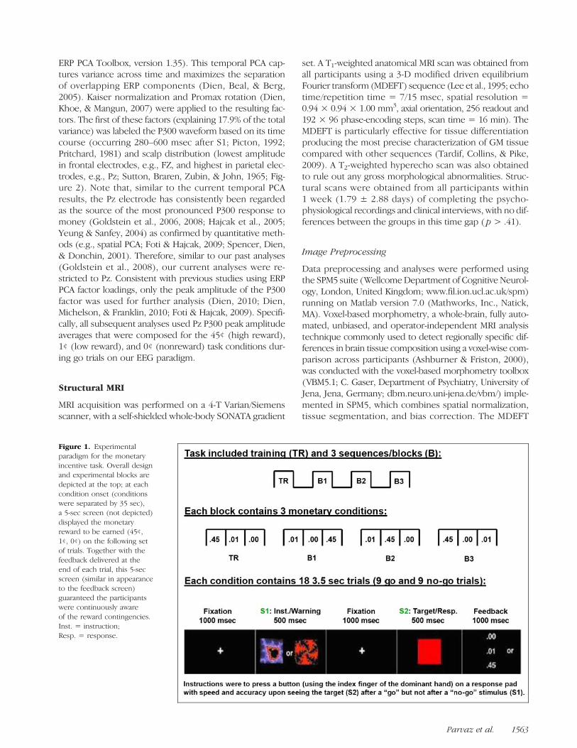
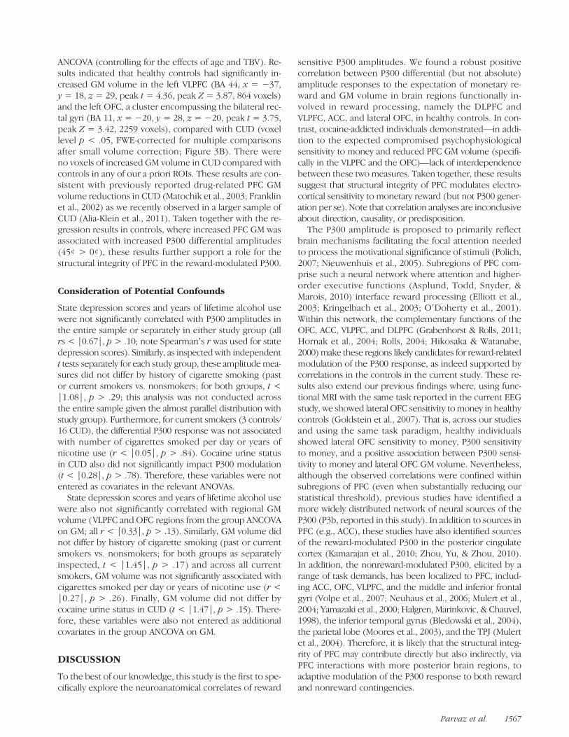
Participants completed amonetary reward paradigm as pre-viously described (Goldstein et al., 2006, 2008). In brief,the task included three blocks, each consisting of threepseudorandomized monetary reward conditions: 45¢, 1¢,or 0¢, separated by a 35-sec fixation cross to prevent carry-over effects. During each monetary condition, there were9 go and 9 no-go trials, pseudorandomized across all trials(no more than three consecutive trials of the same type),for a total of 54 go and 54 no-go trials per monetary condi-tion. Two distinct abstract (fractal) images (Thut et al., 1997)served as the go and no-go warning stimuli (S1: this expec-tation stimulus elicited the P300; see Figure 1).
Participants were instructed to press a button on a re-sponse pad with speed and accuracy upon seeing the tar-get stimulus (S2; a red square) after a go S1 stimulus andto refrain from pressing the button upon seeing S2 after ano-go S1 stimulus. Feedback was presented immediatelyafter responses following the offset of S2 by displayingthe amount of money earned for each correct trial
Parvaz et al. 1561
(45¢, 1¢, or 0¢) and an “X” for an incorrect response. Ashort training session preceded the task, where nomoney could be earned. At the end of the experiment,participants provided task ratings of interest, excitement,and frustration and were informed of their total gain.Compensation of up to $50 was given. There were no groupdifferences in the amount of money earned ( p = .25).
EEG Recordings and Data Reduction
Continuous EEG (Neuroscan, Inc., Sterling, VA) recordingswere obtained in all task conditions using a 64-channelelectrode cap. The digitized, continuous EEG was rerefer-enced to electronically linked mastoid electrodes, trans-formed using a DC offset algorithm, and divided intoepochs extending from 200 msec before the onset of S1
to 1800 msec after. A baseline correction algorithm wasapplied to the epoched EEG with respect to the 200 msecprestimulus baseline. All epochs were then subjected to aband pass filter (0.1–30 Hz). An artifact rejection procedurefollowed: (1) an amplitude threshold of ±75 μV was ap-plied automatically to remove EOG andmovement artifactsand (2) all trials that appeared contaminated by technicalartifacts such as global drifts in EEG were manually rejected.A minimum of 30 epochs per task condition remained afterartifact rejection. Grand averages were composed for eachmonetary condition during go trials on the task (Figure 2A).No-go trials were excluded from this study because oflack of significant reward effects in both our prior studies(Goldstein et al., 2006, 2008).Temporal ROIs in the averaged waveforms were then
chosen quantitatively using temporal PCA (with the Matlab
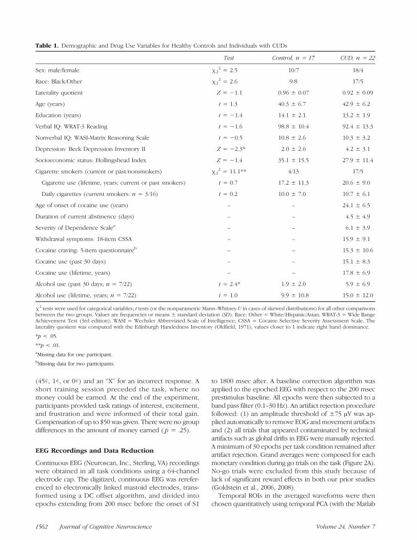
Table 1. Demographic and Drug Use Variables for Healthy Controls and Individuals with CUDs
Test Control, n = 17 CUD, n = 22
Sex: male/female χ22 = 2.5 10/7 18/4
Race: Black/Other χ22 = 2.6 9/8 17/5
Laterality quotient Z = −1.1 0.96 ± 0.07 0.92 ± 0.09
Age (years) t = 1.3 40.3 ± 6.7 42.9 ± 6.2
Education (years) t = −1.4 14.1 ± 2.1 13.2 ± 1.9
Verbal IQ: WRAT-3 Reading t = −1.6 98.8 ± 10.4 92.4 ± 13.3
Nonverbal IQ: WASI-Matrix Reasoning Scale t = −0.5 10.8 ± 2.6 10.3 ± 3.2
Depression: Beck Depression Inventory II Z = −2.3* 2.0 ± 2.6 4.2 ± 3.1
Socioeconomic status: Hollingshead Index Z = −1.4 35.1 ± 15.5 27.9 ± 11.4
Cigarette smokers (current or past/nonsmokers) χ22 = 11.1** 4/13 17/5
Cigarette use (lifetime, years; current or past smokers) t = 0.7 17.2 ± 11.3 20.6 ± 9.0
Daily cigarettes (current smokers: n = 3/16) t = 0.2 10.0 ± 7.0 10.7 ± 6.1
Age of onset of cocaine use (years) – – 24.1 ± 6.5
Duration of current abstinence (days) – – 4.5 ± 4.9
Severity of Dependence Scalea – – 6.1 ± 3.9
Withdrawal symptoms: 18-item CSSA – – 15.9 ± 9.1
Cocaine craving: 5-item questionnaireb – – 15.3 ± 10.6
Cocaine use (past 30 days) – – 15.1 ± 8.3
Cocaine use (lifetime, years) – – 17.8 ± 6.9
Alcohol use (past 30 days; n = 7/22) t = 2.4* 1.9 ± 2.0 5.9 ± 6.9
Alcohol use (lifetime, years; n = 7/22) t = 1.0 9.9 ± 10.8 15.0 ± 12.0
χ2 tests were used for categorical variables; t tests (or the nonparametric Mann–Whitney U in cases of skewed distributions) for all other comparisonsbetween the two groups. Values are frequencies or means ± standard deviation (SD). Race: Other = White/Hispanic/Asian; WRAT-3 = Wide RangeAchievement Test (3rd edition); WASI = Wechsler Abbreviated Scale of Intelligence; CSSA = Cocaine Selective Severity Assessment Scale. Thelaterality quotient was computed with the Edinburgh Handedness Inventory (Oldfield, 1971); values closer to 1 indicate right hand dominance.
*p < .05.
**p < .01.aMissing data for one participant.bMissing data for two participants.
1562 Journal of Cognitive Neuroscience Volume 24, Number 7
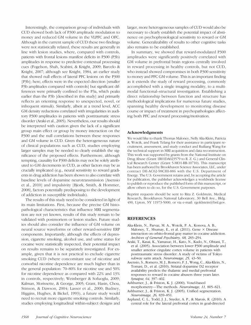
ERP PCA Toolbox, version 1.35). This temporal PCA cap-tures variance across time and maximizes the separationof overlapping ERP components (Dien, Beal, & Berg,2005). Kaiser normalization and Promax rotation (Dien,Khoe, & Mangun, 2007) were applied to the resulting fac-tors. The first of these factors (explaining 17.9% of the totalvariance) was labeled the P300 waveform based on its timecourse (occurring 280–600 msec after S1; Picton, 1992;Pritchard, 1981) and scalp distribution (lowest amplitudein frontal electrodes, e.g., FZ, and highest in parietal elec-trodes, e.g., Pz; Sutton, Braren, Zubin, & John, 1965; Fig-ure 2). Note that, similar to the current temporal PCAresults, the Pz electrode has consistently been regardedas the source of the most pronounced P300 response tomoney (Goldstein et al., 2006, 2008; Hajcak et al., 2005;Yeung & Sanfey, 2004) as confirmed by quantitative meth-ods (e.g., spatial PCA; Foti & Hajcak, 2009; Spencer, Dien,& Donchin, 2001). Therefore, similar to our past analyses(Goldstein et al., 2008), our current analyses were re-stricted to Pz. Consistent with previous studies using ERPPCA factor loadings, only the peak amplitude of the P300factor was used for further analysis (Dien, 2010; Dien,Michelson, & Franklin, 2010; Foti & Hajcak, 2009). Specifi-cally, all subsequent analyses used Pz P300 peak amplitudeaverages that were composed for the 45¢ (high reward),1¢ (low reward), and 0¢ (nonreward) task conditions dur-ing go trials on our EEG paradigm.
Structural MRI
MRI acquisition was performed on a 4-T Varian/Siemensscanner, with a self-shielded whole-body SONATA gradient
set. A T1-weighted anatomical MRI scan was obtained fromall participants using a 3-D modified driven equilibriumFourier transform (MDEFT) sequence (Lee et al., 1995; echotime/repetition time = 7/15 msec, spatial resolution =0.94 × 0.94 × 1.00 mm3, axial orientation, 256 readout and192 × 96 phase-encoding steps, scan time = 16 min). TheMDEFT is particularly effective for tissue differentiationproducing the most precise characterization of GM tissuecompared with other sequences (Tardif, Collins, & Pike,2009). A T2-weighted hyperecho scan was also obtainedto rule out any gross morphological abnormalities. Struc-tural scans were obtained from all participants within1 week (1.79 ± 2.88 days) of completing the psycho-physiological recordings and clinical interviews, with no dif-ferences between the groups in this time gap ( p > .41).
Image Preprocessing
Data preprocessing and analyses were performed usingthe SPM5 suite (WellcomeDepartment of Cognitive Neurol-ogy, London, United Kingdom; www.fil.ion.ucl.ac.uk/spm)running on Matlab version 7.0 (Mathworks, Inc., Natick,MA). Voxel-based morphometry, a whole-brain, fully auto-mated, unbiased, and operator-independent MRI analysistechnique commonly used to detect regionally specific dif-ferences in brain tissue composition using a voxel-wise com-parison across participants (Ashburner & Friston, 2000),was conducted with the voxel-based morphometry toolbox(VBM5.1; C. Gaser, Department of Psychiatry, University ofJena, Jena, Germany; dbm.neuro.uni-jena.de/vbm/) imple-mented in SPM5, which combines spatial normalization,tissue segmentation, and bias correction. The MDEFT
Figure 1. Experimentalparadigm for the monetaryincentive task. Overall designand experimental blocks aredepicted at the top; at eachcondition onset (conditionswere separated by 35 sec),a 5-sec screen (not depicted)displayed the monetaryreward to be earned (45¢,1¢, 0¢) on the following setof trials. Together with thefeedback delivered at theend of each trial, this 5-secscreen (similar in appearanceto the feedback screen)guaranteed the participantswere continuously awareof the reward contingencies.Inst. = instruction;Resp. = response.
Parvaz et al. 1563
scans were first spatially normalized to a standard propor-tional stereotaxic space and segmented into GM, whitematter, and cerebrospinal fluid tissue classes according toa priori tissue probability maps (Ashburner & Friston, 2000,2005). A hidden Markov random field (Cuadra, Cammoun,Butz, Cuisenaire, & Thiran, 2005) was applied to minimizethe noise level by “removing” isolated voxels of one tissueclass that are unlikely to be members of that tissue class,thereby increasing the accuracy of the segmentation.Jacobian modulation was also applied to compensate forthe effect of spatial normalization and to restore the originalabsolute GM volume in the segmented GM images. Totalbrain volume (TBV) was computed as the sum of the ex-tracted total GM and white matter volumes for each partici-pant, calculated as an adjustment factor to account for theeffect of overall head size on regional GM volume. TBV didnot differ between the groups ( p> .21). Statistical analysis
of regional GM volume was performed after smoothingthe normalized and modulated segments with a 10 mm3
FWHM Gaussian kernel.
Statistical Analyses
ERP Analysis
Repeated measures ANOVAs with Money (45¢, 1¢, and 0¢)as the within-subject factor and Group (controls, CUD) asthe between-subject factor were conducted for the task-related measures (accuracy, RT, and post-task ratings)and the PCA peak P300 amplitudes at Pz. Given our priorresults where only controls but not CUD showed modula-tion of the P300 response to money (Goldstein et al.,2008), the current P300 analyses were conducted with ana priori focus on reward sensitivity in controls.
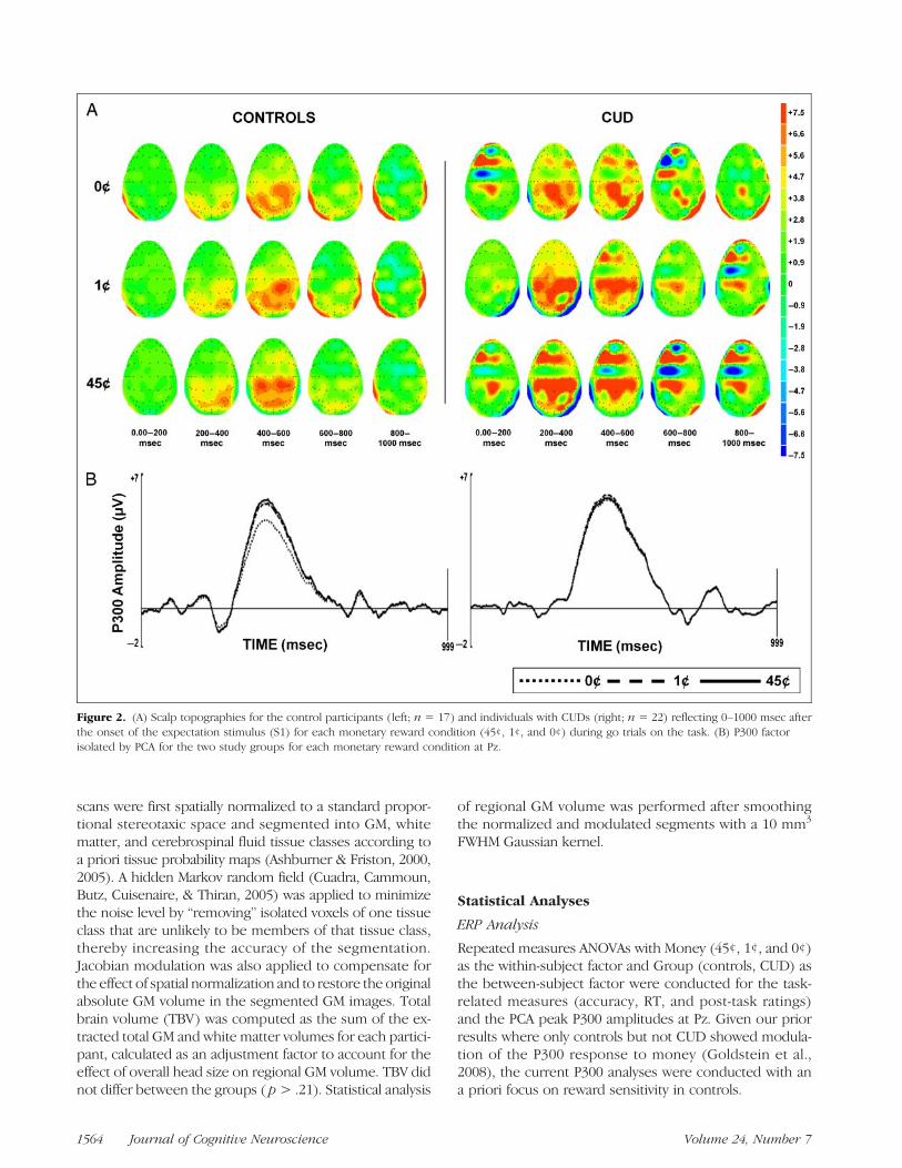
Figure 2. (A) Scalp topographies for the control participants (left; n = 17) and individuals with CUDs (right; n = 22) reflecting 0–1000 msec afterthe onset of the expectation stimulus (S1) for each monetary reward condition (45¢, 1¢, and 0¢) during go trials on the task. (B) P300 factorisolated by PCA for the two study groups for each monetary reward condition at Pz.
1564 Journal of Cognitive Neuroscience Volume 24, Number 7
Morphometry Analyses
Whole-brain regression analyses were performed in SPM5with the peak P300 amplitudes as seed variables regressedagainst participantsʼ regional GM volumes, across all partic-ipants, and separately in healthy controls and CUD. That is,the P300 component amplitudes (computed using PCA) inresponse to 45¢, 1¢, and 0¢ trials separately (Figure 2B)and the differentials 45¢ minus 0¢, 45¢ minus 1¢, and 1¢minus 0¢ that served as the reward-modulated P300 seedvariables were regressed—one at a time—against partici-pantsʼ GM maps. Age and TBV were included as covariatesin all analyses. Statistical maps were thresholded at p <.001 voxel-level uncorrected; clusters that contained atleast 50 contiguous voxels meeting the p< .05 cluster-levelfamily-wise error (FWE) correction for multiple compari-sons using random field theory (Friston, Holmes, Poline,Price, & Frith, 1996) are reported. For completeness, wealso report results of a whole-brain ANCOVA conductedto assess regional differences in PFC GM volume betweenthe groups. Here we used an exploratory voxel-levelthreshold of p < .005 uncorrected and 50 voxels. Signifi-cance for this analysis is reported at p < .05 voxel-levelFWE-corrected after small volume correction. Anatomicalspecificity for all analyses was corroborated with the Anatomytoolbox (Eickhoff et al., 2005), which provides probabilisticcytoarchitectonic neuroanatomical localization maps. Partic-ipantsʼ individual cluster volume measures were extractedusing the EasyROI toolbox in Matlab (www.sbirc.ed.ac.uk/cyril/cp_download.html) and assessed for outliers.
RESULTS
Task Behavior and Ratings
The 3 (Money: 45¢, 1¢, and 0¢) × 2 (Group: control, CUD)ANOVA for RT revealed a main effect of Money (F2, 74 =3.14, p = .049), such that RTs were significantly faster for45¢ than 0¢ across all 39 participants ( p = .02; all othereffects on RT, p> .75). As expected (given our prior resultsand the low level of task difficulty), there were no signifi-cant Money, Group, or interaction effects on accuracy (F<0.76, p > .39). In addition, all participants reported beingfully engaged in the experiment, with significantly higherinterest and excitement ratings and significantly lowerfrustration ratings reported for the high (45¢) than eitherof the two lower (0¢ or 1¢) money conditions (main effectof money for all three rating scales, F > 6.83, p < .01; allother effects, p > .17).
Modulation of the P300 Response to Money
The 3 (Money: 45¢, 1¢, and 0¢) × 2 (Group: controls andCUD) ANOVA for peak P300 amplitude revealed a maineffect of Money (F2, 74 = 5.87, p = .004), such that P300amplitudes were significantly higher for 45¢ than 0¢ acrossall 39 participants ( p= .003; all other effects, p> .64). Fol-
lowing our a priori hypothesis (of P300 amplitude responseto rewardmagnitude within controls but not CUD), we alsotested this Money effect separately for each group. Com-pared with testing an interaction effect, this potentially lessrigorous statistical approach indicated that, in controlsonly, peak P300 amplitudes increased linearly with moneyvalue (45¢ > 1¢ > 0¢, linear contrast for money, p= .003,maximum effect for 45¢ vs. 0¢); this effect was not signif-icant in CUD (45¢ = 1¢ = 0¢, p = .13; quadric contrast,p = ns for both groups; Figure 2B). Similarity of theseresults to our previous findings (where we applied a dif-ferent method to isolate the P300 component amplitudes;Goldstein et al., 2008) argues against a Type I error.
There were no significant differences between thegroups in absolute P300 amplitudes for any of the threemoney conditions (45¢, 1¢, or 0¢) even in follow-up inde-pendent t tests (t < |0.54|, p > .59). Thus, only the ex-pected graded sensitivity to monetary reward magnitudewas compromised in CUD and not the ability to generatea P300 response on the task. Furthermore, given lack ofgroup differences in task performance and ratings, thiscompromised sensitivity in CUD is not explained by lackof task engagement or impaired task performance.
GM Correlates of the Reward-modulatedP300 Response
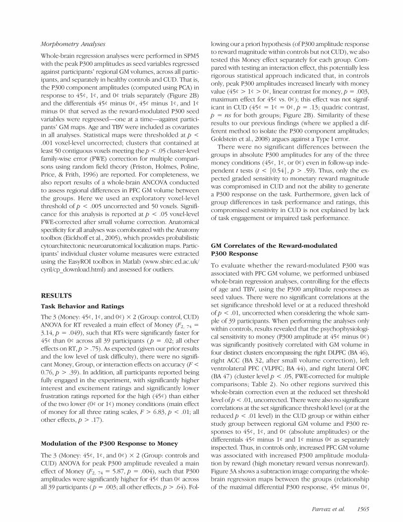
To evaluate whether the reward-modulated P300 wasassociated with PFC GM volume, we performed unbiasedwhole-brain regression analyses, controlling for the effectsof age and TBV, using the P300 amplitude responses asseed values. There were no significant correlations at theset significance threshold level or at a reduced thresholdof p < .01, uncorrected when considering the whole sam-ple of 39 participants. When performing the analyses onlywithin controls, results revealed that the psychophysiologi-cal sensitivity to money (P300 amplitude at 45¢ minus 0¢)was significantly positively correlated with GM volume infour distinct clusters encompassing the right DLPFC (BA 46),right ACC (BA 32, after small volume correction), leftventrolateral PFC (VLPFC; BA 44), and right lateral OFC(BA 47) (cluster level p < .05, FWE-corrected for multiplecomparisons; Table 2). No other regions survived thiswhole-brain correction even at the reduced set thresholdlevel of p< .01, uncorrected. There were also no significantcorrelations at the set significance threshold level (or at thereduced p < .01 level) in the CUD group or within eitherstudy group between regional GM volume and P300 re-sponses to 45¢, 1¢, and 0¢ (absolute amplitudes) or thedifferentials 45¢ minus 1¢ and 1¢ minus 0¢ as separatelyinspected. Thus, in controls only, increased PFC GM volumewas associated with increased P300 amplitude modula-tion by reward (high monetary reward versus nonreward).Figure 3A shows a subtraction image comparing the whole-brain regression maps between the groups (relationshipof the maximal differential P300 response, 45¢ minus 0¢,
Parvaz et al. 1565
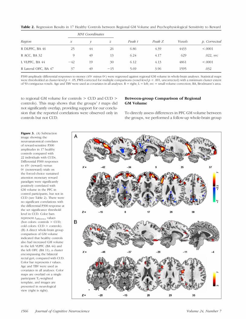
to regional GM volume for controls > CUD and CUD >controls). This map shows that the groupsʼ t maps didnot significantly overlap, providing support for our conclu-sion that the reported correlations were observed only incontrols but not CUD.
Between-group Comparison of RegionalGM Volume
To directly assess differences in PFC GM volume betweenthe groups, we performed a follow-up whole-brain group
Table 2. Regression Results in 17 Healthy Controls between Regional GM Volume and Psychophysiological Sensitivity to Reward
Region
MNI Coordinates
Peak t Peak Z Voxels p, Correctedx y z
R DLPFC, BA 46 25 44 26 6.86 4.39 4433 <.0001
R ACC, BA 32 9 49 13 6.24 4.17 629 .022, svc
L VLPFC, BA 44 −42 19 30 6.12 4.13 4861 <.0001
R Lateral OFC, BA 47 37 49 −15 5.69 3.96 1595 .032
P300 amplitude differential responses to money (45¢ minus 0¢) were regressed against regional GM volume in whole-brain analyses. Statistical mapswere thresholded at cluster-level p< .05, FWE-corrected for multiple comparisons (voxel-level p< .001, uncorrected) with a minimum cluster extentof 50 contiguous voxels. Age and TBV were used as covariates in all analyses. R = right; L = left; svc = small volume correction; BA, Brodmannʼs area.
Figure 3. (A) Subtractionimage showing theneuroanatomical correlatesof reward-sensitive P300amplitudes in 17 healthycontrols compared with22 individuals with CUDs.Differential P300 responsesto 45¢ (reward) versus0¢ (nonreward) trials onthe forced-choice sustainedattention monetary rewardparadigm were significantlypositively correlated withGM volume in the PFC incontrol participants, but not inCUD (see Table 2). There wereno significant correlations withthe differential P300 response atthe set significance thresholdlevel in CUD. Color barsrepresent tdifference values(hot colors: controls > CUD;cold colors: CUD > controls).(B) A direct whole-brain groupcomparison of GM volumeindicated that healthy controlsalso had increased GM volumein the left VLPFC (BA 44) andthe left OFC (BA 11), a clusterencompassing the bilateralrectal gyri, compared with CUD.Color bar represents t values.Age and TBV were used ascovariates in all analyses. Colormaps are overlaid on a singleparticipant T1-weightedtemplate, and images arepresented in neurologicalview (right is right).
1566 Journal of Cognitive Neuroscience Volume 24, Number 7
ANCOVA (controlling for the effects of age and TBV). Re-sults indicated that healthy controls had significantly in-creased GM volume in the left VLPFC (BA 44, x = −37,y = 18, z = 29, peak t = 4.36, peak Z = 3.87, 864 voxels)and the left OFC, a cluster encompassing the bilateral rec-tal gyri (BA 11, x = −20, y = 28, z = −20, peak t = 3.75,peak Z = 3.42, 2259 voxels), compared with CUD (voxellevel p < .05, FWE-corrected for multiple comparisonsafter small volume correction; Figure 3B). There wereno voxels of increased GM volume in CUD compared withcontrols in any of our a priori ROIs. These results are con-sistent with previously reported drug-related PFC GMvolume reductions in CUD (Matochik et al., 2003; Franklinet al., 2002) as we recently observed in a larger sample ofCUD (Alia-Klein et al., 2011). Taken together with the re-gression results in controls, where increased PFC GM wasassociated with increased P300 differential amplitudes(45¢ > 0¢), these results further support a role for thestructural integrity of PFC in the reward-modulated P300.
Consideration of Potential Confounds
State depression scores and years of lifetime alcohol usewere not significantly correlated with P300 amplitudes inthe entire sample or separately in either study group (allrs < |0.67|, p> .10; note Spearmanʼs r was used for statedepression scores). Similarly, as inspected with independentt tests separately for each study group, these amplitudemea-sures did not differ by history of cigarette smoking (pastor current smokers vs. nonsmokers; for both groups, t <|1.08|, p > .29; this analysis was not conducted acrossthe entire sample given the almost parallel distribution withstudy group). Furthermore, for current smokers (3 controls/16 CUD), the differential P300 response was not associatedwith number of cigarettes smoked per day or years ofnicotine use (r < |0.05|, p > .84). Cocaine urine statusin CUD also did not significantly impact P300 modulation(t < |0.28|, p > .78). Therefore, these variables were notentered as covariates in the relevant ANOVAs.State depression scores and years of lifetime alcohol use
were also not significantly correlated with regional GMvolume (VLPFC and OFC regions from the group ANCOVAon GM; all r < |0.33|, p > .13). Similarly, GM volume didnot differ by history of cigarette smoking (past or currentsmokers vs. nonsmokers; for both groups as separatelyinspected, t < |1.45|, p > .17) and across all currentsmokers, GM volume was not significantly associated withcigarettes smoked per day or years of nicotine use (r <|0.27|, p > .26). Finally, GM volume did not differ bycocaine urine status in CUD (t < |1.47|, p > .15). There-fore, these variables were also not entered as additionalcovariates in the group ANCOVA on GM.
DISCUSSION
To the best of our knowledge, this study is the first to spe-cifically explore the neuroanatomical correlates of reward
sensitive P300 amplitudes. We found a robust positivecorrelation between P300 differential (but not absolute)amplitude responses to the expectation of monetary re-ward and GM volume in brain regions functionally in-volved in reward processing, namely the DLPFC andVLPFC, ACC, and lateral OFC, in healthy controls. In con-trast, cocaine-addicted individuals demonstrated—in addi-tion to the expected compromised psychophysiologicalsensitivity to money and reduced PFC GM volume (specifi-cally in the VLPFC and the OFC)—lack of interdependencebetween these two measures. Taken together, these resultssuggest that structural integrity of PFC modulates electro-cortical sensitivity to monetary reward (but not P300 gener-ation per se). Note that correlation analyses are inconclusiveabout direction, causality, or predisposition.
The P300 amplitude is proposed to primarily reflectbrain mechanisms facilitating the focal attention neededto process the motivational significance of stimuli (Polich,2007; Nieuwenhuis et al., 2005). Subregions of PFC com-prise such a neural network where attention and higher-order executive functions (Asplund, Todd, Snyder, &Marois, 2010) interface reward processing (Elliott et al.,2003; Kringelbach et al., 2003; OʼDoherty et al., 2001).Within this network, the complementary functions of theOFC, ACC, VLPFC, and DLPFC (Grabenhorst & Rolls, 2011;Hornak et al., 2004; Rolls, 2004; Hikosaka & Watanabe,2000)make these regions likely candidates for reward-relatedmodulation of the P300 response, as indeed supported bycorrelations in the controls in the current study. These re-sults also extend our previous findings where, using func-tional MRI with the same task reported in the current EEGstudy, we showed lateral OFC sensitivity tomoney in healthycontrols (Goldstein et al., 2007). That is, across our studiesand using the same task paradigm, healthy individualsshowed lateral OFC sensitivity to money, P300 sensitivityto money, and a positive association between P300 sensi-tivity to money and lateral OFC GM volume. Nevertheless,although the observed correlations were confined withinsubregions of PFC (even when substantially reducing ourstatistical threshold), previous studies have identified amore widely distributed network of neural sources of theP300 (P3b, reported in this study). In addition to sources inPFC (e.g., ACC), these studies have also identified sourcesof the reward-modulated P300 in the posterior cingulatecortex (Kamarajan et al., 2010; Zhou, Yu, & Zhou, 2010).In addition, the nonreward-modulated P300, elicited by arange of task demands, has been localized to PFC, includ-ing ACC, OFC, VLPFC, and the middle and inferior frontalgyri (Volpe et al., 2007; Neuhaus et al., 2006; Mulert et al.,2004; Yamazaki et al., 2000; Halgren, Marinkovic, & Chauvel,1998), the inferior temporal gyrus (Bledowski et al., 2004),the parietal lobe (Moores et al., 2003), and the TPJ (Mulertet al., 2004). Therefore, it is likely that the structural integ-rity of PFC may contribute directly but also indirectly, viaPFC interactions with more posterior brain regions, toadaptive modulation of the P300 response to both rewardand nonreward contingencies.
Parvaz et al. 1567
Interestingly, the comparison group of individuals withCUD showed both lack of P300 amplitude modulation tomoney and reduced GM volume in the VLPFC and OFC.Although in the current sample of CUD these two findingswere not statistically related, these results are generally inline with lesion studies, where, compared with controls,patients with frontal lesions manifest deficits in P300 (P3b)amplitudes in response to predictive contextual processingcues (Fogelson, Shah, Scabini, & Knight, 2009; Barcelo &Knight, 2007; although see Knight, 1984, an earlier studythat showed null effects of lateral PFC lesions on the P300[P3b]; here, effects were in the expected direction [smallerP3b amplitudes compared with controls] but significant dif-ferences were primarily confined to the P3a, which peaksearlier than the P3b [described in this study] and primarilyreflects an orienting response to unexpected, novel, orinfrequent stimuli). Similarly, albeit at a trend level, ACCGM density reductions correlated with irregularities in audi-tory P300 amplitudes in patients with posttraumatic stressdisorder (Araki et al., 2005). Nevertheless, our results shouldbe interpreted with caution given the lack of a significantgroup main effect or group by money interaction on theP300 and the null correlations between these responsesand GM volume in CUD. Given the heterogeneous natureof clinical populations such as CUD, studies employinglarger samples may be needed to clearly establish the sig-nificance of the proposed effects. Furthermore, althoughtempting, causality for P300 deficits may not be solely attrib-uted to GM decrements in CUD, as other factors may be ascrucially implicated (e.g., neural sensitivity to reward gradi-ents in drug addiction has been shown to also correlate withbaseline levels of dopamine neurotransmission [Asensioet al., 2010] and impulsivity [Bjork, Smith, & Hommer,2008], factors potentially predisposing to the developmentof addiction in susceptible individuals).
The results of this study need to be considered in light ofits main limitations. First, because the precise GM histo-pathological characteristics that influence MRI segmenta-tion are not yet known, results of this study remain to bevalidated with postmortem or lesion studies. Future stud-ies should also consider the influence of GM volume onneural source waveforms or other reward-sensitive ERPcomponents. Importantly, although the effects of depres-sion, cigarette smoking, alcohol use, and urine status forcocaine were statistically inspected, their potential impacton results remains to be separately investigated. For ex-ample, given that it is not practical to exclude cigarettesmoking CUD (where concomitant use of nicotine andcomorbid nicotine dependence are much higher than inthe general population: 70–80% for nicotine use and 50%for nicotine dependence as compared with 22% and 13%in controls, respectively; Weinberger & Sofuoglu, 2009;Kalman, Morissette, & George, 2005; Grant, Hasin, Chou,Stinson, & Dawson, 2004; Lasser et al., 2000; Budney,Higgins, Hughes, & Bickel, 1993), a future study wouldneed to recruit more cigarette smoking controls. Similarly,studies employing longitudinal within-subject designs and
larger, more heterogeneous samples of CUDwould also benecessary to clearly establish the potential impact of absti-nence on psychophysiological sensitivity to reward or GMvolume. Generalizability of results to other cognitive tasksalso remains to be established.In summary, we showed that reward-modulated P300
amplitudes were significantly positively correlated withGM volume in prefrontal brain regions centrally involvedin reward processing in healthy controls, but not CUD,who instead showed compromises in both P300 sensitivityto money and PFCGM volume. This is an important findingas it extends the study of reward processing, commonlyaccomplished with a single imaging modality, to a multi-modal functional–structural investigation. Establishing adirect relationship between function and structure hasmethodological implications for numerous future studies,spanning healthy development to monitoring diseasecourse or impact of treatment in psychopathologies affect-ing both PFC and reward processing/motivation.
Acknowledgments
We would like to thank Thomas Maloney, Nelly Alia-Klein, PatriciaA. Woicik, and Frank Telang for their assistance in participant re-cruitment, assessment, and study conduct and Ruiliang Wang forthe technical support in MRI acquisition and data reconstruction.This work was supported by grants from the National Institute onDrug Abuse (Grant 1R01DA023579 to R. Z. G.) and General Clin-ical Research Center (Grant 5-MO1-RR-10710). This manuscripthas been authored by Brookhaven Science Associates, LLC, undercontract DE-AC02-98CHI-886 with the U.S. Department ofEnergy. The U.S. Government retains and, by accepting the articlefor publication, the publisher acknowledges a worldwide licenseto publish or reproduce the published form of this manuscript, orallow others to do so, for the U.S. Government purposes.
Reprint requests should be sent to Rita Z. Goldstein, MedicalResearch, Brookhaven National Laboratory, 30 Bell Ave., Bldg.490, Upton, NY 11973-5000, or via e-mail: [email protected].
REFERENCES
Alia-Klein, N., Parvaz, M. A., Woicik, P. A., Konova, A. B.,Maloney, T., Shumay, E., et al. (2011). Gene × Diseaseinteraction on orbito-frontal gray matter in cocaine addiction.Archives of General Psychiatry, 68, 283–294.
Araki, T., Kasai, K., Yamasue, H., Kato, N., Kudo, N., Ohtani, T.,et al. (2005). Association between lower P300 amplitude andsmaller anterior cingulate cortex volume in patients withposttraumatic stress disorder: A study of victims of Tokyosubway sarin attack. Neuroimage, 25, 43–50.
Asensio, S., Romero, M. J., Romero, F. J., Wong, C., Alia-Klein, N.,Tomasi, D., et al. (2010). Striatal dopamine D2 receptoravailability predicts the thalamic and medial prefrontalresponses to reward in cocaine abusers three years later.Synapse, 64, 397–402.
Ashburner, J., & Friston, K. J. (2000). Voxel-basedmorphometry—The methods. Neuroimage, 11, 805–821.
Ashburner, J., & Friston, K. J. (2005). Unified segmentation.Neuroimage, 26, 839–851.
Asplund, C. L., Todd, J. J., Snyder, A. P., & Marois, R. (2010). Acentral role for the lateral prefrontal cortex in goal-directed
1568 Journal of Cognitive Neuroscience Volume 24, Number 7
and stimulus-driven attention. Nature Neuroscience, 13,507–512.
Aston-Jones, G., & Cohen, J. D. (2005a). Adaptive gain andthe role of the locus coeruleus-norepinephrine system inoptimal performance. Journal of Comparative Neurology,493, 99–110.
Aston-Jones, G., & Cohen, J. D. (2005b). An integrative theoryof locus coeruleus-norepinephrine function: Adaptive gainand optimal performance. Annual Review of Neuroscience,28, 403–450.
Barcelo, F., & Knight, R. T. (2007). An information-theoreticalapproach to contextual processing in the human brain:Evidence from prefrontal lesions. Cerebral Cortex, 17(Suppl. 1),i51–i60.
Begleiter, H., Porjesz, B., Chou, C. L., & Aunon, J. I. (1983).P3 and stimulus incentive value. Psychophysiology, 20,95–101.
Bellebaum, C., & Daum, I. (2008). Learning-related changesin reward expectancy are reflected in the feedback-relatednegativity. European Journal of Neuroscience, 27,1823–1835.
Bellebaum, C., Polezzi, D., & Daum, I. (2010). It is less than youexpected: The feedback-related negativity reflects violationsof reward magnitude expectations. Neuropsychologia, 48,3343–3350.
Bjork, J. M., Smith, A. R., & Hommer, D. W. (2008). Striatalsensitivity to reward deliveries and omissions in substancedependent patients. Neuroimage, 42, 1609–1621.
Bledowski, C., Prvulovic, D., Hoechstetter, K., Scherg, M.,Wibral, M., Goebel, R., et al. (2004). Localizing P300generators in visual target and distractor processing: Acombined event-related potential and functional magneticresonance imaging study. Journal of Neuroscience, 24,9353–9360.
Breiter, H. C., Aharon, I., Kahneman, D., Dale, A., & Shizgal, P.(2001). Functional imaging of neural responses to expectancyand experience of monetary gains and losses. Neuron, 30,619–639.
Budney, A. J., Higgins, S. T., Hughes, J. R., & Bickel, W. K.(1993). Nicotine and caffeine use in cocaine-dependentindividuals. Journal of Substance Abuse, 5, 117–130.
Cuadra, M. B., Cammoun, L., Butz, T., Cuisenaire, O., &Thiran, J. P. (2005). Comparison and validation of tissuemodelization and statistical classification methods inT1-weighted MR brain images. IEEE Transactions onMedical Imaging, 24, 1548–1565.
De Pascalis, V., Varriale, V., & DʼAntuono, L. (2010).Event-related components of the punishment and rewardsensitivity. Clinical Neurophysiology, 121, 60–76.
Dien, J. (2010). The ERP PCA Toolkit: An open source programfor advanced statistical analysis of event-related potentialdata. Journal of Neuroscience Methods, 187, 138–145.
Dien, J., Beal, D. J., & Berg, P. (2005). Optimizing principalcomponents analysis of event-related potentials: Matrix type,factor loading weighting, extraction, and rotations. ClinicalNeurophysiology, 116, 1808–1825.
Dien, J., Khoe, W., & Mangun, G. R. (2007). Evaluation of PCAand ICA of simulated ERPs: Promax vs. Infomax rotations.Human Brain Mapping, 28, 742–763.
Dien, J., Michelson, C. A., & Franklin, M. S. (2010). Separatingthe visual sentence N400 effect from the P400 sequentialexpectancy effect: Cognitive and neuroanatomicalimplications. Brain Research, 1355, 126–140.
Donchin, E., Miller, G. A., & Farwell, L. A. (1986). Theendogenous components of the event-related potential—Adiagnostic tool? Progress in Brain Research, 70, 87–102.
Eickhoff, S. B., Stephan, K. E., Mohlberg, H., Grefkes, C.,Fink, G. R., Amunts, K., et al. (2005). A new SPM toolbox for
combining probabilistic cytoarchitectonic maps andfunctional imaging data. Neuroimage, 25, 1325–1335.
Elliott, R., Newman, J. L., Longe, O. A., & Deakin, J. F. (2003).Differential response patterns in the striatum andorbitofrontal cortex to financial reward in humans:A parametric functional magnetic resonance imaging study.Journal of Neuroscience, 23, 303–307.
Fogelson, N., Shah, M., Scabini, D., & Knight, R. T. (2009).Prefrontal cortex is critical for contextual processing:Evidence from brain lesions. Brain, 132, 3002–3010.
Foti, D., & Hajcak, G. (2009). Depression and reducedsensitivity to non-rewards versus rewards: Evidence fromevent-related potentials. Biological Psychology, 81, 1–8.
Franklin, T. R., Acton, P. D., Maldjian, J. A., Gray, J. D., Croft, J. R.,Dackis, C. A., et al. (2002). Decreased gray matterconcentration in the insular, orbitofrontal, cingulate, andtemporal cortices of cocaine patients. Biological Psychiatry,51, 134–142.
Friston, K. J., Holmes, A., Poline, J. B., Price, C. J., & Frith, C. D.(1996). Detecting activations in PET and fMRI: Levels ofinference and power. Neuroimage, 4, 223–235.
Goldstein, R. Z., Alia-Klein, N., Tomasi, D., Zhang, L., Cottone, L. A.,Maloney, T., et al. (2007). Is decreased prefrontal corticalsensitivity to monetary reward associated with impairedmotivation and self-control in cocaine addiction? AmericanJournal of Psychiatry, 164, 43–51.
Goldstein, R. Z., Cottone, L. A., Jia, Z., Maloney, T., Volkow, N. D.,& Squires, N. K. (2006). The effect of graded monetaryreward on cognitive event-related potentials and behaviorin young healthy adults. International Journal ofPsychophysiology, 62, 272–279.
Goldstein, R. Z., Parvaz, M. A., Maloney, T., Alia-Klein, N.,Woicik, P. A., Telang, F., et al. (2008). Compromisedsensitivity to monetary reward in current cocaine users:An ERP study. Psychophysiology, 45, 705–713.
Grabenhorst, F., & Rolls, E. T. (2011). Value, pleasure andchoice in the ventral prefrontal cortex. Trends in CognitiveSciences, 15, 56–67.
Grant, B. F., Hasin, D. S., Chou, S. P., Stinson, F. S., &Dawson, D. A. (2004). Nicotine dependence and psychiatricdisorders in the United States: Results from the nationalepidemiologic survey on alcohol and related conditions.Archives of General Psychiatry, 61, 1107–1115.
Haber, S. N., & Knutson, B. (2010). The reward circuit:Linking primate anatomy and human imaging.Neuropsychopharmacology, 35, 4–26.
Hajcak, G., Holroyd, C. B., Moser, J. S., & Simons, R. F.(2005). Brain potentials associated with expected andunexpected good and bad outcomes. Psychophysiology,42, 161–170.
Halgren, E., Marinkovic, K., & Chauvel, P. (1998). Generatorsof the late cognitive potentials in auditory and visualoddball tasks. Electroencephalography and ClinicalNeurophysiology, 106, 156–164.
Hikosaka, K., & Watanabe, M. (2000). Delay activity of orbitaland lateral prefrontal neurons of the monkey varying withdifferent rewards. Cerebral Cortex, 10, 263–271.
Hornak, J., OʼDoherty, J., Bramham, J., Rolls, E. T., Morris, R. G.,Bullock, P. R., et al. (2004). Reward-related reversal learningafter surgical excisions in orbito-frontal or dorsolateralprefrontal cortex in humans. Journal of CognitiveNeuroscience, 16, 463–478.
Kalman, D., Morissette, S. B., & George, T. P. (2005).Co-morbidity of smoking in patients with psychiatric andsubstance use disorders. American Journal on Addictions,14, 106–123.
Kamarajan, C., Rangaswamy, M., Tang, Y., Chorlian, D. B.,Pandey, A. K., Roopesh, B. N., et al. (2010). Dysfunctional
Parvaz et al. 1569
reward processing in male alcoholics: An ERP studyduring a gambling task. Journal of Psychiatric Research,44, 576–590.
Knight, R. T. (1984). Decreased response to novel stimuli afterprefrontal lesions in man. Electroencephalography andClinical Neurophysiology, 59, 9–20.
Kringelbach, M. L., OʼDoherty, J., Rolls, E. T., & Andrews, C.(2003). Activation of the human orbitofrontal cortex to aliquid food stimulus is correlated with its subjectivepleasantness. Cerebral Cortex, 13, 1064–1071.
Lasser, K., Boyd, J. W., Woolhandler, S., Himmelstein, D. U.,McCormick, D., & Bor, D. H. (2000). Smoking and mentalillness: A population-based prevalence study. Journal of theAmerican Medical Association, 284, 2606–2610.
Lee, J. H., Garwood, M., Menon, R., Adriany, G., Andersen, P.,Truwit, C. L., et al. (1995). High contrast and fast three-dimensional magnetic resonance imaging at high fields.Magnetic Resonance in Medicine, 34, 308–312.
Lesieur, H. R., & Blume, S. B. (1987). The South Oaks GamblingScreen (SOGS): A new instrument for the identification ofpathological gamblers. American Journal of Psychiatry,144, 1184–1188.
Liu, X., Matochik, J. A., Cadet, J. L., & London, E. D. (1998).Smaller volume of prefrontal lobe in polysubstanceabusers: A magnetic resonance imaging study.Neuropsychopharmacology, 18, 243–252.
Matochik, J. A., London, E. D., Eldreth, D. A., Cadet, J. L., &Bolla, K. I. (2003). Frontal cortical tissue composition inabstinent cocaine abusers: A magnetic resonance imagingstudy. Neuroimage, 19, 1095–1102.
Moores, K. A., Clark, C. R., Hadfield, J. L., Brown, G. C., Taylor,D. J., Fitzgibbon, S. P., et al. (2003). Investigating thegenerators of the scalp recorded visuo-verbal P300 usingcortically constrained source localization. Human BrainMapping, 18, 53–77.
Mulert, C., Pogarell, O., Juckel, G., Rujescu, D., Giegling, I.,Rupp, D., et al. (2004). The neural basis of the P300 potential.Focus on the time-course of the underlying corticalgenerators. European Archives of Psychiatry and ClinicalNeuroscience, 254, 190–198.
Neuhaus, A., Bajbouj, M., Kienast, T., Kalus, P., von Haebler, D.,Winterer, G., et al. (2006). Persistent dysfunctional frontallobe activation in former smokers. Psychopharmacology(Berlin), 186, 191–200.
Nieuwenhuis, S., Aston-Jones, G., & Cohen, J. D. (2005).Decision making, the P3, and the locus coeruleus-norepinephrine system. Psychological Bulletin, 131,510–532.
Nieuwenhuis, S., De Geus, E. J., & Aston-Jones, G. (2011).The anatomical and functional relationship between the P3and autonomic components of the orienting response.Psychophysiology, 48, 162–175.
OʼDoherty, J., Kringelbach, M. L., Rolls, E. T., Hornak, J., &Andrews, C. (2001). Abstract reward and punishmentrepresentations in the human orbitofrontal cortex.Nature Neuroscience, 4, 95–102.
Oldfield, R. C. (1971). The assessment and analysis ofhandedness: The Edinburgh inventory. Neuropsychologia,9, 97–113.
Picton, T. W. (1992). The P300 wave of the human event-relatedpotential. Journal of Clinical Neurophysiology, 9, 456–479.
Polich, J. (2007). Updating P300: An integrative theory of P3aand P3b. Clinical Neurophysiology, 118, 2128–2148.
Pritchard, W. S. (1981). Psychophysiology of P300.Psychological Bulletin, 89, 506–540.
Rolls, E. T. (2004). The functions of the orbitofrontal cortex.Brain and Cognition, 55, 11–29.
Rossi, A. F., Pessoa, L., Desimone, R., & Ungerleider, L. G.(2009). The prefrontal cortex and the executive control ofattention. Experimental Brain Research, 192, 489–497.
Sato, A., Yasuda, A., Ohira, H., Miyawaki, K., Nishikawa, M.,Kumano, H., et al. (2005). Effects of value and rewardmagnitude on feedback negativity and P300. NeuroReport,16, 407–411.
Spencer, K. M., Dien, J., & Donchin, E. (2001). Spatiotemporalanalysis of the late ERP responses to deviant stimuli.Psychophysiology, 38, 343–358.
Sutton, S., Braren, M., Zubin, J., & John, E. R. (1965).Evoked-potential correlates of stimulus uncertainty.Science, 150, 1187–1188.
Tanabe, J., Tregellas, J. R., Dalwani, M., Thompson, L., Owens, E.,Crowley, T., et al. (2009). Medial orbitofrontal cortex graymatter is reduced in abstinent substance-dependentindividuals. Biological Psychiatry, 65, 160–164.
Tardif, C. L., Collins, D. L., & Pike, G. B. (2009). Sensitivity ofvoxel-based morphometry analysis to choice of imagingprotocol at 3 T. Neuroimage, 44, 827–838.
Taylor, S. F., Welsh, R. C., Wager, T. D., Phan, K. L., Fitzgerald,K. D., & Gehring, W. J. (2004). A functional neuroimagingstudy of motivation and executive function. Neuroimage, 21,1045–1054.
Thut, G., Schultz, W., Roelcke, U., Nienhusmeier, M., Missimer, J.,Maguire, R. P., et al. (1997). Activation of the human brain bymonetary reward. NeuroReport, 8, 1225–1228.
Ventura, J., Liberman, R. P., Green, M. F., Shaner, A., & Mintz, J.(1998). Training and quality assurance with the StructuredClinical Interview for DSM-IV (SCID-I/P). PsychiatryResearch, 79, 163–173.
Volpe, U., Mucci, A., Bucci, P., Merlotti, E., Galderisi, S., &Maj, M. (2007). The cortical generators of P3a and P3b:A LORETA study. Brain Research Bulletin, 73, 220–230.
Weinberger, A. H., & Sofuoglu, M. (2009). The impact ofcigarette smoking on stimulant addiction. American Journalof Drug and Alcohol Abuse, 35, 12–17.
Wu, Y., & Zhou, X. (2009). The P300 and reward valence,magnitude, and expectancy in outcome evaluation.Brain Research, 1286, 114–122.
Yamazaki, T., Kamijo, K., Kenmochi, A., Fukuzumi, S., Kiyuna, T.,Takaki, Y., et al. (2000). Multiple equivalent current dipolesource localization of visual event-related potentials duringoddball paradigm with motor response. Brain Topography,12, 159–175.
Yeung, N., & Sanfey, A. G. (2004). Independent coding ofreward magnitude and valence in the human brain.Journal of Neuroscience, 24, 6258–6264.
Zhou, Z., Yu, R., & Zhou, X. (2010). To do or not to do? Actionenlarges the FRN and P300 effects in outcome evaluation.Neuropsychologia, 48, 3606–3613.
1570 Journal of Cognitive Neuroscience Volume 24, Number 7