Toward a Witness Recollection Definition of Hearsay - Digital ...
Upload
independentCategory
view
0download
0
www.elsevier.com/locate/ynimg
NeuroImage 33 (2006) 286–295Left prefrontal cortex control of novel occurrences duringrecollection: A psychopharmacological study usingscopolamine and event-related fMRI
M. Bozzali,a,b,⁎ S.E. MacPherson,a R.J. Dolan,b and T. Shallicea
aInstitute of Cognitive Neuroscience, University College London, UKbWellcome Department of Imaging Neuroscience, Institute of Neurology, University College of London, 12 Queen Square, London, WC1N 3BG, UK
Received 1 March 2006; revised 10 June 2006; accepted 15 June 2006Available online 17 August 2006
Recollection and familiarity represent two processes involved inepisodic memory retrieval. We investigated how scopolamine (anantagonist of acetylcholine muscarinic receptors) influenced brainactivity during memory retrieval, using a paradigm that separatedrecollection and familiarity. Eighteen healthy volunteers were recruitedin a randomized, placebo-controlled, double-blind design using event-related fMRI. Participants were required to perform a verbalrecognition memory task within the scanner, either under placebo orscopolamine conditions. Depending on the subcondition, participantswere required to make a simple recognition decision (old/new items) orbase their decision on more specific information related to priorexperience (target/non-target/new items). We show a drug modulationin left prefrontal and perirhinal cortex during recollection. Such aneffect was specifically driven by novelty and showed an inversecorrelation with accuracy performance. Additionally, we show a directcorrelation between drug-related signal change in left prefrontal andperirhinal cortices. We discuss the findings in terms of acetylcholinemediation of the familiarity/novelty signal through perirhinal cortexand the control of the relative signal strength through prefrontalcortex.© 2006 Elsevier Inc. All rights reserved.
Keywords: fMRI; Scopolamine; Episodic memory; Recollection; Familiarity
Introduction
The conscious experience of prior occurrence that accompanieshuman episodic memory retrieval in humans is complex, involvinga number of different neuronal structures. It is widely believed thatat least two types of processes are involved in episodic retrieval,namely recollection, the conscious experience of a previous event,
⁎ Corresponding author. Wellcome Department of Imaging Neuroscience,Institute of Neurology, University College of London, 12 Queen Square,London, WC1N 3BG, UK. Fax: +44 020 7813 1420.
E-mail address: [email protected] (M. Bozalli).Available online on ScienceDirect (www.sciencedirect.com).
1053-8119/$ - see front matter © 2006 Elsevier Inc. All rights reserved.doi:10.1016/j.neuroimage.2006.06.044
and familiarity, where no contextual information is associated withthe memory (Mandler, 1980; Hintzman et al., 1998; Yonelinas andLevy, 2002). Moreover, it is widely asserted that these processeshave separable anatomical and neurochemical substrates within themedial temporal lobe involving hippocampus and perirhinal cortexrespectively (Aggleton and Brown, 1999, 2005; Henson, 2005; butsee Cipolotti et al., 2006). A recent study, using event-related brainpotentials and midazolam (an amnesia inducing benzodiazepine),has provided evidence for separate processes underlying familiarityand recollection (Curran et al., 2006).
Recollection occurs inappropriately in the syndrome ofconfabulation, which typically arises from inferior medialprefrontal lesions, as for instance, following ruptured aneurysmsof the anterior communicating artery. Some authors have arguedfor a double deficit account of confabulation, implicating episodicmemory and executive functions (e.g. DeLuca and Cicerone, 1991;DeLuca, 1993; Fischer et al., 1995). However, meta-analyses ofreports of positive cases (Gilboa and Moscovitch, 2002) do notsupport this hypothesis.
A second possible explanation can be derived by considering astructure frequently found to be damaged in patients whoconfabulate, namely the basal forebrain (Alexander and Freedman,1984, Damasio et al., 1985). In the meta-analysis of Gilboa andMoscovitch (2002), they reported that 32% of the 79 confabulatingpatients had damage to this region and in 10 it was the sole lesiondetected. The basal forebrain is the seat of four overlappingpathways of cholinergic projection systems. This led Damasio et al.(1985), when discussing a patient who manifested bizarreconfabulation, to argue that “damage to the basal forebrain couldlead to significant reduction of cholinergic input to temporal lobestructures and widespread regions of the association cortices. In bothsites, the reduction might contribute to a memory defect” (p. 270).Two of the four basal forebrain cholinergic cell groups – Ch1 andCh2 – project to the hippocampus (Mesulam, 2000). A theoreticalapproach to cholinergic modulation of the hippocampus developedby Hasselmo et al. (1996) suggests that these inputs act as a switch tomove from processing retrieval to encodingmode. Thus, as far as the

287M. Bozzali et al. / NeuroImage 33 (2006) 286–295
hippocampus is concerned, loss of cholinergic input would predictreduced capacity for encoding, whereas confabulation is generallyviewed as a disorder of retrieval (Moscovitch, 1989; Burgess andShallice, 1996). Two other principal cell groups in the basalforebrain include the Ch4 cell group, the major source of cholinergicinput to the cortex. One possibility is that a lesion to this systemgives rise to confabulation. Such a hypothesis could explain why apatient with a severe amnesia, as reported by Phillips et al. (1987),with selective damage to the basal forebrain might not confabulate;the nucleus basalis, the site of origin of Ch4 neurons, was spared inthis patient.
What then are the most critical cortical regions, as far as thecholinergic input is concerned, with respect to confabulation? There-experience of prior episodes and its impairment in confabulationhas been suggested to involve a complex interplay between onlinecore memory retrieval systems involving medial temporal regionsand prefrontal control systems (e.g. Moscovitch, 1989). Thispotential involvement of prefrontal cortex is consistent withimaging studies of episodic memory retrieval that demonstrateextensive prefrontal activation (Tulving et al., 1994; Shallice et al.,1994; Lepage et al., 2000; Fletcher and Henson, 2001). Moreover,it is known from a number of studies that loss of cholinergic inputleads to reduced activation in prefrontal structures (e.g. Chudasamaet al., 2004; Sperling et al., 2002).
One hypothesis for the role of dorsolateral prefrontal cortex inmemory retrieval, particularly in the right hemisphere, is that itconcerns monitoring and checking the products of the ecphoricprocess (Fletcher and Henson, 2001; Shallice, 2002; Cabeza et al.,2003). One possibility is that these structures are deactivated as aresult of loss of cholinergic input to the cortex, and this leads toimpairment in the monitoring of retrieval errors. This can occurespecially if there are additional memory problems with con-sequent production of confabulations. To test such a hypothesis, weinvestigated the effect of scopolamine on a complex memoryretrieval task that strongly activates dorsolateral prefrontal cortex.
Scopolamine is a powerful antagonist of the muscarinic Achreceptors and blocks central cholinergic transmission. To ourknowledge, Mintzer and Griffiths (2001) have conducted the onlystudy to investigate the effect of scopolamine on retrieval using aparadigm separating recollection and familiarity processes. Theyexamined the effects of low and high doses of scopolamine on theso-called Deese–Roediger false recognition paradigm (see Roedi-ger and McDermott, 1995). For low doses of scopolamine,recollection responses were reduced for correct recognition butwere unaffected for the confabulatory so-called ‘theme’ recollec-tion responses. In contrast, familiarity responses were unaffected.Thus, a higher proportion of recollection responses were spurious.At higher scopolamine dosages, all types of retrieval responseswere reduced.
The task we employed in this study was adapted from that usedby Henson et al. (1999a), in turn based on the Jacoby exclusiontask (Jacoby, 1996). This task was chosen for two reasons. First,the contrast between Exclusion and Inclusion conditions poten-tially allows testing of differential effects of scopolamine onrecollection (source memory) and familiarity to be examined.Second, as shown by Henson et al. (1999a), the paradigm leads toextensive activation of lateral prefrontal cortex, both dorsolateraland ventrolateral, particularly on the right, which are likely to testCh4 cholinergic target projection regions.
We recruited a group of young healthy volunteers to study theeffect of scopolamine on this task. Memory retrieval abilities were
separately tested under two different conditions: with and withoutthe administration of scopolamine. The aim was to explore whichbrain regions are modulated by the cholinergic inputs duringmemory recollection. As we were concerned with the effects ofscopolamine on a complex retrieval task and as scopolamineinfluences the efficiency of encoding, we administered the drugafter the study phase, which meant that there was a much longerstudy–test gap compared with the Henson et al. (1999a) study.Furthermore, for simplicity, source memory requirements weresolely based on spatial position, color and font size.
Methods
Subjects
The present event-related functional MR imaging studyinvolved a group of 18 right-handed, native English-speakinghealthy volunteers (11 women and 7 men; mean age=25.9 years;SD=4.3; range=19–33) with no history of medical or psychiatricdisorders. All were recruited from a pool of psychology students.Each volunteer participated in two different sessions (drug andplacebo randomly appointed) separated by a 7- to 10-day interval.Ethical approval from the Joint Ethics Committee of the NationalHospital for Neurology and Neurosurgery and the Institute ofNeurology and written informed consent were obtained beforestudy initiation.
Experimental procedure and cognitive paradigm
For each session (drug or placebo), the cognitive task involvedthe following two experimental conditions: (1) Encoding (study)and (2) Recognition (test) phase. The recognition phase was split intwo different subconditions: (2a) inclusion (I), requiring a simplerecognition decision; (2b) exclusion (E), involving a judgmentbased on the retrieval of more specific information related to priorexperience (context).
After the study phase, 0.4 mg of scopolamine or saline(placebo) was randomly administered to each subject using adouble-blinded placebo-controlled drug administration technique.Each subject received an intravenous cannula into his/her leftcubital fossa, and an infusion of either scopolamine or salinewas administered, depending on the session. All subjects hadtheir blood pressure and pulse frequency checked after injectionand were given questionnaires to document subjective ratings ofpotential side effects. These questionnaires included thesubjective feelings reported in the Bond and Larder scale(Bond and Lader, 1974) and three additional adverse reactionsassociated with scopolamine (dizziness, dry mouth and blurredvision). Subjects were asked to score each item ranging from 0(absence of any abnormality) to 6 (abnormality stronglypresent). Each participant was scanned 90 min after injection,with an event-related fMRI paradigm of the recognition task.
In each condition, the presented words were 4- to 9-letter nounswith imageability values ranging from 550 to 700 (MRC Psycho-linguistic Database; http://www.psy.uwa.edu.au/mrcdatabase/uwa_mrc.htm). The timing of all the events for each trial in thetest phase is schematically summarized in Fig. 1. The encodingcondition was conducted outside the scanner using a laptop. It wascharacterized by the visual presentation of one hundred and twentysequential low and medium frequency [T-LFRQ <150] words (fourmatched blocks with 30-item repetition each). Each item was
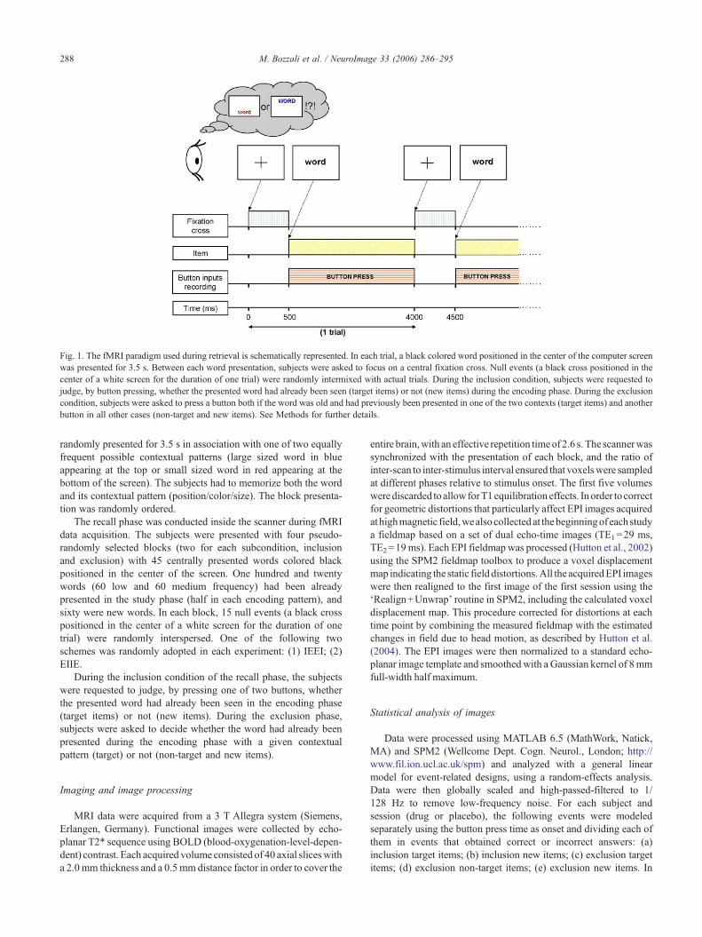
Fig. 1. The fMRI paradigm used during retrieval is schematically represented. In each trial, a black colored word positioned in the center of the computer screenwas presented for 3.5 s. Between each word presentation, subjects were asked to focus on a central fixation cross. Null events (a black cross positioned in thecenter of a white screen for the duration of one trial) were randomly intermixed with actual trials. During the inclusion condition, subjects were requested tojudge, by button pressing, whether the presented word had already been seen (target items) or not (new items) during the encoding phase. During the exclusioncondition, subjects were asked to press a button both if the word was old and had previously been presented in one of the two contexts (target items) and anotherbutton in all other cases (non-target and new items). See Methods for further details.
288 M. Bozzali et al. / NeuroImage 33 (2006) 286–295
randomly presented for 3.5 s in association with one of two equallyfrequent possible contextual patterns (large sized word in blueappearing at the top or small sized word in red appearing at thebottom of the screen). The subjects had to memorize both the wordand its contextual pattern (position/color/size). The block presenta-tion was randomly ordered.
The recall phase was conducted inside the scanner during fMRIdata acquisition. The subjects were presented with four pseudo-randomly selected blocks (two for each subcondition, inclusionand exclusion) with 45 centrally presented words colored blackpositioned in the center of the screen. One hundred and twentywords (60 low and 60 medium frequency) had been alreadypresented in the study phase (half in each encoding pattern), andsixty were new words. In each block, 15 null events (a black crosspositioned in the center of a white screen for the duration of onetrial) were randomly interspersed. One of the following twoschemes was randomly adopted in each experiment: (1) IEEI; (2)EIIE.
During the inclusion condition of the recall phase, the subjectswere requested to judge, by pressing one of two buttons, whetherthe presented word had already been seen in the encoding phase(target items) or not (new items). During the exclusion phase,subjects were asked to decide whether the word had already beenpresented during the encoding phase with a given contextualpattern (target) or not (non-target and new items).
Imaging and image processing
MRI data were acquired from a 3 T Allegra system (Siemens,Erlangen, Germany). Functional images were collected by echo-planar T2* sequence using BOLD (blood-oxygenation-level-depen-dent) contrast. Each acquired volume consisted of 40 axial sliceswitha 2.0mm thickness and a 0.5mm distance factor in order to cover the
entire brain,with an effective repetition timeof 2.6 s. The scannerwassynchronized with the presentation of each block, and the ratio ofinter-scan to inter-stimulus interval ensured that voxelswere sampledat different phases relative to stimulus onset. The first five volumeswerediscarded toallowforT1equilibration effects. Inorder to correctfor geometric distortions that particularly affect EPI images acquiredathighmagnetic field,wealsocollected at thebeginningof eachstudya fieldmap based on a set of dual echo-time images (TE1=29 ms,TE2=19ms). Each EPI fieldmapwas processed (Hutton et al., 2002)using the SPM2 fieldmap toolbox to produce a voxel displacementmap indicating the static fielddistortions.All the acquiredEPI imageswere then realigned to the first image of the first session using the‘Realign+Unwrap’ routine in SPM2, including the calculated voxeldisplacement map. This procedure corrected for distortions at eachtime point by combining the measured fieldmap with the estimatedchanges in field due to head motion, as described by Hutton et al.(2004). The EPI images were then normalized to a standard echo-planar image template and smoothedwith a Gaussian kernel of 8mmfull-width half maximum.
Statistical analysis of images
Data were processed using MATLAB 6.5 (MathWork, Natick,MA) and SPM2 (Wellcome Dept. Cogn. Neurol., London; http://www.fil.ion.ucl.ac.uk/spm) and analyzed with a general linearmodel for event-related designs, using a random-effects analysis.Data were then globally scaled and high-passed-filtered to 1/128 Hz to remove low-frequency noise. For each subject andsession (drug or placebo), the following events were modeledseparately using the button press time as onset and dividing each ofthem in events that obtained correct or incorrect answers: (a)inclusion target items; (b) inclusion new items; (c) exclusion targetitems; (d) exclusion non-target items; (e) exclusion new items. In

289M. Bozzali et al. / NeuroImage 33 (2006) 286–295
those trials in which the answer was missed (separately grouped),the average reaction time calculated over the entire block was usedas onset time. The same conditions modeled in the single subjectanalysis were included in the random-effects analysis using a one-way analysis of variance (ANOVA) design. The baseline includedthe null events and the cross fixation, which separated each trialfrom the following one. Neither was modeled.
To limit the number of statistical tests and the attendant risk offalse-positives, the overall BOLD effects related with the inclusionand exclusion task for each session (placebo or scopolamine) wereinvestigated by collapsing the different subconditions modeled:(1) inclusion condition vs. baseline; (2) exclusion condition vs.baseline; (3) inclusion condition vs. exclusion condition; (4)exclusion condition vs. inclusion condition. The effect of thescopolamine, which represents the main interest in the presentstudy, was separately investigated for inclusion (inclusion-placebovs. exclusion-placebo> inclusion-scopolamine vs. exclusion-sco-polamine) and exclusion (exclusion-placebo vs. inclusion-place-bo>exclusion-scopolamine vs. inclusion-scopolamine). For thecomparisons investigating activation versus baseline, we reportPcorrected<0.05, while differential effects and interactions arereported at Puncorrected<0.001. Stereotaxic coordinates are reportedin Talairach space and correspond to the standard MNI brain(Cocosco et al., 1997). These coordinates bear a close, but notexact, match to the atlas of Talairach and Tournoux (1998).
To investigate the possible correlations between individualsubject behavioral performance and the activation changes ofthose areas that have been found to be modulated by scopolamine,the Spearman Rank Correlation Coefficient was used. To correctfor multiple comparisons and to minimize the risk of type IIerrors, a Bonferroni correction was applied and only Pvalues≤0.0008 were considered statistically significant. For eachsubcondition (target and new for inclusion, and target, non-targetand new for exclusion), the percentage of correct answers wascorrelated with the change in activation (expressed as thepercentage difference from the mean value calculated over theentire population of studied subjects).
Results
Accuracy and reaction time data
None of the subjects reported side effects after placeboadministration. However, 3 of the 18 subjects were excluded fromthe analysis due to excessive sedation during the drug session. Afterscopolamine administration, they expressed a decrease in alertness(mean=4.67; SD=0.58), a moderate dry mouth (mean=3.67;
Table 1Mean percentage accuracy and mean correct reaction time (s) by session (placebo onon-target)
Placebo
Inclusion Exclusion
Targets New Targets New No
Correct Mean 80.62 91.32 82.06 92.91 86SD 13.26 10.24 12.64 12.03 13
Reaction time Mean 1.22 1.32 1.33 1.35 1SD 0.19 0.22 0.20 0.25 0
See text for further details.
SD=0.44) and dizziness (mean=3.41; SD=0.54) on a scale of 0to 6. All other subjects, under scopolamine, reported modest sideeffects (ranging from 1 to 2) that included a dry mouth and dizzinessonly. None of them reported a change in alertness. The performanceof the remaining 15 subjects was analyzed separately for theinclusion and exclusion conditions. A 2 (drug vs. placebo)×2 (targetvs. new) repeated-measures ANOVA was conducted on theinclusion data, and a separate 2 (drug vs. placebo)×3 (target vs.non-target vs. new) ANOVAwas conducted on the exclusion data.Post hoc analyses were performed using Bonferroni t tests.
In the inclusion condition, a 2×2 repeated-measures ANOVAdemonstrated a significant main effect of response [F(1,14)=23.40,P<0.0001] where new responses were identified significantly moreaccurately than target responses. There was no effect of drug orresponse×drug interaction [F(1,14)<0.34]. In the exclusioncondition, a 2×3 ANOVA revealed a significant main effect ofresponse [F(2,28)=11.56, P<0.0001] and post hoc analysisshowed that new items were correctly identified significantlybetter than non-targets and targets. There was no main effect ofdrug [F(1,14)=0.17 or response×drug interaction [F(2,28)=0.17].
In terms of the reaction time data, a 2 (drug vs. placebo)×2(inclusion vs. exclusion)×2 (old vs. new) repeated-measuresANOVA was conducted. There was a significant main effect ofcondition [F(1,14)=41.36,P<0.0001]wherecorrect responses in theexclusionconditionweremadesignificantlymore slowly thancorrectresponses in the inclusion condition. There was also a significantcondition×response interaction [F(1,14)=71.34, P<0.0001]. Posthoc analysis using Bonferroni t tests demonstrated that the responsetimes for correctly identified old items (targets and non targets) weresignificantly slower in the exclusion condition than the inclusioncondition. However, the response times for correctly identified newitemsdidnot significantlydiffer acrossconditions.Therewasnomaineffect of drug [F(1,14)=0.26] or response [F(1,14)=0.33] ordrug×response [F(1,14)=0.30], drug×condition [F(1,14)=0.29]or drug×condition×response interaction [F(1,14)=0.70]. Accuracyand reaction times (RT) data are shown in Table 1.
Imaging findings
Placebo sessionContrasting the inclusion condition against the baseline, we
observed activation in a number of different regions (predomi-nantly left lateralized) that survived after correction for multiplecomparisons (PFWE corrected<0.05). This activation response issummarized in Table 2.
Contrasting the exclusion condition against the baseline, amuch more widespread pattern of activation (again, predominantly
r scopolamine), condition (inclusion or exclusion) and item type (target, new,
Scopolamine
Inclusion Exclusion
n-targets Targets New Targets New Non-targets
.54 82.28 92.26 81.98 93.84 89.00
.07 10.59 6.34 15.71 5.45 7.93
.56 1.24 1.33 1.51 1.35 1.54
.25 0.25 0.27 0.35 0.24 0.19

Table 3Maxima of regions showing significant (P<0.05 corrected) BOLD signalchanges in comparison of exclusion and baseline condition
Brain region Side BA Size Coordinates(mm)
PeakZscore
x y z
Inf/mid occipital gyrus L 17, 18, 19 12,258 −26 −96 −6 8.16Cuneus L 17, 18, 19 −9 −96 −11 7.06Fusiform gyrus L 37 −38 −64 −16 7.01Cerebellum L – −31 −63 −38 5.69Inf/mid occipital gyrus R 18, 19 32 −87 −10 7.46Fusiform gyrus R 37 40 −56 −20 4.83Cuneus R 18, 19 11 −73 3 6.19Cerebellum R – 23 −52 −32 6.84Inf parietal lobule L 7 11,148 −34 −30 50 7.98Precuneus L 7 −6 −76 30 5.19Precuneus R 7 10 −75 36 5.92Sup parietal lobule L 5, 7 −34 −65 46 7.04Angular gyrus L 39 −36 −70 34 5.76Supramarginal gyrus L 40 −37 −61 53 7.53Pre/postcentral gyrus L 1, 2, 3, 4 −35 −53 51 6.84Med frontal gyrus L 8, 9 −2 6 55 6.89Mid frontal gyrus L 8, 9 −14 1 54 4.81Cingulate gyrus L 24, 32 10 12 34 5.46Cingulate gyrus R 24, 32 −5 6 37 6.50Post cingulate L 23, 29, 31 615 −2 −34 24 7.38Post cingulate R 23, 29, 31 3 −32 21 7.35Mid/inf frontal gyrus L 10 80 −44 48 −4 5.26Mid frontal gyrus L 9, 10 37 −38 42 30 5.24Inferior frontal gyrus L 11, 45, 47 2963 −30 24 −6 7.87Insula L – −34 21 6 7.84Parahippocampal gyrus L 27, 28, 35 2723 −23 −30 −10 7.82Thalamus L – −13 −18 2 5.48Substantia nigra L – −9 −12 −13 5.26Caudate head L – −14 8 10 5.20Globus pallidus L – −16 6 1 4.89Mammillary body L – −12 −17 2 4.87Transversetemporal
gyrusL 41 81 −48 −26 16 5.22
Inf frontal gyrus R 47 617 36 24 −8 7.82Insula R – 34 19 6 7.81Putamen R – 334 12 14 2 5.38Globus pallidus R – 15 7 1 4.88Mammillary body R – 11 −15 1 4.79Caudate head R – 14 14 11 5.10Thalamus R – 14 −10 16 5.26
Abbreviations: sup=superior; inf= inferior; ant=anterior; post=posterior;mid=middle; med=medial; R=right; L=left; B=bilateral; BA=Brodmannarea.The extension of each area (size) is expressed in number of voxels. Onlyregions that were statistically significant at uncorrected level (Pcorrected<0.05)with a size of 10 or more voxels have been considered.See text for further details.
Table 2Maxima of regions showing significant (P<0.05 corrected) BOLD signalchanges in comparison of inclusion and baseline condition
Brain region Side BA Size Coordinates(mm)
PeakZscore
x y z
Mid occipital gyrus L 17, 18, 19 2977 −30 −88 −20 7.59Fusiform gyrus L 37 −40 −53 −22 6.57Cerebellum L – −39 −54 −32 7.29Mid/inf occipital gyrus R 17, 18, 19 2582 36 −85 −13 6.07Fusiform gyrus R 37 36 −54 −25 5.69Cerebellum R – 36 −82 −24 7.55Cerebellum R – 25 8 −80 −38 5.07Inf parietal lobule L 5, 7 723 −48 38 48 5.36Supramarginal gyrus L 40 −49 −38 42 4.69Postcentral gyrus L 2, 3, 4 −36 −30 50 6.98Precentral gyrus L 4 −42 −24 58 5.35Sup and inf parietal
lobuleL 7 31 −36 −58 56 5.32
Sup temporal gyrus L 38 −48 15 −10 5.01Inf frontal gyrus L 45, 47 544 −34 22 −4 6.99Insula L – −42 12 −2 5.44Inf frontal gyrus R 47 370 34 26 −8 6.67Insula R – 35 19 6 5.45Med frontal gyrus L 6, 8 −8 5 50 4.91Ant cingulate L 24, 32 −5 14 40 5.81Med frontal gyrus R 6, 8 966 2 16 44 6.97Sup frontal gyrus R 6, 8 8 6 61 4.95Ant cingulate R 32 9 19 34 4.89Inf frontal gyrus L 9, 44, 45 376 −48 6 28 6.45Thalamus L – 75 −12 −20 4 5.12Mammillary body L – 59 −12 −18 1 4.94Subthalamic nucleus L – −11 −1 −5 4.96
Abbreviations: sup=superior; inf= inferior; ant=anterior; post=posterior;mid=middle; med=medial; R=right; L= left; B=bilateral; BA=Brodmannarea.The extension of each area (size) is expressed in number of voxels.Only regions that were statistically significant at uncorrected level(Pcorrected<0.05) with a size of 10 or more voxels have been considered.See text for further details.
290 M. Bozzali et al. / NeuroImage 33 (2006) 286–295
left lateralized) than that obtained in the inclusion condition wasfound (see Table 3).
Contrasting the inclusion with exclusion condition (exclusionminus inclusion) revealed significant effects. These includedbilateral medial frontal gyrus (BA6), anterior (BA 24, 32) andposterior cingulate (BA 23,31), precuneus (BA 7), the angulargyrus (BA 40), occipital lobe (BA 18,19) and cerebellum. Therealso was the unilateral involvement of the left supramarginal gyrus(BA 39), pre- (BA 4, 6) and postcentral (BA 1, 2, 3) gyri, frontalgyri (BA 8, 9, 11), the left fusiform gyrus (BA 20, 37) and theinferior parietal lobe (BA 7). Additional subcortical activationswere noted in the region of the left red nucleus and substantia nigra(see Table 4).
Drug effectsThe interaction between drug and inclusion/exclusion condition
(inclusion-placebo vs. exclusion-placebo>inclusion-scopolaminevs. exclusion-scopolamine) was significant in a number of regions.The interaction effects between drug and inclusion condition(inclusion-placebo vs. exclusion-placebo>inclusion-scopolaminevs. exclusion-scopolamine) indicated that a few regions were
modulated by scopolamine in the inclusion task compared to theexclusion task, most notably the left inferior frontal gyrus (BA 9,45) and left cerebellum.
Conversely, the opposite interaction showed scopolaminemodulated activity in several regions. These regions include themedial frontal gyri (BA6) bilaterally, the left superior (BA9) andmiddle frontal gyrus (BA 6), the right medial frontal gyrus (BA8), the left anterior (BA 24) and right posterior (BA 23,31)cingulate, the left perirhinal cortex (BA 36), the left superior
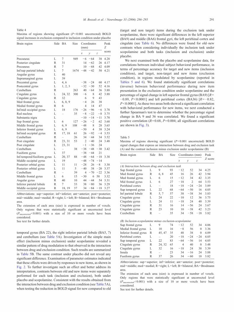
Table 4Maxima of regions showing significant (P<0.001 uncorrected) BOLDsignal increases in exclusion compared to inclusion condition under placebo
Brain region Side BA Size Coordinates(mm)
PeakZscore
x y z
Precuneus L 7 949 −6 −64 38 4.28Posterior cingulate R 31 14 −62 26 4.17Precuneus R 7 10 −68 42 4.09Inf/sup parietal lobule L 7 1674 −48 −42 50 4.21Angular gyrus L 40Supramarginal gyrus L 39Precentral gyrus L 4, 6 −38 −24 60 4.17Postcentral gyrus L 1, 2, 3 −32 −30 52 4.16Cerebellum R 263 40 −64 36 3.88Cingulate gyrus L 24, 32 300 −6 8 42 3.88Cingulate gyrus R 32 6 9 43Med frontal gyrus L 6, 8, 9 −4 26 38Medial frontal gyrus R 6 4 14 47Inf/mid occipital gyrus L 18 176 −26 −96 −6 3.78Red nucleus L – 35 −8 −22 −8 3.75Substantia nigra L −10 −14 −11 3.70Sup frontal gyrus L 6 127 −26 −2 62 3.68Middle frontal gyrus L 6, 9 108 −48 8 28 3.65Inferior frontal gyrus L 6, 9 −50 4 38 3.24Inf/mid occipital gyrus R 17, 18 61 26 −92 −4 3.53Cerebellum R – 26 34 −44 34 3.52Post cingulate R 23, 31 53 2 −40 24 3.48Post cingulate L 23, 31 −1 −36 24Cerebellum L – 38 −38 −48 −30 3.42Fusiform gyrus L 37 −38 −44 21Inf-temporal/fusiform gyrus L 20, 37 88 −48 −64 −18 3.38Middle occipital gyrus L 19 −48 −74 −14Posterior orbital gyrus L 11 11 −28 26 −8 3.38Posterior orbital gyrus L 11 5 −40 38 −20 3.37Cerebellum R – 39 4 −70 −22 3.36Middle frontal gyrus L 6 15 −30 6 50 3.32Angular gyrus R 40 10 44 −44 54 3.31Inferior parietal lobule R 7 33 38 −80 36 3.29Middle occipital gyrus R 18, 19 37 34 −84 −18 3.27
Abbreviations: sup=superior; inf= inferior; ant=anterior; post=posterior;mid=middle; med=medial; R=right; L= left; B=bilateral; BA=Brodmannarea.The extension of each area (size) is expressed in number of voxels.Only regions that were statistically significant at uncorrected level(Puncorrected<0.001) with a size of 10 or more voxels have beenconsidered.See text for further details.
Table 5Maxima of regions showing significant (P<0.001 uncorrected) BOLDsignal changes that express an interaction between drug and exclusion task(A) and the contrast inclusion minus exclusion under scopolamine (B)
Brain region Side BA Size Coordinates (mm) PeakZ score
x y z
(A) Interaction between drug and exclusion taskSup frontal gyrus L 9 93 −12 52 34 4.25Med frontal gyrus R 6, 8 45 16 26 42 3.94Med frontal gyrus L 6 15 −12 34 42 3.35Mid frontal gyrus L 6 27 −18 2 64 3.53Perirhinal cortex L – 18 −18 −24 −24 3.89Sup temporal gyrus L 22 68 −64 −50 16 4.05Inf parietal lobule R 5, 7 57 38 −38 34 3.83Cingulate gyrus L 24 22 −10 12 36 3.75Cingulate gyrus L 24 11 −18 24 40 3.39Cingulate gyrus R 31 16 14 −56 24 3.67Cingulate gyrus R 23 10 10 −38 42 3.23Cerebellum R – 35 34 −58 −18 3.82
(B) Inclusion-scopolamine minus exclusion-scopolamineSup frontal gyrus L 9 74 −12 52 34 4.06Medial frontal gyrus L 10 16 −8 56 0 3.36Inferior frontal gyrus R 45, 47 35 48 38 0 4.09Perirhinal cortex L – 20 −18 −24 −24 4.05Sup temporal gyrus L 22 83 −64 −56 14 4.05Cingulate gyrus R 24, 32 65 4 40 0 3.48Cingulate gyrus L 32 16 −18 24 38 3.39Insula R – 23 34 −16 24 3.88Fusiform gyrus R 37 26 34 \60 18 3.82
Abbreviations: sup=superior; inf= inferior; ant=anterior; post=posterior;mid=middle; med=medial; R=right; L= left; B=bilateral; BA=Brodmannarea.The extension of each area (size) is expressed in number of voxels.Only regions that were statistically significant at uncorrected level(Puncorrected<0.001) with a size of 10 or more voxels have beenconsidered.See text for further details.
291M. Bozzali et al. / NeuroImage 33 (2006) 286–295
temporal gyrus (BA 22), the right inferior parietal lobule (BA5, 7)and cerebellum (see Table 5A). Investigation of the simple maineffect (inclusion minus exclusion) under scopolamine revealed asimilar pattern of drug modulation to that observed in the interactionbetween drug and exclusion condition. Such results are summarizedin Table 5B. The same contrast under placebo did not reveal anysignificant difference. Examination of parameter estimates indicatedthat these effects were driven by responses to new items, as shown inFig. 2. To further investigate such an effect and better address itsinterpretation, contrasts between old and new items were separatelyperformed for each task (inclusion and exclusion), both underplacebo and scopolamine. Consistent with the results obtained fromthe interaction between drug and exclusion condition (see Table 5A),when testing the reduction in BOLD signal for new compared to old
(target and non target) items during the exclusion task underscopolamine, there were significant differences in the left superior(BA9) and middle (BA6) frontal gyri, the left perirhinal cortex andcingulate (see Table 6). No differences were found for old–newcontrasts when considering individually the inclusion task underscopolamine and both tasks (inclusion and exclusion) underplacebo.
We next examined both the placebo and scopolamine data, forcorrelations between individual subject behavioral performance, interms of percentage accuracy for target and new items (inclusioncondition), and target, non-target and new items (exclusioncondition), in regions modulated by scopolamine (reported inTables 5 and 6). We found statistically significant correlations(inverse) between behavioral performance during new itempresentation in the exclusion condition under scopolamine and thepercentage of signal change in left superior frontal gyrus (BA9) [R=−0.89, P<0.0001] and left perirhinal cortex (BA36) [R=−0.82,P<0.0001]. As these two areas both showed a significant correlationwith behavioral performance for new items, we next conducted afurther Spearman's test to determine whether the percentage signalchange in BA 9 and 36 was correlated. We found a significantpositive correlation (R=0.68, P=0.004; all significant correlationsare shown in Fig. 3).
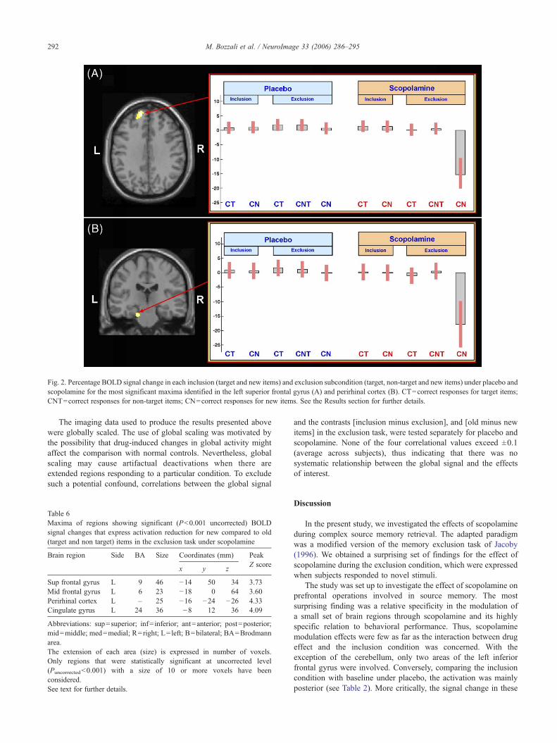
Fig. 2. Percentage BOLD signal change in each inclusion (target and new items) and exclusion subcondition (target, non-target and new items) under placebo andscopolamine for the most significant maxima identified in the left superior frontal gyrus (A) and perirhinal cortex (B). CT=correct responses for target items;CNT=correct responses for non-target items; CN=correct responses for new items. See the Results section for further details.
292 M. Bozzali et al. / NeuroImage 33 (2006) 286–295
The imaging data used to produce the results presented abovewere globally scaled. The use of global scaling was motivated bythe possibility that drug-induced changes in global activity mightaffect the comparison with normal controls. Nevertheless, globalscaling may cause artifactual deactivations when there areextended regions responding to a particular condition. To excludesuch a potential confound, correlations between the global signal
Table 6Maxima of regions showing significant (P<0.001 uncorrected) BOLDsignal changes that express activation reduction for new compared to old(target and non target) items in the exclusion task under scopolamine
Brain region Side BA Size Coordinates (mm) PeakZ score
x y z
Sup frontal gyrus L 9 46 −14 50 34 3.73Mid frontal gyrus L 6 23 −18 0 64 3.60Perirhinal cortex L – 25 −16 −24 −26 4.33Cingulate gyrus L 24 36 −8 12 36 4.09
Abbreviations: sup=superior; inf= inferior; ant=anterior; post=posterior;mid=middle; med=medial; R=right; L= left; B=bilateral; BA=Brodmannarea.The extension of each area (size) is expressed in number of voxels.Only regions that were statistically significant at uncorrected level(Puncorrected<0.001) with a size of 10 or more voxels have beenconsidered.See text for further details.
and the contrasts [inclusion minus exclusion], and [old minus newitems] in the exclusion task, were tested separately for placebo andscopolamine. None of the four correlational values exceed ±0.1(average across subjects), thus indicating that there was nosystematic relationship between the global signal and the effectsof interest.
Discussion
In the present study, we investigated the effects of scopolamineduring complex source memory retrieval. The adapted paradigmwas a modified version of the memory exclusion task of Jacoby(1996). We obtained a surprising set of findings for the effect ofscopolamine during the exclusion condition, which were expressedwhen subjects responded to novel stimuli.
The study was set up to investigate the effect of scopolamine onprefrontal operations involved in source memory. The mostsurprising finding was a relative specificity in the modulation ofa small set of brain regions through scopolamine and its highlyspecific relation to behavioral performance. Thus, scopolaminemodulation effects were few as far as the interaction between drugeffect and the inclusion condition was concerned. With theexception of the cerebellum, only two areas of the left inferiorfrontal gyrus were involved. Conversely, comparing the inclusioncondition with baseline under placebo, the activation was mainlyposterior (see Table 2). More critically, the signal change in these

Fig. 3. Scatterplots of statistically significant correlations between individualsubject behavioral performance on new items and the activation changes inleft superior frontal gyrus (A) and perirhinal cortex (B) are shown. Thepositive correlation of percentage signal change between left superior frontalgyrus and perirhinal cortex is also shown (C). See the Results section forfurther details.
293M. Bozzali et al. / NeuroImage 33 (2006) 286–295
regions was not associated with any behavioral effect whenperforming the inclusion task.
A strikingly different pattern of activation and drug modulationwas found when considering the exclusion task. Under placebo,there were widespread regions of activation in the exclusioncondition compared to baseline (see Table 3). However, theinteraction between drug and exclusion produced a more restrictedset of regions, which showed greater difference in activationbetween exclusion and inclusion under scopolamine as compared
to placebo (see Table 5); the critical regions being predominantlyfrontal and temporal. The region with the greatest Z score – the leftsuperior frontal gyrus (BA 9) – showed a distinctive activationpattern across the different response categories (target, non-targetand new items), and this had a behavioral correlate. There was littledifference in activation between scopolamine and placebo whenparticipants correctly identified targets (old items in the samespatial position) or non-targets (old items in a different spatialposition). Similarly, there was no difference between scopolamineand placebo in either the old or new conditions in the inclusiontask. However, when participants responded to new items in theexclusion condition, there was a significant modulation withscopolamine compared to placebo (see Fig. 2A). Moreover, thismodulation was inversely correlated with accuracy for new itemsacross subjects (see Fig. 3A). This shows that effect is not a mereepiphenomenon created in the imaging analysis procedures. Thesecond of these regions, left perirhinal cortex (BA 36), showed asimilar effect both with respect to the pattern of drug modulationand the correlation with behavioral data (see Figs. 2B and 3B).Moreover, there was a direct correlation between the scopolamine-related signal change in these two regions (see Fig. 3C).
How might this pattern of performance be explained? Thedeactivation occurs in the exclusion task only, not the inclusion task.This is the task where, for one group of stimuli, recollection andfamiliarity processes produce conflicting response tendencies. Thissuggests that the phenomenonmay arise in the processes involved inthe control of to what extent recollection and/or familiarity processesare operative. This explanation would require a second system orsystems to control the relative strength of the familiarity andrecollection signals, and so, the activity of perirhinal cortex. Acandidate structure should have levels of activation that correlatewith those of perirhinal cortex, as was the case for the left inferiorfrontal gyrus (BA9) in the current experiment (see Figs. 2 and 3). Infact, another study contrasting recollection and familiarity directlyreported peak activation in a similar region only 1 cm distant. Thisstudy involved the Remember–Know paradigm, in which subjectsmust decide whether they can recollect having previously con-sciously experienced a stimulus or whether they simply find itfamiliar (Henson et al., 1999b). The region was deactivated duringKnow (familiarity) compared to Remember (recollection) responses.
What specifically could the control signal be doing? Thereseem to be two possibilities. One depends on the way theperirhinal cortex has been held by a number of authors to mediatea familiarity effect. Familiar stimuli lead to overall reduced neuralactivity in perirhinal cortex compared to novel stimuli (Aggletonand Brown, 2005). On this basis, new stimuli should evoke greateractivation in perirhinal cortex than old stimuli do. If the effect ofscopolamine is to produce a reduced activation in the leftperirhinal cortex when new items are presented, the neural repre-sentation of new stimuli would masquerade as familiar stimuli.Therefore, we argue that, due to the effect of scopolamine onperirhinal cortex, it would be difficult under scopolamine torespond to an item as ‘New’ on the basis of a lack of a familiaritysignature. Instead, the presence or absence of any recollectionsignal provides the sole basis for deciding whether a stimulus is infact old or new. Thus, the system would need to base its responseon recollection alone. By contrast, in the placebo condition, thejudgment that an item is new can be based on familiarity as well asrecollection. However, on this explanation, there is no simpleaccount of why the effect occurs only in the exclusion and not alsoin the inclusion condition.
294 M. Bozzali et al. / NeuroImage 33 (2006) 286–295
A second possibility arises from the finding in the behavioralliterature that it is impairments in acquisition rather than retrievalthat are obtained under the administration of scopolamine (GhoneimandMewaldt, 1977; Petersen, 1977; Rosier et al., 1998). Acquisitioneffects operate on new stimuli or at least stimuli or stimuluscombinations that are not yet well-learned. Thus, since the currentphenomenon occurs in the processing of new stimuli, it might beseen as a side effect of acquisition processes. Moreover, it is knownthat, even when scopolamine affects encoding, it impairs subsequentfree recall more than subsequent recognition (see Hasselmo andWyble, 1997). This suggests – but does not demonstrate – thatencoding for subsequent recollection-based judgments is moreimpaired than encoding for subsequent familiarity-based ones. Thealternative possibility thus is the inverse of the first, namely that inthe exclusion condition – which generally requires recollection, theNew stimuli, specifically, are best processed under scopolamineusing the familiarity signal rather than the recollection one andsubjects doing so perform best. This is less of an issue for inclusionsince there are no other stimuli for which the use of a recollectionresponse is necessary. Moreover, in the Henson et al. (1999b) study,this region is deactivated for a ‘Know’, i.e. familiarity-basedresponse. Thus, in this account, deactivation of perirhinal cortex is asign of its being used!
The commonality between these two accounts is that theyinvolve a control system which must oversee the selection ofrecollection and familiarity processes. Cholinergic-mediated in-volvement of the medial prefrontal cortex in this hypotheticalmemory control process may also relate to an analogous cholinergicrole in associated structures — that is vigilance control in the rat(Dalley et al., 2004). Selecting between the two hypotheses wouldrequire further studies.
Another important issue in the study is whether the cholinergicinput potentiates a monitoring process controlled by the dorso-lateral prefrontal cortex. Our experimental paradigm involved asource memory – exclusion – condition that was argued by Hensonet al. (1999a) to require this monitoring process. With certainexceptions, the overall pattern of results found by Henson et al.(1999a) was replicated. However, in the exclusion condition, theregions more activated compared to baseline were considerablymore extensive than in the previous study, but included the mainactivated areas in the equivalent Henson et al. (1999a) contrast.One difference was that the anterior activations were more leftlateralized in the current study, while they were more bilateral inthe Henson et al. (1999a) study. This would fit with the findings ofDobbins et al. (2004) that monitoring based on spatial position, asin the current study, tends to be more left prefrontally based. In theHenson et al. (1999a) study, the source memory task also requiredknowledge of the list a word was presented in (temporal context),as well as its spatial position, but only the latter cue was relevant inthe present study.
Turning to the effects of scopolamine, behaviorally it had littleeffect on accuracy across different conditions, with the exceptiondiscussed above (new item recognition in the exclusion condition).As far as the imaging results were concerned, the most importantfinding was that the set of regions involved in the interactionbetween drug and exclusion task did not overlap with the regionsactivated more in the exclusion task versus baseline contrast in theHenson et al. (1999a) study. Regions held to be involved inconscious monitoring or checking of the retrieval output, namelythe mid-dorsolateral regions, particularly on the right, were notinvolved in the interaction between drug and exclusion task. There
was therefore no support – either behaviorally or from imaging –
for the hypothesis that the basal forebrain cholinergic system mightmodulate the prefrontal structures responsible for monitoring thememory output. Therefore, this study provides no support for thehypothesis that impairments of this process due to lack ofpotentiation by the cholinergic system are involved in the causationof confabulations.
Acknowledgment
This work was supported by the Wellcome Trust.
References
Aggleton, J.P., Brown, M.W., 1999. Episodic memory, amnesia, and thehippocampal–anterior thalamic axis. Behav. Brain Sci. 22, 425–444(discussion 444–489).
Aggleton, J.P., Brown, M.W., 2005. Contrasting hippocampal and perirhinalcortex function using immediate early gene imaging. Q. J. Exp. Psychol.,B 58, 218–233.
Alexander, M.P., Freedman, M., 1984. Amnesia after anterior communicat-ing artery aneurysm rupture. Neurology 34, 752–757.
Bond, A., Lader, M., 1974. The use of analogue scales in rating subjectivefeelings. Br. J. Med. Psychol. 47, 211–218.
Burgess, P.W., Shallice, T., 1996. Confabulation and the control ofrecollection. Memory 4, 359–411.
Cabeza, R., Locantore, J.K., Anderson, N.D., 2003. Lateralization ofprefrontal activity during episodic memory retrieval: evidence for theproduction-monitoring hypothesis. Cogn. Neurosci. 15, 249–259.
Chudasama, Y., Dalley, J.W., Nathwani, F., Bouger, P., Robbins, T.W., 2004.Cholinergic modulation of visual attention and working memory;dissociable effects of basal forebrain 192-IgG-saporin lesions andintraprefrontal infusions of scopolamine. Learn. Mem. 11, 78–86.
Cipolotti, L., Bird, C., Good, T., Macmanus, D., Rudge, P., Shallice, T.,2006. Recollection and familiarity in dense hippocampal amnesia: a casestudy. Neuropsychologia 44, 489–506.
Cocosco, C.A., Kollokian, V., Kwan, R.K.S., Evans, A.C., 1997. BrainWeb:online interface to a 3D MRI simulated brain database. NeuroImage 5,S425.
Curran, T., DeBuse, C., Woroch, B., Hirshman, E., 2006. Combinedpharmacological and electrophysiological dissociation of familiarity andrecollection. J. Neurosci. 15, 1979–1985.
Dalley, J.W., Theobald, D.E., Bouger, P., Chudasama, Y., Cardinal, R.N.,Robbins, T.W., 2004. Cortical cholinergic function and deficits in visualattentional performance in rats following 192 IgG-saporin-inducedlesions of the medial prefrontal cortex. Cereb. Cortex 14, 922–932.
Damasio, A.R., Graff-Radford, N.R., Eslinger, P.J., Damasio, H., Kassell,N., 1985. Amnesia following basal forebrain lesions. Arch. Neurol. 42,263–271.
DeLuca, J., 1993. Predicting neurobehavioral patterns following anteriorcommunicating artery aneurysm. Cortex 29, 639–647.
DeLuca, J., Cicerone, K.D., 1991. Confabulation following aneurysm of theanterior communicating artery. Cortex 27, 417–423.
Dobbins, I.G., Simons, J.S., Schacter, D.L., 2004. FMRI evidence forseparable and lateralized prefrontal memory monitoring processes.J. Cogn. Neurosci. 16, 908–920.
Fischer, R.S., Alexander, M.P., D'Esposito, M., Otto, R., 1995. Neuropsy-chological and neuroanatomical correlates of confabulation. J. Clin. Exp.Neuropsychol. 17, 20–28.
Fletcher, P.C., Henson, R.N., 2001. Frontal lobes and human memory:insights from functional neuroimaging. Brain 124, 849–881.
Ghoneim, M.M., Mewaldt, S.P., 1977. Studies on human memory: theinteractions of diazepam, scopolamine, and physostigmine. Psycho-pharmacology 52, 1–6.
Gilboa, A., Moscovitch, M., 2002. The cognitive neuroscience of

295M. Bozzali et al. / NeuroImage 33 (2006) 286–295
confabulation: a review and a model. In: Baddeley, A.D., Kopelman,M.D., Wilson, B.A. (Eds.), Handbook of Memory Disorders. JohnWiley and Sons, London, pp. 315–342.
Hasselmo, M.E., Wyble, B.P., 1997. Free recall and recognition in a networkmodel of the hippocampus: simulating effects of scopolamine on humanmemory function. Behav. Brain Res. 89, 1–34.
Hasselmo, M.E., Wyble, B.P., Wallenstein, G.V., 1996. Encoding andretrieval of episodic memories: role of cholinergic and GABAergicmodulation in the hippocampus. Hippocampus 6, 693–708.
Henson, R.N.A., 2005. A mini-review of fMRI studies of human medialtemporal lobe activity associated with recognition memory. Q. J. Exp.Psychol., B 58, 340–360.
Henson, R.N., Shallice, T., Dolan, R.J., 1999a. Right prefrontal cortex andepisodic memory retrieval: a functional MRI test of the monitoringhypothesis. Brain 122, 1367–1381.
Henson, R.N., Rugg, M.D., Shallice, T., Josephs, O., Dolan, R.J.,1999b. Recollection and familiarity in recognition memory: an event-related functional magnetic resonance imaging study. J. Neurosci. 19,3962–3972.
Hintzman, D.L., Caulton, D.A., Levitin, D.J., 1998. Retrieval dynamics inrecognition and list discrimination: further evidence of separateprocesses of familiarity and recall. Mem. Cogn. 26, 449–462.
Hutton, C., Bork, A., Josephs, O., Deichmann, R., Ashburner, J., Turner, R.,2002. Image distortion correction in fMRI: a quantitative evaluation.NeuroImage 16, 217–240.
Hutton, C., Deichmann, R., Turner, R., Anderrson, J., 2004. Combinedcorrection for geometric distortions and its interaction with headmovement in fMRI. Proc. of the 12th Scientific Meeting and Exhibitionof the ISMRM, p. 1084.
Jacoby, L.L., 1996. Dissociating automatic and consciously controlled effectof study/test compatibility. J. Mem. Lang. 35, 32–52.
Lepage, M., Ghaffar, O., Nyberg, L., Tulving, E., 2000. Prefrontal cortexand episodic memory retrieval mode. Proc. Natl. Acad. Sci. U. S. A. 97,506–511.
Mandler, G., 1980. Recognizing: the judgment of previous occurrence.Psychol. Rev. 87, 511–519.
Mesulam, M.M., 2000. Behavioural neuroanatomy: large scale networks;association cortex; frontal syndromes; the limbic system, and hemi-spheric specializations. In: Mesulam, M.M. (Ed.), Principles of
Behavioral and Cognitive Neurology. Oxford Univ. Press, New York,pp. 1–120.
Mintzer, M, Griffiths, RR, 2001. Acute dose-effects of scopolamine on falserecognition. Psychopharmacology 153, 425–433.
Moscovitch, M., 1989. Confabulation and the frontal systems: strategicversus associated retrieval in neuropsychological theories of memory. In:RoedigerI.I.I. I.I.I., H.L., Craik, F.I.M. (Eds.), Varieties of Memory andConsciousness: Essays in Honour of Endel Tulving. Erlbaum, Hillsdale,NJ, pp. 133–160.
Petersen, R.C., 1977. Scopolamine induced learning failures in man.Psychopharmacology 52, 283–289.
Phillips, S., Sangalang, V., Sterns, G., 1987. Basal forebrain infarction. Aclinicopathologic correlation. Arch. Neurol. 44, 1134–1138.
Roediger, H.L., McDermott, K.B., 1995. Creating false memories—Remembering words not presented in lists. J. Exp. Psychol. Learn. 21,803–814.
Rosier, A., Cornette, L., Orban, G.A., 1998. Scopolamine-inducedimpairment of delayed recognition of abstract visual shapes. Neuro-psychobiology 37, 98–103.
Shallice, T., 2002. Fractionation of the supervisory system. In: Stuss, D.T.,Knight, R.T. (Eds.), Principles of Frontal Lobe Function. Oxford Univ.Press, New York, pp. 261–277.
Shallice, T., Fletcher, P., Frith, C.D., Grasby, P., Frackowiak, R.S., Dolan,R.J., 1994. Brain regions associated with acquisition and retrieval ofverbal episodic memory. Nature 368, 633–635.
Sperling, R., Greve, D., Dale, A., Killiany, R., Holmes, J., Rosas, H.D.,Cocchiarella, A., Firth, P., Rosen, B., Lake, S., Lange, N., Routledge, C.,Albert, M., 2002. Functional MRI detection of pharmacologicallyinduced memory impairment. Proc. Natl. Acad. Sci. U. S. A. 99,455–460.
Talairach, J., Tournoux, P., 1998. Co-planar Stereotaxic Atlas of the HumanBrain. Thieme Medical Publishers, New York.
Tulving, E., Kapur, S., Craik, F.I., Moscovitch, M., Houle, S., 1994.Hemispheric encoding/retrieval asymmetry in episodic memory: posi-tron emission tomography findings. Proc. Natl. Acad. Sci. U. S. A. 91,2016–2020.
Yonelinas, A.P., Levy, B.J., 2002. Dissociating familiarity from recollectionin human recognition memory: different rates of forgetting over shortretention intervals. Psychon. Bull. Rev. 9, 575–582.
Copyright © 2022 FDOKUMEN