Cognitive aging: a common decline of episodic recollection and spatial memory in rats
10
Behavioral/Systems/Cognitive Cognitive Aging: A Common Decline of Episodic Recollection and Spatial Memory in Rats R. Jonathan Robitsek, 1 Norbert J. Fortin, 1 Ming Teng Koh, 2 Michela Gallagher, 2 and Howard Eichenbaum 1 1 Center for Memory and Brain, Boston University, Boston, Massachusetts 02215, and 2 Department of Psychological and Brain Sciences, Johns Hopkins University, Baltimore, Maryland 21218 In humans, recognition memory declines with aging, and this impairment is characterized by a selective loss in recollection of previously studied items contrasted with relative sparing of familiarity for items in the study list. Rodent models of cognitive aging have focused on water maze learning and have demonstrated an age-associated loss in spatial, but not cued memory. The current study examined odor recognition memory in young and aged rats and compared performance in recognition with that in water maze learning. In the recogni- tion task, young rats used both recollection and familiarity. In contrast, the aged rats showed a selective loss of recollection and relative sparing of familiarity, similar to the effects of hippocampal damage. Furthermore, performance on the recall component, but not the familiarity component, of recognition was correlated with spatial memory and recollection was poorer in aged rats that were also impaired in spatial memory. These results extend the pattern of impairment in recollection and relative sparing of familiarity observed in human cognitive aging to rats, and suggest a common age-related impairment in both spatial learning and the recollective component of nonspatial recognition memory. Key words: aging; hippocampus; recollection; familiarity; spatial memory; receiver operating characteristic Introduction In humans, aging is often associated with memory impairment, and in particular a decline in the capacity for recalling specific recent experiences, that is, in episodic memory (Craik and Byrd, 1982). Since the initial suggestion of age-associated memory im- pairment as a clinical diagnosis (Crook et al., 1986), much re- search has focused on understanding the changes in the brain systems that mediate the specific aspects of the observed deficits in aged humans (Albert, 1997; Buckner, 2004; Hedden and Gab- rieli, 2004) and in animal models of cognitive aging (Gallagher and Rapp, 1997; Wilson et al., 2006). In humans, the pattern of deficits is highlighted by a disproportionate loss in the capacity for episodic recollection, similar to the pattern of deficits associ- ated with a mild form of damage to the medial temporal lobes (Jennings and Jacoby, 1997; Bastin and Van der Linden, 2003; Quamme et al., 2004; Toth and Parks, 2006). In particular, signal detection analyses of recognition memory performance have re- vealed a striking dissociation in the receiver operating character- istic (ROC) function, revealing a pattern of impaired recollection and spared familiarity (Daselaar et al., 2006; Howard et al., 2006; Prull et al., 2006; Duverne et al., 2007). Correspondingly, the deficit in recollection has been associated with decreased hip- pocampal activation (Daselaar et al., 2006). Experimental studies on cognitive aging in rats have also re- vealed age-associated memory impairments that suggest a dete- rioration of hippocampal function (Rosenzweig and Barnes, 2003; Wilson et al., 2006). These analyses have focused on spatial learning and memory and highlight an impairment when perfor- mance requires the use of spatial cues, in contrast with intact memory when performance is guided by nonspatial stimuli (Rapp et al., 1987; Gallagher et al., 1993). Correspondingly, this impairment has been linked to multiple alterations of hippocam- pal anatomy and physiology, including decreases in synaptic den- sities, weakened synaptic plasticity, decreased cholinergic modu- lation, and a rigidity of spatial firing patterns of hippocampal neurons (Rosenzweig and Barnes, 2003; Wilson et al., 2006). In humans and rats, not all aged individuals are impaired in memory relative to young controls, and the degree of impairment is typically associated with dysfunction of the medial temporal lobe. In humans, there is considerable individual variability in the degree of memory impairment and when aged study participants are separated into high- and low-performing groups, hippocam- pal activation in the high-performing elderly subjects is equiva- lent to that observed in young individuals (Duverne et al., 2007). Similarly, the degree of spatial memory impairment is highly variable in aged rats, and the severity of impairment in aged rats is related to the magnitude of change in multiple markers of hip- pocampal integrity (Gallagher and Rapp, 1997; Wilson et al., 2006). Here, we sought to determine whether the age-associated im- pairment in recollection observed in humans extends to rats, and whether there is a relationship between performance on nonspa- Received April 29, 2008; revised June 17, 2008; accepted July 9, 2008. This work was supported by National Institutes of Health Grant AG09973. This manuscript was submitted in partial fulfillment of the requirements for the degree of Doctor of Philosophy in the Psychology Department at Boston University by R.J.R. We thank Lisa Guttentag and Shannon White for their excellent assistance in animal training and data collection. Correspondence should be addressed to Howard Eichenbaum, Center for Memory and Brain, 2 Cummington Street, Boston, MA 02215. E-mail: [email protected]. DOI:10.1523/JNEUROSCI.1893-08.2008 Copyright © 2008 Society for Neuroscience 0270-6474/08/288945-10$15.00/0 The Journal of Neuroscience, September 3, 2008 • 28(36):8945– 8954 • 8945
Transcript of Cognitive aging: a common decline of episodic recollection and spatial memory in rats

Behavioral/Systems/Cognitive
Cognitive Aging: A Common Decline of Episodic Recollectionand Spatial Memory in Rats
R. Jonathan Robitsek,1 Norbert J. Fortin,1 Ming Teng Koh,2 Michela Gallagher,2 and Howard Eichenbaum1
1Center for Memory and Brain, Boston University, Boston, Massachusetts 02215, and 2Department of Psychological and Brain Sciences, Johns HopkinsUniversity, Baltimore, Maryland 21218
In humans, recognition memory declines with aging, and this impairment is characterized by a selective loss in recollection of previouslystudied items contrasted with relative sparing of familiarity for items in the study list. Rodent models of cognitive aging have focused onwater maze learning and have demonstrated an age-associated loss in spatial, but not cued memory. The current study examined odorrecognition memory in young and aged rats and compared performance in recognition with that in water maze learning. In the recogni-tion task, young rats used both recollection and familiarity. In contrast, the aged rats showed a selective loss of recollection and relativesparing of familiarity, similar to the effects of hippocampal damage. Furthermore, performance on the recall component, but not thefamiliarity component, of recognition was correlated with spatial memory and recollection was poorer in aged rats that were alsoimpaired in spatial memory. These results extend the pattern of impairment in recollection and relative sparing of familiarity observedin human cognitive aging to rats, and suggest a common age-related impairment in both spatial learning and the recollective componentof nonspatial recognition memory.
Key words: aging; hippocampus; recollection; familiarity; spatial memory; receiver operating characteristic
IntroductionIn humans, aging is often associated with memory impairment,and in particular a decline in the capacity for recalling specificrecent experiences, that is, in episodic memory (Craik and Byrd,1982). Since the initial suggestion of age-associated memory im-pairment as a clinical diagnosis (Crook et al., 1986), much re-search has focused on understanding the changes in the brainsystems that mediate the specific aspects of the observed deficitsin aged humans (Albert, 1997; Buckner, 2004; Hedden and Gab-rieli, 2004) and in animal models of cognitive aging (Gallagherand Rapp, 1997; Wilson et al., 2006). In humans, the pattern ofdeficits is highlighted by a disproportionate loss in the capacityfor episodic recollection, similar to the pattern of deficits associ-ated with a mild form of damage to the medial temporal lobes(Jennings and Jacoby, 1997; Bastin and Van der Linden, 2003;Quamme et al., 2004; Toth and Parks, 2006). In particular, signaldetection analyses of recognition memory performance have re-vealed a striking dissociation in the receiver operating character-istic (ROC) function, revealing a pattern of impaired recollectionand spared familiarity (Daselaar et al., 2006; Howard et al., 2006;Prull et al., 2006; Duverne et al., 2007). Correspondingly, the
deficit in recollection has been associated with decreased hip-pocampal activation (Daselaar et al., 2006).
Experimental studies on cognitive aging in rats have also re-vealed age-associated memory impairments that suggest a dete-rioration of hippocampal function (Rosenzweig and Barnes,2003; Wilson et al., 2006). These analyses have focused on spatiallearning and memory and highlight an impairment when perfor-mance requires the use of spatial cues, in contrast with intactmemory when performance is guided by nonspatial stimuli(Rapp et al., 1987; Gallagher et al., 1993). Correspondingly, thisimpairment has been linked to multiple alterations of hippocam-pal anatomy and physiology, including decreases in synaptic den-sities, weakened synaptic plasticity, decreased cholinergic modu-lation, and a rigidity of spatial firing patterns of hippocampalneurons (Rosenzweig and Barnes, 2003; Wilson et al., 2006).
In humans and rats, not all aged individuals are impaired inmemory relative to young controls, and the degree of impairmentis typically associated with dysfunction of the medial temporallobe. In humans, there is considerable individual variability in thedegree of memory impairment and when aged study participantsare separated into high- and low-performing groups, hippocam-pal activation in the high-performing elderly subjects is equiva-lent to that observed in young individuals (Duverne et al., 2007).Similarly, the degree of spatial memory impairment is highlyvariable in aged rats, and the severity of impairment in aged rats isrelated to the magnitude of change in multiple markers of hip-pocampal integrity (Gallagher and Rapp, 1997; Wilson et al.,2006).
Here, we sought to determine whether the age-associated im-pairment in recollection observed in humans extends to rats, andwhether there is a relationship between performance on nonspa-
Received April 29, 2008; revised June 17, 2008; accepted July 9, 2008.This work was supported by National Institutes of Health Grant AG09973. This manuscript was submitted in
partial fulfillment of the requirements for the degree of Doctor of Philosophy in the Psychology Department atBoston University by R.J.R. We thank Lisa Guttentag and Shannon White for their excellent assistance in animaltraining and data collection.
Correspondence should be addressed to Howard Eichenbaum, Center for Memory and Brain, 2 CummingtonStreet, Boston, MA 02215. E-mail: [email protected].
DOI:10.1523/JNEUROSCI.1893-08.2008Copyright © 2008 Society for Neuroscience 0270-6474/08/288945-10$15.00/0
The Journal of Neuroscience, September 3, 2008 • 28(36):8945– 8954 • 8945

tial recollection and in spatial memory. We used previously de-veloped methods for characterizing the ROC of rats (Fortin et al.2004) to show that, like young rats with hippocampal damage,aged rats have a selective impairment in recollection and rela-tively spared familiarity. We also exploited the variability in cog-nitive aging to relate the severity of the deficit in recollection tothe impairment in spatial memory performance. These resultssuggest a common fundamental information processing failurethat underlies both episodic recollection and spatial memory,both of which are critically dependent on the integrity of thehippocampus.
Parts of this paper have been presented previously (Robitseket al., 2006).
Materials and MethodsSubjectsSubjects were 10 young (6 – 8 months) and 19 aged (22–24 months) maleLong–Evans rats. The rats were initially tested for spatial learning abilityin the Morris water maze at Johns Hopkins University and then shippedto Boston University for recognition memory testing. All testing oc-curred during the light phase of a 12 h light/dark cycle. Water was avail-able ad libitum when rats were not undergoing testing. Before recogni-tion testing, animals were allowed to acclimate to the laboratory for 1week and then mildly food deprived to 85% of their ad libitum feedingweight for the remainder of the experiment. The experiment was con-ducted in accordance with guidelines set forth by the National Institutesof Health, and protocols were approved by the Johns Hopkins Universityand Boston University Charles River Campus Institutional Care and Useof Animals Committees.
Water maze testingSpatial memory was assessed on the Morris water maze task using peri-odic probe trials to measure learning of the location of a submergedescape platform. Rats received three trials per day for 8 consecutive daysusing a 60 s intertrial interval and a 90 s cutoff to locate the platform ona trial. The location of the platform remained constant in one quadrant ofthe maze, and the starting position for each trial was varied among fourequally spaced positions around the perimeter of the maze. Every sixthtrial was a probe trial in which the platform was retracted for the first 30 s,after which it was raised and made available for escape. Performance wasmeasured by a spatial memory index calculated as the average proximityof the rat to the target platform location on probe trials two through four.Thus, low index values reflect stronger improvement in accurate searchpatterns during learning whereas high index values reflect inaccuratesearching (Gallagher et al., 1993). In addition, on the day after spatialtraining, nonspatial guided performance was also assessed by presentingsix cued training trials in which a visible platform 2 cm above the surfaceof the water was presented, and the location of the platform was variedrandomly among the quadrants of the pool from trial to trial. On eachtrial, the rat was allowed 30 s to reach the platform and then allowed toremain there briefly before being returned to a holding cage for 5 s beforethe next trial.
ROC analysis of recollection and familiarity inrecognition memorySignal detection analysis of recognition memory in humans typicallyinvolves the initial presentation of a list of sample stimuli, then memoryis assessed by asking subjects to distinguish re-presentations of the sam-ple (“old”) stimuli from additional (“new”) stimuli. In addition, charac-terization of the ROC function in recognition requires testing across arange of biases that vary the criterion for classifying test stimuli as old ornew; in experiments with humans subjects this is typically accomplishedby asking subjects to rate their confidence in old and new decisions. TheROC function is then constituted as the probability of “hits,” that is,correct identifications of old items, versus that for “false alarms,” i.e.,incorrect identifications of new items as old, at each bias or confidencelevel (see Fig. 4). The ROC function typically exhibits two major featuresthat have been differentially associated with recollection and familiarity.
First, the ROC curve is asymmetrical such that the hit rate is elevatedtoward the left side of the curve, and this feature has been associated withhigh confidence recollection of items and their associations or sourceunder more conservative bias levels. Second, the shape of the ROC func-tion is typically curvilinear, and the degree of curvature, i.e., the “bow-ing” of the curve, reflects the strength of familiarity. The distinctionbetween these components, and measurement of them in recollectionand familiarity parameter estimates, is supported by a large literature(Yonelinas, 2002) (but see Wixted, 2007; Yonelinas and Parks, 2007).Among the most compelling evidence for these distinctions are studiesshowing that requiring subjects to rely on recollection of associationsbetween studied items results in an ROC curve that is asymmetrical butlinear, whereas requiring subjects to rely on familiarity for rapid recog-nition judgments results in an ROC curve that is curvilinear butsymmetrical.
ROC analysis of recognition memory in ratsWe have developed a version of this task for rats using common house-hold odors as the stimuli and variations in the payoff ratio for correctresponses to manipulate criterion, or bias for old and new responses (Fig.1) (Fortin et al., 2004). Rats were trained to perform this task in a series ofstages beginning with presentation of single sample stimuli after whichthey were required to distinguish a subsequent single test stimulus as oldor new using a nonmatching to sample rule. Progressively longer lists ofsample and test stimuli were then introduced, and finally the subjects’response biases were manipulated by altering the reward payoffs for cor-rect old and new responses (Fig. 1).
Shaping. One week after food deprivation began, 125 ml of Nalgenecups filled with 100 g of sand were baited with 10 buried and 10 half-buried Froot Loop pieces (Kellogg’s). The cups were secured with Velcroon a Plexiglas base and left in the home cage in the vivarium. One hourlater, the cups were checked, and rebaited for a second presentation. Overthe course of the remaining week, the cereal rewards were transitionedtoward being completely buried, and most rats readily approached thecup and obtained all rewards within a few minutes. Once rats wouldreadily dig to obtain the cereal rewards, they were brought to the testingroom and allowed to practice approaching the sand-filled cup to obtainsingle buried rewards. This procedure was repeated until all rats wouldapproach the cup and dig quickly such that they completed 20 trialswithin 10 min.
Training. Subsequently, rats were trained on the nonmatch to samplerule. Each stimulus cup was filled with 100 g of sand that was scented bymixing in 0.5–1 g of a common household spice and baited with one-halfof a Froot Loop. The stimulus cup was presented at the front end of thehome cage and a small empty cup was placed in the back of the cage thatremained there for the duration of testing. At the outset of each trial, therat was forced to the back of the cage using a Plexiglas barrier, a scentedsample cup was placed in the front of the cage, the barrier was removed,the rat was freed to obtain reward, then it was again forced to the back ofthe cage. After a short memory delay (�5 s), either the same odor (a“match”) or a different odor (“nonmatch”) was placed at the front of thecage, and the rat was allowed to approach it. Nonmatching odors werebaited, such that the rat could receive one-half of a Froot Loop for diggingin a nonmatching stimulus. Matching test stimuli were not baited, but ifthe rat refrained from digging in the test cup for three seconds, a wholeFroot Loop was placed in the empty cup at the back of the cage and theanimal was allowed to retrieve it. No reward was given for incorrectresponses, i.e., not digging in test cups with a nonmatching odor ordigging in test cups with a matching odor. Rats were tested on 10 trials perday, each consisting of a sample stimulus and a subsequent test. On eachday, the odors were pseudorandomly selected from a pool of 40 odorsand counterbalanced across days to ensure equal overall familiarity.Training on this stage continued until subjects performed 80% correctover 3 d.
In the next stages of training, the number of sample odors presentedsequentially before testing was increased to five and the test list wasincreased to 10 odors, then subsequently the sample list was increased to10 odors and the test list was increased to 20. At each of these stages oftraining, a criterion of 80% correct over 3 d was required before progress-
8946 • J. Neurosci., September 3, 2008 • 28(36):8945– 8954 Robitsek et al. • Aging, Recollection, and Spatial Memory

ing to the next stage. Multiple lists were used across testing sessions, andsample odors were randomly intermixed with new odors during the testphase so that there was no reliable way for the rats to use the order of odorpresentation during the sample phase to aid performance in the testphase. During training on the 10-odor phase, the delay between thesample and choice phase was gradually increased to 15 min. When ani-mals reached the performance criterion with 10 sample and 20 test odors,new reward payoff assignments were introduced. Five different responsebiases were generated by varying the amount of reward (0.5 or 1 piece ofcereal) and by varying the desirability of the reward (Froot Loop vsCheerio, half vs full piece) (Fig. 1).
In subsequent testing used to generate the ROC functions, the firstfour test odors of each session were used to inform the subject of the biaslevel for that session and were therefore excluded from data analysis. Abehavioral response was scored as a hit when the rat correctly withhelddigging on old test odors and as a false alarm when the rat incorrectlywithheld digging on new test odors. The ratio of hits and false alarms overfour testing sessions was calculated for each bias level for each rat, andaverages of these values were used to construct the ROC functions foreach subject group. In addition, the overall percentage of correct re-sponses [( pHit � (1 � pFA))/2 � 100] was calculated for each rat.
ROC functions and parameter estimates of recollection and familiaritycomponents of the ROC function were derived from the observed hit andfalse alarm rates across the five bias levels using a least-squares methodbased on the dual-process signal-detection model (DPSD) (Yonelinas etal., 1998). Briefly, performance measured at five bias levels yield a systemof 10 simultaneous equations that are combined to produce an ROCfunction that fits the raw hit and false alarm rate data by minimizing thesum of squared error between the predicted functions of the model andthe observed data. The model posits that the probability of a hit (respondold�stimulus � old) is equal to the probability that an old item is recol-lected ( R), or that it is not recollected (1 � R) and the familiarity of theold item exceeds an internal response criterion set by the subject (Fold): P(old�old) � R � (1 � R)(Fold). The probability of a false alarm (respondold�stimulus � new) is equal to the probability that the familiarity of theold item exceeds an internal response criterion (Fnew): P (old�new) �(Fnew). Familiarity (Fold and Fnew) is described as a signal detectionprocess (Macmillan and Creelman, 2005), and the probability that oldand new items will be identified as old based on familiarity is a functionof the degree to which studied items are more familiar than new items
(d�; the distance between the means of the old and new item distribu-tions) and response criterion (the tendency to declare a given stimulusold or new). To facilitate comparisons between estimates of recollection,which is measured as a probability, and familiarity, which is measured asd�, each rat’s d� value was converted to the probability of a hit given a falsealarm rate of 0.032 (Yonelinas, 2002).
Additional analyses calculated linear fits for z-transformed hit andfalse alarm rates, which provide supporting information about the shapeof the ROC curve. The individual hit and false alarm rates at each biaswere transformed into z-scores by taking the inverse of the standardcumulative normal distribution, assuming a mean of 0 and a SD of 1. Theslope of the z-transformed ROC (zROC) provides an index of the degreeof asymmetry present in the recognition ROC, which, according to theDPSD model, is indicative of the degree of the contribution of recollec-tion to recognition performance (Yonelinas et al., 1998). If the empiricalROC is symmetrical about the diagonal, the corresponding zROC willhave a slope not significantly different from 1, suggesting that recollec-tion does not contribute significantly to item recognition performance. Ifthe observed ROC is asymmetrical along the diagonal, appearing to beshifted up and to the left, the zROC slope will be �1 indicating thatrecollection does contribute to recognition performance. Linear regres-sion analysis was performed on the individual and group z-transformedROC data to determine whether the slopes deviated from 1.
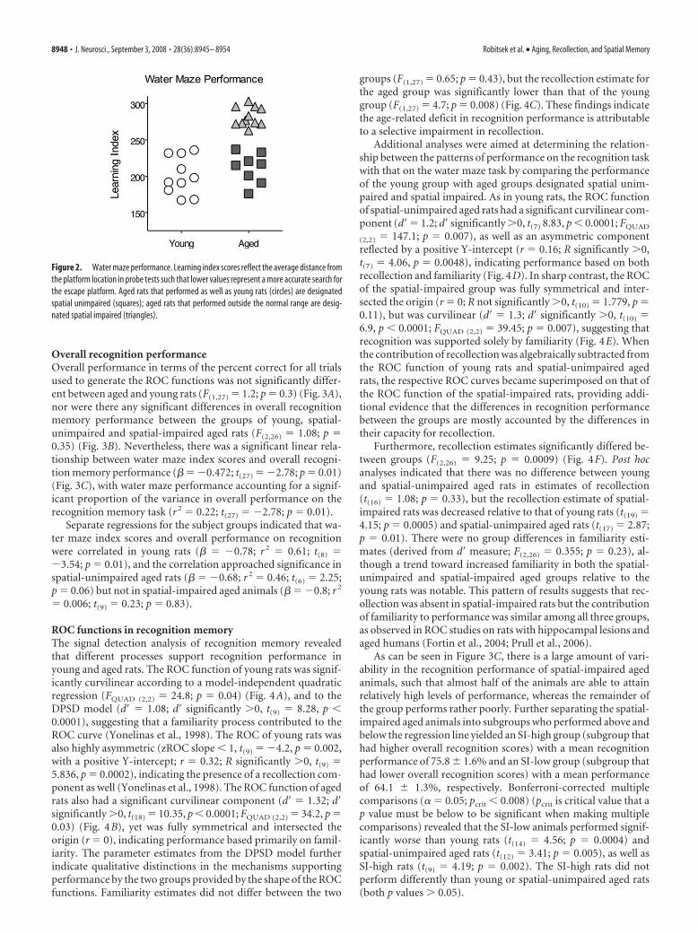
ResultsSpatial learningAs has been shown previously (Gallagher et al., 1993), aged rats(22–24 months) had a significantly higher spatial memory indexcompared with young rats (6 – 8 months), indicative of impairedspatial memory (257 � 11 vs 200 � 8 cm, mean � SEM; t(25) �4.23; p � 0.0003). As shown in Figure 2, some of the aged rats hadscores within the range of young performance, whereas othersperformed outside the normative range (index score, 240) ofthe young subject population (Gallagher et al., 1993). The agedrats that performed outside the range of the young rats weredesignated as “spatial impaired” (n � 11), whereas those whoseperformance was within the range of young rats were designated“spatial unimpaired” (n � 8).
Figure 1. Odor-recognition task. A, During the study phase, rats are presented with a sequence of 10 odors (represented by different patterns) mixed in sand baited with a buried reward. B, Aftera 15 min delay, in the test phase, 10 new odors are singly presented in intermixed order with the 10 old odors. Rats can dig for reward in the new odors and must refrain from digging to obtain rewardelsewhere if the odor is old. C, Five levels of response bias were generated by varying the desirability (Froot Loop vs Cheerio) and amount (0.5 or 1 piece) of reward.
Robitsek et al. • Aging, Recollection, and Spatial Memory J. Neurosci., September 3, 2008 • 28(36):8945– 8954 • 8947
Overall recognition performanceOverall performance in terms of the percent correct for all trialsused to generate the ROC functions was not significantly differ-ent between aged and young rats (F(1,27) � 1.2; p � 0.3) (Fig. 3A),nor were there any significant differences in overall recognitionmemory performance between the groups of young, spatial-unimpaired and spatial-impaired aged rats (F(2,26) � 1.08; p �0.35) (Fig. 3B). Nevertheless, there was a significant linear rela-tionship between water maze index scores and overall recogni-tion memory performance (� � �0.472; t(27) � �2.78; p � 0.01)(Fig. 3C), with water maze performance accounting for a signif-icant proportion of the variance in overall performance on therecognition memory task (r 2 � 0.22; t(27) � �2.78; p � 0.01).
Separate regressions for the subject groups indicated that wa-ter maze index scores and overall performance on recognitionwere correlated in young rats (� � �0.78; r 2 � 0.61; t(8) ��3.54; p � 0.01), and the correlation approached significance inspatial-unimpaired aged rats (� � �0.68; r 2 � 0.46; t(6) � 2.25;p � 0.06) but not in spatial-impaired aged animals (� � �0.8; r 2
� 0.006; t(9) � 0.23; p � 0.83).
ROC functions in recognition memoryThe signal detection analysis of recognition memory revealedthat different processes support recognition performance inyoung and aged rats. The ROC function of young rats was signif-icantly curvilinear according to a model-independent quadraticregression (FQUAD (2,2) � 24.8; p � 0.04) (Fig. 4A), and to theDPSD model (d� � 1.08; d� significantly 0, t(9) � 8.28, p �0.0001), suggesting that a familiarity process contributed to theROC curve (Yonelinas et al., 1998). The ROC of young rats wasalso highly asymmetric (zROC slope � 1, t(9) � �4.2, p � 0.002,with a positive Y-intercept; r � 0.32; R significantly 0, t(9) �5.836, p � 0.0002), indicating the presence of a recollection com-ponent as well (Yonelinas et al., 1998). The ROC function of agedrats also had a significant curvilinear component (d� � 1.32; d�significantly 0, t(18) � 10.35, p � 0.0001; FQUAD (2,2) � 34.2, p �0.03) (Fig. 4B), yet was fully symmetrical and intersected theorigin (r � 0), indicating performance based primarily on famil-iarity. The parameter estimates from the DPSD model furtherindicate qualitative distinctions in the mechanisms supportingperformance by the two groups provided by the shape of the ROCfunctions. Familiarity estimates did not differ between the two
groups (F(1,27) � 0.65; p � 0.43), but the recollection estimate forthe aged group was significantly lower than that of the younggroup (F(1,27) � 4.7; p � 0.008) (Fig. 4C). These findings indicatethe age-related deficit in recognition performance is attributableto a selective impairment in recollection.
Additional analyses were aimed at determining the relation-ship between the patterns of performance on the recognition taskwith that on the water maze task by comparing the performanceof the young group with aged groups designated spatial unim-paired and spatial impaired. As in young rats, the ROC functionof spatial-unimpaired aged rats had a significant curvilinear com-ponent (d� � 1.2; d� significantly 0, t(7) 8.83, p � 0.0001; FQUAD
(2,2) � 147.1; p � 0.007), as well as an asymmetric componentreflected by a positive Y-intercept (r � 0.16; R significantly 0,t(7) � 4.06, p � 0.0048), indicating performance based on bothrecollection and familiarity (Fig. 4D). In sharp contrast, the ROCof the spatial-impaired group was fully symmetrical and inter-sected the origin (r � 0; R not significantly 0, t(10) � 1.779, p �0.11), but was curvilinear (d� � 1.3; d� significantly 0, t(10) �6.9, p � 0.0001; FQUAD (2,2) � 39.45; p � 0.007), suggesting thatrecognition was supported solely by familiarity (Fig. 4E). Whenthe contribution of recollection was algebraically subtracted fromthe ROC function of young rats and spatial-unimpaired agedrats, the respective ROC curves became superimposed on that ofthe ROC function of the spatial-impaired rats, providing addi-tional evidence that the differences in recognition performancebetween the groups are mostly accounted by the differences intheir capacity for recollection.
Furthermore, recollection estimates significantly differed be-tween groups (F(2,26) � 9.25; p � 0.0009) (Fig. 4F). Post hocanalyses indicated that there was no difference between youngand spatial-unimpaired aged rats in estimates of recollection(t(16) � 1.08; p � 0.33), but the recollection estimate of spatial-impaired rats was decreased relative to that of young rats (t(19) �4.15; p � 0.0005) and spatial-unimpaired aged rats (t(17) � 2.87;p � 0.01). There were no group differences in familiarity esti-mates (derived from d� measure; F(2,26) � 0.355; p � 0.23), al-though a trend toward increased familiarity in both the spatial-unimpaired and spatial-impaired aged groups relative to theyoung rats was notable. This pattern of results suggests that rec-ollection was absent in spatial-impaired rats but the contributionof familiarity to performance was similar among all three groups,as observed in ROC studies on rats with hippocampal lesions andaged humans (Fortin et al., 2004; Prull et al., 2006).
As can be seen in Figure 3C, there is a large amount of vari-ability in the recognition performance of spatial-impaired agedanimals, such that almost half of the animals are able to attainrelatively high levels of performance, whereas the remainder ofthe group performs rather poorly. Further separating the spatial-impaired aged animals into subgroups who performed above andbelow the regression line yielded an SI-high group (subgroup thathad higher overall recognition scores) with a mean recognitionperformance of 75.8 � 1.6% and an SI-low group (subgroup thathad lower overall recognition scores) with a mean performanceof 64.1 � 1.3%, respectively. Bonferroni-corrected multiplecomparisons (� � 0.05; pcrit � 0.008) (pcrit is critical value that ap value must be below to be significant when making multiplecomparisons) revealed that the SI-low animals performed signif-icantly worse than young rats (t(14) � 4.56; p � 0.0004) andspatial-unimpaired aged rats (t(12) � 3.41; p � 0.005), as well asSI-high rats (t(9) � 4.19; p � 0.002). The SI-high rats did notperform differently than young or spatial-unimpaired aged rats(both p values 0.05).
Figure 2. Water maze performance. Learning index scores reflect the average distance fromthe platform location in probe tests such that lower values represent a more accurate search forthe escape platform. Aged rats that performed as well as young rats (circles) are designatedspatial unimpaired (squares); aged rats that performed outside the normal range are desig-nated spatial impaired (triangles).
8948 • J. Neurosci., September 3, 2008 • 28(36):8945– 8954 Robitsek et al. • Aging, Recollection, and Spatial Memory

The ROC curves of both SI-high (r � 0; d� � 1.7) (Fig. 4G) andSI-low (r � 0; d� � 0.94) (Fig. 4H) rats were fully symmetricaland intersected the origin, reflecting a complete loss of recollec-tion in both groups. Because of the reduced number of subjects inthose subgroups, the quadratic regression used previously wasnot sensitive enough to show that those ROCs were significantlycurvilinear (SI-high, FQUAD (2,2) � 2.17, p � 0.32; SI-low, FQUAD
(2,2) � 11.5, p � 0.08). However, as for the other ROCs, the groupd� values were significantly 0 (SI-high, t(4) � 7.30, p � 0.002;SI-low, t(5) � 6.82, p � 0.001), confirming the observation thatthe ROCs are indeed curvilinear.
Estimates of recollection were correspondingly low in the SI-high and SI-low rats (t(9) � 0.09; p � 0.93), suggesting a loss ofrecollection in both groups. In contrast, familiarity estimateswere significantly higher in the SI-high than the SI-low group (t(9)
� 3.5; p � 0.007) (Fig. 4 I), and were also higher in the SI-highthan in the young group (t(13) � 3.06; p � 0.009). The meanfamiliarity estimate was lower in the SI-low group than the younggroup, but this difference was not statistically significant (t(12) �1.12; p � 0.28). The combined pattern of results suggests that,although both the SI-high and SI-low groups are severely im-paired in recollection, the SI-high rats successfully compensate byusing a familiarity-based strategy to raise their overall recogni-tion score to a level not different from that of young rats. Theseobservations are consistent with studies showing that some agedhumans rely more heavily on familiarity in making recognitionmemory judgments, and that increased familiarity is associatedwith better performance on recognition memory tasks (Parkinand Walter, 1992; Perfect et al., 1995; Bastin and Van der Linden,2003; Daselaar et al., 2006).
Analyses of the slope of the z-transformed ROC curves as ameasure of ROC asymmetry provide additional evidence indica-tive of differences in the contribution of recollection betweengroups (Fig. 5A). The slopes of the group z-transformed ROCswere significantly different between young and aged rats (youngslope, 0.68; aged slope, 1.11; F(1,6) � 7.17; p � 0.04), supportingthe distinction in the shape of the ROC curves of young and agedanimals, as well as the difference in recollection estimates be-tween the groups. The mean slope of the individual z-transformedROC was significantly less than one for young rats (asymmetricROC; mean slope, 0.68 � 0.08; t(9) � �4.2; p � 0.002), suggesting asignificant contribution of recollection, but was not significantly �1for aged animals (symmetrical ROC; mean slope, 0.88�0.09; t(18) ��1.3; p � 0.23), indicating that recollection did not contribute sig-nificantly to recognition performance.
Comparisons of z-transformed scores among groups sepa-rated based on water maze performance also confirm the ob-
served differences in the amount of recol-lection supporting recognitionperformance (Fig. 5B). The slopes of thegroup z-transformed ROC functions be-tween young, spatial-unimpaired, andspatial-impaired aged rats significantlydiffered (young slope, 0.68; spatial-unimpaired aged group slope, 0.89;spatial-impaired aged group slope, 1.3;F(2,9) � 6.9; p � 0.02). For both young rats(mean slope, 0.68 � 0.08; t(9) � �4.2; p �0.002) and spatial-unimpaired aged rats(mean slope, 0.75 � 0.07; t(7) � �3.6; p �0.001), the mean slope of the individualz-transformed ROCs was significantly �1,indicating asymmetrical ROCs and, thus,
significant contributions of recollection to recognition perfor-mance. In contrast, the mean slope of the individualz-transformed ROC function in spatial-impaired aged rats wasnot significantly different from 1 (symmetrical ROC; mean slope,0.98 � 0.15; t(10) � �0.11; p � 0.92), confirming that recollec-tion did not contribute significantly to performance.
The slopes of the z-transformed ROC functions in the SI-high(slope, 1.3) and SI-low group (slope, 1.05) did not significantly differ(F(1,6) � 0.31; p � 0.6), and the slopes of both the SI-high group(mean slope, 1.3 � 0.47; t(4) � 0.26; p � 0.81) and SI-low group(mean slope, 1.05 � 0.18; t(5) � �1.26; p � 0.26) were not signifi-cantly different from 1, consistent with the selective loss of recollec-tion in aged animals that are impaired on the water maze (Fig. 5C).
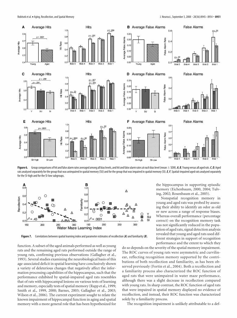
Analyses of hit and false alarm ratesAnalyses of the hit and false alarm rates indicate that the deficit inrecollection in spatial-impaired aged rats is primarily caused byforgetting the sample stimuli. We used separate two-way ANO-VAs to compare the hit and false alarm rates among groups acrossall bias levels and report both the overall average scores and scoresfor each bias level (Fig. 6). Overall, aged rats had lower average hitrates than young rats (F(1,135) � 17.5; p � 0.0001) (Fig. 6A). Posthoc comparisons showed that the hit rates of aged rats were sig-nificantly lower at the middle (t(27) � 3.5; p � 0.001) and lowest(t(27) � 2.84; p � 0.009) bias levels. Aged rats did not differ fromyoung rats in average false alarm rates (F(1,135) � 3.7; p � 0.054),yet there was an interaction between groups and bias level (F(4,135)
� 3.01; p � 0.02) such that aged rats made more false alarms thanyoung rats at the highest bias level (t(27) � 2.4; p � 0.02) andfewer at the lowest bias level (t(27) � �2.24; p � 0.03) (Fig. 6B).
ANOVAs comparing groups separated by performance on thewater maze task also revealed that the young, spatial-unimpaired,and spatial-impaired aged rats differed in hit rates (F(2,130) �9.41; p � 0.0002), and showed that both the spatial unimpaired(t(16) � 3.3; p � 0.004) and spatial-impaired (t(19) � 2.968; p �0.008) aged rats had lower hit rates than young rats (Fig. 6C). Posthoc comparisons revealed that the decrease in hits between youngand spatial-impaired aged animals was significant for the highest(t(19) � 2.32; p � 0.03) and middle (t(19) � �3.04; p � 0.007) biaslevels, and was also significantly lower in spatial-unimpaired agedrats than young animals at the middle bias (t(16) � �3.3; p �0.005). Furthermore, only spatial-impaired aged animals madesignificantly fewer hits than young animals at the highest bias,which reflects the greatest contribution of recollection (Yoneli-nas, 2002; Eichenbaum et al., 2007). The young, spatial-unimpaired, and spatial-impaired aged groups did not differ inoverall false alarm rates (F(2,130) � 2.82; p � 0.06), nor did the
Figure 3. Overall recognition memory performance measured as the percentage correct on all tests (�SEM). A, Overallperformance of young and all aged rats on the recognition memory task. B, Performance of young rats, aged rats that wereunimpaired in spatial memory (SU), and aged rats that were impaired in spatial memory (SI). C, Correlation between overallrecognition memory scores and spatial memory performance.
Robitsek et al. • Aging, Recollection, and Spatial Memory J. Neurosci., September 3, 2008 • 28(36):8945– 8954 • 8949

false alarm rates differ across bias levels(F(4,130) � 1.55; p � 0.14) (Fig. 6D).
The same analyses applied to the sub-groups of spatial-impaired aged rats re-vealed that the better performance of SI-high rats than SI-low rats could beattributed solely to higher hit rates. SI-high rats had a higher average hit rate thanSI-low rats (F(1,45) � 40.4; p � 0.0001),and the post hoc analysis showed that SI-high rats had higher hit rates on the highest(t(9) � 6.7, p � 0.0001), second highest(t(9) � 2.7, p � 0.02), and second lowest (t(9) � 3.0, p � 0.02) biaslevels (Fig. 6E). In contrast, the groups did not differ in overallfalse alarm rate (F(1,45) � 3.6; p � 0.06), nor did false alarm ratesdiffer at any of the bias levels (F(1,45) � 2.2; p � 0.08) (Fig. 6F).
Overall correlation between water maze performance andrecognition memoryIn addition to the above group-wise comparisons, regressionanalyses were performed to compare the degree to which perfor-mance on the water maze could be related to the estimates ofrecollection and familiarity as measured by the DPSD model us-ing the data from all groups combined. Performance on the water
maze was strongly related to the estimates of recollection (� ��0.65; r 2 � 0.43; t(27) � �4.45; p � 0.0001) (Fig. 7A), consistentwith substantial evidence indicating that both the water maze andthe recollection component of recognition memory are depen-dent on the hippocampus. In contrast, there was no relationshipbetween water maze performance and estimates of familiarity(� � 0.02; r 2 � 0.0001; t(27) � 0.08; p � 0.94) (Fig. 7B), suggest-ing distinct bases of spatial memory and familiarity.
DiscussionThe spatial learning ability of young and aged rats was assessedusing the Morris water maze, a well-known assay of hippocampal
Figure 4. ROC functions and recollection and familiarity indices for odor recognition. Each plot shows the group mean hit and false alarm scores at each bias level (circles). Parameter estimates(�SEM) of the contributions of recollection and familiarity calculated for each ROC function are shown. A, B, Young versus all aged rats. C, D, Aged rats analyzed separately for the group that wasunimpaired in spatial memory (SU) and for the group that was impaired in spatial memory (SI). E, F, Spatial-impaired aged rats analyzed separately for the SI-high and SI-low subgroups.
Figure 5. Best-fit linear regressions of z-transformed ROC functions. A, Young versus all aged rats. B, Aged rats analyzedseparately for the group that was unimpaired in spatial memory (SU) and for the group that was impaired in spatial memory (SI).C, Spatial-impaired aged rats analyzed separately for the SI-high and SI-low subgroups.
8950 • J. Neurosci., September 3, 2008 • 28(36):8945– 8954 Robitsek et al. • Aging, Recollection, and Spatial Memory
function. A subset of the aged animals performed as well as youngrats and the remaining aged rats performed outside the range ofyoung rats, confirming previous observations (Gallagher et al.,1993). Several studies examining the neurobiological basis of thisage-associated deficit in spatial learning have conclusively showna variety of deleterious changes that negatively affect the infor-mation processing capabilities of the hippocampus, such that theperformance exhibited by spatial-impaired aged rats resemblesthat of rats with hippocampal lesions on various tests of learningand memory, especially tests of spatial memory (Rapp et al., 1999;Smith et al., 1999, 2000; Barnes, 2003; Gallagher et al., 2003;Wilson et al., 2006). The current experiment sought to relate theknown impairment of hippocampal function in aging and spatialmemory with a more general role that has been hypothesized for
the hippocampus in supporting episodicmemory (Eichenbaum, 2000, 2004; Tulv-ing, 2002; Rosenbaum et al., 2005).
Nonspatial recognition memory inyoung and aged rats was probed by assess-ing their ability to identify an odor as oldor new across a range of response biases.Whereas overall performance (percentagecorrect) on the recognition memory taskwas not significantly reduced in the popu-lation of aged rats, signal detection analysisrevealed that young and aged rats used dif-ferent strategies in support of recognitionperformance and the extent to which they
do so depends on the severity of the spatial memory impairment.The ROC curves of young rats were asymmetric and curvilin-ear, reflecting recognition memory supported by the contri-butions of both recollection and familiarity, as has been ob-served previously (Fortin et al., 2004). Both a recollection anda familiarity process also characterized the ROC function ofaged rats that were unimpaired in water maze performance,although there was a slight decrease in recollection comparedwith young rats. In sharp contrast, the ROC function of aged ratsthat were impaired in spatial memory displayed no evidence ofrecollection, and instead, their ROC function was characterizedsolely by a familiarity process.
The recognition impairment is unlikely attributable to a def-
Figure 6. Group comparisons of hit and false alarm rates averaged among all bias levels, and hit and false alarm rates at each bias level (mean � SEM). A, B, Young versus all aged rats. C, D, Agedrats analyzed separately for the group that was unimpaired in spatial memory (SU) and for the group that was impaired in spatial memory (SI). E, F, Spatial-impaired aged rats analyzed separatelyfor the SI-high and for the SI-low subgroups.
Figure 7. Correlations between spatial learning index and parameter estimates of recollection (A) and familiarity (B).
Robitsek et al. • Aging, Recollection, and Spatial Memory J. Neurosci., September 3, 2008 • 28(36):8945– 8954 • 8951

icit in detection or discrimination of odors, because perceptualimpairments should be evident across all biases and for bothrecollection and familiarity, which was not observed. Further-more, Schoenbaum et al. (2002) evaluated olfactory discrimina-tion thresholds in rats of the same strain and from the samesource and found no age-related changes.
This pattern of performance is similar to that of rats withlesions of the hippocampus (Fortin et al., 2004). These findingsconfirm previous observations of deficits in nonspatial non-matching to sample tasks in rats (Zyzak et al., 1995) and monkeys(Rapp and Amaral, 1989, 1991), as well as other nonspatial learn-ing and memory tests (Winocur, 1992; Gallagher and Rapp, 1997;Scali et al., 1997; Countryman and Gold, 2007; Pieta Dias et al.,2007). These recognition deficits are typically modest in severity,compared with the near total absence of recollection observed inthe present study. Thus, the present findings serve to clarify thatthe previous observations of a modest deficit may reflect the com-bination of a severe impairment in one contribution to recogni-tion (i.e., recollection) and relative sparing of another (i.e., famil-iarity). Furthermore, the present findings indicate the variabilityin the severity of recognition deficits is attributable to variation inthe severity of the recollection deficit and the amount of compen-satory increase in the contribution of familiarity among individ-uals. These findings also parallel data from recognition memorystudies in the human cognitive aging literature, which show thatnormal aging results in a selective loss of recollection and relativesparing of familiarity, and the severity of the deficit is variableamong individuals (Light et al., 2000; Davidson and Glisky, 2002;Bastin and Van der Linden, 2003; Howard et al., 2006).
The current data also suggest an association between hip-pocampal function that supports recognition memory and spa-tial memory. This association was evident in three of our analy-ses. First, performance on the water maze, a task sensitive tohippocampal function, was correlated with recognition memoryperformance for young and spatial-unimpaired aged animals,suggesting a common underlying memory processing function.Second, performance on the recollection component of odor rec-ognition was more severely affected in spatial-impaired thanspatial-unimpaired aged rats; the contribution of familiarity didnot differ between these groups. Third, there was a strong overallcorrelation between water maze performance and recollectionbut not familiarity. Because the hippocampus is also stronglyimplicated in the recollection but not the familiarity componentof recognition (Fortin et al., 2004), the observed relationshipbetween odor recollection and water maze performance in agedrats also suggests a common underlying contribution of the hip-pocampus to normal function in both memory domains. Onepossibility for this common underlying function is the ability torecollect previous experiences, such that performance on probetests in the water maze may rely on recollection of previous suc-cessful routes through the maze, much as recollection contributesto performance in the odor-recognition task.
A neurobiological account of the deficit in recollectionA mechanistic account of the related deficits in recollection andwater maze performance in aged rats is suggested by findingsfrom recording studies in aged rats exploring novel and familiarenvironments (Ikonen et al., 2002; Wilson et al., 2003, 2004,2005). When exploring a novel environment after extensive ex-posure to another (familiar) environment, the place cells ofspatial-impaired aged rats fail to form new representations of thenovel environment and instead often retrieve previously storedrepresentations of the familiar environment. These observations
have been characterized as a failure in pattern separation of thecontextual stimuli from the familiar and novel environments anda consequent tendency toward pattern completion of the repre-sentation of the familiar spatial context (Wilson et al., 2006).
In our odor-recognition protocol, the relevant context asso-ciated with each odor includes the preceding and following odorsin the list each day, and the ability to remember the order of odorsin a list depends on the hippocampus (Fortin et al., 2002). Fur-thermore, a recent study has shown that the hippocampus en-codes a list of odor stimuli in terms of a gradually changing tem-poral context signal (Manns et al., 2007). It is likely thatrecollection of odors involves remembering these temporal con-textual features to distinguish odors on the current list from pre-vious appearances of the same odors on earlier testing sessions.The model proposed by Wilson et al. (2006), which focused onage-associated deficits in pattern separation of spatial contexts,can be extended to understand how a deficit in pattern separationmight also underlie the observed impairment in odor recollec-tion. Thus, a general age-associated reduction in pattern separa-tion operations performed by the hippocampus could compro-mise the animal’s ability to distinguish the temporal context ofodors in the current list from that of previous appearances of thesame odors in earlier lists. The result would be a catastrophicpattern of interference for the hippocampus and a consequentinability to recollect the experience of being presented with aspecific odor in the current study list.
These age-associated abnormalities in pattern separation andcompletion may result directly from deleterious neurobiologicalchanges in the hippocampus (Wilson et al., 2006). In particular,the impairment may be a consequence of age-associated attenu-ated modulation of hippocampal processing by acetylcholine(Chouinard et al., 1995; Gill and Gallagher, 1998; Sugaya et al.,1998; Nicolle et al., 1999). Models of information processing inthe hippocampus suggest that acetylcholine may be important inregulating the dynamics of encoding and retrieval, such that highlevels of acetylcholine bias the dynamics of the network towardencoding, whereas low levels of acetylcholine favor retrieval dy-namics (Hasselmo and Wyble, 1997; Hasselmo, 2006; Giocomoand Hasselmo, 2007). Acetylcholine has also been shown to playa role in modulating interference between memories. Low levelsof acetylcholine, as seen after administration of the cholinergicantagonist scopolamine, result in increased levels of interferencebetween stored memories (De Rosa and Hasselmo, 2000),whereas higher levels of acetylcholine have been suggested todecrease levels of interference between stored activity patterns(Linster et al., 2003). Indeed, reducing cholinergic modulationexperimentally reproduces in young rats the age-associated ten-dency of place cells to pattern complete familiar spatial represen-tations (Ikonen et al., 2002). Also, in a study of human recogni-tion memory, injections of scopolamine selectively modulatesrecollection performance but does not affect familiarity processes(Sherman et al., 2003). Thus, when injected with scopolamine,subjects show a decrease in recollection-based performance, butthis performance is still qualitatively different from familiarityand normal recollection is observed after the drug has worn off.Additionally, reduced synaptic input from the entorhinal cortexthat occurs selectively in aged impaired rats as defined by watermaze performance, may also result in impaired pattern separa-tion in the dentate gyrus and CA3 regions, which both contributeto pattern separation processes in the hippocampus (Wilson etal., 2006; Leutgeb et al., 2007).
8952 • J. Neurosci., September 3, 2008 • 28(36):8945– 8954 Robitsek et al. • Aging, Recollection, and Spatial Memory

ReferencesAlbert MS (1997) The ageing brain: normal and abnormal memory. Philos
Trans R Soc Lond B Biol Sci 352:1703–1709.Barnes CA (2003) Long-term potentiation and the ageing brain. Philos
Trans R Soc Lond B Biol Sci 358:765–772.Bastin C, Van der Linden M (2003) The contribution of recollection and
familiarity to recognition memory: a study of the effects of test format andaging. Neuropsychology 17:14 –24.
Buckner RL (2004) Memory and executive function in aging and AD: mul-tiple factors that cause decline and reserve factors that compensate. Neu-ron 44:195–208.
Chouinard ML, Gallagher M, Yasuda RP, Wolfe BB, McKinney M (1995)Hippocampal muscarinic receptor function in spatial learning-impairedaged rats. Neurobiol Aging 16:955–963.
Countryman RA, Gold PE (2007) Rapid forgetting of social transmission offood preferences in aged rats: relationship to hippocampal CREB activa-tion. Learn Mem 14:350 –358.
Craik FI, Byrd M (1982) Aging and cognitive deficits: the role of attentionalresources. In: Aging and cognitive processes (Craik FI, Trehub S, eds), pp51–110. New York: Plenum.
Crook T, Bartus RT, Ferris SH, Whitehouse P, Cohen GD, Gershon S (1986)Age-associated memory impairment: proposed diagnostic criteria andmeasures of clinical change. Report of a National Institute of MentalHealth Work Group. Dev Neuropsychol 2:261–276.
Daselaar SM, Fleck MS, Dobbins IG, Madden DJ, Cabeza R (2006) Effects ofhealthy aging on hippocampal and rhinal memory functions: an event-related fMRI study. Cereb Cortex 16:1771–1782.
Davidson PS, Glisky EL (2002) Neuropsychological correlates of recollec-tion and familiarity in normal aging. Cogn Affect Behav Neurosci2:174 –186.
De Rosa E, Hasselmo ME (2000) Muscarinic cholinergic neuromodulationreduces proactive interference between stored odor memories during as-sociative learning in rats. Behav Neurosci 114:32– 41.
Duverne S, Habibi A, Rugg MD (2007) Regional specificity of age effects onthe neural correlates of episodic retrieval. Neurobiol Aging, in press.
Eichenbaum H (2000) A cortical-hippocampal system for declarative mem-ory. Nat Rev Neurosci 1:41–50.
Eichenbaum H (2004) Hippocampus: cognitive processes and neural repre-sentations that underlie declarative memory. Neuron 44:109 –120.
Eichenbaum H, Yonelinas AP, Ranganath C (2007) The medial temporallobe and recognition memory. Annu Rev Neurosci 30:123–152.
Fortin NJ, Agster KL, Eichenbaum HB (2002) Critical role of the hippocam-pus in memory for sequences of events. Nat Neurosci 5:458 – 462.
Fortin NJ, Wright SP, Eichenbaum H (2004) Recollection-like memory re-trieval in rats is dependent on the hippocampus. Nature 431:188 –191.
Gallagher M, Rapp PR (1997) The use of animal models to study the effectsof aging on cognition. Annu Rev Psychol 48:339 –370.
Gallagher M, Burwell R, Burchinal M (1993) Severity of spatial learningimpairment in aging: development of a learning index for performance inthe Morris water maze. Behav Neurosci 107:618 – 626.
Gallagher M, Bizon JL, Hoyt EC, Helm KA, Lund PK (2003) Effects of agingon the hippocampal formation in a naturally occurring animal model ofmild cognitive impairment. Exp Gerontol 38:71–77.
Gill TM, Gallagher M (1998) Evaluation of muscarinic M2 receptor sites inbasal forebrain and brainstem cholinergic systems of behaviorally char-acterized young and aged Long-Evans rats. Neurobiol Aging 19:217–225.
Giocomo LM, Hasselmo ME (2007) Neuromodulation by glutamate andacetylcholine can change circuit dynamics by regulating the relative influ-ence of afferent input and excitatory feedback. Mol Neurobiol36:184 –200.
Hasselmo ME (2006) The role of acetylcholine in learning and memory.Curr Opin Neurobiol 16:710 –715.
Hasselmo ME, Wyble BP (1997) Free recall and recognition in a networkmodel of the hippocampus: simulating effects of scopolamine on humanmemory function. Behav Brain Res 89:1–34.
Hedden T, Gabrieli JD (2004) Insights into the ageing mind: a view fromcognitive neuroscience. Nat Rev Neurosci 5:87–96.
Howard MW, Bessette-Symons B, Zhang Y, Hoyer WJ (2006) Aging selec-tively impairs recollection in recognition memory for pictures: evidencefrom modeling and receiver operating characteristic curves. Psychol Ag-ing 21:96 –106.
Ikonen S, McMahan R, Gallagher M, Eichenbaum H, Tanila H (2002) Cho-
linergic system regulation of spatial representation by the hippocampus.Hippocampus 12:386 –397.
Jennings JM, Jacoby LL (1997) An opposition procedure for detecting age-related deficits in recollection: telling effects of repetition. Psychol Aging12:352–361.
Leutgeb JK, Leutgeb S, Moser MB, Moser EI (2007) Pattern separation inthe dentate gyrus and CA3 of the hippocampus. Science 315:961–966.
Light LL, Prull MW, Kennison RF (2000) Divided attention, aging, andpriming in exemplar generation and category verification. Mem Cognit28:856 – 872.
Linster C, Maloney M, Patil M, Hasselmo ME (2003) Enhanced cholinergicsuppression of previously strengthened synapses enables the formation ofself-organized representations in olfactory cortex. Neurobiol Learn Mem80:302–314.
Macmillan NA, Creelman CD (2005) Detection theory: a user’s guide, Ed 2.Mahwah, NJ: Lawrence Erlbaum.
Manns JR, Howard MW, Eichenbaum H (2007) Gradual changes in hip-pocampal activity support remembering the order of events. Neuron56:530 –540.
Nicolle MM, Colombo PJ, Gallagher M, McKinney M (1999) Metabotropicglutamate receptor-mediated hippocampal phosphoinositide turnover isblunted in spatial learning-impaired aged rats. J Neurosci 19:9604 –9610.
Parkin AJ, Walter BM (1992) Recollective experience, normal aging, andfrontal dysfunction. Psychol Aging 7:290 –298.
Perfect TJ, Williams RB, Anderton-Brown C (1995) Age differences in re-ported recollective experience are due to encoding effects, not responsebias. Memory 3:169 –186.
Pieta Dias C, Martins de Lima MN, Presti-Torres J, Dornelles A, Garcia VA,Siciliani Scalco F, Rewsaat Guimaraes M, Constantino L, Budni P, Dal-Pizzol F, Schroder N (2007) Memantine reduces oxidative damage andenhances long-term recognition memory in aged rats. Neuroscience146:1719 –1725.
Prull MW, Dawes LL, Martin AM 3rd, Rosenberg HF, Light LL (2006) Rec-ollection and familiarity in recognition memory: adult age differences andneuropsychological test correlates. Psychol Aging 21:107–118.
Quamme JR, Yonelinas AP, Widaman KF, Kroll NE, Sauve MJ (2004) Recalland recognition in mild hypoxia: using covariance structural modeling totest competing theories of explicit memory. Neuropsychologia42:672– 691.
Rapp PR, Amaral DG (1989) Evidence for task-dependent memory dys-function in the aged monkey. J Neurosci 9:3568 –3576.
Rapp PR, Amaral DG (1991) Recognition memory deficits in a subpopula-tion of aged monkeys resemble the effects of medial temporal lobe dam-age. Neurobiol Aging 12:481– 486.
Rapp PR, Rosenberg RA, Gallagher M (1987) An evaluation of spatial infor-mation processing in aged rats. Behav Neurosci 101:3–12.
Rapp PR, Stack EC, Gallagher M (1999) Morphometric studies of the agedhippocampus: I. Volumetric analysis in behaviorally characterized rats.J Comp Neurol 403:459 – 470.
Robitsek R, Guttentag L, Fortin N, Eichenbaum H (2006) Recognitionmemory in rodent normal aging. Soc Neurosci Abstr 32:66.18.
Rosenbaum RS, Kohler S, Schacter DL, Moscovitch M, Westmacott R, BlackSE, Gao F, Tulving E (2005) The case of K.C.: contributions of amemory-impaired person to memory theory. Neuropsychologia43:989 –1021.
Rosenzweig ES, Barnes CA (2003) Impact of aging on hippocampal func-tion: plasticity, network dynamics, and cognition. Prog Neurobiol69:143–179.
Scali C, Giovannini MG, Bartolini L, Prosperi C, Hinz V, Schmidt B, Pepeu G(1997) Effect of metrifonate on extracellular brain acetylcholine and ob-ject recognition in aged rats. Eur J Pharmacol 325:173–180.
Schoenbaum G, Nugent S, Saddoris MP, Gallagher M (2002) Teaching oldrats new tricks: age-related impairments in olfactory reversal learning.Neurobiol Aging 23:555–564.
Sherman SJ, Atri A, Hasselmo ME, Stern CE, Howard MW (2003) Scopol-amine impairs human recognition memory: data and modeling. BehavNeurosci 117:526 –539.
Smith TD, Calhoun ME, Rapp PR (1999) Circuit and morphological speci-ficity of synaptic change in the aged hippocampal formation. NeurobiolAging 20:357–358, 1999; discussion 359 –360.
Smith TD, Adams MM, Gallagher M, Morrison JH, Rapp PR (2000)Circuit-specific alterations in hippocampal synaptophysin immunoreac-
Robitsek et al. • Aging, Recollection, and Spatial Memory J. Neurosci., September 3, 2008 • 28(36):8945– 8954 • 8953

tivity predict spatial learning impairment in aged rats. J Neurosci20:6587– 6593.
Sugaya K, Greene R, Personett D, Robbins M, Kent C, Bryan D, Skiba E,Gallagher M, McKinney M (1998) Septo-hippocampal cholinergic andneurotrophin markers in age-induced cognitive decline. Neurobiol Aging19:351–361.
Toth JP, Parks CM (2006) Effects of age on estimated familiarity in theprocess dissociation procedure: the role of noncriterial recollection. MemCognit 34:527–537.
Tulving E (2002) Episodic memory: from mind to brain. Annu Rev Psychol53:1–25.
Wilson IA, Ikonen S, McMahan RW, Gallagher M, Eichenbaum H, Tanila H(2003) Place cell rigidity correlates with impaired spatial learning in agedrats. Neurobiol Aging 24:297–305.
Wilson IA, Ikonen S, Gureviciene I, McMahan RW, Gallagher M, Eichen-baum H, Tanila H (2004) Cognitive aging and the hippocampus: howold rats represent new environments. J Neurosci 24:3870 –3878.
Wilson IA, Ikonen S, Gallagher M, Eichenbaum H, Tanila H (2005) Age-associated alterations of hippocampal place cells are subregion specific.J Neurosci 25:6877– 6886.
Wilson IA, Gallagher M, Eichenbaum H, Tanila H (2006) Neurocognitiveaging: prior memories hinder new hippocampal encoding. Trends Neu-rosci 29:662– 670.
Winocur G (1992) A comparison of normal old rats and young adult ratswith lesions to the hippocampus or prefrontal cortex on a test ofmatching-to-sample. Neuropsychologia 30:769 –781.
Wixted JT (2007) Dual-process theory and signal-detection theory of rec-ognition memory. Psychological Rev 114:152–176.
Yonelinas AP (2002) The nature of recollection and familiarity: a review of30 years of research. J Mem Lang 48:441–517.
Yonelinas AP, Parks CM (2007) Receiver operating characteristics (ROCs)in recognition memory: a review. Psychol Bull 133:800 – 832.
Yonelinas AP, Kroll NE, Dobbins I, Lazzara M, Knight RT (1998) Recollec-tion and familiarity deficits in amnesia: convergence of remember-know,process dissociation, and receiver operating characteristic data. Neuro-psychology 12:323–339.
Zyzak DR, Otto T, Eichenbaum H, Gallagher M (1995) Cognitive declineassociated with normal aging in rats: a neuropsychological approach.Learn Mem 2:1–16.
8954 • J. Neurosci., September 3, 2008 • 28(36):8945– 8954 Robitsek et al. • Aging, Recollection, and Spatial Memory