Intentional control and biomechanical exploitation in preparatory handwriting
Upload
independentCategory
view
4download
0
www.elsevier.com/locate/ynimg
NeuroImage 39 (2008) 1314–1323Functional coupling between anterior prefrontal cortex (BA10) andhand muscle contraction during intentional and imitative motor acts
Claudio Babiloni,a,b,c,⁎ Fabrizio Vecchio,a,b Martin Bares,d Milan Brazdil,d Igor Nestrasil,d
Fabrizio Eusebi,a,e,f Paolo Maria Rossini,b,g,h and Ivan Rektord
aDipartimento di Fisiologia Umana e Farmacologia, Università La Sapienza, Rome, ItalybA.Fa.R Dip. Neuroscienze-Ospedale FBF, Isola Tiberina, Rome, ItalycCasa di Cura S. Raffaele Cassino and S. Raffaele Pisana, ItalydFirst Department of Neurology, Masaryk University, St. Anne’s Hospital, Pekarska 53, 65691 Brno, Czech RepubliceIstituto di Medicina e Scienza dello Sport, CONI Servizi, Rome, ItalyfIRCCS Neuromed, Pozzilli (IS), ItalygIRCCS Centro S. Giovanni di Dio-FBF, Brescia, ItalyhClinica Neurologica, Università Campus Biomedico, Rome, Italy
Received 3 January 2007; revised 13 September 2007; accepted 25 September 2007Available online 4 October 2007
The present study tested the hypothesis that functional cortico-muscularcoupling is a putative physiological mechanism by which Brodmann area10 (BA10) of anterior prefrontal cortex controls subjects’ behavior.Intracerebral stereo electroencephalographic (SEEG) data were re-corded from BA10 of epilepsy subjects in the course of pre-surgicalmonitoring. During the SEEG recordings, these subjects were engaged inthree conditions: the execution of intentional hand muscle contractions astriggered by auditory stimuli (“EXE”); the execution of the same musclecontractions as an imitation of a person seated in front of the subject(“IMI”); and the mere observation of the hand muscle contractionsperformed by that person (“OBS”). SEEG frequency bands of interestwere theta (4–7 Hz), alpha (8–12 Hz), beta 1 (13–21 Hz), beta 2 (22–30 Hz), and gamma (31–45 Hz). Results showed that functional cortico-muscular coupling at gamma band was higher in amplitude during theintentional muscle contraction (“EXE”) than the other conditions (“IMI”and “OBS”). Instead, cortico-muscular coupling at theta band washigher in amplitude during the imitative muscle contraction (“IMI”) thanthe other conditions (“EXE” and “OBS”). In parallel, there was anincrease of SEEG gamma band power during the intentional musclecontraction and an increase of SEEG theta band power during itsimitation. The present results suggest that anterior prefrontal cortex(BA10) might control subjects’ behavior by means of functional cortico-
⁎ Corresponding author. Dipartimento di Fisiologia Umana e Farm-acologia, Università degli Studi di Roma La Sapienza, P.le Aldo Moro 5,00185 Rome, Italy. Fax: +39 06 49910917.
E-mail address: [email protected] (C. Babiloni).Available online on ScienceDirect (www.sciencedirect.com).
1053-8119/$ - see front matter © 2007 Elsevier Inc. All rights reserved.doi:10.1016/j.neuroimage.2007.09.043
muscular coupling at selective frequency bands (theta and wide gammarhythms).© 2007 Elsevier Inc. All rights reserved.
Keywords: Stereo electroencephalography (SEEG); Electromyography(EMG); Brain rhythms; Spectral coherence; Event-related desynchroniza-tion/synchronization (ERD/ERS)
Introduction
Brodmann area 10 (BA10) of anterior prefrontal cortex is involvedin integrative activities that include planning, episodic retrieval, andother tasks requiring interaction (i.e., inhibition/facilitation) amongseveral competing sub-goals (Fletcher and Henson, 2001; Ramnaniand Owen, 2004). Such an integrative role is compatible with cyto-architecture of BA10, characterized by cells having very high spinedensity and by low density of cell bodies (Ramnani and Owen, 2004).
Several lines of evidence have shown that BA10 is active duringswitching paradigms in which rules (i.e., set) at the bases of subjects’behavior change in line with external feedback. In these paradigms,previously acquired set needs to be inhibited not only at the first trialafter the change of the rule, but also after a minute or so to avoidperseverative errors (Milner, 1963; Allport et al., 1994;Meiran et al.,2000). In this framework, BA10 would play a crucial role in theneural mechanisms responsible for the inhibition of the prolongedinterference that persists long after the first inhibition trial (Konishiet al., 2005). In parallel, BA10would act to reconfigure andmaintaina set over the task periods (Braver et al., 2003; Sakai andPassingham, 2003) and during task switching under predictabletask order and timing (Dreher et al., 2002).

1315C. Babiloni et al. / NeuroImage 39 (2008) 1314–1323
At the present stage of research, physiological mechanisms bywhich anterior prefrontal cortex exerts its inhibitory/excitatory controlon subjects’ behavior are poorly known. A reasonable hypothesis isthat temporal synchronization of cortical and muscle activity (i.e., theso-called functional cortico-muscular coupling) is implied in the wayBA10 contributes to the selection and implementation of the action tobe performed in line with the prevailing sub-goal.
Temporal synchronization of neural and muscular activity wouldhave deep implication on information processing and motor output.Recent evidence suggests that postsynaptic neuron primarilyresponds to precisely synchronous input volley coming from thepresynaptic neuron (Azouz and Gray, 2000, 2003). Furthermore,neuronal groups increase their impact on target groups throughprecise oscillatory synchronization of the input volley (Fries et al.,2001, 2002). Finally, it has been shown that functional cortico-muscular coupling affects the firing of single corticospinal neuronsfor distal and proximal muscles. This firing constitutes aconnection in a larger web of oscillatory interactions, includingseveral other motor areas in the cortex, thalamus, and cerebellum(Marsden et al., 2001; Salenius and Hari, 2003; Pollok et al.,2004; Timmermann et al., 2004; Schnitzler and Gross, 2005).
From a methodological point of view, previous investigationshave shown that spectral coherence of electroencephalographic(EEG)/magnetoencephalographic overlying primary motor cortexand electromyographic (EMG) data reflects functional cortico-muscular coupling between cortex and motor units firing in thetarget muscle (Salenius et al., 1997, 2002; Mima and Hallett, 1999;Mima et al., 2000, 2001; Salenius and Hari, 2003). Indeed, EEG-EMG coherence would represent a rough index of informationtransfer capability within the operative connection (Babiloni et al.,2004a). That notion is based on the fact that spinal motoneuron andthe corresponding muscle fibers form a motor unit with one-to-onecorrespondence of their action potentials, so that EEG-EMGcoherence roughly measures the temporal synchronization betweencortical neurons having effects on descending motor systems andengaged spinal motoneurons whose activity is to some extentreflected in EMG oscillations (Halliday et al., 1995).
The present study tested the hypothesis that cortico-muscularcoupling might represent a putative physiological mechanism bywhich BA10 of anterior prefrontal cortex controls subjects’ behaviorduring cognitive demands. Intracerebral stereo electroencephalo-graphic (SEEG) data were recorded from BA10 of drug-resistantepilepsy subjects in the course of pre-surgical monitoring. Duringthe SEEG recordings, these subjects were engaged in threeconditions: the execution of intentional hand muscle contractionsas triggered by auditory stimuli (“EXE”); the execution of the samemuscle contractions as an imitation of a person seated in front of thesubject (“IMI”); and the mere observation of the hand musclecontractions performed by that person (“OBS”). It was expected that
Table 1Details on the studied epileptic patients
Patientno.
Sex Age(years)
Age of onset(years)
Epilepti
1 M 27 6 Left par2 M 35 21 Left tem3 M 32 10 Left fro4 M 48 16 Left tem
M: male, F: female, all right-handed. Age (years): age at the time of SEEG exploEngel IA—excellent outcome, no seizures. Engel III—more then 50% reduction o
the spectral SEEG-EMG coherence between BA10 and muscularactivity reflects the control of behavior in the different (“EXE”,“IMI”) conditions and goals. Of course, physiological data inepileptic patients should be evaluated with extreme attention due tothe intrinsic effects of the pathology on cortical neural synchroniza-tion. However, under certain procedural constraints (see thefollowing sections), the heuristic value of intracranial SEEGrecordings from these patients is invaluable for its incomparablehigh spatial and temporal resolution.
Methods
Subjects
Four adult epilepsy right-handed (Edinburgh inventory), drug-resistant subjects (3 males, mean age 34 years, age range 26–39 years)participated to the SEEG experiments in the framework of their pre-surgical routine. The study protocol was approved by the BrnoInstitute’s Ethics Committee. Informed consentwas obtained fromeachsubject prior to the test. He/She could definitively stop the experimentat any moment. Patients were candidates for epilepsy surgery, sincetheywere unresponsive to therapy, andwere recommended by a specialcommission for stereotactic exploration. SEEG electrodes wereimplanted into the left frontal lobe, parietal and opercular regions tolocalize the origin or spread of the ictal epileptic activity. The rationaleof implanting depth electrodes in BA10 area could be based on clinicalpicture of epileptic seizures (short partial complex EEG seizures withsome frontal symptoms) or radiological evidence.
The subjects underwent a careful clinical and neuropsychologicalevaluation, according to the protocol used at Brno First Department ofNeurology prior to epilepsy surgery. Aside from the epilepsy, theclinical neurological status was normal in all patients. The results ofIQWAIS-R (Wechsler Adult Intelligence Scale—Revised), WMS III(Wechsler Memory Scale III), R–O Figure (Rey–Osterrieth ComplexFigure), Stroop test, verbal fluency test, FDT (Figure Drawing Test),BT (Baum Test), test of symbolic functions, and subscales of Lurijaneuropsychological examination were in the normal range, i.e.,largely within one standard deviation of the distribution of healthypopulation (including 68% of the people). In two patients, there wasborderline performance at executive tasks. None of the patientsexpressed praxic disabilities. All subjects were able to fully under-stand and easily perform the experimental tasks.
Magnetic resonance imaging (MRI) showed no morphologiclesions in one patient. The other patients presented left hippocampalsclerosis (one patient), mild diffuse cortical atrophy (one patient),and increased signals in left hippocampus (one patient). In otherwords, the recruited subjects showed no tissue alteration from BA10of the two hemispheres.
For further details on the subjects, see Table 1.
c focus Clinical/MRI data Surgeryoutcome
ietal Normal/Normal Engel IAporal Normal/Left hippocampal sclerosis Engel IAntal Normal/Mild diffuse cortical atrophy Engel IVporal Normal/Increased signal in the left
hippocampusEngel III
ration. Age of onset (years): age of onset of epileptic seizures.f epileptic seizures, Engel IV—no significant improvement.

1316 C. Babiloni et al. / NeuroImage 39 (2008) 1314–1323
Experimental design
During the experiment, the subject from whom SEEG wasrecorded (“observer”) and the subject in front of him/her(“observed” person) seated in comfortable armchairs. The handsof the “observer” and the “observed” person were disposed closeeach other, so that the “observer” could look at both own and“observed” person’s hands while fixing a central target. This ensuredthat the “observer” maintained nearly the same visual field duringown moving hand (“EXE”, “IMI”) and during moving hand of the“observed” person (“OBS”). It should be remarked that both“observer” and “observed” subjects were continuously encouragedby an experimenter to focus attention on the central target and toavoid gaze shift elsewhere.
As mentioned above, the experimental design included threeconditions intermingled in a pseudorandom order across subjects(Fig. 1). In the “EXE” condition, the “observer” briskly performed asustained extension of right fingers. Specifically, subjects’ fingerextension started as a reaction to a first auditory stimulus (1000 Hz,100-ms duration; go stimulus) and ended 6 s later immediately aftera second auditory stimulus (same parameters of the go stimulus; stopstimulus). The interval between stop and subsequent go stimulirandomly varied between 4 and 6 s. In the “OBS” condition, the“observer” passively saw the “observed” person who performed thesame action, namely the finger extension with sustained musclecontraction (stimuli and timing as in the “EXE” condition). In the“IMI” condition, the “observer” executed the above action as animitative behavior as soon as he/she saw the “observed” person whoperformed the same action, namely the sustained finger extensionwith the opposite (left) hand (stimuli and timing as in the “EXE” and“OBS” conditions). The rationale for using this type of experimentaldesign was to investigate movements realized in the right hemifieldof the patient only, namely in the contralateral side of the implantedhemisphere. Before the SEEG recordings, 1–2 training blockslasting 5 min established an approximately stable level of the motorperformance in both “observer” and “observed” subjects, as shownby stable isometric muscle contraction. Of note, the subjects were
Fig. 1. The experimental design included three conditions intermingled in a pseexecuted a brisk extension of right fingers with sustained muscle contractionauditory stimulus (1000 Hz, 100-ms duration; go stimulus) and ended 6 s later immestop stimulus). The interval between stop and subsequent go stimuli randomly varied“observed” person who performed the same action, namely the finger extensioncondition). In the “IMI” condition, the “observer” executed the above action as an imthe same action, namely the finger extension with sustained muscle contraction (s
asked to maintain the same level of strength of muscle contractionstrial by trial. That was controlled by the visual inspection of the“observed” subjects and by “on-line” monitoring of the EMG sig-nals by an experimenter.
Data recording
In the present study, the exploration of SEEG was made strictlyfollowing the ethical guidelines for the pre-surgical evaluation ofbrain regions of interest for the surgery in epilepsy patients resistantto pharmacological treatment. For the sake of brevity, we onlyreported here the description of the electrode implantation for thepurposes of our SEEG study on BA10 functions. Orthogonallyimplanted depth electrodes (15 contact platinum electrodes, eachwith a diameter of 0.8 mm and a length of 2 mm, placed 1.3 mmapart, Microdeep DIXI) were positioned into the BA10 of leftanterior prefrontal cortex, following a standard procedure describedin detail in previous studies (Brazdil et al., 1999; Rektor, 2000; Baresand Rektor, 2001; Bares et al., 2003; Brazdil et al., 2003; Rektor etal., 2003). Correct positioning of the recording contacts in the targetarea was verified via MRI (see Fig. 2). During the SEEG recordings,the patients were on stable and tapered to the lowest possible level(this for the final aim of identifying the eventual focus) ofantiepileptic therapy. Furthermore, they received no sedatives oranalgesic drugs. The SEEG signal was bandpassed to 0.015 Hz–70 Hz and acquired at 256-Hz sampling rate. Eye movements weremonitored by standard electro-oculogram (EOG; same recordingparameters of SEEG), whereas motor performance and possibleinvoluntary mirror movements were evaluated by surface EMGactivity of bilateral extensor digitorum muscles (namely, the EMGsignal was collected by cup electrodes, was bandpassed to 1–300Hz,and acquired at 1-kHz sampling rate). Noteworthy, the SEEG epochsand electrode contacts showing pathological SEEG activity wereexcluded from further evaluation.
The recorded SEEG data were segmented in periods lasting 8 s (2s pre-stimulus). The SEEG segments showing motor or instrumentalartifacts were rejected by a semiautomatic procedure comprising a
udorandom order across subjects. In the “EXE” condition, the “observer”. Specifically, subjects' finger extension started as a reaction to a firstdiately after a second auditory stimulus (same parameters of the go stimulus;between 4 and 6 s. In the “OBS” condition, the “observer” passively saw thewith sustained muscle contraction (stimuli and timing as in the “EXE”itative behavior as soon as he/she saw the “observed” person who performedtimuli and timing as in the “EXE” and “OBS” conditions).

Fig. 2. Position of intracranial stereo electroencephalographic (SEEG)recording electrodes used in the present study. Magnetic resonance image(MRI) in the four epilepsy patients. The real volume of the electrode is about10% of the displayed artifact.
1317C. Babiloni et al. / NeuroImage 39 (2008) 1314–1323
software selection validated by two experiments (Moretti et al.,2003).
Computation of SEEG-EMG coherence
Spectral coherence is a normalized measure of the couplingbetween two signals at any given frequency (Rappelsberger andPetsche, 1988; Halliday et al., 1995). According to the standardinternational guidelines (Halliday et al., 1995), here the SEEG-EMGcoherence values were calculated using the rectified EMG activity,which is especially sensitive to the activation of motoneuronalgroups. The SEEG-EMG coherence for each frequency bin wascomputed by
Cohxy kð Þ ¼ j fxyðkÞj2fxxðkÞfyyðkÞ ;
which is the extension of the Pearson’s correlation coefficient tocomplex number pairs. In this equation, f denotes the spectralestimate of two signals x and y for a given frequency bin. Thenumerator contains the cross-spectrum for x and y (fxy), while thedenominator contains the respective autospectra for x (fxx) andy (fyy). For each frequency bin, the coherence value (Cohxy) isobtained by squaring the magnitude of the complex correlationcoefficient R. This procedure returns a real number between 0 (nocoherence) and 1 (maximal coherence).
The SEEG-EMG coherence was computed between all artifact-free SEEG electrode contacts in left BA10 and electrodes collectingrectified EMG from right extensor digitorum (Halliday et al., 1995).The computation of the SEEG-EMG coherence for each event wasperformed considering the temporal window from +1 s to +4 s afterthe EMG onset (zero time). We discarded the first 1 s from themovement contraction in order to obtain a stable isometriccontraction suitable for the computation of SEEG-EMG coherence.Furthermore, the last 2 s of SEEG-EMG recording was prudentiallydiscarded to minimize the effects of the cortical anticipatoryprogramming for hand relaxation at the end of the isometric musclecontraction as well as to minimize the effects of fatigue. Thefrequency bands of interest were theta (4–7 Hz), alpha (8–12 Hz),beta 1 (13–21 Hz), beta 2 (22–30 Hz), and gamma (31–45 Hz).
Statistical analysis
Statistical analysis of the individual SEEG-EMG coherencevalues strictly followed the procedure reported by Halliday et al.(1995). According to this procedure, the threshold level of thestatistically significant (95%) SEEG-EMG coherence for eachindividual subject was computed on the basis of the number ofartifact-free single SEEG trials used for the analysis. Of note, thepresent statistical analysis only considered SEEG data showingcoherence values above statistical threshold posed at pb0.05, i.e.,statistically significant coherence values. This allowed the evalua-tion of the SEEG-EMG coherence values used as an input for thegroup analysis of the “EXE” and “IMI” conditions in line with theworking hypothesis.
For the group statistical analysis, an ANOVA design for repeatedmeasures evaluated the working hypothesis that SEEG-EMGcoherence (dependent variable) reflecting functional cortico-mus-cular coupling between BA10 and hand muscle activity differed as afunction of the task features. The ANOVA factors were Condition(“EXE”, “IMI”, “OBS”) and Band (theta, alpha, beta 1, beta 2,

1318 C. Babiloni et al. / NeuroImage 39 (2008) 1314–1323
gamma). Mauchley’s test served to evaluate the sphericity assump-tion. Correction of the degrees of freedom was made by Green-house–Geisser procedure when necessary. Duncan test was used forthe post hoc comparisons (pb0.05).
Results
Fig. 3 illustrates the SEEG-EMG coherence spectra relative tothe functional coupling between BA10 and hand muscle activity inthe four subjects, during the three conditions (“EXE,” “IMI,”“OBS”). The individual mean SEEG-EMG coherence values for thefive bands of interest (theta, alpha, beta 1, beta 2, and gamma) areshown in Fig. 4. Note that the “OBS” condition induced very lowvalues of SEEG-EMG spectral coherence as expected by thenegligible level of EMG activity in that condition. The “EXE”condition was associated with a clear preponderance of beta togamma SEEG-EMG coherence, which was statistically significantat individual level (pb0.05). In contrast, the “IMI” conditionpresented prevailing theta SEEG-EMG coherence when comparedto “EXE” and “OBS” conditions; that theta coherence wasstatistically significant at individual level (pb0.05).
ANOVA analysis of the group SEEG-EMG coherence valuesdisclosed a statistically significant main effect Condition (F(2,6)=14.763; pb0.005), showing that these values were lower inamplitude during “OBS” than during “EXE” (pb0.005) and“IMI” (pb0.01). Furthermore, there was a statistical interaction(F(8,24)=4.2618; pb0.0026; Fig. 5) between the factors Condition(“EXE”, “IMI”, “OBS”) and Band (theta, alpha, beta 1, beta 2,
Fig. 3. Coherence spectra between stereo electroencephalographic (SEEG) recordelectromyographic (EMG) activity of right hand in four subjects of the present stu
gamma). Post hoc comparisons indicated that the theta SEEG-EMGcoherence was higher in amplitude during “IMI” than during the“EXE” (pb0.05) and “OBS” (pb0.0005). Instead, the gammaSEEG-EMG coherence was higher in amplitude during “EXE” thanduring “IMI” (pb0.05) and “OBS” (pb0.0005).
Control analyses
A specific control analysis was addressed to evaluate the extentto which the above SEEG-EMG coherence results depended on thedifferent strength of the sustained muscle contraction in the “EXE”and “IMI” conditions. Rectified EMG values were measured from+1 s to +4 s (EMG onset as a zero time). The rectified EMG valueswere 86.8mV (±6 SE) in the “EXE” condition, 89.6mV (±18 SE) inthe “IMI” condition, and 11 mV (±2 SE) in the “OBS” condition.Statistical comparison of these values was performed by an ANOVAanalysis using the factor Condition (“EXE,” “IMI,” “OBS”). Resultsshowed a statistically significant main effect Condition (F(2,6)=22.55; pb0.0016). Duncan post hoc test indicated that the rectifiedEMG values were lower in the “OBS” than in the “EXE” (pb0.01)and “IMI” (pb0.01) conditions. Interestingly, there was nostatistically significant difference between “EXE” and “IMI”conditions (p=0.8), in line with the idea that the difference of theSEEG-EMG coherence between “EXE” and “IMI” conditions didnot depend on the strength of the sustained muscle contraction, asroughly revealed by the rectified EMG activity.
As reported above, the main result of the present study was themodulation of SEEG-EMG coherence at theta and gamma bands
ings from Brodmann area 10 (BA10) of left anterior prefrontal cortex anddy. These spectra refer to the three conditions (“EXE,” “IMI,” “OBS”).

Fig. 5. Means (±SE) of the SEEG-EMG coherence values relative to astatistical interaction (F(8,24)=4.2618; pb0.0026) between the factorsCondition (“EXE”, “IMI”, “OBS”) and Band (theta, alpha, beta 1, beta 2,gamma). Asterisks indicate the statistically significant differences obtainedby Duncan post hoc comparisons (pb0.05 to b0.0005).
1319C. Babiloni et al. / NeuroImage 39 (2008) 1314–1323
during the “EXE” and “IMI” conditions. To evaluate if that mod-ulation was associated with changes of event-related SEEG bandpower in BA10, we addressed a specific statistical analysis.Percentage changes of event-related band SEEG power wereindexed by the so-called event-related desynchronization/synchro-nization (ERD/ERS; Pfurtscheller and Aranibar, 1979; Pfurtschel-ler and Neuper, 1994; Pfurtscheller et al., 1997, 1999). Specifically,the SEEG time series were band-passed (Bartlett function),squared, and averaged across all SEEG single trials. The ERD/ERS was defined as the percentage decrement/increment of instantSEEG power density during the period from +1 to +2 s (EMGonset as a zero time) compared to a pre-stimulus baseline lasting1 s. A statistical ANOVA analysis for repeated measures evaluatedERD/ERS as a dependent variable. The ANOVA factors wereCondition (“EXE”, “IMI”, “OBS”) and Band (theta, gamma).There was a statistical interaction (F(2,6)=8.1; pb0.0197) betweenthe two factors. Duncan test indicated that the theta ERS washigher in the “IMI” than in the “EXE” and “OBS” conditions(pb0.05), whereas the gamma ERS was higher in the “EXE” thanin the “IMI” condition (pb0.05; Fig. 6).
To emphasize the spatial scale of ERD/ERS in the presentconditions, we performed scalp EEG recordings (56 scalp elec-trodes; cephalic reference) in four age-matched healthy subjects inthe same line of the main experiment. LORETA (“low-resolutionelectromagnetic brain topography,” Pascual-Marqui and Michel,1994; Pascual-Marqui et al., 1999, 2002) was used to estimate EEGsources at BA10. Specifically, LORETA computed 3D linearsolutions (LORETA solutions) for the EEG inverse problem withina three-shell spherical head model including scalp, skull, and braincompartments. The brain compartment was restricted to the corticalgray matter/hippocampus and was co-registered to the Talairachprobability brain atlas, digitized at the Brain Imaging Center of theMontreal Neurological Institute (Talairach and Tournoux, 1988).This compartment included 2.394 voxels (7-mm resolution), eachcontaining an equivalent current dipole. LORETA solutions con-
Fig. 4. The individual mean (±standard error, SE) SEEG-EMG coherencevalues for the five bands of interest (theta, alpha, beta 1, beta 2, and gamma)and for the three conditions (“EXE,” “IMI,” “OBS”). The average acrosssubjects of these values is reported at the bottom.
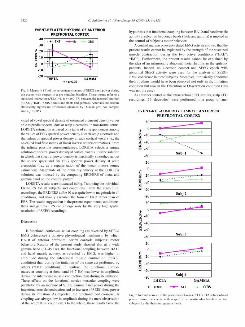
Fig. 6. Means (±SE) of the percentage changes of SEEG band power duringthe events with respect to a pre-stimulus baseline. These means refer to astatistical interaction (F(2,6)=8.1; pb0.0197) between the factors Condition(“EXE”, “IMI”, “OBS”) and Band (theta and gamma). Asterisks indicate thestatistically significant differences obtained by Duncan post hoc compar-isons (pb0.05).
Fig. 7. Individual mean of the percentage changes of LORETA solution bandpower during the events with respect to a pre-stimulus baseline in foursubjects for the theta and gamma bands.
1320 C. Babiloni et al. / NeuroImage 39 (2008) 1314–1323
sisted of voxel spectral density of estimated z-current density valuesable to predict spectral data at scalp electrodes. In non-formal terms,LORETA estimation is based on a table of correspondences amongthe values of EEG spectral power density at each scalp electrode andthe values of spectral power density at each cortical voxel (i.e., theso-called lead field matrix of linear inverse source estimation). Fromthe infinite possible correspondences, LORETA selects a uniquesolution of spectral power density at cortical voxels. It is the solutionin which that spectral power density is maximally smoothed acrossthe source space and fits EEG spectral power density at scalpelectrodes (i.e., as a regularization of the linear inverse sourceestimation). Magnitude of the brain rhythmicity at the LORETAsolutions was indexed by the computing ERD/ERS of theta, andgamma band on the spectral pattern.
LORETA results were illustrated in Fig. 7 showing the individualERD/ERS for all subjects and conditions. From the scalp EEGrecordings, the ERD/ERS at BA10 was quite low in magnitude in allconditions, and mainly assumed the form of ERD rather than ofERS. The results suggest that in the present experimental conditions,theta and gamma ERS can emerge only by the very high spatialresolution of SEEG recordings.
Discussion
Is functional cortico-muscular coupling (as revealed by SEEG-EMG coherence) a putative physiological mechanism by whichBA10 of anterior prefrontal cortex controls subjects’ motorbehavior? Results of the present study showed that at a widegamma band (31–45 Hz), the functional coupling between BA10and hand muscle activity, as revealed by EMG, was higher inamplitude during the intentional muscle contraction (“EXE”condition) than during the imitation of the same act performed byothers (“IMI” condition). In contrast, the functional cortico-muscular coupling at theta band (4–7 Hz) was lower in amplitudeduring the intentional muscle contraction than during its imitation.These effects on the functional cortico-muscular coupling wereparalleled by an increase of SEEG gamma band power during theintentional muscle contraction and an increase of SEEG theta powerduring its imitation. As expected, the functional cortico-muscularcoupling was always low in amplitude during the mere observationof the act (“OBS” condition). On the whole, these results favor the
hypothesis that functional coupling between BA10 and hand muscleactivity at selective frequency bands (theta and gamma) is implied inthe control of subject’s motor behavior.
A control analysis on event-related EMG activity showed that thepresent results cannot be explained by the strength of the sustainedmuscle contraction during the two active conditions (“EXE”,“IMI”). Furthermore, the present results cannot be explained bythe idea of an intrinsically abnormal theta rhythms in the epilepsypatients. Indeed, no electrode contact and SEEG epoch withabnormal SEEG activity were used for the analysis of SEEG-EMG coherence in these subjects. Moreover, intrinsically abnormaltheta rhythms would have been observed not only in the Imitationcondition but also in the Execution or Observation condition (thiswas not the case).
As a further control on the intracerebral SEEG results, scalp EEGrecordings (56 electrodes) were performed in a group of age-
1321C. Babiloni et al. / NeuroImage 39 (2008) 1314–1323
matched healthy subjects. We used the same amount of subjects ofthe SEEG experiments (N=4), and the same experimental tasks.LORETA served to estimate EEG sources at BA10. The results ofthe scalp EEG experiments were quite different with respect tothose obtained with intracerebral SEEG recordings in patients’BA10. From the scalp EEG recordings, the ERD/ERS at LORETAsources modeled in BA10 was quite low in magnitude in allconditions, and mainly assumed the form of theta/gamma ERD(inhibitory indexes) rather than of theta/gamma ERS (excitatoryindexes). The results of the present study globally suggest that thesimple visuo-motor tasks of the current experiments provoked avery selective activation of BA10 as revealed by theta/gamma ERS(intracerebral SEEG recordings). The selectivity of this theta/gamma ERS is indicated by the large theta/gamma ERD of theLORETA sources modeled in BA10 (scalp EEG recordings). It canbe speculated that the present conditions induced in BA10 anexcitatory theta/gamma ERS surrounded by a large inhibitory theta/gamma ERD. In general, the present results emphasize the differentscale of the brain physiological processes in BA10 during taskcontrol, and the complementary contribution given by intracerebralSEEG recordings and LORETA source analysis from scalp EEGdata.
To avoid misleading explanations, it should be remarked thatfunctional coupling between BA10 and hand muscle activity is notbased on direct anatomical connectivity (Ramnani andOwen, 2004),since BA10 is exclusively interconnected with prefrontal, anteriortemporal, and cingulate areas (Morecraft and Van Hoesen, 1993;Ramnani and Owen, 2004). Therefore, BA10 might modulate thecortico-muscular coupling only by means of indirect connectivitywith cortical areas having a direct connection to spinal motoneuronsimpinging upon hand muscles, such as lateral premotor, supple-mentary motor, motor cingulate, primary motor, primary somato-sensory areas, etc. In this regard, it would have been invaluable toinvestigate SEEG-EMG coherence not only from BA10 but alsofrom the mentioned cortical areas, in order to model the wholepattern of functional cortico-muscular coupling in the present ex-perimental conditions. We could not do it for obvious ethicalreasons.
A first issue of the present results is the physiological meaning oftheta and gamma rhythms within functional cortico-muscularcoupling between anterior prefrontal cortex (BA10) and handmuscle contraction. Why was theta band especially modulatedduring the imitation (“IMI” condition)? And why was this true forgamma band during the intentional behavior (“EXE” condition)?Event-related increment of theta and gamma power is supposed to bean important neural correlate of brain processes for the integration ofsensorimotor information (Pfurtscheller et al., 1994, 2003; Brankacket al., 1996; Yordanova et al., 1997, 2000, 2001; Basar, 1998, 1999;Basar et al., 1999; Klimesch, 1999; Pfurtscheller and Lopez da Silva,1999; Tallon-Baudry et al., 1999). It has been proposed that undercertain conditions, cortical pyramidal neurons generate a pattern oftheta (about 4–7 Hz) and gamma (N30 Hz) rhythms. The amplitudeof these rhythms is supposed to be modulated by inputs reachingcortical pyramidal neurons from both cortical and sub-cortical(thalamic/hippocampal) sources (Pfurtscheller and Lopez da Silva,1999; Llinas et al., 1999). In general, theta and gamma rhythmsmight favor temporal synchronization of individual corticospinalneuronal firing for distal and proximal muscles, constitutingfunctional networks of oscillatory nodes in thalamus, cerebellum,and selective cortical regions (Salenius and Hari, 2003; Pollok et al.,2004; Schnitzler and Gross, 2005).
The present findings on the theta cortico-muscular couplingduring imitative behavior (“IMI” condition) agree with and extendprevious evidence reporting that theta rhythms functionallyconnect the activity of hippocampal systems and cortical mantle(Buzsaki and Draguhn, 2004) to sub-serve focused attention,working memory, encoding processes of episodic memory, andcontrol of action (Klimesch, 1999; Gevins and Smith, 2000,Klimesch et al., 2001, 2006). The functional connection with themotor periphery at about theta rhythms would emerge from acerebello-thalamo-premotor–motor cortical network that exerts anoscillatory drive on the spinal motoneurons sustaining physiolo-gical tremor (Timmermann et al., 2004; Pollok et al., 2004;Schnitzler and Gross, 2005). Keeping in mind these data, it can bespeculated that anterior prefrontal cortex (BA10) and hand muscleactivity might be coupled at theta rhythms when subject’s imitativebehavior is guided from external models to be encoded in episodicmemory. Of note, the active behavioral component of the ex-perience seems to be crucial for the relative cortico-muscularcoupling, since the mere observation of the action (“OBS” con-dition) was not able to modulate cortico-muscular coupling at thetarhythms.
The present findings on the gamma cortico-muscular couplingduring intentional behavior (“EXE” condition) complements pre-vious evidence showing that coupling of gamma rhythms as re-vealed by SEEG are implied with cognitive–motor performance(Babiloni et al., 2004b). Furthermore, they agree with previousevidence showing that high-frequency brain oscillations (15 to50 Hz) are synchronized with EMG oscillations accompanyingvoluntary isometric muscle contraction, as a sign of functionalcortico-muscular coupling (Salenius et al., 1997, 2002; Mima andHallett, 1999;Mima et al., 2000, 2001; Babiloni et al., 2004a). Theserhythms are task-specific and generally precede muscle discharge byan interval appropriate for conduction across fast propagating pyra-midal pathways (Brown, 2000). In particular, functional cortico-muscular coupling at beta rhythms is typically found during iso-metric muscle contractions of weak to moderate strength, as apossible mechanism for maintaining a stable motor output with aminimum of computational effort (Conway et al., 1995, Salenius etal., 1997, Halliday et al., 1998, Mima and Hallett, 1999, Gross et al.,2000,Murayama et al., 2001; Kristeva-Feige et al., 2002; Kristeva etal., 2007). Instead, functional cortico-muscular coupling at gammarhythms is most evident during strong isometric muscle contrac-tions, when demands on the motor cortex are likely to be greater,more mutable, and depending on focused attention (Mima et al.,1999; Brown, 2000; Schoffelen et al., 2005; Omlor et al., 2007). Inthis framework, the novel results of the present study suggest thatrhythms at wide gamma (31–45 Hz) but not beta (22–30 Hz) bandsub-serve functional coupling between anterior prefrontal cortex(BA10) and hand muscle activity during intentional over imitativebehavior.
As an alternative explanation, the present results may be ascribedto the confound of joint attention. Indeed, joint attention implies theintegration of perceptual processes concerned with detecting thedirection of other’s and own attention, and is strictly related to theactivity of BA10 of anterior prefrontal cortex (Williams et al., 2005).However, it is unlikely that this is the case here. In the presentintentional (“EXE”), imitative (“IMI”), and observation (“OBS”)conditions, we posed a special care that subjects and observedperson always looked at the central target during the SEEGrecordings, and all SEEG trials associated with eye movementswere rejected.

1322 C. Babiloni et al. / NeuroImage 39 (2008) 1314–1323
Conclusions
In the present study, we tested the working hypothesis thatfunctional coupling between anterior prefrontal cortex (BA10) andhand muscle activity (as revealed by SEEG-EMG coherence) is aphysiological mechanism by which subjects’ motor behavior iscontrolled. Results showed that functional cortico-muscular cou-pling at wide gamma band (31–45 Hz) was higher in amplitudeduring the intentional muscle contraction (“EXE” condition) thanduring the imitation of the same act performed by others (“IMI”condition). The opposite was true at theta band (4–7 Hz). In parallel,there was an increase of SEEG gamma band power during theintentional muscle contraction and an increase of SEEG theta bandpower during its imitation. The present results suggest that anteriorprefrontal cortex (BA10) might control subjects’ behavior by meansof functional cortico-muscular coupling at selective frequency bands(theta and wide gamma rhythms). To complete the functionalpicture, future investigations should disclose the pattern of indirectcortico-cortical functional connectivity between BA10 and primary/non-primary cortical areas directly connected to spinal motoneur-ons. Furthermore, further studies should evaluate the possiblehemispherical effects of the use of dominant vs. non-dominant handfor the motor response.
References
Allport, A., Styles, E.A., Hsieh, S.L., 1994. Shifting intentional set:exploring the dynamic control of tasks. In: Umiltà, C., Moscovitch, M.(Eds.), Attention and Performance, Vol. XV. MIT Press, Cambridge,MA, pp. 421–452.
Azouz, R., Gray, C.M., 2000. Dynamic spike threshold reveals a mechanismfor synaptic coincidence detection in cortical neurons in vivo. Proc. Natl.Acad. Sci. U. S. A. 97 (14), 8110–8115.
Azouz, R., Gray, C.M., 2003. Adaptive coincidence detection and dynamicgain control in visual cortical neurons in vivo. Neuron 37 (3),513–523.
Babiloni, C., Vecchio, F., Babiloni, F., Brunelli, G.A., Carducci, F., Cincotti,F., Pizzella, V., Romani, G.L., Tecchio, F.T., Rossini, P.M., 2004a.Coupling between “hand” primary sensorimotor cortex and lower limbmuscles after ulnar nerve surgical transfer in paraplegia. Behav. Neurosci.118 (1), 214–222.
Babiloni, C., Bares, M., Vecchio, F., Brazdil, M., Jurak, P., Moretti, D.V.,Ubaldi, A., Rossini, P.M., Rektor, I., 2004b. Synchronization of gammaoscillations increases functional connectivity of human hippocampusand inferior-middle temporal cortex during repetitive visuomotor events.Eur. J. Neurosci. 19 (11), 3088–3098.
Bares, M., Rektor, I., 2001. Basal ganglia involvement in sensory andcognitive processing. A depth electrode CNV study in human subjects.Clin. Neurophysiol. 112 (11), 2022–2030.
Bares, M., Rektor, I., Kanovsky, P., Streitova, H., 2003. Cortical andsubcortical distribution of middle and long latency auditory and visualevoked potentials in a cognitive (CNV) paradigm. Clin. Neurophysiol.114, 2447–2460.
Basar, E., 1998. Brain function and oscillations: I. Brain Oscillations:Principles and Approaches. Springer, Berlin, Heidelberg.
Basar, E., 1999. Brain function and oscillations. Integrative Brain Function.Neurophysiology and Cognitive Processes. Springer, Berlin, Heidelberg.
Basar, E., Basar-Eroglu, C., Karakas, S., Schurmann, M., 1999. Arecognitive processes manifested in event-related gamma, alpha, theta anddelta oscillations in the EEG? Neurosci. Lett. 259, 165–168.
Brankack, J., Seidenbecher, T., Muller-Gartner, H.W., 1996. Task-relevantlate positive component in rats: is it related to hippocampal thetarhythm? Hippocampus 6 (5), 475–482.
Braver, T.S., Reynolds, J.R., Donaldson, D.I., 2003. Neural mechanisms of
transient and sustained cognitive control during task switching. Neuron39 (4), 713–726.
Brazdil, M., Rektor, I., Dufek, M., Daniel, P., Jurak, P., Kuba, R., 1999. Therole of frontal and temporal lobes in visual discrimination task-depthERP studies. Neurophysiol. Clin. 29 (4), 339–350.
Brazdil, M., Roman, R., Daniel, P., Rektor, I., 2003. Intracerebralsomatosensory event-related potentials: effect of response type (buttonpressing versus mental counting) on P3-like potentials within the humanbrain. Clin. Neurophysiol. 114 (8), 1489–1496.
Brown, P., 2000. Cortical drives to human muscle: the piper and relatedrhythms. Prog. Neurobiol. 60 (1), 97–108.
Buzsaki, G., Draguhn, A., 2004. Neuronal oscillations in cortical networks.Science 304 (5679), 1926–1929 (Review).
Conway, B.A., Halliday, D.M., Farmer, S.F., Shahani, U., Maas, P., Weir,A.I., Rosenberg, J.R., 1995. Synchronization between motor cortex andspinal motoneuronal pool during the performance of a maintained motortask in man. J. Physiol. 489 (3), 917–924.
Dreher, J.C., Koechlin, E., Ali, S.O., Grafman, J., 2002. The roles of timingand task order during task switching. NeuroImage 17 (1), 95–109.
Fletcher, P.C., Henson, R.N., 2001. Frontal lobes and human memory:insights from functional neuroimaging. Brain 124 (Pt 5), 849–881(May, Review).
Fries, P., Reynolds, J.H., Rorie, A.E., Desimone, R., 2001. Modulation ofoscillatory neuronal synchronization by selective visual attention.Science 291 (5508), 1560–1563.
Fries, P., Schroder, J.H., Roelfsema, P.R., Singer, W., Engel, A.K., 2002.Oscillatory neuronal synchronization in primary visual cortex as acorrelate of stimulus selection. J. Neurosci. 22 (9), 3739–3754.
Gevins, A., Smith, M.E., 2000. Neurophysiological measures of workingmemory and individual differences in cognitive ability and cognitivestyle. Cereb. Cortex 10 (9), 829–839.
Gross, J., Tass, P.A., Salenius, S., Hari, R., Freund, H.J., Schnitzler, A.,2000. Cortico-muscular synchronization during isometric musclecontraction in humans as revealed by magnetoencephalography.J. Physiol. 527 (3), 623–631.
Halliday, D.M., Rosenberg, J.R., Amjad, A.M., Breeze, P., Conway, B.A.,Farmer, S.F., 1995. A framework for the analysis of mixed time series/point process data-theory and application to the study of physiologicaltremor, single motor unit discharges and electromyograms. Prog.Biophys. Mol. Biol. 64 (2-3), 237–278.
Halliday, D.M., Conway, B.A., Farmer, S.F., Rosenberg, J.R., 1998. Usingelectroencephalography to study functional coupling between corticalactivity and electromyograms during voluntary contractions in humans.Neurosci. Lett. 241 (1), 5–8.
Klimesch, W., 1999. EEG alpha and theta oscillations reflect cognitive andmemory performance: a review and analysis. Brain Res. Brain Res. Rev.29 (2-3), 169–195.
Klimesch, W., Doppelmayr, M., Stadler, W., Pollhuber, D., Sauseng, P.,Rohm, D., 2001. Episodic retrieval is reflected by a process specificincrease in human electroencephalographic theta activity. Neurosci. Lett.302 (1), 49–52.
Klimesch, W., Hanslmayr, S., Sauseng, P., Gruber, W., Brozinsky, C.J.,Kroll, N.E., Yonelinas, A.P., Doppelmayr, M., 2006. Oscillatory EEGcorrelates of episodic trace decay. Cereb. Cortex 16 (2), 280–290.
Konishi, S., Chikazoe, J., Jimura, K., Asari, T., Miyashita, Y., 2005. Neuralmechanism in anterior prefrontal cortex for inhibition of prolonged setinterference. Proc. Natl. Acad. Sci. U. S. A. 102 (35), 12584–12588.
Kristeva, R., Patino, L., Omlor, W., 2007. Beta-range cortical motor spectralpower and corticomuscular coherence as a mechanism for effectivecorticospinal interaction during steady-state motor output. NeuroImage36 (3), 785–792 (electronic publication 2007 Mar 28).
Kristeva-Feige, R., Fritsch, C., Timmer, J., Lücking, C., 2002. Effects ofattention and precision of exerted force on beta range EEG-EMGsynchronization during a maintained motor contraction task. Clin.Neurophysiol. 113, 124–131.
Llinas, R.R., Ribary, U., Jeanmonod, D., Kronberg, E., Mitra, P.P., 1999.Thalamocortical dysrhythmia: a neurological and neuropsychiatric

1323C. Babiloni et al. / NeuroImage 39 (2008) 1314–1323
syndrome characterized by magnetoencephalography. Proc. Natl. Acad.Sci. U. S. A. 96 (26), 15222–15227.
Marsden, J.F., Limousin-Dowsey, P., Ashby, P., Pollak, P., Brown, P., 2001.Subthalamic nucleus, sensorimotor cortex and muscle interrelationshipsin Parkinson’s disease. Brain 124 (Pt 2), 378–388.
Meiran, N., Chorev, Z., Sapir, A., 2000. Component processes in taskswitching. Cogn. Psychol. 41 (3), 211–253.
Milner, B., 1963. The effects of different brain lesions on card sorting. Arch.Neurol. 9, 90–100.
Mima, T., Simpkins, N., Oluwatimilehin, T., Hallett, M., 1999. Force levelmodulates human cortical oscillatory activities. Neurosci. Lett. 275 (2),77–80.
Mima, T., Hallett, M., 1999. Corticomuscular coherence: a review. J. Clin.Neurophysiol. 16 (6), 501–511 (Review).
Mima, T., Matsuoka, T., Hallett, M., 2000. Functional coupling of humanright and left cortical motor areas demonstrated with partial coherenceanalysis. Neurosci. Lett. 287 (2), 93–96.
Mima, T., Toma, K., Koshy, B., Hallett, M., 2001. Coherence betweencortical and muscular activities after subcortical stroke. Stroke 32 (11),2597–2601.
Morecraft, R.J., Van Hoesen, G.W., 1993. Frontal granular cortex input tothe cingulate (M3), supplementary (M2) and primary (M1) motorcortices in the rhesus monkey. J. Comp. Neurol. 337 (4), 669–689.
Moretti, D.V., Babiloni, F., Carducci, F., Cincotti, F., Remondini, E.,Rossini, P.M., Salinari, S., Babiloni, C., 2003. Computerized processingof EEG-EOG-EMG artifacts for multicentirc studies in EEG oscillationsand event-related potentials. Int. J. Pshycophysiol. 47 (3), 199–216.
Murayama, N., Lin, YY., Salenius, S., Hari, R., 2001. Oscillatory interactionbetween human motor cortex and trunk muscles during isometriccontraction. NeuroImage 14, 1206–1213.
Omlor, W., Patino, L., Hepp-Reymond, M.C., Kristeva, R., 2007. Gamma-range corticomuscular coherence during dynamic force output. Neuro-Image 34 (3), 1191–1198.
Pascual-Marqui, R.D., Michel, C.M., 1994. LORETA (low resolution brainelectromagnetic tomography): new authentic 3D functional images ofthe brain. ISBET Newsletter ISNN 5, 4–8.
Pascual-Marqui, R.D., Lehmann, D., Koenig, T., Kochi, K., Merlo, M.C.,Hell, D., Koukkou, M., 1999. Low resolution brain electromagnetictomography (LORETA) functional imaging in acute, neurolepticnaive,first-episode, productive schizophrenia. Psychiatry Res. 90 (3), 169–179.
Pascual-Marqui, R.D., Esslen, M., Kochi, K., Lehmann, D., 2002.Functional imaging with low resolution brain electromagnetic tomo-graphy (LORETA): a review. Methods Find. Exp. Clin. Pharmacol. 24,91–95.
Pfurtscheller, G., Aranibar, A., 1979. Evaluation of event-related desyn-chronization (ERD) preceding and following voluntary self-pacedmovement. Electroencephalogr. Clin. Neurophysiol. 46 (2), 138–146.
Pfurtscheller, G., Lopez da Silva, F., 1999. Event-related EEG/MEGsynchronization and desynchronization: basic principles. Clin. Neuro-physiol. 110, 1842–1857.
Pfurtsheller, G., Neuper, C., 1994. Event-related synchronization of the murhythm in the EEG over the cortical hand area in man. Neurosci. Lett.174 (1), 93–96.
Pfurtscheller, G., Flotzinger, D., Neuper, C., 1994. Differentiation betweenfinger, toe and tongue movement in man based on 40 Hz EEG.Electroencephalogr. Clin. Neurophysiol. 90 (6), 456–460.
Pfurtscheller, G., Neuper, C., Flotzinger, D., Pregenzer, M., 1997. EEG-based discrimination between imagination of right and left handmovement. Electroencephalogr. Clin. Neurophysiol. 103 (6), 642–651.
Pfurtscheller, G., Neuper, C., Ramoser, H., Muller-Gerking, J., 1999.Visually guided motor imagery activates sensorimotor areas in humans.Neurosci. Lett. 269 (3), 153–156.
Pfurtscheller, G., Graimann, B., Huggins, J.E., Levine, S.P., Schuh, L.A.,2003. Spatiotemporal patterns of beta desynchronization and gammasynchronization in corticographic data during self-paced movement.Clin. Neurophysiol. 114 (7), 1226–1236.
Pollok, B., Gross, J., Dirks, M., Timmermann, L., Schnitzler, A., 2004. Thecerebral oscillatory network of voluntary tremor. J. Physiol. 554 (Pt 3),871–878.
Ramnani, N., Owen, A.M., 2004. Anterior prefrontal cortex: insights intofunction from anatomy and neuroimaging. Nat. Rev., Neurosci. 5 (3),184–194.
Rappelsberger, P., Petsche, H., 1988. Probability mapping: power and co-herence analyses of cognitive processes. Brain Topogr. 1 (1), 46–54(Fall).
Rektor, I., 2000. Parallel information processing in motor systems:intracerebral recordings of readiness potential and CNV in humansubjects. Neural Plast. 7 (1-2), 65–72.
Rektor, I., Kanovsky, P., Bares, M., Brazdil, M., Streitova, H., Klajblova, H.,Kuba, R., Daniel, P., 2003. A SEEG study of ERP in motor and premotorcortices and in the basal ganglia. Clin. Neurophysiol. 114 (3), 463–471.
Sakai, K., Passingham, R.E., 2003. Prefrontal interactions reflect future taskoperations. Nat. Neurosci. 6 (1), 75–81.
Salenius, S., Hari, R., 2003. Synchronous cortical oscillatory activity duringmotor action. Curr. Opin. Neurobiol. 13 (6), 678–684.
Salenius, S., Portin, K., Kajola, M., Salmelin, R., Hari, R., 1997. Corticalcontrol of human motoneuron firing during isometric contraction.J. Neurophysiol. 77, 3401–3405.
Salenius, S., Avikainen, S., Kaakkola, S., Hari, R., Brown, P., 2002.Defective cortical drive to muscle in Parkinson’s disease and itsimprovement with levodopa. Brain 125 (Pt 3), 491–500.
Schnitzler, A., Gross, J., 2005. Normal and pathological oscillatorycommunication in the brain. Nat. Rev., Neurosci. 6 (4), 285–296.
Schoffelen, R., Oostenveld, P., Fries, 2005. Neuronal coherence as amechanism of effective corticospinal interaction. Science 308,111–113.
Talairach, J., Tournoux, P., 1988. Co-Planar Stereotaxic Atlas of the HumanBrain. Stuttgart: Thieme.
Tallon-Baudry, C., Kreiter, A., Bertrand, O., 1999. Sustained and transientoscillatory responses in the gamma and beta bands in a visual short-termmemory task in humans. Vis. Neurosci. 16 (3), 449–459.
Timmermann, L., Gross, J., Butz, M., Kircheis, G., Haussinger, D.,Schnitzler, A., 2004. Pathological oscillatory coupling within the humanmotor system in different tremor syndromes as revealed by magnetoen-cephalography. Neurol. Clin. Neurophysiol. 26.
Williams, J.H., Waiter, G.D., Perra, O., Perrett, D.I., Whiten, A., 2005. AnfMRI study of joint attention experience. NeuroImage 25 (1),133–140.
Yordanova, J., Kolev, V., Demiralp, T., 1997. The phase-locking of auditorygamma band responses in humans is sensitive to task processing.NeuroReport 8 (18), 3999–4004.
Yordanova, J., Kolev, V., Heinrich, H., Banaschewski, T., Woerner, W.,Rothenberger, A., 2000. Gamma band response in children is related totask-stimulus processing. NeuroReport 11 (10), 2325–2330.
Yordanova, J., Banaschewski, T., Kolev, V., Woerner, W., Rothenberger, A.,2001. Abnormal early stages of task stimulus processing in children withattention-deficit hyperactivity disorder—evidence from event-relatedgamma oscillations. Clin. Neurophysiol. 112 (6), 1096–1108.
Copyright © 2022 FDOKUMEN