Porous ceramics with controllable properties prepared by protein foaming-consolidation method
Upload
independentCategory
view
1download
0
Controllable Surface Modification of Poly(lactic- co-glycolicacid) (PLGA) by Hydrolysis or Aminolysis I: Physical,
Chemical, and Theoretical Aspects
Tristan I. Croll, Andrea J. O’Connor, Geoffrey W. Stevens, and Justin J. Cooper-White*
Department of Chemical and Biomolecular Engineering, The University of Melbourne,Victoria 3010 Australia
Received August 18, 2003; Revised Manuscript Received October 30, 2003
While biodegradable, biocompatible polyesters such as poly (lactic-co-glycolic acid) (PLGA) are popularmaterials for the manufacture of tissue engineering scaffolds, their surface properties are not particularlysuitable for directed tissue growth. Although a number of approaches to chemically modify the PLGA surfacehave been reported, their applicability to soft tissue scaffolds, which combine large volumes, complex shapes,and extremely fine structures, is questionable. In this paper, we describe two wet-chemical methods, basehydrolysis and aminolysis, to introduce useful levels of carboxylic acid or primary and secondary aminegroups, respectively, onto the surface of PLGA with minimal degradation. The effects of temperature,concentration, pH, and solvent type on the kinetics of these reactions are studied by following changes inthe wettability of the PLGA using contact angle measurements. In addition, the treated surfaces are studiedusing X-ray photoelectron spectroscopy (XPS) to determine the effect on the surface chemical structure.Furthermore, we show using XPS analysis that these carboxyl and amine groups are readily activated toallow the covalent attachment of biological macromolecules.
Introduction
Soft tissue engineering offers a number of unique chal-lenges not seen in other tissue engineering applications suchas bone, cartilage or skin. For many soft tissue applications,scaffolds must be large (e.g. breast reconstruction1), veryhighly porous, and soft, yet have enough strength to resistthe contractile forces generated by growing tissue. Scaffoldsproduced from poly(lactic-co-glycolic acid) (PLGA) usingthermally induced phase separation (TIPS)2,3 are able to meetthese goals. However, it is well-known that the surfaceproperties of PLGA are not ideal for cell growth.4-7 PLGAis relatively hydrophobic compared to the natural extracel-lular matrix (ECM), is unable to interact specifically withcells, and does not possess any functional groups for theattachment of biologically active molecules.
Since the mechanical and degradative properties of PLGAare seemingly ideal, and given that PLGA is approved bythe U.S. Food and Drug Administration (FDA) and otherregulatory bodies for implantation into humans, a promisingapproach appears to be the surface modification of PLGAscaffolds post-formation. Ideally, this gives useful surfacecharacteristics to the polymer, without changing the proper-ties of the bulk.
Many approaches have been taken to modify the surfaceof PLGA to date; however, very few of these appearpromising for soft tissue applications. Plasma treatment,although very popular,8-11 appears unable to penetrate morethan a few millimeters into the pores of a scaffold. Surface
entrapment,6,12-14 while apparently promising for 2-dimen-sional applications and possibly the denser bone and cartilagescaffolds, leads to collapse of the thin (<1 µm) walls of asoft tissue scaffold during the swelling step. Partial surfacehydrolysis by acid or base treatment6,15-17 has been muchtoo aggressive in the past, with conditions that destroy atypical soft tissue TIPS scaffold2,3 in less than one minute.
A desirable property of any surface modification techniqueis that it is inherently limited to the very surface of thesubstrate irrespective of reaction rate. As an example, softtissue scaffolds for breast reconstruction are somewhatirregularly shaped, have volumes ranging from 100 mL to 2L,1 and, in the case of PLGA scaffolds produced via the TIPSprocess, have internal wall thicknesses as low as 500 nm.3
In this situation, a relatively slow reaction time on the orderof minutes to hours is desirable to ensure controllabletreatment throughout the scaffold.
In this paper, we report on the use of controlled hydrolysisto produce carboxylic acid functional groups and aminolysisto produce primary and secondary amine groups on thesurface of thin PLGA films in a highly controlled manner,with minimal erosion. In addition, covalent binding of amodel amine-functional macromolecule, chitosan, to thenewly formed functional groups was characterized.
Experimental Section
Materials. Poly(D,L-lactic-co-glycolic acid) (PLGA) witha lactic acid:glycolic acid ratio of 75:25 and an inherentviscosity in chloroform of 0.69 dL/g (molecular weightapproximately 100 kDa) was purchased from Birmingham
* To whom correspondence should be addressed. Phone:+61 3 83444704. E-mail: [email protected].
463Biomacromolecules 2004,5, 463-473
10.1021/bm0343040 CCC: $27.50 © 2004 American Chemical SocietyPublished on Web 01/09/2004

Polymers Inc. Ammonia (LR, 25% aqueous solution),hydrogen peroxide (LR, 30% aqueous solution), chlorotri-methylsilane (CTMS) (>99%), toluene (HPLC grade),acetone (HPLC), ethanol (AR,>99.7%),n-hexane (HPLC),dichloromethane (HPLC), and 2-propanol (iso-propyl alco-hol, IPA) were obtained from Merck.N-Aminoethyl-1,3-propanediamine (AEPDA) (AR), 1-ethyl-3-(3-dimethylami-nopropyl)-carbodiimide (EDAC), andN-hydroxysuccinimide(NHS) were obtained from Sigma-Aldrich. Acetic acid (AR),ethylenediamine (ED) (AR), and triethanolamine (AR) wereobtained from BDH. 2-(N-Morpholino)ethanesulfonic acid(MES) (AR) was obtained from ICN Biomedicals Inc.(Ohio). Low molecular weight chitosan (viscosity @ 0.5%w/v ) 5-20 cP,>80% deacetylated, Wako Fine ChemicalsInc.) was kindly donated by Professor Yoshinari Baba ofMiyazaki University. All reagents were used as supplied,and all water used was purified using a MilliPore Simplicityunit to a resistivity ofg18.2 MΩ.cm. All glassware waswashed sequentially in toluene, acetone, and ethanol anddried using compressed air filtered through activated carbonimmediately prior to use.
Production of Thin Films. PLGA films with a thicknessof <2 µm and an RMS roughness of<2 nm measured viacontact-mode AFM imaging over a 10µm × 10 µm areawere prepared on clean, hydrophobically treated glasscoverslips by a dip-coating method similar to that used byothers.18 Coverslips (22 mm× 22 mm, 100µm thick) werecleaned in a boiling mixture of 9 parts neat hydrogenperoxide solution and 1 part neat ammonia solution. Thesewere rinsed in three washes of water, placed individuallyinto 23 mm internal diameter glass vials so that they stoodvertically, and dried on a hotplate at 80°C. The coverslipswere then made hydrophobic by treatment in a freshlyprepared solution of CTMS inn-hexane (10% v/v) for 10min and rinsed with three washes of freshn-hexane, andallowed to dry by evaporation. These were dipped in asolution of PLGA in dichloromethane (5% w/v) at 0°C andwithdrawn slowly to leave a thin film of polymer solutionon the glass surface. Most of the solvent evaporatedimmediately, leaving a translucent white polymer film onthe glass. The coverslips were then returned to their vials,loosely capped with aluminum foil and placed horizontallyin an oven at 70°C for 24 h to remove the residual solventand anneal the polymer film. At this point the films wereentirely transparent and almost indistinguishable from thecoverslips, except for the presence of refraction fringes,indicating a very low surface roughness on the order ofnanometers.19
Hydrolysis and Aminolysis of Films. Films to behydrolyzed were immersed in a solution of aqueous sodiumhydroxide (NaOH) at suitable concentrations and allowedto react at 20°C for the desired time period, as describedelsewhere.2 Films to be aminolyzed were immersed in EDor AEPDA at various concentrations in either water or IPAand allowed to react at either 20 or 0°C for the desired timeperiod. Treated films were washed three times with ice coldwater, followed by soaking for a further hour in fresh wateron ice, dried under vacuum over silica gel for 12 h, and storedin a desiccator over silica gel until further use.
Activation of Films with Cross-Linking Reagents.Hydrolyzed films were treated in 10 mL of a freshly madesolution of 4 mM EDAC and 100 mM NHS in 10 mM MESbuffer at pH 6.1 for 1 h at room temperature. Aminolysedfilms were treated in freshly prepared 40 mM DMA in 10mM triethanolamine buffer at pH 8.0 for 1 h at roomtemperature. Activated films were rinsed thoroughly withwater and used immediately.
Binding of Chitosan to Films. Chitosan was dissolvedin 0.1 M acetic acid to a concentration of 10 mg/mL. Thissolution was filtered through a 0.25µm syringe filter toremove insoluble particles. A 1 mL aliquot of this solutionwas made up to 90 mL with water, and 10 mL of 100 mMtriethanolamine, 1.4 M NaCl at pH 8.0 or 9.0 (adjusted with1 M NaOH) was added. At this point, the solution turnedslightly milky, indicating the formation of a stable colloidalsuspension of 100µg/mL chitosan with a physiological saltconcentration.
Preactivated surfaces or nonactivated hydrolyzed or ami-nolyzed controls were incubated in 10 mL of this chitosansuspension at pH 8.0 (aminolyzed surfaces) or 9.0 (hydro-lyzed surfaces) for 1 h. The coated surfaces were then washedtwice with 0.1 M acetic acid to remove all weakly boundchitosan, followed by three washes with water.
Scanning Electron Microscope (SEM) Analysis.Samplesto be analyzed via SEM were sputter-coated with gold undervacuum in an argon atmosphere using a sputter current of60 mA (Dynavac Mini Coater, Dynavac USA). SEM analysiswas carried out with a Philips XL30 SEM using an LaB6
electron gun with an accelerating voltage of 20 kV.Contact Angle Measurement.Equilibrium advancing (θa)
and receding (θr) air/water contact angles were measuredwith a contact angle goniometer and drop shape analysissoftware (First Ten Angstroms, Inc) using the captured(sessile) drop method20 on PLGA coated glass coverslips ata temperature of 22.5°C. Water was pumped onto the surfaceof the coverslip through a 23-gauge stainless steel needle ata rate of 2µL/s until the drop diameter was greater than 4times the diameter of the needle. At this point, the drop wasallowed to stand for 2 min before measuringθa. Water wasthen removed from the drop at a rate of 2µL/s until theinterface began to recede. A further 2 min relaxation timewas allowed beforeθr was measured. During the first andsecond relaxation periods, contact angles decreased orincreased by 3-5° respectively; the contact angles changedby <0.5° in the next 3 min. Three drops (six interfaces) wereimaged for each surface; values reported are the mean(1standard deviation.
X-ray Photoelectron Spectroscopy (XPS) Analysis.XPSanalysis was performed using a AXIS HSi spectrometer(Kratos Analytical Ltd) equipped with a monochromatic AlKR source at a power of 300 W. The total pressure in themain vacuum chamber during analysis was typically 2×10⁻⁸ mbar. Elements present were identified from surveyspectra. For further analysis, high-resolution spectra wererecorded from individual peaks at 40 eV pass energy. Atomicconcentrations of each element were calculated by determin-ing the relevant integral peak intensities (using a linear typebackground) and applying the sensitivity factors supplied by
464 Biomacromolecules, Vol. 5, No. 2, 2004 Croll et al.
the instrument manufacturer. A value of 285.0 eV for thebinding energy of the main C 1s component (CHx) was usedto correct for charging of specimens under irradiation.
Results and Discussion
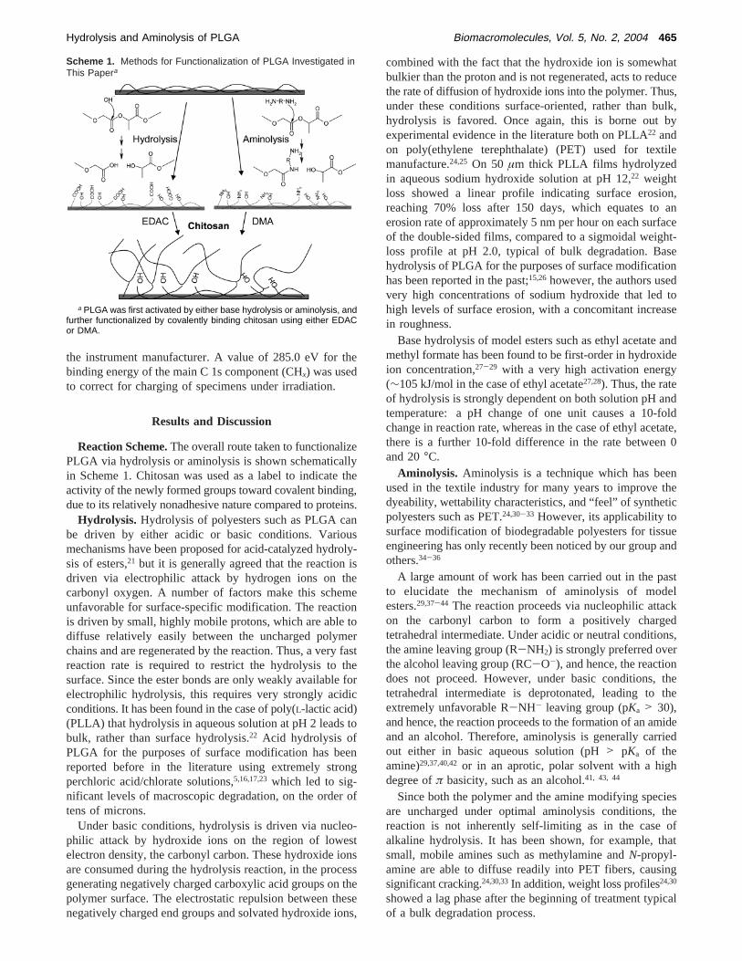
Reaction Scheme.The overall route taken to functionalizePLGA via hydrolysis or aminolysis is shown schematicallyin Scheme 1. Chitosan was used as a label to indicate theactivity of the newly formed groups toward covalent binding,due to its relatively nonadhesive nature compared to proteins.
Hydrolysis. Hydrolysis of polyesters such as PLGA canbe driven by either acidic or basic conditions. Variousmechanisms have been proposed for acid-catalyzed hydroly-sis of esters,21 but it is generally agreed that the reaction isdriven via electrophilic attack by hydrogen ions on thecarbonyl oxygen. A number of factors make this schemeunfavorable for surface-specific modification. The reactionis driven by small, highly mobile protons, which are able todiffuse relatively easily between the uncharged polymerchains and are regenerated by the reaction. Thus, a very fastreaction rate is required to restrict the hydrolysis to thesurface. Since the ester bonds are only weakly available forelectrophilic hydrolysis, this requires very strongly acidicconditions. It has been found in the case of poly(L-lactic acid)(PLLA) that hydrolysis in aqueous solution at pH 2 leads tobulk, rather than surface hydrolysis.22 Acid hydrolysis ofPLGA for the purposes of surface modification has beenreported before in the literature using extremely strongperchloric acid/chlorate solutions,5,16,17,23which led to sig-nificant levels of macroscopic degradation, on the order oftens of microns.
Under basic conditions, hydrolysis is driven via nucleo-philic attack by hydroxide ions on the region of lowestelectron density, the carbonyl carbon. These hydroxide ionsare consumed during the hydrolysis reaction, in the processgenerating negatively charged carboxylic acid groups on thepolymer surface. The electrostatic repulsion between thesenegatively charged end groups and solvated hydroxide ions,
combined with the fact that the hydroxide ion is somewhatbulkier than the proton and is not regenerated, acts to reducethe rate of diffusion of hydroxide ions into the polymer. Thus,under these conditions surface-oriented, rather than bulk,hydrolysis is favored. Once again, this is borne out byexperimental evidence in the literature both on PLLA22 andon poly(ethylene terephthalate) (PET) used for textilemanufacture.24,25 On 50 µm thick PLLA films hydrolyzedin aqueous sodium hydroxide solution at pH 12,22 weightloss showed a linear profile indicating surface erosion,reaching 70% loss after 150 days, which equates to anerosion rate of approximately 5 nm per hour on each surfaceof the double-sided films, compared to a sigmoidal weight-loss profile at pH 2.0, typical of bulk degradation. Basehydrolysis of PLGA for the purposes of surface modificationhas been reported in the past;15,26however, the authors usedvery high concentrations of sodium hydroxide that led tohigh levels of surface erosion, with a concomitant increasein roughness.
Base hydrolysis of model esters such as ethyl acetate andmethyl formate has been found to be first-order in hydroxideion concentration,27-29 with a very high activation energy(∼105 kJ/mol in the case of ethyl acetate27,28). Thus, the rateof hydrolysis is strongly dependent on both solution pH andtemperature: a pH change of one unit causes a 10-foldchange in reaction rate, whereas in the case of ethyl acetate,there is a further 10-fold difference in the rate between 0and 20°C.
Aminolysis. Aminolysis is a technique which has beenused in the textile industry for many years to improve thedyeability, wettability characteristics, and “feel” of syntheticpolyesters such as PET.24,30-33 However, its applicability tosurface modification of biodegradable polyesters for tissueengineering has only recently been noticed by our group andothers.34-36
A large amount of work has been carried out in the pastto elucidate the mechanism of aminolysis of modelesters.29,37-44 The reaction proceeds via nucleophilic attackon the carbonyl carbon to form a positively chargedtetrahedral intermediate. Under acidic or neutral conditions,the amine leaving group (R-NH2) is strongly preferred overthe alcohol leaving group (RC-O-), and hence, the reactiondoes not proceed. However, under basic conditions, thetetrahedral intermediate is deprotonated, leading to theextremely unfavorable R-NH- leaving group (pKa > 30),and hence, the reaction proceeds to the formation of an amideand an alcohol. Therefore, aminolysis is generally carriedout either in basic aqueous solution (pH> pKa of theamine)29,37,40,42or in an aprotic, polar solvent with a highdegree ofπ basicity, such as an alcohol.41, 43, 44
Since both the polymer and the amine modifying speciesare uncharged under optimal aminolysis conditions, thereaction is not inherently self-limiting as in the case ofalkaline hydrolysis. It has been shown, for example, thatsmall, mobile amines such as methylamine andN-propyl-amine are able to diffuse readily into PET fibers, causingsignificant cracking.24,30,33In addition, weight loss profiles24,30
showed a lag phase after the beginning of treatment typicalof a bulk degradation process.
Scheme 1. Methods for Functionalization of PLGA Investigated inThis Papera
a PLGA was first activated by either base hydrolysis or aminolysis, andfurther functionalized by covalently binding chitosan using either EDACor DMA.
Hydrolysis and Aminolysis of PLGA Biomacromolecules, Vol. 5, No. 2, 2004 465
In contrast to base hydrolysis, the overall activation energyfor the aminolysis reaction is very low and in organicsolvents is often negative,44 leading to a low or inversedependence of the reaction rate on temperature. In aqueoussolutions, the reaction rate typically reaches a plateau at pHvalues just above the pKa of the amine, dropping sharply atpH values below this.29,42 It has been found from numerousexperiments39 that in general the log of the aminolysis rateincreases with the pKa of the amine to the 0.8th power.
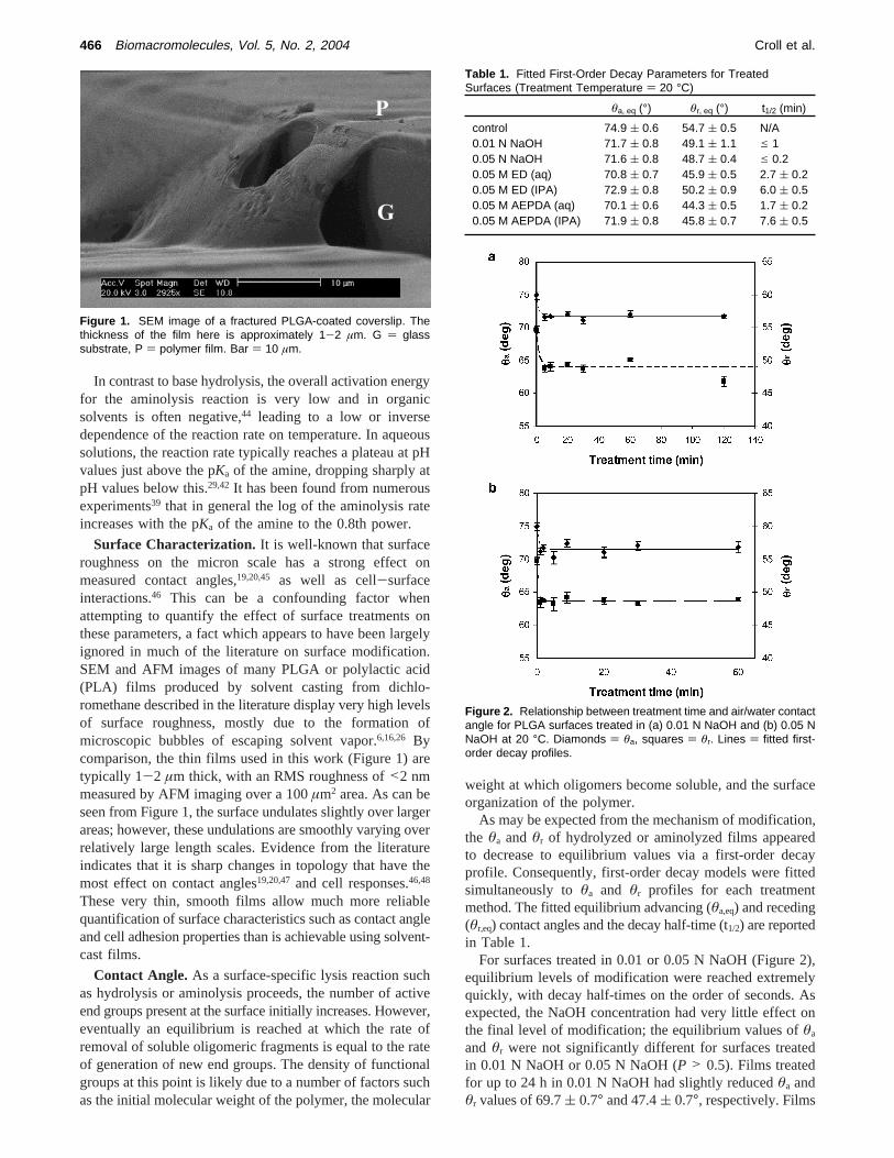
Surface Characterization. It is well-known that surfaceroughness on the micron scale has a strong effect onmeasured contact angles,19,20,45 as well as cell-surfaceinteractions.46 This can be a confounding factor whenattempting to quantify the effect of surface treatments onthese parameters, a fact which appears to have been largelyignored in much of the literature on surface modification.SEM and AFM images of many PLGA or polylactic acid(PLA) films produced by solvent casting from dichlo-romethane described in the literature display very high levelsof surface roughness, mostly due to the formation ofmicroscopic bubbles of escaping solvent vapor.6,16,26 Bycomparison, the thin films used in this work (Figure 1) aretypically 1-2 µm thick, with an RMS roughness of<2 nmmeasured by AFM imaging over a 100µm2 area. As can beseen from Figure 1, the surface undulates slightly over largerareas; however, these undulations are smoothly varying overrelatively large length scales. Evidence from the literatureindicates that it is sharp changes in topology that have themost effect on contact angles19,20,47and cell responses.46,48
These very thin, smooth films allow much more reliablequantification of surface characteristics such as contact angleand cell adhesion properties than is achievable using solvent-cast films.
Contact Angle. As a surface-specific lysis reaction suchas hydrolysis or aminolysis proceeds, the number of activeend groups present at the surface initially increases. However,eventually an equilibrium is reached at which the rate ofremoval of soluble oligomeric fragments is equal to the rateof generation of new end groups. The density of functionalgroups at this point is likely due to a number of factors suchas the initial molecular weight of the polymer, the molecular
weight at which oligomers become soluble, and the surfaceorganization of the polymer.
As may be expected from the mechanism of modification,the θa and θr of hydrolyzed or aminolyzed films appearedto decrease to equilibrium values via a first-order decayprofile. Consequently, first-order decay models were fittedsimultaneously toθa and θr profiles for each treatmentmethod. The fitted equilibrium advancing (θa,eq) and receding(θr,eq) contact angles and the decay half-time (t1/2) are reportedin Table 1.
For surfaces treated in 0.01 or 0.05 N NaOH (Figure 2),equilibrium levels of modification were reached extremelyquickly, with decay half-times on the order of seconds. Asexpected, the NaOH concentration had very little effect onthe final level of modification; the equilibrium values ofθa
and θr were not significantly different for surfaces treatedin 0.01 N NaOH or 0.05 N NaOH (P > 0.5). Films treatedfor up to 24 h in 0.01 N NaOH had slightly reducedθa andθr values of 69.7( 0.7° and 47.4( 0.7°, respectively. Films
Figure 1. SEM image of a fractured PLGA-coated coverslip. Thethickness of the film here is approximately 1-2 µm. G ) glasssubstrate, P ) polymer film. Bar ) 10 µm.
Table 1. Fitted First-Order Decay Parameters for TreatedSurfaces (Treatment Temperature ) 20 °C)
θa, eq (°) θr, eq (°) t1/2 (min)
control 74.9 ( 0.6 54.7 ( 0.5 N/A0.01 N NaOH 71.7 ( 0.8 49.1 ( 1.1 e 10.05 N NaOH 71.6 ( 0.8 48.7 ( 0.4 e 0.20.05 M ED (aq) 70.8 ( 0.7 45.9 ( 0.5 2.7 ( 0.20.05 M ED (IPA) 72.9 ( 0.8 50.2 ( 0.9 6.0 ( 0.50.05 M AEPDA (aq) 70.1 ( 0.6 44.3 ( 0.5 1.7 ( 0.20.05 M AEPDA (IPA) 71.9 ( 0.8 45.8 ( 0.7 7.6 ( 0.5
Figure 2. Relationship between treatment time and air/water contactangle for PLGA surfaces treated in (a) 0.01 N NaOH and (b) 0.05 NNaOH at 20 °C. Diamonds ) θa, squares ) θr. Lines ) fitted first-order decay profiles.
466 Biomacromolecules, Vol. 5, No. 2, 2004 Croll et al.

treated in 0.05 N NaOH were etched through to the glasssubstrate in places after approximately 3 h.
Surfaces treated in 0.05 M ED or AEPDA (Figure 3)displayed somewhat more interesting behavior. The limitingvalues ofθa andθr for surfaces treated with AEPDA in IPAwere significantly lower than those surfaces treated with EDin IPA (P < 0.05 advancing,P < 0.01 receding). Contactangles on surfaces treated with aqueous AEPDA were alsoslightly lower than on surfaces treated with aqueous ED (P) 0.1 advancing,P < 0.05 receding). Furthermore, thelimiting contact angles for either treatment in water weresignificantly lower than the corresponding values for hy-drolysis (P < 0.1 for ED advancing,P , 0.01 for EDreceding and AEPDA). These decreases in contact angle arelikely to be due to extra added hydrophilic groups perreaction site, rather than a higher overall modification density.While hydrolysis yields two hydrophilic species per reaction(a carboxylic acid and an alcohol), ED yields three (amidebond, primary amine, and alcohol) and AEPDA yields four(amide, primary amine, secondary amine, and alcohol).
Under aqueous conditions, the limiting contact angles forboth aminolysis treatments were significantly lower thanthose reached by aminolysis in IPA (P < 0.01). In IPAsolution, aminolysis is the only reaction able to occur at anyappreciable rate.37 The fittedt1/2 values under these conditionswere somewhat higher than those in water. This is likely
due to the lower catalytic ability of IPA compared to water37
as well as the absence of the concurrent hydrolysis reac-tion.39,40
To probe the relative contributions of hydrolysis andaminolysis in aqueous solution and to maximize the amineyield, PLGA surfaces were treated for 2 h with a series ofsolutions of 0.10 or 1.0 M ED at various pH values. To testthe effect of temperature on the reaction selectivity, surfaceswere treated in solutions of identical composition at either0 or 20°C.
The rate equation for aminolysis of PLGA by ED inaqueous solution was assumed to be analogous to thatproposed by Satterthwait and Jencks42 for the reaction ofhydrazine with model esters:
wherera is the overall aminolysis rate (M min-1), [ED]free
and [EDH+] are the concentrations of free and ionized ED,respectively, andkn are rate constants.
[ED]free and [EDH+] can be calculated from theKb of EDand the pH via
where [ED]tot is the total concentration of ED in solution.The competing base hydrolysis reaction is first-order in
hydroxide ion,42 thus
whererh is the hydrolysis rate (M min-1). The total rate,r t,is therefore
and the ratio of amine to carboxylic acid groups on the treatedsurface can be defined by
where
The ratio of the amine group density on the surface,Fa, tothe maximum possible density,Fa,max, is then
wheret is the treatment time andk ) crt/[ester] is the surfacepseudo-first-order decay constant, in units of min-1. The
Figure 3. Relationship between treatment time and air/water contactangle for PLGA surfaces treated in (a) 0.05 M ED and (b) 0.05 MAEPDA at 20 °C. Diamonds ) θa, squares ) θr. Closed symbols )aqueous, open symbols ) IPA solvent. Lines ) fitted first-order decayprofiles.
ra
[ED]free[ester])
k1 + k2[ED]free + k3[OH - ]
1 +k - 1aH
+ + k-2[EDH + ] + k-3
k -
(1)
[ED]free ) [ED]tot - [EDH + ] )
[ED]tot(1 -Kb
[OH - ] + Kb) (2)
rh
[OH - ][ester]) k0 (3)
r t
[ester]) ra[ED]free + rh[OH - ] (4)
φ ) γγ + 1
(5)
γ )ra[ED]free
rh[OH - ](6)
Fa
Fa,max) φ(1 - e-kt) (7)
Hydrolysis and Aminolysis of PLGA Biomacromolecules, Vol. 5, No. 2, 2004 467

constantc is used to convert from bulk to surface-specificrates. Although the bulk rate constants are unknown, it wasassumed as a starting point that the bulk rate of hydrolysisat 20 °C is approximately equal to that of ethyl formate.42
Assuming that the measured change in contact angle is alinear combination of the changes due to hydrolysis andaminolysis
where the second subscript onθ refers to aminolysis (a) orhydrolysis (h), the superscript eq refers to the contact angleafter treatment to equilibrium, and∆θ ) θ0 - θeq refers tothe maximum possible change in contact angle due tohydrolysis or aminolysis.
For each treatment temperature, the above model was fittedsimultaneously to the pH-dependent data for both 0.1 and 1M ED concentration using the least-squares method, withthe squared errors weighted by the inverse of the standarddeviation at each data point. In addition, the half-time andequilibrium contact angles determined for 0.05 M ED at 20°C and its native pH (Figure 3a) were used to constrain the20 °C model fit.
According to the fitted parameters (Table 2), there waslittle evidence of catalysis of the reaction by OH- (k3) or ofinhibition by H+ (k-1/k-). Half-times for pure alkalinehydrolysis at pH 12 (0.01 N NaOH) and pH 12.7 (0.05 NNaOH) and 20°C were calculated using the fitted values ofk0 and c to be 1.0 and 0.2 min, respectively, in goodagreement with the time-dependent data (Figure 2 and Table1).
Plots of contact angles versus treatment pH (Figure 4)displayed three distinct regions. At high pH>12, hydrolysisis the dominating reaction, and hence, the measured contactangles approach those expected from pure hydrolysis. Atintermediate pH, a minimum is reached where aminolysisis the dominant reaction, whereas at low pH, the reaction
kinetics are too slow to give a significant active group densitywithin the treatment time, and hence, the contact angleremains close to control levels.
Although the hydrolysis rate dropped markedly withdecreasing temperature, the aminolysis rate was decreasedby a similar amount, giving no useful gain in selectivity(Figure 5).
The maximum achievable amine group density accordingto the model (Figure 6) was greater than 90% of themaximum possible density for all temperature and concentra-tion conditions tested. At 0°C, however, both the heightand width of the maxima were smaller compared toequivalent amine concentrations at 20°C, indicating thatlowering of the temperature is actually somewhat detrimental.The calculated maximum surface amine density as a functionof amine concentration (Figure 7) showed a steep increaseat low concentration, leveling out at approximately 0.05 MED for 20 °C. At 0.1 M ED, the optimal amine density isapproximately 97% of the maximum; there appears to bevery little gain in increasing the ED concentration furtherthan this.
XPS Analysis. In the case of partial surface hydrolysisby NaOH treatment, no detectable changes in surfacecomposition using XPS are expected. Simple calculationsshow that for every 1% of ester bonds hydrolyzed the amountof oxygen present in the surface should increase by only0.1 atomic % (at. %). Since the random error in XPS isapproximately 1-2% of the measured value for elementspresent in abundance,49 this level of modification is undetect-able. Moreover, since the number and type of carbon-oxygen bonds are unchanged by hydrolysis (Figure 8), nochange in the relative peak areas in the carbon 1s spectrumare expected. Although it is true that alcohols and carboxylicacids display small peak shifts of approximately 0.1 and 0.2eV relative to ethers and esters respectively,50 in practicethese are inseparable from the existing peaks.
In the case of aminolysis, a new element, nitrogen, is addedto the surface, making detection somewhat easier. Onceagain, however, the expected level of nitrogen present is verysmall.
The atomic compositions calculated from the XPS wides-can spectra for untreated, hydrolyzed, and aminolyzedsurfaces are detailed in Table 3. The measured compositionof untreated films was very close to the theoretical composi-tion of 75:25 PLGA. As expected, hydrolysis or aminolysisinduced very little change in the measured compositions,except for a slight increase in the carbon:oxygen ratio,attributed to the CHx peak by analysis of the C1s spectrum(Figure 9). This change is not significant, however, giventhe typical random error in XPS results. The shape of theC1s spectrum for untreated films was very close to theexpected shape for 75:25 PLGA calculated from the atomiccomposition.
PLGA surfaces aminolyzed in aqueous ED or in AEPDAin IPA for 120 min were found to contain approximately0.5 at. % nitrogen within the XPS sampling depth of about5 nm. Thus, calculations based on the empirical formulas ofPLGA, ED, and AEPDA indicate that approximately 1.2 EDmolecules or 0.8 AEPDA molecules were added per 100
Table 2. Fitted Parameters for Kinetic Model of ED Aminolysis inAqueous Solution
parameter unitsvalue
T ) 0 °Cvalue
T ) 20 °C
k0 M-1 min-1 0.33a 4.0b
k1a M-1 min-1 0.032 0.15
k2a M-2 min-1 0.019 0.94
k3a M-2 min-1 0 0
k-1/k-a M-1 0 0k-2/k-a M-1 3.62 7.06k-3/k-a M-1 0 0Kb
c dimensionless 2 × 10⁻⁴ 6.4 × 10⁻⁵
qa0c degrees 75.0 75.0
qr0c degrees 54.8 54.8
qa,heq c degrees 71.6 71.6
qr,heqc degrees 48.8 48.8
qa,aeq a degrees 69.1 67.6
qr,aeqa degrees 44.9 44.9
c dimensionless 17.3 17.3tc min 120b 120*
a Fitted. b Held fixed. c Measured.
θa ) φ(θa,aeq + ∆θa,ae
-kt) + (1 - φ)(θa,heq + ∆θa,he
-kt) (8)
θr ) φ(θr,aeq + ∆θr,ae
-kt) + (1 - φ)(θr,heq + ∆θr,he
-kt) (9)
468 Biomacromolecules, Vol. 5, No. 2, 2004 Croll et al.

monomer units within this depth. Because the C1s bindingenergies for amines (∼285.5) and amides (∼288.5) are veryclose to the binding energies for CHx and C(O)O, respec-tively, this level of modification cannot be deconvoluted fromthe C1s spectrum.
The level of silicon detected on the tested surfaces was ator below the detection limit of the XPS instrument, indicatingthat the PLGA films formed a cohesive layer completelycovering the glass substrate both before and after treatment.
To study the availability of the newly created surfacegroups for covalent binding, low molecular weight chitosanwas used as a naturally nonadhesive amine-functionalbiological macromolecule. Two commonly used cross-linking
systems, EDAC/NHS and DMA, were used to preactivatehydrolyzed and aminolyzed surfaces, respectively. Sampleswere rinsed thoroughly with 0.1 M acetic acid solution aftertreatment to ensure that only strongly bound chitosanremained attached.
The N 1s spectra for these treated surfaces are shown inFigure 10, and the corresponding nitrogen concentrations areshown in Table 4. Surfaces with adsorbed chitosan (c) wereessentially indiscernible from the associated hydrolyzed oraminolyzed surfaces (b), indicating that physical adsorptionof chitosan occurred only to a very low degree on allsurfaces. Preactivation of the surfaces led to distinctly highernitrogen concentrations (d). In all cases, the amount of
Figure 4. Contact angle data for surfaces treated with ED in water for 2 h at (a and b) 20 °C and (c and d) 0 °C, fitted to model described ineqs 1-9. × and - - -, 0.05 M ED; b and s, 0.1 M ED; b and - - - -, 1 M ED.
Figure 5. Calculated rates of aminolysis and alkaline hydrolysis asa function of pH and temperature. Triangles ) aminolysis ([ED] )0.1 M), squares ) hydrolysis. Closed symbols ) 20 °C, open symbols) 0 °C.
Figure 6. Ratio of amine groups to the maximum possible groupdensity on the surface of PLGA treated with ED for 120 min as afunction of pH. Squares ) 1 M ED, triangles ) 0.1 M ED, diamonds) 0.05 M ED. Closed symbols ) 20 °C, open symbols ) 0 °C.
Hydrolysis and Aminolysis of PLGA Biomacromolecules, Vol. 5, No. 2, 2004 469

nitrogen present increased by 0.45-0.6 at. %. The atomicconcentrations of carbon and oxygen also increased anddecreased, respectively, consistent with the presence ofapproximately 5 mol % chitosan within the XPS samplingdepth.
General Discussion.The thick polymer films currentlyused by many groups for testing of surface modificationtechniques are far from an ideal model for most tissueengineering scaffolds. These films, generally with thicknessesranging from tens to hundreds of microns, are generally 1or 2 orders of magnitude thicker than the fine structureswithin such scaffolds. Hence, many treatments which seempromising when tested on such models involve conditionswhich quickly destroy these structures. In addition, thickfilms prepared via the popular solvent casting technique withdichloromethane6,16,26 display surface features on a scale
which has been shown to strongly influence cell-surfaceinteractions,46 as well as contact angle measurements.19,20,45
The dip-coating/annealing technique described here resultsin films with a thickness of 1-2 µm and roughness on thescale of a few nanometers. These very smooth films allowmuch easier determination of the physical and chemicaleffects of treatments, while their small thickness demandsstringent control of conditions similar to those required toavoid structural collapse in a 3-dimensional situation.
Contact angle data showed that the level of modificationquickly reached a limiting value, independent of time andconcentration of modifying species, as expected given thechain-lysis mechanism of hydrolysis and aminolysis. Thesmaller drop in contact angle achieved by aminolysis in IPAcompared to aminolysis in water can be explained by a lowerequilibrium amine group density in the former. This may bedue to increased solubility of PLGA fragments in IPA andpossibly swelling of the molecular structure at the surface,allowing for easier disentanglement and removal of thesefragments.
Using a relatively simple rate equation (eq 1),42 it waspossible to combine all of the collected contact angle datafor aqueous ED aminolysis and hydrolysis into a single self-consistent model. The model shows that, due to competitionbetween aminolysis and hydrolysis at high pH and thenonlinear relationship between aminolysis rate and pH, themaximum amine group density for any given ED concentra-tion is achieved at around pH 9.8, where approximately 50%of the amine is ionized (measured pKa ) 9.8 ( 0.05) andhence unreactive. Furthermore, the concentration of EDrequired to achieve 97% of the theoretical maximum aminegroup density is only 0.1 M.
Reducing the temperature of the aqueous aminolysisreaction from 20 to 0°C results in an approximately 10-fold drop in aminolysis rate, and a 12-fold drop in thehydrolysis rate for any given pH. Although this indicatesthat reduced temperature is not a useful option for surfacemodification, it indicates that storage in neutral solution atreduced temperature may be a valid option for PLGAscaffolds; the calculated half-time for base hydrolysis at pH7 and 0°C is in excess of two years.
Without the use of further labeling techniques, the XPSC1s spectrum is of little or no use in analyzing surface
Figure 7. Ratio of amine groups to the maximum possible groupdensity on the surface of PLGA treated with ED for 120 min as afunction of amine concentration. Solid line, T ) 20 °C; dashed line,T ) 0 °C.
Figure 8. C 1s photoelectrons detected by XPS for hydrolyzed PLGAfilms. 1: CHx (285 eV). 2: C-O (ether or hydroxyl, 287.1 eV). 3:C(O)O (carboxyl or ester, 289. 1 eV). The number and type of carbonbonds is unchanged after hydrolysis, and hence, no change in the C1s spectrum is expected.
Table 3. Atomic Compositions of Untreated and Primary TreatedPLGA as Determined via XPS
O(at. %)
C(at. %)
N(at. %)
Si(at. %)
theoretical 75:25 PLGA 42.2 57.8 0 0theoretical PLA 33.33 66.67 0 0theoretical poly(glycolic acid) 50.0 50.0 0 0untreated 41.1 58.8 <0.1 <0.10.01 N NaOH 120 min 40.4 59.6 <0.1 <0.10.05 M ED 120 min (aq) 39.6 59.9 0.5 <0.10.05 M AEPDA 120 min (IPA) 40.8 58.8 0.5 <0.1
Figure 9. XPS C1s spectra of untreated and primary treatedsamples. (a) Untreated (CHx ) 30.1%), (b) 0.01 N NaOH 120 min(CHx)31.2%), (c) 0.05 M AEPDA in IPA 120 min (CHx ) 30.0%), (d)0.05 M ED (aq) 120 min (CHx ) 31.8%). Theoretical CHx ) 27.3%.
470 Biomacromolecules, Vol. 5, No. 2, 2004 Croll et al.

modifications of this type on ester-based polymers, becausethe very small changes caused are obscured by the underlyingpolymer peaks. The slight enrichment of CHx found in thesurface of most treated samples is not significant enough todraw any conclusions.
The amount of chitosan attached to the surfaces viacovalent binding was somewhat lower than that reportedusing surface entrapment modification of PLLA.14 However,surface entrapment does not appear to be applicable to finethree-dimensional structures.
Although many groups working on surface modificationin the past have aimed for very high levels of surface reactivegroups on substrates such as PLGA, a simple analysis ofthe scale factors involved shows that this may not benecessary. Based on its crystal structure,51,52 human serumalbumin, a relatively small protein, covers an area ofapproximately 100 nm2, equivalent to roughly 400-600PLGA monomer units, and hence, only a very low level ofprimary modification (<1%) is theoretically required to allowthe attachment of a complete monolayer of protein via
covalent binding. The data presented here shows that thelevel of surface coverage with active species achieved byhydrolysis or aminolysis is at least 1%, and possibly as highas 5%, which appears more than adequate for the purposeof further functionalization.
Concluding Remarks
Creation of active groups allowing covalent attachmentof further species on the surface of a polymer such as PLGAis a necessary first step toward the creation of a trulybiomimetic polymer-based scaffold for tissue engineering.Various methods for creation of these groups have beenproposed in the past; however, these have generally displayedmajor problems in translating to a 3-dimensional structureof arbitrary size, shape, and structure. Soft tissue scaffoldsin particular generally have large volumes, complex shapesand internal wall structures of submicron dimensions.
In this study, we have demonstrated the use of twodifferent mild wet chemical techniques, controlled surfacehydrolysis and aminolysis, to activate the surface of poly-(lactic-co-glycolic acid) (PLGA) with either carboxylic acidor primary amine groups. Using contact angle measurements,we have shown that the concentration of these active groupson the surface quickly reaches an equilibrium value inde-pendent of the modifying species concentration. It was shownthat, in aqueous solutions of ethylenediamine (ED), condi-tions can be chosen under which the contribution ofhydrolysis to the surface modification is negligible, even atremarkably low concentrations of ED.
Figure 10. XPS N 1s spectra of surfaces modified by (i) 0.01 N NaOH for 120 min, (ii) 0.05 N NaOH for 30 min, (iii) AEPDA in IPA for 120 min,(iv) aqueous ED for 120 min. (a) Untreated, (b) primary treated, (c) primary + adsorbed chitosan, (d) primary + covalently bound chitosan.
Table 4. Nitrogen Content of Films after Treatment and ChitosanAttachment
treatmentprimary(at. %)
primary + ads.(at. %)
primary + cov.(at. %)
untreated <0.1 N/A N/A0.01 N NaOH <0.1 0.15 0.450.05 N NaOH <0.1 0.1 0.650.05 M AEPDA 0.5 0.4 1.10.05 M ED 0.5 0.5 1.05
Hydrolysis and Aminolysis of PLGA Biomacromolecules, Vol. 5, No. 2, 2004 471

The amine-functional polysaccharide chitosan is regardedby many as a candidate biomaterial in its own right;53-55
however, we have used it here simply as a normallynonadhesive model biomacromolecule for covalent bindingstudies. X-ray photoelectron spectroscopy (XPS) analysisshowed that carboxyl- or amine-functionalized surfaces werereadily available for covalent binding of chitosan using1-ethyl-3-(3-dimethylaminopropyl)-carbodiimide (EDAC)/N-hydroxysuccinimide (NHS) or dimethyl adipimidate (DMA),respectively.
The PLGA films used as model surfaces throughout thisstudy were no more than 2µm thick, with RMS roughnessvalues of<2 nm on untreated films. Although films treatedin 0.01 N NaOH reached equilibrium contact angle valueswithin 5 min, they were still intact after 24 h, indicating anextremely slow etching rate. Hence, these treatments maybe carried out within even the most delicate porous structures,with little loss of mechanical strength.
To direct tissue growth into a tissue engineering scaffold,it is not sufficient to simply provide a surface on which cellsgrow well. Equally important is the retention of the pheno-type of the tissue. To control this, the surface of the scaffoldshould ideally resemble as closely as possible the naturalextracellular matrix of the desired tissue. Thus, specific andpermanent binding of proteins to the scaffold surface appearsto be a necessity. The modifications described here have beenshown to allow this, using chitosan as a model, nonadhesivebiomolecule. Furthermore, since the reaction rates are tunableover a wide range, there is no theoretical limit to the size orshape of scaffolds that can be treated via this method.
Acknowledgment. The authors thank CSIRO MolecularScience for kindly allowing the use of their laboratory andXPS facilities during this work. Thanks also to Dr. DavidSteele for his useful discussions and advice and to RogerCurtain for his expert help with the SEM. We also gratefullyacknowledge the funding support of the Australian ResearchCouncil and the Particulate Fluids Processing Centre.
References and Notes
(1) Patrick, C. W. Adipose tissue engineering: The future of breast andsoft tissue reconstruction following tumor resection.Semin. Surg.Oncol.2000, 19 (3), 302-311.
(2) Cao, Y.; Croll, T. I.; Cooper-White, J. J.; O’Connor, A. J.; Stevens,G. W. Production and surface modification of polylactide-basedpolymeric scaffolds for soft tissue engineering; InBiopolymerMethods in Tissue Engineering; Hollander, A. P.; Hatton, P. V., Eds.;Humana Press: Totowa, NJ, 2003.
(3) Cao, Y.; Davidson, M. R.; Cooper-White, J. J.; O’Connor, A. J.;Stevens, G. W. Architecture control of three-dimensional poly(D,L-lactic-co-glycolic acid) scaffolds for soft tissue engineering I.Establishment and validation of numerical models. In preparation.
(4) Quirk, R. A.; Chan, W. C.; Davies, M. C.; Tendler, S. J. B.;Shakesheff, K. M. Poly(L-lysine)-GRGDS as a biomimetic surfacemodifier for poly(lactic acid).Biomaterials2001, 22, 865-872.
(5) Khang, G.; Lee, S. J.; Jeon, J. H.; Lee, J. H.; Lee, H. B. Interactionof Fibroblast Cell onto Physicochemically Treated PLGA Surfaces.Polym. Korea2000, 24 (6), 869-876.
(6) Cai, K. Y.; Yao, K. D.; Cui, Y. L.; Yang, Z. M.; Li, X. Q.; Xie, H.Q.; Qing, T. W.; Gao, L. B. Influence of different surface modificationtreatments on poly(D,L-lactic acid) with silk fibroin and their effectson the culture of osteoblast in vitro.Biomaterials2002, 23 (7), 1603-1611.
(7) Yang, J.; Shi, G.; Bei, J.; Wang, S.; Cao, Y.; Shang, Q.; Yang, G.;Wang, W. Fabrication and surface modification of macroporous poly-(L-lactic acid) and poly(L-lactic-co-glycolic acid) (70/30) cell scaffoldsfor human skin fibroblast cell culture.J. Biomed. Mater. Res.2002,62, 438-446.
(8) Nitschke, M.; Schmack, G.; Janke, A.; Simon, F.; Pleul, D.; Werner,C. Low-pressure plasma treatment of poly(3-hydroxybutyrate):Toward tailored polymer surfaces for tissue engineering scaffolds.J. Biomed. Mater. Res.2002, 59, 632-638.
(9) Chu, P. K.; Chen, J. Y.; Wang, L. P.; Huang, N. Plasma-surfacemodification of biomaterials.Mater. Sci. Eng. R2002, 36, p 143-206.
(10) Dai, L.; St John, H. A. W.; Bi, J.; Zientek, P.; Chatelier, R. C.;Griesser, H. J. Biomedical coatings by the covalent immobilizationof polysaccharides onto gas-plasma-activated polymer surfaces.Surface Interface Anal.2000, 29, 46-55.
(11) Yang, J.; Bei, J.; Wang, S. Enhanced cell affinity of poly(D,L-lactide)by combining plasma treatment with collagen anchorage.Biomate-rials 2002, 23, 2607-2614.
(12) Quirk, R. A.; Davies, M. C.; Tendler, S. J. B.; Shakesheff, K. M.Surface Engineering of Poly(lactic acid) by Entrapment of ModifyingSpecies.Macromolecules2000, 33, 258-260.
(13) Quirk, R. A.; Davies, M. C.; Tendler, S. J. B.; Chan, W. C.;Shakesheff, K. M. Controlling Biological Interactions with Poly-(lactic acid) by Surface Entrapment Modification.Langmuir2001,17, 2817-2820.
(14) Zhu, H.; Ji, J.; Lin, R.; Gao, C.; Feng, L.; Shen, J. Surface engineeringof poly(D,L-lactic acid) by entrapment of chitosan-based derivativesfor the promotion of chondrogenesis.J. Biomed. Mater. Res.2002,62, 532-539.
(15) Lee, J. Y.; Nam, S. H.; Im, S. Y.; Park, Y. J.; Lee, Y. M.; Seol, Y.J.; Chung, C. P.; Lee, S. J. Enhanced bone formation by controlledgrowth factor delivery from chitosan-based biomaterials.J. ControlledRelease2002, 78 (1-3), 187-197.
(16) Lee, S. J.; Khang, G.; Lee, Y. M.; Lee, H. B. Interaction of humanchondrocytes and NIH/3T3 fibroblasts on chloric acid-treated bio-degradable polymer surfaces.J. Biomater. Sci.-Polym. Ed.2002, 13(2), 197-212.
(17) Khang, G.; Jeon, J. H.; Cho, J. C.; Rhee, J. M.; Lee, H. B.Improvement of wetting property for porous PLGA scaffold byphysicochemical treatment.Polym.-Korea1999, 23 (6), 861-868.
(18) Kiss, E.; Vargha-Butler, E. I. Novel method to characterize thehydrolytic decomposition of biopolymer surfaces.Colloids Surf.B-Biointerfaces1999, 15 (3-4), 181-193.
(19) Kwok, D. Y.; Neumann, A. W. Contact angle measurement andcontact angle interpretation.AdV. Colloid Interface Sci.1999, 81 (3),167-249.
(20) Good, R. J. Contact angle, wetting and adhesion: a critical review;In Contact Angle, Wettability and Adhesion: Festschrift in honor ofProfessor Robert J. Good; Mittal, K. L., Ed.; VSP BV: Utrecht,1993; pp 1-36.
(21) Roberts, I.; Urey, H. C. The Mechanisms of Acid-Catalyzed EsterHydrolysis, Esterification and Oxygen Exchange of Carboxylic Acids.J. Am. Chem. Soc.1939, 61, 2584-87.
(22) Tsuji, H.; Nakahara, K. Poly(L-lactide). IX. Hydrolysis in Acid Media.J. Appl. Polym. Sci.2002, 86, 186-194.
(23) Khang, G.; Choee, J. H.; Rhee, J. M.; Lee, H. B. Interaction ofdifferent types of cells on physicochemically treated poly(L-lactide-co-glycolide) surfaces.J. Appl. Polym. Sci.2002, 85 (6), 1253-1262.
(24) Kish, M. H.; Borhani, S. Degradation Cracking of Poly(ethyleneterephthalate) Filaments by Methyamine andN-Propylamine.J. Appl.Polym. Sci.2000, 78, 1923-1931.
(25) Ellison, M. S.; Fisher, L. D.; Alger, K. W.; Zeronian, S. H. Physicalproperties of polyester fibers degraded by aminolysis and by alkalinehydrolysis.J. Appl. Polym. Sci.1982, 27 (1), 247-57.
(26) Nam, Y. S.; Yoon, J. J.; Lee, J. G.; Park, T. G. Adhesion behavioursof hepatocytes cultured onto biodegradable polymer surface modifiedby alkali hydrolysis process.J. Biomater. Sci.-Polym. Ed.1999, 10(11), 1145-1158.
(27) Flom, D. G.; Elving, P. J. Application of High-Frequency Oscillators- Circuit and Arrangement for Measuring Reaction Rates and Rateof Alkaline Hydrolysis of Lower Aliphatic Esters and Esters ofChloroacetic Acids.Anal. Chem.1953, 25 (4), 541-549.
(28) Brown, L. F.; Robinson, B. A. Using Temperature-ProgrammedReaction for Kinetics Analysis of Liquid-Phase Reactions.Chem.Eng. Sci.1986, 41 (4), 963-970.
(29) Blackburn, G. M.; Jencks, W. P. The mechanism of the aminolysisof methyl formate.J. Am. Chem. Soc.1968, 90 (10), p 2638-45.
472 Biomacromolecules, Vol. 5, No. 2, 2004 Croll et al.

(30) Awodi, Y. W.; Johnson, A.; Peters, R. H.; Popoola, A. V. Theaminolysis of poly(ethylene terephthalate).J. Appl. Polym. Sci.1987,33 (7), 2503-12.
(31) Collins, M. J.; Zeronian, S. H.; Marshall, M. L. Analysis of theMolecular-Weight Distributions of Aminolyzed Poly(ethylene-tere-phthalate) by Using Gel-Permeation Chromatography.J. Macromol.Sci.-Chem.1991, A28 (8), p 775-792.
(32) Holmes, S. A. Aminolysis of poly(ethylene terephthalate) in aqueousamine and amine vapor.J. Appl. Polym. Sci.1996, 61 (2), 255-260.
(33) Wei, Z. H.; Gu, Z. Y. A study of one-bath alkali-amine hydrolysisand silk fibroin finishing of polyester microfiber crepe fabric.J. Appl.Polym. Sci.2001, 81 (6), 1467-1473.
(34) Zhu, Y.; Gao, C.; Liu, X.; Shen, J. Surface modification ofpolycaprolactone membrane via aminolysis and biomacromoleculeimmobilization for promoting cytocompatibility of human endothelialcells.Biomacromolecules2002, 3, 1312-1319.
(35) Zhu, H.; Ji, J.; Tan, Q.; Barbosa, M. A.; Shen, J. Surface engineeringof poly(D,L-lactide) via electrostatic self-assembly of extracellularmatrix-like molecules.Biomacromolecules2003, 4, 378-386.
(36) Zhu, Y.; Gao, C.; He, T.; Liu, X.; Shen, J. Layer-by-layer assemblyto modify poly(L-lactic acid) surface toward improving its cytocom-patibility to human endothelial cells.Biomacromolecules2003, 4,446-452.
(37) Bunnett, J. F.; Davis, G. T. The mechanism of aminolysis of esters.J. Am. Chem. Soc.1960, 82, 665-74.
(38) Chalmet, S.; Harb, W.; Ruiz-Lopez, M. F. Computer simulation ofamide bond formation in aqueous solution.J. Phys. Chem. A2001,105 (51), 11574-11581.
(39) Jencks, W. P.; Gilchrist, M. Nonlinear structure-reactivity correla-tions. The reactivity of nucleophilic reagents toward esters.J. Am.Chem. Soc.1968, 90 (10), 2622-37.
(40) McC. Arnett, E.; Miller, J. G.; Day, A. R. Effect of structure onreactivity. III. Aminolysis of esters with primary amines.J. Am.Chem. Soc.1950, 72, 5635-8.
(41) Menger, F. M.; Smith, J. H. Mechanism of ester aminolyses in aproticsolvents.J. Am. Chem. Soc.1972, 94 (11), 3824-9.
(42) Satterthwait, A. C.; Jencks, W. P. Mechanism of the aminolysis ofacetate esters.J. Am. Chem. Soc.1974, 96 (22), 7018-31.
(43) Talvik, A. T.; Tuulmets, A.; Vaino, E. Kinetics and mechanism ofaminolysis of aliphatic esters in aprotic solvents.J. Phys. Org. Chem.1999, 12 (10), 747-750.
(44) Tuulmets, A.; Talvik, A. T. Activation energies of aminolysis ofaliphatic esters in aprotic media.Ach-Models Chem.2000, 137 (1),111-119.
(45) Vargha-Butler, E. I.; Kiss, E.; Lam, C. N. C.; Keresztes, Z.; Kalman,E.; Zhang, L.; Neumann, A. W. Wettability of biodegradable surfaces.Colloid Polym. Sci.2001, 279 (12), 1160-1168.
(46) Ranucci, C. S.; Moghe, P. V. Substrate microtopography can enhancecell adhesive and migratory responsiveness to matrix ligand density.J. Biomed. Mater. Res.2001, 54 (2), 149-161.
(47) Della Volpe, C.; Maniglio, D.; Morra, M.; Siboni, S. The determi-nation of a ‘stable-equilibrium’ contact angle on heterogeneous andrough surfaces.Colloids Surf. A-Physicochem. Eng. Aspects2002,206 (1-3), 47-67.
(48) Andersson, A.-S.; Ba¨ckhed, F.; Euler, A.v.; Richter-Dahlfors, A.;Sutherland, D.; Kasemo, B. Nanoscale features influence epithelialcell morphology and cytokine production.Biomaterials2003, 24,3427-3436.
(49) Kingshott, P.; Thissen, H.; Griesser, H. J. Effects of cloud-pointgrafting, chain length, and density of PEG layers on competitiveadsorption of ocular proteins.Biomaterials2002, 23 (9), 2043-2056.
(50) Briggs, D.; Beamson, G. Primary and Secondary Oxygen-InducedC1s Binding-Energy Shifts in X-ray Photoelectron-Spectroscopy ofPolymers.Anal. Chem.1992, 64 (15), 1729-1736.
(51) Berman, H. M.; Westbrook, J.; Feng, Z.; Gilliland, G.; Bhat, T. N.;Weissig, H.; Shindyalov, I. N.; Bourne, P. E. The Protein Data Bank.Nucleic Acids Res.2000, 28, 235-242.
(52) Sugio, S.; Kashima, A.; Mochizuki, M.; Noda, M.; Kobayashi, K.Crystal structure of human serum albumin at 2.5 A resolution.ProteinEng.1999, 12, 439.
(53) Okamoto, Y.; Watanabe, M.; Miyatake, K.; Morimoto, M.; Shige-masa, Y.; Minami, S. Effects of chitin/chitosan and their oligomers/monomers on migrations of fibroblasts and vascular endothelium.Biomaterials2002, 23 (9), 1975-1979.
(54) Rao, S. B.; Sharma, C. P. Use of chitosan as a biomaterial: Studieson its safety and hemostatic potential.J. Biomed. Mater. Res.1997,34 (1), 21-28.
(55) VandeVord, P. J.; Matthew, H. W. T.; DeSilva, S. P.; Mayton, L.;Wu, B.; Wooley, P. H. Evaluation of the biocompatibility of achitosan scaffold in mice.J. Biomed. Mater. Res.2002, 59 (3),585-590.
BM0343040
Hydrolysis and Aminolysis of PLGA Biomacromolecules, Vol. 5, No. 2, 2004 473
Copyright © 2022 FDOKUMEN