Control of Muscle Mitochondria by Insulin Entails Activation of Akt2-mtNOS Pathway: Implications for...
13
Control of Muscle Mitochondria by Insulin Entails Activation of Akt2-mtNOS Pathway: Implications for the Metabolic Syndrome Paola Finocchietto 1,3 , Fernando Barreyro 1,3 , Silvia Holod 1,2 , Jorge Peralta 1,3 , Marı´a C. Franco 1 , Carlos Me ´ ndez 4 , Daniela P. Converso 1 , Alvaro Este ´ vez 5 , Maria C. Carreras 1,2 , Juan J. Poderoso 1,3 * 1 Laboratory of Oxygen Metabolism, University Hospital, Buenos Aires, Argentina, 2 Department of Clinical Biochemistry, School of Pharmacy and Biochemistry, University Hospital, University of Buenos Aires, Buenos Aires, Argentina, 3 Department of Medicine, University Hospital, University of Buenos Aires, Buenos Aires, Argentina, 4 Department of Human Biochemistry, School of Medicine, University of Buenos Aires, Buenos Aires, Argentina, 5 Burke Medical Research Institute, Cornell University, Ithaca, New York, United States of America Abstract Background: In the metabolic syndrome with hyperinsulinemia, mitochondrial inhibition facilitates muscle fat and glycogen accumulation and accelerates its progression. In the last decade, nitric oxide (NO) emerged as a typical mitochondrial modulator by reversibly inhibiting citochrome oxidase and oxygen utilization. We wondered whether insulin-operated signaling pathways modulate mitochondrial respiration via NO, to alternatively release complete glucose oxidation to CO 2 and H 2 O or to drive glucose storage to glycogen. Methodology/Principal Findings: We illustrate here that NO produced by translocated nNOS (mtNOS) is the insulin- signaling molecule that controls mitochondrial oxygen utilization. We evoke a hyperinsulinemic-normoglycemic non- invasive clamp by subcutaneously injecting adult male rats with long-lasting human insulin glargine that remains stable in plasma by several hours. At a precise concentration, insulin increased phospho-Akt2 that translocates to mitochondria and determines in situ phosphorylation and substantial cooperative mtNOS activation (+4–8 fold, P,.05), high NO, and a lowering of mitochondrial oxygen uptake and resting metabolic rate (225 to 260%, P,.05). Comparing in vivo insulin metabolic effects on gastrocnemius muscles by direct electroporation of siRNA nNOS or empty vector in the two legs of the same animal, confirmed that in the silenced muscles disrupted mtNOS allows higher oxygen uptake and complete (U- 14 C)- glucose utilization respect to normal mtNOS in the vector-treated ones (respectively 3763 vs 1061 mmolO 2 /h.g tissue and 1361 vs 7.261 mmol 3 H 2 O/h.g tissue, P,.05), which reciprocally restricted glycogen-synthesis by a half. Conclusions/Significance: These evidences show that after energy replenishment, insulin depresses mitochondrial respiration in skeletal muscle via NO which permits substrates to be deposited as macromolecules; at discrete hyperinsulinemia, persistent mtNOS activation could contribute to mitochondrial dysfunction with insulin resistance and obesity and therefore, to the progression of the metabolic syndrome. Citation: Finocchietto P, Barreyro F, Holod S, Peralta J, Franco MC, et al. (2008) Control of Muscle Mitochondria by Insulin Entails Activation of Akt2-mtNOS Pathway: Implications for the Metabolic Syndrome. PLoS ONE 3(3): e1749. doi:10.1371/journal.pone.0001749 Editor: Alessandro Bartolomucci, University of Parma, Italy Received October 18, 2007; Accepted January 24, 2008; Published March 12, 2008 Copyright: ß 2008 Finocchietto et al. This is an open-access article distributed under the terms of the Creative Commons Attribution License, which permits unrestricted use, distribution, and reproduction in any medium, provided the original author and source are credited. Funding: This work was supported with research grants from the Agencia Nacional para la Promocio ´ n Cientı ´fica Tecnolo ´ gica (FONCYT; PICT 8468 and PICT 14199), Ubacyt M063 (University of Buenos Aires), CONICET (PIP 5495), Fundacio ´ n Perez Companc, Fundacio ´ n Florencio Fiorini, Fundacio ´ n Qualitas, and Asociacio ´ n Cooperadora del Hospital de Clı ´nicas, Buenos Aires, Argentina. Competing Interests: The authors have declared that no competing interests exist. * E-mail: [email protected] Introduction The powerhouse of the cell, mitochondria are responsible for sustaining energy levels. However, a number of the new functions discovered for mitochondria in the past decade depend not on energy demand but on adjustments to respiration [1–2]. Critical reduction of respiration takes part as well in the mechanisms of prevalent illnesses; a reduction of mitochondrial activity and the decrease in energy expenditure contribute substantially to metabolic dysfunction in aging, insulin resistance and diabetes and conducts to lipid accumulation [3]. Of the many studies focused on insulin resistance and mitochondrial dysfunction in the last decade [4], few have critically examined mitochondrial activity in terms of alternation between complete substrate oxidation and storage as macromolecular deposits [glycogen or fat stores]. A mutual connection between decreased respiratory rate and the synthesis pathways was reported by Petersen et al who found that 40% reduction in mitochondrial oxidative and phosphorylation activity increased intramyocellular lipid content in elderly volunteers with severe muscle insulin resistance, compared with matched young control subjects [5]. Although lipid and glycogen accumulation further increases insulin resistance [6], a clear mechanism for reduction of mitochondrial oxidations with displacement of substrates to storage was not defined in this context yet. With high affinity for O 2 , cytochrome c oxidase is responsible for the final transference of electrons to oxygen for reduction to PLoS ONE | www.plosone.org 1 2008 | Volume 3 | Issue 3 | e1749
Transcript of Control of Muscle Mitochondria by Insulin Entails Activation of Akt2-mtNOS Pathway: Implications for...

Control of Muscle Mitochondria by Insulin EntailsActivation of Akt2-mtNOS Pathway: Implications for theMetabolic SyndromePaola Finocchietto1,3, Fernando Barreyro1,3, Silvia Holod1,2, Jorge Peralta1,3, Marıa C. Franco1, Carlos
Mendez4, Daniela P. Converso1, Alvaro Estevez5, Maria C. Carreras1,2, Juan J. Poderoso1,3*
1 Laboratory of Oxygen Metabolism, University Hospital, Buenos Aires, Argentina, 2 Department of Clinical Biochemistry, School of Pharmacy and Biochemistry, University
Hospital, University of Buenos Aires, Buenos Aires, Argentina, 3 Department of Medicine, University Hospital, University of Buenos Aires, Buenos Aires, Argentina,
4 Department of Human Biochemistry, School of Medicine, University of Buenos Aires, Buenos Aires, Argentina, 5 Burke Medical Research Institute, Cornell University,
Ithaca, New York, United States of America
Abstract
Background: In the metabolic syndrome with hyperinsulinemia, mitochondrial inhibition facilitates muscle fat and glycogenaccumulation and accelerates its progression. In the last decade, nitric oxide (NO) emerged as a typical mitochondrialmodulator by reversibly inhibiting citochrome oxidase and oxygen utilization. We wondered whether insulin-operatedsignaling pathways modulate mitochondrial respiration via NO, to alternatively release complete glucose oxidation to CO2
and H2O or to drive glucose storage to glycogen.
Methodology/Principal Findings: We illustrate here that NO produced by translocated nNOS (mtNOS) is the insulin-signaling molecule that controls mitochondrial oxygen utilization. We evoke a hyperinsulinemic-normoglycemic non-invasive clamp by subcutaneously injecting adult male rats with long-lasting human insulin glargine that remains stable inplasma by several hours. At a precise concentration, insulin increased phospho-Akt2 that translocates to mitochondria anddetermines in situ phosphorylation and substantial cooperative mtNOS activation (+4–8 fold, P,.05), high NO, and alowering of mitochondrial oxygen uptake and resting metabolic rate (225 to 260%, P,.05). Comparing in vivo insulinmetabolic effects on gastrocnemius muscles by direct electroporation of siRNA nNOS or empty vector in the two legs of thesame animal, confirmed that in the silenced muscles disrupted mtNOS allows higher oxygen uptake and complete (U-14C)-glucose utilization respect to normal mtNOS in the vector-treated ones (respectively 3763 vs 1061 mmolO2/h.g tissue and1361 vs 7.261 mmol 3H2O/h.g tissue, P,.05), which reciprocally restricted glycogen-synthesis by a half.
Conclusions/Significance: These evidences show that after energy replenishment, insulin depresses mitochondrialrespiration in skeletal muscle via NO which permits substrates to be deposited as macromolecules; at discretehyperinsulinemia, persistent mtNOS activation could contribute to mitochondrial dysfunction with insulin resistance andobesity and therefore, to the progression of the metabolic syndrome.
Citation: Finocchietto P, Barreyro F, Holod S, Peralta J, Franco MC, et al. (2008) Control of Muscle Mitochondria by Insulin Entails Activation of Akt2-mtNOSPathway: Implications for the Metabolic Syndrome. PLoS ONE 3(3): e1749. doi:10.1371/journal.pone.0001749
Editor: Alessandro Bartolomucci, University of Parma, Italy
Received October 18, 2007; Accepted January 24, 2008; Published March 12, 2008
Copyright: � 2008 Finocchietto et al. This is an open-access article distributed under the terms of the Creative Commons Attribution License, which permitsunrestricted use, distribution, and reproduction in any medium, provided the original author and source are credited.
Funding: This work was supported with research grants from the Agencia Nacional para la Promocion Cientıfica Tecnologica (FONCYT; PICT 8468 and PICT14199), Ubacyt M063 (University of Buenos Aires), CONICET (PIP 5495), Fundacion Perez Companc, Fundacion Florencio Fiorini, Fundacion Qualitas, andAsociacion Cooperadora del Hospital de Clınicas, Buenos Aires, Argentina.
Competing Interests: The authors have declared that no competing interests exist.
* E-mail: [email protected]
Introduction
The powerhouse of the cell, mitochondria are responsible for
sustaining energy levels. However, a number of the new functions
discovered for mitochondria in the past decade depend not on
energy demand but on adjustments to respiration [1–2]. Critical
reduction of respiration takes part as well in the mechanisms of
prevalent illnesses; a reduction of mitochondrial activity and the
decrease in energy expenditure contribute substantially to metabolic
dysfunction in aging, insulin resistance and diabetes and conducts to
lipid accumulation [3]. Of the many studies focused on insulin
resistance and mitochondrial dysfunction in the last decade [4], few
have critically examined mitochondrial activity in terms of
alternation between complete substrate oxidation and storage as
macromolecular deposits [glycogen or fat stores]. A mutual
connection between decreased respiratory rate and the synthesis
pathways was reported by Petersen et al who found that 40%
reduction in mitochondrial oxidative and phosphorylation activity
increased intramyocellular lipid content in elderly volunteers with
severe muscle insulin resistance, compared with matched young
control subjects [5]. Although lipid and glycogen accumulation
further increases insulin resistance [6], a clear mechanism for
reduction of mitochondrial oxidations with displacement of
substrates to storage was not defined in this context yet.
With high affinity for O2, cytochrome c oxidase is responsible
for the final transference of electrons to oxygen for reduction to
PLoS ONE | www.plosone.org 1 2008 | Volume 3 | Issue 3 | e1749

water in mitochondria. In the last years, cumulative evidence
shows that mitochondrial NO is a typical regulator that binds
cytochrome oxidase at subunit II [7], competes with high-affinity
for oxygen occupancy, and thus reversibly inhibits cytochrome
oxidase and reduces respiration at nanomolar concentrations; at
equal demands, physiologic in vivo oxygen utilization by mito-
chondria depends on the matrix NO/O2 ratio [8].
Although the diffusible NO yielded by cytosol nitric oxide
synthase (NOS) isoforms modulates respiration in vivo [9], an
advantage comes from the mitochondrial compartmentalization of
NOS (mitochondrial nitric oxide synthase; mtNOS) [10] that
directs NO flux vectorially to the matrical space. In particular,
neuronal nitric oxide synthase [nNOSa] binds complexes I [11]
and IV [12] through specific domains; most of the mitochondrial
NO yield is abolished by targeted disruption of the nNOSa gene
[13]. Likewise, activation of mtNOS and enhanced mitochondrial
NO levels were previously demonstrated in normal rat develop-
ment, hypothyroidism, cold acclimation, and hypoxia [14–16]. At
reduced mitochondrial respiratory activity, mtNOS modulation
should therefore be equally important for insulin effects. Matrix
NO reduces oxygen uptake and ATP levels [17], thereby
increasing the level of reduced equivalents and acetyl-S-coenzyme
A (acetyl-CoA), the substrates for anaplerotic reactions and
synthesis pathways stimulated by insulin. Moreover, in rat
mitochondria, mtNOS is phosphorylated in an Akt-sensitive
domain at Ser1412. The Akt/protein kinase B [PKB], a serine/
threonine protein kinase with high homology for protein kinases A
and C, was identified as the cellular homologue of the viral
oncoprotein v-Akt (herein referred to as Akt). It is now clear that
Akt1 and Akt2 have distinct functions, that both kinases are
effectors of the insulin-PI3K pathway and that, Akt2 deficiency
associates to a diabetes mellitus-like syndrome [18].
We therefore link here for the first time the mtNOS activity that
results from nNOS confinement to muscle mitochondria with the
respective rates of oxidative utilization of glucose or of glycogen
synthesis induced by insulin. We postulate that insulin signaling
activates Akt2, which not only activates the GLUT4 recycling
pathway but also increases NOS activity in the organelles in an
attempt to restrain mitochondria, thus favoring the replenishment
of glycogen and fat energy stores. To that, we used a rat model of
hyperinsulinemia with normal glycemia that allowed testing lasting
effects of insulin on mitochondria. We succeeded in creating an
in vivo comparative metabolic condition in which nNOS- and
mtNOS-deficient muscles increase glucose and oxygen utilization
and reciprocally restrict glycogen-synthesis providing molecular
basis for understanding a NO-dependent progression of metabolic
syndrome and type 2 diabetes.
Results
Insulin increases p-Akt2 in skeletal muscle mitochondriaUnder fairly constant stimulation, insulin early increased the
expression of Akt2 and phospho-Akt (p-Akt) in cytosol and
mitochondria from skeletal muscle; thereafter, only mitochondrial
Akt2 and p-Akt remained very high up to twelve hours after
insulin (by about 4 and 6 folds, respectively). In contrast, insulin
effects on Akt1 were poor and non-significant in the different
fractions (Figure 1A). Flow cytometry of the isolated and purified
mitochondria confirmed a net increase of p-Akt fluorescence, with
a similar temporal kinetics to that detected by western blotting.
(Figure1B). To test p-Akt2 activity under these experimental
conditions, we measured the activation of the natural target of Akt,
glycogen synthase kinase (GSK-3a/b), to its phosphorylated form
(p-GSK-3a/b). Early in the experiment, p-GSK-3a/b peaked up
in cytosol and mitochondria and those levels declined for the
remainder of the experiment (Figure 1C). Submitochondrial
fractionation demonstrated that p-Akt is localized at the inner
mitochondrial membrane (Figure 1D). Selective increase of muscle
Akt2 suggests the in vivo accumulation of mitochondrial p-Akt2 in
the insulin-stimulated skeletal muscle.
Insulin boosts nNOS activity in mitochondriaWe next examine the relationship between the enhanced
insulin-p-Akt2 pathway and the mitochondrial nNOS activity.
At maximal mitochondrial p-Akt2, mtNOS activity proportionally
increased by about 8 fold and remained high up to 24 hours
(Figure 2A). Accordingly, high mitochondrial NO concentration
was revealed by flow cytometry of isolated organelles with 4-
amino-5-methyl-amino-29, 79-difluorescein diacetate (DAF-FM)
(Figure 2B). To link NO to metabolic signaling, the muscle was
challenged with insulin and the phosphatidyl-inositide3-kinase
(PI3K) inhibitor LY294002, that resulted in a considerable 75%
reduction in mitochondrial NOS activity and in blocking p-Akt
accumulation in the organelles (Figure 2C and 2D). Taking into
consideration that insulin elicits antagonistic responses, we
searched for the optimal dose to obtain maximal NOS activity
in mitochondria (Figure S1). Interestingly, remarkable effects on
mtNOS were precisely achieved at half-dose insulin while they
were less significant at the highest dose. Increase of NOS activity
was not associated with changes in protein expression (Figure 2E),
and other isoforms were not found in the organelles (not shown).
To quantify the inhibitory effects of NO on the electron transfer
chain, we measured O2 uptake in muscle slices and mitochondria.
Muscle O2 uptake peaked 3 hours after insulin administration
(P,.05 respect control), but significantly decreased at 12–24 h
(P,.05 either respect control or 3 h) (Figure 2F). In accord, at 12–
24 h insulin-administered animals with maximal mtNOS activity
had only 40–50% of the mitochondrial respiratory rate of control
non-treated animals (P,.05) (Figure 2G). Likewise, in those
periods O2 utilization was maximally restricted to 20–40% of the
control values by supplementation of organelles with L-arginine.
Dependence of NO was as well revealed by the fact that O2
inhibition was significantly relieved by the NOS inhibitor N-
monomethyl-L-arginine (L-NMMA) (P,.05) (Figure 2G). To
discern the effect of mtNOS in the different periods, we calculated
the sum of opposite effects of NOS substrate and inhibitor on basal
O2 utilization that determines the mitochondrial NOS functional
activity on respiration (11):
mtNOS functional activity~
% reduction of State 3 O2 uptake with L� arginine½ �zð
% increase of State 3 O2 uptake with L�NMMA½ �Þ
In accord to Figure 2G, mtNOS functional activity on respiration
was maximal at 12 (91%) and 24 h (46%) post insulin treatment
while at 3 h, it did not change respect to the control values (19%).
To test whether these effects actually depend on p-Akt2 and
whether p-Akt2 acts directly on NOS within mitochondria, we next
incubated organelles from control muscle ex vivo with recombinant
active Akt1 and Akt2 phosphorylated at Ser473 and Thr308. In the
presence of ATP and substrate, both phosphorylated Akt1 and
Akt2 translocated to energized mitochondria and were detected
even when incubated with proteinase K, whereas dephosphorylat-
ed Akt1 and Akt2 remained in the supernatant (Figure 3A);
proteinase K removed non-translocated proteins that associate to
the cytosol face of the outer mitochondrial membrane. In addition,
Insulin and Mitochondrial NO
PLoS ONE | www.plosone.org 2 2008 | Volume 3 | Issue 3 | e1749

when mitochondria charged with active Akt1 or Akt2 were
exposed to DAF-FM, a peak of fluorescence was detected only in
those treated with phosphorylated Akt2; this effect was not
observed in mitochondria supplemented with dephosphorylated
Akt and dissapeared by co-incubation with NOS inhibitor L-
NMMA (Figure 3B). These results indicate that, at constant
nNOS expression, subcellular distribution of p-Akt2 accounts for
the temporal kinetics of nNOS activation in cytosol and
mitochondria.
Mitochondrial nNOS is phosphorylated by translocatedp-Akt2: a cooperative kinetics
To confirm that the increase in mtNOS activity was a product
of nNOS phosphorylation by translocated p-Akt, mitochondria
from insulin-administered animals were supplemented with cold
ATP and 15 mC c32P-ATP and additional recombinant Akt2
before being subjected to autoradiography. In these conditions, a
spontaneous signal of phospho-mtNOS was detected in skeletal
muscle mitochondria with a high p-Akt2 charge (Figure 4A), that
was further increased by exogenous recombinant p-Akt2. To
confirm specificity, mitochondria were also isolated from nNOS-
deficient muscle directly electroporated with nNOS siRNA. The
right legs of rats were injected with nNOS siRNA and the left ones
with an empty vector, and both were subjected to direct
electroporation in vivo. These mitochondria exhibited low mtNOS
content and therefore the Akt2-induced phosphorylation signal
substantially decreased (Figure 4B). In accord, direct injection of
Akt2 siRNA produced a dose-dependent inhibitory effect on
cytosol
mitochondria
AKT -1
Den
sito
met
ry(A
.U.)
AKT -2A P-AKT
3
2
1
0
Time (h)
Anti-subunit I complex IV
Anti- -actin
0
2
4
6
*
*
*
Time (h)
Insulin
2412
Contro
l 3
Insulin
2412
Contro
l 3
Insulin
2412
Contro
l 3Insulin
2412
Contro
l 3* *
Insulin
2412
Contro
l 3
Insulin
2412
Contro
l 3
Mitopla
sts
Mitoch
ondr
ia
OM IMS IM
MIT OCHONDRIAL P-AKT
Antisubunit Icomplex IV
D
4
2
0
cytosol
mitochondriaP-GSK 3- /
CB
Eve
nts
3
24
12
C
P-AKT
Contro
l
Insulin24123 Time (h)
cytosolmitochondria
AKT ACTIVITY
P-AKT
Matrix
Contro
l
Den
sito
met
ry(A
.U.)
3
2
1
0
Insulin
2412
Contro
l 3
*
**
Figure 1. Insulin selectively increases p-Akt2 signal in skeletal muscle mitochondria. (A) Representative western blots of Akt1, Akt2, andp-Akt in proteins of rat skeletal muscle during hyperinsulinemic-normoglycemic status as achieved by s.c. administration of human insulin glargineplus oral sucrose (see Methods and Supporting Figure S2). Respective densitometries from 6–7 samples from separate experiments were obtainedafter digital image analysis and normalized to the actin band for cytosol fraction and to the complex IV subunit I band for mitochondria (Akt2mitochondria: F = 39.03, DF = 23, P = 0.000; p-Akt cytosol: F = 111.8, DF = 23, P = 0.000; p-Akt mitochondria: F = 77.48, DF = 25, P = 0.000). Figure showsthat after 12 hours increase of Akt2 and p-Akt2 is selectively confined to mitochondria. (B) Differential flow cytometry histograms of 100 mg ofpurified mitochondria isolated from controls (C) and insulin-treated animals were obtained with 1: 1000 fluorescent antibody anti-p-Akt in amitochondrial population previously delimited with MitoTracker Red 580. Histograms show a kinetics similar to western blotting. (C) An Akt activityassay was performed three times by immunoprecipitation of mitochondria and cytosol proteins with anti-p-Akt antibody. After furtherimmobilization, the proteins were detected with the anti-p-Ser21/9-GSK-3a/b antibody (1:1000). The assay confirms the insulin-induced kinetics of p-Akt in the fractions as in (A) and (B) (Cytosol: F = 8.46, DF = 15, P = 0.003, Mitochondria: F = 8.15, DF = 15, P = 0.003). (D) Western blot showssubmitochondrial localization of p-Akt at the inner mitochondrial membrane, as controlled by duplicate with an antibody specific for subunit IV ofcytochrome oxidase. Data are mean6SEM.; * p,.05 respect to controls by ANOVA and Dunnett test.doi:10.1371/journal.pone.0001749.g001
Insulin and Mitochondrial NO
PLoS ONE | www.plosone.org 3 2008 | Volume 3 | Issue 3 | e1749

mtNOS activity (60% at the highest dose), similar to the reduction
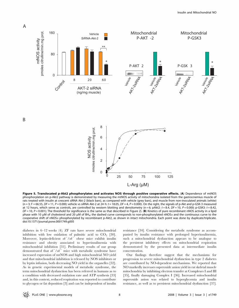
of p-Akt2 and p-GSK3 (Figure 5A).
This effect suggests that the insulin-dependent increase in NOS
activity is based upon the kinetic variations induced by Akt2-
related enzyme phosphorylation in mitochondria (Figure 5A). To
test this hypothesis, His-tagged nNOS was purified from bacteria
transfected with cDNA in pCWori+ vector, and enzyme activity
was assayed for phosphorylation in the presence of p-Akt2 and
c32PATP. The pioneer work by Fulton et al reported that Akt
activates eNOS by phosphorylating Ser1179 in transfected COS
cells [19]. In their study, analogous phosphorylation of nNOS was
observed only when the transfected construct had a myristoylation
A
C
EnNOS
mitochondria
Insulin
2412
Contro
l
cytosol
157 kDa
3
-actin
nNOS 453 bp
nNOS m RNA
Insulin
2412
Contro
l 3
mtN
OS
activ
itypm
oles
citru
lline
/min
.mg
prot
. cytosolmitochondria
Time (h)0
80
120
320
240
Insulin2412
Contro
l
3
Con
trol
Insu
lin
LY+I
nsul
in
NO
Sac
tivity
mt
pmol
esci
trulli
ne/m
in.m
gpr
ot
0
40
80
120
*
D
(µm
olO
/h.g
tissu
e)2
50
25
F
0
157 kDa
565 bp
Insulin
2412
Contro
l 3
*
*
Ly29
4002
Time (h)
Time (h)
Time (h)
G
Stat e 3 O uptake (ngat O/ min.mg prot)2
+ +Basal L-NMMA +L-Arg L-Arg+L-NMMA
Control 94±3 (+8) 2 (+15)90±5 80±2 9 ±6
Insulin 3 94±8 103 (+10) 87±2 85±5 (0)±6
12 35±3 9±2 27±51±4* (+45) 1 * 5 (+43)‡
24 44±2 3 ±2 4757±3* (+30) 5 * ±2 (+29)‡
Complex IV
-actin
*
*
*
****
mitochondria
cytosol
P-AKT
Con
trol
Insu
lin
LY+I
nsul
in
0
2
5
Den
sito
met
ry(A
.U.) *
**43
1
*
*
**
B
Eve
nts
DAF-FM
324
12
C
Con
trol
Insu
lin
LY+I
nsul
in
LY29
4002
LY29
4002
*
Time (h)
0
Insulin and Mitochondrial NO
PLoS ONE | www.plosone.org 4 2008 | Volume 3 | Issue 3 | e1749

consensus site, indicating that attachment to membranes is also
required for enzyme phosphorylation. Elfering et al reported that
mtNOS is myristoylated and has only one Akt-sensitive domain
(RXRXXS/T) at Ser1412, which has been reported to be
phosphorylated in the Maldi/MS analysis [20]. We therefore
measured K’ for L-arginine in phosphorylated or non-phosphor-
ylated recombinant nNOS in the presence of BH4 and cholesterol,
as a lipid phase representative of membrane insertion (Figure 5B).
The S1412G nNOS mutation, which simulates phosphorylation,
increases the heme reduction rate and decreases NO yield
and [21] electron transfer in the calmodulin-bound state. Here,
a sigmoid kinetic curve was noted for both the phosphorylated and
non-phosphorylated nNOS, but with different Hill slopes (3.7 and
1.9), findings that suggest a cooperative behavior (r2 = 0.99 and
0.97); in the lipid phase, p-Akt2 reduced K’ for L-Arg from 19 to
11 mM.
mtNOS is the final insulin/Akt effector that regulatesmuscle utilization of available glucose
A cornerstone of this study is that, after energy replenishment,
mitochondrial oxidations ought to be depressed to allow substrates
to be deposited as macromolecules. We tested the contribution of
mtNOS to the balance maintained between mitochondrial and
intermediary metabolism, by injecting siRNA nNOS into rat
gastrocnemius muscles with high nNOS content. This procedure
reduced nNOS mRNA levels as well as protein expression and
activity in mitochondria by approximately 50–77% in comparison
to constant complex IV (Figure 6A). Under these experimental
conditions and because of the decreased levels of matrix NO
(Figure 2B), oxygen uptake became insensitive to insulin
administration and remained high in the nNOS-silenced muscles.
The increase of glucose uptake elicited by insulin administration
was not modified by siRNA nNOS. Instead, siRNA nNOS
markedly increased glucose utilization evidenced by complete
oxidation to CO2 and H2O. Thus, at fixed insulin with similar
glucose uptake and glycogen synthase activity, high oxygen
utilization with siRNA nNOS was in line with high 3H2O and14CO2 production rates, i.e., 50–100% higher in the right siRNA-
treated muscle than in the left control (Figure 6B). No significant
variations of metabolic rate were detected at low mtNOS activity.
Interestingly, under these conditions the glucose used for glycogen
synthesis was halved in the siRNA nNOS-treated muscle; at
normal mtNOS content similar effects were observed when the
Akt2 gene was silenced, reducing protein expression and
phosphorylation of GSK-3a/b by 56% and 75%, respectively
(Figure 6C). In accordance with previous reports, direct admin-
istration of siRNA Akt2 did not result in significant variations of
glucose uptake or glycogen synthase activity, although the peak of
glucose uptake decreased at 24 h in the deficient muscles
(Figure 6C).
Discussion
We herein demonstrate the modulatory connection between
insulin signaling and mitochondrial function: insulin decreases the
muscle oxidative rate via mitochondrial NO. We confirmed this
finding using a number of different experimental approaches,
showing that the effect relies on p-Akt2-selective phosphorylation
of mitochondrial nNOS, after kinase translocation to the
organelles.
A rapid translocation of p-Akt to the mitochondria and the
phosphorylation of target GSK-3a/b by insulin-PI3K were
previously reported by Bijur and Jope in a variety of cells [22].
It is confirmed here that phosphorylation is absolutely required for
Akt translocation to muscle mitochondria and that, in this
framework, both p-Akt1 and p-Akt2 can be translocated to the
organelles ex vivo (Figure 3A). In vivo however, insulin only increases
p-Akt2 probably because of high expression of SH2-phophatase
that selectively dephosphorylates p-Akt1, but not p-Akt2 in skeletal
muscle [23]. Moreover, solely translocated p-Akt2 and not p-Akt1
induced a robust generation of NO by mtNOS in the isolated
mitochondria (Figure 3B). Considering that eNOS is activated in
the caveolae by insulin/PI3K via p-Akt1, specific functional and
topographic connections between NOS and Akt isoforms become
evident. In this way, a recent study reported p-Akt2 phosphory-
lation of nNOS at Ser1412 in synaptosomes, suggesting the
functioning of this mechanism in different organs and subcellular
fractions [24].
The phosphorylation of nNOS/mtNOS by p-Akt2 involves
cooperative effects centered at the C-terminal tail that contains the
Akt motif (Ser1412; Figures 4B and 7A). Although structural
changes of phosphorylated nNOS are not defined yet, it is worth
noticed that C-terminal tail participates in the regulation of nNOS
activity. Allosteric inhibition of electron transfer from NADPH to
FAD and poor NO release at low Ca2+-calmodulin level result
from electrostatic interactions between acidic negative charges of
29phosphate of NADP and positive charges of basic Arg1400 [25].
On this basis, we surmise that p-Akt2 activation of nNOS/mtNOS
(Figures 4 and 5) promote opposed electrostatic interactions
between Ser1412 and Arg1410 that, in the lipid phase, equilibrates
the oscillation of the C-terminal tail to cooperatively increase the
NO production rate (Figure 7A).
Different approaches confirmed in this study that phosphory-
lation of muscle mtNOS increases mitochondrial NO yield by
several folds and reduces systemic O2 utilization. We show here as
well that the temporal activation of mtNOS and the resulting
decline of mitochondrial O2 uptake via NO drive the muscle
Figure 2. Insulin-PI3K signaling triggers high mitochondrial NOS activity.(A) Time-course of mitochondrial NOS activity after insulinadministration measured by 3H-L-citrulline formation (n = 8; cytosol: F = 30.62, DF = 31, P = 0.000; mitochondria: F = 290.84, DF = 31, P = 0.000). (B)Matrix NO level appraised by DAF-FM fluorescence by flow-cytometry of isolated mitochondria; fluorescence was selectively analyzed in amitochondrial population delimited by previous incubation with Mito Tracker Red 580 (C) mtNOS activity of mitochondria isolated from 200 mg ofmuscle slices preincubated by 30 min with 12 nM of insulin or 100 mM of LY29400 (PI3K-specific inhibitor) (F = 38, DF = 27, P = 0.000). (D)Representative western blot of muscle p-Akt2 distribution under the same conditions as in C. (cytosol: F = 14.75, DF = 15, P = 0.000; mitochondriaF = 13.63, DF = 15, P = 0.000). (E) Expression of mtNOS and nNOS mRNA assessed by western blotting and RT-PCR, under the same conditions ofFigure 1. (F) Polarographic determination of oxygen uptake of 100 mg of sliced gastrocnemius and extensor digitorum longus muscles obtained afterinsulin administration in Robinson buffer with 5 mM of glucose as substrate, pH 7.4 (n = 6; F = 18.32, DF = 22, P = 0.000). (G) State 3 oxygen uptake ofmitochondria isolated from muscles was measured under the same conditions with malate-glutamate and ADP at 30uC in MSHE buffer, pH 7 and inthe presence of L-NMMA, L-Arg alone or together with NOS inhibitor L-NMMA (n = 3–12; 12 h: F = 13.83, DF = 15, P = 0.000; 24 h F = 15.69, DF = 37,P = 0.000, *P,.05 vs respective basal values by Dunnett posthoc test; basal control and insulin-treated groups: F = 45.21, DF = 23, P = 0.000, { P,.05 vscontrol by Dunnett posthoc test). In brackets, the percentage of variation respect to basal or L-arginine, respectively.doi:10.1371/journal.pone.0001749.g002
Insulin and Mitochondrial NO
PLoS ONE | www.plosone.org 5 2008 | Volume 3 | Issue 3 | e1749

insulin-transition from glucose oxidation to glycogen deposition. In
absence of NO or, in nNOS-silenced muscles, mitochondrial O2
uptake is completely released leading to a preferential oxidation of
glucose to CO2 and H2O; in these conditions, prevalent oxidation
of glucose reflects the respective Km of regulatory enzymes
like cytochrome oxidase for O2 and pyruvate dehydrogenase
complex for acetyl-CoA, at least one order of magnitude
lower (1027–1028 M, ref 26), than that of glycogen synthase for
glucose (1025 M, ref 27). In contrast, in the presence of NO,
glucose utilization is delivered to glycogen synthesis. These
results therefore support the notion that a discrete inhibition
of the respiratory chain as produced by NO is required to
+ Phosphatase
C AKT1 AKT2
+ Phosphatase
+ Phosphatase
His-tag
AKT
P-AKT
C AKT1 AKT2
A
B
supernatant mitochondria
P-AKT P-AKT +PhosphataseP-AKT + L-NMMA
Akt2
Akt1
Akt1 Akt2
Control
Control
Akt2
Control
Akt1
DAF-FM
0 0 010 0 10 010 2
10 110 1 10 210 0 10 1 10 2
512512 512
Eve
nts
Eve
nts
Eve
nts
mitochondria
Figure 3. Phosphorylation allows translocation of Akt1 and Akt2 to mitochondria, but only p-Akt2 increases matrix NO. (A)Translocation of Akt1 and 2 was tested ex vivo in purified mitochondria isolated from rat muscle Mitochondria were suspended in MSHE buffer withNADH and ATP (pH7.4), incubated with recombinant His-tagged p-Akt1 and p-Akt2 proteins for 30 min at 30uC and centrifuged at 10000 g by 10 minto separate supernatant from mitochondrial pellet further resuspended in phosphate buffer saline (PBS) and incubated with proteinase K andcompared with non-phosphorylated Akt. Western blots were performed with anti-His-tag (upper), anti-total Akt (middle) and anti-p-Akt antibodies(bottom). (B) To detect mitochondrial NO, flow cytometry histograms from isolated mitochondria (1 mg protein/ml) were obtained with DAF-FM inthe same conditions of (A) and after incubating the organelles for 30 min a 37uC with recombinant phosphorylated and non-phosphorylated Akt1and Akt2 alone or plus 3 mM L-NMMA. The histograms were obtained in a mitochondrial population previously delimited as in Figures 1 and 2.doi:10.1371/journal.pone.0001749.g003
Insulin and Mitochondrial NO
PLoS ONE | www.plosone.org 6 2008 | Volume 3 | Issue 3 | e1749

reduce oxidative rates and to allow glycogen or fat deposition
(Figure 7B).
At ten-fold higher insulin dose, maximal mtNOS activation was
reduced by a half (Figure S1). This fact reminds that insulin
activates other mitochondrial reactions as well; i.e., early activation
of pyruvate dehydrogenase complex through insulin-inhibition of
pyruvate dehydrogenase kinase (PDK4) could transiently compete
with NADH/NADPH availability for NOS in skeletal muscle
mitochondria [28]. In this study, effects at low and high insulin
concentrations thus explain: a) lack of stimulation and null mtNOS
activity in muscle at initial blood insulin peak (3 hours); b) high
respiratory rate at peak insulin level with null mtNOS (Figure 2G);
c) decreased respiratory rate at stable insulin concentration with
full mtNOS and, d) physiological alternation of insulin effects
promoting complete glucose oxidation or glycogen synthesis
(Figures 2 and 6). The increase or decrease of oxygen consumption
was previously reported to occur in muscle at respectively high or
low insulin concentration [29].
The presented data confirm insulin as a normal regulator of
oxidative phosphorylation rate in skeletal muscle but as well they
could explain cardinal features of the progression of metabolic
syndrome and obesity to type 2 diabetes. Zucker Fatty (ZF) rats
with a leptin receptor defect exhibit obesity, insulin resistance with
hyperinsulinemia and normal glycemia that progress to type 2
A
B
+
-
P-Akt2
Akt*
P-Akt2-GST
C Insulin
(kDa)
157198
131
nNOS
40
17
+
-
P-Ak
t 2
Akt*
C Insulin
73Id
+siR
NAnN
OS
Mit
+Ak
t-2
Brai
nho
mog
enat
e
-32P-A TP
157 kDa
Id+
siRNA
nNOS
Mit
+Ak
t-2
nNOS
Autorradiography Western blot
1 2 13 2 3
Brai
nho
mog
enat
e
Figure 4. Insulin promotes mitochondrial NOS phosphorylation. (A) Representative autoradiogram of mtNOS phosphorylation in organellesisolated from the gastrocnemius muscle of animals administered with insulin at p-Akt2 peak concentration (12 h). Mitochondria (2 mg of protein)were incubated in 50 MSHE with 2 mM of cold ATP and 2 mM of NADH and 10 mC of c32P-ATP with or without p-Akt2 or dephosphorylated Akt2 (*),as described in the legend of Figure 3. To the right, we show stable nNOS content in the same membrane of autoradiogram by western blotelectrophoretic run. (B) Left panel: Autoradiogram of mitochondrial proteins isolated from the gastrocnemius muscle electroporated in vivo withempty pRNAT-U6.1/Neo vector (1) or with 10 mg of siARN nNOS (2) as compared with brain homogenate (3). Isolated proteins were processed asdescribed in A; nNOS deficiency confirms insulin-induced mtNOS phosphorylation. Right panel shows a representative western blot of nNOS proteinfrom the same electrophoresis.doi:10.1371/journal.pone.0001749.g004
Insulin and Mitochondrial NO
PLoS ONE | www.plosone.org 7 2008 | Volume 3 | Issue 3 | e1749
diabetes in 6–12 weeks (4); ZF rats have severe mitochondrial
inhibition with low oxidation of palmitic acid to CO2 [30].
Moreover, leptin-deficient ob2/ob2 obese mice exhibit insulin
resistance and obesity associated to hyperinsulinemia with
mitochondrial inhibition [31]. Preliminary results of our group
demonstrated that ob2/ob2 mice with metabolic syndrome have
increased expression of mtNOS and high mitochondrial NO yield
and that mitochondrial inhibition is released by NOS inhibitors or
by leptin infusion, both decreasing NO yield in the organelles [32].
As in genetic experimental models of metabolic syndrome, the
term mitochondrial dysfunction has been referred in humans as to
a condition with decreased oxidation rate and ATP synthesis [33]
and, in this context, reduced respiration was reported to contribute
to glycogen or fat deposition [3] and can be independent of insulin
resistance [34]. Considering the metabolic syndrome as accom-
panied by insulin resistance with prolonged hyperinsulinemia,
such a mitochondrial dysfunction appears to be analogue to
the persistent inhibitory effects on mitochondrial respiration
demonstrated by the presented data at intermediate insulin
concentration.
Our findings therefore suggest that the mechanisms for
progression to severe mitochondrial dysfunction in type 2 diabetes
are contributed by NO-dependent mechanisms. We reported that
NO markedly increases superoxide anion yield in rat skeletal muscle
mitochondria by inhibiting electron transfer at Complexes I and III
[35], finally damaging Complex I [36]. Increased mitochondrial
superoxide anion was related to hyperglycemia and insulin
resistance, as well as to persistent mitochondrial dysfunction [37].
AMitochondrial
P-GSK3
B
MitochondrialP-AKT -2
P-GSK 3
0 25 50 75 100 1250
2
4
6
8
L-Arg ( M)µ
AKT 2 siRNA-(ng/mg muscle)
0
80
160N
OS
activ
itym
tpm
oles
citru
lline
/min
.mg
prot
.
Contro
l 8 20 60
AKT-
2siR
NA
P-AKT 2
NO
Sac
tivity
nmol
esci
trulli
ne/m
in.m
gpr
ot.
* *
**
AKT-
2siR
NA
AKT-
2siR
NA
AKT-
2siR
NA
Vehi
cle
SiRNA-Akt-2Vehicle
Vehi
cle
Vehi
cle
Vehi
cle
*
Figure 5. Translocated p-Akt2 phosphorylates and activates NOS through positive cooperative effects. (A) Dependence of mtNOSphosphorylation on p-Akt2 pathway is demonstrated by measuring the mtNOS activity of mitochondria isolated from the gastrocnemius muscle ofrats treated with insulin at crescent siRNA Akt-2 (black bars), as compared with vehicle (grey bars), and muscle from non-inoculated animals (white)(n = 3; F = 66.55, DF = 11, P = 0.000; vehicle vs siRNA Akt-2 at 24 h: t = 18.05, DF = 4, P = 0.000). On the right, the signals of p-Akt and p-GSK-3 measuredat 12 hours, which serve as controls, are controlled by western blotting and densitometry (n = 6; pAkt2: t = 8.4, DF = 10, P = 0.000; p-GSK3: t = 8.42,DF = 10, P = 0.001); The threshold for significance is the same as that described in Figure 2). (B) Kinetics of pure recombinant nNOS activity in a lipidphase with 10 mM of cholesterol and 20 mM of BH4; the dashed curve corresponds to non-phosphorylated nNOSa and the continuous curve to thecooperative shift of nNOSa phosphorylated by recombinant p-Akt2, as shown in intact mitochondria. Each point was done by duplicate/triplicate.doi:10.1371/journal.pone.0001749.g005
Insulin and Mitochondrial NO
PLoS ONE | www.plosone.org 8 2008 | Volume 3 | Issue 3 | e1749

In the presence of superoxide anion plus nitric oxide, 3-nitrotyrosine
was found as a marker of nitration of mitochondrial proteins in the
ob2/ob2 metabolic syndrome [31] and in experimental and clinical
diabetes [38]. Excessive NO leads both to nitrosylation and
inactivation of the insulin receptor and Akt, increasing insulin
resistance [39]. A number of different NO-based mitochondrial
mechanisms can contribute to insulin resistance as well. First, an
increase of ADP/ATP ratio due to inhibition of electron transfer
[17] can restrict the phosphorylation of insulin receptor substances
(IRSs) and the translocation of GLUT4 to the cell membrane.
Second, accumulation of NADH due to NO-dependent complex I
inhibition can negatively modulate glycolisis and the Krebs cycle
[40]. Finally, nitric oxide stimulates mitochondrial biogenesis by
increasing PGC 1a coactivator and thus could compensate
mitochondrial dysfunction in the early metabolic syndrome, while
reduced biogenesis with further decay of mitochondrial population
has been reported in already established type 2 diabetes [41].
From these perspectives, an additional explanation is required for
progression of diabetes and tissue damage in hypoinsulinemic status,
like type I diabetes, Akt2 deficiency or nNOS knocked out mice. In
contrast to activation of insulin-NOS pathway in mitochondria, in
these models progressive tissue and mitochondrial damage should
not depend on an increased ADP/ATP ratio; it is worth noticed that
Akt2 has extramitochondrial effects on GLUT4 and facilitates per se
the mechanism of glucose uptake. However, it is apparent as well
that weight loss in low-insulin diabetes or severe lypoatrophy in
hypoinsulinemic Akt2-deficient mice could occur because of the
lack of NOS activation, leading to uncontrolled mitochondrial
respiration and to high oxidative stress in the organelles with a
marked reduction of glycogen content [18].
We conclude that increase of NO in the mitochondrial
compartment by insulin is a powerful physiological resource to
properly adjust muscle O2 utilization, while persistence of this
mechanism in hyperinsulinemic states is harmful and could
contribute to diabetic mitochondrial dysfunction.
Materials and Methods
Animals and treatmentsAnimals were housed in a temperature-controlled room,
provided food, and subjected to a 12:12 dark/light cycle. National
Institutes of Health criteria for animal research were followed after
approval by the University Hospital.
To achieve a non-invasive hyperinsulinemic-normoglycemic
clamp, male Wistar rats [250–300g body wt] were subcutaneously
inoculated with long-acting human analogue insulin glargine
(Supporting Figure S2, 42–44) or NaCl 0.9% and skeletal muscle
was excised at 3, 12 and 24 hours. Insulin glargine, with its three
modifications to human insulin (GlyA21, ,ArgB31, ArgB32) is a
stable molecule that is soluble in slightly acidic conditions and
precipitates in the neutral pH of subcutaneous tissue. Because of
these properties, absorption of insulin glargine is delayed and the
analogue provides a fairly constant, basal insulin supply without
peaks in plasma insulin levels for approximately 24 hours, similar
to that achieved by a continuous subcutaneous insulin infusion
(Figure S2). During the 24-hour period, active metabolites sustain
insulin effects after the decay of the hormone. Animals were
allowed to drink water with 5% sucrose ad libitum to avoid
hypoglycemia (Figure S2). Without glucose intake, insulin glargine
results in hypoglycemia 2–4 h after administration (215 to 30% of
basal values; 45). Plasma insulin levels were followed indepen-
dently of endogenous hormone with a specific human immuno-
assay (IMX Microparticle Enzyme Immunoassay, Abbott Labs,
Ramsey, MN). Because the cross reactivity between insulin
glargine and human insulin is approximately 60%, thus resulting
in underestimation of its level in the immune assay, obtained
values were multiplied by 1.8 [43]. All experiments were carried
out in rat extensor digitorum longus and gastrocnemius red-mixed
muscles with appropriate mass (,0.6 g per muscle); adult rat
muscle is comprised of approximately 4–50% slow oxidative fibers
[I] and 20–40% fast oxidative fibers [IIa] with a large
mitochondrial population, high glycogen content, and concomi-
tantly, a high level of nNOS activity [46].
Akt/insulin Signaling Pathway and Nitric Oxide in MuscleMitochondria
Muscle mitochondria were isolated from homogenized muscles
by differential centrifugation and further purified with Percoll
gradients. After purification, the mitochondrial fraction has no
more than 10% activity of cytosol lactic dehydrogenase activity or
5% of calreticulin from SRE (western blot) while cytosolic fraction
did not express complex I proteins or 39 kDa subunit I of
cytochrome oxidase (western blot). Cytosol and mitochondrial
expression of nNOS, p-Akt, Akt1, Akt2 and p-GSK-3 a/b was
assessed by immunoblotting with specific antibodies and by
detecting variations of NO and p-Akt, by flow-cytometry in
freshly isolated mitochondria.
To analyze effects of PI3K pathway on mtNOS, muscles were
dissected out and rapidly cut into 20 to 30 mg strips and incubated
in a shaking water bath at 30uC for 30 min into a 25 ml flask
containing 3 ml of oxygenated Robinson buffer supplemented
with glucose and 0.1% BSA; strips were homogenized and
mitochondria were obtained. Flasks were gassed continuously with
95% O2-5% CO2 throughout the experiment.
NOS activity was followed in cytosol and purified mitochondria
by conversion of [3H]-L-arginine to [3H]-L-citrulline. To detect
variations of NO and p-Akt by flow-cytometry, freshly isolated
mitochondria were previously incubated for 30 min at 37uC with
fluorescent anti-p-Akt or 10 mM of DAF-FM and 0.5 mM of
MitoTracker Red 580 in PBS 1X. Fluorescence intensity was
measured using an Ortho Cytoron Absolute Flow Cytometer
[Johnson and Johnson, Raritan, NY]. Submitochondrial p-Akt
localization was studied by western blot after hypotonic disruption
and differential centrifugation; p-Akt activity was determined with
a commercial kit from Cell Signaling (Beverly, MA.). [3H]-L-
citrulline assay was done in 50 mM of potassium phosphate buffer,
pH 7.5, in the presence of 100 mM of L-arginine, 0.1 mM of [3H]-
L-arginine (NEN, Boston, MA.), 0.1 mM of NADPH, 0.3 mM
CaCl2, 0.1 mM of calmodulin, 10 mM of tetrahydrobiopterin,
1 mM of FAD, 1 mM of FMN, 50 mM of L-valine and 1 mg/ml of
protein [11]. Ca2+-dependent specific activity was calculated by
subtracting the remaining activity in the presence of the NOS
inhibitor L-NG-methyl-L-arginine (5 mM of L-NMMA) or 2 mM
of EGTA 3H arginine assay. NOS activity was measured as the
number of mitochondria isolated from rat muscle injected with
insulin and from muscle incubated with insulin or LY29400
(PI3K-specific inhibitor). Muscles were dissected out and rapidly
cut into 20- to 30-mg strips and incubated in a shaking water bath
at 30uC for 30 min before being transferred into a 25-ml flask
containing 3 ml of oxygenated Krebs-Ringer bicarbonate buffer
(KRB) supplemented with 8 mM of glucose, 32 mM of mannitol
and 0.1% BSA. Flasks were gassed continuously with 95% O2-5%
CO2 throughout the experiment. To measure the mtNOS activity,
muscle mitochondria were isolated and purified after incubation,
as described previously.
Total muscle RNA was extracted with Trizol, and RT-PCR was
performed under standard conditions.
Insulin and Mitochondrial NO
PLoS ONE | www.plosone.org 9 2008 | Volume 3 | Issue 3 | e1749

Translocation of Akts to mitochondriaIsolated organelles at 1 mg/ml were purified with percoll
gradients and incubated ex vivo with pure recombinant active Akt1-
GST or Akt2-GST (100 units/ml) (Cell Signaling) dually
phosphorylated at Thr-308 and Ser-473 or with isoforms
dephosphorylated with acid phosphatase and co-incubated
with the NOS inhibitor L-NMMA. Mitochondria were then
centrifuged and separated from supernatant, co-incubated
with 50 mg/ml proteinase K and DAF-FM, and subjected to
flow cytometry to assess fluorescence intensity. To allow
translocation of Akt2 to the inner mitochondrial membrane
[47], mitochondrial membrane potential was sustained by co-
incubating the isolated organelles with 2 mM NADH and 2 mM
ATP without Mg2Cl.
SiRNA-nNOSVector
2-deoxyglucoseuptake
CO142
(U- C)-glucoseto glycogen14
P-GSK3/
SiRN
A-nN
OS
Vect
or
mt-nNOS
0
90
180
A
90
45
B
SiRNA-Akt-2
CO142
C
(U- C)-glucoseto glycogen
14
Den
sito
met
rym
tNO
S/c
ompl
exIV
0
0.5
1.0
Antisubunit I,complex IV
AKT
2siR
NA-
*
mt-P-GSK3
Vehicle
Antisubunit I,complex IV
mt-P
-GS
K3
GS
K3/
com
plex
IV
mt-nNOS
0
0.5
0
Glycogensynthetase
Glycogensynthetase
nmol
UD
PG/m
in.m
gpr
ot
mtN
OS
activ
itypm
oles
citru
lline
/min
.mg
prot
*
Vect
orSi
RNA-
nNO
S
*
nmol
UD
PG/m
in.m
gpr
ot
0
*
8
4
Contro
l 3 12 24
Insulin
O uptake2
40
20
0
3 12 24
Insulin
(M
ol/h
gtis
sue)
µ.
20
10
0
3 12 24
Insulin
10
5
0
3 12 24
Insulin
12
6
0
12 24
Insulin
12 24
Insulin
6
3
24
Insulin
3 12 24
Insulin
12 24
Insulin
H O32
*
0
8
4
0
(M
ol/h
gtis
sue)
µ.
(M
ol/h
gtis
sue)
µ.
20
10
0
*
10
0
3 12 24
Insulin
15
H O32
5
2-deoxyglucoseuptake
90
45
12 24
Insulin
0
12 24
Insulin
Time (h)
Time (h)
Time (h)
Time (h)
Contro
l
*
**
****
*
********
**** **
*
*
Vehi
cle
Vehi
cle
AKT
2siR
NA-
*
*
**
**
**
**
Insulin and Mitochondrial NO
PLoS ONE | www.plosone.org 10 2008 | Volume 3 | Issue 3 | e1749

nNOS phosphorylation by p-Akt2NOS phosphorylation was evaluated in a mitochondrial free
assay with c32P-ATP and cold ATP in MOPS buffer alone or
together with active human recombinant Akt2 phosphorylated at
Ser473 and Thr308. Recombinant His-tagged nNOS protein was
obtained by magnetic field from E. coli transformed in a pcWori+vector containing the respective cDNA. Kinetics parameters were
assessed with GraphPad Software (San Diego, CA);
K’ = [Vmax6[L-Arg]h/V]-[L-Arg]h, where h is the calculated Hill
coefficient.
Oxygen uptakeOxygen uptake was measured at 220 mM of O2 in a standard
polarograph (IQUIFYB-MADEIC, Buenos Aires, Argentina) in
sliced muscle with Robinson buffer or in isolated mitochondria
with MSHE buffer with 6 mM of malate plus 6 mM of glutamate,
with or without 0.2 mM of ADP, 0.3 mM of L-arginine or 3 mM
of L-NMMA.
In vivo muscle siRNA electroporationAnimals were briefly anesthetized with sodium thiopental
[40 mg/kg weight, intraperitoneal], and gastrocnemius muscles
were percutaneously treated with one proximal and one distal
injection of siRNA nNOS (10 mg/per muscle] [GenScript, New-
Jersey, N.Y.) (right leg) or empty pRNAT-U6.1/Neo vector (left
leg) or with Akt2 siRNA from Santa Cruz Biotechnology (Santa
Cruz, CA) (right leg) or vehicle (left leg), in parallel. Each injection
was performed in 5 ml free-RNAase solution, slowly in 15–20 s
with a Hamilton syringe with a 22 G needle. Around 30 s after
injection, electric pulsation was applied. For pulse delivery to
muscle, two stainless steel connectors were applied on the needles
implanted in muscle. Therefore, the needles were used as
electrodes and connected to the generator; in these conditions
the depth of insertion was about 0.5 cm and the gap between the
two needles was 0.9 cm. The muscle were then electropulsed with
selected parameters using the Gene Pulser II Electropulsator (Bio-
Rad Laboratories,USA).Voltage (120V), pulse duration (20 ms)
and frequency of pulses (1Hz) were all preset on the electro-
pulsator. A train of eight pulses was delivered. [46]. The sequences
of nNOS siRNAs were designed on the structure of rat nNOS g.i.:
16258811 and are provided in Figure S3.
Metabolic studiesThirty six hours after electroporation, insulin was administered
at appropriate times and finally, animals were anesthetized and
gastrocnemius muscles were removed, weighed, and washed with
NaCl (0.9%). Finally, muscles were excised and minced in 0.1-mm
layers with a Thomas slicer and 180–200 mg of tissue were placed
into three different test tubes containing Krebs-Ringer bicarbonate
buffer with 2% BSA pH 7.2 (250 ml/100 mg of tissue) and gassed
with O2:CO2 [95:5 vol/vol] for five minutes at 37uC. To measure
glycogen synthesis and complete glucose oxidation to CO2 and
H2O, muscle was supplemented with 0.5 mCi/ml deoxy-D-
glucose,2 [1,23H[N]]-[25.0 Ci/mmol], 4 mCi/ml glucose, D-
[14C[U]]-[250 mCi/mmol] and 4 mCi/ml glucose D-[5-3H[N] ]-
[10.2 Ci/mmol]. Radioactives were from Perkin-Elmer Life and
Analytical Sciences, Boston, MA, USA.
Determinations were done after 12 hours of fasting; to maintain
both appropriate light-dark cycle and fasting, samples for glycogen
synthesis after 12 hours of insulin administration were obtained at
6:00 PM and for glucose oxidation at 3:00 and 11:00 AM.
To measure glucose uptake, muscle was placed separately in the
appropriate buffers and supplemented with radioactive deoxyglu-
cose [48]. Glycogen synthase activity was measured using the
method developed by Leloir and Goldemberg [49].
Statistical analysisData were compared with ANOVA and the Dunnett post hoc test
or the two-tailed Student’s t test as appropriate; significance was
accepted at P,.05.
Supporting Information
Figure S1 Muscle mtNOS activity at different doses of insulin
glargine. Each value represents separate experiments from animals
with insulin (closed circles) and without insulin (closed squares)
(n = 6, *P,.05 by ANOVA and Dunnett test). Strong activation of
mtNOS selectively occurs at half-maximal insulin dose.
Found at: doi:10.1371/journal.pone.0001749.s001 (0.17 MB EPS)
Figure S2 Effects of long-lasting insulin in rats. (A) A non-
invasive hyperinsulinemic normoglycemic clamp was achieved in
rats by subcutaneous administration of 0.1 U/Kg of insulin
glargine and 5% oral sucrose ad libitum; after an initial peak, level
of insulin+metabolites remains remarkably stable by 20 hours. (B).
The rate of sucrose intake of animals with or without (C) insulin
(white bar), at the same dose but at different times (dark bars);
*P,.05 respect control by ANOVA and Dunnett test).
Found at: doi:10.1371/journal.pone.0001749.s002 (0.24 MB EPS)
Figure S3 Sequence of hairpin siRNAs designed for rat nNOS
(gi: 16258810). siRNAs were cloned in vector pRNAT-U6.1/Neo
with BamH I and Hind III restriction enzymes; in red, poly A tail.
siRNA # 1 was preferentially used in the in vivo studies because of
its maximal inhibitory effect.
Found at: doi:10.1371/journal.pone.0001749.s003 (0.33 MB EPS)
Figure 6. mtNOS Is a final insulin/Akt effector regulating muscle utilization of available glucose. (A) Representative western blot ofdisrupted and normal mtNOS, densitometries, and mtNOS activities of mitochondria isolated from right and left gastrocnemius of the same animal,36 h after direct electroporation of respectively 10 mg of siRNA nNOS or empty pRNAT-U6.1/Neo vector to muscle, as described in ExperimentalProcedures. (n = 6; densitometry: t = 6.55, DF = 10, P = 0.000; activity at 12 h: t = 6.07, DF = 10, P = 0.000; activity at 24 h: t = 2.22, DF = 10, P = 0.05) (B) Totest the interdependence of oxidative and intermediary metabolism as related to activation of mtNOS, metabolic studies including oxygen uptake,glycogen synthesis, complete glucose oxidation to CO2 and H2O, glucose uptake and glycogen synthase activity were performed in nNOS-silencedright gastrocnemius- and vector-administered left muscles of the same animal in parallel under the different conditions by radioactive methods(n = 6; vector: O2 uptake, F = 10.47, DF = 23, P = 0.000; 14CO2, F = 5.06, DF = 23, P = 0.02; (U-14C)-glucose to glycogen, F = 8.61, DF = 23, P = 0.006; 2-deoxyglucose uptake, F = 22.55, DF = 23, P = 0.000; glycogen synthase, F = 3.59, DF = 23, P = 0.032; with siRNAnNOS: O2 uptake, F = 13.78, DF = 23,P = 0.000; 3H2O, F = 17.34, DF = 23, P = 0.000; 14CO2, F = 4.25, DF = 23, P = 0.034; (U-14C)-glucose to glycogen, F = 8.08, DF = 23, P = 0.004; 2-deoxyglucoseuptake, F = 12.90, DF = 23, P = 0.000; glycogen synthase, F = 3.62, DF = 23, P = 0.031). (C) Metabolic studies were performed by previous administrationof crescent siRNAAkt2 or vehicle under conditions analogous to A and B (n = 3, p-GSK3: t = 7.47, DF = 4, P = 0.002; at 24 h, vehicle vs siRNAAkt2, 14CO2:t = 23.23, DF = 4, P = 0.032; (U-14C)-glucose to glycogen: t = 2.85, DF = 4 P = 0.05). The threshold for significance is the same as that described inFigure 4.*P,.05 vs controls or between nNOS or Akt2 silenced and not-silenced muscle samples; ** P,.05 between different times of insulinadministration.doi:10.1371/journal.pone.0001749.g006
Insulin and Mitochondrial NO
PLoS ONE | www.plosone.org 11 2008 | Volume 3 | Issue 3 | e1749

Acknowledgments
We are grateful to Dennis Stuehr (Ph.D) for providing us with
cloned nNOS, to Daniel De Seta for insuline glargine and to Natalia
Riobo (Ph.D) and Juan J. Gagliardino (Ph.D) for their helpful
comments.
Author Contributions
Conceived and designed the experiments: JP SH JP DC MC. Performed
the experiments: PF FB SH MF CM DC. Analyzed the data: JP PF JP CM
AE MC. Contributed reagents/materials/analysis tools: AE. Wrote the
paper: JP MC.
mtNOS- P
CO +H O2 2
Glycogen
Glucose
Acetyl-S-CoA
Pyruvate
mtNOS
PI3KPDK1-2
Akt-2
P-GSK
GSK
P
PNH3
++
PNH3
Ser1412Arg1410Arg1400NADP
4.84.8
COOH
A
B
FMN FAD
Glucose
PAkt-2-
Akt-2-
Insulin Insulin
+
NO
NH3+
ATP
P-GSK
GSK
InsulinReceptorInsulinReceptor
Lipidsmitochondr
P
PP
Figure 7. Scheme of nNOS activation and the modulation of muscle metabolism. (A) A pendulous movement of the nNOS C-terminal tail(in blue) by opposed electrostatic interactions is suggested to explain cooperative positive modulation of nNOS phosphorylated by p-Akt2; nNOS (gi:16258810) was represented with Pymol (DeLano Scientific, Palo Alto, CA, USA). (B) Scheme of the modulation of mitochondrial respiration andglucose metabolism by the Akt2/nNOS couple in rat skeletal muscle.doi:10.1371/journal.pone.0001749.g007
Insulin and Mitochondrial NO
PLoS ONE | www.plosone.org 12 2008 | Volume 3 | Issue 3 | e1749

References
1. Trimmer BA, Aprille JR, Dudzinski DM, Lagace CJ, Lewi SM, et al. (2001)
Nitric oxide and the control of firefly flashing. Science 292: 2486–2488.2. Boveris A, Poderoso JJ (2000) Regulation of oxygen metabolism by nitric oxide.
In LIgnarro, ed (2000) Nitric Oxide Biology and Pathobiology San Diego,California. London, UK: Academic Press. pp 355–368.
3. Auwerx J (2006) Improving metabolism by increasing energy expenditure. Nat
Med 12: 44–45.4. Houstis N, Rose ED, Lander ES (2006) Reactive oxygen species have a causal
role in multiple forms of insulin resistance. Nature 440: 944–948.5. Petersen KF, Dufour S, Befroy D, Garcia R, Shulman GI (2004) Impaired
mitochondrial activity in the insulin-resistant offspring of patients with type 2
diabetes. N Engl J Med 350: 664–671.6. Derave W, Hansen BF, Lund S, Kristiansen S, Richter EA (2000) Muscle
glycogen content affects insulin-stimulated glucose transport and protein kinaseB activity. Am J Physiol Endocrinol Metab 279: E947–955.
7. Carreras MC, Poderoso JJ (2007) Mitochondrial nitric oxide in the signaling ofcell integrated responses. Am J Physiol Cell Physiol. 292: C1569–1580.
8. Poderoso JJ, Lisdero C, Schopfer F, Riobo N, Carreras MC, et al. (1999) The
regulation of mitochondrial oxygen uptake by redox reactions involving nitricoxide and ubiquinol. J. Biol. Chem. 274: 37709–37716.
9. Brown G (2001) Regulation of mitochondrial respiration by nitric oxideinhibition of cytochrome c oxidase. Biochim Biophys Acta 1504: 46–57.
10. Giulivi C, Poderoso JJ, Boveris A (1998) Production of nitric oxide by
mitochondria. J Biol Chem 273: 11038–11043.11. Franco MC, Antico Arciuch VG, Peralta JG, Galli S, Levisman D, et al. (2006)
Hypothyroid phenotype is contributed by mitochondrial complex I inactivationdue to translocated neuronal nitric oxide synthase. J Biol Chem 281: 4779–4786.
12. Persichini T, Mazzone V, Polticelli F, Moreno S, Venturin G, et al. (2005)Mitochondrial type I nitric oxide synthase physically interacts with cytochrome c
oxidase. Neurosci Lett 384: 254–259.
13. Kanai AJ, Pearce LL, Clemens PR, Birder LA, VanBibber MM, et al. (2001)Identification of a neuronal nitric oxide synthase in isolated cardiac
mitochondria using electrochemical detection. Proc Natl Acad Sci 98:14126–31.
14. Peralta JG, Finocchietto PV, Converso D, Schopfer F, Carreras MC, et al.
(2003) Modulation of mitochondrial nitric oxide synthase and energyexpenditure in rats during cold acclimation. Am J Physiol Heart Circ Physiol.
284: H2375–H2383.15. Carreras MC, Converso DP, Lorenti AS, Barbich M, Levisman DM, et al.
(2004) Mitochondrial nitric oxide synthase drives redox signals for proliferationand quiescence in rat liver development. Hepatology 40: 157–166.
16. Zaobornyj T, Valdez LB, La Padula P, Costa LE, Boveris A (2005) Effect of
sustained hypobaric hypoxia during maduration and aging on rat myocardiu-m.II. mtNOS activity. J Appl Physiol 98: 2370–2375.
17. Brookes PS, Bolanos JP, Heales SJ (1999) The assumption that nitric oxideinhibits mitochondrial ATP synthesis is correct. FEBS Lett 446: 261–263.
18. Cho H, Mu J, Kim JK, Thorvaldsen JL, Chu Q, et al. (2001) Insulin resistance
and a diabetes mellitus-like syndrome in mice lacking the protein kinase Akt2(PKBb) Science 242: 1228–31.
19. Fulton D, Gratton JP, McCabe TJ, Fontana J, Fuji Y, et al. (1999) Regulation ofendothelium-derived nitric oxide production by the protein kinase Akt. Nature
399: 597–601.20. Elfering SL, Sarkela TM, Giulivi C (2002) Biochemistry of mitochondrial nitric
oxide. J Biol Chem 277: 38079–38086.
21. Adak S, Santolini J, Tikunova S, Wang Q, Johnson JD, et al. (2001) Neuronalnitric oxide synthase mutant (Ser1412Asp) demonstrates surprising connections
between heme reduction NO complex formation and catalysis. J Biol Chem 276:1244–1252.
22. Bijur G, Jope RS (2003) Rapid accumulation of Akt in mitochondria following
phosphatidylinositol 3-kinase activation. J Neurochem 87: 1427–1435.23. Sasaoka T, Wada T, Fukui K, Murakami S, Ishihara H, et al. (2004) SH2-
containing Inositol Phosphatase 2 predominantly regulates Akt2, and not Akt1,phosphorylation at the plasma membrane in response to insulin in 3T3-L1
adipocytes. J Biol Chem 279: 14835–14843.
24. Rameau GA, Tukey DS, Garcin-Hosfield ED, Titcombe RF, Misra C, et al.(2007) Biphasic coupling of neuronal nitric oxide synthase phosphorylation to
the NMDA receptor regulates AMPA receptor trafficking and neuronal celldeath. J Neurosci 27: 3445–3455.
25. Tiso M, Konas DW, Panda K, Garcin ED, Sharma M, et al. (2005) C-terminal
Tail Residue Arg1400 enables NADPH to regulate electron transfer in neuronalnitric-oxide synthase J Biol Chem 280: 39208–39219.
26. Korochkina LG, Sidhu S, Patel MS (2006) Characterization of testis-specificisoenzyme of human pyruvate dehydrogenase. J Biol Chem 281: 9688–9696.
27. Hargreaves M (2004) Muscle glycogen and metabolic regulation. Proc Nutr Soc
63: 217–220.28. Kim YL, Lee FN, Choi WS, Lee S, Young JH (2006) Insulin regulation of
skeletal muscle PDK4 mRNA expression is impaired in acute insulin-resistantstates. Diabetes 55: 2311–2317.
29. Stump CS, Short KR, Bigelow ML, Schimke JM, Nair KS (2003) Effect of
insulin on human skeletal muscle mitochondrial ATP production, proteinsynthesis, and mRNA transcripts. PNAS 100: 7996–8001.
30. Noland RC, Woodlief TL, Whitfield BR, Manning SM, Evans JR, et al. (2007)Peroxisomal-mitochondrial oxidation in a rodent model of obesity-associated
insulin resistance. Am J Physiol Endocrinol Metab. 293: E986–E1001.31. Garcıa-Ruiz I, Rodrıguez-Juan C, Dıaz-Sanjuan T, del Hoyo P, Colina F, et al.
(2006) Uric acid and anti-TNF antibody improve mitochondrial dysfunction in
ob/ob mice. Hepatology 44: 581–91.32. Barreyro FJ, Finocchietto PV, Franco MC, Holod S, Carreras MC, et al. (2006)
Mitochondrial nitric oxide is increased in the mice Ob-/- model of metabolicsyndrome. Hepatology 44(4) Suppl1: 662A.
33. Lowell BB, Shulman GI (2005) Mitochondrial Dysfunction and Type 2
Diabetes. 384 21 Science 307: 384–387.34. Szendroedi J, Schmid AI, Chmelik M, Toth C, Brehm A, et al. (2007) Muscle
Mitochondrial ATP Synthesis and Glucose Transport/Phosphorylation in Type2 Diabetes. PLOS Med: 4: 858–867.
35. Poderoso JJ, Carreras MC, Lisdero C, Riobo N, Schopfer F, et al. (1996) Nitricoxide inhibits electron transfer and increases superoxide radical production in
rat heart mitochondria and submitochondrial particles. Arch Biochem Biophys
328: 85–92.36. Riobo NA, Clementi E, Melani M, Boveris A, Cadenas E, et al. (2001) Nitric
oxide inhibits mitochondrial NADH : ubiquinone reductase activity throughperoxynitrite formation. Biochem J 359: 139–145.
37. Rask-Madsen C, King GL (2007) Mechanism of disease: endothelial dysfunction
in insulin resistence and diabetes. Nature 3: 46–56.38. Turko IV, Li L, Aulak KS, Stuehr DJ, Chang JY, et al. (2003) Protein tyrosine
nitration in the mitochondria from diabetic mouse heart. J Biol Chem 278:33972–33977.
39. Yasukawa T, Tokunaga E, Ota H, Sugita H, Martyn JA, et al. (2005) S-nitrosylation-dependent inactivation of Akt/PKB in insulin resıstanse. J Biol
Chem 280: 7511–7518.
40. Mohr S, Hallak H, Boitte A, Lapetina EG, Brune B (1999) Nitric oxide-inducedS-glutathionylation and inactivation of glyceraldehydes-3-phosphate dehydro-
genase. J Biol Chem 274: 9427–9430.41. Nisoli E, Clementi E, Carruba MO, Moncada S (2007) Defective mitochondrial
biogenesis. A hallmark of the high cardiovascular risk in the metabolic
syndrome? Circ Res 100: 795–806.42. McKeage K, Goa KL (2001) Insulin glargine: a review of its therapeutic use as a
long-acting agent for the management of type 1 and 2 diabetes mellitus. Drugs61: 1599–1624.
43. Reinhart L, Panning CA (2002) Insulin glargine: a new long-acting product.Am J Health Syst Pharm 59: 643–649.
44. Hofmann T, Horstmann G, Stammberger I (2002) Evaluation of the
reproductive toxicity and embryotoxicity of insulin Glargine in rats and rabbits.Int J Toxicol 21: 181–189.
45. Delp MD, Duan CH (1996) Composition and size of type II, IIA, IID/X andIIB fibers and citrate synthase activity of rat muscle. J Appl Physiol 80: 261–270.
46. Golzio M, Mazzolini L, Moller P, Rols MP, Teisse J (2005) Inhibition of gene
expression in mice muscle by in vivo electrically mediated siRNA delivery. GeneTherapy 12: 246–251.
47. Truscott KN, Bradner K, Pfanner N (2003) Mechanism of protein import intomitochondria. Current Biology 13: R326–R337.
48. Cuendet GS, Loten EG, Jean Renaud B, Renold AE (1976) Decreased basal,
noninsulin-stimulated glucose uptake and metabolism by skeletal muscle isolatedfrom obese-hyperglycemic (ob/ob) mice. J Clin Invest 58: 1078–1088.
49. Leloir LF, Goldemberg SH (1960) Synthesis of glycogen from uridinediphosphate glucose in liver. J Biol Chem 235: 919–923.
Insulin and Mitochondrial NO
PLoS ONE | www.plosone.org 13 2008 | Volume 3 | Issue 3 | e1749