Control of Allergic Rhinitis Chemokine CCL19 and CCL21 in the Regulatory Role of Lymphoid
11
of August 7, 2015. This information is current as Rhinitis CCL19 and CCL21 in the Control of Allergic Regulatory Role of Lymphoid Chemokine Hiroshi Kiyono Dong-Young Kim, Aya Kawamura, Hideyuki Kawauchi and Kaoru Takamura, Satoshi Fukuyama, Takahiro Nagatake, http://www.jimmunol.org/content/179/9/5897 doi: 10.4049/jimmunol.179.9.5897 2007; 179:5897-5906; ; J Immunol References http://www.jimmunol.org/content/179/9/5897.full#ref-list-1 , 25 of which you can access for free at: cites 50 articles This article Subscriptions http://jimmunol.org/subscriptions is online at: The Journal of Immunology Information about subscribing to Permissions http://www.aai.org/ji/copyright.html Submit copyright permission requests at: Email Alerts http://jimmunol.org/cgi/alerts/etoc Receive free email-alerts when new articles cite this article. Sign up at: Print ISSN: 0022-1767 Online ISSN: 1550-6606. Immunologists All rights reserved. Copyright © 2007 by The American Association of 9650 Rockville Pike, Bethesda, MD 20814-3994. The American Association of Immunologists, Inc., is published twice each month by The Journal of Immunology by guest on August 7, 2015 http://www.jimmunol.org/ Downloaded from by guest on August 7, 2015 http://www.jimmunol.org/ Downloaded from
-
Upload
shimaneuniversity -
Category
Documents
-
view
0 -
download
0
Transcript of Control of Allergic Rhinitis Chemokine CCL19 and CCL21 in the Regulatory Role of Lymphoid
of August 7, 2015.This information is current as
RhinitisCCL19 and CCL21 in the Control of Allergic Regulatory Role of Lymphoid Chemokine
Hiroshi KiyonoDong-Young Kim, Aya Kawamura, Hideyuki Kawauchi and Kaoru Takamura, Satoshi Fukuyama, Takahiro Nagatake,
http://www.jimmunol.org/content/179/9/5897doi: 10.4049/jimmunol.179.9.5897
2007; 179:5897-5906; ;J Immunol
Referenceshttp://www.jimmunol.org/content/179/9/5897.full#ref-list-1
, 25 of which you can access for free at: cites 50 articlesThis article
Subscriptionshttp://jimmunol.org/subscriptions
is online at: The Journal of ImmunologyInformation about subscribing to
Permissionshttp://www.aai.org/ji/copyright.htmlSubmit copyright permission requests at:
Email Alertshttp://jimmunol.org/cgi/alerts/etocReceive free email-alerts when new articles cite this article. Sign up at:
Print ISSN: 0022-1767 Online ISSN: 1550-6606. Immunologists All rights reserved.Copyright © 2007 by The American Association of9650 Rockville Pike, Bethesda, MD 20814-3994.The American Association of Immunologists, Inc.,
is published twice each month byThe Journal of Immunology
by guest on August 7, 2015
http://ww
w.jim
munol.org/
Dow
nloaded from
by guest on August 7, 2015
http://ww
w.jim
munol.org/
Dow
nloaded from

Regulatory Role of Lymphoid Chemokine CCL19 and CCL21in the Control of Allergic Rhinitis1
Kaoru Takamura,*† Satoshi Fukuyama,* Takahiro Nagatake,* Dong-Young Kim,*‡
Aya Kawamura,* Hideyuki Kawauchi,† and Hiroshi Kiyono2*
The lymphoid chemokines CCL19 and CCL21 are known to be crucial both for lymphoid cell trafficking and for the structuralorganization of lymphoid tissues such as nasopharynx-associated lymphoid tissue (NALT). However, their role in allergic re-sponses remains unclear, and so our current study aims to shed light on the role of CCL19/CCL21 in the development of allergicrhinitis. After nasal challenge with OVA, OVA-sensitized plt (paucity of lymph node T cells) mice, which are deficient in CCL19/CCL21, showed more severe allergic symptoms than did identically treated wild-type mice. OVA-specific IgE production, eosin-ophil infiltration, and Th2 responses were enhanced in the upper airway of plt mice. Moreover, in plt mice, the number ofCD4�CD25� regulatory T cells declined in the secondary lymphoid tissues, whereas the number of Th2-inducer-typeCD8��CD11b� myeloid dendritic cells (m-DCs) increased in cervical lymph nodes and NALT. Nasal administration of theplasmid-encoding DNA of CCL19 resulted in the reduction of m-DCs in the secondary lymphoid tissues and the suppression ofallergic responses in plt mice. These results suggest that CCL19/CCL21 act as regulatory chemokines for the control of airwayallergic disease and so may offer a new strategy for the control of allergic disease. The Journal of Immunology, 2007, 179:5897–5906.
T he CCR7 ligands CCL19 and CCL21 are lymphoid che-mokines involved in the chemotaxis of lymphoid cellssuch as leukocytes and dendritic cells (DC)3 (1, 2). In-
deed, these chemokines play important roles in the formation ofappropriate cellular microcompartmentalization and homeostasisin lymphoid tissues (3–5). CCL19 is produced primarily by stro-mal cells in the thymus and by the T cell area of secondary lym-phoid tissues (1, 6). CCL21 is encoded by two genes, Scya21a(CCL21-Ser) and Scya21b (CCL21-Leu) (7). CCL21-Ser is pro-duced by stromal cells in the T cell area and by the high endothe-lial venules of the secondary lymphoid tissues, CCL21-Leu by thelymphatic endothelium alone (1). Paucity of lymph node T cells( plt) mice have a genomic deletion that includes the CCL19/
CCL21-Ser gene, leading to defective homing of naive T cells tothe secondary lymphoid tissues and thereby to insufficient archi-tectural development of nasopharynx-associated lymphoid tissue(NALT) and of other organized lymphoid tissues such as spleen,cervical lymph nodes (CLN), and Peyer’s patches (2, 5, 7, 8).Among these CCL19/CCL21-associated lymphoid tissues, NALTis considered one of the most important mucosal inductive sites forthe initiation and regulation of both mucosal and systemic immuneresponses to inhaled Ags (9). NALT has been shown to be rich inTh0-type CD4� T cells, which are capable of differentiating intoTh1 or Th2 cells based on the nature of the nasally administeredAg (10). For example, nasal immunization of the fimbrial proteinof anaerobe together with cholera toxin as mucosal adjuvant re-sulted in the induction of Ag-specific Th2-mediated IgA responses(11). Nasal administration of soluble protein together with a mu-cosal delivery molecule or immunomodulator (e.g., Escherichiacoli heat-labile toxin B subunit, cholera toxin) can suppress patho-genic responses (12, 13). Further, nasal administration of Ag caninduce Ag-specific regulatory T cells (Tregs) for the establishmentof immunological homeostatic conditions (14). Respiratory-asso-ciated lymphoid tissues including NALT have thus been shown toplay a pivotal role in the regulation of both active and quiescentmucosal immune responses in the respiratory tract as well as insystemic immune responses. However, their contribution to theinduction and regulation of allergic responses in the upper airwayremains to be elucidated.
Allergic rhinitis (AR) is a Th2-mediated disorder characterizedby Ag-specific IgE production, infiltration of inflammatory cellsincluding eosinophils into the nasal mucosa, and several nasalsymptoms such as sneezing, nasal congestion, itching, and rhinor-rhea (15). Exposure to allergens following allergic presensitizedconditions leads to the cross-linking of allergen-specific mast-cellsurface-bound IgE and basophils via the Fc�R, to the degranula-tion of these cells and to the release of histamine and other allergy-associated chemical mediators responsible for the early phases ofallergic responses. When released by mast cells and other cells,
*Division of Mucosal Immunology, Department of Microbiology and Immunology,Institute of Medical Science, The University of Tokyo, Tokyo, Japan; †Department ofOtorhinolaryngology, The Shimane University School of Medicine, Izumo, Japan;and ‡Department of Otorhinolaryngology, Seoul National University College of Med-icine, Seoul, Korea
Received for publication February 23, 2007. Accepted for publication August14, 2007.
The costs of publication of this article were defrayed in part by the payment of pagecharges. This article must therefore be hereby marked advertisement in accordancewith 18 U.S.C. Section 1734 solely to indicate this fact.1 This work was supported by the Core Research for Evolutional Science and Tech-nology (CREST) Program, the Japan Science and Technology Corporation, a Grant-in-Aid from the Ministry of Education, Science, Sports and Culture, and the Ministryof Health. S.F. and T.N. were supported by research fellowships from the JapanSociety for the Promotion of Science for Young Scientists. D.-Y.K. was supported byresearch fellowships from the Japan Society for the Promotion of Science for ForeignResearchers.2 Address correspondence and reprint requests to Dr. Hiroshi Kiyono, Division ofMucosal Immunology, Department of Microbiology and Immunology, The Instituteof Medical Science, The University of Tokyo, 4-6-1 Shirokanedai, Minato-ku, Tokyo,Japan. E-mail address: [email protected] Abbreviations used in this paper: DC, dendritic cell; plt, paucity of lymph node Tcells; NALT, nasopharynx-associated lymphoid tissue; CLN, cervical lymph nodes;Treg, regulatory T cell; AR, allergic rhinitis; m-DC, myeloid DC; NP, nasal passage;l-DC, lymphoid DC; WT, wild type.
Copyright © 2007 by The American Association of Immunologists, Inc. 0022-1767/07/$2.00
The Journal of Immunology
www.jimmunol.org
by guest on August 7, 2015
http://ww
w.jim
munol.org/
Dow
nloaded from

chemokines such as CCL5 (RANTES), CCL11 (eotaxin), CCL17(thymus and activation-regulated chemokine), and CCL22 (mac-rophage-derived chemokine) trigger recruitment of inflammatorycells such as eosinophils and Th2 cells, thereby contributing to theinduction of late-phase allergic responses (16). Inasmuch as thechemokine family has been shown to play a critical role in mostphysiological and pathological immune scenarios, we thought itlogical next to determine whether the NALT-associated lymphoidchemokines CCL19/CCL21 are involved in the development ofallergic responses in the upper respiratory compartment.
DCs are the front-line sentinels for Ag detection in both orga-nized lymphoid tissues such as Peyer’s patches and NALT and thediffused connective tissues of the lamina propria region in mucosalcompartments. Mucosal DCs have been shown to play an impor-tant role in the induction of both physiological and pathologicalTh1/Th2 polarization in protective immunity, inflammation, andallergy (17, 18). Mucosal DCs are also involved in the induction ofTregs for the creation of immunologically quiescent conditions inthe harsh environment of the aero-digestive mucosa (19, 20). Tregsare a distinct population of CD4� T cells constitutively expressingIL-2 receptor �-chains (CD25) (21). Tregs play a central role in theregulation of autoimmune, infectious, and allergic diseases by cell-to-cell contact-dependent inhibition and by the secretion of anti-inflammatory cytokines such as IL-10 and TGF-� (22). Th2 re-sponses have been shown to be down-regulated by naturallyoccurring CD4�CD25� Tregs expressing forkhead/winged-helixfamily transcription factor P3 (Foxp3) and by inducible popula-tions of Ag-specific IL-10-secreting Tregs (23, 24).
In this study, we examine whether the NALT-associated lym-phoid chemokines CCL19/CCL21 help regulate T cell-mediatedcontrol of allergic responses in nasopharyngeal tissue. plt miceshow aggravated allergic symptoms with aberrant Th2 responses,increased numbers of CD8��CD11b� myeloid DCs (m-DCs), anda reduction in CCR7-expressing Tregs in the NALT and CLN. Theworsened allergic responses in plt mice could be reversed by nasaladministration of plasmids encoding CCL19/CCL21-Ser DNA toreduce Th2-inducer-type m-DCs. Our results suggest that the lym-phoid chemokines CCL19 and CCL21 play a role in the control ofAR by reducing the numbers of m-DCs and thereby the likelihoodof inhibiting a Th2 environment.
Materials and MethodsMice
BALB/c mice were purchased from Japan SLC. Plt mice on a BALB/cbackground were provided by Dr. Terutaka Kakiuchi (Toho UniversitySchool of Medicine). Mice transgenic for a TCR that recognizes theOVA323–339 peptide in the context of I-Ad (DO11.10 TCR-�� transgenicmice) on a BALB/c background were purchased from The Jackson Labo-ratory Animal Resources Center. These mice were maintained under spe-cific pathogen-free conditions in the Laboratory Animal Research Centerof The Institute of Medical Science (The University of Tokyo). All micewere 6–7 wk of age at the beginning of individual experiments.
Induction of AR
For the induction of AR, we employed a previously described protocol withsome modifications in the quantity of Ags and the sensitization schedule(25). In brief, female BALB/c mice and plt mice were presensitized bymeans of an i.p. injection of 25 �g of OVA (Grade V; Sigma-Aldrich) with1 mg of aluminum hydroxide hydrate gel (Alum) (LSL Co.) in 200 �l ofPBS on days 0, 7, and 14. Thereafter, mice were challenged by nasaladministration of either 500 �g of OVA in 20 �l of PBS (for AR group)or 20 �l of PBS alone (for control group) for 14 consecutive days from day21 to 34.
Assessment of allergic symptoms
On days 20 (after three rounds of i.p. sensitization) and on days 27 and 34(after 7 and 14 nasal challenges, respectively), the instances of sneezing
and nasal rubbing in a 5-min period were counted by investigators in ablinded fashion after the last nasal challenge (25). At the same time, thebehavior of the mice was recorded by video camera.
ELISA for the analysis of IgE Abs and histamine in serum
For the analysis of total and OVA-specific IgE levels in serum, a sandwichELISA system was employed in accordance with the manufacturer’s pro-tocol (26). Ninety-six-well plates were coated with purified anti-mouse IgEmAb (clone R35–72; BD Pharmingen) and a purified mouse IgE isotype(27–74; BD Pharmingen) was used as a standard. HRP-conjugated anti-mouse IgE (23G3; Southern Biotechnology) (for total IgE) and HRP-labeled anti-biotin (Vector Laboratories) following biotin-labeled OVA(for OVA-specific IgE) were added to the plates as detection enzymes. Thereaction was developed by 3,3�,5,5�-tetramethylbenzidine (Moss) and ter-minated by the addition of 2 N H2SO4. OD was recorded by a luminometer(iEMS Reader; Labsystems) set at 450 nm. End-point titers of OVA-spe-cific IgE were expressed as the reciprocal log2 of the last dilution of asample giving an OD value 0.1 higher than background. Serum was col-lected within 10 min after the last nasal challenge and its histamine levelsanalyzed using a histamine immunoassay kit (Immunotech) (27).
Histological analysis for eosinophil infiltration
After the analysis of nasal symptoms, mice were sacrificed and their headsfixed in 4% paraformaldehyde at 4°C for 16 h. Fixed tissues were thendecalcified in EDTA solution at 4°C for 10 days and embedded in paraffin.Samples were sliced into 5 �m coronal sections and the sections subjectedto H&E staining (28). The number of eosinophils that had infiltrated intothe nasal septal mucosa was counted using a high magnification (�400)microscope.
Isolation of mononuclear cells
Spleen, CLN, and thymus were removed, and single-cell suspensions wereprepared by mechanical dissociation (10). Mononuclear cells of NALT andnasal passage (NP) were isolated as previously described with some mod-ifications (10). In brief, the palatine plate containing NALT was removedand then NALT was dissected out. NP tissues without NALT were alsoextracted from the nasal cavity, and mononuclear cells from individualtissues were isolated by gentle teasing using needles through 40-�mnylon mesh.
Analysis of cytokine production by CD4� T cells
For the purification of CD4� T cells, isolated mononuclear cells wereincubated with CD4 (L3T4) MicroBeads (Miltenyi Biotec) at 4°C for 30min. CD4� cells were sorted by autoMACS (Miltenyi Biotec) and sus-pended in complete RPMI 1640 medium containing 10% FBS, 5 �M2-ME, 10 U/ml of penicillin, and 100 �g/ml streptomycin. Cells were thencultured at a density of 1 � 105 cells/well in the presence of 1 mg/ml OVAwith T cell-depleted and irradiated splenic feeder cells (5 � 105 cells/well)in round-bottom 96-well microculture plates for 48 to 96 h (11).
To determine whether each DC subset preferentially directed naiveCD4� T cells to develop a Th1 or Th2 cytokine profile, mononuclear cellsharvested from mice with AR were first incubated with CD11c MicroBeads(Miltenyi Biotec) and sorted using autoMACS to enrich the CD11c� pop-ulation. Cells were then stained with allophycocyanin-conjugated anti-CD11c (HL3; BD Pharmingen), PE-conjugated anti-CD8� (53-6.7; BDPharmingen), and FITC-conjugated anti-CD11b (M1/70; BD Pharmingen)to collect CD11c�CD8��CD11b� cells (m-DCs) and CD11c�CD8��
CD11b� cells (lymphoid DCs: l-DCs) by FACSAria (BD Biosciences).Naive T cells were isolated from the spleen of DO11.10 TCR transgenicmice and stained with FITC-conjugated anti-CD62L (MEL-14; BD Pharm-ingen), PE-conjugated anti-CD44 (IM7; BD Pharmingen), and allophyco-cyanin-conjugated anti-CD4 (L3T4) (RM4–5; BD Pharmingen) usingFACSAria. Tregs were sorted from spleen cells as a CD3�CD4�CD25�
population using FACSAria by staining with FITC-conjugated anti-CD3�(145-2C11; BD Pharmingen), PE-conjugated anti-CD4 (BD PharMingen)and allophycocyanin-conjugated anti-CD25 (PC61; BD Pharmingen). DCsof each subset were initially cultured at a density of 1 � 104 cells/well,with naive CD4�CD44intCD62Lhigh cells (1 � 105 cells/well) isolatedfrom the spleen of DO11.10 TCR transgenic mice in the presence of humanIL-2 and OVA (1 mg/ml) with or without Tregs (2 � 104 cells/well) for 7days. Cells were then washed and re-stimulated with OVA in the presenceof irradiated splenic feeder cells (9 � 105 cells/well) for 48 h (29, 30). Forthe cytokine neutralization assay, anti-IL-10 mAb (JES5-2A5) (10 �g/ml),anti-TGF-� mAb (1D11) (10 �g/ml) or rat IgG (Sigma-Aldrich) (10 �g/ml) was added to the culture. Culture supernatants were collected and
5898 REGULATORY ROLE OF CCL19 AND CCL21 IN ALLERGIC RHINITIS
by guest on August 7, 2015
http://ww
w.jim
munol.org/
Dow
nloaded from

examined for the production of cytokines (IL-4, IL-5, IL-13, and IFN-�) bycytokine ELISA kits (R&D Systems).
RT-PCR
Total RNA was extracted using TRIzol reagent (Invitrogen). DNase diges-tion of the extracted RNA was performed before cDNA synthesis. Weconducted reverse transcription using Omniscript Reverse Transcriptase(Qiagen) and Oligo d(T)16 (Applied Biosystems), as well as quantitativereal-time PCR using LightCycler (Roche Diagnostics) with LightCycler-FastStart DNA Master Hybridization probes (Roche Diagnostics). Theprimers and hybrid probes for real-time PCR were as follows: the oligo-nucleotide primers specific for IL-13 (sense, 5�-AGCATGGTATGGAGTGTGGA-3�; antisense, 5�-GTGGGCTACTTCGATTTTGG-3�): theIL-13 detection FITC-labeled probe (5�-TGCAATGCCATCTACAGGACCCAGAGG-3�) and the Lightcycler Red 640-labeled hybrid probe (5�-TATTGCATGGCCTCTGTAACCGCAAGG-3�); the oligonucleotideprimers specific for GATA-3 (sense, 5�-CATGCGTGAGGAGTCTCCAA-3�; antisense, 5�-GGAATGCAGACACCACCTCG-3�): the GATA-3 de-tection FITC-labeled probe (5�-GGGCTTCATGATACTGCTCCTGCGAAA-3�) and the Lightcycler Red 640-labeled hybrid probe (5�-ACGCAAGTAGAAGGGGTCGGAGGAACTC-3�); and the oligonucleotide
primers specific for GAPDH (sense, 5�-TGAACGGGAAGCTCACTGG-3�; antisense, 5�-TCCACCACCCTGTTGCTGTA-3�): the GAPDH detec-tion FITC-labeled probe (5�-CTGAGGACCAGGTTGTCTCCTGCGA-3�)and the Lightcycler Red 640-labeled hybrid probe (5�-TTCAACAGCAACTCCCACTCTTCCACC-3�). They were designed and produced byNihon Gene Research Laboratories. A Lightcycler-primer/probes set (NihonGene Research Laboratories) was used for the amplification of the cDNA ofIL-4, IFN-�, and T-bet. Messenger RNA expression levels for specific geneswere normalized as a ratio relative to GAPDH.
Flow cytometric analysis
For the flow cytometric analysis, mononuclear cells isolated from severaltissues were first incubated with anti-CD16/CD32 (2.4G2; BD Pharmin-gen) to block nonspecific binding of Abs to the Fc� ��� and Fc� �� recep-tors, and then stained with each Ab. Allophycocyanin- (or PE)-conjugatedanti-CD11c, FITC-conjugated anti-CD11b, and PE-conjugated anti-CD8�were used to analyze DCs. For the analysis of Tregs, cells were stainedwith FITC-conjugated anti-CD3�, FITC- or PE-conjugated anti-CD4, andallophycocyanin-conjugated anti-CD25. In some experiments, a PE anti-mouse/rat Foxp3 staining set (FJK-16s; eBioscience) or biotin-conjugatedanti-CCR7 (4B12; eBioscience) with a streptavidin-PE conjugate (BD
FIGURE 1. Nasal symptoms and Ag-specific aller-gic responses in mice with allergic rhinitis (AR). WTmice and plt mice were nasally challenged with OVAfor 14 consecutive days following systemic sensitiza-tion. A, Sneezing and nasal rubbing were observed andcounted for 5 min at 4 different time points: 1) before re-ceiving any sensitization (naive, on day �1); 2) after threerounds of i.p. sensitization (on day 20); 3) after seven nasalchallenges (“in 7”, on day 27); and 4) after the last nasalchallenge (“in 14”, on day 34). B, Total and OVA-specificIgE levels in serum were assayed by sandwich ELISA.OVA-specific IgE Abs are expressed as reciprocal log2
titers. C, Serum histamine levels were determined by sand-wich ELISA. D, Nasal tissue was subjected to H&E stain-ing. The arrowheads point to eosinophils. The scale of thebar is 20 �m. E, Total numbers of eosinophils infiltratedinto the bottom of the nasal septum were counted. Thesedata are representative of three independent experimentscontaining three to five mice in each group. Significancewas evaluated by an unpaired t test. �, p � 0.05; ��, p �0.01 vs WT mice.
5899The Journal of Immunology
by guest on August 7, 2015
http://ww
w.jim
munol.org/
Dow
nloaded from

Pharmingen) was used. Compensation was carefully performed in eachtissue in accordance with the published instructions (31, 32). Nonviablecells were excluded using a Via-Probe (7-amino-actinomycin D; BDPharmingen). Stained cells were then analyzed using a FACSCalibur flowcytometer (BD Biosciences) with CellQuest software (BD Biosciences).
Nasal CCL19/CCL21 DNA treatment
CCL19 and CCL21-Ser cDNAs were amplified by PCR using cDNA fromwhole spleen cells of naive BALB/c mice as a template. The oligonucle-otide primers were as follows: the primers specific for CCL19 (sense, 5�-CCTTGTCTCGAGCCACCATGGCCCCCCGTGTGACCCCAC-3�; anti-sense, 5�-AGCCTCGAATTCTCAAGACACAGGGCTCCTTCTGG-3�)and CCL21-Ser (sense, 5�-CCTTGTCTCGAGCCTCAACTCAACCACAATCATGGC-3�; antisense, 5�-AGCCTCGAATTCCTATCCTCTTGAGGGCTGTGTC-3�, with underlining indicating the XhoI and EcoRI restric-tion enzyme site). Plasmid DNA encoding either CCL19 or CCL21-Serwas constructed by the ligation of CCL19 or CCL21-Ser cDNA, respec-tively, into a pIRES2-EGFP vector (BD Biosciences Clontech). The emptyvector pIRES2-EGFP (mock DNA) was used as a control. The plasmidDNAs and the mock DNA control were amplified in E. coli and purified
using an EndoFree Plasmid Maxi kit (Qiagen). For the detection of CCL19/CCL21 expression, each plasmid was transfected into COS-7 (CRL-1651;ATCC) in Opti-MEM (Invitrogen) by electrophoresis using Gene PulserXcell (Bio-Rad). After 48 h, culture supernatants were collected andchemokine levels were determined using a commercial ELISA kit(R&D Systems). Mice were sensitized by i.p. injection of 25 �g ofOVA with 1 mg of Alum on days 0, 7, and 14, followed by nasalchallenge with 500 �g of OVA for 14 consecutive days from day 21 to34 for the induction of AR. As a nasal CCL19/CCL21 DNA treatment,mice were nasally administered with an additional 100 �g of plasmid,mock DNA, or PBS on days �1, 6, and 13 (24 h before systemicsensitization) and from day 20 to 33 (before nasal challenge). Nasalsymptoms were observed, and sera and mononuclear cells in severaltissues were harvested for further examination.
Statistical analysis
Data were expressed as mean � SE and evaluated by an unpaired Student’st test. Values of p � 0.05 were assumed to be statistically significant.
FIGURE 2. Th2 cytokine production, and Th1/Th2cytokine and associated transcriptional factor mRNAexpression from CD4� T cells isolated from mice withAR. A, Culture supernatants of CD4� T cells of NALT,NP, CLN, and spleen from mice with AR were assessedfor Th2 cytokine production levels by ELISA. B, Th1and Th2 cytokine and associated transcriptional factor-specific mRNA expression in spleen, and NALT wasdetermined by quantitative real-time PCR analysis. Theexpression of each cytokine was normalized to the ex-pression of GAPDH. Representative results from threeindependent experiments containing three mice in eachgroup are shown. Significance was evaluated by an un-paired t test. �, p � 0.05; ��, p � 0.01 vs WT mice.N.D., not detected. P, PBS-treated; O, OVA-treated.
5900 REGULATORY ROLE OF CCL19 AND CCL21 IN ALLERGIC RHINITIS
by guest on August 7, 2015
http://ww
w.jim
munol.org/
Dow
nloaded from
ResultsInduction of severe allergic symptoms and Ag-specific IgEproduction in plt mice
To clarify the role played by the lymphoid chemokines CCL19 andCCL21 in the control of allergic diseases in the upper respiratorytract, the murine AR model was employed (25). Systemicallyprimed wild-type (WT) BALB/c mice and plt mice were nasallychallenged with OVA for 14 consecutive days with no significantsymptomatic difference between WT and plt mice seen through theseventh nasal challenge (Fig. 1A). After 14 days of continuousexposure, however, markedly more severe nasal symptoms wereobserved in plt than in WT mice (Fig. 1A). As would be expectedgiven the worsened nasal symptoms observed in plt mice, the se-rum of these mice showed significantly higher levels of OVA-specific IgE and of total IgE Abs than did that of identically treatedWT mice (Fig. 1B). To assess the extent of immediate AR-asso-ciated reactions, serum histamine levels were measured by ELISAand plt mice were found to produce significantly higher levels ofhistamine than WT mice (Fig. 1C). When the nasal tissues of na-sally challenged plt and WT mice were histologically compared,plt nasal tissue showed higher numbers of infiltrated eosinophils, asignature trait of the delayed phase of the allergic reaction (Fig. 1,D and E). These findings suggest that the clinical symptoms ofinhaled Ag-induced AR escalate in the absence of the lymphoidchemokines CCL19 and CCL21.
Th2 responses were enhanced in plt mice
Given the exacerbated Ag-specific allergic responses observed inplt mice to nasally administered Ag, we hypothesized that Th2responses would also be enhanced in these mice. To test this hy-pothesis, we compared the Th1/Th2 cytokine synthesis profiles ofplt and WT mice after chronic exposure to nasal allergens. Thus,Th1 and Th2 cytokine production was measured in vitro by OVAstimulation of CD4� T cells isolated from the CLN, the regionalLN of the upper airway, in nasally challenged plt and WT mice. As
one might expect, no evidence of Th1 cytokine IFN-� synthesiswas found in plt mice with AR (data not shown). In contrast,significantly higher levels of the Th2 cytokines IL-5 and IL-13were observed in CLN isolated from nasally challenged plt thanfrom WT mice (Fig. 2A). Higher levels of IL-5 and IL-13 werealso noted in CD4� T cells isolated from the site of allergic reac-tions, i.e., NALT and NP, of plt than of WT mice. An identicalpattern of elevated IL-5 and IL-13 production was also noted insystemically (spleen-) derived CD4� T cells of plt mice with AR.As a rule, levels of IL-4, the other known Th2 cytokine, tended tobe higher in plt mice nasally exposed to allergens, but that increasedid not reach statistical significance when compared with WT mice(Fig. 2A). However, the NP of plt mice, where major local allergicresponses were occurring, showed a more vigorous synthesis ofIL-4 and an increase over WT IL-4 levels that reached statisticalsignificance. The hypothesis that Th2 responses were dominant innasally challenged plt mice received further support from the anal-ysis of the levels of Th1-/Th2-associated transcription factor andof cytokine-specific mRNA. Higher levels of GATA-3- and IL-13-specific mRNA expression were noted in spleen of plt micethan in that of WT mice, although no significant difference wasobserved in IL-4-specific mRNA expression levels (Fig. 2B). Incontrast, the mRNA expression of Th1 transcription factor T-betand IFN-� was low or undetectable in plt mice with AR. Theseresults suggest that inhaled allergens trigger an aberrant Th2 im-munological environment at both inductive (e.g., NALT) and ef-fector (e.g., NP) sites in plt mice.
Low numbers of naturally occurring Tregs but elevated numbersof m-DCs under allergic conditions are characteristic of plt mice
To further elucidate the immunopathological mechanisms under-lying the exacerbated allergic responses observed in plt mice, wenext set out to determine whether the regulatory network formedby Tregs and DCs was altered in nasally challenged plt mice. Flowcytometric analysis revealed a lower frequency of and decreased
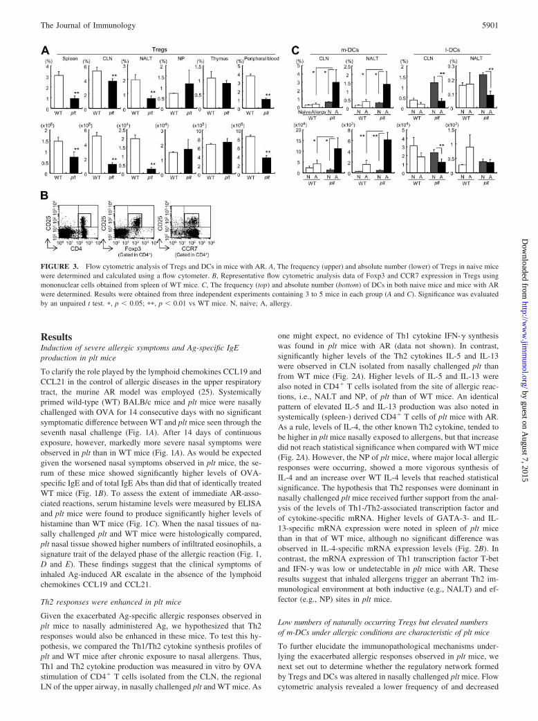
FIGURE 3. Flow cytometric analysis of Tregs and DCs in mice with AR. A, The frequency (upper) and absolute number (lower) of Tregs in naive micewere determined and calculated using a flow cytometer. B, Representative flow cytometric analysis data of Foxp3 and CCR7 expression in Tregs usingmononuclear cells obtained from spleen of WT mice. C, The frequency (top) and absolute number (bottom) of DCs in both naive mice and mice with ARwere determined. Results were obtained from three independent experiments containing 3 to 5 mice in each group (A and C). Significance was evaluatedby an unpaired t test. �, p � 0.05; ��, p � 0.01 vs WT mice. N, naive; A, allergy.
5901The Journal of Immunology
by guest on August 7, 2015
http://ww
w.jim
munol.org/
Dow
nloaded from

numbers of CD4�CD25� T cells in the secondary lymphoid tis-sues such as NALT and in the peripheral blood of naive plt than ofnaive WT mice (Fig. 3A). Because these CD4�CD25� T cellsexpressed Foxp3, they were considered to be Tregs (Fig. 3B). In-terestingly, most of these Tregs observed in both plt and WT miceexpressed CCR7 (Fig. 3B). As Treg levels did not change afterthe induction of AR (data not shown), these Tregs were con-sidered to be naturally occurring. As CD8��CD11b� m-DCsand CD8��CD11b� l-DCs are reported to have immunomodula-tory roles in Th1/Th2 cytokine production (21–23), our next flowcytometric analysis was aimed at DC subsets located in the varioustissues of plt mice with severe AR. The most significant changesobserved were an increased frequency of m-DCs residing in sec-ondary lymphoid tissues such as CLN and NALT of nasally chal-lenged plt mice with AR (0.66 � 0.08% to 3.02 � 1.51% and0.30 � 0.02% to 1.41 � 0.74%, respectively; Fig. 3C) and anelevated total number of m-DCs (Fig. 3C). In contrast, the fre-quency and number of m-DCs in nonsensitized plt mice were com-parable to those seen in WT mice (Fig. 3C). Taken together, thesefindings suggest that, under CCL19- and CCL21-deficient condi-tions, severe AR is associated with a reduction in naturally occur-ring Tregs and an increase in the frequency and the number ofm-DCs in the nasal mucosa-associated lymphoid tissues.
m-DCs induce a predominantly Th2 environment
Inasmuch as the secondary lymphoid tissues of plt mice with se-vere nasal allergic responses showed increased numbers of m-DCs,we focused our next experiment on the role of this DC subset in thedevelopment of AR. When cultured with naive CD4� T cells iso-lated from OVA-TCR transgenic mice in the presence of OVA,m-DCs isolated from the CLN of WT mice with AR producedsignificantly higher levels of IL-4 and IL-13 than did l-DCs iso-lated from the same mice (Fig. 4). When Tregs isolated from WTmice were added to Th2-leaning cultures of m-DC and CD4� T
FIGURE 4. Role played by each DC subset in the induction of Ag-specific T cell responses. The production levels of IL-4, IL-13, and IFN-�were measured by ELISA in culture supernatants of naive T cells fromDO11.10 OVA-TCR transgenic mice cocultured with or without Tregsand/or DCs. Data are representative of three separate experiments. Signif-icance was evaluated by an unpaired t test. �, p � 0.05; ��, p � 0.01. N.S.,not significant.
FIGURE 5. Allergic symptoms and the number of Tregs and DCs in plt mice treated by nasal administration of plasmids encoding CCL19/CCL21-SerDNA. Nasal symptoms (A) and total and OVA-specific IgE levels in serum (B) were assessed as previously described at two different time points: 1) after7 nasal challenges (“in 7”, on day 27) and 2) after the last nasal challenge (“in 14”, on day 34). C, The final frequency (top) and absolute number (bottom)of Tregs were determined and calculated by flow cytometric analysis. D, The final frequency (top) and absolute number (bottom) of DCs in CLN and NALTwas determined. These data were obtained from two to three independent experiments containing 3 to 5 mice in each group. Significance was evaluatedby an unpaired t test. �, p � 0.05; ��, p � 0.01. P, PBS-treated; M, mock DNA-treated; 19, pCCL19-treated; 21, pCCL21-treated.
5902 REGULATORY ROLE OF CCL19 AND CCL21 IN ALLERGIC RHINITIS
by guest on August 7, 2015
http://ww
w.jim
munol.org/
Dow
nloaded from

cells, Th2 cytokine production was suppressed. Tregs from pltmice possessed a similar capacity to suppress m-DC-induced IL-4and IL-13 production (data not shown). The addition of two neu-tralizing Abs, anti-IL-10 Ab and anti-TGF-� Ab, did not inhibitTreg function, suggesting that Tregs suppress Th2 production in-dependently of suppressive cytokines such as IL-10 and TGF-�(data not shown). No significant difference was observed in IFN-�production between CD4� T cells cocultured with m-DCs andthose cocultured with l-DCs (Fig. 4). These data demonstrate thatm-DCs are key players in Th2 cytokine production and Tregs arekey players in its suppression.
Nasal administration of plasmids encoding CCL19 DNA alteredDC population and suppressed allergic symptoms in plt mice
Because plt mice show enhanced allergic responses, we nextsought to determine whether artificial reconstitution of lymphoidchemokines using plasmids encoding CCL19 DNA (pCCL19) andCCL21-Ser DNA (pCCL21) would lead to the inhibition of nasalallergic responses. For the assessment of protein production,pCCL19 and pCCL21 were transfected into COS-7 cells and theproduction of CCL19 and CCL21 was confirmed in culture super-natants (data not shown). When plt mice were treated with controls(PBS or mock DNA) or these chemokine plasmids together withAR induction, significantly milder nasal clinical symptoms wereobserved in plt mice treated with pCCL19 than in mice treatedwith PBS or mock DNA (Fig. 5A). These milder clinical symptomswere similar to those observed in AR-induced WT mice withoutany treatment. Both total IgE- and OVA-specific IgE Abs werealso significantly lower in pCCL19-treated plt mice than in controlmice (Fig. 5B). Following pCCL21 treatment, some lessening ofexaggerated allergic symptoms and IgE production were noted,but, with the exception of the level of Ag-specific IgE, observed
differences did not reach statistical significance when comparedwith control-treated mice (Fig. 5, A and B).
Flow cytometric analysis revealed that the frequency of Tregswas higher in spleen, CLN and NALT of plt mice treated withpCCL21 than in control plt mice (Fig. 5C). In mice treated withpCCL19, the frequency of Tregs also increased in CLN and NALTcompared with control-treated mice, but no statistical differencewas observed (Fig. 5C). The frequency of m-DCs in CLN of pltmice treated with pCCL19 was considerably reduced when com-pared with control-treated mice (Fig. 5D). The frequency of m-DCs was also lower in NALT of mice treated with pCCL19 and inCLN of mice treated with pCCL21 than in control mice, but thereduction did not reach statistical differences (Fig. 5D).
When the nasal chemokine plasmid treatment was also tested inWT mice, the reduction of nasal symptoms could be observed;however, serum IgE levels did not change when WT mice weretreated with pCCL19/pCCL21 (Fig. 6, A and B). Nasal pCCL19treatment induced a higher frequency and increased number ofTregs in spleen as well as CLN (Fig. 6C). The total number ofm-DCs in NALT was reduced when WT mice were treated withpCCL19/pCCL21 (Fig. 6D). Taken together, these data suggestthat CCL19 and CCL21 increase the number of Tregs and simul-taneously inhibit the pathological function of m-DCs in allergicdiseases.
DiscussionBecause our recent study demonstrated that lymphoid chemokinesplay a crucial role in the maturation of NALT, a major commandertissue for the upper respiratory mucosal immune system (5), we setout in this study to elucidate the roles of the lymphoid chemokinesCCL19 and CCL21 in the development of allergic diseases asso-ciated with the nasal cavity and other upper airway tract tissues
FIGURE 6. Allergic symptoms and the number of Tregs and DCs in WT mice treated by nasal administration of plasmids encoding CCL19/CCL21-Ser DNA.Nasal symptoms (A) and total and OVA-specific IgE levels in serum (B) were assessed at two different time points: 1) after 7 nasal challenges (“in 7”, on day 27)and 2) after the last nasal challenge (“in 14”, on day 34). C, The final frequency (top) and absolute number (bottom) of Tregs were determined by flow cytometricanalysis. D, The final frequency (top) and absolute number (bottom) of DCs in CLN and NALT were determined. These data were obtained from three independentexperiments containing 3 to 6 mice in each group. Significance was evaluated by an unpaired t test. �, p � 0.05; ��, p � 0.01.
5903The Journal of Immunology
by guest on August 7, 2015
http://ww
w.jim
munol.org/
Dow
nloaded from

using a murine AR model. Constitutively produced by stromalcells in the T cell area of lymphoid tissues, in the endothelial cellsof high endothelial venules, and in lymphatic vessels, these twochemokines are involved in homeostatic lymphocyte traffickingand DC migration as well as in the distribution of cells in and thedevelopment of organized lymphoid tissues (1). After up-regulat-ing CCR7 expression, DCs migrate via lymphatic vessels fromperipheral tissues into T cell areas of the secondary lymphoid tis-sues, which constitutively express CCL19 and CCL21 for thepriming of naive T cells expressing CCR7 (1–3). Primed CD4� Tcells down-regulate CCR7 expression and differentiate into Th1,Th2 or nonpolarized memory T cells before circulating or migrat-ing to the periphery for the initiation of Ag-specific immune re-sponse (33). Despite such major immunological contributions bythese chemokines to secondary lymphoid tissue development andimmune cell trafficking, limited information exists as to their in-volvement in allergic states of the upper respiratory tract such asAR. By using plt mice defective in CCL19 and CCL21-Ser but stillcapable of producing CCL21-Leu from the endothelium of lym-phatic vessels (8), we have directly shown that CCL19 and CCL21regulate the inhibition of nasal allergic responses. It has been fur-ther demonstrated that plt mice show delayed but enhanced T cellresponses in a contact sensitivity model (8). It has also been shownthat enhanced Th2-mediated allergen-induced lung inflammationis observed in plt mice using an asthma model (34, 35). Our cur-rent study is in agreement with these previous findings and dem-onstrates in a murine model of allergy in the upper respiratory tractthat Th2 allergic responses are aggravated when CCL19 andCCL21-Ser are deficient. It also shows that AR development isprevented by the reintroduction of the chemokines using the cor-responding plasmids via the nasal route. Together with the originalstudies (8, 34, 35), our study suggests that the lymphoid chemo-kine family of CCL19 and CCL21 regulates the inhibition of hy-persensitivity responses including allergy.
Under CCL19- and CCL21-deficient conditions, m-DCs weresignificantly increased in the upper airway-associated lymphoidtissues of the CLN and NALT of mice with AR. Indeed, m-DCsisolated from the CLN of WT mice with AR induced considerablyhigher levels of IL-4 and IL-13 but not of IFN-� production fromcocultured naive T cells when compared with l-DCs. Interestingly,the nature of the immune response (Th1 and/or Th2 responses)depends in part on the specific subsets of DCs involved and theirpoint of origin (17). For example, m-DCs isolated from the spleenand/or Peyer’s patches are capable of inducing Ag-specific T cellsto produce Th2 cytokines, while l-DCs induce Th1 responses (29,36). Furthermore, Th2 responses are generated when bonemarrow-derived m-DCs are transferred into the airway, leadingto eosinophil infiltration in the asthma model (37). Lymphoidchemokines can also enhance the functioning of DCs; bonemarrow-derived DCs stimulated with CCL19/CCL21 produceinflammatory cytokines such as IL-1�, TNF-� and IL-12 at al-most comparable levels to those stimulated with LPS and anti-CD40 Ab (38). Indeed, CCL19-activated DCs selectively me-diate the induction of Th1 responses (38). Taken together, thesefindings strongly suggest that the absence of CCL19/CCL21during DC Ag presentation strongly favors the creation of aTh2-dominant environment that is conducive to the establish-ment of airway allergy.
Chemokines other than CCL19/CCL21 may help account for theincrease in m-DCs noted in plt mice. For instance, CCR6 is re-quired for the recruitment of m-DCs toward mucosal surfaces ex-pressing its ligand CCL20 (39), and the CCL20-CCR6 signal iscrucial for airway immune responses. The proinflammatory cyto-kines TNF-� and IL-1� and the Th2 cytokines IL-4 and IL-13 can
stimulate human bronchial epithelial cells to produce CCL20 (40).CCR6 knockout mice demonstrate a diminished allergic responseand reduced peribronchial eosinophil accumulation and IgE pro-duction (41). These findings suggest that other chemokines in ad-dition to CCL19/CCL21 may be involved in the recruitment ofm-DCs in plt mice with AR.
In general, undesired T cell-mediated responses are down-reg-ulated by Ag-specific inducible Tregs and/or naturally occurringCD4�CD25�Foxp3� Tregs. Down-regulation is mediated by theanti-inflammatory cytokines IL-10 and TGF-� and/or by cell-to-cell contact with coinhibitory molecules such as cytotoxic T lym-phocyte-associated Ag-4 (CTLA-4), B and T lymphocyte attenu-ator, and PD-1 (22–24, 42). The frequency of Ag-specific Tregsexpressing the surface molecules CTLA-4 and PD-1 and secretingIL-10 and TGF-� is higher in healthy individuals than in allergicindividuals (43). Indeed, healthy immune responses to allergensdepend upon a proper balance between allergen-specific Tregs andallergen-specific Th2 cells, with a disruption of that balance char-acterizing disease states like allergies (43). The murine colitismodel can be used to demonstrate that the mediation of the in-flammatory immune response by naturally occurring Tregs de-pends upon CTLA-4 (44). B and T lymphocyte attenuator andPD-1 are crucial in limiting the duration of acute allergic airwayinflammation and act as terminators of established immune re-sponses (42). In addition, naturally occurring Tregs are capable ofinhibiting DC function directly. Depletion of these Tregs resultedin worsening airway hyperresponsiveness as the result of the ex-aggerated Th2 cytokine production caused by altered pulmonaryDC function (45). In the murine asthma model, in vivo transfer ofAg-specific Tregs reduced airway hyperresponsiveness, eosinophilrecruitment and Th2 responses in an IL-10-dependent manner (46).Th2 responses were elevated when m-DCs and naive T cells werecocultured in the presence of Ag, but suppressed upon the additionof Tregs (Fig. 4). These data suggest that naturally occurring Tregsplay a critical role in inhibiting the Th2 response, probably bydirectly suppressing T cell responses and/or by indirectly suppress-ing the m-DC function that favors Th2 responses. Moreover, nei-ther anti-IL-10 Ab nor anti-TGF-� Ab treatment impaired Treg-mediated Th2 suppression, suggesting that this suppression wasindependent of the inhibitory cytokines IL-10 and TGF-� (our un-published observation). However, cell-to-cell interaction is re-quired for the suppression of aberrant Th2 responses by Tregs.
CCL19/CCL21-Ser is not produced by plt mice, but CCL21-Leuis produced by their lymphatic vessels (1). As a result, activatedm-DCs and naive T cells expressing CCR7 can migrate to andaccumulate in secondary lymphoid tissues in plt mice. To this end,CCL21-Leu has been shown to act as a chemoattractant for CCR7-expressing DCs (2, 7). Because Tregs are significantly reduced inplt mice with AR (Fig. 3A), the capacity of Tregs to inhibit m-DCfunction and thereby suppress Th2 responses may also be im-paired. However, a reduction in Tregs was observed in both naiveand diseased plt mice, suggesting that the remnant Treg popula-tions might be Ag-nonspecific, naturally occurring Tregs. The or-igin and development of naturally occurring Tregs in the thymusare still poorly understood. Generally, upon TCR-mediated posi-tive selection, developing thymocytes relocate within the thymusfrom the cortex to the medulla for further differentiation and se-lection before export to the periphery (47). The CCR7 signal isessential for the migration of single-positive thymocytes from thecortex to the medulla and for the optimal emigration of T cellsfrom the thymus to the periphery in newborn but not in adult mice(47). In plt mice, mature single-positive thymocytes rarely migratefrom the cortex to the medulla, but, paradoxically T cell export
5904 REGULATORY ROLE OF CCL19 AND CCL21 IN ALLERGIC RHINITIS
by guest on August 7, 2015
http://ww
w.jim
munol.org/
Dow
nloaded from

from the thymus into peripheral blood is not impaired (47). There-fore, one can speculate that plt mice have an impaired ability tomaintain naturally occurring Tregs in the periphery (blood circu-lation) once they exit the thymus, but further studies are needed toshed light on this issue.
Nasal administration of pCCL19 results in the inhibition of ARdevelopment in plt mice. To the best of our knowledge, the currentstudy provides the first evidence that intranasal pCCL19 treatmentsuppresses AR-associated allergic responses. In recent studies,plasmid DNA can be used in vivo as an adjuvant to enhance Ag-specific immune response (48) or as a therapeutic tool to alterundesired immunopathological conditions (49, 50). Intranasalcodelivery of plasmids encoding the DNA of CCR7 ligands andplasmid DNA or recombinant vaccinia virus encoding HSV-gBincreases HSV-gB-specific serum IgG and vaginal IgA levels,thereby enhancing protective immunity against HSV-1 infection(48). In contrast, nasally administered plasmids encoding IL-12DNA can be used to treat not only airway hyperresponsiveness inasthma but even large intestinal inflammation in allergic diarrhea(49, 50). Hino et al. (50) have also demonstrated that GFP� signalsare preferentially colocalized within DCs in the NALT, spleen, andintestine after nasal administration of GFP-DNA. This finding sug-gests that mucosal DCs take up plasmids deposited in the nasalcavity and then migrate to distant lymphoid tissues. Our model hasnot yet enabled us to elucidate the mechanism underlying the de-crease in m-DCs in the CLN and NALT following pCCL19/pCCL21 treatment. However, it is possible to speculate that re-placement of these chemokines in plt mice enhanced Tregfunction, thereby inhibiting the accumulation of m-DCs in theCLN and NALT. Alternatively, it is also possible that the admin-istration of the lymphoid chemokine plasmid either triggers a shiftfrom Th2 to benign responses in m-DCs or simply restores theircapacity for migration.
In summary, we demonstrated enhanced allergic responses in pltmice lacking the lymphoid chemokines CCL19 and CCL21-Ser.We also showed that these lymphoid chemokines are involved inthe recruitment of CCR7 expressing naturally occurring Tregs inthe secondary lymphoid tissues and help suppress the pathologicalTh2 environment induced by m-DCs during the development ofAR. Taken together, these findings underline the importance of thelymphoid chemokines CCL19/CCL21 as regulatory molecules forthe control of allergic disease.
AcknowledgmentsWe thank Dr. H. Nakano in Department of Medicine, Division of Cardi-ology, Duke University Medical Center for technical advice on mating pltmice. We thank the members of our laboratory for their technical adviceand discussions. We also thank Dr. K. McGhee, Medical College of Geor-gia for editorial help.
DisclosuresThe authors have no financial conflict of interest.
References1. Gunn, M. D., K. Tangemann, C. Tam, J. G. Cyster, S. D. Rosen, and
L. T. Williams. 1998. A chemokine expressed in lymphoid high endothelialvenules promotes the adhesion and chemotaxis of naive T lymphocytes. Proc.Natl. Acad. Sci. USA 95: 258–263.
2. Gunn, M. D., S. Kyuwa, C. Tam, T. Kakiuchi, A. Matsuzawa, L. T. Williams, andH. Nakano. 1999. Mice lacking expression of secondary lymphoid organ che-mokine have defects in lymphocyte homing and dendritic cell localization.J. Exp. Med. 189: 451–460.
3. Forster, R., A. Schubel, D. Breitfeld, E. Kremmer, I. Renner-Muller, E. Wolf, andM. Lipp. 1999. CCR7 coordinates the primary immune response by establishingfunctional microenvironments in secondary lymphoid organs. Cell 99: 23–33.
4. Ohl, L., G. Henning, S. Krautwald, M. Lipp, S. Hardtke, G. Bernhardt, O. Pabst,and R. Forster. 2003. Cooperating mechanisms of CXCR5 and CCR7 in devel-opment and organization of secondary lymphoid organs. J. Exp. Med. 197:1199–1204.
5. Fukuyama, S., T. Nagatake, D. Y. Kim, K. Takamura, E. J. Park, T. Kaisho,N. Tanaka, Y. Kurono, and H. Kiyono. 2006. Cutting edge: uniqueness of lym-phoid chemokine requirement for the initiation and maturation of nasopharynx-associated lymphoid tissue organogenesis. J. Immunol. 177: 4276–4280.
6. Kim, C. H., L. M. Pelus, J. R. White, and H. E. Broxmeyer. 1998. Differentialchemotactic behavior of developing T cells in response to thymic chemokines.Blood 91: 4434–4443.
7. Nakano, H., and M. D. Gunn. 2001. Gene duplications at the chemokine locus onmouse chromosome 4: multiple strain-specific haplotypes and the deletion ofsecondary lymphoid-organ chemokine and EBI-1 ligand chemokine genes in theplt mutation. J. Immunol. 166: 361–369.
8. Mori, S., H. Nakano, K. Aritomi, C. R. Wang, M. D. Gunn, and T. Kakiuchi.2001. Mice lacking expression of the chemokines CCL21-ser and CCL19 ( pltmice) demonstrate delayed but enhanced T cell immune responses. J. Exp. Med.193: 207–218.
9. Kiyono, H., and S. Fukuyama. 2004. NALT- versus Peyer’s-patch-mediated mu-cosal immunity. Nat. Rev. Immunol. 4: 699–710.
10. Hiroi, T., K. Iwatani, H. Iijima, S. Kodama, M. Yanagita, and H. Kiyono. 1998.Nasal immune system: distinctive Th0 and Th1/Th2 type environments in murinenasal-associated lymphoid tissues and nasal passage, respectively. Eur. J. Immu-nol. 28: 3346–3353.
11. Yanagita, M., T. Hiroi, N. Kitagaki, S. Hamada, H. O. Ito, H. Shimauchi,S. Murakami, H. Okada, and H. Kiyono. 1999. Nasopharyngeal-associated lym-phoreticular tissue (NALT) immunity: fimbriae-specific Th1 and Th2 cell-regu-lated IgA responses for the inhibition of bacterial attachment to epithelial cellsand subsequent inflammatory cytokine production. J. Immunol. 162: 3559–3565.
12. Tamura, S., E. Hatori, T. Tsuruhara, C. Aizawa, and T. Kurata. 1997. Suppressionof delayed-type hypersensitivity and IgE antibody responses to ovalbumin byintranasal administration of Escherichia coli heat-labile enterotoxin B subunit-conjugated ovalbumin. Vaccine 15: 225–229.
13. Yura, M., I. Takahashi, S. Terawaki, T. Hiroi, M. N. Kweon, Y. Yuki, andH. Kiyono. 2001. Nasal administration of cholera toxin (CT) suppresses clinicalsigns of experimental autoimmune encephalomyelitis (EAE). Vaccine 20:134–139.
14. Unger, W. W., W. Jansen, D. A. Wolvers, A. G. van Halteren, G. Kraal, andJ. N. Samsom. 2003. Nasal tolerance induces antigen-specific CD4�CD25� reg-ulatory T cells that can transfer their regulatory capacity to naive CD4� T cells.Int. Immunol. 15: 731–739.
15. Gelfand, E. W. 2004. Inflammatory mediators in allergic rhinitis. J. Allergy Clin.Immunol. 114: S135–S138.
16. Skoner, D. P. 2001. Allergic rhinitis: definition, epidemiology, pathophysiology,detection, and diagnosis. J. Allergy Clin. Immunol. 108: S2–S8.
17. Kelsall, B. L., and M. Rescigno. 2004. Mucosal dendritic cells in immunity andinflammation. Nat. Immunol. 5: 1091–1095.
18. Kalinski, P., and M. Moser. 2005. Consensual immunity: success-driven devel-opment of T-helper-1 and T-helper-2 responses. Nat. Rev. Immunol. 5: 251–260.
19. McGuirk, P., C. McCann, and K. H. Mills. 2002. Pathogen-specific T regulatory1 cells induced in the respiratory tract by a bacterial molecule that stimulatesinterleukin 10 production by dendritic cells: a novel strategy for evasion of pro-tective T helper type 1 responses by Bordetella pertussis. J. Exp. Med. 195:221–231.
20. Kelsall, B. L., and F. Leon. 2005. Involvement of intestinal dendritic cells in oraltolerance, immunity to pathogens, and inflammatory bowel disease. Immunol.Rev. 206: 132–148.
21. Sakaguchi, S., N. Sakaguchi, M. Asano, M. Itoh, and M. Toda. 1995. Immuno-logic self-tolerance maintained by activated T cells expressing IL-2 receptor�-chains (CD25): breakdown of a single mechanism of self-tolerance causesvarious autoimmune diseases. J. Immunol. 155: 1151–1164.
22. Taylor, A., J. Verhagen, C. A. Akdis, and M. Akdis. 2005. T regulatory cells andallergy. Microbes Infect. 7: 1049–1055.
23. Hawrylowicz, C. M., and A. O’Garra. 2005. Potential role of interleukin-10-secreting regulatory T cells in allergy and asthma. Nat. Rev. Immunol. 5:271–283.
24. Shevach, E. M. 2002. CD4�CD25� suppressor T cells: more questions thananswers. Nat. Rev. Immunol. 2: 389–400.
25. Iwasaki, M., K. Saito, M. Takemura, K. Sekikawa, H. Fujii, Y. Yamada,H. Wada, K. Mizuta, M. Seishima, and Y. Ito. 2003. TNF-� contributes to thedevelopment of allergic rhinitis in mice. J. Allergy Clin. Immunol. 112: 134–140.
26. Kweon, M. N., M. Yamamoto, M. Kajiki, I. Takahashi, and H. Kiyono. 2000.Systemically derived large intestinal CD4� Th2 cells play a central role inSTAT6-mediated allergic diarrhea. J. Clin. Invest. 106: 199–206.
27. Morafo, V., K. Srivastava, C. K. Huang, G. Kleiner, S. Y. Lee, H. A. Sampson,and A. M. Li. 2003. Genetic susceptibility to food allergy is linked to differentialTH2-TH1 responses in C3H/HeJ and BALB/c mice. J. Allergy Clin. Immunol.111: 1122–1128.
28. Fukuyama, S., T. Hiroi, Y. Yokota, P. D. Rennert, M. Yanagita, N. Kinoshita,S. Terawaki, T. Shikina, M. Yamamoto, Y. Kurono, and H. Kiyono. 2002. Ini-tiation of NALT organogenesis is independent of the IL-7R, LT�R, and NIKsignaling pathways but requires the Id2 gene and CD3�CD4�CD45� cells. Im-munity 17: 31–40.
29. Iwasaki, A., and B. L. Kelsall. 2001. Unique functions of CD11b�, CD8��,and double-negative Peyer’s patch dendritic cells. J. Immunol. 166: 4884–4890.
30. Sato, A., M. Hashiguchi, E. Toda, A. Iwasaki, S. Hachimura, andS. Kaminogawa. 2003. CD11b� Peyer’s patch dendritic cells secrete IL-6 andinduce IgA secretion from naive B cells. J. Immunol. 171: 3684–3690.
5905The Journal of Immunology
by guest on August 7, 2015
http://ww
w.jim
munol.org/
Dow
nloaded from

31. Herzenberg Leonore, A., J. Tung, W. A. Moore, A. Leonard Herzenberg, andD. R. Parks. 2006. Interpreting flow cytometry data: a guide for the perplexed.Nat. Immunol. 7: 681–685.
32. Henri, S., D. Vremec, A. Kamath, J. Waithman, S. Williams, C. Benoist,K. Burnham, S. Saeland, E. Handman, and K. Shortman. 2001. The dendritic cellpopulations of mouse lymph nodes. J. Immunol. 167: 741–748.
33. Bromley, S. K., S. Y. Thomas, and A. D. Luster. 2005. Chemokine receptorCCR7 guides T cell exit from peripheral tissues and entry into afferent lymphat-ics. Nat. Immunol. 6: 895–901.
34. Yamashita, N., H. Tashimo, Y. Matsuo, H. Ishida, K. Yoshiura, K. Sato,N. Yamashita, T. Kakiuchi, and K. Ohta. 2006. Role of CCL21 and CCL19 inallergic inflammation in the ovalbumin-specific murine asthmatic model.J. Allergy Clin. Immunol. 117: 1040–1046.
35. Grinnan, D., S. S. Sung, J. A. Dougherty, A. R. Knowles, M. B. Allen,C. E. Rose, H. Nakano, M. D. Gunn, S. M. Fu, and C. E. Rose. 2006. Enhancedallergen-induced airway inflammation in paucity of lymph node T cell ( plt) mu-tant mice. J. Allergy Clin. Immunol. 118: 1234–1241.
36. Maldonado-Lopez, R., T. De Smedt, P. Michel, J. Godfroid, B. Pajak,C. Heirman, K. Thielemans, O. Leo, J. Urbain, and M. Moser. 1999. CD8�� andCD8�� subclasses of dendritic cells direct the development of distinct T helpercells in vivo. J. Exp. Med. 189: 587–592.
37. Lambrecht, B. N., M. De Veerman, A. J. Coyle, J. C. Gutierrez-Ramos,K. Thielemans, and R. A. Pauwels. 2000. Myeloid dendritic cells induce Th2responses to inhaled antigen, leading to eosinophilic airway inflammation.J. Clin. Invest. 106: 551–559.
38. Marsland, B. J., P. Battig, M. Bauer, C. Ruedl, U. Lassing, R. R. Beerli,K. Dietmeier, L. Ivanova, T. Pfister, L. Vogt, et al. 2005. CCL19 and CCL21induce a potent proinflammatory differentiation program in licensed dendriticcells. Immunity 22: 493–505.
39. Iwasaki, A., and B. L. Kelsall. 2000. Localization of distinct Peyer’s patch den-dritic cell subsets and their recruitment by chemokines macrophage inflammatoryprotein (MIP)-3�, MIP-3�, and secondary lymphoid organ chemokine. J. Exp.Med. 191: 1381–1394.
40. Reibman, J., Y. Hsu, L. C. Chen, B. Bleck, and T. Gordon. 2003. Airway epi-thelial cells release MIP-3�/CCL20 in response to cytokines and ambient par-ticulate matter. Am. J. Respir. Cell Mol. Biol. 28: 648–654.
41. Lukacs, N. W., D. M. Prosser, M. Wiekowski, S. A. Lira, and D. N. Cook. 2001.Requirement for the chemokine receptor CCR6 in allergic pulmonary inflamma-tion. J. Exp. Med. 194: 551–555.
42. Deppong, C., T. I. Juehne, M. Hurchla, L. D. Friend, D. D. Shah, C. M. Rose,T. L. Bricker, L. P. Shornick, E. C. Crouch, T. L. Murphy, et al. 2006. Cuttingedge: B and T lymphocyte attenuator and programmed death receptor-1 inhibi-tory receptors are required for termination of acute allergic airway inflammation.J. Immunol. 176: 3909–3913.
43. Akdis, M., J. Verhagen, A. Taylor, F. Karamloo, C. Karagiannidis, R. Crameri,S. Thunberg, G. Deniz, R. Valenta, H. Fiebig, et al. 2004. Immune responses inhealthy and allergic individuals are characterized by a fine balance between al-lergen-specific T regulatory 1 and T helper 2 cells. J. Exp. Med. 199: 1567–1575.
44. Read, S., R. Greenwald, A. Izcue, N. Robinson, D. Mandelbrot, L. Francisco,A. H. Sharpe, and F. Powrie. 2006. Blockade of CTLA-4 on CD4�CD25� reg-ulatory T cells abrogates their function in vivo. J. Immunol. 177: 4376–4383.
45. Lewkowich, I. P., N. S. Herman, K. W. Schleifer, M. P. Dance, B. L. Chen,K. M. Dienger, A. A. Sproles, J. S. Shah, J. Kohl, Y. Belkaid, and M. Wills-Karp.2005. CD4�CD25� T cells protect against experimentally induced asthma andalter pulmonary dendritic cell phenotype and function. J. Exp. Med. 202:1549–1561.
46. Kearley, J., J. E. Barker, D. S. Robinson, and C. M. Lloyd. 2005. Resolution ofairway inflammation and hyperreactivity after in vivo transfer of CD4�CD25�
regulatory T cells is interleukin 10 dependent. J. Exp. Med. 202: 1539–1547.47. Ueno, T., F. Saito, D. H. Gray, S. Kuse, K. Hieshima, H. Nakano, T. Kakiuchi,
M. Lipp, R. L. Boyd, and Y. Takahama. 2004. CCR7 signals are essential forcortex-medulla migration of developing thymocytes. J. Exp. Med. 200: 493–505.
48. Toka, F. N., M. Gierynska, and B. T. Rouse. 2003. Codelivery of CCR7 ligandsas molecular adjuvants enhances the protective immune response against herpessimplex virus type 1. J. Virol. 77: 12742–12752.
49. Lee, Y. L., Y. L. Ye, C. I. Yu, Y. L. Wu, Y. L. Lai, P. H. Ku, R. L. Hong, andB. L. Chiang. 2001. Construction of single-chain interleukin-12 DNA plasmid totreat airway hyperresponsiveness in an animal model of asthma. Hum. Gene Ther.12: 2065–2079.
50. Hino, A., S. Fukuyama, K. Kataoka, M. N. Kweon, K. Fujihashi, and H. Kiyono.2005. Nasal IL-12p70 DNA prevents and treats intestinal allergic diarrhea. J. Im-munol. 174: 7423–7432.
5906 REGULATORY ROLE OF CCL19 AND CCL21 IN ALLERGIC RHINITIS
by guest on August 7, 2015
http://ww
w.jim
munol.org/
Dow
nloaded from