Confidential: For Review Only - The BMJ
98
Confidential: For Review Only Use of artificial intelligence for image analysis in breast cancer screening programmes – a systematic review of test accuracy Journal: BMJ Manuscript ID BMJ-2021-065673.R1 Article Type: Research Date Submitted by the Author: 18-Jun-2021 Complete List of Authors: Freeman, Karoline; University of Warwick, Warwick Medical School Geppert, Julia; University of Warwick, Warwick Medical School Stinton, Chris; University of Warwick, Warwick Medical School Todkill, Dan; University of Warwick, Warwick Medical School Johnson, Samantha; University of Warwick, Warwick Medical School Clarke, Aileen; University of Warwick, Warwick Medical School Taylor-Phillips, Sian; University of Warwick, Warwick Medical School Keywords: artificial intelligence, breast cancer, test accuracy, Screening https://mc.manuscriptcentral.com/bmj BMJ
-
Upload
khangminh22 -
Category
Documents
-
view
0 -
download
0
Transcript of Confidential: For Review Only - The BMJ

Confidential: For Review OnlyUse of artificial intelligence for image analysis in breast
cancer screening programmes – a systematic review of test accuracy
Journal: BMJ
Manuscript ID BMJ-2021-065673.R1
Article Type: Research
Date Submitted by the Author: 18-Jun-2021
Complete List of Authors: Freeman, Karoline; University of Warwick, Warwick Medical SchoolGeppert, Julia; University of Warwick, Warwick Medical SchoolStinton, Chris; University of Warwick, Warwick Medical SchoolTodkill, Dan; University of Warwick, Warwick Medical SchoolJohnson, Samantha; University of Warwick, Warwick Medical SchoolClarke, Aileen; University of Warwick, Warwick Medical SchoolTaylor-Phillips, Sian; University of Warwick, Warwick Medical School
Keywords: artificial intelligence, breast cancer, test accuracy, Screening
https://mc.manuscriptcentral.com/bmj
BMJ

Confidential: For Review Only
1
Use of artificial intelligence for image analysis in breast cancer screening programmes – a systematic review of test accuracy
Karoline Freeman (0000-0002-9963-2918), Julia Geppert (0000-0001-6446-6094), Chris Stinton (0000-0001-9054-1940), Daniel Todkill (0000-0002-4325-4786), Samantha Johnson (0000-0002-4716-1229), Aileen Clarke, Sian Taylor-Phillips (0000-0002-1841-4346)
Karoline Freeman, Senior Research Fellow, Division of Health Sciences, University of
Warwick, Coventry, CV4 7AL, UK
Julia Geppert, Research Fellow, Division of Health Sciences, University of Warwick,
Coventry, CV4 7AL, UK
Chris Stinton, Senior Research Fellow, Division of Health Sciences, University of Warwick,
Coventry, CV4 7AL, UK
Daniel Todkill, Clinical Research Fellow, Division of Health Sciences, University of Warwick,
Coventry, CV4 7AL, UK
Samantha Johnson, Academic Support Librarian, Division of Health Sciences, University of
Warwick, Coventry, CV4 7AL, UK
Aileen Clarke, Professor of Public Health & Health Services Research, Division of Health
Sciences, University of Warwick, Coventry, CV4 7AL, UK
Sian Taylor-Phillips, Professor of Population Health, Division of Health Sciences, University
of Warwick, Coventry, CV4 7AL, UK
Correspondence to: Sian Taylor-Phillips [email protected]
Warwick Medical School
University of Warwick
Coventry
CV4 7AL
Word Count: 5678
Page 1 of 97
https://mc.manuscriptcentral.com/bmj
BMJ
123456789101112131415161718192021222324252627282930313233343536373839404142434445464748495051525354555657585960

Confidential: For Review Only
2
ABSTRACT
ObjectiveTo examine the accuracy of artificial intelligence (AI) for the detection of breast cancer in
mammography screening practice.
DesignSystematic review of test accuracy studies.
Data sourcesMEDLINE ALL, EMBASE, Web of Science, and Cochrane Database of Systematic Reviews
from 1st January 2010 to 17th May 2021.
Eligibility criteriaStudies reporting test accuracy of AI algorithms, alone or in combination with radiologists, to
detect cancer in women’s digital mammograms in screening practice, or in test sets. The
reference standard was biopsy with histology or follow up (for screen negative women).
Outcomes included test accuracy and cancer type detected.
Study selection and synthesisTwo reviewers independently assessed articles for inclusion and assessed the
methodological quality of included studies using QUADAS-2. A single reviewer extracted
data, checked by a second reviewer. Narrative data synthesis was performed.
ResultsTwelve studies totalling 131,942 screened women were included. There were no prospective
studies that measured test accuracy of AI in screening practice. Studies were of poor
methodological quality. They used enriched populations and reading under ‘laboratory’
conditions, lacked pre-specified test thresholds, and/or had differential verification. Three
retrospective studies compared AI systems to the original radiologist clinical decisions,
including 79,910 women, of which 1,878 had screen detected cancer or interval cancer
within 12 months of screening. 34/36 (94%) of AI systems evaluated in these studies were
less accurate than a single radiologist, and all were less accurate than consensus of two or
more radiologists (European standard practice). Five smaller studies (1,086 women, 520
cancers) at high risk of bias and low generalisability to the clinical context reported that 5/5
of evaluated AI systems (stand-alone to replace radiologist or reader aid) were more
accurate than a single radiologist reading a test set in the laboratory. In three studies, AI for
Page 2 of 97
https://mc.manuscriptcentral.com/bmj
BMJ
123456789101112131415161718192021222324252627282930313233343536373839404142434445464748495051525354555657585960

Confidential: For Review Only
3
triage screened out 53%, 45% and 50% of low-risk women but also 10%, 4% and 0% of
cancers detected by radiologists.
ConclusionsThe current evidence base for AI in breast cancer screening does not allow us to judge the
accuracy of AI in breast cancer screening programmes yet and it is not yet clear where on
the clinical pathway AI may be of most benefit. AI systems are not sufficiently specific to
replace radiologist double reading in screening programmes. Promising results in smaller
studies are not replicated in larger studies. Prospective studies are required to measure the
impact of AI in clinical practice. Such studies will require clear stopping rules to ensure AI
does not reduce programme specificity.
RegistrationProtocol registered as PROSPERO CRD42020213590.
Page 3 of 97
https://mc.manuscriptcentral.com/bmj
BMJ
123456789101112131415161718192021222324252627282930313233343536373839404142434445464748495051525354555657585960

Confidential: For Review Only
4
What is already known on this topic
A recent scoping review of 23 studies on AI for the early detection of breast cancer highlighted evidence gaps and methodological concerns about published studies.
Published opinion pieces claim that the replacement of radiologists by AI is imminent.
Current mammography screening is boring and repetitive work for radiologists and misses 15%–35% of cancers - a prime example of the sort of role we would expect AI to be fulfilling.
What this study adds
This systematic review of test accuracy identified ten studies, of which only one was included in the previous review.
The current evidence on the use of AI systems in breast cancer screening is of insufficient quality and quantity for implementation into clinical practice.
In retrospective test accuracy studies 94% of AI systems were less accurate than the original radiologist, and all were less accurate than original consensus of two radiologists. Prospective evaluation is required.
Page 4 of 97
https://mc.manuscriptcentral.com/bmj
BMJ
123456789101112131415161718192021222324252627282930313233343536373839404142434445464748495051525354555657585960

Confidential: For Review Only
5
INTRODUCTION
Breast cancer is a leading cause of death amongst women worldwide. Approximately 2.4
million women were diagnosed with breast cancer in 2015 and 523,000 women died.1 Breast
cancer is more amenable to treatment when detected early,2 so many countries have
introduced breast cancer screening programmes. Breast cancer screening involves one or
two radiologists examining women’s mammograms for signs of pre-symptomatic cancer,
with the aim to reduce breast cancer-related morbidity and mortality. Breast cancer
screening is also associated with harms, such as overdiagnosis and overtreatment of cancer
which would not have become symptomatic within the woman’s lifetime. There is
disagreement on the extent of overdiagnosis, from 1% to 54% of screen detected cancers,
and on the balance of benefits and harms of screening.2 The spectrum of disease detected
at screening is associated with women’s outcomes. For example, detection of low grade
ductal carcinoma in situ (DCIS) is more associated with overdiagnosis,3 4 whereas detection
of grade 3 cancer is more likely to be associated with mortality benefit.5 Between 0.6% and
0.8% of screened women have a cancer detected during screening.6 7 Breast screening
programmes also miss between 15%–35% of cancers present in screened women due to
either error or because the cancer is not visible or perceptible to the radiologist. Some of
these missed cancers present symptomatically as interval cancers.8
There is great interest in artificial intelligence (AI) either complementing the work of, or
replacing humans; in 2019, 3.8% of all peer-reviewed scientific publications worldwide on
Scopus related to AI.9 It has been claimed that image recognition using AI within breast
screening programmes outperforms experienced radiologists and will address some of the
limitations of current programmes.10-13 For instance, fewer cancers may be missed because
an AI algorithm is unaffected by fatigue or subjective diagnosis,14 15 and AI could reduce
workload or replace radiologists completely.11 12 However, AI may also exacerbate harm
from screening. For example, AI could alter the spectrum of disease detected at breast
screening if AI differentially detects more microcalcifications which are associated with lower
grade DCIS. This could increase rates of over- diagnosis and over- treatment and would
alter the balance of benefits and harms for women screened. Autopsy studies suggest that
around 4% of women die with, not of, breast cancer,16 so there is a ‘reservoir’ of clinically
insignificant disease including incidental in-situ carcinoma, which potentially could be
detected by AI. Spectrum of disease is correlated with mammographic features (for example
DCIS is often associated with microcalcifications). Therefore, the cases AI systems were
trained on, and the structures within the AI system, have the potential to significantly affect
the spectrum of disease detected. These structures and algorithms within an AI are not
Page 5 of 97
https://mc.manuscriptcentral.com/bmj
BMJ
123456789101112131415161718192021222324252627282930313233343536373839404142434445464748495051525354555657585960

Confidential: For Review Only
6
always transparent or explainable, making interpretability a potential problem. Unlike human
interpretation, it can be difficult to understand how or why an algorithm has made a decision
(known as the ‘black box’ problem.17 This is especially problematic as unlike human
decision-makers, algorithms do not understand the context, mode of collection or meaning of
viewed images. This can lead to the problem of ‘shortcut’ learning;18 whereby deep neural
networks reach a conclusion to a problem via a shortcut, rather than the intended solution.
This is well illustrated by DeGrave et.al19 who demonstrate how some deep learning systems
detect COVID-19 via confounding factors, rather than pathology, leading to poor
generalisability. Although this problem does not preclude the use of deep learning, it
highlights the importance of avoiding potential confounders in training data, understanding of
algorithm decision making and the critical role of rigorous evaluation.
This review was commissioned by the UK National Screening Committee to determine
whether there is sufficient evidence to implement AI for image analysis in breast screening
practice. Our aim was to assess the accuracy of AI to detect breast cancer when integrated
into breast screening programmes, with a focus on cancer type detected.
METHODS
Data sourcesOur systematic review was reported in accordance with the PRISMA-DTA statement.20 The
review protocol is registered on PROSPERO (registration number CRD42020213590).
We conducted literature searches for studies published in English between 01 January 2010
and 09 September 2020 and updated our searches on 17 May 2021. The search comprised
of four themes – breast cancer, artificial intelligence, mammography and test accuracy or
RCTs. A number of additional synonyms were identified for each theme. Searches were
limited to 2010 onwards and English only. Databases searched were MEDLINE ALL (Ovid);
EMBASE (Ovid); Web of Science, and the Cochrane Database of Systematic Reviews
(CENTRAL) (Wiley). A copy of the search strategies is provided in online supplementary
appendix 1. We screened the reference lists of systematic reviews and included studies for
additional relevant studies and contacted experts in the field.
Study selectionTwo reviewers independently reviewed the titles and abstracts of all retrieved records
against the inclusion criteria, and subsequently all full text publications. Disagreements were
resolved by consensus or discussion with a third reviewer.
Page 6 of 97
https://mc.manuscriptcentral.com/bmj
BMJ
123456789101112131415161718192021222324252627282930313233343536373839404142434445464748495051525354555657585960

Confidential: For Review Only
7
We applied strict inclusion/exclusion criteria to focus on the evaluation of the integration of AI
into a breast cancer screening programme rather than the development of AI systems.
Studies were eligible for inclusion if they reported test accuracy of AI algorithms applied to
women’s digital mammograms to detect breast cancer as part of a pathway change or a
complete read (reading + decision resulting in classification). Eligible study designs were
prospective test accuracy studies, randomised controlled trials (RCTs), retrospective test
accuracy studies using geographical validation only, comparative cohort studies and
enriched test set multiple reader multiple case (MRMC) laboratory studies. The latter include
retrospective data collection of images and prospective classification by stand-alone AI or AI
assisted radiologists. The reference standard was cancer confirmed by histological analysis
of biopsy samples from women referred for further tests at screening and preferably also
from symptomatic presentation during follow-up. All studies will necessarily have differential
verification, because not all women can or should be biopsied. In prospective test accuracy
studies this will not introduce significant bias because those positive on either index or
comparator test will receive follow-up tests. In retrospective studies and enriched test set
studies (with prospective readers) the decision whether women receive biopsy or follow-up is
based on the original reader decision. This introduces bias because cancer, when present, is
more likely to be found if the person receives follow-up tests after recall from screening. We
assessed this using the QUADAS-2 tool. When AI is used as a pre-screen to triage which
mammograms need to be examined by a radiologist and which do not, we also accepted a
definition of a normal mammogram as free of screen-detected cancer based on human
consensus reading, as this allows estimation of accuracy in the triage role.
We excluded studies which reported the validation of AI systems using internal validation
test sets (e.g. x-fold cross-validation, leave-one out method), split validation test sets and
temporal validation test sets as they are prone to overfitting and insufficient to assess the
generalisability of the AI system. Furthermore, studies were excluded if less than 90% of
included mammograms were complete full field digital mammography screening
mammograms. Additional exclusions included: if the AI system was used to predict future
cancer risk, if only detection of cancer subtypes was reported, if traditional computer aided
detection (CAD) systems without deep learning were used, or if test accuracy measures
were not expressed at any clinically relevant threshold (e.g. area under the curve only) or not
characterising the trade-off between false positives and false negative results (e.g. sensitivity
for cancer positive samples only). Finally, we excluded simulation results of the hypothetical
integration of AI with radiologists’ decisions as they do not reliably estimate radiologist
behaviour when AI is applied.
Page 7 of 97
https://mc.manuscriptcentral.com/bmj
BMJ
123456789101112131415161718192021222324252627282930313233343536373839404142434445464748495051525354555657585960

Confidential: For Review Only
8
Data extraction and quality assessmentOne reviewer extracted data on a pre-designed data collection form. Data extraction sheets
were checked by a second reviewer and any disagreements were resolved by discussion.
Study quality was assessed independently by two reviewers using the Quality Assessment
of Diagnostic Accuracy Studies 2 tool (QUADAS-221) tailored to the review question (see
online supplementary appendix 2).
Data analysisThe unit of analysis was the woman. Data were analysed by where AI was used in the
pathway (for example stand-alone AI to replace one or all readers, or reader aid to support
human reader decision making) and by outcome. The primary outcome was test accuracy.
Important secondary outcomes were cancer type and interval cancers. Cancer type (e.g. by
grade, stage, size, prognosis, nodal involvement) is important in order to estimate the impact
of cancer detection on benefits and harms of screening. Interval cancers are important
because they have worse average prognosis than screen detected cancers,22 and by
definition are not associated with overdiagnosis at screening. We synthesised studies
narratively due to small numbers of studies and extensive heterogeneity. We plotted
reported sensitivity and specificity for the AI systems and any comparators in a receiver
operating characteristic (ROC) plot using the package “ggplot2”23 in R version 3.6.1 (Vienna,
Austria).24
Patient involvementThe review was commissioned on behalf of the UK National Screening Committee (UKNSC),
and the scope was determined by the UKNSC adult reference group, which includes lay
members. The results were discussed with patient contributors.
RESULTS
Study selectionDatabase searches yielded 4,016 unique results of which 464 potentially eligible full texts
were assessed. Four additional articles were identified: one through screening the reference
lists of relevant systematic reviews, one through contact with experts and two by hand-
searches. Overall, 13 articles25-37 reporting 12 studies were included in this review (see
online supplementary figure 1 for full PRISMA flow diagram). Exclusions on full text are listed
in online supplementary appendix 2.
Characteristics of included studies
Page 8 of 97
https://mc.manuscriptcentral.com/bmj
BMJ
123456789101112131415161718192021222324252627282930313233343536373839404142434445464748495051525354555657585960

Confidential: For Review Only
9
The characteristics of the 12 included studies are presented in table 1 and in online
supplementary appendix 3. They comprised a total of 131,942 screened women. Four
studies evaluated datasets from Sweden,26 27 35 36 three of which had largely overlapping
populations,26 35 36 one from the US and Germany,32 one from Germany,25 one from the
Netherlands,33 one from Spain31 and four from the US.28-30 37 There were no prospective test
accuracy studies in clinical practice, only retrospective test accuracy studies25-27 29 31 35 36 and
enriched test set multiple reader multiple case (MRMC) laboratory studies.28 30 32 33 37 Of
these, three enriched test set MRMC laboratory studies reported test accuracy for a single
read of AI as a reader aid.30 32 37 Another nine studies reported test accuracy for a single AI
read as a stand-alone system in a retrospective test accuracy study 25-27 29 31 35 36 or an
enriched test set MRMC laboratory study.28 33
In studies of stand-alone systems, the AI algorithms provided a cancer risk score which can
be turned into a binary operating point to classify women into high risk (recall) or low risk (no
recall). The in-house or commercial stand-alone AI systems (table 1) were evaluated in five
studies as a replacement for one or all radiologists. Three studies compared the
performance of the AI system to the original decision on recall recorded in the database
based on either a US single radiologist29 or two radiologists with consensus within the
Swedish screening programme.35 36 Two studies compared the performance of the AI system
to the average performance of nine Dutch single radiologists33 and five US single
radiologists,28 respectively, who read the images under laboratory conditions. Four
commercial AI systems were evaluated as a pre-screen to remove normal cases25-27 31 or
were used as a post-screen of negative mammograms following double reading to predict
interval and next round screen detected cancers.26
In studies of assistive AI, the commercial AI systems provided the radiologist with a level of
suspicion for the area clicked. All three studies compared the test accuracy of the AI-
assisted read with an unassisted read by the same radiologists under laboratory
conditions.30 32 37 The experience of the radiologists in the reader assisted studies ranged
from 3-25 years (median 9.5 years) in 14 radiologists,32 from 0-25 years (median 8.5 years)
in 14 American Board of Radiology and Mammography Quality Standards Act (MQSA)
certified radiologists,30 and from <5-42 years in 7 MQSA certified radiologists.37
The 12 studies are summarised in figure 1 in terms of their role in the screening pathway.
++Figure 1++
Page 9 of 97
https://mc.manuscriptcentral.com/bmj
BMJ
123456789101112131415161718192021222324252627282930313233343536373839404142434445464748495051525354555657585960

Confidential: For Review Only
10
Table 1. Summary of study characteristics
STUDY Study design Population Mammography vendor
Index test Comparator Reference standard
Stand-alone AI (5 studies)Lotter 202128
Enriched test set MRMC laboratory study (accuracy of a read)
285 women from 1 US health system with 4 centres (46.0% screen-detected cancer), 405 exams: 131 index cancer, 120 pre-index cancer, 154 non-cancer.Age and ethnicity NR
Hologic 100% In-house AI system (DeepHealth Inc)Threshold: NR (set to match readers’ sensitivity and specificity, respectively)
5 MQSA-certified radiologists (USA), single reading.Threshold: BI-RADS 3, 4 and 5 considered recall.
Cancer: Pathology-confirmed cancer within 3 months of screening.
Confirmed negative: a negative exam followed by an additional BI-RADS 1 or 2 interpretation at the next screening exam 9–39 months later.
McKinney 202029
Retrospective test accuracy study (accuracy of a read)
3,097 women from 1 US centre (22.2% cancer within 27 months of screening), age <40: 181 (5.9%)40-49: 1,259 (40.8%)50-59: 800 (26.1%)60-69: 598 (19.0%)>=70: 259 (8.2%)
Hologic / Lorad branded: >99%; Siemens or GeneralElectric: <1%
In-house AI system (Google Health).Threshold: to achieve superiority for both sensitivity and specificity compared to original single reading using validation set.
Original single radiologist decision (USA).Threshold: BI-RADS scores 0, 4, 5 were treated as positive.
Cancer: Biopsy-confirmed cancer within 27 months of imaging.
Non-cancer: One follow-up non-cancer screen or biopsied negative (benign pathologies) after ≥21 months.
Rodriguez-Ruiz 201933
Enriched test set MRMC laboratory study (accuracy of a read)
199 exams from a Dutch digital screening pilot project (39.7% cancer), age range 50-74
Hologic 100% Transpara v1.4.0 (Screenpoint Medical BV, Nijmegen, the Netherlands).Threshold: 8.26/10; corresponding to the average radiologist’s specificity.
9 Dutch radiologists, single reading, as part of a previously completed MRMC study38
No threshold.
Cancer: Histopathology-proven cancer.
Non-cancer: ≥1 normal follow-up screening examination (2-year screening interval).
Salim202035
Retrospective test accuracy study (accuracy of a read)
8,805 women selected from a Swedish cohort study (8.4% cancer within 12 months of screening), median age 54.5 (interquartile range, 47.4-63.5)
Hologic 100% 3 commercial AI systems (anonymised: AI-1, AI-2 and AI-3)Threshold: corresponding to the specificity of the first reader.
Original radiologist decision (Sweden)1) single reader (R1; R2), (2) Consensus reading.No threshold.
Cancer: Pathology-confirmed cancer within 12 months of screening.
Non-cancer: ≥2 years cancer free follow-up.
Schaffter 202036
Retrospective test accuracy study (accuracy of a read)
68,008 consecutive women from 1 Swedish centre (1.1% cancer within 12 months of screening), mean age 53.3 (SD 9.4)
NR 4 in-house AI systems:1 top performing model submitted to the DREAM challenge,1 ensemble method of the 8 best performing models (CEM),CEM combined with reader decision (single reader or consensus reading)Threshold: corresponding to the sensitivity of single and consensus reading, respectively.
Original radiologist decision (Sweden)1) single reader (R1; R2), (2) Consensus reading.No threshold
Cancer: Tissue diagnosis within 12 months of screening.
Non-cancer: No cancer diagnosis ≥12 months after screening.
AI for triage (4 studies)
Page 10 of 97
https://mc.manuscriptcentral.com/bmj
BMJ
123456789101112131415161718192021222324252627282930313233343536373839404142434445464748495051525354555657585960

Confidential: For Review Only
11
STUDY Study design Population Mammography vendor
Index test Comparator Reference standard
Balta 202025
Retrospective cohort study (accuracy of classifying into low- and high-risk categories)
18,015 consecutively acquired screening exams from 1 centre in Germany (0.64% screen-detected cancer), age NR
Siemens 70% Hologic 30%
TransparaTM v1.6.0 (Screenpoint Medical BV, Nijmegen, Netherlands).Pre-selection of likely normal mammograms.Transpara risk score of 1–10, different cut-offs evaluated.Optimal cut-off: ≤7 low risk.
No comparator as human consensus reading decisions used as reference standard for screen-negatives.
Cancer: Biopsy-proven screen-detected cancers.
Non-cancer: No information about follow-up for the normal exams was available.For this review, a normal mammogram was defined as free of screen-detected cancer based on human consensus reading.
Dembrower 202026
Retrospective case-control study (accuracy of classifying into low- and high-risk categories)
7,364 women with screening exams obtained during 2 consecutive screening rounds in 1 centre in Sweden (7.4% cancer: 347 screen-detected in current round, 200 interval cancer within 30 months of previous screening round), median age 53.6 years (IQR 47.6-63.0)
Hologic 100% Lunit (Seoul, South Korea, v5.5.0.16).1) AI for pre-selection of likely normal mammograms.
2) AI as post-screen after negative double reading to recall women at highest risk of undetected cancer.
AI risk score: decimal between 0 and 1, different cut-offs evaluated.
None Cancer: Diagnosed with breast cancer at current screening round or within ≤30 months of previous screening round.
Non-cancer: >2 years follow up.
Lang 202027
Retrospective cohort study (accuracy of classifying into low- and high-risk categories)
9,581 women attending screening at 1 centre in Sweden, consecutive sub-cohort of Malmö Breast Tomosynthesis Screening Trial (0.71% screen-detected cancers), mean age 57.6 (range 40-74)
Siemens 100% Transpara v1.4.0 (Screenpoint Medical BV, Nijmegen, Netherlands).Pre-selection of likely normal mammograms.Transpara risk score of 1–10, different cut-offs evaluated.Chosen cut-off: ≤5 low risk.
No comparator as human consensus reading decisions used as reference standard for screen-negatives.
Cancer: Histology of surgical specimen or core-needle biopsies with a cross reference to a regional cancer register.
Non-cancer: A normal mammogram was defined as free of screen-detected cancer based on human consensus reading.
Raya-Povedano 202131
Retrospective cohort study (accuracy of classifying into low- and high-risk categories)
15,986 consecutive women from the Córdoba Tomosynthesis Screening Trial, 1 Spanish centre (0.7% cancer:98 screen-detected (FFDM or DBT), 15 interval cancer within 24 months of screening);Mean age 58 (SD 6), range 50-69 years.
Hologic (Selenia Dimensions) 100%
Transpara, v1.6.0 (ScreenPoint Medical BV, Nijmegen, Netherlands).Pre-selection of likely normal mammograms.Transpara risk score of 1–10,Cut-off: ≤7 low risk (chosen based on previous research by Balta 20201)
Original radiologist decision from Córdoba Tomosynthesis Screening Trial (double reading without consensus or arbitration)
Cancer: Histopathologic results of biopsy, screen-detected via FFDM or DBT and interval cancers within 24 months of screening.
Non-cancer: Normal reading with 2-year follow-up.
AI as reader aid (3 studies)
Page 11 of 97
https://mc.manuscriptcentral.com/bmj
BMJ
123456789101112131415161718192021222324252627282930313233343536373839404142434445464748495051525354555657585960

Confidential: For Review Only
12
STUDY Study design Population Mammography vendor
Index test Comparator Reference standard
Pacilè 202030
Enriched test set MRMC laboratory study,counterbalance design (accuracy of a read)
240 women from 1 US centre (50.0% cancer), mean age 59 (range 37-85)
NR 14 MQSA-certified radiologists (USA) with AI support (MammoScreen v1, Therapixel, Nice, France).Threshold: Level of suspicion (0-100) > 40.
14 MQSA-certified radiologists (USA) without AI support, single reading.Threshold: Level of suspicion (0-100) > 40.
Cancer: Histopathology.
Non-cancer: Negative biopsy or negative result at follow-up for ≥18 months.
Rodriguez-Ruiz 201932
Enriched test set MRMC laboratory study, fully crossed (accuracy of a read)
240 women (120 from 1 US centre and 120 from 1 German centre) (41.7% cancer), median age 62 (range 39-89)
Hologic 50%Siemens 50%
14 MQSA-certified radiologists (USA) with AI support (Transpara v 1.3.0, Screenpoint Medical BV, Nijmegen, the Netherlands).Threshold: BI-RADS score ≥3.
14 MQSA-certified radiologists (USA) without AI support, single reading.Threshold: BI-RADS score ≥3.
Cancer: Histopathology-confirmed cancer.
False positives: Histopathologic evaluation or negative follow-up for ≥1 year.
Non-cancer: ≥1 year of negative follow-up.Watanabe 201937
Enriched test set MRMC laboratory study, first without AI support, then AI-aided (accuracy of a read)
122 women from 1 US centre (73.8% cancer, all false negative mammograms), mean age 65.4(range 40-90)
NR 7 MQSA-certified radiologists (USA) with AI support (cmAssistTM, CureMetrix, Inc., La Jolla, CA).No threshold.
7 MQSA-certified radiologists (USA) without AI support, single reading.No threshold.
Cancer: Biopsy-proven cancer.
Non-cancer: BI-RADS 1 and 2 women with a 2-year follow-up of negative diagnosis.
BI-RADS, Breast Imaging-Reporting and Data System; DBT, Digital breast tomosynthesis; DREAM, Dialogue on Reverse Engineering Assessment and Methods; FFDM, Full-field digital mammography; MQSA, Mammography Quality Standards Act; MRMC, multireader multicase; NR, Not reported; R1, First reader; R2, Second reader.
Page 12 of 97
https://mc.manuscriptcentral.com/bmj
BMJ
123456789101112131415161718192021222324252627282930313233343536373839404142434445464748495051525354555657585960
Confidential: For Review Only
13
Assessment of risk of bias and applicability The evidence base for the accuracy of AI to detect breast cancer was of low quality and
applicability across all studies (figure 2) according to QUADAS-2 (supplementary appendix
4). Only four studies (albeit the four largest comprising 85% of all 131,942 women in the
review) enrolled women consecutively or randomly with a cancer prevalence between 0.64%
and 1.1%.25 27 31 36 The remaining studies used enrichment leading to breast cancer
prevalence (ranging from 7.4%26 to 73.8%37) which is atypical of screening populations. Five
studies28 30 32 33 37 used reading under ‘laboratory’ conditions at risk of introducing bias
because radiologists act differently when reading mammograms in a retrospective laboratory
experiment than in clinical practice.39 Only one of the studies used a pre-specified test
threshold which was internal to the AI system to classify mammogram images.31
The reference standard was at high (n=8) or unclear (n=3) risk of bias in 11/12 studies.
Follow-up of screen-negative women was less than two years in seven studies25-28 30 32 36
which may have underestimated the number of missed cancers and overestimated test
accuracy.
Furthermore, in retrospective studies of routine data the choice of patient management
(biopsy or follow-up) to confirm the disease status was based on the decision of the original
radiologist(s) but not on the decision of the AI system. Cases classified as positive by AI who
did not receive biopsy based on the original radiologists’ decision only received follow-up to
confirm disease status. Therefore, cancers with a lead time from screen to symptomatic
detection longer than the follow-up time in these studies will be misclassified as false
positives for the AI test, and cancers which would have been overdiagnosed and overtreated
after detection by AI would not be identified as such because the type of cancer, which can
act as an overdiagnosis indicator, is unknown. The direction and magnitude of bias is
complex and dependent on the positive and negative concordance between AI and
radiologists but is more likely to be in the direction of overestimation of sensitivity and
underestimation of specificity.
The applicability to European or UK breast cancer screening programmes was low (figure 2).
None of the studies described the accuracy of AI integrated into a clinical breast screening
pathway. None of the studies evaluated the accuracy of AI prospectively in clinical practice
in any country. Only two studies compared AI performance to the decision from human
consensus reading.35 36 However, the studies only included interval cancers within 12
months of screening which is not typical for screening programmes. Therefore, there is no
direct evidence on how AI may affect accuracy if integrated into breast screening practice.
Page 13 of 97
https://mc.manuscriptcentral.com/bmj
BMJ
123456789101112131415161718192021222324252627282930313233343536373839404142434445464748495051525354555657585960
Confidential: For Review Only
14
+++Figure 2+++
AnalysisAI as a stand-alone system to replace radiologist(s)
There were no prospective test accuracy studies, RCTs, or cohort studies which addressed
this question. Test accuracy of the stand-alone AI systems and the human comparators from
retrospective cohort studies is summarised in table 2. All point estimates of accuracy of AI
systems were inferior to those of consensus of two radiologists in screening practice, with
mixed results in comparison to a single radiologist (figure 3). Three studies compared AI
accuracy to that of the original radiologist in clinical practice29 35 36, of which two were
enriched with extra cancer cases. The DREAM challenge of 68,008 consecutive women
from the Swedish screening programme found the top performing AI system (by Therapixel
in a competition between 31 AI systems evaluated in the competitive phase on the
independent Swedish dataset) had inferior specificity compared to the original first
radiologist (88% vs 96.7%) and inferior specificity compared to the original consensus
decision (81% vs 98.5%) when the AI threshold was set to match the first reader’s sensitivity
and the consensus of readers’ sensitivity, respectively.36 The specificity of an ensemble
method of the eight top performing AI systems remained inferior to that of the original first
radiologist (92.5% vs 96.7%, p<0.001), even in the same dataset that was used to choose
the top eight. An enriched Swedish cohort study (which overlapped that of the DREAM
challenge, n=8,805, 8.4% cancer) used three commercially available AI systems with
thresholds set to match the specificity of the original radiologists. The study found that one
commercially available AI system had superior sensitivity (81.9%, p=0.03) and two had
inferior sensitivity (67%, 67.4%) to the original first radiologist (77.4%).35 All had inferior
sensitivity to the original consensus decision (85%, p=0.11 for best AI versus consensus).
The manufacturer and identity were not reported for any of the three AI systems. An
enriched retrospective cohort from the US (n=3,097, 22.2% cancer) found the AI system
outperformed the original single radiologist in sensitivity (56% vs 48%, p=0.0006) and
specificity (84% vs 81%, p=0.0212), although absolute values for the radiologist were lower
than those found in clinical practice in the US and Europe.29 Two enriched test set MRMC
laboratory studies reported that AI outperformed average single radiologist reading in a
laboratory setting, but the generalisability to clinical practice is unclear.28 33
+++Figure 3+++
Page 14 of 97
https://mc.manuscriptcentral.com/bmj
BMJ
123456789101112131415161718192021222324252627282930313233343536373839404142434445464748495051525354555657585960

Confidential: For Review Only
15
AI as a stand-alone system for triage
Four studies used the Transpara v1.4.0.and v1.6.0 and Lunit v5.5.0.16 AI systems,
respectively, as a pre-screen to identify low risk women whose mammograms require less or
no radiological review.25-27 31 In this use case AI systems require very high sensitivity so that
few cancer cases are excluded from radiological review, and only moderate specificity,
which determines the radiology caseload saved. In a retrospective consecutive German
cohort (n=17,895, 0.64% cancer) the Transpara v1.6.0 AI system achieved a sensitivity of
92% and a specificity of 66% at the Transpara score 7 to remove low risk cases from double
reading and 96% sensitivity (45% specificity) at a Transpara score of 5.25 A Transpara v1.4.0
score of 5 had 90% sensitivity and 53% specificity in a Swedish cohort (n=9,581, 0.71%
cancer).27 Both studies reported 100% sensitivity at a score of 2 (and specificities of 15%
and 19%, respectively). The threshold for classification (725 and 527) was determined by
exploring the full range of Transpara scores from 1-10 in the same dataset (figure 4A). In
these studies, there was no follow-up of screen negative women, so sensitivity is to detect
cancers which were detected by the original radiologists. One study pre-defined the
Transpara score of 7 to identify low risk women in a Spanish cohort (n=15,986, 0.7% cancer
including 15 interval cancers within 24 months follow-up) and achieved 88% sensitivity and
72% specificity.31 A Swedish case-control study (n=7,364, 7.4% cancer) used a range of
thresholds to consider use of the Lunit v5.5.0.16 AI system as a pre-screen to remove
normal cases (figure 4A) and then as a post-screen of negative cases following double
reading to identify additional cancer cases (interval cancers and next round screen detected
cancers) (figure 4B).26 Using 11-times up-sampling of healthy women to simulate a
screening population, they reported that use of AI alone with no subsequent radiologist
assessment in the 50% and 90% of women with the lowest AI scores had 100% and 96%
sensitivity and 50% and 90% specificity, respectively. AI assessment of negative
mammograms following double reading detected 19% (103/547) of interval and next round
screen-detected cancers if the 2% women with the highest AI scores are post-screened (with
a hypothetical perfect follow-up test).26 None of these studies reported any empirical data on
the impact of integrating AI into the screening pathway on radiologist behaviour.
+++Figure 4+++
AI as a reader aid
There were no RCTs, test accuracy studies or cohort studies which evaluated AI as a reader
aid in clinical practice. The only three studies of AI as a reader aid reported accuracy of
Page 15 of 97
https://mc.manuscriptcentral.com/bmj
BMJ
123456789101112131415161718192021222324252627282930313233343536373839404142434445464748495051525354555657585960

Confidential: For Review Only
16
radiologists reading an enriched test set in a laboratory environment, with limited
generalisability to clinical practice. Sensitivity and specificity were reported as an average of
14,30 1432 or seven37 radiologists with and without the AI reader aid. Point estimates of the
average sensitivity were higher for radiologists with AI support compared to unaided reading
(absolute difference +3.0%, p=0.046,32 +3.3%, p=0.02130 and +11%, p=0.03037) in all three
studies of 240,30 24032 and 12237 women. The effect of AI support on average reader
specificity in a laboratory setting was small (absolute difference +2.0%, p=0.06,32 +1.0%,
p=0.63430 and -0.9%,37 no p value reported) (table 2).
Page 16 of 97
https://mc.manuscriptcentral.com/bmj
BMJ
123456789101112131415161718192021222324252627282930313233343536373839404142434445464748495051525354555657585960

Confidential: For Review Only
17
Table 2. Summary of test accuracy outcomes
STUDY Index test (manufacturer) (white rows) / comparator (grey rows)
TP FP FN TN % Sensitivity[95% CI]
Δ % Sensitivity,p value or [95% CI]
% Specificity[95% CI]
Δ % Specificity,p value or [95% CI]
Stand-alone AI (5 studies)AI (in-house) at reader’s specificity 126 51 5 103 96.2 [91.7 - 99.2] +14.2, p<0.0001 66.9 Set to be equalAI (in-house) at reader’s sensitivity 107 14 24 140 82.0 Set to be equal 90.9 [84.9 - 96.1] +24.0, p<0.0001
Lotter 2021,28
Index cancer
Average single reader† NA NA NA NA 82.0 66.9AI (in-house) NR NR NR NR 56.24 +8.1, p=0.0006 84.29 +3.46, p=0.0212McKinney 202029*Original single reader NR NR NR NR 48.1 80.83AI (Transpara v1.4.0) 63 25 16 95 80 [70 - 90] +3 [-6.2,12.6] 79 [73 - 86] Set to be equalRodriguez-Ruiz
201933Average single reader§ NA NA NA NA 77 [70 - 83] 79 [73 - 86]AI-1 (anonymised) 605 NR NR NR 81.9 [78.9 - 84.6] See below 96.6 [96.5 - 96.7] Set to be equalAI-2 (anonymised) 495 NR NR NR 67.0 [63.5 - 70.4] -14.9 vs AI-1 (p<0.001) 96.6 [96.5 - 96.7] Set to be equalAI-3 (anonymised) 498 NR NR NR 67.4 [63.9 - 70.8] -14.5 vs AI-1 (p<0.001) 96.7 [96.6 - 96.8] Set to be equalOriginal reader 1 572 NR NR NR 77.4 [74.2 - 80.4] -4.5 vs AI-1 (p=0.03) 96.6 [96.5 - 96.7]Original reader 2 592 NR NR NR 80.1 [77.0 - 82.9] -1.8 vs AI-1 (p=0.40) 97.2 [97.1 - 97.3] +0.6 vs AI-1 (NR)
Salim 202035**
Original consensus reading 628 NR NR NR 85.0 [82.2 - 87.5] +3.1 vs AI-1 (p=0.11) 98.5 [98.4 - 98.6] +1.9 vs AI-1 (NR)Top-performing AI (in-house) NR NR NR NR 77.1 Set to be equal 88 -8.7 vs reader 1 (NR)Schaffter 202036†Ensemble method (CEM) (in-house) NR NR NR NR 77.1 Set to be equal 92.5 -4.2 vs reader 1 (NR)Original reader 1 NR NR NR NR 77.1 96.7 [96.6 - 96.8]
Schaffter 202036Top-performing AI (in-house) NR NR NR NR 83.9 Set to be equal 81.2 -17.3 vs consensus
(NR)Original consensus reading NR NR NR NR 83.9 98.5
AI for triage pre-screen (4 studies)AI as pre-screen (Transpara v1.6.0)AI score ≤2: ~15% low risk 114 15,028 0 2,754 100.0 NA 15.49 NAAI score ≤5: ~45% low risk 109 9,791 5 7,991 95.61 NA 44.94 NA
Balta 202025
AI score ≤7: ~65% low risk 105 6,135 9 11,647 92.11 NA 65.50 NAAI as pre-screen (Transpara v1.4.0)AI score ≤2: ~19% low risk 68 7,684 0 1,829 100.0 NA 19.23 NAAI score ≤5: ~53% low risk 61 4,438 7 5,075 89.71 NA 53.35 NA
Lang 202027
AI score ≤7: ~73% low risk 57 2,541 11 6,972 83.82 NA 73.29 NARaya-Povedano 202131
AI as pre-screen (Transpara v1.6.0)AI score ≤7: ~72% low risk 100 4,450 13 11,424 88.5 [81.1, 93.7] NA 72.0 [71.3, 72.7] NA
AI as pre-screen (Lunit v5.5.0.16)AI score ≤0.0293: 60% low risk‡‡ 347 29,787 0 45,200 100.0 NA 60.28 NA
Dembrower 202026‡
AI score ≤0.0870: 80% low risk‡‡ 338 14,729 9 60,258 97.41 NA 80.36 NA
Page 17 of 97
https://mc.manuscriptcentral.com/bmj
BMJ
123456789101112131415161718192021222324252627282930313233343536373839404142434445464748495051525354555657585960

Confidential: For Review Only
18
STUDY Index test (manufacturer) (white rows) / comparator (grey rows)
TP FP FN TN % Sensitivity[95% CI]
Δ % Sensitivity,p value or [95% CI]
% Specificity[95% CI]
Δ % Specificity,p value or [95% CI]
AI for triage - post-screen (1 study)AI as post-screen (Lunit v5.5.0.16)Prediction of interval cancers:
Dembrower 202026‡
AI score ≥0.5337: ~2% high risk 32 1,413 168 73,921 16 NA 98.12 NA
AI as post-screen (Lunit v5.5.0.16) Prediction of interval and next-round screen-detected cancers:
Dembrower 202026‡
AI score ≥0.5337: ~2% high risk 103 1,342 444 73,645 19 NA 98.21 NA
AI as reader aid (3 studies)AI support§ (MammoScreen v1) NA NA NA NA 69.1 [60.0 - 78.2] +3.3, p=0.021 73.5 [65.6 - 81.5] +1.0, p=0.634Pacilè 202030
Average single reader§ NA NA NA NA 65.8 [57.4 - 74.3] 72.5 [65.6 - 79.4]AI support (Transpara v1.3.0) 86 29 14 111 86 [84 - 88] +3, p=0.046 79 [77 - 81] +2, p=0.06Rodriguez-Ruiz
201932Average single reader 83 32 17 108 83 [81 - 85] 77 [75 - 79]
AI support§ (cmAssistTM) NA NA NA NA 62 [Range 41-75] +11, p=0.030 77.2 -0.9 (NR)Watanabe 201937
Average single reader§ NA NA NA NA 51 [Range 25-71] 78.1AI, Artificial intelligence system; CEM, Challenge ensemble method of 8 top-performing AIs from DREAM challenge; FN, False negatives; FP, False positives; NA, Not applicable; NR, Not reported; TN, True negatives; TP, True positives.* Inverse probability weighting: Negative cases were upweighted to account for the spectrum enrichment of the study population. Cases associated with negative biopsies were downweighted by 0.64. Cases that were not biopsied were upweighted by 23.61.** Applied an inverse probability weighted bootstrapping (1,000 samples) with a 14:1 ratio of healthy women to women receiving a diagnosis of cancer to simulate a study population with a cancer prevalence matching a screening cohort.† In addition, the CEM (Challenge ensemble method) prediction was combined with the original radiologist assessment. At the first reader’s sensitivity of 77.1%, CEM+Reader 1 resulted in a specificity of 98.5% (95%CI, 98.4%-98.6%), higher than the specificity of the first reader alone of 96.7% (95%CI, 96.6%-96.8%; P < .001). At the consensus readers’ sensitivity of 83.9%, CEM+Consensus did not significantly improve the consensus interpretations alone (98.1%vs 98.5% specificity, respectively). These simulated results of the hypothetical integration of AI with radiologists’ decisions were excluded as they did not incorporate radiologist behaviour when AI is applied.‡ Applied 11-times up-sampling of the 6,817 healthy women, resulting in 74,987 healthy women and a total simulated screening population of 75,534.‡‡ Specificity estimates not based on exact numbers; the numbers were calculated by reviewers from reported proportions applied to 75,334 women (347 screen-detected cancers and 74,987 healthy women).§ In enriched test set MRMC laboratory studies where multiple readers asses the same images, there are significant issues in summing 2x2 test data across readers.Numbers in italics have been calculated by the reviewers.
Page 18 of 97
https://mc.manuscriptcentral.com/bmj
BMJ
123456789101112131415161718192021222324252627282930313233343536373839404142434445464748495051525354555657585960

Confidential: For Review Only
19
Cancer type
Limited data were reported on types of cancer detected, with some evidence of systematic
differences between different AI systems. Of the three retrospective cohort studies
investigating AI as a stand-alone system to replace radiologist(s), only one reported
measuring whether there was a difference between AI and radiologists in type of cancer
detected. One anonymised AI system detected more invasive cancers (82.8%) than a
radiologist (radiologist 1: 76.7%; radiologist 2: 79.7%, n=640) and less DCIS (83.5%) than a
radiologist (radiologist 1: 89.4%; radiologist 2: 89.4%, n=85), though the grade of DCIS and
invasive cancer was not reported.35 This same AI system detected more stage 2 or higher
invasive cancers (n=204, 78.4% in comparison to radiologist 1: 68.1% and radiologist 2:
68.1%).35 The other two anonymised AI systems detected less stage 2 or higher invasive
cancers (58.3% and 60.8%) than the radiologists. In an enriched test set MRMC laboratory
study, a stand-alone in-house AI model (DeepHealth Inc.) detected more invasive cancer
(+12.7%, 95%CI 8.5-16.5) and more DCIS (+16.3%, 95%CI 10.9-22.2) compared to the
average single reader.28 The trend of higher model performance was also observed when
considering factors such as lesion type, cancer size and breast density. In an enriched test
set MRMC laboratory study, addition of the CureMetrix AI system to assist readers increased
detection of microcalcifications (n=17, +20%) preferentially in comparison to other
mammographic abnormalities such as masses (n=73, +9%).37 Whilst microcalcifications are
known to be more associated with DCIS than invasive cancer, the spectrum of disease was
not directly reported. Eighty-seven percent (47/54) of screen-detected invasive cancers were
classified as high risk using Transpara v.1.4.0 with a threshold of 5, in comparison to 100%
(14/14) microcalcifications.27 Using Transpara v.1.6.0 with a threshold of 7 as pre-screen,
four additional cancers were classified as high risk by AI that were missed by original double
reading without consensus (two DCIS, one low-grade invasive ductal cancer, and one was a
high-grade invasive ductal cancer).31 No information on cancer type is reported for the two
screen-detected cancers that were classed by the AI as low risk.
DISCUSSION
Main findingsIn this systematic review of AI systems for image analysis in routine breast screening, we
identified 12 studies which evaluated commercially available or in-house AI systems, of
which nine included a comparison to radiologists. None of the studies reported that they
followed STARD or any other recognised diagnostic reporting guidelines. The six smallest
studies (total 4,183 women) found that AI was more accurate than single radiologists.28-30 32
33 37 The radiologists in 5/6 of these studies were examining cases (n=932 women) in a
Page 19 of 97
https://mc.manuscriptcentral.com/bmj
BMJ
123456789101112131415161718192021222324252627282930313233343536373839404142434445464748495051525354555657585960

Confidential: For Review Only
20
laboratory setting which is not generalisable to clinical practice, and in the remaining study,
the comparison was to single reading in the US whose accuracy was below that expected in
usual clinical practice.29 It is not clear if this lower accuracy is due to case mix or radiologist
expertise. In two of the largest retrospective cohort studies of AI to replace radiologists in
Europe (n=76,813 women)35 36 all AI systems were less accurate than consensus of two
radiologists, and 34/35 AI systems were less accurate than a single reader. One
unpublished study is in line with these findings.40 This large retrospective study (n= 275,900
women) reported higher sensitivity of AI compared to the original first reader decision but
lower specificity, and the AI system was less accurate than consensus reading.40
Four retrospective studies25-27 31 indicated that at lower thresholds, AI can achieve very high
sensitivity so may be suitable for triaging which women receive radiological review. Further
research is required to determine the most appropriate threshold as the only study which
pre-specified the threshold for triage achieved 88.5% sensitivity.31 There is sufficient
evidence to suggest variability in accuracy and spectrum of disease detected between
different AI systems. The overlap of populations in three Swedish studies means that they
represent only one rather than three separate cohorts.26 35 36 There is a chance that the
performance of the AI system was overestimated if the same AI system read the same
dataset more than once and, therefore, may had the opportunity to learn. We could not
confirm this as the three AI systems used by Salim et al. were anonymised.35 The included
studies have some variation in reference standard in the definition of normal cases, from
simply consensus decision of radiologists at screening, to 1 to 3 years follow up. This
inconsistency means accuracy estimates are comparable within, but not between studies.
The current evidence is a long way from the quality and quantity required for implementation
into clinical practice.
Strengths and limitationsWe followed standard methodology for conducting systematic reviews, used stringent
inclusion criteria and tailored the quality assessment tool for included studies. The stringent
inclusion criteria meant that we only included geographical validation of test sets in the
review, i.e. at different centres in the same or different countries. This resulted in exclusion
of a large number of studies which used some form of internal validation (where the same
dataset is used for training and validation, for example using cross validation or
bootstrapping). Internal validation overestimates accuracy and has limited generalisability.41
It may also result in overfitting and loss of generalisability as the model fits the trained data
extremely well but to the detriment of its ability to perform with new data. The split-sample
approach similarly does not accurately reflect a model’s generalisability.42 Temporal
validation is regarded as an approach that lies midway between internal and external
Page 20 of 97
https://mc.manuscriptcentral.com/bmj
BMJ
123456789101112131415161718192021222324252627282930313233343536373839404142434445464748495051525354555657585960

Confidential: For Review Only
21
validation42 and has been reported by others to be sufficient in meeting the expectations of
an external validation set to evaluate the effectiveness of AI.41 However, in a screening
context, temporal validation may introduce bias because for instance the same women may
attend repeat screens, and be screened by the same personnel using the same machines.
Only geographical validation offers the benefits of external validation and generalisability.41
We also excluded computer aided detection (CAD) for breast screening using systems that
were categorised as traditional CAD. The definition was based on expert opinion as well as
the literature.14 However, the distinction is not clear cut and this approach may have
excluded relevant studies which poorly reported the AI methods or used a combination of
methods.
We extracted binary classifications from AI systems, and do not know how other information
on a recall to assessment form from a radiologist, such as mammographic characteristics or
BI-RADS score/level of suspicion may affect the provision of follow-up tests. In addition, AI
algorithms are short lived and constantly improve. Reported assessments of AI systems may
be out of date by the time of study publication and their assessments may not be applicable
to AI systems available at the time. The exclusion of non-English studies may have excluded
relevant evidence. However, the available methodological evidence suggests that this is
unlikely to have biased the results or affected the conclusions of our review.43 44 Finally, the
QUADAS-2 adaptation was a first iteration and needs further refinement taking into
consideration the QUADAS-2 AI version and AI reporting guides such as STARD-AI and
CONSORT-AI which are expected to be published in due course.
Strengths and limitations in comparison to previous studiesThe findings from our systematic review disagree with the publicity some studies have
received and opinions published in various journals which claim that AI outperforms humans
and may soon be used instead of experienced radiologists.10-13 This different conclusion is
based on our rigorous and systematic evaluation of study quality. We did not extract the
‘simulation’ parts of studies, which were often used as the headline numbers in the original
papers, and often estimated higher accuracy for AI than the empirical data parts of the
studies. In these simulations various assumptions were made about how radiologist
arbitrators would behave in combination with AI, without any clinical data on behaviour in
practice with AI. Although a great number of studies report the development and internal
validation of AI systems for breast screening, our study demonstrates that this high volume
of published studies does not reflect commercially available AI systems fit for purpose for
integration into screening programmes. Our emphasis on comparisons to radiologists’
Page 21 of 97
https://mc.manuscriptcentral.com/bmj
BMJ
123456789101112131415161718192021222324252627282930313233343536373839404142434445464748495051525354555657585960

Confidential: For Review Only
22
accuracy in clinical practice explains why our conclusions are more cautious than many of
the included papers.
A recent scoping review from 2019 with a similar research question but broader scope
reported a potential role for AI in in the breast screening context but identified evidence gaps
which revealed a lack of readiness of AI for breast screening programmes.45 The 23 included
studies were mainly small, retrospective and used publicly available and institutional image
datasets which often overlapped. The evidence included only one study with a consecutive
cohort, one study with a commercially available AI system and five studies which compared
AI to radiologists. There was only overlap of one study between the scoping review and our
review despite the same start date of the searches probably because of our focus on higher
study quality. Our review identified nine more recent eligible studies. This may be an
indication that the quality of evidence is improving, however, there is still no prospective
evaluations of AI reported in clinical practice settings.
Possible explanations and implications for clinicians and policymakersOur systematic review should be considered in the wider context of the increasing proposed
use of AI in healthcare and screening. The majority of the literature focuses, understandably,
on those screening programmes where image recognition and interpretation are central
components, and this is indicated a number of reviews recently published describing studies
of AI and deep learning for diabetic retinopathy screening.46 47 Beyond conventional
screening programmes, the use of deep learning in medicine is increasing, and has been
considered in the diagnosis of melanoma,48 ophthalmic diseases (age-related macular
degeneration49 and glaucoma50 as well as in histological,51 radiological52 and
electrocardiogram53 image interpretation.
There is not enough evidence on accuracy or clinical impact to implement AI to examine
mammograms anywhere on the screening pathway. It is not yet clear where on the clinical
pathway AI may be of most benefit, however novel use cases which redesign the pathway
using AI to complement rather than compete with radiologists represent a potentially
promising way forward. Examples of this include using AI to pre-screen easy normal
mammograms for no further review and post-screen for missed cases. Similarly, in diabetic
eye screening there is a growing evidence base that AI can sift which images need to be
viewed by human grader, and which can be reported as normal immediately to the patient.54
55. However, AI making medical decisions independent of humans may have medico-legal
implications.56 57
Page 22 of 97
https://mc.manuscriptcentral.com/bmj
BMJ
123456789101112131415161718192021222324252627282930313233343536373839404142434445464748495051525354555657585960

Confidential: For Review Only
23
Implications for research Prospective research is required to measure the effect of AI in clinical practice. While the
retrospective comparative test accuracy studies, which compared AI performance with the
original decision of the radiologist, have the advantage of not being biased by the
‘laboratory’ effect, the readers were ‘gatekeepers’ for biopsy. This means that we do not
know the true cancer status of women whose mammograms were AI positive and radiologist
negative. Follow-up to interval cancers does not fully resolve this issue, as lead times to
symptomatic presentation are often longer than the study follow-up time. Prospective studies
can answer this question by recalling to further assessment women whose mammograms
test positive by AI or radiologist. Additionally, evidence is needed on the types of cancers
detected by AI to allow an assessment of potential changes to the balance of benefits and
harms including potential overdiagnosis. We need evidence for specific subgroups according
to age, breast density, prior breast cancer and breast implants. Evidence is also needed on
radiologist views and understanding and on how radiologist arbitrators behave in
combination with AI.
Finally, evidence is needed on the direct comparison of different AI systems, the impact of
different mammogram machines on the accuracy of AI systems, the impact of differences in
screening programmes on cancer detection with AI, or on how the AI system may work
within specific breast screening IT systems, and the impact of making available additional
information to AI systems for decision making. Commercially available AI systems should not
be anonymised in research papers, as this renders the data useless for clinical and policy
decision-makers. The most applicable evidence to address this question would come from
prospective comparative studies where the index test is the AI system integrated into the
screening pathway, as it would be used in screening practice. These studies would need to
report the change to the whole screening pathway when AI is added either as a second
reader, as the only reader, as a pre-screen or as a reader aid. No studies of this type or
prospective studies of test accuracy in clinical practice were available for this review.
However, we identified two ongoing RCTs; one investigating AI as pre-screen with the
replacement of double reading for low risk women with single reading (randomising to
AI‐integrated mammography screening versus conventional mammography screening) and
one investigating AI as a post-screen (randomising women with the highest probability of
having had a false negative screening mammogram to MRI or standard‐of‐care.)58 59
CONCLUSIONS
The current evidence on the use of AI systems in breast cancer screening is a long way from
the quality and quantity required for implementation into clinical practice. Well-designed
Page 23 of 97
https://mc.manuscriptcentral.com/bmj
BMJ
123456789101112131415161718192021222324252627282930313233343536373839404142434445464748495051525354555657585960

Confidential: For Review Only
24
comparative test accuracy studies, RCTs and cohort studies in large screening populations
are needed which evaluate commercially available AI systems in combination with
radiologists. This will enable an understanding of potential changes to the performance of
breast screening programmes with an integrated AI system. By highlighting the
shortcomings, we hope to encourage future users, commissioners and other decision
makers to press for high quality evidence on test accuracy when considering the future
integration of AI into breast cancer screening programmes.
Page 24 of 97
https://mc.manuscriptcentral.com/bmj
BMJ
123456789101112131415161718192021222324252627282930313233343536373839404142434445464748495051525354555657585960

Confidential: For Review Only
25
REFERENCES
1. Global Burden of Disease Cancer C, Fitzmaurice C, Allen C, et al. Global, Regional, and National Cancer Incidence, Mortality, Years of Life Lost, Years Lived With Disability, and Disability-Adjusted Life-years for 32 Cancer Groups, 1990 to 2015: A Systematic Analysis for the Global Burden of Disease Study. JAMA Oncol 2017;3(4):524-48. doi: 10.1001/jamaoncol.2016.5688 [published Online First: 2016/12/06]
2. Youlden DR, Cramb SM, Dunn NA, et al. The descriptive epidemiology of female breast cancer: an international comparison of screening, incidence, survival and mortality. Cancer Epidemiol 2012;36(3):237-48. doi: 10.1016/j.canep.2012.02.007 [published Online First: 2012/03/31]
3. van Luijt PA, Heijnsdijk EA, Fracheboud J, et al. The distribution of ductal carcinoma in situ (DCIS) grade in 4232 women and its impact on overdiagnosis in breast cancer screening. Breast Cancer Res 2016;18(1):47. doi: 10.1186/s13058-016-0705-5 [published Online First: 2016/05/11]
4. Yen MF, Tabar L, Vitak B, et al. Quantifying the potential problem of overdiagnosis of ductal carcinoma in situ in breast cancer screening. Eur J Cancer 2003;39(12):1746-54. doi: 10.1016/s0959-8049(03)00260-0 [published Online First: 2003/07/31]
5. Tabar L, Chen TH, Yen AM, et al. Effect of Mammography Screening on Mortality by Histological Grade. Cancer Epidemiol Biomarkers Prev 2018;27(2):154-57. doi: 10.1158/1055-9965.EPI-17-0487 [published Online First: 2017/11/19]
6. Baines CJ, Miller AB, Wall C, et al. Sensitivity and specificity of first screen mammography in the Canadian National Breast Screening Study: a preliminary report from five centers. Radiology 1986;160(2):295-8. doi: 10.1148/radiology.160.2.3523590 [published Online First: 1986/08/01]
7. Houssami N, Macaskill P, Bernardi D, et al. Breast screening using 2D-mammography or integrating digital breast tomosynthesis (3D-mammography) for single-reading or double-reading--evidence to guide future screening strategies. Eur J Cancer 2014;50(10):1799-807. doi: 10.1016/j.ejca.2014.03.017 [published Online First: 2014/04/22]
8. Houssami N, Hunter K. The epidemiology, radiology and biological characteristics of interval breast cancers in population mammography screening. NPJ Breast Cancer 2017;3:12. doi: 10.1038/s41523-017-0014-x [published Online First: 2017/06/27]
9. AI Index Steering Committee. The AI Index 2021 Annual Report: Human-Centered AI Institute, Stanford University, Stanford, CA,, 2021.
10. Dustler M. Evaluating AI in breast cancer screening: a complex task. Lancet Digit Health 2020;2(3):e106-e07. doi: 10.1016/S2589-7500(20)30019-4 [published Online First: 2020/12/19]
11. Obermeyer Z, Emanuel EJ. Predicting the Future - Big Data, Machine Learning, and Clinical Medicine. N Engl J Med 2016;375(13):1216-9. doi: 10.1056/NEJMp1606181 [published Online First: 2016/09/30]
12. Chockley K, Emanuel E. The End of Radiology? Three Threats to the Future Practice of Radiology. J Am Coll Radiol 2016;13(12 Pt A):1415-20. doi: 10.1016/j.jacr.2016.07.010 [published Online First: 2016/09/23]
13. Stower H. AI for breast-cancer screening. Nat Med 2020;26(2):163. doi: 10.1038/s41591-020-0776-9 [published Online First: 2020/02/13]
14. Hosny A, Parmar C, Quackenbush J, et al. Artificial intelligence in radiology. Nat Rev Cancer 2018;18(8):500-10. doi: 10.1038/s41568-018-0016-5 [published Online First: 2018/05/20]
15. McDonald RJ, Schwartz KM, Eckel LJ, et al. The effects of changes in utilization and technological advancements of cross-sectional imaging on radiologist workload. Academic radiology 2015;22(9):1191-98.
Page 25 of 97
https://mc.manuscriptcentral.com/bmj
BMJ
123456789101112131415161718192021222324252627282930313233343536373839404142434445464748495051525354555657585960
Confidential: For Review Only
26
16. Thomas ET, Del Mar C, Glasziou P, et al. Prevalence of incidental breast cancer and precursor lesions in autopsy studies: a systematic review and meta-analysis. BMC Cancer 2017;17(1):808. doi: 10.1186/s12885-017-3808-1 [published Online First: 2017/12/05]
17. Castelvecchi D. Can we open the black box of AI? Nature News 2016;538(7623):20.18. Geirhos R, Jacobsen J-H, Michaelis C, et al. Shortcut learning in deep neural networks. Nature
Machine Intelligence 2020;2(11):665-73. doi: 10.1038/s42256-020-00257-z19. DeGrave AJ, Janizek JD, Lee SI. AI for radiographic COVID-19 detection selects shortcuts over
signal. medRxiv 2020 doi: 10.1101/2020.09.13.20193565 [published Online First: 2020/10/01]
20. McInnes MDF, Moher D, Thombs BD, et al. Preferred Reporting Items for a Systematic Review and Meta-analysis of Diagnostic Test Accuracy Studies: The PRISMA-DTA Statement. JAMA 2018;319(4):388-96. doi: 10.1001/jama.2017.19163 [published Online First: 2018/01/25]
21. Whiting PF, Rutjes AW, Westwood ME, et al. QUADAS-2: a revised tool for the quality assessment of diagnostic accuracy studies. Ann Intern Med 2011;155(8):529-36. doi: 10.7326/0003-4819-155-8-201110180-00009 [published Online First: 2011/10/19]
22. Niraula S, Biswanger N, Hu P, et al. Incidence, Characteristics, and Outcomes of Interval Breast Cancers Compared With Screening-Detected Breast Cancers. JAMA Netw Open 2020;3(9):e2018179. doi: 10.1001/jamanetworkopen.2020.18179 [published Online First: 2020/09/26]
23. Wickham H. ggplot2: Elegant Graphics for Data Analysis. New York: Springer-Verlag 2016.24. R: A language and environment for statistical computing. [program]. Vienna, Austria: R
Foundation for Statistical Computing, 2017.25. Balta C, Rodriguez-Ruiz A, Mieskes C, et al. Going from double to single reading for screening
exams labeled as likely normal by AI: what is the impact? Proceedings of SPIE 2020;11513:115130D.
26. Dembrower K, Wahlin E, Liu Y, et al. Effect of artificial intelligence-based triaging of breast cancer screening mammograms on cancer detection and radiologist workload: a retrospective simulation study. Lancet Digital Health 2020;2(9):E468-E74.
27. Lang K, Dustler M, Dahlblom V, et al. Identifying normal mammograms in a large screening population using artificial intelligence. European Radiology 2020;02:02. doi: https://dx.doi.org/10.1007/s00330-020-07165-1
28. Lotter W, Diab AR, Haslam B, et al. Robust breast cancer detection in mammography and digital breast tomosynthesis using an annotation-efficient deep learning approach. Nature medicine 2021;27(2):244-49. doi: https://dx.doi.org/10.1038/s41591-020-01174-9
29. McKinney SM, Sieniek M, Godbole V, et al. International evaluation of an AI system for breast cancer screening. Nature 2020;577(7788):89-94. doi: https://doi.org/10.1038/s41586-019-1799-6)
30. Pacilè S, Lopez J, Chone P, et al. Improving Breast Cancer Detection Accuracy of Mammography with the Concurrent Use of an Artificial Intelligence Tool. Radiology: Artificial Intelligence 2020;2(6):e190208. doi: 10.1148/ryai.2020190208
31. Raya-Povedano JL, Romero-Martin S, Elias-Cabot E, et al. AI-based Strategies to Reduce Workload in Breast Cancer Screening with Mammography and Tomosynthesis: A Retrospective Evaluation. Radiology 2021:203555. doi: https://dx.doi.org/10.1148/radiol.2021203555
32. Rodriguez-Ruiz A, Krupinski E, Mordang JJ, et al. Detection of Breast Cancer with Mammography: Effect of an Artificial Intelligence Support System. Radiology 2019;290(2):305-14. doi: https://dx.doi.org/10.1148/radiol.2018181371
33. Rodriguez-Ruiz A, Lang K, Gubern-Merida A, et al. Stand-Alone Artificial Intelligence for Breast Cancer Detection in Mammography: Comparison With 101 Radiologists. J Natl Cancer Inst 2019;111(9):916-22. doi: https://dx.doi.org/10.1093/jnci/djy222
Page 26 of 97
https://mc.manuscriptcentral.com/bmj
BMJ
123456789101112131415161718192021222324252627282930313233343536373839404142434445464748495051525354555657585960

Confidential: For Review Only
27
34. Rodriguez-Ruiz A, Mordang JJ, Karssemeijer N, et al. Can radiologists improve their breast cancer detection in mammography when using a deep learning based computer system as decision support? Proceedings of SPIE 2018;10718:1071803. doi: 10.1117/12.2317937
35. Salim M, Wahlin E, Dembrower K, et al. External Evaluation of 3 Commercial Artificial Intelligence Algorithms for Independent Assessment of Screening Mammograms. JAMA Oncol 2020;27:27. doi: https://dx.doi.org/10.1001/jamaoncol.2020.3321
36. Schaffter T, Buist DSM, Lee CI, et al. Evaluation of Combined Artificial Intelligence and Radiologist Assessment to Interpret Screening Mammograms. JAMA netw 2020;3(3):e200265. doi: https://dx.doi.org/10.1001/jamanetworkopen.2020.0265
37. Watanabe AT, Lim V, Vu HX, et al. Improved Cancer Detection Using Artificial Intelligence: a Retrospective Evaluation of Missed Cancers on Mammography. J Digit Imaging 2019;32(4):625-37. doi: https://dx.doi.org/10.1007/s10278-019-00192-5
38. Hupse R, Samulski M, Lobbes M, et al. Standalone computer-aided detection compared to radiologists' performance for the detection of mammographic masses. European Radiology 2013;23(1):93-100. doi: https://dx.doi.org/10.1007/s00330-012-2562-7
39. Gur D, Bandos AI, Cohen CS, et al. The "laboratory" effect: comparing radiologists' performance and variability during prospective clinical and laboratory mammography interpretations. Radiology 2008;249(1):47-53. doi: 10.1148/radiol.2491072025 [published Online First: 2008/08/07]
40. Sharma N, Ng AY, James JJ, et al. Large-scale evaluation of an AI system as an independent reader for double reading in breast cancer screening. medRxiv 2021:2021.02.26.21252537. doi: 10.1101/2021.02.26.21252537
41. Park SH, Han K. Methodologic Guide for Evaluating Clinical Performance and Effect of Artificial Intelligence Technology for Medical Diagnosis and Prediction. Radiology 2018;286(3):800-09. doi: 10.1148/radiol.2017171920 [published Online First: 2018/01/09]
42. Ramspek CL, Jager KJ, Dekker FW, et al. External validation of prognostic models: what, why, how, when and where? Clinical Kidney Journal 2020 doi: 10.1093/ckj/sfaa188
43. Nussbaumer-Streit B, Klerings I, Dobrescu AI, et al. Excluding non-English publications from evidence-syntheses did not change conclusions: a meta-epidemiological study. J Clin Epidemiol 2020;118:42-54. doi: 10.1016/j.jclinepi.2019.10.011 [published Online First: 2019/11/08]
44. Morrison A, Polisena J, Husereau D, et al. The effect of English-language restriction on systematic review-based meta-analyses: a systematic review of empirical studies. Int J Technol Assess Health Care 2012;28(2):138-44. doi: 10.1017/S0266462312000086 [published Online First: 2012/05/09]
45. Houssami N, Kirkpatrick-Jones G, Noguchi N, et al. Artificial Intelligence (AI) for the early detection of breast cancer: a scoping review to assess AI’s potential in breast screening practice. Expert review of medical devices 2019;16(5):351-62.
46. Nielsen KB, Lautrup ML, Andersen JKH, et al. Deep Learning-Based Algorithms in Screening of Diabetic Retinopathy: A Systematic Review of Diagnostic Performance. Ophthalmol Retina 2019;3(4):294-304. doi: 10.1016/j.oret.2018.10.014 [published Online First: 2019/04/25]
47. Grzybowski A, Brona P, Lim G, et al. Artificial intelligence for diabetic retinopathy screening: a review. Eye (Lond) 2020;34(3):451-60. doi: 10.1038/s41433-019-0566-0 [published Online First: 2019/09/07]
48. Codella NCF, Nguyen Q, Pankanti S, et al. Deep learning ensembles for melanoma recognition in dermoscopy images. IBM Journal of Research and Development 2017;61(4/5):5:1-5:15. doi: 10.1147/JRD.2017.2708299
49. Lee CS, Baughman DM, Lee AY. Deep learning is effective for the classification of OCT images of normal versus Age-related Macular Degeneration. Ophthalmol Retina 2017;1(4):322-27. doi: 10.1016/j.oret.2016.12.009 [published Online First: 2017/07/01]
Page 27 of 97
https://mc.manuscriptcentral.com/bmj
BMJ
123456789101112131415161718192021222324252627282930313233343536373839404142434445464748495051525354555657585960

Confidential: For Review Only
28
50. Xiangyu C, Yanwu X, Damon Wing Kee W, et al. Glaucoma detection based on deep convolutional neural network. Annu Int Conf IEEE Eng Med Biol Soc 2015;2015:715-8. doi: 10.1109/embc.2015.7318462 [published Online First: 2016/01/07]
51. Sirinukunwattana K, Ahmed Raza SE, Yee-Wah T, et al. Locality Sensitive Deep Learning for Detection and Classification of Nuclei in Routine Colon Cancer Histology Images. IEEE Trans Med Imaging 2016;35(5):1196-206. doi: 10.1109/tmi.2016.2525803 [published Online First: 2016/02/11]
52. McBee MP, Awan OA, Colucci AT, et al. Deep Learning in Radiology. Acad Radiol 2018;25(11):1472-80. doi: 10.1016/j.acra.2018.02.018 [published Online First: 2018/04/03]
53. Pourbabaee B, Roshtkhari MJ, Khorasani K. Deep Convolutional Neural Networks and Learning ECG Features for Screening Paroxysmal Atrial Fibrillation Patients. IEEE Transactions on Systems, Man, and Cybernetics: Systems 2018;48(12):2095-104. doi: 10.1109/TSMC.2017.2705582
54. Tufail A, Rudisill C, Egan C, et al. Automated Diabetic Retinopathy Image Assessment Software: Diagnostic Accuracy and Cost-Effectiveness Compared with Human Graders. Ophthalmology 2017;124(3):343-51. doi: 10.1016/j.ophtha.2016.11.014 [published Online First: 2016/12/28]
55. Heydon P, Egan C, Bolter L, et al. Prospective evaluation of an artificial intelligence-enabled algorithm for automated diabetic retinopathy screening of 30 000 patients. Br J Ophthalmol 2021;105(5):723-28. doi: 10.1136/bjophthalmol-2020-316594 [published Online First: 2020/07/02]
56. Carter SM, Rogers W, Win KT, et al. The ethical, legal and social implications of using artificial intelligence systems in breast cancer care. The Breast 2020;49:25-32.
57. Gerke S, Minssen T, Cohen G. Ethical and legal challenges of artificial intelligence-driven healthcare. Artificial Intelligence in Healthcare 2020:295-336. doi: 10.1016/B978-0-12-818438-7.00012-5 [published Online First: 2020/06/26]
58. Nct. Mammography Screening With Artificial Intelligence (MASAI). https://clinicaltrialsgov/show/NCT04838756 2021
59. Nct. Using AI to Select Women for Supplemental MRI in Breast Cancer Screening. https://clinicaltrialsgov/show/NCT04832594 2021
60. Lynge E, Bak M, von Euler-Chelpin M, et al. Outcome of breast cancer screening in Denmark. BMC Cancer 2017;17(1):897. doi: 10.1186/s12885-017-3929-6 [published Online First: 2017/12/29]
61. Bennett RL, Sellars SJ, Moss SM. Interval cancers in the NHS breast cancer screening programme in England, Wales and Northern Ireland. Br J Cancer 2011;104(4):571-7. doi: 10.1038/bjc.2011.3 [published Online First: 2011/02/03]
62. Screening & Immunisations Team. Breast Screening Programme: England, 2019-20: National Statistics; NHS Digital; 2021 [updated 28 January 2021. Available from: https://files.digital.nhs.uk/F9/98C8E3/breast-screening-programme-eng-2019-20-report.pdf accessed 15 March 2021.
63. Breast Cancer Surveillance Consortium (BCSC). Screening Mammography Sensitivity, Specificity & False Negative Rate 2017 [updated 23 March 2017. Available from: https://www.bcsc-research.org/statistics/screening-performance-benchmarks/screening-sens-spec-false-negative accessed 15 March 2021.
Page 28 of 97
https://mc.manuscriptcentral.com/bmj
BMJ
123456789101112131415161718192021222324252627282930313233343536373839404142434445464748495051525354555657585960

Confidential: For Review Only
29
Figure legend
Figure 1 Overview of published evidence in relation to proposed role in screening pathway
Green shade current pathway, grey shade AI add-on to pathway, AI artificial intelligence, +/- high/low risk of breast cancer, radiologist reading mammograms as single, first or second reader, sens sensitivity, spec specificity
Figure 2 Overview of risk of bias and applicability concerns of included studies
* Low applicability concerns for consensus reading; high applicability concerns for single reading as comparator test.
† Low risk of bias and low applicability concerns for the previous screening round (biopsy-proven cancer or at least 2-year follow-up); high risk of bias and high applicability concerns for the current screening round (biopsy-proven cancer but no follow-up of test negatives).
Figure 3 Study estimates of sensitivity and false positive rate (1-specificity) in ROC space by index test (AI) and comparator (radiologist) for 8 included studies
Comparators are defined as consensus of two readers and arbitration (radiologist consensus), or single reader decision/average of multiple readers (radiologist single/average).
Vertical lines represent specificity for screening programmes for Denmark (2% false positive rate)60, UK (3% false positive rate)61 62 and US (11% false positive rate)63
Retrospective test accuracy studies: Salim et al.,35 Schaffter et al.,36 and McKinney et al.29 Enriched test set MRMC laboratory studies: Pacilè et al.,30 Watanabe et al., 37 Rodriguez-Ruiz et al.33 – Rodriguez-Ruiz 2019a in diagram, Lotter 202128 and Rodriguez-Ruiz et al.32 – Rodriguez-Ruiz 2019b in diagram.
Figure 4 Study estimates of sensitivity and false positive rate (1-specificity) in ROC space for studies of AI as a pre-screen (A) or post-screen (B). Pre-screen requires very high sensitivity, but can have modest specificity, post-screen requires very high specificity, but can have modest sensitivity.
Reference standard for test negatives was double reading not follow-up.(A) Dembrower 2020a – retrospective study using AI (Lunit v5.5.0.16) for pre-screen (point estimates not based on exact numbers). Reference standard only includes screen detected cancers. No data reported for radiologists.26
Balta 2020 (Transpara v1.6.0),25 Raya-Povedano 2021 (Transpara v1.6.0)31 and Lang 2020 (Transpara v1.4.0)27 – retrospective studies using AI as pre-screen. Reference standard only includes screen detected cancers.(B) Dembrower 2020b – retrospective study using AI (Lunit v5.5.0.16) for post-screen detecting interval cancers26
Dembrower 2020c – retrospective study using AI (Lunit v5.5.0.16) for post-screen detecting interval cancers and next round screen-detected cancers26
Thresholds highlighted represent thresholds specified in studies. Radiologist double reading fort his cohort would be 100% specificity and 0% sensitivity as this was only in a cohort of women with screen (true and false) negative mammograms.
Page 29 of 97
https://mc.manuscriptcentral.com/bmj
BMJ
123456789101112131415161718192021222324252627282930313233343536373839404142434445464748495051525354555657585960

Confidential: For Review Only
30
Author contributionsKF, JG, SJ and CS undertook the review. SJ devised and managed the search strategy in
discussion with the other authors. KF, JG, CS, DT, AC, STP contributed to the conception of
the work and interpretation of the findings. KF drafted the manuscript. All authors critically
revised the manuscript and approved the final version. STP takes responsibility for the
integrity and accuracy of the data analysis. STP acts as guarantor. The corresponding
author attests that all listed authors meet authorship criteria and that no others meeting the
criteria have been omitted.
FundingThis study was funded by the UK National Screening Committee (UKNSC). The funder had
no role in the study design; in the collection, analysis, and interpretation of data; in the
writing of the report; and in the decision to submit the article for publication.
Conflict of interestAll authors have completed the ICMJE uniform disclosure form at icmje_1.pdf (bmj.com)
and declare: CS, STP, KF, JG and AC have received funding from the UK National
Screening Committee for the conduct of the review. STP is funded by the NIHR through a
career development fellowship. AC is partly supported by the National Institute for Health
Research (NIHR) Applied Research Collaboration (ARC) West Midlands. The views
expressed are those of the author(s) and not necessarily those of the UKNSC, NIHR or the
Department of Health and Social Care. SJ and DT have nothing to declare.
Data sharing statementNo additional data available
TransparencyThe lead author and manuscript’s guarantor (STP) affirms that the manuscript is an honest,
accurate and transparent account of the study being reported; that no important aspects of
the study have been omitted; and that any discrepancies from the study as planned have
been explained.
Ethical approvalEthical approval not required
Dissemination declarationThe results will be discussed with patient contributors.
Page 30 of 97
https://mc.manuscriptcentral.com/bmj
BMJ
123456789101112131415161718192021222324252627282930313233343536373839404142434445464748495051525354555657585960

Confidential: For Review Only
31
Exclusive licenceI, Sian Taylor-Phillips, have the right to grant on behalf of all authors and does grant on
behalf of all authors, an exclusive licence (or non exclusive for government employees) on a
worldwide basis to the BMJ Publishing Group Ltd to permit this article (if accepted) to be
published in BMJ and any other BMJPGL products and sublicences such use and exploit all
subsidiary rights, as set out in our licence
(http://group.bmj.com/products/journals/instructions-for-authors/licence-forms).
Page 31 of 97
https://mc.manuscriptcentral.com/bmj
BMJ
123456789101112131415161718192021222324252627282930313233343536373839404142434445464748495051525354555657585960
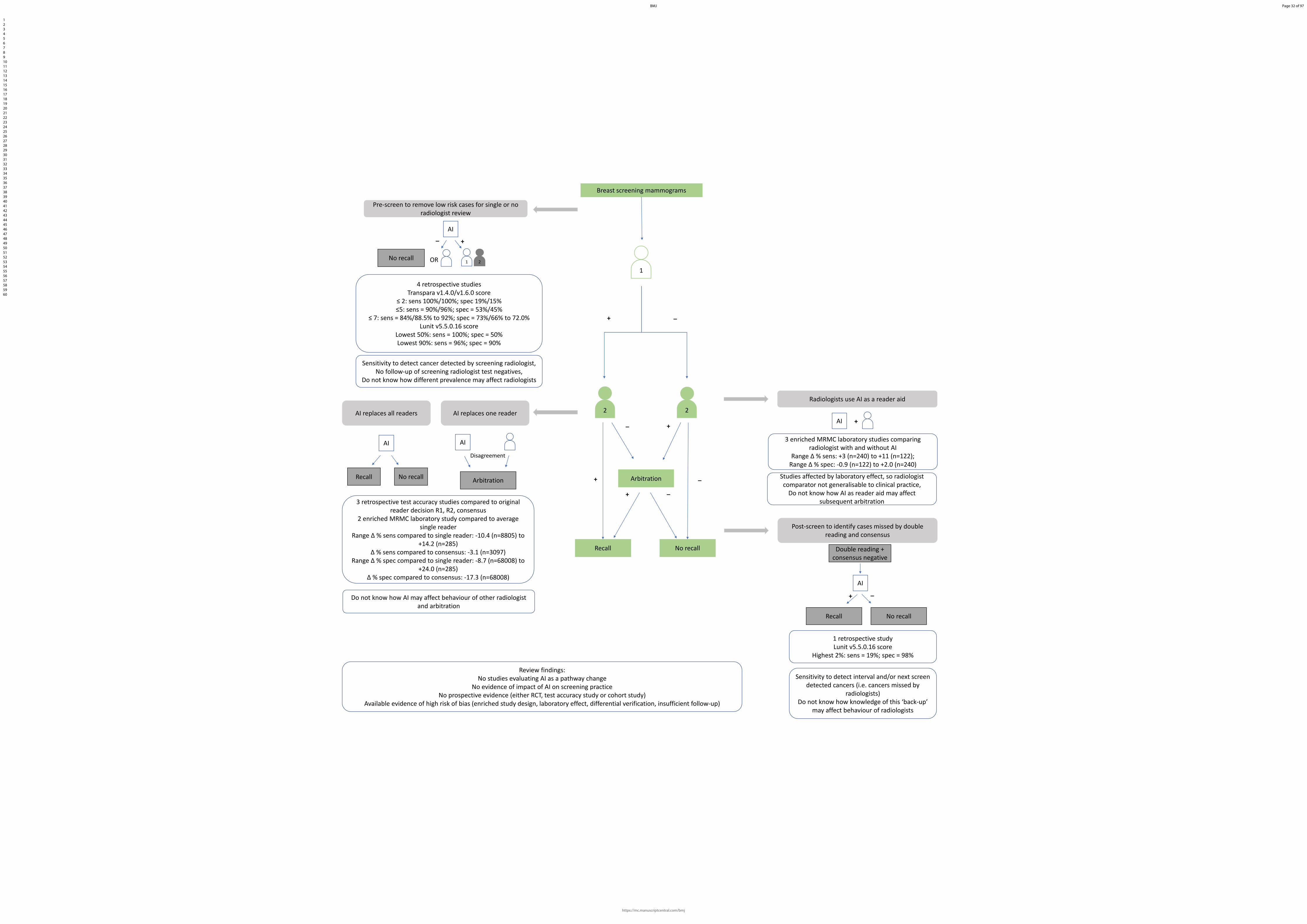
Confidential: For Review Only
Breast screening mammograms
_+
Pre-screen to remove low risk cases for single or no radiologist review
AI
No recall
_
1
+
Sensitivity to detect cancer detected by screening radiologist,No follow-up of screening radiologist test negatives,
Do not know how different prevalence may affect radiologists
2
Arbitration
No recallRecall
+
+
_
_
_ +
Radiologists use AI as a reader aid
AI +
3 enriched MRMC laboratory studies comparing radiologist with and without AI
Range Δ % sens: +3 (n=240) to +11 (n=122); Range Δ % spec: -0.9 (n=122) to +2.0 (n=240)
Post-screen to identify cases missed by double reading and consensus
Double reading + consensus negative
AI
Studies affected by laboratory effect, so radiologist comparator not generalisable to clinical practice,
Do not know how AI as reader aid may affect subsequent arbitration
2 2AI replaces one readerAI replaces all readers
AI
Arbitration
Disagreement
AI
3 retrospective test accuracy studies compared to original reader decision R1, R2, consensus
2 enriched MRMC laboratory study compared to average single reader
Range Δ % sens compared to single reader: -10.4 (n=8805) to +14.2 (n=285)
Δ % sens compared to consensus: -3.1 (n=3097)Range Δ % spec compared to single reader: -8.7 (n=68008) to
+24.0 (n=285)Δ % spec compared to consensus: -17.3 (n=68008)
No recallRecall
Review findings:No studies evaluating AI as a pathway change
No evidence of impact of AI on screening practiceNo prospective evidence (either RCT, test accuracy study or cohort study)
Available evidence of high risk of bias (enriched study design, laboratory effect, differential verification, insufficient follow-up)
Do not know how AI may affect behaviour of other radiologist and arbitration
+ _
No recallRecall
1 retrospective studyLunit v5.5.0.16 score
Highest 2%: sens = 19%; spec = 98%
Sensitivity to detect interval and/or next screen detected cancers (i.e. cancers missed by
radiologists)Do not know how knowledge of this ‘back-up’
may affect behaviour of radiologists
1
4 retrospective studiesTranspara v1.4.0/v1.6.0 score
≤ 2: sens 100%/100%; spec 19%/15%≤5: sens = 90%/96%; spec = 53%/45%
≤ 7: sens = 84%/88.5% to 92%; spec = 73%/66% to 72.0%Lunit v5.5.0.16 score
Lowest 50%: sens = 100%; spec = 50%Lowest 90%: sens = 96%; spec = 90%
OR
Page 32 of 97
https://mc.manuscriptcentral.com/bmj
BMJ
123456789101112131415161718192021222324252627282930313233343536373839404142434445464748495051525354555657585960

Confidential: For Review Only
Study reference
Risk of bias Applicability concerns Patient
selection Index test Comparator
test Reference standard
Flow and timing
Patient selection
Index test Comparator test
Reference standard
Stand-alone AI systems (5 studies) Lotter 202128
High High High Unclear Unclear High High High High
McKinney 202029
High High Low High High High High High Low
Rodriguez-Ruiz 201933
High High High Unclear Unclear High High High Unclear
Salim 202035
High High Low High High High High Low* High* High
Schaffter 202036
Low High Low High High Unclear High Low* High* High
AI as reader aid (3 studies) Pacilè 202030
High High High High Unclear High High High High
Rodriguez-Ruiz 201932,34
High High High High Unclear High High High High
Watanabe 201937
High High High Unclear Unclear High High High Low
AI for triage (4 studies) Balta 202025
Low High None High High Low High None High
Dembrower 202026
High High None Low† High† High High High None Low† High†
Lang 202027
Low High None High High Low High None High
Raya-Povedano 202131
Low Low Low Low High Low High High Low
Page 33 of 97
https://mc.manuscriptcentral.com/bmj
BMJ
123456789101112131415161718192021222324252627282930313233343536373839404142434445464748495051525354555657585960

Confidential: For Review Only
Figure 3 Study estimates of sensitivity and false positive rate (1-specificity) in ROC space by index test (AI) and comparator (radiologist) for 8 included studies
Comparators are defined as consensus of two readers and arbitration (radiologist consensus), or single reader decision/average of multiple readers (radiologist single/average).
Vertical lines represent specificity for screening programmes for Denmark (2% false positive rate)60, UK (3% false positive rate)61 62 and US (11% false positive rate)63
Retrospective test accuracy studies: Salim et al.,35 Schaffter et al.,36 and McKinney et al.29 Enriched test set MRMC laboratory studies: Pacilè et al.,30 Watanabe et al., 37 Rodriguez-Ruiz et al.33 – Rodriguez-Ruiz 2019a in diagram, Lotter 202128 and Rodriguez-Ruiz et al.32 – Rodriguez-Ruiz 2019b in
diagram
580x407mm (38 x 38 DPI)
Page 34 of 97
https://mc.manuscriptcentral.com/bmj
BMJ
123456789101112131415161718192021222324252627282930313233343536373839404142434445464748495051525354555657585960

Confidential: For Review Only
A B
Page 35 of 97
https://mc.manuscriptcentral.com/bmj
BMJ
123456789101112131415161718192021222324252627282930313233343536373839404142434445464748495051525354555657585960

Confidential: For Review OnlyOnline supplementary appendices
Appendix 1 Search strategies
Table 1. Summary of electronic database searches and datesDatabase Platform Searched on
dateDate range of search
Update search
MEDLINE, MEDLINE In-Process, MEDLINE Daily, Epub Ahead of Print
Ovid SP 09 September 2020
2010 to Present 17 May 2021
Embase Ovid SP 09 September 2020
2010 to 2020 Week 36
17 May 2021
The Cochrane Library, including:- Cochrane
Database of Systematic Reviews (CDSR)
- Cochrane Central Register of Controlled Trials (CENTRAL)
- Database of Abstracts of Reviews of Effects (DARE)
Wiley Online 09 September 2020
January 2010 to September 2020
17 May 2021
Web of Science Ovid SP 09 September 2020
2010 to 2020 17 May 2021
Database: Ovid MEDLINE(R) ALL <1946 to September 08, 2020>Search Strategy:--------------------------------------------------------------------------------1 exp Breast Neoplasms/ (293428)2 (breast adj5 (cancer* or neoplasm* or tumor* or tumour*or carcino* or malignan*or disease*)).mp. [mp=title, abstract, original title, name of substance word, subject heading word, floating sub-heading word, keyword heading word, organism supplementary concept word, protocol supplementary concept word, rare disease supplementary concept word, unique identifier, synonyms] (394921)3 1 or 2 (395368)4 exp artificial intelligence/ or exp machine learning/ or exp deep learning/ or exp supervised machine learning/ or exp support vector machine/ or exp unsupervised machine learning/ (99304)5 ai.mp. (28888)6 ((artificial or machine or deep) adj5 (intelligence or learning or reasoning)).mp. [mp=title, abstract, original title, name of substance word, subject heading word, floating sub-heading word, keyword heading word, organism supplementary concept word, protocol supplementary concept word, rare disease supplementary concept word, unique identifier, synonyms] (70647)7 exp Neural Networks, Computer/ or exp Algorithms/ or neural network*.mp. (354996)8 exp Diagnosis, Computer-Assisted/ (83632)9 4 or 5 or 6 or 7 or 8 (452038)10 3 and 9 (10492)11 exp Mammography/ (30025)12 mammogra*.mp. (41086)13 screen*.mp. or exp Mass Screening/ (844672)
Page 36 of 97
https://mc.manuscriptcentral.com/bmj
BMJ
123456789101112131415161718192021222324252627282930313233343536373839404142434445464748495051525354555657585960

Confidential: For Review Only14 exp "Early Detection of Cancer"/ or early detect*.mp. (86182)15 11 or 12 or 13 or 14 (921015)16 10 and 15 (3324)17 exp "Sensitivity and Specificity"/ or sensitivity.mp. or specificity.mp. (1898406)18 exp "Predictive Value of Tests"/ (203774)19 exp roc curve/ or roc.mp. or receiver operating characteristic*.mp. (119948)20 exp Area Under Curve/ or auc.mp. (96772)21 exp False Positive Reactions/ (27763)22 exp False Negative Reactions/ (17783)23 exp Observer Variation/ (42540)24 exp Diagnostic Errors/ (116740)25 (false adj4 (negativ* or positiv*)).mp. [mp=title, abstract, original title, name of substance word, subject heading word, floating sub-heading word, keyword heading word, organism supplementary concept word, protocol supplementary concept word, rare disease supplementary concept word, unique identifier, synonyms] (100096)26 (true adj4 (positiv* or negativ*)).mp. [mp=title, abstract, original title, name of substance word, subject heading word, floating sub-heading word, keyword heading word, organism supplementary concept word, protocol supplementary concept word, rare disease supplementary concept word, unique identifier, synonyms] (10701)27 likelihood ratio*.mp. (15918)28 ((predict* or test*) adj1 (value* or accura* or error*)).mp. [mp=title, abstract, original title, name of substance word, subject heading word, floating sub-heading word, keyword heading word, organism supplementary concept word, protocol supplementary concept word, rare disease supplementary concept word, unique identifier, synonyms] (342222)29 exp Reproducibility of results/ (403133)30 17 or 18 or 19 or 20 or 21 or 22 or 23 or 24 or 25 or 26 or 27 or 28 or 29 (2397952)31 Randomized controlled trials as Topic/ (135939)32 Randomized controlled trial/ (512638)33 Random allocation/ (103549)34 Double blind method/ (159672)35 Single blind method/ (28987)36 Clinical trial/ (524613)37 exp Clinical Trials as Topic/ (345470)38 (clinic$ adj trial$1).tw. (373312)39 ((singl$ or doubl$ or treb$ or tripl$) adj (blind$3 or mask$3)).tw. (174345)40 Randomly allocated.tw. (29185)41 (allocated adj2 random).tw. (802)42 (test-treat trial* or test treat trial*).mp. (1)43 or/31-42 (1423959)44 30 or 43 (3676443)45 3 and 9 and 15 and 44 (2179)46 Case report.tw. (316143)47 Letter/ (1098543)48 Historical article/ (359991)49 Review of reported cases.pt. (0)50 Review, multicase.pt. (0)51 or/46-50 (1758549)52 45 not 51 (2164)53 30 or 52 (2398016)54 3 and 9 and 15 and 44 (2179)55 54 not 51 (2164)56 limit 55 to (english language and yr="2010 -Current") (1228)
Page 37 of 97
https://mc.manuscriptcentral.com/bmj
BMJ
123456789101112131415161718192021222324252627282930313233343536373839404142434445464748495051525354555657585960

Confidential: For Review OnlyDatabase: Embase <1980 to 2020 Week 36>Search Strategy:--------------------------------------------------------------------------------1 exp breast tumor/ (522987)2 exp breast cancer/ (459643)3 (breast adj5 (neoplasm* or cancer* or tumor* or tumour* or malignanc* or carcino* or disease*)).mp. [mp=title, abstract, heading word, drug trade name, original title, device manufacturer, drug manufacturer, device trade name, keyword, floating subheading word, candidate term word] (609633)4 or/1-3 (617527)5 exp artificial intelligence/ (41063)6 exp machine learning/ (215028)7 exp deep learning/ (9250)8 exp supervised machine learning/ (1511)9 exp support vector machine/ (22089)10 exp unsupervised machine learning/ (745)11 ai.mp. (37967)12 ((artificial or machine or deep) adj5 (intelliegence or learning or reasoning)).mp. [mp=title, abstract, heading word, drug trade name, original title, device manufacturer, drug manufacturer, device trade name, keyword, floating subheading word, candidate term word] (64514)13 exp artificial neural network/ or neural network*.mp. (76598)14 exp algorithm/ (381894)15 exp computer assisted diagnosis/ (1123074)16 or/5-15 (1645988)17 exp mammography/ or mammogra*.mp. (62455)18 screen*.mp. (1308438)19 exp mass screening/ or exp screening/ (661930)20 exp early cancer diagnosis/ or early detect*.mp. (97077)21 or/17-20 (1419229)22 exp "sensitivity and specificity"/ or sensitivity.mp. or specificity.mp. (1803827)23 exp reproducibility/ (217747)24 exp receiver operating characteristic/ or exp roc curve/ or roc.mp. (163201)25 exp predictive value/ or ((predict* or test*) adj1 (value* or error* or accura*)).mp. (420596)26 auc.mp. or exp area under the curve/ (211311)27 exp false positive result/ (30242)28 exp false negative result/ (18705)29 exp observer variation/ (19992)30 exp diagnostic error/ (97269)31 (false adj4 (negativ* or positiv*)).mp. [mp=title, abstract, heading word, drug trade name, original title, device manufacturer, drug manufacturer, device trade name, keyword, floating subheading word, candidate term word] (120409)32 (true adj4 (positiv* or negativ*)).mp. [mp=title, abstract, heading word, drug trade name, original title, device manufacturer, drug manufacturer, device trade name, keyword, floating subheading word, candidate term word] (15519)33 likelihood ratio.mp. (16617)34 or/22-33 (2487428)35 clinical trial/ (971925)36 Randomized controlled trial/ (614311)37 Randomization/ (87593)38 Single blind procedure/ (40000)39 Double blind procedure/ (172538)
Page 38 of 97
https://mc.manuscriptcentral.com/bmj
BMJ
123456789101112131415161718192021222324252627282930313233343536373839404142434445464748495051525354555657585960
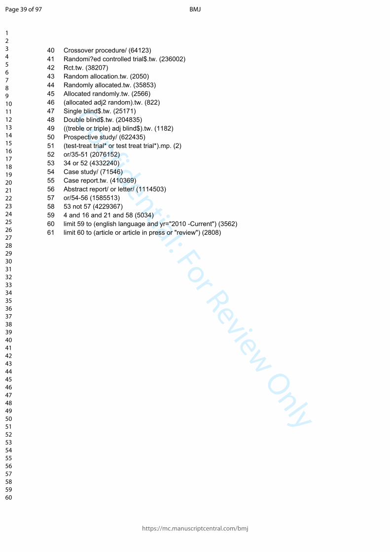
Confidential: For Review Only40 Crossover procedure/ (64123)41 Randomi?ed controlled trial$.tw. (236002)42 Rct.tw. (38207)43 Random allocation.tw. (2050)44 Randomly allocated.tw. (35853)45 Allocated randomly.tw. (2566)46 (allocated adj2 random).tw. (822)47 Single blind$.tw. (25171)48 Double blind$.tw. (204835)49 ((treble or triple) adj blind$).tw. (1182)50 Prospective study/ (622435)51 (test-treat trial* or test treat trial*).mp. (2)52 or/35-51 (2076152)53 34 or 52 (4332240)54 Case study/ (71546)55 Case report.tw. (410369)56 Abstract report/ or letter/ (1114503)57 or/54-56 (1585513)58 53 not 57 (4229367)59 4 and 16 and 21 and 58 (5034)60 limit 59 to (english language and yr="2010 -Current") (3562)61 limit 60 to (article or article in press or "review") (2808)
Page 39 of 97
https://mc.manuscriptcentral.com/bmj
BMJ
123456789101112131415161718192021222324252627282930313233343536373839404142434445464748495051525354555657585960

Confidential: For Review OnlyDatabase: Web of Science (Ovid SP)
Database: Cochrane Library (CENTRAL) (Wiley online)
Search Name: AI and Breast CancerLast Saved: 09/09/2020 14:10:48Comment: Numbers for individual search lines are not captured by the saved search strategy.
ID Search#1 MeSH descriptor: [Breast Neoplasms] explode all trees#2 ((breast NEAR/5 (cancer* or neoplasm* or carcino* or malignan* or tumor* or tumour* or disease*))):ti,ab,kw#3 #1 or #2#4 MeSH descriptor: [Artificial Intelligence] explode all trees#5 MeSH descriptor: [Machine Learning] explode all trees#6 MeSH descriptor: [Deep Learning] explode all trees#7 MeSH descriptor: [Supervised Machine Learning] explode all trees#8 MeSH descriptor: [Support Vector Machine] explode all trees#9 MeSH descriptor: [Unsupervised Machine Learning] explode all trees#10 (ai):ti,ab,kw#11 ((artificial or machine or deep) NEAR/5 (intelligence or learning or reasoning)):ti,ab,kw#12 MeSH descriptor: [Neural Networks, Computer] explode all trees#13 MeSH descriptor: [Algorithms] explode all trees#14 (neural network*):ti,ab,kw#15 MeSH descriptor: [Diagnosis, Computer-Assisted] explode all trees#16 #4 or #5 or #6 or #7 or #8 or #9 or #10 or #11 or #12 or #13 or #14 or #15#17 MeSH descriptor: [Mammography] explode all trees#18 (mammogra*):ti,ab,kw#19 MeSH descriptor: [Mass Screening] explode all trees#20 (screen*):ti,ab,kw#21 MeSH descriptor: [Early Detection of Cancer] explode all trees#22 (early detect*):ti,ab,kw#23 #17 or #18 or #19 or #20 or #21 or #22#24 #3 and #16 and #23#25 MeSH descriptor: [Sensitivity and Specificity] explode all trees#26 (sensitivity or specificity):ti,ab,kw#27 MeSH descriptor: [Predictive Value of Tests] explode all trees#28 MeSH descriptor: [ROC Curve] explode all trees#29 (roc or "receiver operating characteristic"):ti,ab,kw#30 MeSH descriptor: [Area Under Curve] explode all trees
Page 40 of 97
https://mc.manuscriptcentral.com/bmj
BMJ
123456789101112131415161718192021222324252627282930313233343536373839404142434445464748495051525354555657585960

Confidential: For Review Only#31 (auc):ti,ab,kw#32 MeSH descriptor: [False Positive Reactions] explode all trees#33 MeSH descriptor: [False Negative Reactions] explode all trees#34 MeSH descriptor: [Observer Variation] explode all trees#35 MeSH descriptor: [Diagnostic Errors] explode all trees#36 (false NEAR/4 (negativ* or positiv*)):ti,ab,kw#37 (true NEAR/4 (positiv* or negativ*)):ti,ab,kw#38 (liklihood ratio*):ti,ab,kw#39 ((predict* or test*) NEAR/1 (value* or accura* or error*)):ti,ab,kw#40 MeSH descriptor: [Reproducibility of Results] explode all trees#41 #25 or #26 or #27 or #28 or #29 or #30 or #31 or #32 or #33 or #34 or #35 or #36 or #37 or #38 or #39 or #40#42 #24 and #41
Page 41 of 97
https://mc.manuscriptcentral.com/bmj
BMJ
123456789101112131415161718192021222324252627282930313233343536373839404142434445464748495051525354555657585960

Confidential: For Review Only
Supplementary Figure 1. PRISMA diagram. Summary of publications included and excluded at each stage of the review (original searches / update searches)FFDM, Full field digital mammogram; NA, Not applicable; ND, Not done
Records identified from database searches (n = 4,696 / 713)
Medline ALL (n = 1,228 / 153)Embase (n= 2,808 / 339)Web of Science (n = 881 / 214)CENTRAL) (n = 52 / 7)
Records removed before screening:
Duplicate records removed (n = 1,335 / 331)Records marked as ineligible by automation tools (ND / ND)Records removed for other reasons (ND / ND)
Records screened(n = 3,634 / 382)
Records excluded(n = 3,211 / 341)
Reports sought for retrieval(n = 423 / 41)
Reports not retrieved(n = 7 / 2)
Reports assessed for eligibility(n = 416 / 39)
Reports excluded:(n = 409 / 36)
Population (n = 179 / 24)Not external validation (n = 152 / 9)Intervention (n = 42 / 1)Outcomes (n = 24 / 1) Publication type (n = 8 / 0)Other (n = 4 / 0)Already identified in original search (n = NA / 2)
Records identified from:Websites (ND / ND)Organisations (ND / ND)Citation searching (n = 489 / ND)Reference list screen (n = 205 / 9)Expert suggestions (n = 2 / ND)
Reports assessed for eligibility(n = 147 / 2)
Reports excluded:(n = 143 / 2)
Population (n = 53 / 0)Not external validation (n = 42 / 0)Intervention (n = 1 / 0)Outcomes (n = 8 / 2) Publication type (n = 32 / 0)Language (n = 1 / 0)Already identified in database searches (6 / 0)Studies included in review
(n = 10 / 2)Reports of included studies(n = 11 / 2)
Identification of studies via databases and registers Identification of studies via other methods
Iden
tific
atio
nSc
reen
ing
Incl
uded
Reports sought for retrieval(n = 148 / 2)
Reports not retrieved(n = 1 / 0)
Page 42 of 97
https://mc.manuscriptcentral.com/bmj
BMJ
123456789101112131415161718192021222324252627282930313233343536373839404142434445464748495051525354555657585960

Confidential: For Review OnlyAppendix 2 Publications and sub-studies excluded after review of full-text articles
Key to reasons for exclusions and justificationsPopulation – Image type: Studies evaluating AI on image types other than full field digital mammography (FFDM) (mainly digitised film images). Screening mammography is typically undertaken using FFDM in women attending breast screening. Other imaging techniques not classed as mammography or not digital are not relevant. Results from imaging other than FFDM may not be applicable to FFDM in screening programmes.
Population – Mammography type not reported: Studies evaluating AI on image types which were not clearly identified as FFDM. Screening mammography is typically undertaken using full field digital mammography (FFDM) in women attending breast screening. Other imaging techniques not classed as mammography or not digital are not relevant. Results from imaging other than FFDM may not be applicable to FFDM in screening programmes.
Population – Incomplete images: Studies evaluating AI using regions of interest (ROIs) of images. Images of part of a mammogram do not represent the use case in the screening context, which requires recall or not decisions to be made on women’s (craniocaudal and mediolateral oblique) screening mammograms for both breasts.
Population – Subpopulation: Studies only including images with cancer. These are not sufficient to estimate test accuracy of AI for screening mammograms, as it excludes specificity, and the trade-off between sensitivity and specificity. Studies on images of subpopulations by screening risk or screening outcome. They do not represent the screening population and no inference on the performance of the AI system in a screening population can be drawn (however, subpopulations by ethnicity or socioeconomic status are included as the impact of any change on equity is important). This does not apply to populations which represent a group of women at any stage within the screening pathway (e.g. recalled women without selection on final diagnosis) on the assumption that AI could be incorporated for this subgroup only.
Population – <90% screening mammograms or unclear proportion: Studies on images of diagnostic mammograms, with >10% diagnostic mammograms or an unclear proportion of diagnostic mammograms. These do not represent the use case in the screening context.
Internal validation – Cross validation, Leave-one out, Split sample: Studies using internal validation whereby the validation dataset used to assess a model uses data which were used to develop that model. Cross validation and leave-one out are resampling techniques which use the original data to assist with preliminary assessment and fine-tuning of the model during validation. The issue of using data on which an algorithm was trained with is that models can be prone to overfitting; whereby the model fits the trained data extremely well, but to the detriment of the model’s ability to perform when presented with new data, which is known as poor generalization. The split-sample approach is generally an
Page 43 of 97
https://mc.manuscriptcentral.com/bmj
BMJ
123456789101112131415161718192021222324252627282930313233343536373839404142434445464748495051525354555657585960

Confidential: For Review Onlyinefficient form of internal validation because it does not accurately reflect a model’s generalisability.
Intervention – Detecting subtypes: Studies using AI to detect cancer subtypes such as microcalcifications or architectural distortions only. The detection of cancer subtypes does not present the complete picture of cancer detection (e.g. microcalcifications are associated mainly with DCIS and not with cancer; detecting microcalcifications only will miss some types of cancers). On their own, these AI systems do not provide the information on cancer present/ not present to inform a decision whether to recall or not recall. Systems reporting single features could be combined to provide a more complete picture, however, studies would need to report an overall outcome of test accuracy of the combination of systems.
Intervention – No detection/classification: Studies using AI for lesion enhancement or segmentation of pectoral muscle regions. These studies do not report on the classification of images into recall or no recall and are uninformative in terms of test performance of AI.
Intervention – Not AI: Studies using traditional computer aided detection without deep learning features for the detection of lesions in images. In AI the layers of features are not designed by human expertise; instead they are learnt from the underlying data. Therefore, studies reporting the test accuracy and effectiveness of traditional CAD systems were considered significantly different from studies assessing deep learning AI systems.
Intervention – Prediction of cancer: Studies using AI for the prediction of future cancer risk including the detection of breast density and parenchymal patterns as risk factors. These studies did not consider the detection of cancer present on screening mammograms.
No relevant outcomes: Studies reporting accuracy without outcomes characterising the trade-off between false positive and false negative results including global measures such as the area under the curve (AUC). The trade-off between false positive and false negative results is critical to test accuracy.
Study type – Systematic reviews with no relevant outcomes: Studies reporting systematic reviews that were off topic and did not provide additional references for the review.
Document Supply cancelled request: no location found: Studies unavailable following internet searches, contacting authors and pursuing interlibrary requests.
Page 44 of 97
https://mc.manuscriptcentral.com/bmj
BMJ
123456789101112131415161718192021222324252627282930313233343536373839404142434445464748495051525354555657585960

Confidential: For Review Only
Publications excluded with reason – Original database searchesReference Main reason for exclusionPopulation – Image type (e.g. digitised film images; not FFDM images) (n=150)
1. Abbas Q, Fondo'n I, Celebi E. A Computerized System for Detection of Spiculated Margins based on Mammography. International Arab Journal of Information Technology. 2015;12(6):582-8.
Population – Image type
2. Agnes SA, Anitha J, Pandian SIA, Peter JD. Classification of Mammogram Images Using Multiscale all Convolutional Neural Network (MA-CNN). J Med Syst. 2019;44(1):30.
Population – Image type
3. Anitha J, Dinesh Peter J, Immanuel Alex Pandian S. A dual stage adaptive thresholding (DuSAT) for automatic mass detection in mammograms. Computer Methods and Programs in Biomedicine. 2017;138:93-104.
Population – Image type
4. Bartolotta TV, Orlando A, Cantisani V, Matranga D, Ienzi R, Cirino A, et al. Focal breast lesion characterization according to the BI-RADS US lexicon: role of a computer-aided decision-making support. La Radiologia medica. 2018;123(7):498-506.
Population – Image type
5. Beheshti SM, AhmadiNoubari H, Fatemizadeh E, Khalili M. An efficient fractal method for detection and diagnosis of breast masses in mammograms. J Digit Imaging. 2014;27(5):661-9.
Population – Image type
6. Chakraborty J, Midya A, Mukhopadhyay S, Rangayyan RM, Sadhu A, Singla V, et al. Computer-Aided Detection of Mammographic Masses Using Hybrid Region Growing Controlled by Multilevel Thresholding. Journal of Medical and Biological Engineering. 2019;39(3):352-66.
Population – Image type
7. Chithra Devi M, Audithan S. Analysis of different types of entropy measures for breast cancer diagnosis using ensemble classification. Biomedical Research (India). 2017;28(7):3182-6.
Population – Image type
8. Choi JY, Ro YM. Multiresolution local binary pattern texture analysis combined with variable selection for application to false-positive reduction in computer-aided detection of breast masses on mammograms. Phys Med Biol. 2012;57(21):7029-52.
Population – Image type
9. Chougrad H, Zouaki H, Alheyane O. Deep Convolutional Neural Networks for breast cancer screening. Comput Methods Programs Biomed. 2018;157:19-30.
Population – Image type
Page 45 of 97
https://mc.manuscriptcentral.com/bmj
BMJ
123456789101112131415161718192021222324252627282930313233343536373839404142434445464748495051525354555657585960

Confidential: For Review Only
Reference Main reason for exclusion
10. Chowdhary CL, Mittal M, P K, Pattanaik PA, Marszalek Z. An Efficient Segmentation and Classification System in Medical Images Using Intuitionist Possibilistic Fuzzy C-Mean Clustering and Fuzzy SVM Algorithm. Sensors (Basel). 2020;20(14):13.
Population – Image type
11. Cunningham CA, Drew T, Wolfe JM. Analog Computer-Aided Detection (CAD) information can be more effective than binary marks. Atten Percept Psychophys. 2017;79(2):679-90.
Population – Image type
12. de Oliveira Silva LC, Barros AK, Lopes MV. Detecting masses in dense breast using independent component analysis. Artif Intell Med. 2017;80:29-38.
Population – Image type
13. Dheeba J, Albert Singh N, Tamil Selvi S. Computer-aided detection of breast cancer on mammograms: a swarm intelligence optimized wavelet neural network approach. J Biomed Inform. 2014;49:45-52.
Population – Image type
14. Dheeba J, Jaya T, Singh NA. Breast cancer risk assessment and diagnosis model using fuzzy support vector machine based expert system. Journal of Experimental & Theoretical Artificial Intelligence. 2017;29(5):1011-21.
Population – Image type
15. Dheeba J, Tamil Selvi S. An improved decision support system for detection of lesions in mammograms using Differential Evolution Optimized Wavelet Neural Network. J Med Syst. 2012;36(5):3223-32.
Population – Image type
16. Dubey AK, Gupta U, Jain S. Analysis of k-means clustering approach on the breast cancer Wisconsin dataset. Int. 2016;11(11):2033-47.
Population – Image type
17. El Fahssi K, Elmoufidi A, Abenaou A, Jai-Andaloussi S, Sekkaki A. Novel approach to classification of Abnormalities in the mammogram image. International Journal of Biology and Biomedical Engineering. 2016;10:72-9.
Population – Image type
18. El-Shazli AMA, Youssef SM, Elshennawy M. Computer-aided model for breast cancer detection in mammograms. International Journal of Pharmacy and Pharmaceutical Sciences. 2016;8(Supplement 2):31-4.
Population – Image type
19. Elmoufidi A, El Fahssi K, Jai-andaloussi S, Sekkaki A, Gwenole Q, Lamard M. Anomaly classification in digital mammography based on multiple-instance learning. Iet Image Processing. 2018;12(3):320-8.
Population – Image type
20. Eltoukhy MM, Faye I, Samir BB. Breast cancer diagnosis in digital mammogram using multiscale curvelet transform. Comput Med Imaging Graph. 2010;34(4):269-76.
Population – Image type
Page 46 of 97
https://mc.manuscriptcentral.com/bmj
BMJ
123456789101112131415161718192021222324252627282930313233343536373839404142434445464748495051525354555657585960
Confidential: For Review Only
Reference Main reason for exclusion21. Ericeira DR, Silva AC, de Paiva AC, Gattass M. Detection of masses based on
asymmetric regions of digital bilateral mammograms using spatial description with variogram and cross-variogram functions. Comput Biol Med. 2013;43(8):987-99.
Population – Image type
22. Ganesan K, Acharya RU, Chua CK, Min LC, Mathew B, Thomas AK. Decision support system for breast cancer detection using mammograms. Proc Inst Mech Eng [H]. 2013;227(7):721-32.
Population – Image type
23. Garma FB, Hassan MA. Classification of breast tissue as normal or abnormal based on texture analysis of digital mammogram. Journal of Medical Imaging and Health Informatics. 2014;4(5):647-53.
Population – Image type
24. Gedik N. Breast cancer diagnosis system via contourlet transform with sharp frequency localization and least squares support vector machines. Journal of Medical Imaging and Health Informatics. 2015;5(3):497-505.
Population – Image type
25. Gedik N, Atasoy A. Performance evaluation of the wave atom algorithm to classify mammographic images. Turkish Journal of Electrical Engineering and Computer Sciences. 2014;22(4):957-69.
Population – Image type
26. Gedik N, Atasoy A, Sevim Y. Investigation of wave atom transform by using the classification of mammograms. Applied Soft Computing. 2016;43:546-52.
Population – Image type
27. Gorgel P, Sertbas A, Ucan ON. Mammographical mass detection and classification using Local Seed Region Growing-Spherical Wavelet Transform (LSRG-SWT) hybrid scheme. Computers in Biology and Medicine. 2013;43(6):765-74.
Population – Image type
28. Guan JS, Lin LY, Ji GL, Lin CM, Le TL, Rudas IJ. Breast Tumor Computer-aided Diagnosis using Self-Validating Cerebellar Model Neural Networks. Acta Polytechnica Hungarica. 2016;13(4):39-52.
Population – Image type
29. Hu K, Gao XP, Li F. Detection of Suspicious Lesions by Adaptive Thresholding Based on Multiresolution Analysis in Mammograms. Ieee Transactions on Instrumentation and Measurement. 2011;60(2):462-72.
Population – Image type
30. James JJ, Gilbert FJ, Wallis MG, Gillan MG, Astley SM, Boggis CR, et al. Mammographic features of breast cancers at single reading with computer-aided detection and at double reading in a large multicenter prospective trial of computer-aided detection: CADET II. Radiology. 2010;256(2):379-86.
Population – Image type
Page 47 of 97
https://mc.manuscriptcentral.com/bmj
BMJ
123456789101112131415161718192021222324252627282930313233343536373839404142434445464748495051525354555657585960

Confidential: For Review Only
Reference Main reason for exclusion31. Jebamony J, Jacob D. Classification of Benign and Malignant Breast Masses on
Mammograms for Large Datasets using Core Vector Machines. Curr Med Imaging. 2020;16(6):703-10.
Population – Image type
32. Kadam VJ, Jadhav SM, Vijayakumar K. Breast Cancer Diagnosis Using Feature Ensemble Learning Based on Stacked Sparse Autoencoders and Softmax Regression. J Med Syst. 2019;43(8):263.
Population – Image type
33. Kanadam KP, Chereddy SR. Mammogram classification using sparse-ROI: A novel representation to arbitrary shaped masses. Expert Systems with Applications. 2016;57:204-13.
Population – Image type
34. Kanchana M, Varalakshmi P. Computer aided system for breast cancer in digitized mammogram using shearlet band features with LS-SVM classifier. International Journal of Wavelets Multiresolution and Information Processing. 2016;14(3).
Population – Image type
35. Kanchanamani M, Perumal V. Performance evaluation and comparative analysis of various machine learning techniques for diagnosis of breast cancer. Biomedical Research (India). 2016;27(3):623-31.
Population – Image type
36. Kashyap KL, Bajpai MK, Khanna P. Globally supported radial basis function based collocation method for evolution of level set in mass segmentation using mammograms. Comput Biol Med. 2017;87:22-37.
Population – Image type
37. Kashyap KL, Bajpai MK, Khanna P. An efficient algorithm for mass detection and shape analysis of different masses present in digital mammograms. Multimedia Tools and Applications. 2018;77(8):9249-69.
Population – Image type
38. Kashyap KL, Bajpai MK, Khanna P, Giakos G. Mesh-free based variational level set evolution for breast region segmentation and abnormality detection using mammograms. Int j numer method biomed eng. 2018;34(1):01.
Population – Image type
39. Kayode AA, Akande NO, Adegun AA, Adebiyi MO. An automated mammogram classification system using modified support vector machine. Medical Devices: Evidence and Research. 2019;12:275-84.
Population – Image type
40. Kelder A, Zigel Y, Lederman D, Zheng B. A new computer-aided detection scheme based on assessment of local bilateral mammographic feature asymmetry - a preliminary evaluation. Conf Proc IEEE Eng Med Biol Soc. 2015;2015:6394-7.
Population – Image type
Page 48 of 97
https://mc.manuscriptcentral.com/bmj
BMJ
123456789101112131415161718192021222324252627282930313233343536373839404142434445464748495051525354555657585960

Confidential: For Review Only
Reference Main reason for exclusion41. Khan AA, Arora AS. Computer aided diagnosis of breast cancer based on level set
segmentation of masses and classification using ensemble classifiers. Biomedical Research (India). 2018;29(19):3610-5.
Population – Image type
42. Khan HN, Shahid AR, Raza B, Dar AH, Alquhayz H. Multi-View Feature Fusion Based Four Views Model for Mammogram Classification Using Convolutional Neural Network. Ieee Access. 2019;7.
Population – Image type
43. Krishnan MMR, Banerjee S, Chakraborty C, Chakraborty C, Ray AK. Statistical analysis of mammographic features and its classification using support vector machine. Expert Systems with Applications. 2010;37(1):470-8.
Population – Image type
44. Li JB, Wang YH, Tang LL. Mammogram-based discriminant fusion analysis for breast cancer diagnosis. Clin Imaging. 2012;36(6):710-6.
Population – Image type
45. Li P, Bi T, Huang J, Li S. Breast cancer early diagnosis based on hybrid strategy. Biomed Mater Eng. 2014;24(6):3397-404.
Population – Image type
46. Li Z, Sun J, Zhang J, Hu D, Wang Q, Peng K. Quantification of acoustic radiation force impulse in differentiating between malignant and benign breast lesions. Ultrasound Med Biol. 2014;40(2):287-92.
Population – Image type
47. Lo CM, Moon WK, Huang CS, Chen JH, Yang MC, Chang RF. INTENSITY-INVARIANT TEXTURE ANALYSIS FOR CLASSIFICATION OF BI-RADS CATEGORY 3 BREAST MASSES. Ultrasound in Medicine and Biology. 2015;41(7):2039-48.
Population – Image type
48. Onan A. A stochastic gradient descent based SVM with fuzzy-rough feature selection and instance selection for breast cancer diagnosis. Journal of Medical Imaging and Health Informatics. 2015;5(6):1233-9.
Population – Image type
49. Singh WJ, Nagarajan B. Automatic diagnosis of mammographic abnormalities based on hybrid features with learning classifier. Comput Methods Biomech Biomed Engin. 2013;16(7):758-67.
Population – Image type
50. Valarmathi P, Robinson S. An Improved Neural Network for Mammogram Classification Using Genetic Optimization. Journal of Medical Imaging and Health Informatics. 2016;6(7):1631-5.
Population – Image type
51. Liu X, Zeng Z. A new automatic mass detection method for breast cancer with false positive reduction. Neurocomputing. 2015;152:388-402.
Population – Image type
Page 49 of 97
https://mc.manuscriptcentral.com/bmj
BMJ
123456789101112131415161718192021222324252627282930313233343536373839404142434445464748495051525354555657585960

Confidential: For Review Only
Reference Main reason for exclusion52. Liu XM, Tang JS. Mass Classification in Mammograms Using Selected Geometry and
Texture Features, and a New SVM-Based Feature Selection Method. Ieee Systems Journal. 2014;8(3):910-20.
Population – Image type
53. Liu XM, Zhai LL, Zhu T, Liu J, Zhang K, Hu W. Multiple TBSVM-RFE for the detection of architectural distortion in mammographic images. Multimedia Tools and Applications. 2018;77(12):15773-802.
Population – Image type
54. Mahersia H, Boulehmi H, Hamrouni K. Development of intelligent systems based on Bayesian regularization network and neuro-fuzzy models for mass detection in mammograms: A comparative analysis. Comput Methods Programs Biomed. 2016;126:46-62.
Population – Image type
55. Mencattini A, Salmeri M. Breast masses detection using phase portrait analysis and fuzzy inference systems. Int. 2012;7(4):573-83.
Population – Image type
56. Meselhy Eltoukhy M, Faye I, Belhaouari Samir B. A statistical based feature extraction method for breast cancer diagnosis in digital mammogram using multiresolution representation. Comput Biol Med. 2012;42(1):123-8.
Population – Image type
57. Midya A, Rabidas R, Sadhu A, Chakraborty J. Edge Weighted Local Texture Features for the Categorization of Mammographic Masses. Journal of Medical and Biological Engineering. 2018;38(3):457-68.
Population – Image type
58. Milosevic M, Jovanovic Z, Jankovic D. A comparison of methods for three-class mammograms classification. Technol Health Care. 2017;25(4):657-70.
Population – Image type
59. Mohammadi-Sardo S, Labibi F, Shafiei SA. A new approach for detecting abnormalities in mammograms using a computer-aided windowing system based on Otsu's method. Radiol Phys Technol. 2019;12(2):178-84.
Population – Image type
60. Mohammed SHA, Yousuf SEK. A computer-aided diagnosis system for the detection and classification of breast cancer. Journal of Clinical Engineering. 2016;41(2):97-100.
Population – Image type
61. Mohammed SHAA, Mustafa ZA. Breast Tumors Classification Using Adaptive Neuro-Fuzzy Inference System. Journal of Clinical Engineering. 2017;42(2):68-72.
Population – Image type
62. Mohanty F, Rup S, Dash B. Automated diagnosis of breast cancer using parameter optimized kernel extreme learning machine. Biomedical Signal Processing and Control. 2020;62 (no pagination).
Population – Image type
Page 50 of 97
https://mc.manuscriptcentral.com/bmj
BMJ
123456789101112131415161718192021222324252627282930313233343536373839404142434445464748495051525354555657585960

Confidential: For Review Only
Reference Main reason for exclusion63. Mohanty F, Rup S, Dash B, Majhi B, Swamy MNS. Mammogram classification using
contourlet features with forest optimization-based feature selection approach. Multimedia Tools and Applications. 2019;78(10):12805-34.
Population – Image type
64. Mohanty F, Rup S, Dash B, Majhi B, Swamy MNS. A computer-aided diagnosis system using Tchebichef features and improved grey wolf optimized extreme learning machine. Applied Intelligence. 2019;49(3):983-1001.
Population – Image type
65. Mohanty F, Rup S, Dash B, Majhi B, Swamy MNS. An improved scheme for digital mammogram classification using weighted chaotic salp swarm algorithm-based kernel extreme learning machine. Applied Soft Computing. 2020;91.
Population – Image type
66. Mohanty F, Rup S, Dash B, Majhi B, Swamy MNS. Digital mammogram classification using 2D-BDWT and GLCM features with FOA-based feature selection approach. Neural Computing & Applications. 2020;32(11):7029-43.
Population – Image type
67. Moslemi H, Kazerouni IA, Hourali F. Breast cancer diagnosis in mammogram images using coordinate logic filters. Biomedical Research (India). 2017;28(22):10108-11.
Population – Image type
68. Muduli D, Dash R, Majhi B. Automated breast cancer detection in digital mammograms: A moth flame optimization based ELM approach. Biomedical Signal Processing and Control. 2020;59 (no pagination).
Population – Image type
69. Mughal B, Muhammad N, Sharif M. Adaptive hysteresis thresholding segmentation technique for localizing the breast masses in the curve stitching domain. Int J Med Inf. 2019;126:26-34.
Population – Image type
70. Nagarajan V, Britto EC, Veeraputhiran SM. Feature extraction based on empirical mode decomposition for automatic mass classification of mammogram images. Medicine in Novel Technology and Devices. 2019;1 (no pagination).
Population – Image type
71. Nagthane DK, Rajurkar AM. An improved diagnosis technique for breast cancer using LCFS and TreeHiCARe classifier model. Sensor Review. 2019;39(1):107-20.
Population – Image type
72. Nanni L, Brahnam S, Lumini A. A very high performing system to discriminate tissues in mammograms as benign and malignant. Expert Systems with Applications. 2012;39(2):1968-71.
Population – Image type
73. Narvaez F, Alvarez J, Garcia-Arteaga JD, Tarquino J, Romero E. Characterizing Architectural Distortion in Mammograms by Linear Saliency. J Med Syst. 2017;41(2):26.
Population – Image type
Page 51 of 97
https://mc.manuscriptcentral.com/bmj
BMJ
123456789101112131415161718192021222324252627282930313233343536373839404142434445464748495051525354555657585960

Confidential: For Review Only
Reference Main reason for exclusion74. Naseem MT, Sulong GZB, Jaffar MA. MRT letter: Quantum noise removal and
classification of breast mammogram images. Microscopy Research and Technique. 2012;75(12):1609-12.
Population – Image type
75. Naveed N, Jaffar MA, Choi TS. MRT Letter: Segmentation and Texture-Based Classification of Breast Mammogram Images. Microscopy Research and Technique. 2011;74(11):985-7.
Population – Image type
76. Neto OPS, Silva AC, Paiva AC, Gattass M. Automatic mass detection in mammography images using particle swarm optimization and functional diversity indexes. Multimedia Tools and Applications. 2017;76(18):19263-89.
Population – Image type
77. Nishikawa RM, Schmidt RA, Linver MN, Edwards AV, Papaioannou J, Stull MA. Clinically missed cancer: how effectively can radiologists use computer-aided detection? AJR Am J Roentgenol. 2012;198(3):708-16.
Population – Image type
78. Nugroho HA, Fajrin HR, Soesanti I, Budiani RL. Analysis of texture for classification of breast cancer on mammogram images. International Journal of Medical Engineering and Informatics. 2018;10(4):382-91.
Population – Image type
79. P S, R T. Aiding the Digital Mammogram for Detecting the Breast Cancer Using Shearlet Transform and Neural Network. Asian Pac J Cancer Prev. 2018;19(9):2665-71.
Population – Image type
80. Pak F, Kanan HR, Alikhassi A. Breast cancer detection and classification in digital mammography based on Non-Subsampled Contourlet Transform (NSCT) and Super Resolution. Comput Methods Programs Biomed. 2015;122(2):89-107.
Population – Image type
81. Panetta K, Zhou Y, Agaian S, Jia H. Nonlinear unsharp masking for mammogram enhancement. IEEE Trans Inf Technol Biomed. 2011;15(6):918-28.
Population – Image type
82. Paquerault S, Hardy PT, Wersto N, Chen J, Smith RC. Investigation of optimal use of computer-aided detection systems: the role of the "machine" in decision making process. Acad Radiol. 2010;17(9):1112-21.
Population – Image type
83. Paramkusham S, Rao KMM, Prabhakar Rao BVVSN, Sharma S. Application of TAR signature for breast mass analysis. Biomedical Research (India). 2018;29(10):2030-4.
Population – Image type
84. Parmeggiani D, Avenia N, Sanguinetti A, Ruggiero R, Docimo G, Siciliano M, et al. Artificial intelligence against breast cancer (A.N.N.E.S-B.C.-Project). Ann Ital Chir. 2012;83(1):1-5.
Population – Image type
Page 52 of 97
https://mc.manuscriptcentral.com/bmj
BMJ
123456789101112131415161718192021222324252627282930313233343536373839404142434445464748495051525354555657585960

Confidential: For Review Only
Reference Main reason for exclusion85. Patel BC, Sinha GR, Soni D. Detection of masses in mammographic breast cancer images
using modified histogram based adaptive thresholding (MHAT) method. International Journal of Biomedical Engineering and Technology. 2019;29(2):134-54.
Population – Image type
86. Pawar MM, Talbar SN, Dudhane A. Local Binary Patterns Descriptor Based on Sparse Curvelet Coefficients for False-Positive Reduction in Mammograms. J. 2018;2018:5940436.
Population – Image type
87. Perre AC, Alexandre LA, Freire LC. Lesion classification in mammograms using convolutional neural networks and transfer learning. Computer Methods in Biomechanics and Biomedical Engineering: Imaging and Visualization. 2019;7(5-6):550-6.
Population – Image type
88. Pezeshki H, Rastgarpour M, Sharifi A, Yazdani S. Extraction of spiculated parts of mammogram tumors to improve accuracy of classification. Multimedia Tools and Applications. 2019;78(14):19979-20003.
Population – Image type
89. Povyakalo AA, Alberdi E, Strigini L, Ayton P. How to discriminate between computer-aided and computer-hindered decisions: a case study in mammography. Med Decis Making. 2013;33(1):98-107.
Population – Image type
90. Qasim KR, Ouda AJ. An accurate breast cancer detection system based on deep learning cnn. Medico Legal Update. 2020;20(1).
Population – Image type
91. Quellec G, Lamard M, Cozic M, Coatrieux G, Cazuguel G. Multiple-Instance Learning for Anomaly Detection in Digital Mammography. IEEE Trans Med Imaging. 2016;35(7):1604-14.
Population – Image type
92. Rabidas R, Arif W. Characterization of mammographic masses based on local photometric attributes. Multimedia Tools and Applications. 2020;79(29-30):21967-85.
Population – Image type
93. Rabidas R, Chakraborty J, Midya A. Analysis of 2D singularities for mammographic mass classification. Iet Computer Vision. 2017;11(1):22-32.
Population – Image type
94. Rabidas R, Midya A, Chakraborty J. Neighborhood Structural Similarity Mapping for the Classification of Masses in Mammograms. IEEE j. 2018;22(3):826-34.
Population – Image type
95. Rabottino G, Mencattini A, Salmeri M, Caselli F, Lojacono R. Performance evaluation of a region growing procedure for mammographic breast lesion identification. Computer Standards & Interfaces. 2011;33(2):128-35.
Population – Image type
96. Ragab DA, Sharkas M, Marshall S, Ren J. Breast cancer detection using deep convolutional neural networks and support vector machines. Peerj. 2019;7:e6201.
Population – Image type
Page 53 of 97
https://mc.manuscriptcentral.com/bmj
BMJ
123456789101112131415161718192021222324252627282930313233343536373839404142434445464748495051525354555657585960

Confidential: For Review Only
Reference Main reason for exclusion97. Raghavendra U, Acharya UR, Fujita H, Gudigar A, Tan JH, Chokkadi S. Application of
Gabor wavelet and Locality Sensitive Discriminant Analysis for automated identification of breast cancer using digitized mammogram images. Applied Soft Computing. 2016;46:151-61.
Population – Image type
98. Rangayyan RM, Banik S, Chakraborty J, Mukhopadhyay S, Desautels JE. Measures of divergence of oriented patterns for the detection of architectural distortion in prior mammograms. Int. 2013;8(4):527-45.
Population – Image type
99. Rangayyan RM, Banik S, Desautels JE. Computer-aided detection of architectural distortion in prior mammograms of interval cancer. J Digit Imaging. 2010;23(5):611-31.
Population – Image type
100. Rangayyan RM, Banik S, Desautels JE. Detection of architectural distortion in prior mammograms via analysis of oriented patterns. J. 2013;78:30.
Population – Image type
101. Rangayyan RM, Oloumi F. Fractal analysis and classification of breast masses using the power spectra of signatures of contours. Journal of Electronic Imaging. 2012;21(2).
Population – Image type
102. Rangayyan RM, Oloumi F, Nguyen TM. Fractal analysis of contours of breast masses in mammograms via the power spectra of their signatures. Conf Proc IEEE Eng Med Biol Soc. 2010;2010:6737-40.
Population – Image type
103. Rauch T, Rieger J, Pelzer G, Horn F, Erber R, Wunderle M, et al. Discrimination analysis of breast calcifications using x-ray dark-field radiography. Med Phys. 2020;47(4):1813-26.
Population – Image type
104. Reyad YA, Berbar MA, Hussain M. Comparison of statistical, LBP, and multi-resolution analysis features for breast mass classification. J Med Syst. 2014;38(9):100.
Population – Image type
105. Roberts T, Newell M, Auffermann W, Vidakovic B. Wavelet-based scaling indices for breast cancer diagnostics. Stat Med. 2017;36(12):1989-2000.
Population – Image type
106. Roseline R, Manikandan S. Determination of Breast Cancer Using KNN Cluster Technique. Indian Journal of Public Health Research and Development. 2018;9(2):418-23.
Population – Image type
107. Rouhi R, Jafari M. Classification of benign and malignant breast tumors based on hybrid level set segmentation. Expert Systems with Applications. 2016;46:45-59.
Population – Image type
108. Rouhi R, Jafari M, Kasaei S, Keshavarzian P. Benign and malignant breast tumors classification based on region growing and CNN segmentation. Expert Systems with Applications. 2015;42(3):990-1002.
Population – Image type
Page 54 of 97
https://mc.manuscriptcentral.com/bmj
BMJ
123456789101112131415161718192021222324252627282930313233343536373839404142434445464748495051525354555657585960

Confidential: For Review Only
Reference Main reason for exclusion109. S RS, Rajaguru H. Comparison Analysis of Linear Discriminant Analysis and Cuckoo-
Search Algorithm in the Classification of Breast Cancer from Digital Mammograms. Asian Pac J Cancer Prev. 2019;20(8):2333-7.
Population – Image type
110. Safdar Gardezi SJ, Faye I. Mammogram classification based on morphological component analysis (MCA) and Curvelet decomposition. Neuroscience and Biomedical Engineering. 2015;3(1):27-33.
Population – Image type
111. 5Saki F, Tahmasbi A, Soltanian-Zadeh H, Shokouhi SB. Fast opposite weight learning rules with application in breast cancer diagnosis. Comput Biol Med. 2013;43(1):32-41.
Population – Image type
112. Salazar-Licea LA, Pedraza-Ortega JC, Pastrana-Palma A, Aceves-Fernandez MA. Location of mammograms ROI's and reduction of false-positive. Comput Methods Programs Biomed. 2017;143:97-111.
Population – Image type
113. Sampaio WB, Diniz EM, Silva AC, de Paiva AC, Gattass M. Detection of masses in mammogram images using CNN, geostatistic functions and SVM. Comput Biol Med. 2011;41(8):653-64.
Population – Image type
114. Samulski M, Karssemeijer N. Optimizing Case-based detection performance in a multiview CAD system for mammography. IEEE Trans Med Imaging. 2011;30(4):1001-9.
Population – Image type
115. Saranyaraj D, Manikandan M, Maheswari S. A deep convolutional neural network for the early detection of breast carcinoma with respect to hyper- parameter tuning. Multimedia Tools and Applications. 2020;79(15-16):11013-38.
Population – Image type
116. Saraswathi D, Srinivasan E. A CAD system to analyse mammogram images using fully complex-valued relaxation neural network ensembled classifier. J Med Eng Technol. 2014;38(7):359-66.
Population – Image type
117. Saybani MR, Teh YW, Aghabozorgi SR, Shamshirband S, Kiah MLM, Balas VE. Diagnosing breast cancer with an improved artificial immune recognition system. Soft Computing. 2016;20(10):4069-84.
Population – Image type
118. Schonmeyer R, Athelogou M, Sittek H, Ellenberg P, Feehan O, Schmidt G, et al. Cognition Network Technology prototype of a CAD system for mammography to assist radiologists by finding similar cases in a reference database. Int. 2011;6(1):127-34.
Population – Image type
119. Selvamani I, Arasu GT. Computer aided system for detection and classification of breast cancer. Current Medical Imaging Reviews. 2015;11(2):77-84.
Population – Image type
Page 55 of 97
https://mc.manuscriptcentral.com/bmj
BMJ
123456789101112131415161718192021222324252627282930313233343536373839404142434445464748495051525354555657585960
Confidential: For Review Only
Reference Main reason for exclusion120. Selvi C, Suganthi M. A Novel Enhanced Gray Scale Adaptive Method for Prediction of
Breast Cancer. J Med Syst. 2018;42(11).Population – Image type
121. Senthilkumar B, Umamaheswari G. Combination of novel enhancement technique and fuzzy C means clustering technique in breast cancer detection. Biomedical Research (India). 2013;24(2):252-6.
Population – Image type
122. Sha ZJ, Hu L, Rouyendegh BD. Deep learning and optimization algorithms for automatic breast cancer detection. International Journal of Imaging Systems and Technology. 2020;30(2):495-506.
Population – Image type
123. Shahin OR, Alruily M, Alsmarah M, Alruwaill M. Breast cancer detection using modified hough transform. Biomedical Research (India). 2018;29(16):3188-91.
Population – Image type
124. Sharma S, Khanna P. Computer-aided diagnosis of malignant mammograms using Zernike moments and SVM. J Digit Imaging. 2015;28(1):77-90.
Population – Image type
125. Shen LZ, He MF, Shen N, Yousefi N, Wang C, Liu GQ. Optimal breast tumor diagnosis using discrete wavelet transform and deep belief network based on improved sunflower optimization method. Biomedical Signal Processing and Control. 2020;60.
Population – Image type
126. Shirazinodeh A, Noubari HA, Rabbani H, Dehnavi AM. Detection and classification of breast cancer in wavelet sub-bands of fractal segmented cancerous zones. Journal of Medical Signals and Sensors. 2015;5(3):162-70.
Population – Image type
127. Shobha Rani N, Rao CS. Exploration and evaluation of efficient pre-processing and segmentation technique for breast cancer diagnosis based on mammograms. International Journal of Research in Pharmaceutical Sciences. 2019;10(3):2071-81.
Population – Image type
128. Singh B, Kaur M. An approach for classification of malignant and benign microcalcification clusters. Sadhana-Academy Proceedings in Engineering Sciences. 2018;43(3).
Population – Image type
129. Singh L, Jaffery ZA. Computer-aided diagnosis of breast cancer in digital mammograms. International Journal of Biomedical Engineering and Technology. 2018;27(3):233-46.
Population – Image type
130. Singh SP, Urooj S. An Improved CAD System for Breast Cancer Diagnosis Based on Generalized Pseudo-Zernike Moment and Ada-DEWNN Classifier. J Med Syst. 2016;40(4):105.
Population – Image type
131. Singh SP, Urooj S, Lay-Ekuakille A. Breast Cancer Detection Using PCPCET and ADEWNN: A Geometric Invariant Approach to Medical X-Ray Image Sensors. Ieee Sensors Journal. 2016;16(12):4847-55.
Population – Image type
Page 56 of 97
https://mc.manuscriptcentral.com/bmj
BMJ
123456789101112131415161718192021222324252627282930313233343536373839404142434445464748495051525354555657585960
Confidential: For Review Only
Reference Main reason for exclusion132. Singh VP, Srivastava S, Srivastava R. Effective mammogram classification based on
center symmetric-LBP features in wavelet domain using random forests. Technology and Health Care. 2017;25(4):709-27.
Population – Image type
133. Singh WJ, Nagarajan B. Automatic diagnosis of mammographic abnormalities based on hybrid features with learning classifier. Comput Methods Biomech Biomed Engin. 2013;16(7):758-67.
Population – Image type
134. Soulami KB, Saidi MN, Honnit B, Anibou C, Tamtaoui A. Detection of breast abnormalities in digital mammograms using the electromagnetism-like algorithm. Multimedia Tools and Applications. 2019;78(10):12835-63.
Population – Image type
135. Sriramkumar D, Malmathanraj R, Mohan R, Umamaheswari S. Mammogram tumour classification using modified segmentation techniques. International Journal of Biomedical Engineering and Technology. 2013;13(3):218-39.
Population – Image type
136. Suganthi M, Madheswaran M. An improved medical decision support system to identify the breast cancer using mammogram. J Med Syst. 2012;36(1):79-91.
Population – Image type
137. Suvetha K, Sultana M. Analysis of breast cancer using tetrolet transform. Indian Journal of Public Health Research and Development. 2017;8(3 Supplement):19-21.
Population – Image type
138. Tahmasbi A, Saki F, Shokouhi SB. Classification of benign and malignant masses based on Zernike moments. Comput Biol Med. 2011;41(8):726-35.
Population – Image type
139. Tai SC, Chen ZS, Tsai WT. An automatic mass detection system in mammograms based on complex texture features. IEEE j. 2014;18(2):618-27.
Population – Image type
140. Tan M, Pu J, Zheng B. Optimization of breast mass classification using sequential forward floating selection (SFFS) and a support vector machine (SVM) model. Int. 2014;9(6):1005-20.
Population – Image type
141. Thivya KS, Sakthivel P, Sai PMV. Analysis of framelets for breast cancer diagnosis. Technology and Health Care. 2016;24(1):21-9.
Population – Image type
142. Timp S, Varela C, Karssemeijer N. Computer-aided diagnosis with temporal analysis to improve radiologists' interpretation of mammographic mass lesions. IEEE Trans Inf Technol Biomed. 2010;14(3):803-8.
Population – Image type
143. Ting FF, Tan YJ, Sim KS. Convolutional neural network improvement for breast cancer classification. Expert Systems with Applications. 2019;120:103-15.
Population – Image type
Page 57 of 97
https://mc.manuscriptcentral.com/bmj
BMJ
123456789101112131415161718192021222324252627282930313233343536373839404142434445464748495051525354555657585960

Confidential: For Review Only
Reference Main reason for exclusion144. Uppal MTN. Classification of mammograms for breast cancer detection using fusion of
discrete cosine transform and discrete wavelet transform features. Biomedical Research (India). 2016;27(2):322-7.
Population – Image type
145. Vaijayanthi N, Caroline BE, Murugan VS. Automatic detection of masses in mammograms using bi-dimensional empirical mode decomposition. Journal of Medical Imaging and Health Informatics. 2018;8(7):1326-41.
Population – Image type
146. Velikova M, Lucas PJ, Karssemeijerb N. Using local context information to improve automatic mammographic mass detection. Stud Health Technol Inform. 2010;160(Pt 2):1291-5.
Population – Image type
147. Vikhe PS, Thool VR. Mass Detection in Mammographic Images Using Wavelet Processing and Adaptive Threshold Technique. J Med Syst. 2016;40(4):82.
Population – Image type
148. Wang H, Feng J, Bu Q, Liu F, Zhang M, Ren Y, et al. Breast Mass Detection in Digital Mammogram Based on Gestalt Psychology. J. 2018;2018:4015613.
Population – Image type
149. Wang X, Li L, Liu W, Xu W, Lederman D, Zheng B. An interactive system for computer-aided diagnosis of breast masses. J Digit Imaging. 2012;25(5):570-9.
Population – Image type
150. Wei J, Chan HP, Zhou C, Wu YT, Sahiner B, Hadjiiski LM, et al. Computer-aided detection of breast masses: four-view strategy for screening mammography. Med Phys. 2011;38(4):1867-76.
Population – Image type
Population – Mammography type not reported (n=8)
151. Li Y, Chen H, Yang Y, Cheng L, Cao L. A bilateral analysis scheme for false positive reduction in mammogram mass detection. Comput Biol Med. 2015;57:84-95.
Population – Mammography type not reported
152. Moin P, Deshpande R, Sayre J, Messer E, Gupte S, Romsdahl H, et al. An observer study for a computer-aided reading protocol (CARP) in the screening environment for digital mammography. Acad Radiol. 2011;18(11):1420-9.
Population – Mammography type not reported
153. Mutasa S, Chang P, Nemer J, Van Sant EP, Sun M, McIlvride A, et al. Prospective Analysis Using a Novel CNN Algorithm to Distinguish Atypical Ductal Hyperplasia From Ductal Carcinoma in Situ in Breast. Clinical Breast Cancer. 2020.
Population – Mammography type not reported
154. Padmavathy TV, Vimalkumar MN, Bhargava DS. Adaptive clustering based breast cancer detection with ANFIS classifier using mammographic images. Cluster Computing-the Journal of Networks Software Tools and Applications. 2019;22:13975-84.
Population – Mammography type not reported
Page 58 of 97
https://mc.manuscriptcentral.com/bmj
BMJ
123456789101112131415161718192021222324252627282930313233343536373839404142434445464748495051525354555657585960

Confidential: For Review Only
Reference Main reason for exclusion155. Ribli D, Horvath A, Unger Z, Pollner P, Csabai I. Detecting and classifying lesions in
mammograms with Deep Learning. Sci. 2018;8(1):4165.Population – Mammography type not reported
156. Sasikala S, Bharathi M, Ezhilarasi M, Reddy MR, Arunkumar S. Fusion of MLO and CC view binary patterns to improve the performance of breast cancer diagnosis. Current Medical Imaging Reviews. 2018;14(4):651-8.
Population – Mammography type not reported
157. Sasikala S, Ezhilarasi M. Comparative analysis of serial and parallel fusion on texture features for improved breast cancer diagnosis. Current Medical Imaging Reviews. 2018;14(6):957-68.
Population – Mammography type not reported
158. Vimalkumar MN, Helenprabha K. Adaptive neuro-fuzzy inference system for classification of mammographic image using electromagnetism-like optimisation. International Journal of Biomedical Engineering and Technology. 2018;26(3-4):376-84.
Population – Mammography type not reported
Population – Incomplete images (e.g. regions of interest) (n=8)
159. Sun W, Tseng TL, Zhang J, Qian W. Computerized breast cancer analysis system using three stage semi-supervised learning method. Comput Methods Programs Biomed. 2016;135:77-88.
Population – Incomplete images
160. Tan M, Pu J, Zheng B. A new and fast image feature selection method for developing an optimal mammographic mass detection scheme. Med Phys. 2014;41(8):081906.
Population – Incomplete images
161. Wang XH, Park SC, Zheng B. Assessment of performance and reliability of computer-aided detection scheme using content-based image retrieval approach and limited reference database. J Digit Imaging. 2011;24(2):352-9.
Population – Incomplete images
162. Yu X, Kang C, Guttery DS, Kadry S, Chen Y, Zhang YD. ResNet-SCDA-50 for breast abnormality classification. IEEE/ACM transactions on computational biology and bioinformatics. 2020;13.
Population – Incomplete images
163. Zhang Y, Tomuro N, Furst J, Raicu DS. Building an ensemble system for diagnosing masses in mammograms. Int. 2012;7(2):323-9.
Population – Incomplete images
164. Zyout I, Togneri R. Empirical mode decomposition of digital mammograms for the statistical based characterization of architectural distortion. Conf Proc IEEE Eng Med Biol Soc. 2015;2015:109-12.
Population – Incomplete images
165. Zyout I, Togneri R. A new approach for the detection of architectural distortions using textural analysis of surrounding tissue. Conf Proc IEEE Eng Med Biol Soc. 2016;2016:3965-8.
Population – Incomplete images
Page 59 of 97
https://mc.manuscriptcentral.com/bmj
BMJ
123456789101112131415161718192021222324252627282930313233343536373839404142434445464748495051525354555657585960

Confidential: For Review Only
Reference Main reason for exclusion166. Zyout I, Togneri R. A computer-aided detection of the architectural distortion in digital
mammograms using the fractal dimension measurements of BEMD. Comput Med Imaging Graph. 2018;70:173-84.
Population – Incomplete images
Population – Subpolulation (e.g. only cancer cases) (n=3)
167. Bolivar AV, Gomez SS, Merino P, Alonso-Bartolome P, Garcia EO, Cacho PM, et al. Computer-aided detection system applied to full-field digital mammograms. Acta Radiol. 2010;51(10):1086-92.
Population - Subpopulation
168. Cho KR, Seo BK, Woo OH, Song SE, Choi J, Whang SY, et al. Breast Cancer Detection in a Screening Population: Comparison of Digital Mammography, Computer-Aided Detection Applied to Digital Mammography and Breast Ultrasound. Journal of Breast Cancer. 2016;19(3):316-23.
Population - Subpopulation
169. Hamza AO, El-Sanosi MD, Habbani AK, Mustafa NA, Khider MO. Computer-aided detection of benign tumors of the female breast. Journal of Clinical Engineering. 2013;38(1):32-7.
Population - Subpopulation
Population – <90% screening mammograms or unclear proportion (n=10)
170. Al-Najdawi N, Biltawi M, Tedmori S. Mammogram image visual enhancement, mass segmentation and classification. Applied Soft Computing. 2015;35:175-85.
Population – <90% screening mammograms or unclear proportion
171. Angayarkanni N, Kumar D, Arunachalam G. The application of image processing techniques for detection and classification of cancerous tissue in digital mammograms. Journal of Pharmaceutical Sciences and Research. 2016;8(10):1179-83.
Population – <90% screening mammograms or unclear proportion
172. Cascio D, Fauci F, Iacomi M, Raso G, Magro R, Castrogiovanni D, et al. Computer-aided diagnosis in digital mammography: Comparison of two commercial systems. Imaging in Medicine. 2014;6(1):13-20.
Population – <90% screening mammograms or unclear proportion
173. Diz J, Marreiros G, Freitas A. Applying Data Mining Techniques to Improve Breast Cancer Diagnosis. J Med Syst. 2016;40(9).
Population – <90% screening mammograms or unclear proportion
174. Langarizadeh M, Mahmud R, Bagherzadeh R. Detection of masses and microcalcifications in digitalmammogram images using fuzzy logic. Asian Biomedicine. 2016;10(4):345-50.
Population – <90% screening mammograms or unclear proportion
175. Mutasa S, Chang P, Van Sant EP, Nemer J, Liu M, Karcich J, et al. Potential Role of Convolutional Neural Network Based Algorithm in Patient Selection for DCIS Observation Trials Using a Mammogram Dataset. Acad Radiol. 2020;27(6):774-9.
Population – <90% screening mammograms or unclear proportion
Page 60 of 97
https://mc.manuscriptcentral.com/bmj
BMJ
123456789101112131415161718192021222324252627282930313233343536373839404142434445464748495051525354555657585960

Confidential: For Review Only
Reference Main reason for exclusion176. Sasaki M, Tozaki M, Rodriguez-Ruiz A, Yotsumoto D, Ichiki Y, Terawaki A, et al. Artificial
intelligence for breast cancer detection in mammography: experience of use of the ScreenPoint Medical Transpara system in 310 Japanese women. Breast Cancer. 2020;27(4):642-51.
Population – <90% screening mammograms or unclear proportion
177. Rodriguez-Ruiz A, Lang K, Gubern-Merida A, Teuwen J, Broeders M, Gennaro G, et al. Can we reduce the workload of mammographic screening by automatic identification of normal exams with artificial intelligence? A feasibility study. Eur Radiol. 2019;29(9):4825-32.
Population – <90% screening mammograms or unclear proportion
178. Soulami KB, Kaabouch N, Saidi MN, Tamtaoui A. An evaluation and ranking of evolutionary algorithms in segmenting abnormal masses in digital mammograms. Multimedia Tools and Applications. 2020;79(27-28):18941-79.
Population – <90% screening mammograms or unclear proportion
179. Zheng J, Lin DA, Gao ZJ, Wang S, He MJ, Fan JP. Deep Learning Assisted Efficient AdaBoost Algorithm for Breast Cancer Detection and Early Diagnosis. Ieee Access. 2020;8:96946-54.
Population – <90% screening mammograms or unclear proportion
Internal validation – Cross validation (n=91)
180. Abdar M, Zomorodi-Moghadam M, Zhou XJ, Gururajan R, Tao XH, Barua PD, et al. A new nested ensemble technique for automated diagnosis of breast cancer. Pattern Recognition Letters. 2020;132:123-31.
Internal validation – Cross validation
181. Agarwal R, Diaz O, Yap MH, Llado X, Marti R. Deep learning for mass detection in Full Field Digital Mammograms. Comput Biol Med. 2020;121:103774.
Internal validation – Cross validation
182. Ahmadi A, Afshar P. Intelligent breast cancer recognition using particle swarm optimization and support vector machines. Journal of Experimental & Theoretical Artificial Intelligence. 2016;28(6):1021-34.
Internal validation – Cross validation
183. Al-Antari MA, Al-Masni MA, Choi MT, Han SM, Kim TS. A fully integrated computer-aided diagnosis system for digital X-ray mammograms via deep learning detection, segmentation, and classification. Int J Med Inf. 2018;117:44-54.
Internal validation – Cross validation
184. Al-Masni MA, Al-Antari MA, Park JM, Gi G, Kim TY, Rivera P, et al. Simultaneous detection and classification of breast masses in digital mammograms via a deep learning YOLO-based CAD system. Comput Methods Programs Biomed. 2018;157:85-94.
Internal validation – Cross validation
185. Aminikhanghahi S, Shin S, Wang W, Jeon SI, Son SH. A new fuzzy Gaussian mixture model (FGMM) based algorithm for mammography tumor image classification. Multimedia Tools and Applications. 2017;76(7):10191-205.
Internal validation – Cross validation
Page 61 of 97
https://mc.manuscriptcentral.com/bmj
BMJ
123456789101112131415161718192021222324252627282930313233343536373839404142434445464748495051525354555657585960

Confidential: For Review Only
Reference Main reason for exclusion
186. Arzehgar A, Khalilzadeh MM, Varshoei F. Assessment and classification of mass lesions based on expert knowledge using mammographic analysis. Current Medical Imaging Reviews. 2019;15(2):199-208.
Internal validation – Cross validation
187. Ayer T, Alagoz O, Chhatwal J, Shavlik JW, Kahn CE, Jr., Burnside ES. Breast cancer risk estimation with artificial neural networks revisited: discrimination and calibration. Cancer. 2010;116(14):3310-21.
Internal validation – Cross validation
188. Azar AT, El-Metwally SM. Decision tree classifiers for automated medical diagnosis. Neural Computing & Applications. 2013;23(7-8):2387-403.
Internal validation – Cross validation
189. Azar AT, El-Said SA. Probabilistic neural network for breast cancer classification. Neural Computing & Applications. 2013;23(6):1737-51.
Internal validation – Cross validation
190. Azar AT, El-Said SA. Performance analysis of support vector machines classifiers in breast cancer mammography recognition. Neural Computing & Applications. 2014;24(5):1163-77.
Internal validation – Cross validation
191. Beura S, Majhi B, Dash R, Roy S. Classification of mammogram using two-dimensional discrete orthonormal S-transform for breast cancer detection. Healthc. 2015;2(2):46-51.
Internal validation – Cross validation
192. Bouyer A. Breast cancer diagnosis using data mining methods, cumulative histogram features, and gary level co-occurrence matrix. Current Medical Imaging Reviews. 2017;13(4):460-70.
Internal validation – Cross validation
193. Cao P, Liu X, Bao H, Yang J, Zhao D. Restricted Boltzmann machines based oversampling and semi-supervised learning for false positive reduction in breast CAD. Bio-Medical Materials and Engineering. 2015;26(Supplement 1):S1541-S7.
Internal validation – Cross validation
194. Carneiro G, Nascimento J, Bradley AP. Automated Analysis of Unregistered Multi-View Mammograms With Deep Learning. IEEE Trans Med Imaging. 2017;36(11):2355-65.
Internal validation – Cross validation
195. Casti P, Mencattini A, Salmeri M, Ancona A, Lorusso M, Pepe ML, et al. Towards localization of malignant sites of asymmetry across bilateral mammograms. Computer Methods and Programs in Biomedicine. 2017;140:11-8.
Internal validation – Cross validation
196. Casti P, Mencattini A, Salmeri M, Ancona A, Mangeri F, Pepe ML, et al. Contour-independent detection and classification of mammographic lesions. Biomedical Signal Processing and Control. 2016;25:165-77.
Internal validation – Cross validation
Page 62 of 97
https://mc.manuscriptcentral.com/bmj
BMJ
123456789101112131415161718192021222324252627282930313233343536373839404142434445464748495051525354555657585960

Confidential: For Review Only
Reference Main reason for exclusion197. Casti P, Mencattini A, Salmeri M, Ancona A, Mangieri F, Rangayyan RM. Development
and validation of a fully automated system for detection and diagnosis of mammographic lesions. Conf Proc IEEE Eng Med Biol Soc. 2014;2014:4667-70.
Internal validation – Cross validation
198. Celaya-Padilla J, Martinez-Torteya A, Rodriguez-Rojas J, Galvan-Tejada J, Trevino V, Tamez-Pena J. Bilateral Image Subtraction and Multivariate Models for the Automated Triaging of Screening Mammograms. Biomed Res Int. 2015;2015:231656.
Internal validation – Cross validation
199. Celaya-Padilla JM, Guzman-Valdivia CH, Galvan-Tejada CE, Galvan-Tejada JI, Gamboa-Rosales H, Garza-Veloz I, et al. Contralateral asymmetry for breast cancer detection: A CADx approach. Biocybernetics and Biomedical Engineering. 2018;38(1):115-25.
Internal validation – Cross validation
200. Chakraborty J, Midya A, Rabidas R. Computer-aided detection and diagnosis of mammographic masses using multi-resolution analysis of oriented tissue patterns. Expert Systems with Applications. 2018;99:168-79.
Internal validation – Cross validation
201. Chen HL, Yang B, Liu J, Liu DY. A support vector machine classifier with rough set-based feature selection for breast cancer diagnosis. Expert Systems with Applications. 2011;38(7):9014-22.23.
Internal validation – Cross validation
202. Chen HL, Yang B, Wang G, Wang SJ, Liu J, Liu DY. Support vector machine based diagnostic system for breast cancer using swarm intelligence. J Med Syst. 2012;36(4):2505-19.
Internal validation – Cross validation
203. Chen X, Zargari A, Hollingsworth AB, Liu H, Zheng B, Qiu Y. Applying a new quantitative image analysis scheme based on global mammographic features to assist diagnosis of breast cancer. Comput Methods Programs Biomed. 2019;179:104995.
Internal validation – Cross validation
204. Choi JY. A generalized multiple classifier system for improving computer-aided classification of breast masses in mammography. Biomedical Engineering Letters. 2015;5(4):251-62.
Internal validation – Cross validation
205. Choi JY, Kim DH, Plataniotis KN, Ro YM. Combining multiple feature representations and AdaBoost ensemble learning for reducing false-positive detections in computer-aided detection of masses on mammograms. Conf Proc IEEE Eng Med Biol Soc. 2012;2012:4394-7.
Internal validation – Cross validation
206. Choi JY, Kim DH, Plataniotis KN, Ro YM. Computer-aided detection (CAD) of breast masses in mammography: combined detection and ensemble classification. Phys Med Biol. 2014;59(14):3697-719.
Internal validation – Cross validation
Page 63 of 97
https://mc.manuscriptcentral.com/bmj
BMJ
123456789101112131415161718192021222324252627282930313233343536373839404142434445464748495051525354555657585960

Confidential: For Review Only
Reference Main reason for exclusion207. Costa DD, Campos LF, Barros AK. Classification of breast tissue in mammograms using
efficient coding. Biomed. 2011;10:55.Internal validation – Cross validation
208. Dhungel N, Carneiro G, Bradley AP. A deep learning approach for the analysis of masses in mammograms with minimal user intervention. Med Image Anal. 2017;37:114-28.
Internal validation – Cross validation
209. do Nascimento MZ, Martins AS, Neves LA, Ramos RP, Flores EL, Carrijo GA. Classification of masses in mammographic image using wavelet domain features and polynomial classifier. Expert Systems with Applications. 2013;40(15):6213-21.
Internal validation – Cross validation
210. Dong M, Lu X, Ma Y, Guo Y, Ma Y, Wang K. An Efficient Approach for Automated Mass Segmentation and Classification in Mammograms. J Digit Imaging. 2015;28(5):613-25.
Internal validation – Cross validation
211. Drukker K, Giger ML, Joe BN, Kerlikowske K, Greenwood H, Drukteinis JS, et al. Combined Benefit of Quantitative Three-Compartment Breast Image Analysis and Mammography Radiomics in the Classification of Breast Masses in a Clinical Data Set. Radiology. 2019;290(3):621-8.
Internal validation – Cross validation
212. Eltrass AS, Salama MS. Fully automated scheme for computer-aided detection and breast cancer diagnosis using digitised mammograms. Iet Image Processing. 2020;14(3):495-505.
Internal validation – Cross validation
213. Esmaeili M, Ayyoubzadeh SM, Ahmadinejad N, Ghazisaeedi M, Nahvijou A, Maghooli K. A decision support system for mammography reports interpretation. Health Inf Sci Syst. 2020;8(1):17.
Internal validation – Cross validation
214. Fanizzi A, Basile TMA, Losurdo L, Bellotti R, Bottigli U, Dentamaro R, et al. A machine learning approach on multiscale texture analysis for breast microcalcification diagnosis. BMC Bioinformatics. 2020;21(Suppl 2):91.
Internal validation – Cross validation
215. Ganesan K, Acharya UR, Chua CK, Lim CM, Abraham KT. One-Class Classification of Mammograms Using Trace Transform Functionals. Ieee Transactions on Instrumentation and Measurement. 2014;63(2):304-11.
Internal validation – Cross validation
216. Ganesan K, Acharya UR, Chua CK, Min LC, Abraham TK. Automated diagnosis of mammogram images of breast cancer using discrete wavelet transform and spherical wavelet transform features: a comparative study. Technol Cancer Res Treat. 2014;13(6):605-15.
Internal validation – Cross validation
217. Gao F, Wu T, Li J, Zheng B, Ruan L, Shang D, et al. SD-CNN: A shallow-deep CNN for improved breast cancer diagnosis. Comput Med Imaging Graph. 2018;70:53-62.
Internal validation – Cross validation
Page 64 of 97
https://mc.manuscriptcentral.com/bmj
BMJ
123456789101112131415161718192021222324252627282930313233343536373839404142434445464748495051525354555657585960

Confidential: For Review Only
Reference Main reason for exclusion218. Garcia-Manso A, Garcia-Orellana CJ, Gonzalez-Velasco H, Gallardo-Caballero R, Macias
MM. Consistent performance measurement of a system to detect masses in mammograms based on blind feature extraction. Biomed. 2013;12:2.
Internal validation – Cross validation
219. Ghasemzadeh A, Azad SS, Esmaeili E. Breast cancer detection based on Gabor-wavelet transform and machine learning methods. International Journal of Machine Learning and Cybernetics. 2019;10(7):1603-12.
Internal validation – Cross validation
220. Ghosh A. Artificial Intelligence Using Open Source BI-RADS Data Exemplifying Potential Future Use. J. 2019;16(1):64-72.
Internal validation – Cross validation
221. Gomez-Flores W, Hernandez-Lopez J. Assessment of the invariance and discriminant power of morphological features under geometric transformations for breast tumor classification. Computer Methods and Programs in Biomedicine. 2020;185.
Internal validation – Cross validation
222. Ha R, Chang P, Karcich J, Mutasa S, Pascual Van Sant E, Liu MZ, et al. Convolutional Neural Network Based Breast Cancer Risk Stratification Using a Mammographic Dataset. Acad Radiol. 2019;26(4):544-9.
Internal validation – Cross validation
223. Hai J, Tan H, Chen J, Wu M, Qiao K, Xu J, et al. Multi-level features combined end-to-end learning for automated pathological grading of breast cancer on digital mammograms. Computerized Medical Imaging and Graphics. 2019;71:58-66.
Internal validation – Cross validation
224. Heidari M, Mirniaharikandehei S, Liu W, Hollingsworth AB, Liu H, Zheng B. Development and Assessment of a New Global Mammographic Image Feature Analysis Scheme to Predict Likelihood of Malignant Cases. IEEE Trans Med Imaging. 2020;39(4):1235-44.
Internal validation – Cross validation
225. Huynh BQ, Li H, Giger ML. Digital mammographic tumor classification using transfer learning from deep convolutional neural networks. J Med Imaging (Bellingham). 2016;3(3).
Internal validation – Cross validation
226. Jacomini RS, Nascimento MZ, Dantas RD, Ramos RP. Classification of mass in two views mammograms: Use of analysis of variance (ANOVA) for reduction of the features. Recent Patents on Medical Imaging. 2013;3(1):80-8.
Internal validation – Cross validation
227. Keles A, Keles A. Extracting fuzzy rules for the diagnosis of breast cancer. Turkish Journal of Electrical Engineering and Computer Sciences. 2013;21(5):1495-503.
Internal validation – Cross validation
228. Khan S, Hussain M, Aboalsamh H, Mathkour H, Bebis G, Zakariah M. Optimized Gabor features for mass classification in mammography. Applied Soft Computing. 2016;44:267-80.
Internal validation – Cross validation
Page 65 of 97
https://mc.manuscriptcentral.com/bmj
BMJ
123456789101112131415161718192021222324252627282930313233343536373839404142434445464748495051525354555657585960

Confidential: For Review Only
Reference Main reason for exclusion229. Khan S, Khan A, Maqsood M, Aadil F, Ghazanfar MA. Optimized Gabor Feature
Extraction for Mass Classification Using Cuckoo Search for Big Data E-Healthcare. Journal of Grid Computing. 2019;17(2):239-54.
Internal validation – Cross validation
230. Kilic N, Gorgel P, Ucan ON, Sertbas A. Mammographic mass detection using wavelets as input to neural networks. J Med Syst. 2010;34(6):1083-8.
Internal validation – Cross validation
231. Kim DH, Choi JY, Ro YM. Region based stellate features combined with variable selection using AdaBoost learning in mammographic computer-aided detection. Computers in Biology and Medicine. 2015;63:238-50.
Internal validation – Cross validation
232. Kim DH, Lee SH, Ro YM. Mass type-specific sparse representation for mass classification in computer-aided detection on mammograms. Biomed. 2013;12 Suppl 1:S3.
Internal validation – Cross validation
233. Kim S. Margin-maximised redundancy-minimised SVM-RFE for diagnostic classification of mammograms. Int J Data Min Bioinform. 2014;10(4):374-90.
Internal validation – Cross validation
234. Kooi T, van Ginneken B, Karssemeijer N, den Heeten A. Discriminating solitary cysts from soft tissue lesions in mammography using a pretrained deep convolutional neural network. Med Phys. 2017;44(3):1017-27.
Internal validation – Cross validation
235. Kozegar E, Soryani M. A cost-sensitive Bayesian combiner for reducing false positives in mammographic mass detection. Biomed Tech (Berl). 2019;64(1):39-52.
Internal validation – Cross validation
236. Kozegar E, Soryani M, Minaei B, Domingues I. Assessment of a novel mass detection algorithm in mammograms. J Cancer Res Ther. 2013;9(4):592-600.
Internal validation – Cross validation
237. Kyono T, Gilbert FJ, van der Schaar M. Improving Workflow Efficiency for Mammography Using Machine Learning. J. 2020;17(1 Pt A):56-63.
Internal validation – Cross validation (UK Tommy dataset)
238. Lakshmanan R, Shiji TP, Jacob SM, Pratab T, Thomas C, Thomas V. Detection of architectural distortion in mammograms using geometrical properties of thinned edge structures. Intelligent Automation and Soft Computing. 2017;23(1):183-97.
Internal validation – Cross validation
239. Lee J, Nishikawa RM. Detecting mammographically occult cancer in women with dense breasts using deep convolutional neural network and Radon Cumulative Distribution Transform. J Med Imaging (Bellingham). 2019;6(4):044502.
Internal validation – Cross validation
240. Li H, Zhuang S, Li DA, Zhao J, Ma Y. Benign and malignant classification of mammogram images based on deep learning. Biomedical Signal Processing and Control. 2019;51:347-54.
Internal validation – Cross validation
Page 66 of 97
https://mc.manuscriptcentral.com/bmj
BMJ
123456789101112131415161718192021222324252627282930313233343536373839404142434445464748495051525354555657585960

Confidential: For Review Only
Reference Main reason for exclusion241. Shan LH, Faust O, Yu W. Data mining framework for breast cancer detection in
mammograms: A hybrid feature extraction paradigm. Journal of Medical Imaging and Health Informatics. 2014;4(5):756-65.
Internal validation – Cross validation
242. Liu N, Qi ES, Xu M, Gao B, Liu GQ. A novel intelligent classification model for breast cancer diagnosis. Information Processing & Management. 2019;56(3):609-23.
Internal validation – Cross validation
243. Luo ST, Cheng BW. Diagnosing breast masses in digital mammography using feature selection and ensemble methods. J Med Syst. 2012;36(2):569-77.
Internal validation – Cross validation
244. Mednikov Y, Nehemia S, Zheng B, Benzaquen O, Lederman D. Transfer Representation Learning using Inception-V3 for the Detection of Masses in Mammography. Conf Proc IEEE Eng Med Biol Soc. 2018;2018:2587-90.
Internal validation – Cross validation
245. Melendez J, Sanchez CI, van Ginneken B, Karssemeijer N. Improving mass candidate detection in mammograms via feature maxima propagation and local feature selection. Med Phys. 2014;41(8):081904.
Internal validation – Cross validation
246. Milosevic M, Jankovic D, Peulic A. Comparative analysis of breast cancer detection in mammograms and thermograms. Biomed Tech (Berl). 2015;60(1):49-56.
Internal validation – Cross validation
247. Min H, Chandra SS, Crozier S, Bradley AP. Multi-scale sifting for mammographic mass detection and segmentation. Biomedical Physics and Engineering Express. 2019;5(2).
Internal validation – Cross validation
248. Naghibi S, Teshnehlab M, Shoorehdeli MA. Breast cancer classification based on advanced multi dimensional fuzzy neural network. J Med Syst. 2012;36(5):2713-20.
Internal validation – Cross validation
249. Nassif H, Wu Y, Page D, Burnside E. Logical Differential Prediction Bayes Net, improving breast cancer diagnosis for older women. AMIA Annu Symp Proc. 2012;2012:1330-9.
Internal validation – Cross validation
250. Nilashi M, Ibrahim O, Ahmadi H, Shahmoradi L. A knowledge-based system for breast cancer classification using fuzzy logic method. Telematics and Informatics. 2017;34(4):133-44.
Internal validation – Cross validation
251. Oliver A, Freixenet J, Marti J, Perez E, Pont J, Denton ER, et al. A review of automatic mass detection and segmentation in mammographic images. Med Image Anal. 2010;14(2):87-110.
Internal validation – Cross validation
252. Peng J, Bao C, Hu C, Wang X, Jian W, Liu W. Automated mammographic mass detection using deformable convolution and multiscale features. Medical and Biological Engineering and Computing. 2020;58(7):1405-17.
Internal validation – Cross validation
Page 67 of 97
https://mc.manuscriptcentral.com/bmj
BMJ
123456789101112131415161718192021222324252627282930313233343536373839404142434445464748495051525354555657585960

Confidential: For Review Only
Reference Main reason for exclusion253. Perez NP, Guevara Lopez MA, Silva A, Ramos I. Improving the Mann-Whitney statistical
test for feature selection: an approach in breast cancer diagnosis on mammography. Artif Intell Med. 2015;63(1):19-31.
Internal validation – Cross validation
254. Qiu Y, Yan S, Gundreddy RR, Wang Y, Cheng S, Liu H, et al. A new approach to develop computer-aided diagnosis scheme of breast mass classification using deep learning technology. Journal of X-Ray Science and Technology. 2017;25(5):751-63.
Internal validation – Cross validation
255. Ragab DA, Sharkas M, Attallah O. Breast cancer diagnosis using an efficient CAD system based on multiple classifiers. Diagnostics (Basel). 2019;9(4).
Internal validation – Cross validation
256. Sapate S, Talbar S, Mahajan A, Sable N, Desai S, Thakur M. Breast cancer diagnosis using abnormalities on ipsilateral views of digital mammograms. Biocybernetics and Biomedical Engineering. 2020;40(1):290-305.
Internal validation – Cross validation
257. Sapate SG, Mahajan A, Talbar SN, Sable N, Desai S, Thakur M. Radiomics based detection and characterization of suspicious lesions on full field digital mammograms. Comput Methods Programs Biomed. 2018;163:1-20.
Internal validation – Cross validation
258. Suresh A, Udendhran R, Balamurgan M, Varatharajan R. A Novel Internet of Things Framework Integrated with Real Time Monitoring for Intelligent Healthcare Environment. J Med Syst. 2019;43(6):165.
Internal validation – Cross validation
259. Tan M, Aghaei F, Wang Y, Zheng B. Developing a new case based computer-aided detection scheme and an adaptive cueing method to improve performance in detecting mammographic lesions. Phys Med Biol. 2017;62(2):358-76.
Internal validation – Cross validation
260. Tan M, Pu JT, Zheng B. Reduction of false-positive recalls using a computerized mammographic image feature analysis scheme. Physics in Medicine and Biology. 2014;59(15):4357-73.
Internal validation – Cross validation
261. Torabi M, Razavian SM, Vaziri R, Vosoughi-Vahdat B. A Wavelet-packet-based approach for breast cancer classification. Conf Proc IEEE Eng Med Biol Soc. 2011;2011:5100-3.
Internal validation – Cross validation
262. Velikova M, Lucas PJ, Samulski M, Karssemeijer N. A probabilistic framework for image information fusion with an application to mammographic analysis. Med Image Anal. 2012;16(4):865-75.
Internal validation – Cross validation
263. Velikova M, Lucas PJ, Samulski M, Karssemeijer N. On the interplay of machine learning and background knowledge in image interpretation by Bayesian networks. Artif Intell Med. 2013;57(1):73-86.
Internal validation – Cross validation
Page 68 of 97
https://mc.manuscriptcentral.com/bmj
BMJ
123456789101112131415161718192021222324252627282930313233343536373839404142434445464748495051525354555657585960

Confidential: For Review Only
Reference Main reason for exclusion264. Wang Z, Huang Y, Li M, Zhang H, Li C, Xin J, et al. Breast mass detection and diagnosis
using fused features with density. Journal of X-Ray Science and Technology. 2019;27(2):321-42.
Internal validation – Cross validation
265. Wang Z, Yu G, Kang Y, Zhao Y, Qu Q. Breast tumor detection in digital mammography based on extreme learning machine. Neurocomputing. 2014;128:175-84.
Internal validation – Cross validation
266. Xie W, Li Y, Ma Y. Breast mass classification in digital mammography based on extreme learning machine. Neurocomputing. 2016;Part 3. 173:930-41.
Internal validation – Cross validation
267. Yang LY, Xu ZS. Feature extraction by PCA and diagnosis of breast tumors using SVM with DE-based parameter tuning. International Journal of Machine Learning and Cybernetics. 2019;10(3):591-601.
Internal validation – Cross validation
268. Zadeh HG, Seryasat OR, Haddadnia J. Assessment of a novel computer aided mass diagnosis system in mammograms. Biomedical Research (India). 2017;28(7):3129-35.
Internal validation – Cross validation
269. Zeng JM, Gimenez F, Burnside ES, Rubin DL, Shachter R. A Probabilistic Model to Support Radiologists' Classification Decisions in Mammography Practice. Med Decis Making. 2019;39(3):208-16.
Internal validation – Cross validation
270. Zhang C, Zhao J, Niu J, Li D. New convolutional neural network model for screening and diagnosis of mammograms. PLoS ONE. 2020;15(8):e0237674.
Internal validation – Cross validation
Internal validation – Leave-one out (n=12)
271. Casti P, Mencattini A, Salmeri M, Rangayyan RM. Analysis of structural similarity in mammograms for detection of bilateral asymmetry. IEEE Trans Med Imaging. 2015;34(2):662-71.
Internal validation – Leave-one out
272. Dhahbi S, Barhoumi W, Zagrouba E. Breast cancer diagnosis in digitized mammograms using curvelet moments. Comput Biol Med. 2015;64:79-90.
Internal validation – Leave-one out
273. Drukker K, Duewer F, Giger ML, Malkov S, Flowers CI, Joe B, et al. Mammographic quantitative image analysis and biologic image composition for breast lesion characterization and classification. Med Phys. 2014;41(3):031915.
Internal validation – Leave-one out
274. Kelder A, Lederman D, Zheng B, Zigel Y. A new computer-aided detection approach based on analysis of local and global mammographic feature asymmetry. Med Phys. 2018;45(4):1459-70.
Internal validation – Leave-one out
275. Kendall EJ, Barnett MG, Chytyk-Praznik K. Automatic detection of anomalies in screening mammograms. BMC med. 2013;13:43.
Internal validation – Leave-one out
Page 69 of 97
https://mc.manuscriptcentral.com/bmj
BMJ
123456789101112131415161718192021222324252627282930313233343536373839404142434445464748495051525354555657585960
Confidential: For Review Only
Reference Main reason for exclusion276. Kendall EJ, Flynn MT. Automated breast image classification using features from its
discrete cosine transform. PLoS ONE. 2014;9(3):e91015.Internal validation – Leave-one out
277. Liang C, Bian Z, Lv W, Chen S, Zeng D, Ma J. A computer-aided diagnosis scheme of breast lesion classification using GLGLM and shape features: Combined-view and multi-classifiers. Phys Med. 2018;55:61-72.
Internal validation – Leave-one out
278. Muramatsu C, Hara T, Endo T, Fujita H. Breast mass classification on mammograms using radial local ternary patterns. Comput Biol Med. 2016;72:43-53.
Internal validation – Leave-one out
279. Ramos-Pollan R, Guevara-Lopez MA, Suarez-Ortega C, Diaz-Herrero G, Franco-Valiente JM, Rubio-Del-Solar M, et al. Discovering mammography-based machine learning classifiers for breast cancer diagnosis. J Med Syst. 2012;36(4):2259-69.
Internal validation – Leave-one out
280. Rangayyan RM, Nguyen TM, Ayres FJ, Nandi AK. Effect of pixel resolution on texture features of breast masses in mammograms. J Digit Imaging. 2010;23(5):547-53.
Internal validation – Leave-one out
281. Wang X, Lederman D, Tan J, Wang XH, Zheng B. Computerized detection of breast tissue asymmetry depicted on bilateral mammograms: a preliminary study of breast risk stratification. Acad Radiol. 2010;17(10):1234-41.
Internal validation – Leave-one out
282. Wang Y, Aghaei F, Zarafshani A, Qiu Y, Qian W, Zheng B. Computer-aided classification of mammographic masses using visually sensitive image features. Journal of X-Ray Science and Technology. 2017;25(1):171-86.
Internal validation – Leave-one out
Internal validation – Split sample (n=49)
283. Aboutalib SS, Mohamed AA, Berg WA, Zuley ML, Sumkin JH, Wu S. Deep Learning to Distinguish Recalled but Benign Mammography Images in Breast Cancer Screening. Clin Cancer Res. 2018;24(23):5902-9.
Internal validation – Split sample
284. Akselrod-Ballin A, Chorev M, Shoshan Y, Spiro A, Hazan A, Melamed R, et al. Predicting Breast Cancer by Applying Deep Learning to Linked Health Records and Mammograms. Radiology. 2019;292(2):331-42.
Internal validation – Split sample
285. Akselrod-Ballin A, Karlinsky L, Alpert S, Hashoul S, Ben-Ari R, Barkan E. A CNN based method for automatic mass detection and classification in mammograms. Computer Methods in Biomechanics and Biomedical Engineering: Imaging and Visualization. 2019;7(3):242-9.
Internal validation – Split sample
Page 70 of 97
https://mc.manuscriptcentral.com/bmj
BMJ
123456789101112131415161718192021222324252627282930313233343536373839404142434445464748495051525354555657585960

Confidential: For Review Only
Reference Main reason for exclusion286. Alqudah AM, Algharib AMS, Algharib HMS. Computer aided diagnosis system for
automatic two stages classification of breast mass in digital mammogram images. Biomedical Engineering - Applications, Basis and Communications. 2019;31(1).
Internal validation – Split sample
287. Andreadis, II, Spyrou GM, Nikita KS. A CADx scheme for mammography empowered with topological information from clustered microcalcifications' atlases. IEEE j. 2015;19(1):166-73.
Internal validation – Split sample
288. Arevalo J, Gonzalez FA, Ramos-Pollan R, Oliveira JL, Guevara Lopez MA. Representation learning for mammography mass lesion classification with convolutional neural networks. Comput Methods Programs Biomed. 2016;127:248-57.
Internal validation – Split sample
289. Bakkouri I, Afdel K. Multi-scale CNN based on region proposals for efficient breast abnormality recognition. Multimedia Tools and Applications. 2019;78(10):12939-60.
Internal validation – Split sample
290. Banaem HY, Dehnavi AM, Shahnazi M. Ensemble Supervised Classification Method Using the Regions of Interest and Grey Level Co-Occurrence Matrices Features for Mammograms Data. Iranian Journal of Radiology. 2015;12(3).
Internal validation – Split sample
291. Bandeira Diniz JO, Bandeira Diniz PH, Azevedo Valente TL, Correa Silva A, de Paiva AC, Gattass M. Detection of mass regions in mammograms by bilateral analysis adapted to breast density using similarity indexes and convolutional neural networks. Comput Methods Programs Biomed. 2018;156:191-207.
Internal validation – Split sample
292. Barkana BD, Saricicek I. Classification of breast masses in mammograms using 2D homomorphic transform features and supervised classifiers. Journal of Medical Imaging and Health Informatics. 2017;7(7):1566-71.
Internal validation – Split sample
293. Beura S, Majhi B, Dash R. Mammogram classification using two dimensional discrete wavelet transform and gray-level co-occurrence matrix for detection of breast cancer. Neurocomputing. 2015;154:1-14.
Internal validation – Split sample
294. Bhardwaj A, Tiwari A. Breast cancer diagnosis using Genetically Optimized Neural Network model. Expert Systems with Applications. 2015;42(10):4611-20.
Internal validation – Split sample
295. Boumaraf S, Liu X, Ferkous C, Ma X. A New Computer-Aided Diagnosis System with Modified Genetic Feature Selection for BI-RADS Classification of Breast Masses in Mammograms. Biomed Res Int. 2020;2020:7695207.
Internal validation – Split sample
296. Cha KH, Petrick N, Pezeshk A, Graff CG, Sharma D, Badal A, et al. Evaluation of data augmentation via synthetic images for improved breast mass detection on mammograms using deep learning. J Med Imaging (Bellingham). 2020;7(1).
Internal validation – Split sample
Page 71 of 97
https://mc.manuscriptcentral.com/bmj
BMJ
123456789101112131415161718192021222324252627282930313233343536373839404142434445464748495051525354555657585960

Confidential: For Review Only
Reference Main reason for exclusion297. Chinnasamy VA, Shashikumar DR. Breast cancer detection in mammogram image with
segmentation of tumour region. International Journal of Medical Engineering and Informatics. 2020;12(1):1-18.
Internal validation – Split sample
298. Choi JY, Kim DH, Plataniotis KN, Ro YM. Classifier ensemble generation and selection with multiple feature representations for classification applications in computer-aided detection and diagnosis on mammography. Expert Systems with Applications. 2016;46:106-21.
Internal validation – Split sample
299. Chu J, Min H, Liu L, Lu W. A novel computer aided breast mass detection scheme based on morphological enhancement and SLIC superpixel segmentation. Med Phys. 2015;42(7):3859-69.
Internal validation – Split sample
300. de Nazare Silva J, de Carvalho Filho AO, Correa Silva A, Cardoso de Paiva A, Gattass M. Automatic Detection of Masses in Mammograms Using Quality Threshold Clustering, Correlogram Function, and SVM. J Digit Imaging. 2015;28(3):323-37.
Internal validation – Split sample
301. de Sampaio WB, Silva AC, de Paiva AC, Gattass M. Detection of masses in mammograms with adaption to breast density using genetic algorithm, phylogenetic trees, LBP and SVM. Expert Systems with Applications. 2015;42(22):8911-28.
Internal validation – Split sample
302. Dhas AS, Vijikala V. An improved CAD system for abnormal mammogram image classification using SVM with linear kernel. Biomedical Research (India). 2017;28(12):5499-505.
Internal validation – Split sample
303. Duggento A, Aiello M, Cavaliere C, Cascella GL, Cascella D, Conte G, et al. An Ad Hoc Random Initialization Deep Neural Network Architecture for Discriminating Malignant Breast Cancer Lesions in Mammographic Images. Contrast Media Mol Imaging. 2019;2019:5982834.
Internal validation – Split sample
304. Duraisamy S, Emperumal S. Computer-aided mammogram diagnosis system using deep learning convolutional fully complex-valued relaxation neural network classifier. Iet Computer Vision. 2017;11(8):656-62.
Internal validation – Split sample
305. Ferreira P, Fonseca NA, Dutra I, Woods R, Burnside E. Predicting malignancy from mammography findings and image-guided core biopsies. Int J Data Min Bioinform. 2015;11(3):257-76.
Internal validation – Split sample
306. Gao X, Wang Y, Li X, Tao D. On combining morphological component analysis and concentric morphology model for mammographic mass detection. IEEE Trans Inf Technol Biomed. 2010;14(2):266-73.
Internal validation – Split sample
Page 72 of 97
https://mc.manuscriptcentral.com/bmj
BMJ
123456789101112131415161718192021222324252627282930313233343536373839404142434445464748495051525354555657585960

Confidential: For Review Only
Reference Main reason for exclusion307. Gastounioti A, Oustimov A, Hsieh MK, Pantalone L, Conant EF, Kontos D. Using
Convolutional Neural Networks for Enhanced Capture of Breast Parenchymal Complexity Patterns Associated with Breast Cancer Risk. Acad Radiol. 2018;25(8):977-84.
Internal validation – Split sample
308. Hinton B, Ma L, Mahmoudzadeh AP, Malkov S, Fan B, Greenwood H, et al. Deep learning networks find unique mammographic differences in previous negative mammograms between interval and screen-detected cancers: a case-case study. Cancer Imaging. 2019;19(1):41.
Internal validation – Split sample
309. Ibrahim IM, Wahed MA. Visual versus statistical features selection applied to mammography mass detection. Journal of Medical Imaging and Health Informatics. 2014;4(2):237-44.
Internal validation – Split sample
310. Kim EK, Kim HE, Han K, Kang BJ, Sohn YM, Woo OH, et al. Applying Data-driven Imaging Biomarker in Mammography for Breast Cancer Screening: Preliminary Study. Sci. 2018;8(1):2762.
Internal validation – Split sample
311. Kooi T, Karssemeijer N. Classifying symmetrical differences and temporal change for the detection of malignant masses in mammography using deep neural networks. J Med Imaging (Bellingham). 2017;4(4):044501.
Internal validation – Split sample
312. Kooi T, Litjens G, van Ginneken B, Gubern-Merida A, Sanchez CI, Mann R, et al. Large scale deep learning for computer aided detection of mammographic lesions. Med Image Anal. 2017;35:303-12.
Internal validation – Split sample
313. Lesniak JM, Hupse R, Blanc R, Karssemeijer N, Szekely G. Comparative evaluation of support vector machine classification for computer aided detection of breast masses in mammography. Phys Med Biol. 2012;57(16):5295-307.
Internal validation – Split sample
314. Li H, Meng X, Wang T, Tang Y, Yin Y. Breast masses in mammography classification with local contour features. Biomed. 2017;16(1).
Internal validation – Split sample
315. Liu B, Jiang Y. A multitarget training method for artificial neural network with application to computer-aided diagnosis. Med Phys. 2013;40(1):011908.
Internal validation – Split sample
316. Mao N, Yin P, Wang Q, Liu M, Dong J, Zhang X, et al. Added Value of Radiomics on Mammography for Breast Cancer Diagnosis: A Feasibility Study. J. 2019;16(4 Pt A):485-91.
Internal validation – Split sample
317. Memon MH, Li JP, Ul Haq A, Memon MH, Zhou W. Breast Cancer Detection in the IOT Health Environment Using Modified Recursive Feature Selection. Wireless Communications & Mobile Computing. 2019;2019.
Internal validation – Split sample
Page 73 of 97
https://mc.manuscriptcentral.com/bmj
BMJ
123456789101112131415161718192021222324252627282930313233343536373839404142434445464748495051525354555657585960

Confidential: For Review Only
Reference Main reason for exclusion318. Raj JR, Rahman SMK, Anand S. Preliminary evaluation of differentiation of benign and
malignant breast tumors using non-invasive diagnostic modalities. Biomedical Research (India). 2016;27(3):596-603.
Internal validation – Split sample
319. Ramos-Pollan R, Franco JM, Sevilla J, Guevara-Lopez MA, de Posada NG, Loureiro J, et al. Grid infrastructures for developing mammography CAD systems. Conf Proc IEEE Eng Med Biol Soc. 2010;2010:3467-70.
Internal validation – Split sample
320. Sasikala S, Ezhilarasi M. Fusion of k-Gabor features from medio-lateral-oblique and craniocaudal view mammograms for improved breast cancer diagnosis. J Cancer Res Ther. 2018;14(5):1036-41.
Internal validation – Split sample
321. Shankar RS, Gupta VM, Murthy KVSS, Rao CS. Breast cancer data classification using machine learning mechanisms. Indian Journal of Public Health Research and Development. 2019;10(5):214-20.
Internal validation – Split sample
322. Shen L, Margolies LR, Rothstein JH, Fluder E, McBride R, Sieh W. Deep Learning to Improve Breast Cancer Detection on Screening Mammography. Sci. 2019;9(1):12495.
Internal validation – Split sample
323. Teare P, Fishman M, Benzaquen O, Toledano E, Elnekave E. Malignancy Detection on Mammography Using Dual Deep Convolutional Neural Networks and Genetically Discovered False Color Input Enhancement. J Digit Imaging. 2017;30(4):499-505.
Internal validation – Split sample
324. Wang J, Yang X, Cai H, Tan W, Jin C, Li L. Discrimination of Breast Cancer with Microcalcifications on Mammography by Deep Learning. Sci. 2016;6:27327.
Internal validation – Split sample
325. Wang Y, Shi H, Ma S. A new approach to the detection of lesions in mammography using fuzzy clustering. Journal of International Medical Research. 2011;39(6):2256-63.
Internal validation – Split sample
326. Wu N, Phang J, Park J, Shen Y, Huang Z, Zorin M, et al. Deep Neural Networks Improve Radiologists' Performance in Breast Cancer Screening. IEEE Trans Med Imaging. 2020;39(4):1184-94.
Internal validation – Split sample
327. Yala A, Schuster T, Miles R, Barzilay R, Lehman C. A Deep Learning Model to Triage Screening Mammograms: A Simulation Study. Radiology. 2019;293(1):38-46.
Internal validation – Split sample
328. Zadeh Shirazi A, Seyyed Mahdavi Chabok SJ, Mohammadi Z. A novel and reliable computational intelligence system for breast cancer detection. Med Biol Eng Comput. 2018;56(5):721-32.
Internal validation – Split sample
Page 74 of 97
https://mc.manuscriptcentral.com/bmj
BMJ
123456789101112131415161718192021222324252627282930313233343536373839404142434445464748495051525354555657585960

Confidential: For Review Only
Reference Main reason for exclusion329. Zeiser FA, da Costa CA, Zonta T, Marques NMC, Roehe AV, Moreno M, et al.
Segmentation of Masses on Mammograms Using Data Augmentation and Deep Learning. J Digit Imaging. 2020;23:23.
Internal validation – Split sample
330. Zhang YD, Pan CC, Chen XQ, Wang FB. Abnormal breast identification by nine-layer convolutional neural network with parametric rectified linear unit and rank-based stochastic pooling. Journal of Computational Science. 2018;27:57-68.
Internal validation – Split sample
331. Zhou L, Ding M, Xu L, Zhou Y, Zhang X. Automated segmentation of malignant mass in mammography using the principal component analysis network based deep learning model. Journal of Medical Imaging and Health Informatics. 2018;8(8):1678-83.
Internal validation – Split sample
Intervention – Detecting subtypes (n=17)
332. Bekker AJ, Shalhon M, Greenspan H, Goldberger J. Multi-view probabilistic classification of breast microcalcifications. IEEE Trans Med Imaging. 2016;35(2):645-6536.
Intervention – Detecting subtypes
333. Berks M, Chen Z, Astley S, Taylor C. Detecting and classifying linear structures in mammograms using random forests. Inf. 2011;22:510-24.
Intervention – Detecting subtypes
334. Devisuganya S, Suganthe RC. A wrapper based binary shuffled frog algorithm for efficient classification of mammograms. Current Signal Transduction Therapy. 2016;11(2):105-13.
Intervention – Detecting subtypes
335. Du GM, Dong M, Sun Y, Li SY, Mu XM, Wei HB, et al. A New Method for Detecting Architectural Distortion in Mammograms by NonSubsampled Contourlet Transform and Improved PCNN. Applied Sciences-Basel. 2019;9(22).
Intervention – Detecting subtypes
336. Huang ML, Hung YH, Lee WM, Li RK, Wang TH. Usage of case-based reasoning, neural network and adaptive neuro-fuzzy inference system classification techniques in breast cancer dataset classification diagnosis. J Med Syst. 2012;36(2):407-14.
Intervention – Detecting subtypes
337. Jing H, Yang Y, Nishikawa RM. Retrieval boosted computer-aided diagnosis of clustered microcalcifications for breast cancer. Med Phys. 2012;39(2):676-85.
Intervention – Detecting subtypes
338. Kamra A, Jain VK, Singh S, Mittal S. Characterization of Architectural Distortion in Mammograms Based on Texture Analysis Using Support Vector Machine Classifier with Clinical Evaluation. J Digit Imaging. 2016;29(1):104-14.
Intervention – Detecting subtypes
339. Keles A, Keles A, Yavuz U. Expert system based on neuro-fuzzy rules for diagnosis breast cancer. Expert Systems with Applications. 2011;38(5):5719-26.
Intervention – Detecting subtypes
340. Magna G, Casti P, Jayaraman SV, Salmeri M, Mencattini A, Martinelli E, et al. Identification of mammography anomalies for breast cancer detection by an ensemble of
Intervention – Detecting subtypes
Page 75 of 97
https://mc.manuscriptcentral.com/bmj
BMJ
123456789101112131415161718192021222324252627282930313233343536373839404142434445464748495051525354555657585960

Confidential: For Review Only
Reference Main reason for exclusionclassification models based on artificial immune system. Knowledge-Based Systems. 2016;101:60-70.
341. Matsubara T, Ito A, Tsunomori A, Hara T, Muramatsu C, Endo T, et al. An automated method for detecting architectural distortions on mammograms using direction analysis of linear structures. Conf Proc IEEE Eng Med Biol Soc. 2015;2015:2661-4.
Intervention – Detecting subtypes
342. Mordang JJ, Gubern-Merida A, Bria A, Tortorella F, den Heeten G, Karssemeijer N. Improving computer-aided detection assistance in breast cancer screening by removal of obviously false-positive findings. Med Phys. 2017;44(4):1390-401.
Intervention – Detecting subtypes
343. Mordang JJ, Gubern-Merida A, Den Heeten G, Karssemeijer N. Reducing false positives of microcalcification detection systems by removal of breast arterial calcifications. Med Phys. 2016;43(4):1676-87.
Intervention – Detecting subtypes
344. Scaranelo AM, Eiada R, Bukhanov K, Crystal P. Evaluation of breast amorphous calcifications by a computer-aided detection system in full-field digital mammography. Br J Radiol. 2012;85(1013):517-22.
Intervention – Detecting subtypes
345. Shao YZ, Liu LZ, Bie MJ, Li CC, Wu YP, Xie XM, et al. Characterizing the Clustered Microcalcifications on Mammograms to Predict the Pathological Classification and Grading: A Mathematical Modeling Approach. J Digit Imaging. 2011;24(5):764-71.
Intervention – Detecting subtypes
346. Tiedeu A, Daul C, Kentsop A, Graebling P, Wolf D. Texture-based analysis of clustered microcalcifications detected on mammograms. Digital Signal Processing. 2012;22(1):124-32.
Intervention – Detecting subtypes
347. Wang X, Li L, Xu W, Liu W, Lederman D, Zheng B. Improving the performance of computer-aided detection of subtle breast masses using an adaptive cueing method. Phys Med Biol. 2012;57(2):561-75.
Intervention – Detecting subtypes
348. Wang X, Li L, Xu W, Liu W, Lederman D, Zheng B. Improving performance of computer-aided detection of masses by incorporating bilateral mammographic density asymmetry: an assessment. Acad Radiol. 2012;19(3):303-10.
Intervention – Detecting subtypes
Intervention – No detection/classification (n=7)
349. Angayarkanni SP, Kamal NB, Thangaiya RJ. Dynamic graph cut based segmentation of mammogram. Springerplus. 2015;4.
Intervention – No detection/classification
Page 76 of 97
https://mc.manuscriptcentral.com/bmj
BMJ
123456789101112131415161718192021222324252627282930313233343536373839404142434445464748495051525354555657585960

Confidential: For Review Only
Reference Main reason for exclusion350. Dabagov AR, Gorbunov VA, Filist SA, Malyutina IA, Kondrashov DS. An Automated
System for Classification of Radiographs of the Breast. Biomedical Engineering. 2020;53(6):425-8.
Intervention – No detection/classification
351. James JJ, Giannotti E, Chen Y. Evaluation of a computer-aided detection (CAD)-enhanced 2D synthetic mammogram: comparison with standard synthetic 2D mammograms and conventional 2D digital mammography. Clin Radiol. 2018;73(10):886-92.
Intervention – No detection/classification
352. Mayo RC, Kent D, Sen LC, Kapoor M, Leung JWT, Watanabe AT. Reduction of False-Positive Markings on Mammograms: a Retrospective Comparison Study Using an Artificial Intelligence-Based CAD. J Digit Imaging. 2019;32(4):618-24.
Intervention – No detection/classification
353. Patel BC, Sinha GR. Abnormality detection and classification in computer-aided diagnosis (CAD) of breast cancer images. Journal of Medical Imaging and Health Informatics. 2014;4(6):881-885.
Intervention – No detection/classification
354. Shen R, Yan K, Xiao F, Chang J, Jiang C, Zhou K. Automatic Pectoral Muscle Region Segmentation in Mammograms Using Genetic Algorithm and Morphological Selection. J Digit Imaging. 2018;31(5):680-91.
Intervention – No detection/classification
355. Sujatha K, Shalini Punithavathani D, Mary Sowbaghya P. Model based non-rigid registration framework for high dynamic range mammography. WSEAS Transactions on Biology and Biomedicine. 2014;11(1):126-32.
Intervention – No detection/classification
Intervention – Not AI (“old” CAD) (n=16)
356. Bargallo X, Santamaria G, Del Amo M, Arguis P, Rios J, Grau J, et al. Single reading with computer-aided detection performed by selected radiologists in a breast cancer screening program. Eur J Radiol. 2014;83(11):2019-23.
Intervention – Not AI
357. Bargallo X, Velasco M, Santamaria G, Del Amo M, Arguis P, Sanchez Gomez S. Role of computer-aided detection in very small screening detected invasive breast cancers. J Digit Imaging. 2013;26(3):572-7.
Intervention – Not AI
358. Cole EB, Zhang Z, Marques HS, Hendrick RE, Yaffe MJ, Pisano ED. Impact of computer-aided detection systems on radiologist accuracy with digital mammography. American Journal of Roentgenology. 2014;203(4):909-16.
Intervention – Not AI
359. Fenton JJ, Xing G, Elmore JG, Bang H, Chen SL, Lindfors KK, et al. Short-term outcomes of screening mammography using computer-aided detection a population-based study of medicare enrollees. Ann Intern Med. 2013;158(8):580-7.
Intervention – Not AI
Page 77 of 97
https://mc.manuscriptcentral.com/bmj
BMJ
123456789101112131415161718192021222324252627282930313233343536373839404142434445464748495051525354555657585960

Confidential: For Review Only
Reference Main reason for exclusion360. Guerriero C, Gillan MG, Cairns J, Wallis MG, Gilbert FJ. Is computer aided detection
(CAD) cost effective in screening mammography? A model based on the CADET II study. BMC Health Serv Res. 2011;11:11.
Intervention – Not AI
361. Hupse R, Samulski M, Lobbes M, den Heeten A, Imhof-Tas MW, Beijerinck D, et al. Standalone computer-aided detection compared to radiologists' performance for the detection of mammographic masses. Eur Radiol. 2013;23(1):93-100.
Intervention – Not AI
362. Hupse R, Samulski M, Lobbes MB, Mann RM, Mus R, den Heeten GJ, et al. Computer-aided detection of masses at mammography: interactive decision support versus prompts. Radiology. 2013;266(1):123-9.
Intervention – Not AI
363. Jung NY, Kang BJ, Kim HS, Cha ES, Lee JH, Park CS, et al. Who could benefit the most from using a computer-aided detection system in full-field digital mammography? World J Surg Oncol. 2014;12:168.
Intervention – Not AI
364. Lehman CD, Wellman RD, Buist DS, Kerlikowske K, Tosteson AN, Miglioretti DL, et al. Diagnostic Accuracy of Digital Screening Mammography With and Without Computer-Aided Detection. JAMA Intern Med. 2015;175(11):1828-37.
Intervention – Not AI
365. Onega T, Aiello Bowles EJ, Miglioretti DL, Carney PA, Geller BM, Yankaskas BC, et al. Radiologists' perceptions of computer aided detection versus double reading for mammography interpretation. Acad Radiol. 2010;17(10):1217-26.
Intervention – Not AI
366. Romero C, Varela C, Munoz E, Almenar A, Pinto JM, Botella M. Impact on breast cancer diagnosis in a multidisciplinary unit after the incorporation of mammography digitalization and computer-aided detection systems. AJR Am J Roentgenol. 2011;197(6):1492-7.
Intervention – Not AI
367. Sato M, Kawai M, Nishino Y, Shibuya D, Ohuchi N, Ishibashi T. Cost-effectiveness analysis for breast cancer screening: Double reading versus single + CAD reading. Breast Cancer. 2014;21(5):532-41.
Intervention – Not AI
368. Singh S, Maxwell J, Baker JA, Nicholas JL, Lo JY. Computer-aided classification of breast masses: performance and interobserver variability of expert radiologists versus residents. Radiology. 2011;258(1):73-80.
Intervention – Not AI
369. Skaane P, Kshirsagar A, Hofvind S, Jahr G, Castellino RA. Mammography screening using independent double reading with consensus: is there a potential benefit for computer-aided detection? Acta Radiol. 2012;53(3):241-8.
Intervention – Not AI
Page 78 of 97
https://mc.manuscriptcentral.com/bmj
BMJ
123456789101112131415161718192021222324252627282930313233343536373839404142434445464748495051525354555657585960

Confidential: For Review Only
Reference Main reason for exclusion370. Sohns C, Angic BC, Sossalla S, Konietschke F, Obenauer S. CAD in full-field digital
mammography-influence of reader experience and application of CAD on interpretation of time. Clin Imaging. 2010;34(6):418-24.
Intervention – Not AI
371. Zheng B, Sumkin JH, Zuley ML, Lederman D, Wang X, Gur D. Computer-aided detection of breast masses depicted on full-field digital mammograms: a performance assessment. Br J Radiol. 2012;85(1014):e153-61.
Intervention – Not AI
Intervention – Prediction of cancer (n=2)
372. Chen X, Moschidis E, Taylor C, Astley S. Breast cancer risk analysis based on a novel segmentation framework for digital mammograms. Med Image Comput Comput Assist Interv Int Conf Med Image Comput Comput Assist Interv. 2014;17(Pt 1):536-43.
Intervention – Prediction of cancer
373. Timmers JM, Verbeek AL, IntHout J, Pijnappel RM, Broeders MJ, den Heeten GJ. Breast cancer risk prediction model: a nomogram based on common mammographic screening findings. Eur Radiol. 2013;23(9):2413-9.
Intervention – Prediction of cancer
Outcomes – No relevant outcomes (n=24)
374. Antropova N, Huynh BQ, Giger ML. A deep feature fusion methodology for breast cancer diagnosis demonstrated on three imaging modality datasets. Med Phys. 2017;44(10):5162-71.
No relevant outcomes
375. Benndorf M. Conditional non-independence of radiographic image features and the derivation of post-test probabilities - A mammography BI-RADS example. Radiography. 2012;18(3):201-5.
No relevant outcomes
376. Benndorf M, Burnside ES, Herda C, Langer M, Kotter E. External validation of a publicly available computer assisted diagnostic tool for mammographic mass lesions with two high prevalence research datasets. Med Phys. 2015;42(8):4987-96.
No relevant outcomes
377. Clancy K, Aboutalib S, Mohamed A, Sumkin J, Wu S. Deep Learning Pre-training Strategy for Mammogram Image Classification: an Evaluation Study. J Digit Imaging. 2020;30:30.
No relevant outcomes
378. Cole EB, Zhang Z, Marques HS, Nishikawa RM, Hendrick RE, Yaffe MJ, et al. Assessing the stand-alone sensitivity of computer-aided detection with cancer cases from the Digital Mammographic Imaging Screening Trial. AJR Am J Roentgenol. 2012;199(3):W392-401.
No relevant outcomes
379. Li Z, Yu L, Wang X, Yu H, Gao Y, Ren Y, et al. Diagnostic Performance of Mammographic Texture Analysis in the Differential Diagnosis of Benign and Malignant Breast Tumors. Clin Breast Cancer. 2018;18(4):e621-e7.
No relevant outcomes
Page 79 of 97
https://mc.manuscriptcentral.com/bmj
BMJ
123456789101112131415161718192021222324252627282930313233343536373839404142434445464748495051525354555657585960
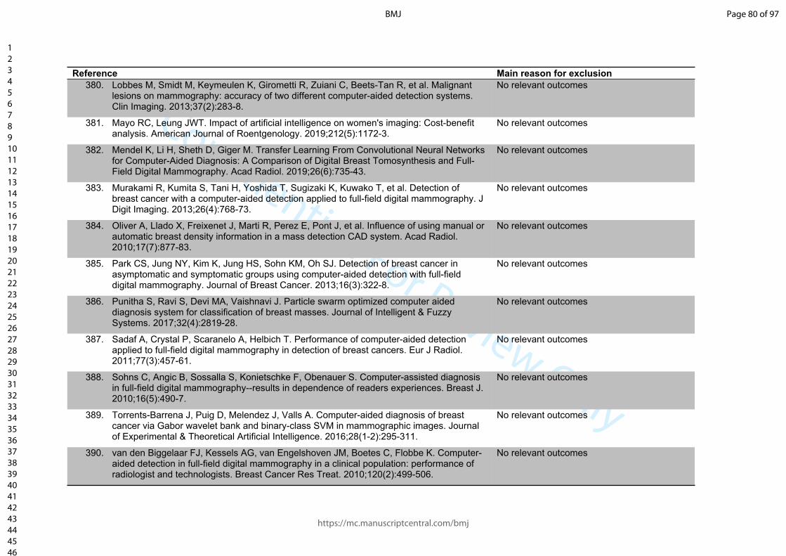
Confidential: For Review Only
Reference Main reason for exclusion380. Lobbes M, Smidt M, Keymeulen K, Girometti R, Zuiani C, Beets-Tan R, et al. Malignant
lesions on mammography: accuracy of two different computer-aided detection systems. Clin Imaging. 2013;37(2):283-8.
No relevant outcomes
381. Mayo RC, Leung JWT. Impact of artificial intelligence on women's imaging: Cost-benefit analysis. American Journal of Roentgenology. 2019;212(5):1172-3.
No relevant outcomes
382. Mendel K, Li H, Sheth D, Giger M. Transfer Learning From Convolutional Neural Networks for Computer-Aided Diagnosis: A Comparison of Digital Breast Tomosynthesis and Full-Field Digital Mammography. Acad Radiol. 2019;26(6):735-43.
No relevant outcomes
383. Murakami R, Kumita S, Tani H, Yoshida T, Sugizaki K, Kuwako T, et al. Detection of breast cancer with a computer-aided detection applied to full-field digital mammography. J Digit Imaging. 2013;26(4):768-73.
No relevant outcomes
384. Oliver A, Llado X, Freixenet J, Marti R, Perez E, Pont J, et al. Influence of using manual or automatic breast density information in a mass detection CAD system. Acad Radiol. 2010;17(7):877-83.
No relevant outcomes
385. Park CS, Jung NY, Kim K, Jung HS, Sohn KM, Oh SJ. Detection of breast cancer in asymptomatic and symptomatic groups using computer-aided detection with full-field digital mammography. Journal of Breast Cancer. 2013;16(3):322-8.
No relevant outcomes
386. Punitha S, Ravi S, Devi MA, Vaishnavi J. Particle swarm optimized computer aided diagnosis system for classification of breast masses. Journal of Intelligent & Fuzzy Systems. 2017;32(4):2819-28.
No relevant outcomes
387. Sadaf A, Crystal P, Scaranelo A, Helbich T. Performance of computer-aided detection applied to full-field digital mammography in detection of breast cancers. Eur J Radiol. 2011;77(3):457-61.
No relevant outcomes
388. Sohns C, Angic B, Sossalla S, Konietschke F, Obenauer S. Computer-assisted diagnosis in full-field digital mammography--results in dependence of readers experiences. Breast J. 2010;16(5):490-7.
No relevant outcomes
389. Torrents-Barrena J, Puig D, Melendez J, Valls A. Computer-aided diagnosis of breast cancer via Gabor wavelet bank and binary-class SVM in mammographic images. Journal of Experimental & Theoretical Artificial Intelligence. 2016;28(1-2):295-311.
No relevant outcomes
390. van den Biggelaar FJ, Kessels AG, van Engelshoven JM, Boetes C, Flobbe K. Computer-aided detection in full-field digital mammography in a clinical population: performance of radiologist and technologists. Breast Cancer Res Treat. 2010;120(2):499-506.
No relevant outcomes
Page 80 of 97
https://mc.manuscriptcentral.com/bmj
BMJ
123456789101112131415161718192021222324252627282930313233343536373839404142434445464748495051525354555657585960

Confidential: For Review Only
Reference Main reason for exclusion391. Vedanarayanan V, Nandhitha NM. Advanced image segmentation techniques for accurate
isolation of abnormality to enhance breast cancer detection in digital mammographs. Biomedical Research (India). 2017;28(6):2753-7.
No relevant outcomes
392. Warren LM, Given-Wilson RM, Wallis MG, Cooke J, Halling-Brown MD, Mackenzie A, et al. The effect of image processing on the detection of cancers in digital mammography. AJR Am J Roentgenol. 2014;203(2):387-93.
No relevant outcomes
393. Warren LM, Halling-Brown MD, Looney PT, Dance DR, Wallis MG, Given-Wilson RM, et al. Image processing can cause some malignant soft-tissue lesions to be missed in digital mammography images. Clin Radiol. 2017;72(9):799.e1-.e8.
No relevant outcomes
394. Wu Y, Vanness DJ, Burnside ES. Using multidimensional mutual information to prioritize mammographic features for breast cancer diagnosis. AMIA Annu Symp Proc. 2013;2013:1534-43.
No relevant outcomes
395. Yang X, Cao A, Song Q, Schaefer G, Su Y. Vicinal support vector classifier using supervised kernel-based clustering. Artif Intell Med. 2014;60(3):189-96.
No relevant outcomes
396. Yu SD, Liu LL, Wang ZY, Dai GZ, Xie YQ. Transferring deep neural networks for the differentiation of mammographic breast lesions. Science China-Technological Sciences. 2019;62(3):441-7.
No relevant outcomes
397. Zheng K, Harris C, Bakic P, Makrogiannis S. Spatially localized sparse representations for breast lesion characterization. Computers in Biology and Medicine. 2020;123 (no pagination).
No relevant outcomes
Study type – Systematic reviews (n=7)
398. Azavedo E, Zackrisson S, Mejare I, Heibert Arnlind M. Is single reading with computer-aided detection (CAD) as good as double reading in mammography screening? A systematic review. BMC med. 2012;12:22.
Study type – Systematic reviews with no relevant outcomes
399. Eadie LH, Taylor P, Gibson AP. A systematic review of computer-assisted diagnosis in diagnostic cancer imaging. Eur J Radiol. 2012;81(1):e70-6.
Study type – Systematic reviews with no relevant outcomes
400. Gruppo di studio G-S, Chersevani R, Ciatto S, Del Favero C, Frigerio A, Giordano L, et al. "CADEAT": considerations on the use of CAD (computer-aided diagnosis) in mammography. Radiol Med (Torino). 2010;115(4):563-70.
Study type – Systematic reviews with no relevant outcomes
Page 81 of 97
https://mc.manuscriptcentral.com/bmj
BMJ
123456789101112131415161718192021222324252627282930313233343536373839404142434445464748495051525354555657585960

Confidential: For Review Only
Reference Main reason for exclusion401. Henriksen EL, Carlsen JF, Vejborg IM, Nielsen MB, Lauridsen CA. The efficacy of using
computer-aided detection (CAD) for detection of breast cancer in mammography screening: a systematic review. Acta Radiol. 2019;60(1):13-8.
Study type – Systematic reviews with no relevant outcomes
402. Houssami N, Kirkpatrick-Jones G, Noguchi N, Lee CI. Artificial Intelligence (AI) for the early detection of breast cancer: a scoping review to assess AI's potential in breast screening practice. Expert Rev Med Devices. 2019;16(5):351-62.
Study type – Systematic reviews with no relevant outcomes
403. Sadoughi F, Kazemy Z, Hamedan F, Owji L, Rahmanikatigari M, Azadboni TT. Artificial intelligence methods for the diagnosis of breast cancer by image processing: a review. Breast Cancer (Dove Med Press). 2018;10:219-30.
Study type – Systematic reviews with no relevant outcomes
404. Yassin NIR, Omran S, El Houby EMF, Allam H. Machine learning techniques for breast cancer computer aided diagnosis using different image modalities: A systematic review. Comput Methods Programs Biomed. 2018;156:25-45.
Study type – Systematic reviews with no relevant outcomes
Full text not available via Document Supply (n=7)
405. Bhavani SR, Chilambuchelvan A, Senthilkumar J, Manjula D, Krishnamoorthy R, Kannan A. A secure cloud-based multi-agent intelligent system for mammogram image diagnosis. International Journal of Biomedical Engineering and Technology. 2018;28(2):185-202.
Document Supply cancelled request: no location found.
406. Grout S, Dheeraj Suryaa SR, Hitesh, Venkatesan DH, Sumanth S, Vishnu Vardhan Reddy M. Anomaly detection in digital mammography using neural networks. Journal of International Pharmaceutical Research. 2019;46(3):750-4.
Document Supply cancelled request: no location found.
407. Saraswathi D, Srinivasan E. An ensemble approach to diagnose breast cancer using fully complex-valued relaxation neural network classifier. International Journal of Biomedical Engineering and Technology. 2014;15(3):243-60.
Document Supply cancelled request: no location found.
408. Selvan VP, Suganthi M. Clinical support system for classification of tumor in mammogram images using multiple features and neural network classifier. Journal of Pure and Applied Microbiology. 2015;9(Special Edition):253-61.
Document Supply cancelled request: no location found.
409. Singh B, Jain VK, Singh S. Mammogram mass classification using support vector machine with texture, shape features and hierarchical centroid method. Journal of Medical Imaging and Health Informatics. 2014;4(5):687-96.
Document Supply cancelled request: no location found.
410. Srivastava S, Sharma N, Singh SK, Srivastava R. Quantitative analysis of a general framework of a CAD tool for breast cancer detection from mammograms. Journal of Medical Imaging and Health Informatics. 2014;4(5):654-74.
Document Supply cancelled request: no location found.
Page 82 of 97
https://mc.manuscriptcentral.com/bmj
BMJ
123456789101112131415161718192021222324252627282930313233343536373839404142434445464748495051525354555657585960

Confidential: For Review Only
Reference Main reason for exclusion411. Zhou L, Ding M, Xu L, Zhou Y, Zhang X. The automatic segmentation of mammographic
mass using the end-to-end convolutional network based on dense-prediction. Journal of Medical Imaging and Health Informatics. 2019;9(7):1429-34.
Document Supply cancelled request: no location found.
Other reasons (n=5)
412. Becker AS, Marcon M, Ghafoor S, et al. Deep Learning in Mammography: Diagnostic Accuracy of a Multipurpose Image Analysis Software in the Detection of Breast Cancer. Invest Radiol 2017;52(7):434-40. doi: https://dx.doi.org/10.1097/RLI.0000000000000358
Study 1: BCDR database; unclear proportion of screening mammograms
Study 2: Temporal validation
413. da Silva R, de Carvalho A. Automatic classification of breast lesions usingTransfer Learning. Ieee Latin America Transactions. 2019;17(12):1964-9.
Language – Not available in English
414. Kim HE, Kim HH, Han BK, et al. Changes in cancer detection and false-positive recall in mammography using artificial intelligence: a retrospective, multireader study. The Lancet Digital Health 2020;2(3):e138-e48. doi: http://dx.doi.org/10.1016/S2589-7500%2820%2930003-0
Evaluation study: unclear proportion of screening mammograms.
Reader study: In parts temporal validation confirmed by corresponding author via email.
415. Polat K. Application of Attribute Weighting Method Based on Clustering Centers to Discrimination of Linearly Non-Separable Medical Datasets. J Med Syst. 2012;36(4):2657-73.
Separation of two different mage datasets (liver and breast)
416. Sechopoulos I, Mann RM. Stand-alone artificial intelligence - The future of breast cancer screening? Breast. 2020;49:254-60.
Narrative review
Page 83 of 97
https://mc.manuscriptcentral.com/bmj
BMJ
123456789101112131415161718192021222324252627282930313233343536373839404142434445464748495051525354555657585960

Confidential: For Review Only
Publications excluded after review of full-text articles – Update database searches
PublicationsReference Main reason for exclusionPopulation – Image type (e.g. digitised film images; not FFDM images) (n=18)
1. Abubacker NF, Hashem IAT, Hui LK. Mammographic Classification Using Stacked Ensemble Learning with Bagging and Boosting Techniques. Journal of Medical and Biological Engineering 2020;40(6):908-16. doi: http://dx.doi.org/10.1007/s40846-020-00567-y
Population – Image type
2. Arora R, Rai PK, Raman B. Deep feature-based automatic classification of mammograms. Medical & biological engineering & computing 2020;58(6):1199-211. doi: https://dx.doi.org/10.1007/s11517-020-02150-8
Population – Image type
3. Bakthavachalam MD, Albert Antony Raj S. A study on breast cancer analysis by using k-nearest neighbor with different distances and classification rules using machine learning. European Journal of Molecular and Clinical Medicine 2020;7(3):4842-51.
Population – Image type
4. Gautam N, Singh A, Kumar K, et al. Investigation on performance analysis of support vector machine for classification of abnormal regions in medical image. Journal of Ambient Intelligence and Humanized Computing doi: 10.1007/s12652-021-02965-9
Population – Image type
5. Graewingholt A, Duffy S. Retrospective comparison between single reading plus an artificial intelligence algorithm and two-view digital tomosynthesis with double reading in breast screening. Journal of medical screening 2021:969141320984198. doi: https://dx.doi.org/10.1177/0969141320984198
Population – Image type
6. Jahangeer GSB, Rajkumar TD. Early detection of breast cancer using hybrid of series network and VGG-16. Multimedia Tools and Applications 2021;80(5):7853-86. doi: 10.1007/s11042-020-09914-2
Population – Image type
7. Kakileti ST, Madhu HJ, Krishnan L, et al. Observational Study to Evaluate the Clinical Efficacy of Thermalytix for Detecting Breast Cancer in Symptomatic and Asymptomatic Women. JCO global oncology 2020;6:1472-80. doi: https://dx.doi.org/10.1200/GO.20.00168
Population – Image type
8. Ketabi H, Ekhlasi A, Ahmadi H. A computer-aided approach for automatic detection of breast masses in digital mammogram via spectral clustering and support vector machine. Physical and Engineering Sciences in Medicine 2021;44(1):277-90. doi: http://dx.doi.org/10.1007/s13246-021-00977-5
Population – Image type
Page 84 of 97
https://mc.manuscriptcentral.com/bmj
BMJ
123456789101112131415161718192021222324252627282930313233343536373839404142434445464748495051525354555657585960

Confidential: For Review Only
Reference Main reason for exclusion9. Khamparia A, Bharati S, Podder P, et al. Diagnosis of breast cancer based on modern
mammography using hybrid transfer learning. Multidimensional systems and signal processing 2021:1-19. doi: https://dx.doi.org/10.1007/s11045-020-00756-7
Population – Image type
10. Melekoodappattu JG, Kadan AB, Anoop V. Early detection of breast malignancy using wavelet features and optimized classifier. International Journal of Imaging Systems and Technology doi: 10.1002/ima.22537
Population – Image type
11. Melekoodappattu JG, Subbian PS. Automated breast cancer detection using hybrid extreme learning machine classifier. Journal of Ambient Intelligence and Humanized Computing doi: 10.1007/s12652-020-02359-3
Population – Image type
12. Melekoodappattu JG, Subbian PS, Queen MPF. Detection and classification of breast cancer from digital mammograms using hybrid extreme learning machine classifier. International Journal of Imaging Systems and Technology 2021;31(2):909-20. doi: 10.1002/ima.22484
Population – Image type
13. Rao PMM, Singh SK, Khamparia A, et al. Multi-class Breast Cancer Classification using Ensemble of Pretrained models and Transfer Learning. Current medical imaging 2021 doi: https://dx.doi.org/10.2174/1573405617666210218101418
Population – Image type
14. Shaikh TA, Ali R. An intelligent healthcare system for optimized breast cancer diagnosis using harmony search and simulated annealing (HS-SA) algorithm. Informatics in Medicine Unlocked 2020;21:100408. doi: http://dx.doi.org/10.1016/j.imu.2020.100408
Population – Image type
15. Solanki YS, Chakrabarti P, Jasinski M, et al. A Hybrid Supervised Machine Learning Classifier System for Breast Cancer Prognosis Using Feature Selection and Data Imbalance Handling Approaches. Electronics 2021;10(6) doi: 10.3390/electronics10060699
Population – Image type
16. Thawkar S. A hybrid model using teaching-learning-based optimization and Salp swarm algorithm for feature selection and classification in digital mammography. Journal of Ambient Intelligence and Humanized Computing doi: 10.1007/s12652-020-02662-z
Population – Image type
17. Thawkar S, Ingolikar R. Classification of masses in digital mammograms using Biogeography-based optimization technique. Journal of King Saud University-Computer and Information Sciences 2020;32(10):1140-48. doi: 10.1016/j.jksuci.2018.01.004
Population – Image type
18. Xiao M, Zhao C, Li J, et al. Diagnostic Value of Breast Lesions Between Deep Learning-Based Computer-Aided Diagnosis System and Experienced Radiologists: Comparison the
Population – Image type
Page 85 of 97
https://mc.manuscriptcentral.com/bmj
BMJ
123456789101112131415161718192021222324252627282930313233343536373839404142434445464748495051525354555657585960

Confidential: For Review Only
Reference Main reason for exclusionPerformance Between Symptomatic and Asymptomatic Patients. Frontiers in Oncology 2020;10:1070. doi: http://dx.doi.org/10.3389/fonc.2020.01070
Population – Subpolulation (e.g. only cancer cases) (n=3)
19. Graewingholt A, Rossi PG. Retrospective analysis of the effect on interval cancer rate of adding an artificial intelligence algorithm to the reading process for two-dimensional full-field digital mammography. Journal of medical screening 2021:969141320988049. doi: https://dx.doi.org/10.1177/0969141320988049
Population - Subpopulation
20. Lang K, Hofvind S, Rodriguez-Ruiz A, et al. Can artificial intelligence reduce the interval cancer rate in mammography screening? European radiology 2021 doi: https://dx.doi.org/10.1007/s00330-021-07686-3
Population - Subpopulation
21. Lee SE, Han K, Kim E-K. Application of artificial intelligence-based computer-assisted diagnosis on synthetic mammograms from breast tomosynthesis: comparison with digital mammograms. European radiology 2021 doi: https://dx.doi.org/10.1007/s00330-021-07796-y
Population - Subpopulation
Population – <90% screening mammograms or unclear proportion (n=3)
22. Ashiba HI. A proposed framework for diagnosis and breast cancer detection. Multimedia Tools and Applications 2021;80(6):9333-69. doi: 10.1007/s11042-020-10131-0
Population – <90% screening mammograms or unclear proportion
23. Cui Y, Li Y, Xing D, et al. Improving the Prediction of Benign or Malignant Breast Masses Using a Combination of Image Biomarkers and Clinical Parameters. Frontiers in oncology 2021;11:629321. doi: https://dx.doi.org/10.3389/fonc.2021.629321
Population – <90% screening mammograms or unclear proportion
24. Kalyani K. CNN analysis for mammogramdisease detection. European Journal of Molecular and Clinical Medicine 2020;7(9):1540-43.
Population – <90% screening mammograms or unclear proportion
Internal validation – Cross validation (n=5)
25. Africano G, Arponen O, Sassi A, et al. A Comparison of Regions of Interest in Parenchymal Analysis for Breast Cancer Risk Assessment. Annual International Conference of the IEEE Engineering in Medicine and Biology Society IEEE Engineering in Medicine and Biology Society Annual International Conference 2020;2020:1136-39. doi: https://dx.doi.org/10.1109/EMBC44109.2020.9176200
Internal validation – Cross validation
26. Hassan SA, Sayed MS, Abdalla MI, et al. Breast cancer masses classification using deep convolutional neural networks and transfer learning. Multimedia Tools and Applications 2020;79(41-42):30735-68. doi: 10.1007/s11042-020-09518-w
Internal validation – Cross validation
Page 86 of 97
https://mc.manuscriptcentral.com/bmj
BMJ
123456789101112131415161718192021222324252627282930313233343536373839404142434445464748495051525354555657585960

Confidential: For Review Only
Reference Main reason for exclusion27. Heidari M, Lakshmivarahan S, Mirniaharikandehei S, et al. Applying a random projection
algorithm to optimize machine learning model for breast lesion classification. IEEE Transactions on Biomedical Engineering 2021 doi: http://dx.doi.org/10.1109/TBME.2021.3054248
Internal validation – Cross validation
28. Hsu CH, Chen X, Lin WW, et al. Effective multiple cancer disease diagnosis frameworks for improved healthcare using machine learning. Measurement 2021;175 doi: 10.1016/j.measurement.2021.109145
Internal validation – Cross validation
29. Jiang MK, Han L, Sun H, et al. Cross-modality image feature fusion diagnosis in breast cancer. Physics in Medicine and Biology 2021;66(10) doi: 10.1088/1361-6560/abf38b
Internal validation – Cross validation
Internal validation – Split sample (n=4)
30. Gnanasekaran VS, Joypaul S, Sundaram PM, et al. Deep learning algorithm for breast masses classification in mammograms. Iet Image Processing 2020;14(12):2860-68. doi: 10.1049/iet-ipr.2020.0070
Internal validation – Split sample
31. Guan Y, Wang X, Li H, et al. Detecting Asymmetric Patterns and Localizing Cancers on Mammograms. Patterns (New York, NY) 2020;1(7) doi: https://dx.doi.org/10.1016/j.patter.2020.100106
Internal validation – Split sample
32. Shen Y, Wu N, Phang J, et al. An interpretable classifier for high-resolution breast cancer screening images utilizing weakly supervised localization. Medical image analysis 2021;68:101908. doi: https://dx.doi.org/10.1016/j.media.2020.101908
Internal validation – Split sample
33. Suh YJ, Jung J, Cho B-J. Automated Breast Cancer Detection in Digital Mammograms of Various Densities via Deep Learning. Journal of personalized medicine 2020;10(4) doi: https://dx.doi.org/10.3390/jpm10040211
Internal validation – Split sample
Intervention – Detecting subtypes (n=1)
34. Vedalankar AV, Gupta SS, Manthalkar RR. Addressing architectural distortion in mammogram using AlexNet and support vector machine. Informatics in Medicine Unlocked 2021;23:100551. doi: http://dx.doi.org/10.1016/j.imu.2021.100551
Intervention – Detecting subtypes
Outcomes – No relevant outcomes (n=1)
35. Nct. Mammography Screening With Artificial Intelligence (MASAI). https://clinicaltrialsgov/show/NCT04838756 2021
Registered study protocol, no relevant outcomes
Full text not available via Document Supply (n=23)
Page 87 of 97
https://mc.manuscriptcentral.com/bmj
BMJ
123456789101112131415161718192021222324252627282930313233343536373839404142434445464748495051525354555657585960

Confidential: For Review Only
Reference Main reason for exclusion36. Sathya Priya T, Ramaprabha T. Deep learning based image segmentation with alexnet
feature extraction for classification of mammogram images. International Journal of Pharmaceutical Research 2021;13(1):4995-5009. doi: http://dx.doi.org/10.31838/ijpr/2021.13.01.690
Document Supply cancelled request: no location found.
37. Yi XC, Hou J. Segmentation of Medical Image Based on Superpixel Boundary Perceptual Convolutional Network in Cancer Diagnosis and Treatment. Journal of Medical Imaging and Health Informatics 2021;11(1):254-60. doi: 10.1166/jmihi.2021.3425
Document Supply cancelled request: no location found.
Already picked up by original searches (n=2)
38. Lindholm P, Eklund M, Dembrower K, et al. Effect of artificial intelligence-based triaging of breast cancer screening mammograms on cancer detection and radiologist workload: a retrospective simulation study. The Lancet Digital Health 2020;2(9):e468-e74. doi: http://dx.doi.org/10.1016/S2589-7500%2820%2930185-0
Same article as Dembrower (2020), already picked up by original search
39. Pacile S, Lopez J, Chone P, et al. Improving Breast Cancer Detection Accuracy of Mammography with the Concurrent Use of an Artificial Intelligence Tool. Radiology Artificial intelligence 2020;2(6):e190208. doi: https://dx.doi.org/10.1148/ryai.2020190208
Already picked up by original search
Page 88 of 97
https://mc.manuscriptcentral.com/bmj
BMJ
123456789101112131415161718192021222324252627282930313233343536373839404142434445464748495051525354555657585960
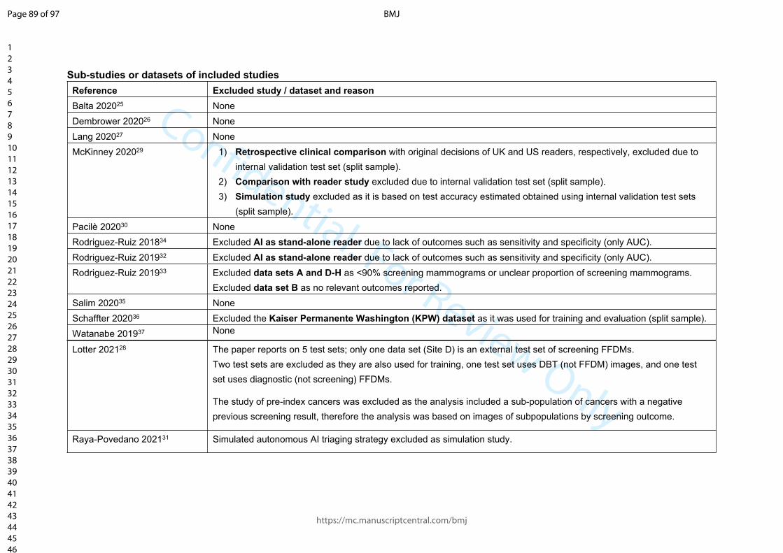
Confidential: For Review Only
Sub-studies or datasets of included studiesReference Excluded study / dataset and reasonBalta 202025 NoneDembrower 202026 NoneLang 202027 NoneMcKinney 202029 1) Retrospective clinical comparison with original decisions of UK and US readers, respectively, excluded due to
internal validation test set (split sample).2) Comparison with reader study excluded due to internal validation test set (split sample).3) Simulation study excluded as it is based on test accuracy estimated obtained using internal validation test sets
(split sample).Pacilè 202030 NoneRodriguez-Ruiz 201834 Excluded AI as stand-alone reader due to lack of outcomes such as sensitivity and specificity (only AUC).Rodriguez-Ruiz 201932 Excluded AI as stand-alone reader due to lack of outcomes such as sensitivity and specificity (only AUC).Rodriguez-Ruiz 201933 Excluded data sets A and D-H as <90% screening mammograms or unclear proportion of screening mammograms.
Excluded data set B as no relevant outcomes reported.Salim 202035 NoneSchaffter 202036 Excluded the Kaiser Permanente Washington (KPW) dataset as it was used for training and evaluation (split sample).Watanabe 201937 None
Lotter 202128 The paper reports on 5 test sets; only one data set (Site D) is an external test set of screening FFDMs.Two test sets are excluded as they are also used for training, one test set uses DBT (not FFDM) images, and one test set uses diagnostic (not screening) FFDMs.
The study of pre-index cancers was excluded as the analysis included a sub-population of cancers with a negative previous screening result, therefore the analysis was based on images of subpopulations by screening outcome.
Raya-Povedano 202131 Simulated autonomous AI triaging strategy excluded as simulation study.
Page 89 of 97
https://mc.manuscriptcentral.com/bmj
BMJ
123456789101112131415161718192021222324252627282930313233343536373839404142434445464748495051525354555657585960

Confidential: For Review Only
Appendix 3 Additional baseline characteristics of included studies
Study Confirmed cancer (prevalence) n (%)
Cancer type n (%) Cancer size/grade (invasive only)
Breast density n (%)
Stand-alone AILotter 202128 131 (46.0%) ILC or IDC: 88 (67.2%)
DCIS: 38 (29.0%)Other: 5 (3.8%)
Lesion typeSoft tissue 87Calcifications 53
For all 131 cancers:0-1cm: 45 (34.35%)1-2cm: 27 (20.6%)2-5cm: 11 (8.4%)>5cm: 3 (2.3%)Unknown: 45 (34.35%)
Non-cancer (n=154):Non-dense (A&B) 96 (62.3%)Dense (C&D) 58 (37.7%)
Cancer (n=131):Non-dense (A&B) 81 (61.8%)Dense (C&D) 50 (38.2%)
McKinney 202029 686 (22.2%) Data for 553/686 with BI-RADS score: Invasive 364/553 (65.8%)DCIS 163/553 (29.5%)Other 26/553 (4.7%)
NR NR
Rodriguez-Ruiz 201933
79 (39.7%) NR NR NR
Salim 202035 739 (8.4%) Invasive 640 (86%) IDC 514/640 (80%)ILC 82/640 (13%)other 43/640 (7%)missing 1/640 (0.15%)
In situ 85 (12%) Missing information 14 (2%)
Median 15mm (IQR 10-21mm)
Mammographic percent density, %Median 21.9IQR 13.8-32.1
Schaffter 202036 780 (1.1%) Invasive 681 (87.3%)DCIS 99 (12.7%)
NR NR
AI for triageBalta 202025 114 (0.64%) NR NR NRDembrower 202026 547 (7.4%) NR NR NRLang 202027 68 (0.71%) IDC 33/68 (48.5%)
ILC 11/68 (16.2%)ITC 10/68 (14.7%)DCIS 11/68 (16.2%)Other (e.g. papillary carcinoma, apocrine tumour) 3/68 (4.4%)
Grade 1 24/56 (42.9%)Grade 2 25/56 (44.6%)Grade 3 7/56 (12.5%)
NR
Page 90 of 97
https://mc.manuscriptcentral.com/bmj
BMJ
123456789101112131415161718192021222324252627282930313233343536373839404142434445464748495051525354555657585960

Confidential: For Review Only
Study Confirmed cancer (prevalence) n (%)
Cancer type n (%) Cancer size/grade (invasive only)
Breast density n (%)
Rava-Povedano 202131
113 (0.7%) Cancer type:Mass 67 (59.3%)Architectural distortion21 (18.6%)Asymmetry 4 (3.5%)Calcification 21 (18.6%)
Histologic type:IDC 80 (70.8%)ILC 5 (4.4%)Other invasive 1 (0.9%)DCIS 27 (23.9%)
For all 113 cancers:Grade 1 49 (43.4%)Grade 240 (35.4%)Grade 324 (21.2%)
Category A 3,648/15,986 (22.8%)Category B 8,153/15,986 (51.0%)Category C 3,749/15,986 (23.5%)Category D 436/15,986 (2.7%)
AI as reader aidPacilè 202030 120 (50%) Histologic type:
IDC 75 (62.5%)DCIS 27 (22.5%)ILC 6 (5.0%)Other 12 (10.0%)
Lesion type:Mass 64 (53.3%)Calcification 30 (25.0%)Asymmetry 13 (10.8%)Architectural distortion 13 (10.8%)
NR Category A 15.00% (36/240)Category B43.75% (105/240)Category C 34.58% (83/240)Category D 6.67% (16/240)
Page 91 of 97
https://mc.manuscriptcentral.com/bmj
BMJ
123456789101112131415161718192021222324252627282930313233343536373839404142434445464748495051525354555657585960
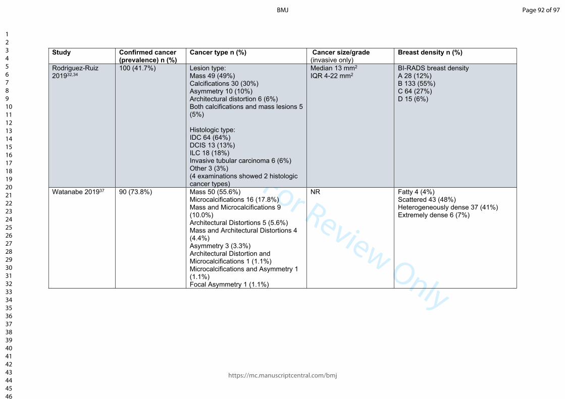
Confidential: For Review Only
Study Confirmed cancer (prevalence) n (%)
Cancer type n (%) Cancer size/grade (invasive only)
Breast density n (%)
Rodriguez-Ruiz 201932,34
100 (41.7%) Lesion type:Mass 49 (49%)Calcifications 30 (30%)Asymmetry 10 (10%)Architectural distortion 6 (6%)Both calcifications and mass lesions 5 (5%)
Histologic type:IDC 64 (64%)DCIS 13 (13%)ILC 18 (18%)Invasive tubular carcinoma 6 (6%)Other 3 (3%)(4 examinations showed 2 histologic cancer types)
Median 13 mm2
IQR 4-22 mm2BI-RADS breast densityA 28 (12%)B 133 (55%)C 64 (27%)D 15 (6%)
Watanabe 201937 90 (73.8%) Mass 50 (55.6%)Microcalcifications 16 (17.8%) Mass and Microcalcifications 9 (10.0%)Architectural Distortions 5 (5.6%)Mass and Architectural Distortions 4 (4.4%)Asymmetry 3 (3.3%) Architectural Distortion and Microcalcifications 1 (1.1%) Microcalcifications and Asymmetry 1 (1.1%)Focal Asymmetry 1 (1.1%)
NR Fatty 4 (4%)Scattered 43 (48%)Heterogeneously dense 37 (41%)Extremely dense 6 (7%)
Page 92 of 97
https://mc.manuscriptcentral.com/bmj
BMJ
123456789101112131415161718192021222324252627282930313233343536373839404142434445464748495051525354555657585960

Confidential: For Review OnlyReferences for Appendices 2 and 3
25. Balta C, Rodriguez-Ruiz A, Mieskes C, et al. Going from double to single reading for screening exams labeled as likely normal by AI: what is the impact? Proceedings of SPIE 2020;11513:115130D.
26. Dembrower K, Wahlin E, Liu Y, et al. Effect of artificial intelligence-based triaging of breast cancer screening mammograms on cancer detection and radiologist workload: a retrospective simulation study. Lancet Digital Health 2020;2(9):E468-E74.
27. Lang K, Dustler M, Dahlblom V, et al. Identifying normal mammograms in a large screening population using artificial intelligence. European Radiology 2020;02:02. doi: https://dx.doi.org/10.1007/s00330-020-07165-1
28. Lotter W, Diab AR, Haslam B, et al. Robust breast cancer detection in mammography and digital breast tomosynthesis using an annotation-efficient deep learning approach. Nature medicine 2021;27(2):244-49. doi: https://dx.doi.org/10.1038/s41591-020-01174-9
29. McKinney SM, Sieniek M, Godbole V, et al. International evaluation of an AI system for breast cancer screening. Nature 2020;577(7788):89-94. doi: https://doi.org/10.1038/s41586-019-1799-6)
30. Pacilè S, Lopez J, Chone P, et al. Improving Breast Cancer Detection Accuracy of Mammography with the Concurrent Use of an Artificial Intelligence Tool. Radiology: Artificial Intelligence 2020;2(6):e190208. doi: 10.1148/ryai.2020190208
31. Raya-Povedano JL, Romero-Martin S, Elias-Cabot E, et al. AI-based Strategies to Reduce Workload in Breast Cancer Screening with Mammography and Tomosynthesis: A Retrospective Evaluation. Radiology 2021:203555. doi: https://dx.doi.org/10.1148/radiol.2021203555
32. Rodriguez-Ruiz A, Krupinski E, Mordang JJ, et al. Detection of Breast Cancer with Mammography: Effect of an Artificial Intelligence Support System. Radiology 2019;290(2):305-14. doi: https://dx.doi.org/10.1148/radiol.2018181371
33. Rodriguez-Ruiz A, Lang K, Gubern-Merida A, et al. Stand-Alone Artificial Intelligence for Breast Cancer Detection in Mammography: Comparison With 101 Radiologists. J Natl Cancer Inst 2019;111(9):916-22. doi: https://dx.doi.org/10.1093/jnci/djy222
34. Rodriguez-Ruiz A, Mordang JJ, Karssemeijer N, et al. Can radiologists improve their breast cancer detection in mammography when using a deep learning based computer system as decision support? Proceedings of SPIE 2018;10718:1071803. doi: 10.1117/12.2317937
35. Salim M, Wahlin E, Dembrower K, et al. External Evaluation of 3 Commercial Artificial Intelligence Algorithms for Independent Assessment of Screening Mammograms. JAMA Oncol 2020;27:27. doi: https://dx.doi.org/10.1001/jamaoncol.2020.3321
36. Schaffter T, Buist DSM, Lee CI, et al. Evaluation of Combined Artificial Intelligence and Radiologist Assessment to Interpret Screening Mammograms. JAMA netw 2020;3(3):e200265. doi: https://dx.doi.org/10.1001/jamanetworkopen.2020.0265
37. Watanabe AT, Lim V, Vu HX, et al. Improved Cancer Detection Using Artificial Intelligence: a Retrospective Evaluation of Missed Cancers on Mammography. J Digit Imaging 2019;32(4):625-37. doi: https://dx.doi.org/10.1007/s10278-019-00192-5
Page 93 of 97
https://mc.manuscriptcentral.com/bmj
BMJ
123456789101112131415161718192021222324252627282930313233343536373839404142434445464748495051525354555657585960

Confidential: For Review OnlyAppendix 4 QUADAS-2
Item Response
PARTICIPANT SELECTION - A. RISK OF BIASWas a consecutive or random sample of patients enrolled?
Yes - RCTs and cohort studies (prospective or retrospective) with unenriched (consecutive or random) sampling Unclear - If not stated No - other studies
Did the study avoid inappropriate exclusions?
Yes – If inappropriate exclusions were avoided Unclear – if not clearly reportedNo - Exclusion of more than 10% of the samples for any reason, for example retrospective studies with missing dataNo - Systematic exclusion of types of women / images (e.g. of dense breasts)No - Exclusion based on outcomes (e.g. exclusion of cancer types, exclusion of interval cancers, exclusion/inclusion based on recall decision)
Were the women and mammograms included in the study independent of those used to train the AI algorithm?
For test set studies, this translates as has the test set been clearly described as an external (geographically) validation set?No - Any internal validation (e.g. split sample, cross-validation) or temporal validationUnclear - No details stated about the training set and tuning setYes - External geographical validation (Test set was sample from a different centre; can be in another country or the same country)
For prospective applied studies in a clinical context:Yes - If the study is located at different centre(s) to those who provided mammograms used to train and tune the AI algorithmUnclear - If not stated No - If there is any overlap
PARTICIPANT SELECTION - B. CONCERNS REGARDING APPLICABILITYIs there concern that the included patients do not match the review question?
High - If ‘yes’ for any of the following statementsUnclear - If no details are providedLow - If ‘no’ for all the following statements
Not a consecutive or random sample of women attending screening;
Enriched sample / cancer prevalence doesn’t match screening context (>3%);
Mammograms not from full-field digital mammography;
Mammograms not from screening (e.g. diagnostic or symptomatic) or only subset such as recalled cases or false-negatives included
Page 94 of 97
https://mc.manuscriptcentral.com/bmj
BMJ
123456789101112131415161718192021222324252627282930313233343536373839404142434445464748495051525354555657585960

Confidential: For Review Only(cancer might be easier or more difficult to detect);
Women/women’s mammograms not representative of UK population (ethnicity, age)
INDEX TESTS – A. RISK OF BIASWere the index test results interpreted without knowledge of the results of the reference standard?
For index tests where a human is involved (either human read comparator, AI as reader aid, or included otherwise on the AI testing pathway, e.g. arbitration):Yes - Require clear statement of blinding, or clear temporal relationships where the human read occurred before the reference standardNo - Otherwise
For index test where AI is used without any human element:Yes - AI system has not previously been trained on these mammograms or learned from these mammograms or other mammograms from the same womenNo - If any repeat use of the same cases then (unless explicit that the AI algorithm was pre-set and did not change upon repeat use, and the study did not select one of several AI systems based on use with the same cases)Unclear - If not explicit that there has been no repeat within same or previous studies
Were the index test results interpreted without knowledge of the results of any other index tests?
No - If human readers were not blinded to AI (unless that AI is specifically part of the same index test) No - If AI systems are trained or calibrated using decisions from human readers in same casesYes - Otherwise
If a threshold was used, was it pre- specified?
Yes - If using a commercially available AI system which gives a yes/no result, or threshold clearly pre-specified in methodsYes - For systems giving a risk score and study explicitly states the pre-specified thresholdNo - Using sensitivity / specificity of the reader as benchmark using the same datasetNo - Setting the threshold with the validation set without temporal evidence (e.g. published protocol) that threshold was truly pre-specifiedNA - Human readers or human/AI combinations
Where human readers are part of the test, were their decisions made in a clinical practice context? (i.e. avoidance of the laboratory effect)
Yes - If the readers made decisions in the clinical context, and those decisions were used to decide whether to recall women (either prospectively as part of a trial or test accuracy study or retrospective studies using the original decision) No - If readers examined a test set (of any prevalence) outside clinical practice, or any other context likely to result in the laboratory effect1
Page 95 of 97
https://mc.manuscriptcentral.com/bmj
BMJ
123456789101112131415161718192021222324252627282930313233343536373839404142434445464748495051525354555657585960

Confidential: For Review OnlyINDEX TESTS - B. CONCERNS REGARDING APPLICABILITYIs there concern that the index test(s) or comparator, its conduct, or interpretation differ from the review question?
High - If ‘yes’ for any of the followingUnclear – If no details are providedLow - If ‘no’ for all of the following
AI system not yet commercially available, e.g. in house systems;
Study did not use a pre-specified threshold for AI system;
Not a complete testing pathway applicable to clinical practice (for example AI accuracy for single read, but not integrated into screening centre decisions, e.g. arbitration);
Human comparator not a complete testing pathway applicable to clinical practice (human double reading with arbitration at clinical threshold);
AI system / reader had no access to prior mammograms / not 4 views available
REFERENCE STANDARD – A. RISK OF BIASIs the reference standard likely to correctly classify the target condition?
Yes - If the reference standard is histopathology results from biopsy (cancer present or absent) with at least 2 years follow up to interval cancersNo - If the reference standard is histopathology results from biopsy (cancer present or absent) with no follow up
Were the reference standard results interpreted without knowledge of the results of the index test?
Yes - Retrospective studies where readers read mammograms prospectively (enriched test sets) No - For retrospective studies (if we include the human reader comparator as an index test)No - For prospective studies if the investigators did not blind the clinicians undertaking the follow up tests to which index test examined the mammograms, for example by putting location marks in the same format for AI and human readers
REFERENCE STANDARD - B. CONCERNS REGARDING APPLICABILITYIs there concern that the target condition as defined by the reference standard does not match the review question?
High - If ‘yes’ for any of the followingUnclear - If no details are providedLow - If ‘no’ for all of the following
Length of screening rounds <2 years for follow-up / definition of interval cancers;
Classification not by biopsy/follow-upFLOW AND TIMING – A. RISK OF BIASDid all patients receive a reference standard?
No - If there was significant (>10%) loss to follow up for reference standards of interval cancers or subsequent screening resultsNo - If any women who should have received a biopsy or follow-up tests after index test positive results did not receive one or results were unavailableYes - otherwise
Page 96 of 97
https://mc.manuscriptcentral.com/bmj
BMJ
123456789101112131415161718192021222324252627282930313233343536373839404142434445464748495051525354555657585960
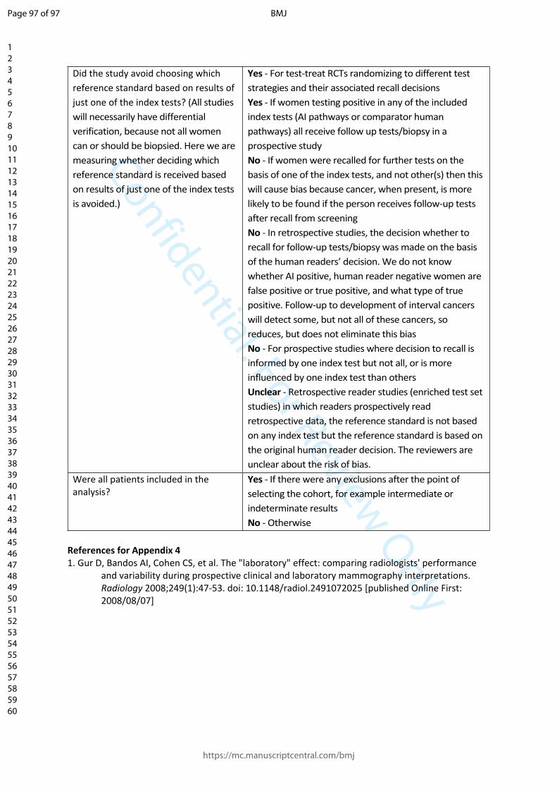
Confidential: For Review OnlyDid the study avoid choosing which reference standard based on results of just one of the index tests? (All studies will necessarily have differential verification, because not all women can or should be biopsied. Here we are measuring whether deciding which reference standard is received based on results of just one of the index tests is avoided.)
Yes - For test-treat RCTs randomizing to different test strategies and their associated recall decisionsYes - If women testing positive in any of the included index tests (AI pathways or comparator human pathways) all receive follow up tests/biopsy in a prospective studyNo - If women were recalled for further tests on the basis of one of the index tests, and not other(s) then this will cause bias because cancer, when present, is more likely to be found if the person receives follow-up tests after recall from screening No - In retrospective studies, the decision whether to recall for follow-up tests/biopsy was made on the basis of the human readers’ decision. We do not know whether AI positive, human reader negative women are false positive or true positive, and what type of true positive. Follow-up to development of interval cancers will detect some, but not all of these cancers, so reduces, but does not eliminate this biasNo - For prospective studies where decision to recall is informed by one index test but not all, or is more influenced by one index test than othersUnclear - Retrospective reader studies (enriched test set studies) in which readers prospectively read retrospective data, the reference standard is not based on any index test but the reference standard is based on the original human reader decision. The reviewers are unclear about the risk of bias.
Were all patients included in the analysis?
Yes - If there were any exclusions after the point of selecting the cohort, for example intermediate or indeterminate resultsNo - Otherwise
References for Appendix 41. Gur D, Bandos AI, Cohen CS, et al. The "laboratory" effect: comparing radiologists' performance
and variability during prospective clinical and laboratory mammography interpretations. Radiology 2008;249(1):47-53. doi: 10.1148/radiol.2491072025 [published Online First: 2008/08/07]
Page 97 of 97
https://mc.manuscriptcentral.com/bmj
BMJ
123456789101112131415161718192021222324252627282930313233343536373839404142434445464748495051525354555657585960