Mechanisms of acquired resistance to cetuximab: role of HER (ErbB) family members
Upload
u-bourgogneCategory
view
1download
0
Annals of Oncology 20 (Supplement 8): viii113–viii130, 2009
doi:10.1093/annonc/mdp445session L: gastrointestinalcancer-colon-rectalL1* FOLFOXIRI (IRINOTECAN, OXALIPLATIN, INFUSIONAL 5FU/LV)VS FOLFIRI AS FIRST-LINE TREATMENT OF METASTATICCOLORECTAL CANCER (MCRC): UPDATED RESULTS AFTER 5YEARS FOLLOW UP AND RISK-STRATIFIED ANALYSIS
Masi G1, Vasile E1, Loupakis F1, Cupini S2, Fornaro L1, Baldi G2, Salvatore L1,Stasi I1, Brunetti I3, Ricci S3, Palazzo A4, Pellegrino A4, Truscelli K4, Garrone M5,Chiara S6, Ribecco A7, Crino L8, Andreuccetti M1, Falcone A1,2
1U.O. Oncologia Medica 2 Universitaria, Azienda Ospedaliera-UniversitariaPisana, Istituto Toscano Tumori, Pisa (IT); 2U.O. Oncologia Medica, AziendaUSL 6 di Livorno, Istituto Toscano Tumori, Livorno (IT); 3U.O. Oncologia Medica1, Azienda Ospedaliera-Universitaria Pisana, Istituto Toscano Tumori, Pisa (IT);4U.O. Oncologia B, Dipartimento di Medicina Sperimentale, Universita di Roma‘‘Sapienza’’, Roma (IT); 5U.O. Oncologia Medica, Ospedale S. Croce e Carle,Cuneo (IT); 6U.O. Oncologia Medica, Istituto Nazionale Tumori, Genova (IT); 7S.C.Oncologia Medica, Azienda Sanitaria di Firenze, Istituto Toscano Tumori, Firenze(IT); 8U.O. Oncologia Medica, Ospedale S. Andrea delle Fratte, Perugia (IT)
Background: As we previously reported the GONO-FOLFOXIRI regimen compared toFOLFIRI demonstrated significant improvements in responses (60% vs 34%, p<0.001),secondary radical (R0) resection of metastases (15% vs 6%, p=0.03), progression-freesurvival (9.8 vs 6.9 months, p=0.006) and overall survival (22.6 vs 16.7 months,p=0.03) after a median follow up of 18.4 months.
Methods:We updated overall survival (OS) and progression-free survival (PFS) data ofthe 244 randomized patients after a median follow up of 60.6 months (mos) and weused a risk-stratified analysis according to the Kohne prognostic model to determine iftreatment outcomes differ in specific patient subgroups.
Results: The updated results confirm a significant improvement for FOLFOXIRI interms of PFS (median 9.8 vs 6.8 mos, HR=0.59, p<0.0001) and OS (median 23.4 vs 16.7mos, HR=0.74, p=0.026 and 5-years survival rate 15% vs 8%). There is a PFS andOS benefit from FOLFOXIRI also excluding from the analysis the R0 patients (medianPFS 9.5 vs 6.6 mos, p=0.0001 and median OS 20.2 vs 15.9 months, p=0.12). Withregard to the risk-stratified analysis, FOLFOXIRI results in longer PFS and OS thanFOLFIRI in all risk subgroups with Hazard Ratios for low, intermediate and high riskgroups respectively of 0.68, 0.56 and 0.44 for PFS and of 0.90, 0.58 and 0.78 for OS.
Conclusions: These results demonstrate that the GONO-FOLFOXIRI regimen isassociated also with a better long term outcome compared to FOLFIRI (with anabsolute benefit in survival at 5 years of 7%) and that the superiority of FOLFOXIRI isnot only related to the increased rate of R0 surgery of metastases, but also to a betterpalliative effect which does not seem to be limited to some specific subgroup.
L2* VEGF GENE POLYMORPHISMS IN THE PREDICTION OFBENEFIT FROM FIRST-LINE FOLFIRI PLUS BEVACIZUMAB (BV) INMETASTATIC COLORECTAL CANCER (MCRC) PATIENTS (PTS)
Loupakis F1, Ruzzo A2, Salvatore L1, Canestrari E2, Cremolini C1, Masi G1,Schirripa M1, Spoto C3, Galluccio N2, Vincenzi B3, Santini D3, Bencardino K4,Ricci V4, Catalano V5, Manzoni M6, Danova M6, Tonini G3, Magnani M2,Falcone A1,7, Graziano F5
1U.O. Oncologia Medica 2 Universitaria, Azienda Ospedaliera-UniversitariaPisana Istituto Toscano Tumori, Pisa, Italy; 2Dipartimento di ScienzeBiomolecolari, Universita degli Studi ‘‘Carlo Bo’’, Urbino, Italy; 3U.O. OncologiaMedica, Universita Campus Biomedico, Roma, Italy; 4U.O. Oncologia Medica,Istituto Scientifico Universitario San Raffaele, Milano, Italy; 5U.O. OncologiaMedica, Ospedale di Pesaro, Italy; 6U.O. Oncologia Medica, Fondazione IRCCSPoliclinico S. Matteo, Pavia, Italy; 7Dipartimento di Oncologia dei Trapianti e delleNuove Tecnologie in Medicina, Universita di Pisa, Italy
Introduction: Addition of BV to first-line irinotecan plus 5FU improves PFS and OS ofmCRC pts. Meanwhile, the anti-VEGF causes specific toxicities and increases costs oftreatment. At the same time, not all pts derive an equal benefit from the VEGF inhibitor.Molecular predictors of BV efficacy have not yet been identified. Specific VEGFpolymorphisms may affect gene transcription, thus indirectly influencing BV efficacy.
Methods: Peripheral blood samples for genomic DNA extraction were collected fromconsecutive mCRC pts receiving FOLFIRI plus BV as first-line treatment (BV-group).VEGF-2578A/C, -460C/T, +405C/G, +936C/T polymorphisms were analysed by means of PCR-RFLP.One-hundred-sevenpts, treatedwithFOLFIRIalone, servedashistorical control group.
Results: One-hundred-eleven pts were included in the BV-group. M/F=57/54, medianage=63 (34-82), Kohne score (low/intermediate/high/data missing)=57/39/12/3. Sixty-nine out of 111 pts achieved response (RR=62%). Median PFS (mPFS) and median OS(mOS) were 10.2 and 22.2 months, respectively. -460C/C, C/T and T/T allelic variantswere found in 20%, 54% and 26% of pts, respectively. -460 T allele demonstrated
shorter PFS and OS with an additive effect of each T allele (PFS:HR=2.65[1.49-6.62],p=0.003; OS:2.47[0.91-7.66],p=0.074). -460C/C pts achieved longer PFS and OSin comparison to pts carrying at least one T allele (mPFS:12.8 vs 9.8months;HR=0.48[0.28-0.85],p=0.012; mOS:27.3 vs 20.5 months; HR=0.38[0.19-0.94],p=0.034). In the control group mPFS and mOS were 8.2 and 20.6 months; -460C/C, C/T and T/T variants were found in 23%, 52% and 25% of pts; there was nosignificant association with PFS or OS. Other polymorphisms did not affect outcomeneither in BV-group nor in the control group.
Conclusions: At our knowledge this is the first report of a pharmacogeneticdeterminant of improved PFS and OS for mCRC pts treated with first-line BV-containing therapy. The observation that VEGF -460C/T variants did not influence theoutcome in the control group led to hypothesize a predictive other than a prognosticrole for such genetic signature. These preliminary data deserve investigation inprospective, randomized, validating trials.
L3* CONCOMITANT ANALYSIS OF KRAS, BRAF, PIK3CAMUTATIONS AND LOSS OF PTEN EXPRESSION ENHANCESPREDICTION OF CLINICAL OUTCOME TO CETUXIMAB ORPANITUMUMAB IN METASTATIC COLORECTAL CANCER
Salvatore Siena1, Andrea Sartore-Bianchi1, Federica Di Nicolantonio2,Michele Nichelatti3, Francesca Molinari4, Sara De Dosso5, Piercarlo Saletti5,Miriam Martini2, Federico Pozzi1, Giovanna Marrapese1, Carolina Sarnataro1,Luca Mazzucchelli4, Simona Lamba2, Silvio Veronese6, Milo Frattini4,Alberto Bardelli2,7
1The Falck Division of Medical Oncology, 3Service of Biostatistics, and 6 Divisionof Pathology, Ospedale Niguarda Ca’ Granda, Milan, Italy; 2Laboratory ofMolecular Genetics, The Oncogenomics Center, Institute for Cancer Researchand Treatment (IRCC), University of Torino Medical School, Candiolo, Turin, Italy;4Laboratory of Molecular Diagnostic, Istituto Cantonale di Patologia, Locarno,Switzerland; 5Oncology Institute of Southern Switzerland, Ospedale San Giovanni,Bellinzona, Switzerland; 7FIRC Institute of Molecular Oncology, Milan, Italy
Presented in part at the 100th annual meeting of the American Association for CancerResearch, April 18-22, 2009, Denver, CO.
Despite the recent recommendations by EMEA as well as by ASCO of KRAS testingas a diagnostic prerequisite for EGFR-targeted cetuximab- or panitumumab-basedtherapies for metastatic colorectal cancer (mCRC), the response rate to either of thesedrugs is limited to <20% in wild-type KRAS patients. Recent data indicate that BRAF orPIK3CA mutations may contribute for additional 20-30% of resistance. However, therelative and overall contribution of each of these molecular alterations to clinicaldecision making remains unclear. Furthermore, whether and to what extent theoccurrence of multiple molecular alterations affects clinical response and survival ispresently unknown. We present here a comprehensive analysis of KRAS, BRAF,PIK3CAmutations, and PTEN expression in mCRC patients treated with cetuximab orpanitumumab, with the aim of clarifying the relative contribution of these molecularalterations to resistance. We retrospectively analyzed objective tumor response,progression-free (PFS) and overall survival (OS) together with the mutational status ofKRAS, BRAF, PIK3CA and expression of PTEN in 132 tumors from cetuximab orpanitumumab treated mCRC patients. Among the 106 non-responsive patients, 74(70%) had tumors with at least one molecular alteration in the four markers. Theprobability of response was 51% (22/43) among patients with no alterations, 4% (2/47)among patients with 1 alteration, and 0% (0/24) for patients with ‡ 2 alterations(p<0.0001). Accordingly, PFS and OS were increasingly worse for patients with tumorsharboring none, 1, or ‡2 molecular alteration(s) (p<0.001). When expression of PTENand mutations of KRAS, BRAF and PIK3CA are concomitantly ascertained, up to 70%of mCRC patients unlikely to respond to anti-EGFR therapies can be identified.Comprehensive molecular dissection of the EGFR signaling pathways should beconsidered to select mCRC patients for cetuximab- or panitumumab-based therapies.
L4* RESECTABILITY OF COLORECTAL LIVER METASTASES (CLM)WITH CETUXIMAB PLUS CPT-11/5-FLUOROURACIL (5-FU)/LEUCOVORIN(FA)/OXALIPLATIN (L-OHP) (CPT-11-FFL) ASNEOADJUVANT TREATMENT (POCHER TRIAL)
Torsello A, Garufi C, Tumolo S, Melucci E, Conti S, Campanella C, Zeuli M,Pizzi G, Tropea G, Sperduti I, Ettorre GM, Cognetti FIstituto Regina Elena, Rome, Italy; Ospedale S.M. degli Angeli, Pordenone, Italy;Ospedale S. Camillo, Rome, Italy
Background: Addiction of Cmab to standard chemotherapy seem to be beneficial inadvanced colorectal cancer pts K-RAS wild type (Crystal and Opus trials). Tripletcombination of CPT-11/(5-FU)/FA/L-OHP, (Falcone, 2007), is more effective than
se
ssio
nL
ª The Author 2009. Published by Oxford University Press on behalf of the European Society for Medical Oncology.
All rights reserved. For permissions, please email: [email protected]
by guest on March 22, 2015
http://annonc.oxfordjournals.org/D
ownloaded from
doublets. Aim of the study was to evaluate Cmab+CPT-11-FFL in pts with unresectableCLM, with primary end-point resectability.
Method: Unresectability criteria: size>5 cm (a); multinodular (b); ilar location (c);extrahepatic disease (d); >3 stable mts after chemotherapy before surgery (e). Aim: tohave at least 30% resection rate (power 80%, p0=10% and p1=25%). Pts, irrespectiveof EGFR and k-RAS status, received weekly Cmab plus CPT-11 130 mg/m2/d1, 6-hinfusion (peak 13:00), and 12-h, days 2-5, infusion of L-OHP 20/mg/m2/d (peak at16:00), FA 150 mg/m2/d plus 5-FU 600 mg/m2/d (peak at 4:00), q2 weeks; after first17 pts L-OHP and 5-FU were reduced to 15 and 550 mg/m2 respectively due totoxicity. The trial was disegned in 2006, thus we retrospectively evaluated EGFR andK-RAS status.
Results: From 07/20/2006 to 09/01/2008 we enrolled 43 pts,: M/F 27/16; median age60,7y (33-76), median PS 0. Primary tumor colon/rectum 34/9; primary tumorresected 39 pts (79%), synchronous metastases 35 pts (81%); liver involvement <25%/>25% 9/34 (21/79%); pre-treatment median CEA/Ca19-9 55ng/ml (1-6600)/91.8 U/L(2-66440); unresectability: (a) 9 (21%); (b) 14 (33%); (c) 1; (d) 4 (9%); (e) 15 (35%).EGFR and K-RAS status was retrospectively evaluated in 70% of pts in liver biopsies.EGFR staining: 0 18%, 1+ 7%, 2+ 57%, 3+ 18%. K-RAS wt 75% and mut 25%. We had34 PR (79%, CI 79.1-87), 5 SD and 4 pts not valuable because of toxicity. Completeresection of CLM in 25 pts (58%), 2 pts still to be resected. Median number (n) ofcourses (c) per pt was 10 (2-18) with median n of c before surgery (s)=5 (3-10) andafter s=6 (1-6); median time from last c to s was 2 wks (2-4), from s to recovery chemowas 10 wks (2-16). Median: follow-up 14 months (range1-34), PFS 13 months (CI95%6-20); OS not reached with 2-y survival of 63%; 14 pts alive without recurrence (32%),13 deaths (30%).
Major limiting toxicity was diarrhea: G2 6.3%; G3 81% and G4 12.5%, but it wassignificantly reduced after dose modification: G2 26%, G3 35% (p0.005), G4 1%(p0.006). Abdominal pain also resulted significantly reduced: G2 31/25%; G3 31,3/7%(p 0.05). No significant differences pre/after dose modification for other toxicities:fatigue G2 43/37%, G3 8/12%; nausea G2 50/44% G3 12/10%; afebrile neutropenia G212/4%, G3 7/6%; rush G2 50/66%, G3 20/15%. We observed a trend towards more G3/4 toxicities in pts with stable disease respect to partial response (p 0.06). Toxicity didnot affect time to surgery (p0.23).
Conclusions: This study showed that a triplet chemotherapy combination plusCmab is able to obtain tumour shrinkage in 79% of pts with 58% of complete liverresection. Major limiting toxicity was diarrhea improved with dose modification.Definitive results on molecular analysis and prognostic factors are in progress.
L5* AIOM GUIDELINES: RESULTS OF EXTENDED PHASE RIGHT-2IN COLORECTAL CANCER (CRC)
Beretta GD1, Barni S2, Maiello E3, Carnaghi C4, Cosimelli M5, Faggiuolo R6,Valvo F7, Sgarbi S8
1Ospedale Sant’Orsola-FBF Brescia; 2Ospedale Treviglio; 3Casa Sollievo dellasofferenza San Giovanni Rotondo; 4Istituto Clinico Humanitas Milano; 5IstitutoRegina Elena Roma; 6Ospedale Alba; 7Istituto Nazionale Tumori Milano;8Medidata
AIOM Guidelines task force was created in 2002. In that year many evidence-basedguidelines in cancer management and treatment were developed. Update of guidelinesis only one of the aspect for improvement of quality of care. Diffusion andverification of applicability are also very important. In this context, the RIGHT(Research for the Identification of the most effective and hIGHly accepted clinicalguidelines for cancer Treatment) program was developed for these reasons. Pilotphase, named RIGHT-1, validated Process Indicators of agreement between guidelinesand clinical practice. RIGHT-2 used the same Process Indicators in an extendedcontext.Method: RIGHT-2 is a cross-sectional survey for colorectal cancer care performed ina sample of 37 centre. The RIGHT-2 study centre were randomly selected to bea representative sample of the 319 AIOM centre that were identified by the 2003 AIOMcensus. Centre were selected for inclusion into the study after stratification bygeographical area and kind of centre. The medical charts of patients with stage III coloncancer (CC), stage II-III rectal cancer (RC) and advanced colorectal cancer (ACRC)who had the first visit at the selected centre between October 2005 and November 2006were retrospectively analyzed according to AIOM 2005 colo -rectal guidelines. Theprocess indicators regarded diagnosis, surgery, therapy and follow up.
Results: 129 CC, 80 RC and 117 ACRC patients were eligible for the analysis.AIOM guidelines were correctly applied to 78% in CC, 68% in RC and 77% in ACRC.The best adherence were observed for diagnostic colonoscopy or barium enema(CC,RC), starting adjuvant chemotherapy on time (CC), correct use of oralfluoropyrimidine (ACRC). The worse adherence were: proportion of patientreceiving preoperative treatment (RC), time from diagnosis and the start ofpreoperative treatment (RC), preoperative CEA determination (CC), correct useof biological drugs (ACRC). Analysis for geographical area and kind of centre isongoing.
Conclusion: RIGHT-2 survey showed percentage of adherence similar between thatreported in international studies and in the representative sample of AIOM Centre
L6* CIRCULATING ENDOTHELIAL CELLS (CECS) AND FDG-PETFOR EARLY PREDICTION OF RESPONSE IN HIGH- RISK LOCALLYADVANCED RECTAL CANCER (H-LARC) PATIENTS (PTS) TREATEDWITH TWO DIFFERENT SCHEDULES OF BEVACIZUMAB (BEV) INCOMBINATION WITH PREOPERATIVE CHEMO-RADIOTHERAPY(CT-RT)
Antonio Avallone1, Paolo Delrio2, Biagio Pecori3, Elena Di Gennaro3,Fabiana Tatangelo4, Corradina Caraco5, Antonella Petrillo7,Claudia Sandomenico1, Alfredo Budillon3, Pasquale Comella1
1Department of Medical Oncology; 2Surgical Oncology; 3ExperimentalPharmacology Unit; 4Radiotherapy; 5Pathology; 6Nuclear Medicine; 7Radiology,National Cancer Institute Fondazione G. Pascale, Naples, Italy
Background: Vascular endothelial growth factor (VEGF) has a crucial role in tumorangiogenesis, and its inhibition leads to the normalization of tumor vessels andincreases tumor oxygenation and drug delivery. However, the clinical benefits ofcurrent anti-VEGF treatments have thus far been rather modest, stimulating interest indeveloping more effective ways to combine anti-VEGF drugs and CT and in theidentification of valid predictive biomarkers of clinical benefit We have previouslyshown that pre-operative oxaliplatin (OXA), raltitrexed (RTX), fluorouracil (5FU),and folinic acid (LFA) during pelvic RT have high rate of complete (TRG1) or subtotal(TRG2) tumor regression in HR-LARC. Therefore, we planned to add BEV to primaryCH-RT in two different schedules, to evaluate the relevance of the timing of BEVrelative to CT and RT. Changes of CECs and glucose metabolism evaluated by flowcytometry and FDG-PET were used as early surrogate marker of tumor response.
Methods: Twenty-eight pts (inclusion criteria: cT4, cN+, cT3£ 5 cm from the analverge and/or +ve CRM, M1 resectable/initially unresectable) received 3 biweeklycourses of OXA (100 mg/m2)/RTX (2.5 mg/m2) on day 1, and 5FU (800 mg/m2)/LFA(250 mg/m2) on day 2 during pelvic RT (45 Gy). In schedule A (16 pts) BEV (5 mg/kg)was given biweekly from day -14 for 4 courses, while in schedule B (12 pts) it was givenfrom day -4 for 2 courses. Toxicity was graded with NCI-CTC version 3. According tothe Simon’s two-stage design, assuming a hypothesis of a 50% TRG1 (a error=0.05, berror=0.20), at least 6/16 TRG1 should be obtained (first stage) to continue pts accrual
Results: No treatment-related or perioperative death occurred. As in the previousphase II study without BEV, Grade 3 /
4neutropenia was the most common adverse eventwith the schedule A (7 pts, 44%), but it was lower with the schedule B (2 pts, 17%). Pre-treatment basal CEC amounts showed significantly increased levels in the pts samplescompared to the healthy controls. After 1st course of CT median CEC levels werereduced compare to basal levels in both schedules. However, a significant differencewas observed between the two schedules with median -78% and -29%, reduction, inschedule B and A, respectively (p<0.05). Moreover, we observed in the schedule Ba different CECs kinetic compared with schedule A. Likewise, a major reduction ofmedian tumor metabolic volume was observed in schedule B compared to schedule A(-78% vs -50%, p<0.05). All but one pt ( refused surgery) in schedule A, and 11 pts inschedule B have proceeded to surgery. In the schedule A only 2 (12%) pts obtaineda TRG 1. On the contrary the number of TRG 1 required by statistical design wasalready reached in the first 11 pts treated. in the schedule B( 6pts; 55%).
Conclusion: Overall these data suggest the relevance of the BEV schedule to optimizethe feasibility and efficacy of combination treatment, as well as the potential role ofCEC evaluation and FDG-PET for the early prediction of responses to anti-angiogenesis therapeutical approaches.
L7 PHASE II STUDY OF TAILORED CHEMOTHERAPY FORELDERLY PATIENTS WITH ADVANCED COLORECTAL CANCER(ACRC) BASED ON THE EXPRESSION OF THYMIDYLATE SYNTHASE(TS), DIHIDROPYRIMIDINE DEHYDROGENASE (DPD), EXCISIONREPAIR CROSS-COMPLEMENTING-1 (ERCC-1) AND UDP-GLUCURONYL-TRANSFERASE (UGT 1A1)
Mario Scartozzi1, Cristian Loretelli2, Rossana Berardi1, Rosa Rita Silva3,Davide Mari3, Riccardo Giampieri4, Stefano Cascinu1
1Clinica di Oncologia Medica, AO Ospedali Riuniti-Universita Politecnica delleMarche, Ancona-Italy; 2Dipartimento di Medicina Clinica e BiotecnologieApplicate, Universita Politecnica delle Marche, Ancona-Italy; 3OncologiaMedica, Ospedale di Fabriano, Italy; 4Scuola di Specializzazione in Oncologia,Universita Politecnica delle Marche, Ancona-Italy
Although therapeutic options for ACRC have expanded, it is not uncommon toobserve toxic side-effects with lack of efficacy. Elderly patients, a growing proportion ofall ACRC, are more prone to severe treatment-related toxicity and may be morefrequently excluded from a potentially effective treatment on this basis. Howeverretrospective studies have suggested that biological markers may be able to identifyresponding tumours and individuals with a greater chance of severe toxicity. We thenconducted a phase II study in previously untreated, elderly (‡ 70 year old) ACRCpatients in which the selection of chemotherapy was based on genotyping expression.Aim of the study was to obtain a 70% response rate with a 10% (or lower) rate of grade3-4 toxicity.Patients and Methods: Patients with histologically-proven ACRC aged ‡ 70 year wereeligible. Genotyping for TS, DPD, ERCC-1 and UGT1A1 was conducted with PCR-based techniques on genomic DNA from peripheral blood.
viii114 | session L: gastrointestinal cancer-colon-rectal Volume 20 | Supplement 8 | October 2009
session L Annals of Oncology
by guest on March 22, 2015
http://annonc.oxfordjournals.org/D
ownloaded from
Results: Twenty-four patients were included in our study (20 males, median age 76,range 70-84). According to genotyping results 12 patients (50%) were treated withthe FOLFIRI regimen, 11 patients (46%) with the FOLFOX-6 regimen and 1 (4%)with 5FULV according to the De Gramont regimen. Overall a partial remission wasobtained in 4 cases (17%), stable disease in 8 cases (33%) and progressive disease inthe remaining 12 cases (50%). Grade 3-4 toxicities included neutropenia in 7 patients(29%)and diarrhoea in 3 cases (12%). A toxic death (4%) was observed. The trial wasthen interrupted according to study design which required 13 partial remissions outof the first 24 patients enrolled as the necessary response rate level in order tocontinue.
Discussion: Prospective selection of a chemotherapy regimen based on TS, DPD,ERCC-1 and UGT1A1 expression in elderly patients with ACRC failed to confirm theencouraging results reported in retrospective analyses. A more accurate validation ofdata deriving from retrospective series should be performed before prospectivelyselecting the appropriate treatment for the appropriate patient.
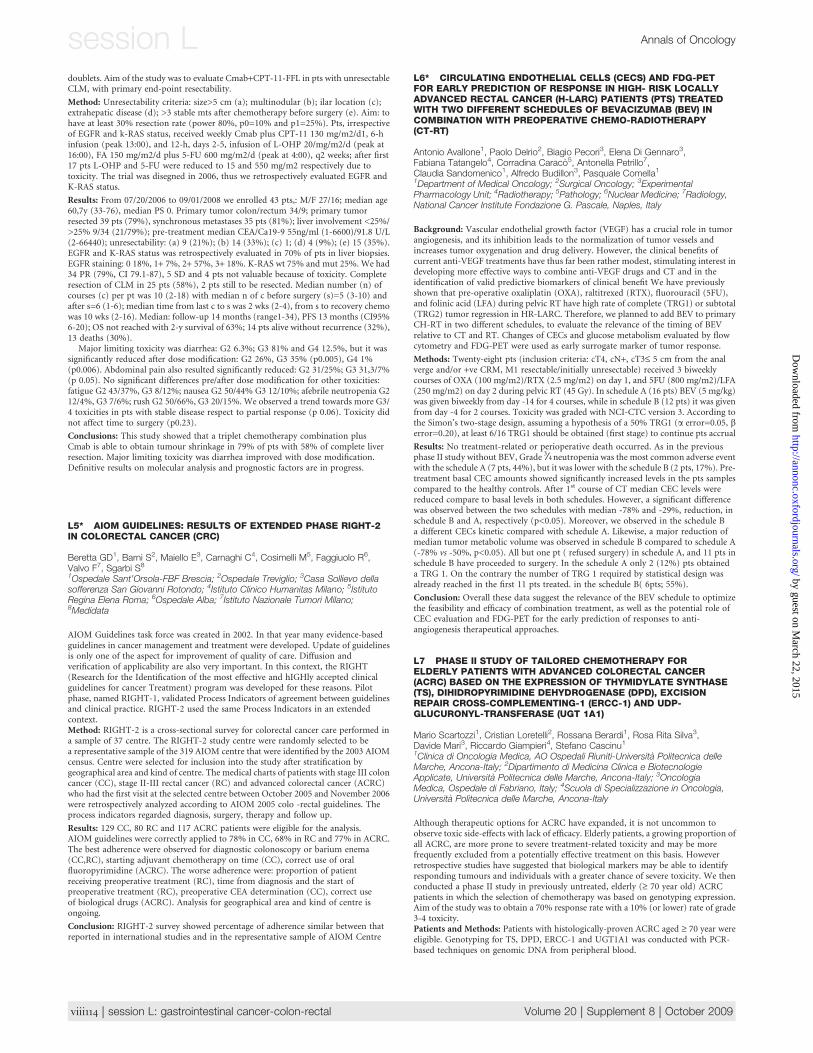
L8 SYSTEMIC CHEMOTHERAPY(SCT) IN RESECTABLECOLORECTAL LIVER METASTASIS(RCLM): SYSTEMATIC REVIEWOF LITERATURE AND METAANALYSIS OF RANDOMIZED CLINICALTRIALS (RCTS)
Emiliano Tamburini1, Davide Tassinari1, Maximiliam Papi1, Stefania Nicoletti1,Manuela Fantini1, Cinzia Possenti1, Fabrizio Drudi1, Giovanni Oliverio1,Lorenzo Gianni1, Enzo Pasquini2, Alberto Ravaioli11U.O. di Oncologia ed Oncoematologia Ospedale Infermi di Rimini; 2U.O. diOncologia Ospedale Cervesi di Cattolica; IRST, istituto scientifico Romagnoloper lo studio e cura dei tumori, Meldola
Background: At present surgery is considered the only potentially curative approachfor patients with colorectal cancer liver metastasis. In this context the role of systemicchemotherapy remains controversial.
Materials and Methods: A systematic review of MEDLINE, EMBASE and CochraneSystematic reviews databases from January 1966 to April 2009 was performedindependently by two authors. All randomized phase III trials comparing one medicalapproach with surgery alone in RCLM were considered eligible and included into theanalysis. Primary end point was the Progression free survival (PFS). Secondary endpoints was overall survival (OS). Heterogeneity between the trials was assessed usingthe Mantel-Haenszel test, and the pooled Odds Ratios (ORs) were calculated usinga fixed effects model. An alpha error<5% was assumed as index of statisticalsignificance. All selected trials were analyzed and pondered using the Nicolucci andJadad score.
Results: The electronic searches retrieved 865 citations (390 reviews, 463 clinical trialsand 12 systematic revisions with meta-analysis). 642 patients of three trials wereincluded into the analysis. No statistically significant heterogeneity between studies wasobserved. The study showed a not significant trend favouring adding of sCT to surgeryalone: HR PFS = 0,76 (95% CI; 0,55-1,06: p=0,11).
Conclusion: Our data suggest that addition of sCT to surgery alone in RCLM notimprove PFS. However the trend observed in favour of chemotherapy group, thestrength of clinical and biological rationales and the lack of properly designed trialsdo not allow us to find any unanimous conclusions in merit of the role of sCT inRCLM.
Clinical trials properly conducted and designed are therefore necessary for theoptimization of an integrated therapeutic approach.
L9 ROLE OF SYSTEMIC CHEMOTHERAPY IN THE MANAGEMENTOF RESECTED COLORECTAL LIVER METASTASES: A SYSTEMATICREVIEW AND META-ANALYSIS OF RANDOMIZED CONTROLLEDTRIALS
Domenico Ciliberto1, Maria Saveria Rotundo1, Nicoletta Staropoli1,Angela Costantino1, Danula Garigliano1, Pierosandro Tagliaferri1,Pierfrancesco Tassone1
1Medical Oncology Unit, University of Magna Græcia, Catanzaro, Italy
Liver metastases represent a frequent event in patients with colorectal cancer andsurvival is estimated <20% at five years. Surgical resection, if feasible, producesa survival benefit. It is believed that the systemic chemotherapy reduce the rates ofrecurrence if combined to optimal surgery of liver metastases. By using the mostcommon databases (Pubmed, Embase, Cochrane, etc.) and selecting the abstractspresented at the most important oncology meetings, we have done a systematic reviewin order to perform a meta-analysis and clarify if perioperative (neoadjuvant and/oradjuvant) chemotherapy has an impact on survival and general outcome of patients.We considered the period included between 1982 and April 2008 and research criterialed to identification of three studies for a total of 666 patients (642 evaluable forsurvival analysis). The meta-analysis was carried out with the fixed-effects model. Theresults were evaluated by the heterogeneity test. The software for performing the meta-analysis was STATATM SE v. 10.0. We analysed the Hazard ratios (HRs), with theirconfidence intervals, as presented in the studies, referred to the disease- and/orprogression-free (DFS or PFS) and overall survival (OS). Substantial data in favour of
chemotherapy benefit come out from the HR analysis in both terms of DFS (pooledHR: 0.71; CI: 0.582-0.878; p=0.001), PFS (pooled HR: 0.75; CI: 0.620-0.910; p=0.003),but without reaching a statistical significance in terms of OS (pooled HR: 0.743; CI:0.527-1.045; p=0.088). We can conclude that chemotherapy combined to resection ofcolorectal liver metastases improves DFS and PFS, but at present the effect on OS is notevident. New prospective trials in the era of targeted therapy are therefore needed onthis specific topic.
L10 KRAS AND BRAF MUTATIONAL ANALYSES IN A PHASE IITRIAL OF FIRST-LINE FOLFOXIRI PLUS BEVACIZUMAB (BV) INMETASTATIC COLORECTAL CANCER (MCRC) PATIENTS (PTS)
Salvatore L1, Loupakis F1, Cremolini C1, Lupi C2, Masi G1, Sensi E2, Stasi I3,Fornaro L1, Vasile E1, Schirripa M1, Truscelli K4, Giannini R2, Baldi GG3,Altavilla A4, Ciarlo A5, Granetto C6, Fea E6, Fontanini G2, Basolo F2, Falcone A1,8
1U.O. Oncologia Medica 2 Universitaria, Azienda Ospedaliera-UniversitariaPisana Istituto Toscano Tumori, Pisa, Italy; 2U.O Anatomia PatologicaSperimentale Universitaria, Azienda Ospedaliera-Universitaria Pisana, Pisa, Italy;3U.O. Oncologia Medica, Azienda USL 6, Istituto Toscano Tumori, Livorno, Italy;4U.O. Oncologia Medica, Dipartimento di Medicina Sperimentale e Patologia,Universita La Sapienza, Roma; 5U.O. Oncologia Medica, Ospedale Misericordiae Dolce, Prato; 6U.O. Oncologia Medica, Ospedale S. Croce e Carle, Cuneo;7Dipartimento di Oncologia dei Trapianti e delle Nuove Tecnologie in Medicina,Universita di Pisa, Italy
Introduction: KRAS codon 12 and 13 mutations have recently acquired a strategicimportance for the therapeutic algorithm of mCRC pts, since their presence determinesresistance to anti-EGFR antibodies. BRAF V600E mutation seems to play a similar role.Moreover, a negative prognostic effect of KRAS and BRAF mutations has beenobserved in mCRC pts receiving first-line chemotherapy +/- biologics. On the otherhand, benefit from BV seems independent from BRAF/KRAS alterations.
Methods: Fifty-seven previously untreated mCRC pts were enrolled in a multicenterphase II single-arm study of FOLFOXIRI+BV. DNA was extracted from formalin-fixedparaffin-embedded samples of primary tumour after microdissection. Mutationalanalyses of KRAS codons 12-13 and BRAF codon 600 were conducted by means of PCRand direct sequencing.
Results: Analyses were successfully performed in 54 cases. KRAS and BRAF weremutated (mut) in 21 (39%) and 10 (18,5%) cases, respectively. One sample beared bothKRAS and BRAF mutations. Examined mutations were not associated with response(RR: 27/33, 82% in KRAS wild-type (wt) pts vs 15/21, 71% in KRASmut, p=0.371; 33/44, 75% in BRAF wt pts vs 9/10, 90% in BRAF mut, p=0.426). KRAS mutated pts hada PFS similar to that obtained by KRAS wt pts, (median PFS 13.1 vs 12.2 months;HR=1.27, p=0.474). Similar results were obtained for BRAF. Combined analysisshowed that KRAS and/or BRAF mut pts had a PFS comparable to that of wt pts (13.1vs 12.0 months; HR=1.26, p=0.456). OS data are still immature.
Conclusions: The outcome of mCRC pts treated with first-line FOLFOXIRI+BV doesnot differ on the basis of KRAS and/or BRAF status. Therefore it could be suggested thatthe triplet combination may counterbalance the negative prognostic impact of suchmutations. These preliminary data need confirmation in larger prospective studies.
Meta-analysis of HRs in terms of DFS in patients estimable in the studies Test ofHR=1 : z= 3.19 p = 0.001
Volume 20 | Supplement 8 | October 2009 doi:10.1093/annonc/mdp445 | viii115
Annals of Oncology session L by guest on M
arch 22, 2015http://annonc.oxfordjournals.org/
Dow
nloaded from
L11 SECOND-LINE TREATMENTS IN PATIENTS WITHMETASTATIC COLORECTAL CANCER PROGRESSED AFTER FIRST-LINE FOLFOXIRI
Lorenzo Fornaro1, Enrico Vasile1, Gianluca Masi1, Fotios Loupakis1,Lisa Salvatore1, Irene Stasi1, Giacomo Giulio Baldi1, Samanta Cupini1,Cecilia Barbara1, Elisabetta Pfanner2, Isa Maura Brunetti2, Samantha Di Donato2,Sara Caponi1, Giacomo Allegrini3, Andrea Antonuzzo1, Sergio Ricci2,Silvana Chiara4, Stefano Vitello5, Michele Andreuccetti1, Alfredo Falcone1,6
1Dipartimento Interaziendale di Oncologia Medica, Azienda Ospedaliera-Universitaria Pisana - Azienda USL 6 di Livorno, Istituto Toscano Tumori; 2U.O.Oncologia Medica 1, Azienda Ospedaliera-Universitaria Pisana, Istituto ToscanoTumori, Pisa; 3U.O. Oncologia Medica, Azienda USL 5, Pontedera (PI); 4U.O.Oncologia Medica, Istituto Nazionale Tumori, Genova; 5U.O. Oncologia Medica,Azienda Ospedaliera Sant’Elia, Caltanissetta; 6Dipartimento di Oncologia, deiTrapianti e delle Nuove Tecnologie in Medicina, Universita di Pisa
Background: The GONO-FOLFOXIRI regimen demonstrated significantimprovements in response rate (RR), progression-free survival (PFS) and overallsurvival (OS) compared to FOLFIRI in metastatic colorectal cancer (mCRC) patients.However, with the use of the three active cytotoxics upfront, some concerns may ariseabout the activity of second-line treatments. In this retrospective analysis, we evaluatedthe outcome of second-line treatments in patients treated with first-line FOLFOXIRIenrolled in two consecutive phase II and one phase III studies.
Material and Methods: Overall, a total of 196 initially unresectable mCRC patientswere treated with first-line FOLFOXIRI administered for a maximum of 12 cycles.Among the 185 patients so far progressed, 136 (74%) received a second-line treatment.Thirty-nine patients (26%) did not receive second-line treatments mainly because ofdeterioration of performance status (PS) or liver function.
Results: Patients’ characteristics at second-line treatment included: M/F=88/48patients, median age 63 yrs (range 27–76), ECOG PS ‡1=52 patients (38%).
Three (2.2%) complete and 28 (20.6%) partial responses were observed for an overallRR of 22.8%; 35.3% of patients obtained a stable disease while 41.9% progressed.
The table reports the regimens used in second-line and the RR obtained:
Number of patients RR (%)
Overall 136 22.8FOLFOXIRI 32 37.5FOLFIRI 35 31.4FOLFOX 14 28.6Mitomycin plus 5-Fluorouracil/Capecitabine 19 5.3Infusional 5-Fluorouracil/Capecitabine 14 14.3Cetuximab-containing regimens 7 0Bevacizumab-containing regimens 3 0Other 12 8.3
After a median follow up of 48 months from the start of salvage treatment, the medianPFS and OS were 5.93 and 13.2 months, respectively.
At an explorative analysis, patients treated with second-line FOLFOXIRI, FOLFIRIor FOLFOX had a higher RR (33.3% vs 7.3%, p=0.0003), PFS (6.9 vs 3.5 months,p=0.001) and OS (15.2 vs 9.2 months, p=0.004) compared to patients treated withother regimens.
Conclusions: First-line FOLFOXIRI does not impair the possibility to obtain objectiveresponses and to delay tumour progression with second-line treatments containing thesame agents used upfront.
L12 KRAS CODON 61, 146 AND BRAF V600E MUTATIONSPREDICT RESISTANCE TO CETUXIMAB PLUS IRINOTECAN IN KRASCODON 12 AND 13 WILD-TYPE METASTATIC COLORECTALCANCER
Annamaria Ruzzo1, Fotios Loupakis2, Emanuele Canestrari1, Chiara Cremolini2,Bruno Vincenzi3, Lisa Salvatore2, Daniele Santini3, Gianluca Masi2, Irene Stasi2,Eliana Rulli4, Irene Floriani4, Katia Bencardino5, Nadia Galluccio1, Silvia D’Emidio1,Vincenzo Catalano6, Giuseppe Tonini3, Mauro Magnani11, Alfredo Falcone2,Francesco Graziano6
1Department of Biomolecular Sciences, Section of Biochemistry and MolecularBiology ‘‘G. Fornaini’’, University of Urbino, Italy; 2Unit of Medical Oncology 2,Azienda-Ospedaliero Universitaria Pisana, Istituto Toscano Tumori andDepartment of Oncology, Transplantes and New Technologies in MedicineUniversity of Pisa, Italy; 3Unit of Medical Oncology, University CampusBiomedico, Rome, Italy; 4Mario Negri Institue for Pharmacological Research,Milan, Italy; 5Unit of Medical Oncology, San Raffaele Scientific Institute, Milan,Italy; 6Unit of Medical Oncology, Hospital of Pesaro, Italy
Introduction:Mutations in codons 12 and 13 of KRAS gene predict resistance to anti-EGFR monoclonal antibodies (moAbs) in metastatic colorectal cancer (mCRC). Anti-EGFR moAbs seem not effective also in patients bearing mutation V600E of BRAFgene. Additional KRAS mutations have been described in CRC. In this study, thepredictive role of mutations in KRAS codon 61 and 146 and the V600E mutation inBRAF gene was investigated for treatment of mCRC patients with cetuximab.
Methods: Tissue samples from irinotecan-refractory mCRC patients treated withcetuximab plus irinotecan, were analysed to detect KRAS mutations in codons 12 and13 and patients resulted wild-type, were screened for KRAS codons 61 and 146 andBRAF V600E mutations to assess the association with clinical outcomes.
Results: Among 87 KRAS wild-type patients for codons 12 and 13, we found 7 patientsmutated in codon 61 and 1 patient in codon 146 of KRAS gene. None of the mutatedpatients responded versus 22/68 wild-type. KRAS mutations were associated withshorter progression-free survival (PFS) (3.8 vs 5.1 months; HR: 0.46, p=0.028). None of13 BRAF-mutated patients responded versus 50/74 wild-type (p=0.016), and BRAFmutation was associated with a trend toward shorter PFS (2.6 vs 4.4 months; HR: 0.59,p=0.073). In the BRAF wild-type subgroup, KRAS mutations in codons 61 and 146determined a lower response rate (RR) (0% vs 63%, p=0.047) and worse PFS (3.8 vs5.3; HR: 0.45, p=0.023). Patients bearing KRAS or BRAFmutations had poorer RR (0%vs 63%, p=0.0005) and PFS (3.3 vs 5.3 months; HR: 0.51, p=0.006) compared withKRAS and BRAF wild-type patients.
Conclusions: Assessing KRASmutations also in codons 61 and 146 and the mutationsBRAF V600E might help to optimize the selection of patients to be treated with an anti-EGFR moAb.
L13 PROGNOSIS OF MUCINOUS HISTOLOGY FOR PATIENTS WITHRADICALLY RESECTED STAGE II AND III COLON CANCER
Catalano V1, Loupakis F2, Graziano F1, Bisonni R3, Torresi U4, Vincenzi B5,Mari D6, Giordani P1, Baldi G2, Santini D5, Giustini L3, Silva RR6, Falcone A2,D’Emidio S1, Rocchi M7, Luzi Fedeli S1
1Medical Oncology, A.O. Ospedale San Salvatore, Pesaro; 2Oncologia 2Universitaria, Azienda Ospedaliero-Universitaria Pisana; 3Ospedale di Fermo;4Ospedale di Macerata; 5Universita Campus BioMedico, Roma; 6Ospedale diFabriano; 7Istituto di Biomatematica, Universita di Urbino; Italy
Background: Previous studies investigating the prognostic role of mucinous histologyof colorectal cancer have shown conflicting results. Many factors may explain suchdiffering results: geographical variations in the epidemiology of mucinous colorectalcarcinoma (MC), differences in diagnostic histopathological criteria used to defineMC, and heterogeneity of population investigated, including colon and rectal cancers,a variety of stages of disease, curative and non-curative resected tumours. Thisretrospective analysis was conducted to explore whether MC is associated with a worseprognosis than non-mucinous carcinoma (NMC) for patients undergoing curativeresection for stage II and III colon cancer.
Material and Methods: We investigated 1,025 unselected patients who underwentcurative surgery for sporadic colon cancer and followed-up at six OncologyDepartments between 1998 and 2006. MC was defined according to the WHOclassification. Results: MC accounted for 17.4% (n=178) of tumours, stage II/III 98/80compared to NMC stage II/III 393/454; 105 (59%) patients with MC received adjuvantchemotherapy compared to 523 (62%) patients with NMC. MC were more frequentlylocated in the proximal colon (67% versus 42% for NMC; p<0.0001). Patients with MChad 5-year and 8-year disease-free survival (DFS) of 66.3% and 63.3%, respectively,compared to 61.8% and 57.7%, respectively, for patients with NMC (hazard ratio,HR = 1.26; 95% CI, 0.90-1.73; p=0.77). Patients with MC had 5-year and 8-year overallsurvival (OS) of 78.6% and 68.8%, respectively, compared to 72.3% and 63.8%,respectively, for patients with NMC (HR = 1.22; 95% CI, 0.57-1.12; p=0.20). Afterstratification by stage of disease, MC and NMC had no statistically significantdifference in 5-year OS (stage II: 85.7% versus 81.8%, p=0.14; stage III: 68.9% versus64.0%, p=0.73). Multivariate analysis using the Cox proportional hazards modelshowed that the clinically significant prognostic factors were stage of disease (HR =2.60; 95% CI, 1.99-3.39; P<0.0001) and adjuvant chemotherapy (HR = 2.73; 95% CI,1.71-3.78; p=0.0002). No statistically significant interaction between adjuvantchemotherapy and mucinous histology was found.
Conclusions: these findings show that for patients with stage II and stage III coloncancer who underwent curative surgery, mucinous histology has no significantcorrelation with prognosis compared to NMC.
L14 INCREASED SENSITIVITY OF K-RAS MUTATIONS DETECTIONENHANCES PREDICTION OF CLINICAL RESISTANCE TO ANTI-EGFRMONOCLONAL ANTIBODIES (MOABS) IN METASTATICCOLORECTAL CANCER (MCRC)
Sara De Dosso1, Francesca Molinari2, Lara Felicioni3, Michela Buscarino4,Fiamma Buttitta3, Sara Malatesta3, Piercarlo Saletti1, Stefano Crippa2,Luca Mazzucchelli2, Antonio Marchetti3, Alberto Bardelli4,5, Milo Frattini21Oncology Institute of Southern Switzerland, Bellinzona, Switzerland; 2Instituteof Pathology, Locarno, Switzerland; 3Clinical Research Center, Center ofExcellence on Aging, University-Foundation, Chieti, Italy; 4Laboratory ofMolecular Genetics, The Oncogenomics Center, Institute for Cancer Researchand Treatment (IRCC), University of Torino Medical School, Candiolo, Italy;5FIRC Institute of Molecular Oncology (IFOM), Milan, Italy
Background: Deregulations of EGFR downstream proteins (K-Ras, BRAF, PI3K,PTEN) are predictors of resistance to anti-EGFR monoclonal antibodies cetuximaband panitumumab in patients with mCRC. Direct sequencing, the main technique
viii116 | session L: gastrointestinal cancer-colon-rectal Volume 20 | Supplement 8 | October 2009
session L Annals of Oncology
by guest on March 22, 2015
http://annonc.oxfordjournals.org/D
ownloaded from
currently used to analyze K-Ras mutations in tumor samples, may skip the detection ofmutated minor clones. Several methods have been proposed to investigate K-Rasmutations. We focused our attention on three more sensitive methods.
Methods:We retrospectively evaluated objective tumor response, the mutational statusof K-Ras, BRAF and PIK3CA by direct sequencing, and PTEN protein expression byimmunohistochemistry in mCRC patients treated with cetuximab or panitumumab;mutational status of K-Ras by sequenom, mutant-enriched PCR (ME-PCR) and ME-PCR engineered was also analyzed in K-Ras and BRAF wild-type patients. All tissuesamples contained at least 70% of neoplastic cells.
Results: Fifty-six patients were considered for analysis. A partial response was observedin 13 cases (23%). By direct sequencing, K-Ras mutations were detected in 17/56(30%), BRAF mutations in 4/56 (7%), and PIK3CA mutations in 6/56 (11%) patients.PTEN loss was identified in 17/54 (31%) cases. These alterations were restricted to non-responders. Cumulatively, 31 out of 43 (72%) non responders contained at least onealteration. Thirty-five patients exhibited K-Ras and BRAF wild-type status. In thissubgroup the sequenom method identified K-Ras mutations in 3 (9%) cases, the ME-PCR in 7 (20%) cases and the ME-PCR engineered in 8 (23%) cases. K-Ras mutationsfound by the three methods were identified only in non-responders.
Conclusion: The analyses of EGFR downstream pathways alterations are able to selectmCRC patients resistant to anti-EGFR MoAbs in up to 72% of cases. More sensitivemethods for the detection of K-Ras mutations could be useful for this purpose and mayallow to identify up to 81% of non-responders.
L15 BRAF V600E MUTATION AND AMPHIREGULIN (AR)IMMUNOHISTOCHEMICAL EXPRESSION IN THE PREDICTION OFBENEFIT FROM CETUXIMAB PLUS IRINOTECAN IN KRAS WILD-TYPE METASTATIC COLORECTAL CANCER (MCRC) PATIENTS
Cremolini C1, Loupakis F1, Perrone G2, Ruzzo A3, Rulli E4, Garavaglia D4,Bencardino K5, Fornaro L1, Vincenzi B2, Stasi I6, Masi G1, Santini D2, Vasile E1,Salvatore L1, Baldi GG6, Spoto C2, Tonini G2, Floriani I4, Graziano F7, Falcone A8
1U.O. Oncologia Medica 2 Universitaria, Azienda Ospedaliera-UniversitariaPisana Istituto Toscano Tumori, Pisa, Italy; 2U.O. Oncologia Medica, UniversitaCampus Biomedico, Roma, Italy; 3Dipartimento di Scienze BiomolecolariSezione di Biochimica e Biologia Molecolare ‘‘G. Fornaini’’, Universita di Urbino,Italy; 4Istituto di Ricerche Farmacologiche Mario Negri, Milano, Italy; 5OncologiaMedica, Ospedale San Raffaele, Milano, Italy; 6U.O. Oncologia Medica, AziendaUSL 6, Istituto Toscano Tumori, Livorno, Italy; 7U.O. Oncologia Medica,Ospedale di Pesaro, Italy; 8Dipartimento di Oncologia dei Trapianti e delle NuoveTecnologie in Medicina, Universita di Pisa, Italy
Background: BRAF V600E mutation is suggested to predict resistance to anti-EGFRmonoclonal antibodies, in KRAS wild-type (wt) mCRC patients. Also the expression ofthe endogenous EGFR ligand AR might play a predictive/prognostic role.
Methods: We retrospectively assessed KRAS, BRAF mutations and AR expression atimmunohistochemistry (IHC) in 86 mCRC patients treated with cetuximab plusirinotecan. KRAS and BRAFmutations were detected by PCR and sequencing and AR-IHC was performed on tissue sections from paraffin-embedded tumors.
The correlation among BRAF mutations, AR expression (as continuous variable)and clinical outcome was investigated in the subgroup of KRAS-wt patients.
Results: Eighty-six patients were included. M/F=44/42, median age=67 (41-78), mediannumber of previous lines of CT=2 (1-5). In the subgroup of 52 (60%) KRAS-wt patients,BRAF mutation was associated with a trend toward lower response rate (RR 10% vs29%; OR:3.86 [95%CI:0.44-33.88], p=0.224) and with significantly shorter PFS(HR:2.33 [95%CI:1.12-4.84], p=0.023) and OS (HR:3.51 [95%CI:1.55-7.98], p=0.003).KRAS-wt patients with higher AR expression showed a trend toward better RR (OR:0.94[95%CI:0.88-1.02], p=0.119) and PFS (HR:0.971 [95%CI:0.938-1.005], p=0.095) thattranslated into significantly longer OS (HR: 0.950 [95%CI:0.907-0.995], p=0.030. Astrong association between BRAFmutations and lower AR levels was found both in theoverall population (t-test;p=0.0005) and in KRAS-wt subgroup (t-test;p=0.0023). In thesubgroup of 40 (47%) KRAS- and BRAF-wt patients AR expression did not predictRR (OR:0.969 [95%CI:0.898-1.046], p=0.422) nor PFS (HR:0.983 [95%CI:0.948-1.019],p=0.345) nor OS (HR:0.968 [95%CI:0.924-1.014], p=0.175).
In KRAS-wt subgroup, at the multivariate analysis BRAF mutation retained itspredictive value both in PFS (HR:2.577 [95%CI:1.103-6.022], p=0.029) and OS(HR:3.472 [95%CI:1.417-8.506], p=0.007), while AR expression did not predict PFS(HR:0.982 [95%CI:0.947-1.018], p=0.320) nor OS (HR:0.968 [95%CI:0.924-1.014],p=0.17).
Conclusions: KRAS and BRAF mutations are confirmed as predictors of resistance tocetuximab/irinotecan. The significant association of BRAF mutations with lower ARexpression suggests that decreasing levels of AR may be an epiphenomenon of BRAFmutations. Future studies of potential predictors of benefit should consider theirpossible overlap.
L16 REINTRODUCTION OF FOLFOXIRI TREATMENT INMETASTATIC COLORECTAL CANCER PATIENTS PROGRESSEDAFTER FIRST-LINE FOLFOXIRI
Giacomo Giulio Baldi1, Enrico Vasile1, Gianluca Masi1, Fotios Loupakis1,Irene Stasi1, Lisa Salvatore1, Lorenzo Fornaro1, Samanta Cupini1,Elisabetta Pfanner2, Isa Maura Brunetti2, Samantha Di Donato2, Sara Caponi1,Giacomo Allegrini3, Andrea Antonuzzo1, Sergio Ricci2, Michele Andreuccetti1,Alfredo Falcone1,4
1Dipartimento Interaziendale di Oncologia Medica, Azienda Ospedaliera-Universitaria Pisana - Azienda USL 6 di Livorno, Istituto Toscano Tumori; 2U.O.Oncologia Medica 1, Azienda Ospedaliera-Universitaria Pisana, Istituto ToscanoTumori, Pisa; 3U.O. Oncologia Medica, Azienda USL 5, Pontedera (PI);4Dipartimento di Oncologia, dei Trapianti e delle Nuove Tecnologie in Medicina,Universita di Pisa
Background: The triple drug combination FOLFOXIRI developed by GONO hasdemonstrated higher activity and efficacy compared to FOLFIRI in metastaticcolorectal cancer (mCRC) patients. A positive impact of second-line chemotherapy inmCRC was demonstrated, but the use of the three active cytotoxics upfront mightcompromise the activity of second-line chemotherapy. However, recent studiessuggested that reintroduction after progression of disease of drugs received upfrontmay be associated with improved survival. The objective of this retrospective analysiswas to evaluate the outcome of mCRC patients treated with first-line FOLFOXIRI whoreceived, after disease progression, retreatment with FOLFOXIRI.
Material and Methods: Overall, a total of 196 initially unresectable mCRC patientswere treated with first-line FOLFOXIRI administered for a maximum of 12 cycles intwo phase II and one phase III studies. Among the 185 patients so far progressed,137 patients (74%) received a second-line treatment; 32 of these patients (23%)received as second-line retreatment with FOLFOXIRI.
Results: The main characteristics of these patients were: M/F 26/6 patients, median age62 years (range 38-74), ECOG PS 0/1 = 21/11 patients, primary tumor site colon/rectum 20/12. Twenty-nine patients had obtained a PR and 3 a SD with first-lineFOLFOXIRI that was administered for a median of 9 cycles (range 3-12). Median timefrom the end of first-line FOLFOXIRI to the beginning of second-line was 6.2 months(range 3.3-25.3).
Retreatment with FOLFOXIRI at progression obtained 1 (3.1%) CR and 11 (34.4%)PR for an overall response rate of 37.5%; sixteen (50%) patients presented SD while4 (12.5%) progressed. The median PFS and OS from the reintroduction of FOLFOXIRIwere 8.2 and 19.3 months, respectively. The median time from the beginning offirst-line to definitive failure of FOLFOXIRI was 20.1 months.
Conclusions: Retreatment with FOLFOXIRI as in a stop-and-go fashion is an activetreatment for selected patients having received the same drugs in first-line and mayrepresent an option for second-line treatment of selected mCRC patients.
L17 BEVACIZUMAB (BV) IN COMBINATION WITH FOLFOXIRI(IRINOTECAN, OXALIPLATIN AND INFUSIONAL 5FU/LV) INMETASTATIC COLORECTAL CANCER (MCRC): UPDATED RESULTSOF A PHASE II G.O.N.O. TRIAL
Enrico Vasile1,Gianluca Masi1, Fotios Loupakis1, Lisa Salvatore1,Lorenzo Fornaro1, Samanta Cupini2, Roberta Di Marsico2, Andrea Antonuzzo2,Giacomo Giulio Baldi2, Irene Stasi2, Elisabetta Pfanner3, Andrea Ciarlo4,Francesca Del Monte4, Davide Conte5, Roberto Iacovelli5, Claudia Sonaglio6,Cristina Granetto7, Sara Donati8, Michele Andreuccetti1, Alfredo Falcone1,2
1U.O. Oncologia Medica 2 Universitaria, Azienda Ospedaliera-UniversitariaPisana, Istituto Toscano Tumori, Pisa (IT); 2U.O. Oncologia Medica, AziendaUSL 6 di Livorno, Istituto Toscano Tumori, Livorno (IT); 3U.O. Oncologia Medica1, Azienda Ospedaliera-Universitaria Pisana, Istituto Toscano Tumori, Pisa (IT);4U.O. Oncologia Medica, Ospedale Misericordia e Dolce, Prato, Istituto ToscanoTumori, Livorno (IT); 5U.O. Oncologia B, Dipartimento di Medicina Sperimentale,Universita di Roma ‘‘Sapienza’’, Roma (IT); 6U.O. Oncologia Medica, IstitutoNazionale Tumori, Genova (IT); 7U.O. Oncologia Medica, Ospedale S. Croce eCarle, Cuneo; 8U.O. Oncologia Medica, Ospedale Unico della Versilia, Lido diCamaiore, Istituto Toscano Tumori, Pisa (IT)
Background: The combination of BV with fluoropyrimidines and oxaliplatin/irinotecan based doublets is a safe and effective treatment of MCRC. The FOLFOXIRIregimen developed by the GONO group significantly improved response-rate (RR),progression-free survival (PFS), overall survival (OS) and post-CT surgical resection ofmetastases compared to FOLFIRI in a phase III study.
Methods: This phase II trial evaluates the combination of bevacizumab 5 mg/kg on d1with the GONO-FOLFOXIRI regimen (irinotecan 165 mg/sqm d1, oxaliplatin 85 mg/sqm d1, l-LV 200 mg/sqm d1 and 5FU 3200 mg/sqm 48-h flat continuous infusionstarting on d1) repeated every 2 weeks, as first-line treatment of initially unresectablemCRC patients (pts). After a maximum of 12 cycles of induction treatmenta maintenance treatment with bevacizumab +/- 5FU/LV was planned.
Results: A total of 57 pts have been enrolled. Main pts characteristics are:M/F=60%/40%, median age=61 years (range 34-75), ECOG-PS 0/1/2=68%/26%/5%,primary colon/rectum=72%/28%, primary on site=23%, sites of disease single/multiple=58%/42%, liver only mts=53%. The maximum grade (G) 3-4 observed
Volume 20 | Supplement 8 | October 2009 doi:10.1093/annonc/mdp445 | viii117
Annals of Oncology session L by guest on M
arch 22, 2015http://annonc.oxfordjournals.org/
Dow
nloaded from
toxicities per pt during the induction treatment were: neutropenia 50% (febrileneutropenia 2%), diarrhea 14%, nausea 4%, stomatitis 4%, neurotoxicity 2%, deepvenous thrombosis 5% and hypertension 11%. No toxic deaths have occurred.Seven CR, 37 PR (ORR = 77%) and 13 SD (disease control rate = 100%) have beenobserved. So far, 18 pts underwent to secondary surgery on mts and 15 R0 resections(26%) have been performed, 13 of which among the 30 pts with liver-only mts (43%).After a median follow up of 18.4 months, 39 pts (68%) have progressed with a medianPFS of 13.4 months and a 10-months PFS of 72%. Median OS has not yet been reached.
Conclusion: BV can be safely combined with the GONO-FOLFOXIRI regimen withmanageable toxicities. Results in term of RR, secondary R0 resection of mts and PFS arevery promising. A phase III trial comparing this regimen with FOLFIRI+ BV isongoing. Partially supported by ARCO Foundation.
L18 XELOX-2 (BI-WEEKLY ADMINISTRATION OF CAPECITABINE 1OXALIPLATIN) AS FIRST-LINE THERAPY OF ADVANCEDCOLORECTAL CANCER (ACRC): A PHASE II STUDY OF THE GRUPPOONCOLOGICO DELL’ITALIA MERIDIONALE (GOIM)
Di Maggio G*, Leo S�, Nanni L*, Giuliani F^, Sponziello F§, Biglietto Mk,Latiano TP*, Lorusso V�, Cinieri S§, Pisconti S#, Maiello E*, Colucci G^
*Casa Sollievo della Sofferenza, San Giovanni Rotondo; �Vito Fazzi Hospital,Lecce; ^Giovanni Paolo II Institute, Bari; §Perrino Hospital, Brindisi; kCardarelliHospital, Napoli; #Moscati Hospital, Taranto - Italy
Introduction: Combination therapy (XELOX) with oral capecitabine (XEL) andoxaliplatin (OHP) has been shown to be a convenient and well tolerated treatment inACRC pts. However, the questions about the most appropriate dosage and schedule ofXEL have not yet been completely resolved. Taking into account these data the GOIMstarted a phase II study to evaluate the activity and the toxicity of biweeklyadministration of XEL plus OHP (XELOX-2) in ACRC pts.
Methods: Previously untreated metastatic colorectal cancer pts with measurable disease,ECOG PS<2, and age 18-75 yrs entered into this trial. The schedule of treatment was asfollows: OHP at 100 mg/mq i.v. on days 1 and XEL at 2000 mg/mq p.o. in two dailyadministrations from the 1 to the 7 day, every 2 weeks. RECIST and NCI criteria wereemployed to determine the activity and the toxicity of this regimen, respectively.
Results: Seventy-four pts have been enrolled and up to now 59 are evaluable for activityand toxicity. The main characteristics of the entered pts were as follows: sex (M/F) 52/22, median age 66 yrs (range 38-75 yrs), site of disease: liver in 55 pts (74%), lymph-nodes in 23 (31%), and lung in 14 (19%), single vs multiple 43 (58%) vs 31 (42%) .Three CR (5%), 27 PR (46%), 15 SD (25%) and 14 PD (24%) were observed, with anoverall response rate of 51% and a disease control rate (CR+PR+SD) of 76%. MedianTTP was 6+ months (range 2-19). A total of 551 cycles were administered. Maintoxicity rate (G1-2 vs G3-4) in the evaluable pts were: thrombocytopenia 56/6, anemia44/0, neutropenia 17/0, nausea/vomiting 40/2, diarrhea 24/2, neurotoxicity 62/0,andasthenia 40/2. Only one pts presented G4 toxicity (diarrhea).
Conclusions: Preliminary results of XELOX-2 administration in ACRC pts show thatthis combination is active and well tolerated.
L19 PROSPECTIVE STUDY ON PRE-OPERATIVE DIAGNOSTICACCURACY OF DIFFERENT IMAGING TECHNIQUES INCOLORECTAL CANCER PATIENTS WITH LIVER METASTASESCANDIDATES TO SURGICAL RESECTION (ITALIAN PROMETEOSTUDY)
Fabiola Lorena Rojas Llimpe1, Francesca Di Fabio1, Giorgio Ercolani2,Emanuela Giampalma3, Carla Serra4, Paolo Castellucci5, Sara Pini1, Vita Mutri1,Andrea Angelo Martoni1, Carmine Pinto1
1Medical Oncology Unit, S.Orsola-Malpighi Hospital, Bologna, Italy; 2Generaland Liver Surgery Unit, S.Orsola-Malpighi, Bologna, Italy; 3Radiology Unit,S.Orsola-Malpighi Hospital, Bologna, Italy; 4Medicine Unit, S.Orsola-MalpighiHospital, Bologna, Italy; 5Nuclear Medicine Unit, S.Orsola-Malpighi Hospital,Bologna, Italy
Background: The aim of the study was to compare different available imagingtechniques in patients (pts) with colorectal cancer liver metastasis (CLMs) in order todefine the best diagnostic accuracy before liver surgery.
Methods: Consecutive pts with potentially resectable CLMs afferent to theMultidisciplinary Liver Team of S.Orsola Malpighi Hospital in Bologna were studied,with computed tomography scan (CT), magnetic resonance diffusion-weighted (MR-DW), 18F-FDG-PET and liver contrast-enhanced ultrasonography (CEUS1) in the 3weeks prior to liver surgery. CEUS was also performed intra-operatively (CEUS2). Allthe imaging exams were performed according to the standard operative procedures.
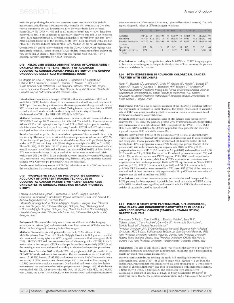
Results: From December 2007 to March 2009, thirty-eight out of 65 pts enrolled in thePROMETEO study underwent liver resection. The pt characteristics were: 23 (60.5%)males, 15 (39.5%) females; 25 (65.8%) synchronousmetastasis, 13 (34.2%)metachronousmetastasis; 19 (50%) neoadjuvant chemotherapy; 8 (21.1%) previous liver surgery; 4(10.5%) previous loco-regional treatment. One-hundred and twenty-nine liver lesionswere resected; the median number lesions per patient was 2 (range 1-15). All the lesionswere studied with CT, 109 (84.5%) with MR-DW, 119 (92.2%) with PET, 116 ( 89.9%)with CEUS1, and 126 (97.7%) with CEUS2. Five lesions (4%) at pathological examination
were non-metastasis (1 hamartoma, 1 steatosis, 1 giant-cell reaction, 2 necrosis). The tablereports diagnostic values of different imaging techniques:
CT % MR-DW % PET % CEUS1 % CEUS2 %
Accuracy 102/129 79 105/109 96 69/119 58 87/116 75 117/126 93Sensitivity 101/124 81.5 104/116 90 68/116 59 86/111 77.5 114/121 94Positive Predictivevalue
101/105 96 104/106 98 68/70 97 86/90 95.5 114/116 98
Specificity 1/5 20 1/3 33 1/3 33 1/5 20 3/5 60Negative predictivevalue
1/24 4 1/13 8 1/49 2 1/26 4 3/10 30
Conclusions: According to this preliminary data, MR-DW and CEUS2 imaging appearto be very accurate imaging techniques in the detection of liver metastasis in patientswho are candidates for resection.
L20 PTEN EXPRESSION IN ADVANCED COLORECTAL CANCERTREATED WITH CETUXIMAB
Negri F1, Bozzetti C1, Lagrasta C2, Crafa P2, Graiani G2, Naldi N1, Bortesi B3,Azzoni C2, Ruzzo A4, Camisa R1, Bonasoni MP5, Bisagni G6, Ardizzoni A1
1Oncologia Medica; 2Anatomia Patologica; 3Unita di Genetica Medica, AziendaOspedaliero-Universitaria di Parma; 4Istituto di Biochimica ‘‘G Fornaini’’,Universita di Urbino; 5Anatomia Patologica; 6Oncologia Medica, Arcispedale ‘‘S.Maria Nuova’’, Reggio Emilia
Background: PTEN is a major negative regulator of the PI3K/AKT signalling pathwaythat may results in resistance to EGFR-blockade. The present study aimed to assess therole of PTEN and other potential predictive factors in relation to cetuximab-basedtreatment in advanced colorectal cancer.
Methods: Both primary and metastatic sites of fifty patients were retrospectivelyanalyzed for PTEN loss at the gene and protein levels by immunohistochemistry (IHC)and fluorescence in situ hybridization (FISH) respectively, for PAKT (IHC), EGFR(FISH) and KRAS mutations. We defined responders those patients who obtaineda partial response (PR) or a stable disease (SD).
Results: Eighty percent (40/50) of the patients received ‡3 lines of chemotherapy.Thirty-six patients were treated with cetuximab and irinotecan and 14 with cetuximaband oxaliplatin. Twelve patients (24%) experienced PR, fourteen (28%) SD andtwenty-four (48%) a progressive disease (PD). Seventy-two percent (36/50) of thepatients with skin rash showed a higher response rate (88% vs 12%; p<0.05),progression free survival (PFS) (6.5 months vs 2.1; p<0.005) and overall survival (OS)(12.7 months vs 2.9; p<0.005). Five out of the 43 evaluable primary tumours (12%) and4/24 (17%) of the metastases were PTEN negative. PTEN IHC tested on primarieswas not predictive of response, while loss of PTEN expression on metastases wasnegatively associated with response (pd 100% in PTEN negative cases vs 30% in PTENpositive; p<0.05), PFS (2.0 months vs 4.1; p<0.05) and OS (2.9 months vs 14.2;p<0.001). KRAS mutations were assessed in 47/50 cases: 8/47 (17%) of the cases weremutated and of these only one (12%) experienced a PR. pAKT was not predictive ofresponse nor pfs and os, neither was EGFR.
Conclusions: a correlation between response to cetuximab-based therapy and theEGFR pathway has been reported. A possible functional interaction of PTEN activitywith EGFR tyrosine kinase signalling and potential role for PTEN in the antitumouractivity of cetuximab could be hypothesized.
L21 PHASE II STUDY WITH PANITUMUMAB, 5-FLUOROURACIL,OXALIPLATIN AND CONCURRENT RADIOTHERAPY IN LOCALLYADVANCED RECTAL CANCER (STARPAN - STAR-02 STUDY).SAFETY ANALYSIS
Francesca Di Fabio1, Carmine Pinto1, Evaristo Maiello2, Sara Pini1,Tiziana Latiano2, Carlo Aschele3, Carlo Garufi4, Annamaria Bochicchio5,Davide Tassinari6, Andrea Angelo Martoni11Medical Oncology Unit, S.Orsola-Malpighi Hospital, Bologna, Italy; 2MedicalOncology, IRCCS Casa Sollievo della Sofferenza, San Giovanni Rotondo (FG),Italy; 3Medical Oncology, Galliera Hospital, Genoa, Italy; 4Medical Oncology,Regina Elena Institute, Rome, Italy; 5Medical Oncology, CROB, Rio Nero inVulture (PZ), Italy; 6Medical Oncology, ‘‘Degli Infermi’’ Hospital, Rimini, Italy
Background: The aim of this phase II study was to assess the activity of preoperativeexternal radiotherapy combined with panitumumab, oxaliplatin and 5-fluorouracil inlocally advanced rectal cancer patients (pts).
Materials and Methods: Pts entering the study had histologically-proven rectaladenocarcinoma, either cT3N+ or cT4N-/+ stage, with location <12 cm from theanal margin. Panitumumab was administered at a dose of 6 mg/kg IV, 2 weeks beforethe start of chemoradiotherapy, and then in combination with chemoradiotherapy,3 times every 2 weeks. 5-fluorouracil and oxaliplatin were administeredaccording to established schedule of STAR-01 Study (oxaliplatin 60 mg/m2 IVweekly six times, 1h after the panitumumab infusion, and 5-fluorouracil 225 mg/m2/
viii118 | session L: gastrointestinal cancer-colon-rectal Volume 20 | Supplement 8 | October 2009
session L Annals of Oncology
by guest on March 22, 2015
http://annonc.oxfordjournals.org/D
ownloaded from
day continuous infusion IV days 1-38). Radiotherapy was delivered at a dose of50.4 Gy in daily fractions of 1.8 Gy. Rectal surgery was performed 7-8 weeks afterthe end of neoadjuvant treatment. Eight courses of adjuvant chemotherapywith FOLFOX4 plus panitumumab at the dose of 6 mg/kg, every 2 weeks, weregiven post-surgery. The main study endpoint was complete pathological responserate.
Results: From February 2007 to April 2009 fifty-one pts were enrolled (9 too earlypts). Characteristics of the 42 evaluated pts were: male 28 (66.7%), female 14(33.3%); median age 60 (37-78); median Karnofsky PS 100 (70-100); stage: cT3N+31 (73.8%), cT4N- 3 (7.1%), cT4N+ 8 (19.1%). Thirty-three pts have completedneoadjuvant treatment and 30 have undergone surgery (12 pts ongoing). The mostfrequent grade 1-2 side effects were acneiform rash (56.7%), diarrhea (27%) andfatigue (8%). Grade 3-4 diarrhea was found in 32.4% of pts, and grade 3 cutaneoustoxicity in 43.3%. No grade 3 hematological toxicity was found. The mediancumulative dose of delivered radiotherapy was 50.4 Gy. The planned dose ofpanitumumab, 5-fluourouracil and oxaliplatin was administered in 78.8%, 63.6%and 69.6% of pts, respectively.
Conclusions: These early results demonstrate that panitumumab can be added to 5-fluorouracil/oxaliplatin-based chemoradiotherapy without compromising theconcurrent radiotherapy dose. This combination treatment is associated with highincidence of grade 3-4 diarrhea.
L22 BEVACIZUMAB RELATED ADVERSE EVENTS IN PATIENTSAFFECTED BY METASTATIC COLORECTAL CANCER: A META-ANALYSIS
Michela Cinquini1, Elena Galfrascoli2, Antonio Rossi3, Andrea Manazza4,Giovanna Damia5, Giuseppe Banna6, Alicia Tosoni7, Marcello Tiseo8,Gabriella Farina9, Marina Chiara Garassino9, on behalf of ORION CollaborativeGroup1Mario Negri Institute, New Drug Development Strategies Lab., Milano, Italy;2Fatebenefratelli and Ophthalmic Hospital, Hospital Pharmacy, Milano, Italy;3S.G. Moscati Hospital, Oncology, Avellino, Italy; 4CeRMS, Molecular OncologyLab., Torino, Italy; 5Mario Negri Institute, Molecular Pharmacology Lab., Milano,Italy; 6Vittorio Emanuele Hospital, Oncology, Catania, Italy; 7Maggiore Hospital,Oncology, Bologna, Italy; 8Maggiore Hospital, Oncology, Parma, Italy;9Fatebenefratelli and Ophthalmic Hospital, Oncology, Milano, Italy
Background: Bevacizumab, a recombinant humanized monoclonal antibody targetingthe vascular endothelial growth factor, is widely used in patients with metastaticcolorectal cancer. Bevacizumab suffers by several adverse events which may be differentaccording to the diverse kind of treated tumours. We performed a systematic reviewand meta-analysis of published randomized clinical trials (RCTs) investigatingbevacizumab in the treatment of patients affected by advanced colorectal cancer tobetter understand the overall risk of side effects.
Methods: PubMed, Medline, CancerLit, and Embase databases were searched forRCTs, comparing chemotherapy plus bevacizumab versus chemotherapy alone inmetastatic colorectal cancer patients. Also abstracts presented at the main internationalmeetings until April 2009 were analyzed. Odds ratios (ORs) and Number Needed toHarm (NNH) for main side effects were calculated with their 95% confidence intervals(CI) using fixed-effects model.
Results: Nine controlled trials encompassing 7,132 patients, were eligible for thepresent analysis. Patients receiving bevacizumab plus chemotherapy have a risk twicesuperior (OR 1,92 95% CI 1.51–2.44) of developing all-grade hypertensioncorresponding to a NNH 9 and seven times superior of developing grade 3-4hypertension (OR 6.94 95% CI 5,07-9,52; NNH 11). Moreover, the risk of the othergrade 3-4 toxicities were: bleedings (OR 1,83 95% CI 1.11-3.01 NNH 83), proteinuria(OR 4.20 95% CI 2.17-8.12 NNH 73), thromboembolic events (OR 1,19 95% CI 0.98-1.45 NNH 77), cardiac events (OR 1,72 95% CI 0.72 -4.13 NNH 167), and oxaliplatin-related neuropathy events (OR 1.55 95% CI 1.29-1.87 NNH 17).
Conclusions: Patients affected by metastatic colorectal cancer and treated withchemotherapy plus bevacizumab have a significant increased risk of developing severehypertension, proteinuria, and bleedings. Surprisingly, in our analysis, bevacizumab isnot associated with higher onset of thromboembolism events, but it increases theoxaliplatin-related neurotoxicity.
L23 CHRONOCHEMOTHERAPY (CCT) WITH FLUOROURACIL,OXALIPLATIN, IRINOTECAN,, FOLINIC ACID (FOLFOXIRI)1/-BEVACIZUMAB (BEV) IN METASTATIC COLORECTAL CANCER(MCRC): A PHASE 2 TRIAL
Salvatore Tumolo*, Massimo Boccalon*, Bernardo Marzano+, Gianni Fanti+,Armando Scata�, Davide Adriano Santeufemia*, Alessandro Del Conte*,Giordano Chiara+, Giancarlo Tosolini�, Walli Marus§, Sandro Sulfaro§
*Oncology Unit; +�Surgery Divisions; §Pathology, Santa Maria degli Angeli GH,Pordenone, Italy
Background: FOLFOXIRI induce a 60 % of response rate and 22,6 mos of overallsurvival (OS) in mCRC with heavy G3-4 haematological toxicity (Falcone A. JCO,2007). CCT may represent an alternative modality of administration in order to
improve safety and toxicity. BRiTE study seems to demonstrate an improvement of OS(Grothey A. JCO,2008). Aim of our study was to evaluate feasibility, efficacy andtoxicity of chrono-FOLFOXIRI +/- Bev in mCRC
Methods: we enrolled since January 2003 46 naıve/previous treated (N/PT) pts withhistological diagnosis of mCRC and good organ function. Treatment:CPT-11180 mg/m2/d1, 6 h infusion, (peak at 17:00) and a 12-h, days 2-5, infusion of L-OHP20 mg/m2/day (peak at 16:00), FA 150 mg/m2/day plus 5-FU 700 mg/m2/d (peak at4:00)+/- Bev 5 mg/kg q 2 wks until PD or unacceptable toxicity.
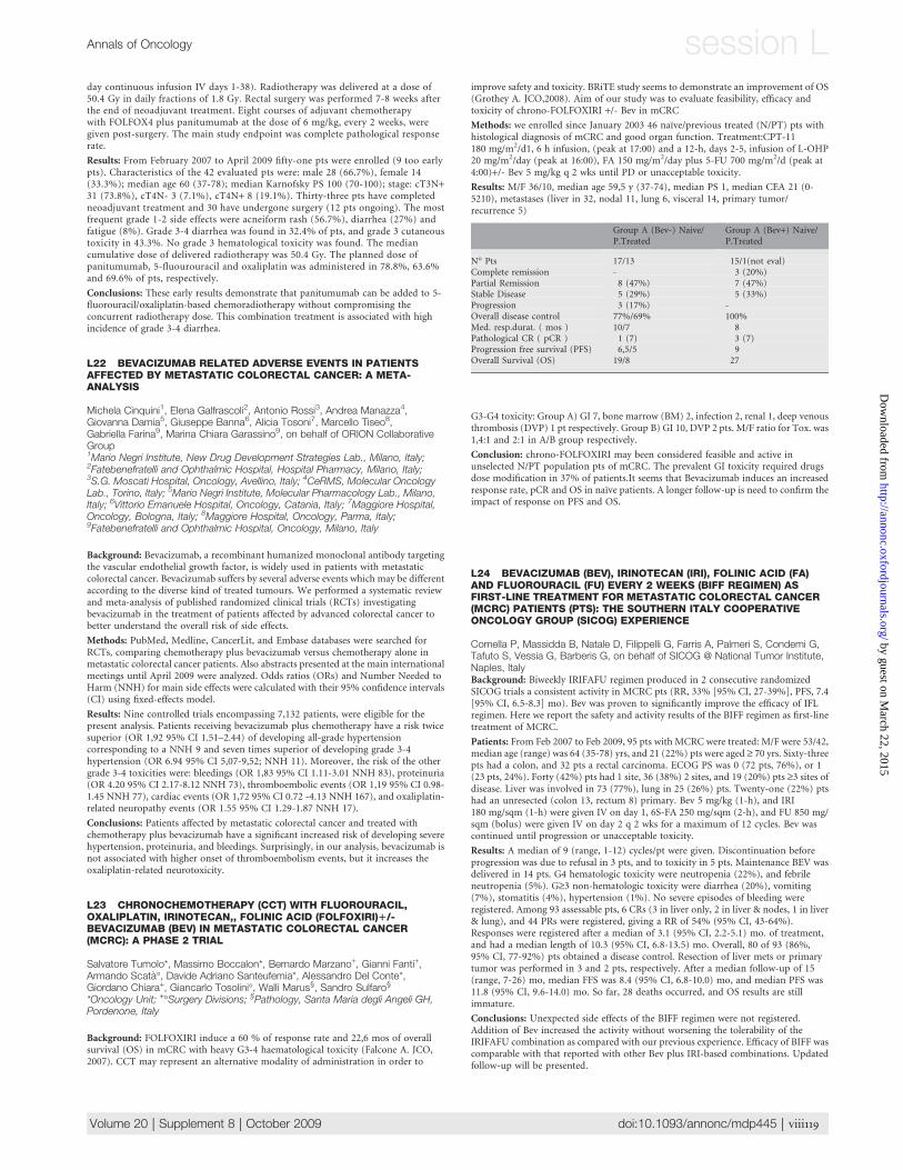
Results: M/F 36/10, median age 59,5 y (37-74), median PS 1, median CEA 21 (0-5210), metastases (liver in 32, nodal 11, lung 6, visceral 14, primary tumor/recurrence 5)
Group A (Bev-) Naive/P.Treated
Group A (Bev+) Naive/P.Treated
N� Pts 17/13 15/1(not eval)Complete remission - 3 (20%)Partial Remission 8 (47%) 7 (47%)Stable Disease 5 (29%) 5 (33%)Progression 3 (17%) -Overall disease control 77%/69% 100%Med. resp.durat. ( mos ) 10/7 8Pathological CR ( pCR ) 1 (7) 3 (7)Progression free survival (PFS) 6,5/5 9Overall Survival (OS) 19/8 27
G3-G4 toxicity: Group A) GI 7, bone marrow (BM) 2, infection 2, renal 1, deep venousthrombosis (DVP) 1 pt respectively. Group B) GI 10, DVP 2 pts. M/F ratio for Tox. was1,4:1 and 2:1 in A/B group respectively.
Conclusion: chrono-FOLFOXIRI may been considered feasible and active inunselected N/PT population pts of mCRC. The prevalent GI toxicity required drugsdose modification in 37% of patients.It seems that Bevacizumab induces an increasedresponse rate, pCR and OS in naıve patients. A longer follow-up is need to confirm theimpact of response on PFS and OS.
L24 BEVACIZUMAB (BEV), IRINOTECAN (IRI), FOLINIC ACID (FA)AND FLUOROURACIL (FU) EVERY 2 WEEKS (BIFF REGIMEN) ASFIRST-LINE TREATMENT FOR METASTATIC COLORECTAL CANCER(MCRC) PATIENTS (PTS): THE SOUTHERN ITALY COOPERATIVEONCOLOGY GROUP (SICOG) EXPERIENCE
Comella P, Massidda B, Natale D, Filippelli G, Farris A, Palmeri S, Condemi G,Tafuto S, Vessia G, Barberis G, on behalf of SICOG @ National Tumor Institute,Naples, ItalyBackground: Biweekly IRIFAFU regimen produced in 2 consecutive randomizedSICOG trials a consistent activity in MCRC pts (RR, 33% [95% CI, 27-39%], PFS, 7.4[95% CI, 6.5-8.3] mo). Bev was proven to significantly improve the efficacy of IFLregimen. Here we report the safety and activity results of the BIFF regimen as first-linetreatment of MCRC.
Patients: From Feb 2007 to Feb 2009, 95 pts with MCRC were treated: M/F were 53/42,median age (range) was 64 (35-78) yrs, and 21 (22%) pts were aged ‡ 70 yrs. Sixty-threepts had a colon, and 32 pts a rectal carcinoma. ECOG PS was 0 (72 pts, 76%), or 1(23 pts, 24%). Forty (42%) pts had 1 site, 36 (38%) 2 sites, and 19 (20%) pts ‡3 sites ofdisease. Liver was involved in 73 (77%), lung in 25 (26%) pts. Twenty-one (22%) ptshad an unresected (colon 13, rectum 8) primary. Bev 5 mg/kg (1-h), and IRI180 mg/sqm (1-h) were given IV on day 1, 6S-FA 250 mg/sqm (2-h), and FU 850 mg/sqm (bolus) were given IV on day 2 q 2 wks for a maximum of 12 cycles. Bev wascontinued until progression or unacceptable toxicity.
Results: A median of 9 (range, 1-12) cycles/pt were given. Discontinuation beforeprogression was due to refusal in 3 pts, and to toxicity in 5 pts. Maintenance BEV wasdelivered in 14 pts. G4 hematologic toxicity were neutropenia (22%), and febrileneutropenia (5%). G‡3 non-hematologic toxicity were diarrhea (20%), vomiting(7%), stomatitis (4%), hypertension (1%). No severe episodes of bleeding wereregistered. Among 93 assessable pts, 6 CRs (3 in liver only, 2 in liver & nodes, 1 in liver& lung), and 44 PRs were registered, giving a RR of 54% (95% CI, 43-64%).Responses were registered after a median of 3.1 (95% CI, 2.2-5.1) mo. of treatment,and had a median length of 10.3 (95% CI, 6.8-13.5) mo. Overall, 80 of 93 (86%,95% CI, 77-92%) pts obtained a disease control. Resection of liver mets or primarytumor was performed in 3 and 2 pts, respectively. After a median follow-up of 15(range, 7-26) mo, median FFS was 8.4 (95% CI, 6.8-10.0) mo, and median PFS was11.8 (95% CI, 9.6-14.0) mo. So far, 28 deaths occurred, and OS results are stillimmature.
Conclusions: Unexpected side effects of the BIFF regimen were not registered.Addition of Bev increased the activity without worsening the tolerability of theIRIFAFU combination as compared with our previous experience. Efficacy of BIFF wascomparable with that reported with other Bev plus IRI-based combinations. Updatedfollow-up will be presented.
Volume 20 | Supplement 8 | October 2009 doi:10.1093/annonc/mdp445 | viii119
Annals of Oncology session L by guest on M
arch 22, 2015http://annonc.oxfordjournals.org/
Dow
nloaded from
L25 RECURRENCE ANALYSIS IN 133 CONSECUTIVE PATIENTSTREATED WITH NEOADJUVANT CHEMO-RADIOTHERAPYFOLLOWED BY TME SURGERY FOR LOCALLY ADVANCED RECTALCANCER (LARC)
Piergiorgio Di Tullio1, Carmine Pinto1, Francesca Di Fabio1, Sara Pini1, DajanaCuicchi2, Bruno Iacopino3, Claudio Ceccarelli4, Raffaele Lombardi2, Stefano Neri3,GianPaolo Ugolini2, Maria Lucia Tardio4, Bruno Cola2, Andrea Angelo Martoni11Medical Oncology Unit; 2General Surgery Unit; 3Radiotherapy Unit; 5PathologyUnit, S.Orsola-Malpighi University Hospital, Bologna, Italy
Background: Chemo-radiotherapy (CRT) represents the standard treatment forpatients (pts) with LARC. The aim of this analysis was to evaluate the factors correlatedto disease relapse in 133 consecutive pts treated with CRT and surgery by theMultidisciplinary Rectal Cancer Team (MRCT) at the S.Orsola-Malpighi Hospital inBologna.
Methods: From November 2001 to December 2008, 194 pts rectal cancer wereevaluated by MRCT. One hundred and thirty-three pts with LARC underwent theintegrated treatment programme. Pt. characteristics were: 86 (64.6%) males, 47(35.4%) females; median age 69 years (range 33-89); 70 (52.6%) stage II and 63(47.4%) stage III. The chemotherapy regimens were: fluoropyrimidines alone (5-fluorouracil/capecitabine) in 43 (32.2%) pts, 5-fluorouracil plus oxaliplatin in 90(67.8%). Total Mesorectal Excision (TME) surgery was the standard treatment.Ninety pts (67.7%) underwent anterior rectal resection and 36 (7.1%) abdominal-perineal resection. Endorectal transanal excision was performed in 7 (5.2%) pts.Twenty-one (15.8 %) pts achieved a ypCR. Adjuvant chemotherapy was performed in102 (76.7 %) pts.
Results: At the median follow-up time of 40 months (range 3-84), nineteen (14.2%) ptsshowed disease relapse: 7 (5.2%) local recurrence, 9 (6.8%) distant, 3 (2.2%) both localand distant. The median time to disease relapse was 17 months (range 3-36). The sitesof distant metastasis were: 6 lung, 3 liver and one peritoneum. Two pts with diseaserelapse achieved a ypCR. No significant differences were observed between the pts withrelapsed disease in terms of clinical stage at diagnosis (p=0.228), chemotherapyregimen (p=0.583), or adjuvant chemotherapy (p=0.532).
Conclusions: These results indicate a low recurrence rate in pts with LARC afterneoadjuvant CRT and TME surgery. The disease relapse does not appear to be relatedto clinical stage, chemotherapy pre-operatory regimen or adjuvant chemotherapy.
L26 PRELIMINARY DATA OF INCREASED RATE OF POTENTIALLYCURATIVE RESECTION OF LIVER METASTASES AFTER INTENSIVECHEMOTHERAPY IN METASTATIC COLO-RECTAL CANCERPATIENTS
Santomaggio A1, Cannita K1, Lanfiuti Baldi P1, Porzio G1, Mancini M1, Tudini M1,Bruera G1, Pelliccione M1, Sarno I1, De Galitiis F3, Morelli MF3, Marchetti P3,4,Antonucci A2, Ficorella C1, Ricevuto E1
1Medical Oncology, S. Salvatore Hospital, University of L’Aquila; 2GeneralSurgery, S. Salvatore Hospital, University of L’Aquila; 3Medical Oncology, IDI,Rome; 4Medical Oncology, S. Andrea Hospital, University La Sapienza, Rome
Background: Integration of surgical resection of metastatic lesions after chemotherapy(CT) is reported in 15-20% metastatic colorectal cancer (mCRC) patients (pts), thuscontributing to extend survival. We describe preliminary data concerning surgicalresections performed in consecutive mCRC pts after first line CT with weeklyalternating 5-Fluorouracil (5-FU)/Irinotecan (CPT-11)/Bevacizumab (BEV) and 5-FU/Oxaliplatin (OHP) combination (FIr-B/FOx).
Methods: Forty-eight pts were enrolled in a previously reported phase II study showingapproximately 80% RR (Proc. AIOM’08). Surgery was recommended 5 to 8 weeks afterBEV discontinuation. Resectability of metastatic lesions for liver-only disease wasdefined according to 4 categories: I ‘‘resectable low risk’’ with conventional surgery(single metastasis £ 5 cm, CEA £ 200 ng/ml, metachronous metastases, N0 at primarytumor); II ‘‘resectable high risk’’ with conventional surgery (multiple metastases, N+ atprimary tumor, involvement of < 4 segments); III ‘‘potentially resectable high risk’’with advanced surgery (portal vein embolization, necessity of two stage hepatectomy, >1 hepatic vein involvement, necessity of intraoperative ablation); IV ‘‘non resectable’’(> 70% of liver involvement, < 25% remnant after resection).
Results: liver metastases and no extrahepatic disease, 19/48 (40%) pts: 9 single and 10multiple. Patients’distribution before and after treatment, respectively: IV, 6 then 2(32% then 10%); III, 2 then 2 (10% then 10%); II, 6 then 8 (32% then 43%); I, 5 then 7(26% then 37%).
Thirteen pts (27%) underwent surgery with curative intent: 10 pts for liver-onlydisease (21%), single in 6 and multiple in 4; 1 for liver and lung metastases, in two steps(2%); 2 pts at the primary tumor and retroperitoneal nodes (4%). All 13 pts had noresidual disease following surgery (R0 resection) and 1 pathological CR (2%). Overallsurvival is not mature.
Conclusion: Preliminary data show 40% prevalence of liver-only disease in MCRC,mostly objectively downstaged with FIr-B/FOx; 23% pts become NED after liversurgery (1 pCR), 53% if liver-only mCRC; overall, 27% mCRC pts NED after surgery.
L27 OBSERAVATIONS AND RESULTS OF THE FIRST ROUND OFCOLORECTAL CANCER SCREENING PROGRAMME IN RIMINI
Giovanardi M*, Santilli F*, Di Marco M*, Casale C�, Fava C�, Canuti D�, Panzini I�,Balducci C�, Giuliani O^, Ravaioli A�, Desiderio F�*U.O. di Gastroenterologia ed Endoscopia Digestiva, Ospedale ‘‘Infermi’’ Rimini;�U.O. di Oncologia ed Oncoematologia, Ospedale ‘‘Infermi’’ Rimini; ^I.R.S.T.Meldola
Cancer of the large bowel (colorectal cancer) is the third most common form of cancer(after lung and prostate cancer) in males in Italy and the second form in females afterthe cancer of the breast.
Colorectal cancer is registered as the underlying cause of approximately 20.000deaths in Italy every year. In our Region, Emilia Romagna, the incidence of coloncancer is 2.5 times more frequent then rectal cancer and in 2003 we registered 1.606deaths (1.147 for colon cancer and 458 for the rectal one) due to colorectal cancer(11.5% of all tumors).
In Rimini, instead, in 2005, we registered 223 tumors of the colon (129 males and 94females) and 94 rectal cancer (61 males and 33 females).
In 2005 in Rimini, we started the first Round of the Colorectal Cancer ScreeningProgramme; we invited from march 2005 to march 2007, 75.464 individuals aged 50-69, offering FOBT (Faecal Occult Blood Testing) every two years (biennal screening),and in case of positive test we offered pancolonscopy as second level test.
The aim of this study is the comparise of the histopathologic profile (TNM) oftumors diagnosis before the beginning of colorectal cancer screening programme(2003-2004) and those found in the first round of Screening.Materials and Methods: in this case-control study we considered ‘‘cases’’ allindividuals with colorectal cancer consecutively diagnosed by Screening and ‘‘controls’’the pre-screening diagnosed one. Cases and controlls were matched by sex and age withratio1:2. We enrolled 110 cases and 202 controlls, aged 50-69 years, registered inRimini’s Hospital. The statistical analysis was performed with STATA 9.2 program.
Results: Univariate analysis showed a significative difference between cases andcontrols (Pearson chi square test) for ‘‘T’’ - Tumor size in TNM definition - (p< 0.001)and Dukes stage (p<0.001). Instead we found a non significative difference forlocalization (colon vs rectum p>0.01): in cases colon cancer were about 3 times morefrequent then rectum, in colntrols colon cancer were about 5 times more frequent thenthe rectum one.
In the multivariate analysis (logistic regression analysis) we found a significativeassociation for ‘‘T’’ and localization.
Conclusions: We observed a significative difference in ‘‘T’’ and Dukes Stage betweencases and controlls; this probably suggests that screening programme based on biennalFOBT, has an objective benefit in detecting tumors at early stage with obviousrebounds on the kind of surgery, chemotherapy and quality of life. We know that weneed further studies and long follow up period to investigate differences in mortalityand survival in the two groups.
L28 THE LYMPH NODE RATIO IS A POWERFUL PROGNOSTICFACTOR IN NODE POSITIVE COLON CANCERS UNDERGOINGPOTENTIALLY CURATIVE SURGERY
Gennaro Galizia1, Michele Orditura2, Francesca Ferraraccio3, Paolo Castellano1,Margherita Pinto1, Anna Zamboli1, Sabrina Cecere2, Ferdinando De Vita2,Carlo Pignatelli1, Giuseppe Catalano2, Eva Lieto1
Divisions of Surgical Oncology1, Medical Oncology2, and Pathology 3‘‘F.Magrassi - A. Lanzara’’ Department of Clinical and Experimental Medicine andSurgery Second University of Naples School of Medicine, Naples, Italy
Objective: To investigate the role of the lymph node ratio (LNR: the ratio betweenmetastatic and resected nodes) as a prognostic factor in node positive colon cancers.
Summary Background Data: The number of harvested (LNs) and metastatic nodes(LNs+) represents the most significant factor to define postoperative treatment andprognosis in colon cancer. However, its assessment may be inadequate causing anincorrect cancer staging. LNR has been demonstrated to hold prognostic significance inmany tumors; however, its role in colon cancer is not yet clearly elucidated.
Methods: 145 consecutive node positive colon cancer patients undergoing potentiallycurative surgery and adjuvant chemotherapy in a single oncologic unit entered thisstudy.
Results: The number of LNs+ did not increase by harvesting more LNs, but patientswith fewer than twelve LNs had fewer LNs+. LNR ranged from 0.0416 to 0.9; it wasclearly lower in pN1 than pN2 patients, and increased as tumor stage worsened. ROCanalysis selected 0.1818 as the best LNR cut-off value. Low LNR patients didsignificantly better than high LNR patients; this difference was not dependent on thenumber of LNs and stronger than differences observed by grouping patients accordingto LNs or LNs+. When stratified by low and high LNR value, pN1 and pN2 patients, aswell as stage III subgroups, were shown to display substantially different outcomes.Finally, LNR was an independent prognostic factor for disease-specific survival, and theonly covariate related to disease-free survival.
Conclusions: LNR was a robust prognostic indicator for metastatic node colon cancerpatients undergoing potentially curative surgery. Since this ratio-based staging wasdemonstrated to reduce stage migration and to aid in identifying high-risk patients, itshould be proposed as a standard tool for colon cancer staging.
viii120 | session L: gastrointestinal cancer-colon-rectal Volume 20 | Supplement 8 | October 2009
session L Annals of Oncology
by guest on March 22, 2015
http://annonc.oxfordjournals.org/D
ownloaded from
L29 DETECTION OF CIRCULATING TUMOR CELLS (CTCS) INSTAGE T3-4 AND/OR N POSITIVE RECTAL CANCER PATIENTSUNDERGOING NEOADJUVANT THERAPY FOLLOWED BY CURATIVESURGERY. PRELIMINARY DATA
Giulia Zampino M1, Elena Magni1, Gianmarco Contino2, Antonio Chiappa2,Fabrizio Luca2, Laura Zorzino3, Franco Nole1, Teresa Sandri M3
1Medicine Department, European Institute of Oncology, Milan; 2SurgeryDepartment; 3Laboratory Unit European Institute of Oncology, Milan
Background: CTCs detected at baseline and at disease-evaluation time-point duringtreatment seems to be an independent prognostic factor in metastatic colorectal cancer(Cohen S.J. JCO, 2008) while its role in early stages after radical surgery is underinvestigation. Nowadays no information are available in naıve patients with rectalcancer suitable for neoadjuvant chemoradiotherapy (CT-RT).
Aim of the Study: To investigate the role of CTCs in locally advanced rectal cancerpatients undergoing neo-adjuvant CT-RT.
Patients andMethods: In a prospective single institution study, cT3-4 and/or N+ rectalcancer patients staged by rectal ultrasound and/or pelvic MRI and chest-abdomen CTscan, are submitted to capecitabine (825 mg/mq, orally, twice daily continuously) withconcomitant radiotherapy (50.4 Gy/28 fractions to the primary tumor and perirectalnodes), followed by two cycles of capecitabine (1250 mg/mq, orally, tid 14/21 days).Primary endpoints are evaluation in 120 patients of CTCs at baseline (t0), afterneoadjuvant therapy, before surgery (t1), within 7 days after surgery (t2), and at 6-month follow-up (t3) and its correlation with disease free survival and overall survival.CTCs are enumerated with immunomagnetic separation in 7,5ml peripheral blood atover-mentioned time-points (CellSearch System, Veridex Inc).
Results: Twenty-six patients (16M; 10F; median age:63 yrs; range: 44-83 yrs)underwent t0 sampling, 8 pts completed CT/RT and therefore underwent t1 and t2sampling. At baseline (t0) three pts presented 1 CTC (12%), one 2 CTCs (3.5%), one 27CTCs (3.5%) while in twenty-one (81%) no CTCs were detected. At t1 and t2 none ofthe eight pts analyzed showed CTCs. Correlation between t0 CTCs number andbaseline clinical staging is reported in the following table.
Conclusions: CTCs ‡ 1 are present in 15% of our patients, but the sample is too smallfor clinical and/or statistical analysis. The study recruitment is still open for achievingsample size objective
Baseline CTCs n� uT2N1 uT3N0 uT3N1 uT3N2 uT4N2 uTxN1
0 1 pts 4 pts 7 pts 7 pts 1 pts 1 pts1 3 pts2 1 pt27 1 pt
L30 THROMBOCYTOSIS IS ASSOCIATED WITH RESPONSE TOFOLFIRI PLUS BEVACIZUMAB (BVZ) IN METASTACTICCOLORECTAL CANCER (MCC) PATIENTS
Latteri F1, Soto Parra H1, Cordio S1, Pumo V1, Bordonaro R1, Palermo F2,Magnano V3, Pizzone A4, Strano G4
1UO Medical Oncology, Ospedale Garibaldi, Catania, Italy; 2Internal Medicineand Specialist Medicine University of Catania; 3Department of Radiology,Ospedale Garibaldi, Catania; 4Department of Clinical Pathology, OspedaleGaribaldi, CataniaBackground: Thrombocytosis (TH) is a poor prognostic factor in many types of cancerincluding colon cancer. Platelets (PLT) are the main source of VEGF in blood andcontribute to angiogenesis by release of growth factors, including VEGF. Preclinicaland clinical models have shown that that PLT may mediate BVZ activity in cancerpatients by taking up BVZ and neutralizing VEGF (HM Verheul et al. Clin Cancer Res.2007). We aimed to investigate whether TH defined as a PLT count > 400x109/l mayaffect response rate and outcome in metastatic colorectal cancer (MCC) pts treatedwith first-line FOLFIRI-BVZ.
Methods: From 04/06 to 11/08, 57 consecutive MCC pts were treated with FOLFIRI-BVZ in our institution and 53 fulfilled criteria for this retrospective analysis (first-lineFOLFIRI-BVZ, response evaluation by CT scan, complete follow-up): median age, 58-yrs; PS0, 77%; Tumor site, colon 67.9%. Twenty-seven pts (50.9%) achieved objectiveresponse, 19 (35.8%) stable disease (SD) and 7 (13.3%) progression (PD). Median TTPand OS were respectively 10.9 and 22.8 months.
Results: Twelve out of 53 (22%) pts had TH before treatment and 10 out of 27 (37%)of responsive pts presented TH. Patients with TH presented 10 partial remission (PR),1 SD and 1 PD with an overall response rate of 83% which was significantly better thanthat observed in those without TH (41.4%), P= 0.001. TTP for pts with TH was 10.7months and for those without 11 (P=.3). OS for patients with TH was not reached and23.1 months for those without TH (P=.6) . About possible causes of TH: 2 ptsunderwent palliative surgery 30 days before chemotherapy, 3 presented moderateanemia, 7 unknown (paraneoplastic).
Conclusions: This first report identified a subgroup of MCC pts highly sensible toFOLFIRI-BVZ. This association may correspond to impaired platelet tumorangiogenesis by BVZ. TH was not a poor prognostic factor in MCC treated withFOLFIRI-BVZ. Further studies are warranted.
L31 IN VIVO IMMUNOLOGICAL EFFECTS OF ANTI-EPIDERMALGROWTH FACTOR RECEPTOR (EGFR) THERAPY IN METASTATICCOLORECTAL CANCER (MCRC) PATIENTS
Chatzileontiadou S1, Rovati B1, Mariucci S1, Loupakis F2, Bencardino K3,Manzoni M1, Delfanti S1, Valentino F1, Ricci V3, Brugnatelli S1, Ronzoni M3,Falcone A2, Danova M1
1S.C. Oncologia Medica, Fondazione IRCCS S. Matteo, PAVIA; 2Azienda USL-6LIVORNO e Universita di PISA; 3 HSR San Raffaele, MILANO, ITALIA
Background: Multiple links have been found between the EGFR signalling pathwayand the immunoresponse in human tumors. Cetuximab, the recombinant human-murine chimeric monoclonal antibody against EGFR was found to exert antibodydependent cellular cytotoxicity in several tumor cell lines. In order to get deeper insideinto the immunological mechanisms of action of Cetuximab we have focused on its invivo impact on both the peripheral blood (PB) lymphocyte and dendritic cell (DC)immunophenotype in mCRC pts.
Methods:We analyzed by multiparameter flow cytometry the PB lymphocytes and DCsubsets in 17 pts (M/F:12/5, median age: 63 yrs; range 43-78) treated with Cetuximab-based therapy, in absence of clinically relevant infections. Baseline data were comparedwith reference values obtained by 50 healthy subjects (M/F: 25/25, median age: 43 yrs,range 21-65).
Results:With respect to normal donors in our pts at baseline we observed a significantlower level of the absolute lymphocyte number, CD19, CD20 and CD23 B-lymphocytesand DCs (in both DC1 and DC2 subsets) while NK cells and CD4 T-lymphocytesshowed a higher level. After 3 courses of CT + Cetuximab, a trend was shown towarda progressive increase of all the lymphocyte subsets, as well as of the count of total DCsand of their subsets. This trend was confirmed after 6, 9 courses and at the time ofdisease evaluation.
Conclusions: To our knowledge these are the first data showing that Cetuximab-basedtreatment improves in vivo the T-cell mediated response in pretreated in mCRC pts.This provides new insight into its possible antitumor mechanism and may be helpful inthe design of combination therapy for mCRC pts.
L32 IS A NEW LINE WITH CETUXIMAB RETREATMENT FEASIBLEAND ACTIVE IN METASTATIC COLORECTAL CANCER (CRC)PATIENTS?
Daniele Santini1, Bruno Vincenzi1, Gianluca Masi2, Raffaele Addeo3,Lorenzo Fornaro2, Mario Scartozzi4, Evaristo Maiello5, Salvatore Del Prete3,Alfredo Falcone2, Giuseppe Tonini11Universita Campus Bio-Medico di Roma; 2Azienda Sanitaria di Livorno;3Ospedale San Giovanni Di Dio ASLNA3, Frattamaggiore (NA); 4UniversitaPolitecnica delle Marche, Ancona; 5Ospedale Casa Sollievo della Sofferenza,San Giovanni Rotondo (FG)
Background: the aim of this ongoing study is to evaluate the feasibility and activity ofa retreatment with a cetuximab-based therapy in CRC patients.
Material and Methods: we included 22 irinotecan refractory CRC patients whoobtained a stable disease or a response to a first cetuximab based therapy, thenprogressed and, for this reason, have been treated with a following line of therapywithout cetuximab. All these patients received, for a second time, a cetuximab basedtherapy.
Results: female/male: 7/15; median age (range): 61 (41-79); KRAS status: WT/mutated(19/3). Median number of previous therapy lines (range): 4 (3-6). Partial response(PR), according to RECIST criteria, was detected in 9 patients out of 22. Moreover,additional 7 patients showed a stable disease (SD). As a consequence the tumourcontrol rate (TCR) was 72.7%. In particular, considering the patients who obtaineda PR (15) to the previous cetuximab treatment, 7 patients (46.6%) re-showed the samePR and 6 patients (40%) a SD with a TCR of 86.6%. Both patients with completeresponse (CR) to the previous cetuximab treatment obtained a PR with the followinganti-EGFR line. Two of the 3 KRAS mutated patients showed a progression diseasewith cetuximab retreatment. Median time to progression in the entire population was5.8 months (95%CI: 3.45 – 8.40). Final data about overall survival will be presentedduring the meeting. The safety profile was nor significantly different between the twocourses of cetuximab-based anticancer treatment.
Conclusions: This study represents a preliminary evidence of the utility to retreat withcetuximab based therapy patients progressed with a previous line including the sameanti-EFFR drug.
L33 PREDICTIVE VALUE OF MOLECULAR MARKERS EXPRESSIONIN LOCALLY ADVANCED RECTAL CANCER (LARC) TREATED WITH
Volume 20 | Supplement 8 | October 2009 doi:10.1093/annonc/mdp445 | viii121
Annals of Oncology session L by guest on M
arch 22, 2015http://annonc.oxfordjournals.org/
Dow
nloaded from
NEOADJUVANT BEVACIZUMAB AND CAPECITABINE 1 STANDARDRADIOTHERAPY
Torino F1, Ueno T2, Berardi R3, Troiani T4, Ballestrero A5, Lencioni M6,Martignetti A7, Granetto C8, Yoshizawa A2, Nagayama S2, Mori Y2, Sarmiento R1,Toi M2, Gasparini G1*
1Oncol.Med.Osp. S.FilippoNeri, Roma; 2Graduate School of Medicine, Kyoto,Japan; 3Oncol. Med.Univ. Marche/Osp. UmbertoI, AN; 4Oncol. Med. IIUniversita, NA; 5Dip. Med. Int. Univ., GE; 6Oncol. Med. Osp. S.Chiara, PI; 7Dip.Oncol. AUSL7, SI; 8Oncol. Med. Osp. S.Croce & Carle, Cuneo
Purpose: The aim of this study was to determine the value of a panel of molecularmarkers as predictors of complete pathologic tumor regression (pCR) or residualtumor area (RTA) in patients undergoing preoperative chemoradiotherapy (pre-CRT)and antiangiogenic therapy for LARC.
Methods: Microvascular density (CD31, CD34), CD68, M30, VEGFR2, thymidinephosphorylase, thymidine synthase and Ki67 were evaluated byimmunohistochemistry. Pre- and post-treatment tumor samples were obtained from27/43 patients (pts) treated with pelvic radiotherapy (50.4 Gy) plus concurrentcapecitabine (825mg/mq bid concurrently with RT) and bevacizumab (5mg/Kg every 2weeks for 4 courses, the first administration 2 weeks before pre-CRT). Pathologicalassessment was performed by central review blinded of clinicopathologic data. Residualtumor was assessed based on the RTA and fibrosis. Tumor necrosis area (TNA) wasevaluated as necrosis area/necrosis area + fibrosis area.
Results: A pCR was observed in 6 pts (13.9%); 12 pts (27.9%) had only focal RTA(£10%). Pre-treatment level of CD34-positive vessel density (VD) was associated totreatment response. Patients with RTA £10% showed higher CD34-positive VD thanthose with larger RTA (>10%) (p = 0.037), suggesting that tumors highly vascularizedrespond better to treatment. There was no correlation between treatment response andpre-treatment levels of the other markers, including CD31-positive VD and VEGFR2expression. In addition, the association between post-treatment levels of each markerand RTA or TNA was examined. A significant correlation between post-treatment Ki67labeling index (LI) and RTA was observed (p<0.0001), suggesting that residual cancercells may have aggressive phenotype. Post-treatment Ki67 LI also correlated with TNA(p=0.009). A significant correlation between post-treatment levels of VEGFR2expression in cancer cells and RTA (p=0.007) was found.
Conclusion: These results suggest that pre-treatment level of CD34-positive VD ispredictive of response of pre-CRT including bevacizumab and that post-treatmentVEGFR2 and Ki67 index correlate to TNA and RTA, respectively.
L34 DOES BENEFIT FROM FIRST-LINE THERAPY PREDICT THEOUTCOME OF FOLLOWING TREATMENT LINES IN ADVANCEDCOLORECTAL CANCER PATIENTS?
Federica De Pauli1, Giuseppe Aprile2, Federica Edith Pisa2, Silvia Di Terlizzi3,Elisa Zanon4, Aldo Iop3, Enrico Vigevani4, Liliana Foghin1, Gianna Adami5,Vincenzo de Pangher Manzini1, Luisa Foltran2, Micol Mazzer2, Fabio Puglisi2,Gianpiero Fasola2
1Medical Oncology, General Hospital, Gorizia; 2Department of Oncology,University Hospital, Udine; 3Medical Oncology, General Hospital, Palmanovaand Latisana; 4Medical Oncology, General Hospital, Tolmezzo; 5MedicalOncology, General Hospital, San Daniele
Introduction: In developing new cancer treatment, the key issue is whether theyimprove overall survival (OS) or a validated surrogate survival endpoint. Since gain inprogression-free survival (PFS) may predict improvement in OS, PFS to first-linechemotherapy was accepted as a surrogate endpoint for OS in clinical trials enrollingadvanced colorectal cancer (CRC) patients. Moreover, tumor shrinkage may correlatewith prolonged survival, as well as having received all the active agents during thetreatment course. However, there is limited data on the impact of first-line treatmenton the outcome of following treatment lines.
Methods:We reviewed electronic medical charts of 316 consecutive CRC patients whohad failed at least two lines of chemotherapy for metastatic disease, retrievingdemographics and clinical data, information on chemotherapy regimens, dosereductions and number of cycles, and responses. PFS to first- (PFS1) and second-line(PFS2) chemotherapy were calculated as the interval between the start date of eachtreatment and the date of disease progression. To estimate the effects of PFS1 andresponse to first-line treatment on PFS2, a Cox proportional hazard regression survivalanalysis was performed.
Results: PFS1 did not influence PFS2 (HR 0.99, 95% CI 0.99-1.00), although patientswho did not respond to first-line treatment were more likely to have a shorter PFS2(HR 1.23, 95% CI 0.94-1.61) compared to responding patients. Gender, age, PS, diseaseextension or type of treatment received (doublet vs single agent; oxaliplatin-basedchemotherapy vs not; bevacizumab-based chemotherapy vs not) did not impact on theresults. However, patients with PS >80 at the beginning of second-line treatment hadsignificantly prolonged PFS2 compared to those with PS<80 (HR=0.61, 95% CI 0.46-0.82).
Conclusions: In this community-based, retrospective, observational study, second-linetreatment outcome was independent of PFS1 and marginally influenced by response to
first-line treatment. CRC patients should be offered second-line chemotherapyregardless outcome of first-line treatment, especially those with optimal PS.
L35 COLORECTAL CANCER LIVER METASTASES TREATED WITHTARGET THERAPIES: MONITORING RESPONSE WITH CONTRAST-ENHANCED ULTRASONOGRAPHY (CE-US)
Recine F1, Di Seri M1, De Sanctis R1, Quadrini S1, Stumbo L1, Del Signore E1,Adua D1, Gori B1, Falbo PT1, Fulvi A1, Manna A1, Cantisani V2, Ricci P2, Longo F1
1Oncologia Medica A, Policlinico Umberto I, Roma; 2Radiologia Centrale,Policlinico Umberto I, Roma
Background: Since anti-angiogenic treatment induces necrosis with no change in thevolume of the tumor, new imaging technologies are particularly suitable for the earlyassessment of the response, for which the RECIST size criteria appear inappropriate.CE-US has recently been proposed for evaluating therapeutic response, demonstratingchanges in tumor parenchymal perfusion and emergence of necrosis with no change intumor volume. The aim of the study was to compare CE-US and CT (the goldstandard) in assessing therapeutic response to antiangiogenic-based therapies for livermetastases from colorectal cancer.
Patients and Methods: Both CE-US and CT were used to prospectively evaluate 48hepatic lesions in 21 patients (male 13, female 8; age range 44-78 years, mean 58.2years) with colorectal adenocarcinoma receiving antiangiogenic therapy since January2008 through February 2009. CE-US was performed the day before (day -1) startingtarget therapy (bevacizumab or cetuximab) and at days 28 and 90. The percentage ofcontrast uptake (Sonovue) before treatment and at follow-up was evaluated, thusrating all patients as responders or poor/non-responders. Re-evaluation total-body CTscan was performed at 90 days.
Results: Based on RECIST criteria, at the CT scan, 7/21 patients demonstrated stabledisease (SD), 8/21 had partial response (PR) and 6/21 showed progressive disease (PD).When assessed by CE-US, 16 patients (33 lesions) were considered good respondersand 5 patients (15 lesions) poor or non-responders. Of note, 4/7 patients with SD(57%) were categorized as good responder when evaluated by CE-US. A good responseat CE-US preceded PR based in 5/8 patients by 2 months. Of the 6 patientscharacterized with PD, 4/6 (66.6%) demonstrated a corresponding lack of decrease intumor contrast enhancement within the tumor.
Conclusion: In patients treated with anti-angiogenic drugs, CE-US identifies moregood-responder patients compared with RECIST at an earlier time-point. Whenmonitoring tumor response in patients treated with anti-angiogenic therapy, CE-USmight need to be added to CT scan at different time of follow-up.
L36 IDENTIFICATION OF PHARMACOGENOMIC MARKERS OF5-FLUOROURACIL-BASED CHEMOTHERAPY IN COLORECTALCANCER
Enrico Mini1, Stefania Nobili1, Cristina Napoli1, Ida Landini1, Maria Morganti1,Fabio Cianchi2, Rosa Valanzano3, Francesco Tonelli3, Teresita Mazzei11Department of Pharmacology; 2Department of Medical and Surgical CriticalCare; 3Department of Clinical Physiopathology, University of Florence, Florence,Italy
Background: Although adjuvant chemotherapy has significantly increased overallsurvival (OS) in resected stage III colorectal cancer (CRC), disease recurrence is stillhigh (30-40%). 20-25% of stage II CRC patients also develop recurrent disease, andalthough adjuvant chemotherapy in stage II patients is debatable, high risk patientsmay benefit from chemotherapy. Since patient response to standard chemotherapyvaries, the study of molecular differences in pharmacologically relevant genes may helpto understand interpatient variability and tailor therapy.
Methods: The expression of 26 5-fluorouracil pathway genes in tumors from 53 stageII-III CRC patients who underwent 5-fluorouracil adjuvant chemotherapy wasinvestigated by quantitative real-time RT-PCR. Patients were dichotomized into highand low mRNA expression level groups using the median value of gene mRNA levels.
Results: A high degree of interpatient variation in relative tumor gene expression wasobserved. DPYS showed the largest range (�1100-fold), and RRM1 the smallest (�18-fold). Multiple gene correlations (Spearman correlation >0.600) were found (p<0.001),suggesting possible coregulation mechanisms. Unsupervised hierarchical clusteringanalysis showed at least two groups of patients and genes.
Correlations between experimental data, baseline clinical-pathologicalcharacteristics and clinical outcome showed an inverse relationship between disease-free survival (DFS) and DUT expression: higher gene expression was associated withshorter DFS. No significant association was observed between gene expression and OS.
Also, by threshold analysis, inverse relationships between DFS and FDXR or TP53 inthe entire case series were observed: patients whose tumors expressed higher levels ofthese genes had a shorter DFS.
There were inverse relationships between DUT or NME23-H1 (p=0.0019 andp=0.032, respectively), and TYMS or RRM1 (only a statistical trend) and DFS in stageIII patients. Higher expression of these genes was associated with shorter DFS.
Conclusions: This study provides data on relationships between expression of 5-fluorouracil pathway genes and clinical outcome of CRC patients undergoing adjuvant
viii122 | session L: gastrointestinal cancer-colon-rectal Volume 20 | Supplement 8 | October 2009
session L Annals of Oncology
by guest on March 22, 2015
http://annonc.oxfordjournals.org/D
ownloaded from
5-fluorouracil chemotherapy and underscores the predictive role of specific genes.Validation in an independent case series is warranted.
L37 INTRA-ARTERIAL HEPATIC RADIOTHERAPY WITH 90YTTRIUMSIR-SPHERES IN UNRESECTABLE, COLORECTAL LIVERMETASTASES REFRACTORY TO SYSTEMIC CHEMOTHERAPY: THEEXPERIENCE OF S. ORSOLA-MALPIGHI HOSPITAL - BOLOGNA
Bruna Angelelli1, Sara Cassani1, Emanuela Giampalma2, Annabella Blotta3,Cinzia Pettinato4, Maurizio Lavorato5, Rita Golfieri, Andrea Angelo Martoni11Medical Oncology Unit; 2Radiology Unit; 3Radiotherapy Unit; 4Medical PhysicsUnit; 5Nuclear Medicine Unit, S.Orsola-Malpighi Hospital, Bologna, Italy
Background: Although resistant or no longer responsive to standard systemicantitumor treatments, a fraction of patients with metastatic colorectal cancer presentisolated unresectable liver involvement and, at the same time, a good performancestatus. Selective internal radiotherapy (SIRT) by intra-arterial hepatic administrationof resin 90yttrium microspheres (SIR-spheres) could be an appropriate palliativetreatment for these patients.
Methods: Patients with unresectable colorectal isolated liver metastases, with at leasttwo previous lines of chemotherapy for metastatic disease, KPS ‡70 and normal liverfunction tests were potentially eligible. Pre-treatment evaluation included CT scan,PET scan, arteriography and liver scintigraphy. Exclusion criteria were: extra-hepaticmetastases, >50% of liver volume involved by the tumor, extra-hepatic uptakes andpulmonary shunts by liver scintigraphy more than 20%. Eligible patients receiveda single intra-arterial infusion of SIR-spheres after embolization of the gastroduodenalartery. The objective response was evaluated by CT-scan following the RECIST criteria,one and three months after the treatment, and subsequently every three months.
Results: Twenty patients were treated. Their main characteristics were: males/females75%/25%; median age 62.5 years (range 42-74); median KPS 100 (range 80-100), 2/>2prior chemotherapy lines 50%/50%, <25% or 25%-50% liver involvement 75%/25%.The median SIRT dose delivered was 1.69 GBq. One pt received a second SIRTtreatment at a 4-month interval. Toxicity was represented by fever within 48 hours in 2pts, abdominal pain in 6 and nausea-vomiting in 2. No operative mortality occurred.The best objective response in 19 evaluable pts was: partial remission in 6 (30%), stabledisease in 5 (20%), disease progression 8 (40%). One responder patient wassubsequently operated on by liver resection. Median PFS was 4 months (95% c.l. 3-5)and median overall survival was 8 months (95% c.l. 3-13). At the moment 7 pts arestill alive.
Conclusions: These results confirm that SIRT is a valid therapeutic option for selectedpatients with refractory unresectable isolated liver metastases from colorectal cancer.Its integration with systemic treatment in an earlier phase in the course of the diseasewarrants to be prospectively studied.
L38 NEOADJUVANT TREATMENT OF EXTRAPERITONEAL RECTALCANCER WITH CHRONOMODULATED OXALIPLATIN (L-OHP) ANDCAPECITABINE COMBINED TO RADIOTHERAPY (RT)
Campanella C, Garufi C, Ambesi-Impiombato F, Torsello A, Diodoro MG,Stigliano V, Cosimelli M, Assisi D, Caterino M, Sperduti I, Cognetti FRegina Elena institute, Rome, Italy
Introduction: Chemoradiation is considered treatment options for locally advancedrectal cancer. In pts with advanced colorectal cancer chronomodulated treatmentcompared to flat infusion of fluoropyrimidines plus L-OHP has been shown to besignificantly more active in men than in women (Giacchetti S, JCO 2006). Here weevaluated chronomodulated L-OHP and capecitabine plus RT as neoadjuvanttreatment of locally advanced rectal cancer respect to toxicity and pathologicalresponse rates.
Methods: We assigned T3/T4-N0 and anyT-N+ rectal cancer patients to receiveneoadjuvant chemoradiotherapy. The extent of the disease was assessed by rigidproctoscopy, endoscopic ultrasound of the rectum, TC scan of the abdomen/pelvis andpelvis RMN. The chemotherapy schedule was modified for L-OHP from the originalpaper of Santini D et al (Canc Chemother Pharmacol 2007): L-OHP 100 mg/m2 12-hour ci from 10:00 am to 10:00 pm, days 1, 14, 28 plus Capecitabine 1300 mg/m2chronomodulated delivey (h 8.00 a.m. 25%, h 6.00 p.m. 25%, h 11.00 p.m. 50% of totaldose) during the whole radiotherapic treatment. Radiotherapy consisted of 45 Gy tothe whole pelvis, 1.8 Gy daily, 5 times per week and 5.4 Gy delivered to the mesorectum(total dose of 50.4 Gy in 5 weeks). No adjuvant treatment was planned.
Results: We enrolled 33 patients (M=20 F=13); median age 65 years. Tumor waslocated within 5 cm of the anal verge in 11 pts, between 5-10 cm in 18 pts and over than10 cm in 1 pt. Clinical staging was: T2N0 2 pts, T2N1 2 pts, T3N0 12 pts, T3N1 11 ptsand T4N0 in 2 pts. G3/G4 toxicity was: anemia 0%, neutropenia 6%, piastrinopenia0%, diarrhea 18%, nausea/vomiting 17%, rectal mucositis 12%, asthenia 21%. Highertoxicity was observed in female patients (5/13: 38%), with one sepsis and one fatalcardiac toxicity. Thirty pts were evaluated for response, 3 are under evaluation.Surgery: 21 anterior resections; 5 local excisions; 3 Miles. Tumor TRG: 1: 10 pts, 2: 4pts, 3: 7 pts, 4: 1 pt; 5: 1 pt; NE 9. Tumor downstaging was observed in 23/30 pts (77%,)and all pts were pN0, with complete pathological response rate in 6/23 pts (26%); 3
stable; 4 progressions (including the toxic death). Median follow up was 24.7 months(0.2-39.4) and two yrs PFS was of 85,9%.
Conclusions: This study shows that preoperative chronomodulated oxaliplatin andcapecitabine combined to radiotherapy seems to be an active treatment in rectal cancerpatients. This schedule displays different toxicity according to gender being more toxicin female than in males.
L39 K-RAS STATUS IN ANAL SQUAMOUS CELL CARCINOMA: NEWERA FOR TARGET-ORIENTED TREATMENT?
Elena Magni1, Giuseppe Renne2, Nicola Fazio1, Maria Giulia Zampino1
1Medicine Department; 2Pathology Department, European Institute ofOncology, Milan
Introduction: Anal squamous cell carcinoma (SCC) is an uncommon diseasecomprising only 1 to 5 % of all intestinal tumours. SCC is considered the prototype forthe successful application of conservative treatment as chemo-radiotherapy instead ofaggressive surgery in locally extended disease. The EGFR status and k-ras mutations inanal SCC has not been well investigated. The purpose of our evaluation is to giveinformation about this issue.
Patients and Methods: From June 1999 to December 2008, 32 patients affected by analSCC were treated in our institution with chemotherapy containing fluoropyrimidineand platinum salt concomitant with pelvic radiotherapy. Samples of anal tumour wereembedded in paraffin and immunohistochemical analysis was performed for EGFR andamplification of the k-ras gene exon 1 by laser capture micro-dissection. Then, DNAextraction, PCR and sequencing were evaluated for possible mutation in codons 12and 13.
Results: Characteristics of patients and tumours were described in the following table.In 24 patients was possible to review histology for biological objective. Among 30patients with locally-extended disease, 2 relapsed, one on lung and the other on liverand cervical nodes; both of them are still alive. K-ras mutation text was performed in24 cases, while EGFR analysis was evaluable in 12 patients. In all cases of our serieswild-type K-ras was observed.
Conclusion: Such information is, in our knowledge, the first reported in literature onthis setting of patients. This observation previously reported in other SCC of differentorigin, may suggest the effective use of EGFR-inhibitors in locally extended and inadvanced disease.
Variables N
Median age (yr) 59 (range 39-75)GenderMales/Females 5/27Performance status (ECOG)0/1/2 24/7/1GradingG1/G2/G3 0/8/12unknown 12Clinical stagingT1/T2 2/13T3/T4 8/7N0/N+ 10/20M1 2EGFRPositive/negative/N.A. 7/5/20K-ras statusWild type/ Mutant/N.A. 24/0/8
N.A. :not assessable.
L40 YKL-40 AND P53 EXPRESSIONS IN ANAL CARCINOMAPREDICT SHORTER OVERALL AND DISEASE FREE SURVIVAL
Massimiliano Mistrangelo, Isabella Castellano*, Paola Cassoni*, Valentina Crudo*,Luigi Chiusa*, Rosanna Lupo*, Umberto Ricardi^, Enrica Milanesi^^,Patrizia Racca^^, Libero Ciuffreda^^, Giancarlo Beltramo^, Antonio Mussa**,Mario MorinoMolinette Hospital, Department of Surgery and Centre of Minimal InvasiveSurgery, University of Turin, Italy, Chief Prof. M. Morino; *Department ofbiomedical Sciences and Human Oncology, University of Turin, Italy, Chief Prof.G. Bussolati; ^Molinette Hospital, Department of Radiotherapy, University ofTurin, Italy, Chief Prof. U. Ricardi; ^^Molinette Hospital, Oncological Centre forGastrointestinal Neoplasms, University of Turin, Italy, Chief Dr. Ciuffreda;Molinette Hospital, Surgical and Oncological Department, University of Turin,Italy, Chief Prof. A. Mussa
Purpose: to assess the potential prognostic/predictive value of histopathologicalfeatures and of various immunophenotypical markers, including YKL-40, in a series ofanal carcinomas treated by chemo-radiotherapy (CM-RT).
Experimental Design: A series of 34 biopsies of AN were obtained from a consecutivecohort of patients between January 2003 and December 2006. Clinical andhistopathological data such as age, gender, HIV status, histotype, tumour stage and
Volume 20 | Supplement 8 | October 2009 doi:10.1093/annonc/mdp445 | viii123
Annals of Oncology session L by guest on M
arch 22, 2015http://annonc.oxfordjournals.org/
Dow
nloaded from
grade were collected. Immunohistochemistry with antibodies raised against Ki67, p53,p63, epidermal growth factor receptor (EGFR) and Ykl40 was performed. Statisticalcorrelations between these parameters and clinical outcome were achieved.
Results: 35,2 % of patients developed recurrences after CM-RT treatment No statisticaldifferences were noted in patients with recurrence regarding clinical andhistopathological data. HPV, p63, Ki67 and EGFr expressions were unrelated toprognosis, whereas higher levels of p53 expression were significantly associated withpoorer radio-chemotherapy response. Interestingly, in our study, we found that Ykl40expression can predict overall survival and disease-free survival and that its expressionis strongly related with histotype; in fact, squamous keratinizing carcinomas showedsignificantly higher expression of YKL40 than non keratinizing ones. In contrast, only50% of the basaloid carcinomas expressed Ykl-40, and these patients had a worseprognosis than negative ones. In addition, p53 and Ykl40 expressions were stronglyrelated.
Conclusions: Ykl40 and p53 co-expression in the majority of recurrences bearsa relevant significance in predicting poor response to CM-RT. Moreover, Ykl40expression in basaloid carcinoma selects a group of patients in which surgical treatmentis more suitable than CM-RT.
L41 MANAGEMENT OF COLORECTAL CANCER (CRC) PATIENTS INTHE CLINICAL SETTING: RESULTS FROM A LARGE ITALIAN EXPERTPANEL
Rosti G^, Barni S**, Scartozzi M*, Morosini P�^Oncology, Treviso; **Oncology Dept, Treviglio; *Oncology Dept, AnconaUniversity, Italy; �Roche Italy
In 2008, 13 meetings were held in Italy involving a total of 135 Medical Oncologists, GIcancer specialists. Before each meeting an anonymous questionnaire was administeredaddressing hot topics linked to the management of CRC, here we present global resultsfor this survey.
Primary end-points for the treatment of advanced colorectal cancer patients resultedimprovement in OS (80%) and response rate (60%), followed by symptoms relief(50%), QoL and response duration (40%). In the daily practice adjuvant treatment isadministered within 8 weeks from surgery in as few as 33% of all cases. All meetingparticipants agreed on the opportunity to treat high risk stage II colon cancer patients.Folfox/Xelox resulted the most used regimens (59%) followed by fluoropyrimidinesalone in 33% of cases. Neoadjuvant treatment for liver metastases includes, in 65% ofcases, a target therapy with a prevalence for bevacizumab. In case of R0 resection, 66%would continue with the chemotherapy regimen used preoperatively, 13% wouldchange the treatment and 15% would not offer further therapy. First-line treatmentpreferred in inoperable cases, is a doublet + a molecularly target drug in 97% of cases.However 50% of Medical Oncologists consider the impact of molecular target therapyto be less relevant than expected, while 48% consider it to be clinically meaningful. Inmost of the cases (72%) medical oncologists would continue the treatment witha molecularly target drug up to progression. The economic cost of the drugs isconsidered a crucial issue in as few as 9% of the medical oncologists participating. Thisinvestigation reveals a good Medical Oncologist-Surgical Oncologist relationship, butit still highlights procedural difficulties. The clinical impact of target therapies,although not entirely satisfactory, is considered relevant. We believe that these data,originating from a large number of experts in GI cancer treatment, may be useful bothin the planning of new clinical trials and in the decision making process driven byRegulatory Authorities.
L42 CHANGING MONOCLONAL ANTIBODY KEEPING UNALTEREDTHE CHEMOTHERAPY REGIMEN IN METASTATIC COLORECTALCANCER (MCRC) PATIENTS (PTS): IS EFFICACY MANTAINED?
Roberta Grande1, Giovanni Cianci1, Giulia Grassi1, Isabella Sperduti2,Filomena Narducci1, Alain Gelibter3, Carmen Nuzzo3, Lucia Mentuccia1,Alessandro Tuzi4, Teresa Gamucci11Medical Oncology Unit – ASL Frosinone, Italy; 2Department of Bio-StatisticsRegina Elena National Cancer Institute – Rome, Italy; 3Department of MedicalOncology Regina Elena National Cancer Institute – Rome, Italy; 4PoliclinicoUmberto I – Roma, Italy
Background: Bevacizumab (B) and Cetuximab (C) both improve overall survival (OS),progression free survival (PFS) and overall response rate (ORR) when combined withirinotecan-containing regimens. The optimal sequence of these monoclonal antibodiesin combination with chemotherapy (CT) is controversial. In this study we analysed theefficacy of C associated with irinotecan-based CT (FOLFIRI) after progression with thesame regimen plus B in pts with MCRC.
Materials and Methods: Eligibility criteria: progression disease (PD) afterchemotherapy with Folfiri-B (FB)->B; ECOG PS 0-1. Primary endpoints: ORR anddisease control rate (DCR:ORR plus stable disease > 6 months); secondary endpoints:PFS, duration of response, OS and toxicity. ORR and DCR were reported with theirconfidence interval at 95 %, Kaplan-Meier method was used for PFS/OS evaluation.
Results: 41 pts were enrolled to receive Folfiri-C (FC)->C after progression to FB->B.Median age was 67 (44-80), M/F 25/16, ECOG PS 0/1 was 14/ 27, WT Kras 37 (90%).Median cycles of first line FB->B was 12 (range 4-16)/8 (range 1-23) and median cycles
of second line FC->C was 7 (range 1-12)/6 (range 2-12). The ORR was 22% (CI 95%9.3-34.6), DCR was 36.6% (CI 95% 21.-51.3); 43.8% of pts responders to FB->Bobtained a new response to FC->C and 10% of non responders obtained a response.Median duration of response was 4 months (range 1-8) and clinical benefit 6 months(CI 95% 2-13). PFS and OS are shown in table.
Endpoints 6 months (%) 12 months (%) 24 months (%) Median (CI 95%)
PFS 47.9 19.5 - 6 (3-9)OS 91.4 54.1 21.2 13 (6-20)
Acne-like rash occurred in 68.3% of pts (34.1% grade‡2). No grade 4 toxicity wasobserved.
Conclusions: Efficacy in response and survival is maintained in pts with mCRC treatedwith the same chemotherapy regimen changing monoclonal antibodies. Data collectionis ongoing, and update results will be presented.
L43 5-FLUOROURACIL, LEUCOVORIN AND IRINOTECAN (FOLFIRI)PLUS BEVACIZUMAB (BV) IN PATIENTS WITH METASTATIC COLO-RECTAL CANCER (MCRC): A MULTICENTER PHASE II TRIAL
Cordio S1, Soto Parra HJ1, Pumo V1, Condorelli S1, Latteri F1, Sambataro D1,Rosati G2, Aprile G4, Fassari G1, De Pauli F4, Rimassa L3, Bordonaro R1
1Medical Oncology, ‘‘Garibaldi’’ Hospital, Catania; 2Medical Oncology,‘‘S. Carlo’’ Hospital, Potenza; 3Medical Oncology, ‘‘Humanitas Hospital’’, Milan;4Medical Oncology, Udine HospitalIntroduction: Aim of the trial was to investigate efficacy, safety and progression freesurvival (PFS) of first-line chemotherapy FOLFIRI plus Bevacizumab (BV) in MCRCpts. Secondary aim was overall survival (OS).
Patients and Methods: From July 2005 a total of 119 chemotherapy-naive pts withmeasurable disease have been treated with FOLFIRI-BV. Pts characteristics were asfollows: male 71 pts; median age 62 (range 31-78); PS ECOG 0-1 82%, colon/rectum81/38; 58 pts had ‡ 2 sites of disease. Treatment consisted of CPT-11 180mg/m2 on day1, LV 100mg/m2 followed by 5-FU 400 mg/m2 (bolus) and 5-FU 600 mg/m2 (22-hcontinuous infusion) on days 1 and 2. After the end of 5-FU infusion pts received BV ata dose of 5 mg/kg with 90 and 60 minute initial infusion. Courses were repeated every 2weeks until progressive disease or unacceptable toxicity.
Results: As today 116 pts have been evaluated for response according to RECISTcriteria. Eight pts (6.9%) presented complete remission (CR) and 44 (37.9) had partialremission (PR) with an overall response rate of (44.8%). Forty-three pts (37.1%) hadstable disease (SD) and 21 progression (18.1%). Overall tumour growth control(CR+PR+SD) was obtained in 81.9% of pts. Median TTP and OS were respectively 10.7and 28.5 months. TTP according to responses were: CR+PR, 13 mos; SD, 11 mos; PD,3.4 mos (P=0.001). 1169 cycles were delivered (median 12, range 2-21) in 102 ptsassessable for toxicity. The most common G3-G4 adverse events (AEs) wereneutropenia (20%) and diarrhoea (10%). AEs BV-related were moderate and includingbleeding (35%, mainly mild epistaxis), hypertension (19%), proteinuria (3.6%) whiletwo wound healing complication and 7 venous thromboembolic event were developed.One death related to pulmonary bleeding was observed.
Conclusion: Our trial with FOLFIRI-BV showed a disease control-rate and safetyprofile consistent with that observed in randomized phase III clinical trials. Updatedata will be presented at the meeting.
L44 INFLUENCE OF TYMS GENE EXPRESSION AND GENOTYPE ONTHE CLINICAL OUTCOME OF COLORECTAL CANCER PATIENTSTREATED WITH 5-FLUOROURACIL
Marina Vignoli1,3, Stefania Nobili2, Cristina Napoli2, Anna L. Putignano3,Maria Morganti2, Laura Papi3, Rosa Valanzano4, Fabio Cianchi5,Francesco Tonelli4, Teresita Mazzei2, Maurizio Genuardi3, Enrico Mini21Fondazione Farmacogenomica Fiorgen, Sesto Fiorentino, Italy; 2Dipartimentodi Farmacologia, Unita‘ di Chemioterapia, Universita‘ degli Studi di Firenze,Firenze, Italy; 3Dipartimento di Fisiopatologia Clinica, Sezione di GeneticaMedica, Universita‘ degli Studi di Firenze, Firenze, Italy; 4Dipartimento diFisiopatologia Clinica, Sezione di Chirurgia, Universita‘ degli Studi di Firenze,Firenze, Italy; 5Dipartimento di Area Critica Medico Chirurgica, Universita‘ degliStudi di Firenze, Firenze, Italy
Thymidylate synthase (TS) expression levels appear to be related to clinical outcomeand response to 5-FU chemotherapy. Three polymorphisms have been proposed asmodulators of TS mRNA transcriptional and translational efficiency: a tandemlyrepeated sequence (2 or 3 repeats; 2R and 3R), a SNP within the 3R allele (G>C) in the59 UTR and a 6 bp deletion in the 39 UTR.
Our aim was to evaluate the influence of TYMS gene expression and genotype on theclinical outcome of 63 colorectal cancer patients treated with 5-fluorouracil (5-FU)-based chemotherapy.
TS expression levels were analyzed in healthy and tumor tissues, the coding sequenceand 59 and 39 UTR genotypes were determined on genomic DNA from normal colonicmucosa. In addition, LOH analysis was performed to determine the genotype of tumortissues.
viii124 | session L: gastrointestinal cancer-colon-rectal Volume 20 | Supplement 8 | October 2009
session L Annals of Oncology
by guest on March 22, 2015
http://annonc.oxfordjournals.org/D
ownloaded from
A marked, although not significant, difference in disease-free survival (DFS) wasobserved between high and low TS mRNA expression levels: patients with low TSmRNA levels showed longer DFS. This finding is in keeping with results obtained inmost other studies.
No significant associations were observed between all polymorphisms analyzed (2R/3R, G>C, and 6bp deletion) and TYMS gene expression. The analysis of alleliccombinations at the polymorphic sites showed the presence of linkage disequilibriumbetween 2R/3R and 6 bp deletion polymorphisms: the 3RG allele was associated withthe 6 bp deletion, and, on the other hand, the 2R allele showed an association withabsence of the 6bp deletion. Since the alleles associated on these haplotypes exertopposite effects, this observation may explain the lack of association between TYMSgenotype and mRNA levels. These results are in agreement with the growing evidencethat the control of TS may require multiple mechanisms acting in close coordinationwith one another and suggest that TYMS genotyping alone is not sufficient toaccurately predict response to 5-FU.
L45 TRANSANAL ENDOSCOPIC MICROSURGERY FOR RECTALNEOPLASMS
Maro Ettore Allaix, Massimiliano Mistrangelo, Mario Caldart, Federico Festa,Alberto Arezzo, Mario MorinoDepartment of Surgery and Centre of Minimal Invasive Surgery, University ofTurin, Italy, Chief Prof. M. Morino
Background: Transanal Endoscopic Microsurgery (TEM) is an effective minimallyinvasive technique for local excision of rectal cancers in highly selected patients.
Methods: From January 1993 to June 2008, 333 patients underwent TEM, witha median age of 66 (range, 25-94) years. Preoperative assessment included digitalexamination, proctoscopy, colonoscopy, transanal ultrasound, CT scan and ananorectal manometry. Distal tumor’s margin ranged from 2 to 20 cm from the analverge. Preoperative indications were 243 (73.0%) adenomas, 70 (21.0%) carcinomas, 3(0.9%) carcinoids and 17 minor pathologies. The parameters evaluated includedoperating time, morbidity and mortality rate, hospital stay, histological and stagingdiscrepancy and oncological results.
Results: Median operative time was 60 (range 15-320) minutes. In 13 cases theperitoneum was inadvertently opened The rate of conversion to abdominal surgery was0.9% (3/333). No patients required intraoperative blood transfusion. There was nomortality. Postoperative morbidity was 6.9% (23/333). Median hospital stay was 5(range 2-14) days. At final histology there were 194 adenomas, 46 pT1, 40 pT2 and 15pT3. 21 patients who underwent TEM with radical intent following endoscopicpolypectomy of misdiagnosed cancer, showed no residual disease. Therefore, thehistological discrepancy was 20.2%. Median follow-up was 100 months for adenomasand 36 for carcinomas, with a recurrence rate of 5.7% in adenomas, 10.8% in pT1 (0%in ‘‘low risk’’ patients), 22.5% in pT2 and 50% in pT3. Overall estimated 5-yearssurvival was 87.9%; the disease-free survival rate was 75.8%.
Conclusion(s): TEM is safe and effective in the treatment of adenomas and pT1carcinomas with a lower morbidity than conventional surgery and a recurrence ratecomparable to conventional surgery.
L46 DIFFERENT PATHOGENETIC MECHANISMS AREUNDERLYING MOLECULAR ALTERATIONS IN FANCONI ANDMISMATCH DNA REPAIR GENES AMONG PATIENTS WITHCOLORECTAL CARCINOMA
Maria Colombino1, Milena Casula1, Antonio Cossu2, Mario Budroni3,Antonio Avallone4, Francesco Izzo4, Fabiana Tatangelo4, MariaCristina Sini1,Grazia Palomba1, Giuseppe Palmieri11Istituto di Chimica Biomolecolare-C.N.R., Sassari; 2Servizio di AnatomiaPatologica, Azienda Ospedaliero Universitaria, Sassari; 3Servizio diEpidemiologia, Azienda Sanitaria Locale 1, Sassari; 4Istituto Nazionale TumoriFondazione Pascale, Napoli
Background: Genetic instability, which has been demonstrated to play a crucial role incolorectal cancer (CRC), has been associated with the presence of mutations in the twoprincipal DNA mismatch repair genes, MLH1 and MSH2. Genes involved into thepathogenesis of the Fanconi anemia (FA) have been demonstrated to regulate thehomologous recombination DNA repair mechanisms. The purpose of this study was tocompare the role of MLH1/MSH2 and FA genes in colorectal tumorigenesis.
Materials and Methods: A cohort of 137 consecutively-collected CRC patients wasevaluated for germline mutations in MLH1/MSH2 and FA genes (Fanc-A, Fanc-C,Fanc-E, Fanc-F, and Fanc-G) by DHPLC analysis and automated sequencing. Somaticexpression of candidate genes by immunohistochemistry was investigated on subsets oftissue samples from primary tumors and liver metastases of CRC patients.
Results: Fourteen (10.2%) patients had a family history of CRC; among them, 4(28.6%) cases presented three differentMLH1 germline mutations. Conversely, none ofthe familial CRC cases presented a germline mutation in FA genes. Among theremaining 123 sporadic CRC cases, two Fanc-C, one Fanc-E, and one Fanc-G germlinemutations were detected in 4 (3.3%) patients. NoMLH1/MSH2 germline mutation wasobserved in the 123 sporadic CRC cases. Immunohistochemistry revealed
a downregulation of the MLH1 and MSH2 genes in 21/69 (30%) and 17/74 (23%)primary CRC lesions. No alteration of the FA gene protein levels was observed in 67analyzed primary tumors. Among 34 available liver CRC metastases from the sameseries, downregulated expression was observed in 8 (24%) samples, for Fanc-C protein,and in 7 (21%) samples, for Fanc-G protein; no additional FA gene expression wasfound altered in CRC metastases from the same series. No difference in prevalencebetween primary and secondary CRCs was instead observed for the MLH1/MSH2protein expression.
Conclusions: Our findings indicate that alterations in mismatch DNA repair and FApathways may represent two unrelated phenomena in CRC, due to differentpathogenetic mechanisms.
L47 PML AS A POTENTIAL PREDICTIVE FACTOR OF EFFICACY OFOXALIPLATIN BASED FIRST LINE CHEMOTHERAPY INCOLORECTAL CANCER PATIENTS
Bruno Vincenzi, Giuseppe Perrone, Daniele Santini, Fotios Loupakis, FrancescoGraziano, Annamaria Ruzzo, Olga Venditti, Andrea Onetti Muda, Alfredo Falcone,Giuseppe ToniniIntroduction: PML plays a major part in the progression of cell cycle, in checkpointresponses upon chemical and physical DNA damage, in the response to oxidative stress,in cellular senescence process, etc. PML expression has been examined in many solidtumors of multiple histologic origins and a modulation has been found. The aim of thepresent study is the evaluation of PML down-expression as a predictive factor ofefficacy to oxaliplatin based anticancer therapy in advanced colorectal cancer patients.
Patients and Methods: 74 consecutive (40 males and 34 females, median age 61 years)advanced colorectal cancer patients were included in the present study. All patientsreceived an oxalipaltin based first line regimen for advanced disease (in 22 patientsFOLFOX IV regimen and in 52 patients XELOX regimen). Treatment was continueduntil the occurrence of radiologic progression as defined by RECIST criteria or theoccurrence of either unacceptable adverse events or death. PML protein expressionpatterns were divided into three mutually exclusive categories: no loss, partial loss, andcomplete loss. Complete loss was defined as undetectable levels of PML, and partial losswas defined by two or fewer PML nuclear bodies per cell in less than 50% of cells. Wecompared response rate and PFS between patients with PML down regulation or not.
Results: In 39 patients PML down-regulation (14 complete and 25 partial PML loss)was detected. In remaining 35 patients PML tumor expression was preserved. Patientswith PML down-regulation showed a response rate of 28.2% vs 56.4% in the patientswith preserved PML expression (P=0.006). The median time to progression was 5.50(95%CI: 2.78 – 7.43) months in patients with PML down-regulation vs 11.90 (95%CI:6.56 – 14.90) months in patients without PML reduced protein expression (P<0.0001).Finally, also in terms of overall survival a statistical significant difference was detectedbetween the groups of patients (15.60 and 24.50 months respectively, P=0.003).
Conclusions: The present study provides the first evidence of the role of PMLexpression in prediction of efficacy of oxaliplatin-based first line therapy in advancedcolorectal cancer patients.
L48 TARGET THERAPY OF METASTATIC COLORECTAL CANCERIN THE CLINICAL PRATICE: A MONOINSTITUTIONAL OUTCOMESTUDY
Ilaria Carandina, Marina Marzola, Benedetta Urbini, Laura Matteucci, JacopoGiuliani, Giorgio LelliClinical Oncology Unit, S. Anna Hospital, Ferrara
Randomized trials demonstrated the impact on overall survival (OS) of new cytotoxicagents (oxaliplatin and irinotecan) and of target therapies in the treatment ofmetastatic colorectal cancer (M-CRC).
We analysed the main clinical and pathological parameters related to OS ofconsecutive patients with M-CRC treated outside of clinical protocols (1994-2008).
355 patients were included, 180 (50.7%) received only old-therapies(fluoropirymidines ev or orally, mitomycin), 115 (32.4%) new-therapies (irinotecanand/or oxaliplatin) and 60 (16.9%) chemotherapy combined with target therapies(cetuximab and/or bevacizumab). Patients receiving new and target therapies hada longer median OS than the others (31 vs. 20 months, p=0.005). Moreover, OS of new-therapies’ group and target therapies’ one isn’t significantly different (31 vs. 26.7months, p<0.704). However, in the second group 43,3% of patients are still alive,whereas only 9,6% in the first one (p<0.0001): this could explain the absence of survivalgain in the univariate analysis. On the contrary, ECOG PS ‡ 2 (median 13 vs. 34months, p<0.0001), weight loss (median 18 vs. 29 months, p=0.0269), colon vs. rectaltumors (median 29 vs. 22 months, p=0.0513), hepatic metastases (median 19.3 vs. 37months, p<0.0001), peritoneal metastases (median 16 vs. 25 months, p=0.0011), highpre-treatment levels of CEA (median 21.8 vs. 29 months, p=0.0195), Ca 19.9 (median22 vs. 33.3 months, p=0.0067) and alkaline phosphatase (median 14 vs. 25 months,p=0.0016) have a negative impact on OS. Age doesn’t seem to influence prognosis (age£ 70 years, median OS 23 months vs. age > 70 years, 23 months, p=0,539), but theanalysis of the effect of new and target therapies vs. old ones highlight that survival gainis smaller in patients older than 70 years (median OS 25 vs. 17 months, p=0,294) thanin younger (median OS 36 vs. 16 months, p<0.0001).
Volume 20 | Supplement 8 | October 2009 doi:10.1093/annonc/mdp445 | viii125
Annals of Oncology session L by guest on M
arch 22, 2015http://annonc.oxfordjournals.org/
Dow
nloaded from
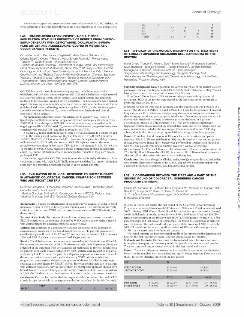
New cytotoxic agents and target therapies increased survival in M-CRC. Perhaps, insome subgroups of patients, target therapies are not as effective as in trials populations.
L49 IMMUNE-REGULATORY (FOXP31)-T-CELL TUMORINFILTRATION STATUS IS PREDICTIVE OF BENEFIT FROM CHEMO-IMMUNOTHERAPY WITH GEMCITABINE, OXALIPLATIN, 5-FU/FAPLUS GM-CSF AND ALDESLEUKINE (GOLFIG) IN METASTATICCOLON CANCER PATIENTS
Cinzia Remondo1, Pierosandro Tagliaferri2, Maria Teresa Del Vecchio3,Cristina Migali1, Kwong Y Tsang4, Maria Saveria Rotundo5, PierfrancescoTassone2,5, Guido Francini1, Pierpaolo Correale1
1Section of Medical Oncology, Department ‘‘Giorgio Segre’’ of Pharmacology,Siena University School of Medicine, Siena, Italy; 3Pathology Section, HumanPathology and Oncology, Siena University School of Medicine, Italy; 2MedicalOncology Unit and 5Referral Center for Genetic Counseling, ‘‘Campus SalvatoreVenuta’’, ‘‘Magna Graecia’’ University, School of Medicine Catanzaro, Italy;4Laboratory of Tumor Immunology and Biology, National Cancer Institute,National Institute of Health, Bethesda, MD,USA.
GOLFIG is a novel chemo-immunotherapy regimen, combining gemcitabine,oxaliplatin, 5-FU/FA with immunoadjuvant GM-CSF and aldesleukine, which resultedsafe and very active in colon-cancer patients. Anti-tumor activity and immunityfeedback to the treatment resulted strictly correlated. The best outcome was observedin patients showing autoimmunity signs, rise in central-memory-T cells, and decline inperipheral and tumor infiltrating immuno-regulatory T (Treg) cells. We investigateda possible correlation between Treg-tumor-infiltration at diagnosis and clinicaloutcome of these patients.
An immunohistochemistry study was carried out to quantify Treg (FoxP3+)
lymphocytes-infiltration in tumor samples of 41 colon cancer patients who receivedFOLFOX-4 chemotherapy or GOLFIG chemo-immunotherapy as enrolled in theongoing phase III GOLFIG-2 trial. Treg-tumor-infiltration score (range 0 to 5) was thencorrelated with survival (OS) and time to progression (TTP).
A higher Treg-tumor-infiltration score (score 3-5) was associated to a longer OS andTTP in the whole patient population (high vs low score; TTP=18 vs 9.4 months;P=0.002; OS=55.7 vs 28.9 months; P=0.001), however, those patients with high tumor-infiltration of FoxP3+-T cells who received GOLFIG regimen showed the mostfavorable outcome (high vs low score; TTP=20.8 vs 11.6 months; P=0.04; OS=68.1 vs41 months; P=0.04). A COX-regression model demonstrated in these patients thata high Treg-tumor-infiltration score is an independent variable of long survival andprolonged TTP.
Our results suggest that GOLFIG chemoimmunotherapy is highly effective in colon-carcinoma patients with high FoxP3+ infiltration score and that Treg-tumor-infiltrationscore may be a favorable prognostic marker in colon cancer patients.
L50 EVALUATION OF CLINICAL RESPONSE TO CHEMOTHERAPYIN ADVANCED COLORECTAL CANCER: COMPARISON BETWEENWHO AND RECIST CRITERIA
Berenice Brocadello1, Francesca Bergamo1, Antonio Jirillo1, Umberto Basso1,Carlo Aschele2, Sara Lonardi11Medical Oncology Unit, Istituto Oncologico Veneto – IRCCS, Padova, Italy;2Medical Oncology Unit, E.O. Ospedali Galliera, Genova, Italy
Background: To assess the effectiveness of chemotherapy is essential in order to avoidunnecessary both in terms of toxicity and economic costs. Two criteria are currentlyavailable to this purpose: WHO criteria (two-dimensional) and RECIST criteria (one-dimensional).
Purpose of the Study: To compare the evaluation of response in accordance withRECIST criteria with the response obtained by WHO criteria in 149 patients treatedwith chemotherapy for advanced colorectal cancer (CRC).
Material and Methods: In a retrospective analysis we compared the response tochemotherapy, according to the two different criteria, of 149 patients prospectivelyenrolled in 4 phase II trials of 1st, 2nd and 3rd line treatment of advanced CRC, between2000 and 2003. For data comparison we used Kappa statistical.
Results: The global response rate to treatment assessed by WHO criteria was 37% whilethe response rate recalculated by RECIST criteria was 38%. Only 15 patients (10%) areredefined in the transition from two-dimensional methodical to the one-dimensional:six patients with stable disease evaluated by WHO criteria were reclassified as partialresponse by RECIST criteria; five patients with partial response were reassessed as stabledisease; one patient assessed with stable disease by WHO criteria switched toprogression; three patients defined as progression of disease by WHO criteria werereassessed as stable disease by RECIST criteria. However despite there are 15 patientswith different evaluation, only in four of them the therapeutic approach should havebeen different. The value of Kappa statistic for the correlation of the two sets of criteriais 0,845 which indicate an excellent agreement between the two measurement systems.
Conclusion: Our results confirm that the response evaluation defined by the RECISTcriteria is super imposable to the response evaluation as defined by the WHO criteria.
WHORECIST RC RP SD PD Totale
RC 3 3RP 47 6 53SD 5 57 3 65PD 1 27 28Totale 3 52 64 30 149
L51 EFFICACY OF CHEMORADIOTHERAPY FOR THE TREATMENTOF LOCALLY ADVANCED SQUAMOUS CELL CARCINOMA OF THERECTUM
Maria Chiara Tronconi1, Roberto Doci2, Mario Bignardi3, Francesco Sclafani1,Silvia Bozzarelli1, Nicola Personeni1, Tiziana Pressiani1, Lorenza Rimassa1,Mariagrazia Di Rocco4, Armando Santoro1, Carlo Carnaghi11Department of Oncology and Hematology; 2Surgical Oncology Unit,3Radiotherapy and Radiosurgery Unit; 4Department of Pathology, Istituto ClinicoHumanitas, Rozzano, Milano, Italy
Summary Background Data: Squamous cell carcinoma (SCC) of the rectum is a rarepathologic entity, accounting for only 0.1% to 0.25% of all rectal cancers. Only 57 caseshaving been reported over a period of more than 60 years.
From June 2006 to August 2008, six consecutive patients with squamous cellcarcinoma (SCC) of the rectum were treated at the same Institution, according toprotocols used for anal SCC.
Methods: All tumors were locally advanced and the clinical stage was T3N0M0 in 2cases, T3N1M0 in 1, T4N1M0 in 2 and T3N2M1 in 1 case for the presence of bilaterallung metastases. Five patients received primary chemoradiotherapy and one receivedchemotherapy only due to previous pelvic irradiation. Chemotherapy regimens were 5-fluorouracil-based with (4 cases) or without (1 case) platinum. In 1 patienta continuous infusion of 5-fluorouracil (5-FU) was given during radiotherapy.Radiation treatment also involved the external iliac nodes and in two patients with lowrectal cancer it also included the anal region. The minimum dose was 5.040 cGy;a boost dose to the primary tumor up to 5.940 cGy was given to three patients.
Results: Complete clinical response (CR) was achieved in 3 patients and partialresponse (PR) in 2. Disease stabilization (SD) was obtained in 1 case and no patientsshowed progressive disease (PD). Surgery was performed in 1 patient with PR and in 1with SD. The patient with lung metastases received 4 courses of systemicchemotherapy. As of the last follow-up (FU) patients with CR were free of recurrence at17 (cT4N1), 31 and 28 months (cT3N0). At a median FU of 18 months all patients arealive and all but the patient with metastasis are disease free.
Conclusions:Our data, though in a limited series, strongly support the conclusion thatconcomitant chemoradiotherapy of rectal SCC can achieve a complete response ina relevant proportion of patients, avoiding demolitive surgery.
L52 A COMPARISON BETWEEN THE FIRST AND A PART OF THESECOND ROUND OF COLORECTAL SCREENING CANCERPROGRAMME IN RIMINI
Casale C*, Antonioli G�, Di Marco M�, Giovanardi M�, Miracolo A�, Onorato GD�,Santilli F�, Scarpulla R�, Solmi L�, Fava C*, Canuti D**U.O. di Oncologia ed Oncoematologia; �U.O. di Gastroenterologia edEndoscopia Digestiva
In 2005 in Rimini, we started the first round of the Colorectal Cancer ScreeningProgramme; we invited, frommarch 2005 to march 2007 about 75.464 individuals aged50-69, offering FOBT (Faecal Occult Blood Test) every two years (biennal screening).33.098 individuals responded to our invite (43.9%); 1097 males (7%) and 838 (5%)females were positive at the firts level test (FOBT). Consequently we made 1478 firstpancolonscopy and 260 follow up colonscopy. We found 433 adenomas at high risk,and 112 tumors. The first round ended on march 2007. From april 2007 to december2008 (21 months of the secon round) we invited 68.815 and with a compliance of51.5% . In the same period we found 60 tumors.
We would compare the histopatological profile of the tumors and the detection ratebetween the first (prevalence round) and the second round (21 months).Materials and Methods: The Screening Center collected data - by oracle software -from gatroenterolgists on colonscopy results for people who were screened positive.Then we compared cancer screen-detected in the first round with cancer
Results: The main differences between the first and the second round are sinthetisedbelow (see the attacched file). We analized sex, age, T, Dukes Stage and Detection Rate(D.R.) for screen-detected cancers in the two groups.
SEX M F
FIRST ROUND 65 (59%) 47(41%)SECOND ROUND 35 (58%) 25 (42%)
50-54 55-59 60-64 65-69
First Round 14 (12.5%) 25 (22.3%) 35 (31.3%) 38 (33.9%)Second Round 5 (8.3%) 15 (25.0%) 11 (18.0%) 29 (48.3%)
viii126 | session L: gastrointestinal cancer-colon-rectal Volume 20 | Supplement 8 | October 2009
session L Annals of Oncology
by guest on March 22, 2015
http://annonc.oxfordjournals.org/D
ownloaded from
T T1 T2 T3 T4 yT Tx
First Round 30 (26.8%) 33 (39.4%) 33 (29.4%) 2 (1.6%) 3 (2.7%) 14 (12.5%)Second Round 24 (40.0%) 9 (15.0%) 19 (31.6%) 1 (1.6%) 2 (3.3%) 5 (8.5%)
Dukes A B1 B2 B3 C1 C2 C3 D n.n.
First Round 42 21 6 1 14 11 1 2 14Second Round 25 7 11 0 3 7 0 1 6
DETECTION RATE
FIRST ROUND 3.4 · 1000SECOND ROUND 2.0 · 1000
We observed a significant increase in T1 cancer from the first to the second round from26.8% to 40.0% and in Dukes A stage from 37.5% to 41.6%. Nevertheless detection rateconfirm a significative difference in the round of prevalence vs the second round from3.4 x 1000 to 2.0 x1000.These results, even if are not completed, suggest that indicatorsare consistent with international references. We need further studies to complete ourconsiderations at the end of the second round.
L53 IMMUNOHEMOLYTIC ANEMIA FOLLOWING OXALIPLATINADMINISTRATION FOR COLON CANCER
Giglio G1, Antuzzi G2, Musacchio M2, Carrozza F2
1U.O.S. di Onco-ematologia; 2U.O.C. di Oncologia Ospedale Civile ‘‘A.Cardarelli’’ - ASREM - Campobasso – Italy
Introduction: Oxaliplatin (L-OHP) is a third generation platinum derivative withproven effectiveness in the treatment of colorectal cancer. Combined with 5-fluorouracil (5-FU) and leucovorin as a 48 hour infusion every two weeks (FOLFOXregimen), L-OHP is drastic in the adjuvant, first and second line setting. We reporthere a case of significant post-infusion hemolysis in a patient undergoing adjuvantchemotherapy with combination chemotherapy with FOLFOX regimen for stage IIIcolon cancer, and review the literature.
Case Report: A 64 year-old-male was administered adjuvant chemotherapy withFOLFOX 4 regimen for a Duke’s stage C colorectal carcinoma. Eight cycles ofchemotherapy had been administered without complications. On the first day of 9th
cycle, and after L-OHP infusion was completed, the patient noticed urine discolorationand immediate urine analysis demonstrated hematuria with hemoglobinuria. Thepatient developed clinical and laboratory abnormalities consistent with thedevelopment of the hemolytic syndrome (AIHA). A direct Coombs test was positive.Treatment was discontinued. Given the moderate size of anemia the patient was notsubjected to transfusion or treatment with cortisonic. Ten days after the generalconditions gradually improved together with increase of hemoglobin and decrease ofreticulocytes, bilirubin and LDH; fifteen days after laboratory values were returning tonormal.
Discussion: The induction of autoimmune hemolysis is a rare side effect of oxaliplatin.An immune-complex type hemolytic anemia, similar to that induced by penicillin, hasbeen postulated as a possible pathogenetic mechanism. In licterature only six patientswith adequately documented oxaliplatin–induced autoimmune haemolytic anaemiahave previously been reported. The direct’s test represents the essential hallmark of thediagnosis. Oxaliplatin-induced AIHA require an immediate diagnosis sincereexposition results in a more severe hemolysis.
Conclusion: Acute hemolysis is a rare but potentially life-threatening complication ofL-OHP based chemotherapy. Thus, increased vigilance on behalf of the oncologiccommunity is required.
referencesd Desame J, Broustet H, Darodes de Tailly P et al. Oxaliplatin-induced haemolytic
anaemia. Lancet 1999; 354: 1179–80.d Garufi C, Vaglio S, Brienza S et al. Immunohemolytic anemia following oxaliplatin
administration. Ann Oncol 2000; 11: 497.d Hofheinz R.-D, Nguyen X-D, Buchheidt D et al. Two potential mechanisms of
oxaliplatin-induced haemolytic anaemia in a single patient. Cancer ChemotherPharmacol 2004; 53: 276–7.
d Sørbye H, Bruserud Ø, Dahl O. Oxaliplatin-induced haematological emergency withan immediate severe thrombocytopenia and haemolysis. Acta Oncol 2001; 40:882–3.
d Earle CC, Chen WY, Ryan DP et al. Oxaliplatin-induced Evan’s syndrome. Br JCancer 2001; 84: 441.
d Brian HC LE, Surender JUNEJA and Peter GIBBS Hemolysis and thrombocytopeniafollowing oxaliplatin administration. Asia-Pacific J Clin Oncol
d Oxaliplatin-induced acute-onset thrombocytopenia, hemorrhage and hemolysis.Oncology. 2004;67(2):179-82.
d An immediate hemolytic reaction induced by repeated administration of oxaliplatin
L54 THE TIMING OF SURGERY FOR RESECTABLEMETACHRONOUS LIVER METASTASES FROM COLORECTALCANCER: BETTER SOONER THAN LATER
Eva Galizia1 Mario Scartozzi2, Walter Siquini3, Cristina Marmorale3,Rossana Berardi2, Aroldo Fianchini3, Alessandro Bittoni4, Stefano Cascinu2
1Oncologia Medica, Ospedale Profili, Fabriano, ITALY; 2Clinica di OncologiaMedica, AO Ospedali Riuniti-Universita Politecnica delle Marche, Ancona,ITALY; 3Clinica Chirurgica, AO Ospedali Riuniti-Universita Politecnica delleMarche, Ancona, ITALY; 4Scuola di Specializzazione in Oncologia, UniversitaPolitecnica delle Marche, Ancona-Italy
In patients with clearly unresectable liver metastases from colorectal cancer, few woulddispute the utility of chemotherapy. However, in patients who are initially resectable,the benefit of preoperative chemotherapy is still matter of debate. Aim of our analysiswas to evaluate the role of neoadjuvant chemotherapy on the global outcome forpatients with colorectal cancer metachronous liver metastases undergoing potentiallycurative liver resection.
One hundred and four patients were available for analysis. Tested variables includedage, sex, primary tumor TNM stage, location and grading, number of liver metastases,monolobar or bilobar location, interval time between liver metastases diagnosis andliver resection, Fong Clinical Risk Score (CRS). Neoadjuvant chemotherapy wasadministered according to the FOLFOX4 regimen.
Forty-four patients (42.3%) underwent liver resection without receivingneoadjuvant chemotherapy (group A); sixty patients (57.7%) received neoadjuvantchemotherapy (group B). At univariate analysis, only administration of neoadjuvantchemotherapy seemed to affect overall survival. In fact patients in group A showeda median survival time significantly superior to that of patients in group B (165 vs 43months; p=0.0016).
Our findings seem to suggest that, when feasible, resection of liver metastases shouldbe considered as an initial approach in this setting. Further studies are needed to betterdelineate innovative therapeutic strategies that may lead to an improved outcome forcolorectal cancer patients with surgically resectable liver metastases.
L55 CLINICAL PREDICTORS FON IN-HOSPITAL ADMISSION OFCOLORECTAL CANCER PATIENTS RECEIVING CHEMOTHERAPY
Luisa Foltran, Giuseppe Aprile, Micol Mazzer, Emiliana Iaiza, Paola Ermacora,Giovanni Cardellino, Nicoletta Pella, Federica De Pauli, Alessandro Follador,Magda Fanzutti, Gianpiero FasolaDepartment of Medical Oncology, University Hospital of Udine, Italy
Objective: Although cancer symptoms and treatment-related toxicities increase therate of in-hospital admission, there is no accepted guideline to identify those patientswho are at higher risk for hospitalization. Aim of this analysis was to identify reliableclinical predictors for hospitalization among those colorectal cancer (CRC) patients onactive treatment who ask for an unplanned visit
Methods: Electronic medical records of 268 consecutive CRC patients accessing a 12-hour open dedicated ward from October 2006 to March 2008 were reviewed. Dataregarding tumor stage, patients’ age and demographics, reasons for visiting, as well asdate and type of last oncologic treatment were all recorded. Considering hospitalizationas the dependent variable, non conditional logistic regression uni- and multivariatemodels were used to identify risk factors for hospitalization. Analyses were performedusing SAS 9.1.
Results: 268 patients (132f/136m) receiving irinotecan- (45%), oxaliplatin- (35%) or5-FU-based chemotherapy with or without biologics, accessed the service. Median agewas 70 years, KPS 0/1/2/3 patients were respectively 81/131/51 and 5. Overall, 16patients were admitted to the hospital with an hospitalization rate of 6%. Univariatelogistic regression demonstrated that presenting with multiple (>3) symptoms (OR5.8, 95%CI 1.5-22.4, p<0.001) or with declined ECOG PS (2/3 versus 0/1) (OR 10.1,3.3-20.5, p<0.001) were significant risk factors for hospitalization. Patients’ age andtype of chemotherapy did not predict the risk of hospitalization.
Conclusions: The growing proportion of CRC patients on active treatment oftenproduce an increased number of unplanned visits, that should be adequately managedand selected for appropriate hospital admission. Both number of referred toxicities,and ECOG PS at the time of the visit are simple clinical predictors for in-hospitaladmission.
L56 POSITRON EMISSION TOMOGRAPHY (FDG-PET) SCAN ANDCOMPUTERIZED TOMOGRAPHY (CT) SCAN FOR EVALUATION OFCOLORECTAL LIVER METASTASES (CRLM) AFTER FOLFOX-6 ANDBEVACIZUMAB NEOADJUVANT THERAPY
Norma Malavasi1, Federica Bertolini1, Roberta Depenni1, Federica Fiocchi2,Pietro Torricelli2, Sandra Zironi1, Bruno Bagni3, Antonella Franceschetto3,Annalisa Fontana1, Laura Scartabelli1, Gabriele Luppi1, Pier Franco Conte1
Volume 20 | Supplement 8 | October 2009 doi:10.1093/annonc/mdp445 | viii127
Annals of Oncology session L by guest on M
arch 22, 2015http://annonc.oxfordjournals.org/
Dow
nloaded from
1Oncology and Haematology Department, University of Modena and ReggioEmilia; 2Radiology Department, University of Modena and Reggio Emilia;3Nuclear Medicine Division, University of Modena and Reggio Emilia
Introduction: The present approach at our institution for patients (pts) with CRLM isneoadjuvant Folfox-6 chemotherapy and Bevacizumab. Efficacy evaluated by CT scanis based on anatomic imaging and complete response is defined as disappearance of alltarget lesions. FDG-PET has the advantage to integrate functional and anatomicscanner; complete metabolic response (CMR) is defined as normalization of FDG-uptake. Correlation between imaging and pathologic status is not clear; we evaluate therelationship between FDG-uptake, CT scan and pathologic complete response (pCR)of CRLM after neoadjuvant treatment
Methods: Pts with initially unresectable CRLM with regard to number, size of tumouror locoregional invasion and no evidence of extra-hepatic disease, received Folfox-6chemotherapy and Bevacizumab 5mg/Kg, every 2 weeks for 3 months. CRLM wereinitially documented by biopsy and FDG-PET/CT performed both at baseline and afterthe completion of neoadjuvant therapy; images were analyzed by measuring the meanstandardized uptake value. Elective surgery was planned at least 6 weeks after the lastdose of Bevacizumab and 4 weeks after last dose of chemotherapy
Results: Up to now, 10/20 pts undergoing surgery are valuable; they all presented basalCT scan and FDG-avid hepatic lesions.
Four (40%) pts achieved CMR on FDG-PET; of these, one (10%) was found to bepCR without evidence of liver lesions also on CT scan. In the others (30%), still viabletumour was found by pathologist and liver metastases persistent also on CT scan.
Six (60%) pts didn’t achieve CMR on FDG-PET; of these, one (10%) was found tobe pCR while in the others (50%) viable tumour was found by pathologists.
Conclusions: FDG-PET might be not superior to traditional CT scan after neoadjuvanttreatment; even if few cases are available, FDG-uptakedoesnot seemtopredict pCR.Whenfeasible, the site of disappeared lesion should be considered for pathologic evaluation.
FDG-PET could be enhanced if considered as possible marker for early assessmentof response to neaodjuvant treatment.
L57 K-RAS AND B-RAF MUTATION AND CETUXIMAB THERAPY INADVANCED COLORECTAL CANCER PATIENTS : A RETROSPECTIVEMONOISTITUTIONAL STUDY.
Frisinghelli M, Soini B, Ambrosini G, Caffo O, Caldara A, Girlando S*,Barbareschi M*, Cuorvo LV*, Dalla Palma P*, Galligioni EMedical Oncology and *Pathology S.Chiara Hospital Trento ItalyBackground: It has been reported that k-rasmutations in k-ras or b-raf gene negativelyaffect response to anti-epidermal growth factor receptor (EGFR) monoclonalantibodies in metastatic colorectal cancer (mCRC) patients. Our study was aimed toevaluate the role of k-ras or b-raf in predicting objective tumor response to cetuximabamong 28 metastatic colorectal cancer (mCRC) patients.
Patients and Methods: 28 consecutive mCRC patients, treated with Cetuximab in ourinstitution, were retrospectively analyzed for the mutational status of k-ras and b-rafand objective tumor responses: they were 17 men and 11 women with a median age of57 years (range 22 – 80 years). Major metastatic sites were: liver in 12 patients (43%),lung in 2 patients (7%), and both in 14 patients (50%). Genomic DNA was extractedfrom paraffin-embedded tissue blocks of the primary tumors, amplified and sequencedfor k-ras and b-raf genes using the ABIPrism 310 analyzer.
Results: One cases was excluded because the DNA was poorly preserved. Among theremaining 27 patients k-ras was mutated in 14 cases (50%) and was associated withresistance to cetuximab (7 % ORR compared to 33 % in non mutated patients). Twenty-three cases were analyzed for b-raf genemutation: one was mutated and 22 cases were wildtype. Of them: 7 were also k-ras wild type, 14 k-ras mutated and 1 not valuable. Noobjective responses were observed among 14 patients b-raf wild type/ k-ras mutated, while3 OR were observed among 7 patients b-raf wild type/k-ras wild type (43%)
Conclusions: Among k-ras wild type patients, b-raf mutation status can therefore helpidentify patients who are most likely to benefit from treatment with the anti-EGFRmonoclonal antibodies
L58 HAEMOLYTIC UREMIC SYNDROME INDUCED BY INFUSION OFOXALIPLATIN: TWO CASES REPORTS
Patrizia Racca, Giuliana Ritorto, Rosella Spadi, Ivan Facilissimo, Laura Fanchini,Gabriella Monasterolo, Patrizia Lista, Libero CiuffredaSC Oncologia Medica 1 A.O.U. San Giovanni Battista di Torino
Background: Oxaliplatin (L-OHP) is a third generation platinum compound withproven antitumor activity in the treatment of colorectal cancer (CRC). Acute onsethaemolysis and thrombocytopenia associated with this drug has been rarely reported.We describe two patients, with advanced CRC, who developed different degree ofseverity reaction.
Patients: A 49 years-old woman was administered palliative chemotherapy with L-OHP and capecitabine for unresectable metastasis of CRC. The reaction appearedduring the 17th cycle at the beginning of the L-OHP infusion the patient developedback pain, hematuria, upper gastrointestinal bleeding and haemolysis. Chemotherapywas immediately discontinued:the immediate urine analysis demonstrated haematuriawith haemoglobinuria;the day after the patient’s complete blood count showed offfindings consistent with acute haemolysis and thrombocytopenia (Hb8,7g/dl PLTS33x
10 9/L D-dimmers>40lg/mL). A haemolytic reaction was pointed by elevated LDH(4074 UI/l), indirect bilirubin indices (9,3 mg/dL), severe reduction of haptoglobin(<3 mg/dL) and fibrinogen (<50mg/dL) and strongly positive Coomb’s Test. Thepatient also exhibited signs of acute renal failure (creatinine3,2mg/dL BUC11 ml/minBCC23 ml/min) and moderate hepatic failure (AST338 UI/L; ALT109 UI/L; GGT119UI/L; INR7,7 ).The patient was managed with monitored i.v. hydration and loopdiuretics, higth dose corticosteroids and daily frozen plasma infusion. The patientgradually recovered and successive haematological and biochemical findings confirmedher improvement. A month later after haemolytic uremic syndrome onset laboratoryvalues were returning to normal and the patient was asymptomatic. The second case wereported is a 70 years-old women with CRC with unresectable lung metastasis of CRC .The patient developed the reaction at the beginning of the fourth cycle ofchemotherapy (FOLFOX4) with the same symptoms and laboratory findings but withless intensity and faster recovered.
Conclusion: The haemolytic uremic syndrome may be a rare but potentially life-threatening complication of oxaliplatin-based chemoterapy. Oncologist should beaware to diagnose and treat this adverse event.
L59 BEVACIZUMAB-INDUCED HYPERTENSION IS A PREDICTIVEMARKER OF RESPONSE AND PROGRESSION FREE SURVIVAL INPATIENTS TREATED FOR METASTATIC COLORECTAL CANCER
Alfonso De Stefano, Chiara Carlomagno, Lucia Cannella, Anna Crispo,Stefano Pepe, Salvatore Alfieri, Maria Colantuoni, Chiara Cella, Lucia Raimondo,Sabino De PlacidoDipartimento di Endocrinologia ed Oncologia Molecolare e Clinica. Universitadegli Studi di Napoli Federico II
Background: Patients (pts) with metastatic colorectal cancer (mCRC) receiving allthree active drugs (Irinotecan, Oxaliplatin and Fluorouracil) achieve the best outcome.Bevacizumab (Bev) further improved progression-free (PFS) and overall survival.
Aim: Arterial hypertension (aHT) has been reported in all studies involving Bev.Hypothesizing that Bev-induced aHT may be a potential predictive marker, weretrospectively analyzed the modifications of arterial blood pressure among patientstreated with Bev according to response rate and PFS.
Patients: Pts with histologically proven, mCRC receiving Bev as first-line treatment incombination with chemotherapy were eligible.
Results: 39 pts were considered for the present analysis: 29 M and 10 F; median age was62.5 years (range 31.7-74.5). Twenty-five pts were diagnosed at stage IV (16 pts liver, 1pt lung, 4 pts liver and lung, 3 pts pelvic mass, 1 pts lung and bone). Fourteen pts werestage II/III at diagnosis and developed lung (4 pts), liver (3 pts), peritoneal (3 pts)metastasis; 4 pts had multiple sites of disease. Chemotherapy regimens were: FOLFIRI(34 pts), XELOX (3 pts), XELIRI (1 pts) and FOLFOX (1 pt). Eleven patients had basalhypertension. Ten pts (25.6%) with controlled blood pressure values (3 had basal aHT)developed G2 aHT during the administration of chemotherapy + Bev. After startingadequate anti-hypertensive therapy, they continued Bev until disease progression.Thirty-seven pts were evaluable for response: 22 (59.4%) had complete (1) or partial(21) response. The following table shows the distribution of response according toinduced-aHT. Among pts with induced-aHT, 80% achieved a complete or partialresponse, as compared with 52% of pts who did non have aHT. Kaplan-Myers analysisshowed a statistically significant improvement in PFS for pts with induced-aHT (15.2vs 8.0 months, p=0.05).
Conclusions:Our data suggest that Bev-related aHTnmay represent a predictive factorof response and prolonged DFS in pts with mCRC receiving first-line Bev.
L60 CASE REPORT: LIVER METASTASIS FROM DONOR‘S CELL INA TRANSPLANT RECIPIENT
Giuliana Ritorto, Laura Fanchini, Rosella Spadi, Patrizia Racca, Ivan Facilissimo,Nadia Birocco, Raffaella Napoletano, Libero CiuffredaSC Oncologia Medica AOU San Giovanni Battista Torino
Background: The incidence of donor-derived malignancy is thought to be of the orderof 0.02%-0.2%. There have been reports of transmission from donor to recipient ofa variety of malignancies, including malignant melanoma, choriocarcinoma,neuroendocrine tumors, and adenocarcinomas of the lung, prostate, and pancreas.Identifying the origin of a malignancy post transplant as being either donor-derived orrecipient-derived has important clinical consequences, including withdrawal ofimmunosuppression and retransplantation. It also has implications for other recipientsof donor organs.
Case Report: we present the case of a 53 years old male patient, transplanted foralcoholic cirrhosis in 2007, who after 6 months developed multiple secondary liverlesions detected by US before and confirmed by TC scan. Colonoscopy and gastroscopywere performed, and revealed no evidence of primary carcinoma.The patientunderwent liver biopsy: citology confirmed presence of adenocarcinoma cells andimmunohistochemistry seemed to indicate a colorectal origin of the lesions. Thereforea F-18 FDG PET/CT was performed, showing abnormal uptake limited to the liver.Asecond biopsy to evaluate DNA origin revealed that 80% of cancer cells derived fromthe donor, even if it had no risk factors for occult carcinoma identified on routine
viii128 | session L: gastrointestinal cancer-colon-rectal Volume 20 | Supplement 8 | October 2009
session L Annals of Oncology
by guest on March 22, 2015
http://annonc.oxfordjournals.org/D
ownloaded from
questionnaire screening. 3 months later (July2008) the patient underwent secondortothopic liver tranplantation. Histologic examination of the explanted organconfirmed the diagnosis of adenocarcinoma liver metastasis from colorectal primarycancer. During September2008 the patient referred abdominal pain with CT scan andUS evidence of multiple nodular areas looking like angiomas . Further surveillance wasadvised. One month later US was suspicious for new metastatic lesions, so that thepatient underwent a second liver biopsy. Histologic examination found the presence ofundifferentiated carcinoma cells with desmoplastic aspects and focal mucusproduction, probably deriving from biliary tract. He started palliative chemotherapywith weekly oxaliplatin and 5-fluorouracil folinic acid; after 3 weeks the patientsdeveloped hepatic failure, and died after 5 months from the second transplantation.
L61 NEW ONCO-GERIATRIC SCREENING TEST FORGASTROINTESTINAL CANCER IN ELDERLY PATIENTS.PRELIMINARY DATA
Sara Pini, Francesca Di Fabio, Carmine Pinto, Fabiola Lorena Rojas Llimpe,Vita Mutri, Piergiorgio Di Tullio, Ciro Longobardi, Andrea Angelo MartoniMedical Oncology Unit, S.Orsola-Malpighi Hospital, Bologna, Italy
Background: More than half of new gastrointestinal cancers are diagnosed in elderlypatients (pts) in which treatment-related toxicity could be increased. ComprehensiveGeriatric Assessment (CGA) has been proposed to help oncologists identify vulnerablepts and better customize therapy, but this appears to be time-consuming andredundant. A brief screening test could be more useful in clinical practice to identify ptsin which a complete CGA would be appropriate.
Methods:We developed a computer-assisted ‘‘basal’’ assessment including some of themain CGA scales: ADL/IADL, CIRS-G/S, psychological status (GDS-5), social support,and some clinical and laboratory items such as weight loss, KPS, polypharmacy,creatinine clearance, Hb and albumin blood levels. Each item contributes to creatinga score ranging from 11 to 33. A second ‘‘follow up’’ assessment detecting treatmenttoxicity and compliance was administered at the end of adjuvant chemotherapy, orafter 3 months of treatment in an advanced setting. Seventy-year old pts or older withgastrointestinal tumors, admitted to the Medical Oncology Outpatients’ Hospital Unitof Sant’Orsola-Malpighi Hospital, Bologna, were eligible.
Results: From June 2006 to February 2009, ninety-seven pts joined the study. Ptscharacteristics were: male 52 (53.6%), female 45 (46.3%), median age 78 (70-87 range),colorectal cancer 85 (87.6%), gastro-esophageal cancers 12 (12.3%), adjuvant setting44 (45.3%), advanced setting 53 (54.6). Mean time to fill out the form was 8 minutes(range 5-10 mins). Patients were arbitrarily divided into 2 groups, good risk (score 11-15) and poor risk (score 16-33), according to the median score at basal assessment of15. The distribution of toxicity and compliance parameters in good and poor risk wasas follows: grade 3-4 toxicity 21.9% vs. 27.5%; treatment dose-intensity £80% 28.5%vs. 41.4%; age >80 11.1% vs. 42.3%; treatment never started or was lost early to follow-up 8.8% vs. 23%.
Conclusions: This onco-geriatric screening test is feasible in clinical practice. However,it has to be prospectively validated and benchmarked with other screening tests.
L62 COMPARISON OF POSITRON EMISSION TOMOGRAPHYSCANNING AND SENTINEL NODE BIOPSY IN THE DETECTION OFINGUINAL NODE METASTASES IN PATIENTS WITH ANAL CANCER
Enrica Milanesi^^, Massimiliano Mistrangelo, Ettore Pelosi*, Marilena Bello**,Isabella Castellano***, Paola Cassoni***, Umberto Ricardi^, Fernando Munoz^,Patrizia Racca^^, Viviana Contu^^, Giancarlo Beltramo^, Libero Ciuffreda,Mario Morino, Antonio Mussa****Molinette Hospital, Department of Surgery and Centre of Minimal InvasiveSurgery, University of Turin, Italy, Chief Prof. M. Morino; *PET Center IRMET spa,Turin, Italy; **Molinette Hospital, Department of Nuclear Medicine, University ofTurin, Italy, Chief Prof. G. Bisi; ***Molinette Hospital, Anatomo-PathologicalDepartment, University of Turin, Italy, Chief Prof. G. Bussolati; ^MolinetteHospital, Department of Radiotherapy, University of Turin, Italy, Chief Prof. U.Ricardi; ^^Molinette Hospital, Oncological Centre for GastrointestinalNeoplasms, University of Turin, Italy, Chief Dr. Racca; ****Molinette Hospital,Surgical and Oncological Department, University of Turin, Italy, Chief Prof. A.Mussa
Background: Inguinal lymph node metastases in patients with anal cancer are anindependent prognostic factor for local failure and overall mortality. Inguinal lymphnode status can be adequately assessed with sentinel node biopsy and radiotherapystrategy subsequently changed. We compared the technique versus dedicated 18F-FDGPET in order to determine which was the better tool for staging inguinal lymph nodes.
Patients and Methods: At our Department 27 patients (9 men and 18 women)underwent both inguinal sentinel node biopsy and PET-CT. PET-CT was performedbefore treatment and then at 1 and 3 months after treatment.
Results: PET-CT scans detected no inguinal metastases in 20/27 patients andmetastases in the remaining 7. Histologic analysis of the sentinel lymph node detectedmetastases in only 3 patients (4 PET-CT false positives). HIV status was not found toinfluence the results. None of the patients negative at sentinel node biopsy developed
metastases during the follow-up period. PET-CT had a sensitivity of 100%, witha negative predictive value of 100%. Owing to the high number of false positives, PET-CT specificity was 83% and positive predictive value was 43%.
Conclusions: In this series of patients with anal cancer, inguinal sentinel node biopsywas superior to PET-CT for staging inguinal lymph nodes.
L63 CHOICE OF CHEMOTHERAPY SCHEDULES IN ELDERLYPATIENTS (>75 YEARS) WITH METASTATIC COLORECTAL CANCER(MCRC) ACCORDING TO COMORBIDITY
Tudini M1, Palluzzi E1, K Cannita1, Santomaggio A1, Mancini M1, Bruera G1,Lanfiuti Baldi P1, Porzio G1, Aielli F1, Brollo M1, Paradisi S1, Ficorella C1,Ricevuto E1
1Department of Medical Oncology, S. Salvatore Hospital, University of L’Aquila,L’Aquila
Background: treatment of elderly mCRC patients (pts) is still debated due tocomorbidities and effect of polipharmacology.
Patients and Methods: MCRC elderly pts ‡75 age have been processed with ADL,IADL, CIRS and treated according to stage of co-morbidity: primary, standardizedchemotherapy; intermediate and secondary, modified chemotherapy; tertiary, notreatment.
Results: 34 MCRC pts (M/F=24/10) were evaluated; median age 80 y (range 75-86 y).Pts distribution: stable stage, 4 pts (12%); intermediate stage, 6 pts (17%); secondarystage, 20 pts (59%) and tertiary stage, 4 pts (12%). Thirty pts (88%) were treated withfirst-line chemotherapy. Stable stage: triplet schedule FIR-FOX (Irinotecan 160 mg/mqd1, 15; Oxaliplatin 80 mg/mq d8, 22; 12-h flat infusional 5FU 900 mg/mq d1-2, 8-9, 15-16, 21-22; q28 d) in 2 pts (6%); FOLFIRI in 1 pt (6%); FOX (Oxaliplatin 80 mg/mg d1,15- 5FU 900 mg/mq/die d1-4, 5-18; q28) in 1 pt (3%). Intermediate stage: FOX-Cetuximab combination (Oxaliplatin 40 mg/mq/w; 5FU 1800 mg/mq/w; Cetuximab)in 2 pts (6%); modified FIR-FOX (Irinotecan 140 mg/mq, Oxaliplatin 70 mg/mq, 5FU800 mg/mq/die) in 3 pts (9%). Secondary stages: FOX regimens bi- or weekly in 16 pts(47%); Irinotecan 80 mg/mq and 5FU 800 mg/mq/die g1-2 weekly in 2 pts (6%);modified FIR-FOX in 1 pt (3%); monochemotherapy (UFT or OXP 40 mg/mq/w) in 2pts (6%). Treatment discontinued after one/two cycles in 3 pts not for toxicity; 2 ptslost to follow-up; 2 pts, no measurable disease. Different treatments were toleratedwithout limiting toxicities. Median administered cycles 6 (range 1-9); 5 pts on-treatment to date. 20 valuable patients: 1 cCR (5%), 6 PR (30%), 7 SD (35%), 6 PD(30%). Median follow-up, 12 months.
Conclusions: the prevalence (>70%) elderly >75y metastatic colon cancer patientsshow secondary/tertiary comorbidity stage; 88% were treated with first-linechemotherapy; doublet or triplet combinations were safely administered in 82%patients; preferred modified doublet schedule was FOX (47%), OR were observed in35% pts.
L64 ANAL CANAL METASTASIS OF BREAST CANCER A CASEREPORT AND REVIEW OF LITERATURE
Bochicchio AM, Ignomirelli O, Latorre G, Digiovannantonio L, Romano C,Tartarone A, Coccaro M, Capobianco A, Di Leo P, Ardito R, Cangiano R, Fusco V,Aieta MO.U. of Medical Oncology, Gastroenterology, Surgery, Histopatology,Radiotherapy CROB-IRCCS, Rionero in Vulture-Italy
Gastrointestinal metastases with anal involvement are rare in breast cancer. Mostfrequent site of the gastrointestinal tract involved are stomach and small intestine,while the rectal or anal localization is extremely rare. We report a case of a patient withbreast cancer metastases to the anus.
The patient, a 74-year old woman had undergone left modified radical mastectomyand axillary lymph node dissection on April 2002 due to a breast cancer. Histologicalexamination revealed a pT4 infiltrating lobular adenocarcinoma with 7 on 8 resectednodes, G2 (Er and PgR 70% positive, c-Erb-B2 resulted negative). The patient receivedfive cycles of adjuvant chemotherapy with FEC followed by toremifene 60 mg daily.
On November 2002 there was a local recurrence, surgically excised, with cutaneousinfiltration treated with radiotherapy and hormonal treatment with exemestane. OnDecember 2004, metastatic bone disease was documented by a bone scan that showeda pathologic concentration of tracer on skull. A computed tomography showed livermetastasis, axillary, epigastric lymphonodes and nodular thoracic lesion. The patientrefused chemotherapy and continued hormonal treatment.
On August to November 2005, for bone progressive disease, the patient received 5cycles of chemotherapy with capecitabine and intravenous vinorelbine without benefit,then received radiotherapy on symptomatic bone lesions, tamoxifene 20 mg orally dailyand iv. zoledronic acid. On April 2006, the appearance of tenesmus and fecalincontinence secondary to perianal skin and external rectum orifice infiltration,required endoscopic and histopathological study. Endoscopy showed: hyperaemiclesion of the anal mucosae with stenosis of the anorectal wall, like as infiltratingneoplastic process. The patient was submitted to surgical treatment of left colostomy,minimal sigmoid resection, biopsies, Hartman rectal amputation. Histopatholology
Volume 20 | Supplement 8 | October 2009 doi:10.1093/annonc/mdp445 | viii129
Annals of Oncology session L by guest on M
arch 22, 2015http://annonc.oxfordjournals.org/
Dow
nloaded from
confirmed: metastasis from breast cancer infiltrating the derma and rectal wall. On july2006, the patient died for abdominal and bone progressive disease.
This is one of the three cases reported into literature of the anal metastasessecondary to breast cancer.
L65 CHOICE OF CETUXIMAB-CONTAINING CHEMOTHERAPY INPRE-TREATED METASTATIC COLORECTAL CANCER PATIENTS INCLINICAL PRACTICE
Bruera G1, Cannita K1, Santomaggio A1, Tudini M1, Lanfiuti Baldi P1, Mancini M1,Spagnuolo M1, Ricevuto E1, Ficorella C1
1Department of Medical Oncology, S. Salvatore Hospital, University of L’Aquila,L’Aquila
Background: We describe our experience in the treatment of patients (pts) affected bymetastatic colorectal cancer (MCRC) with associations Cetuximab-containing assecond line treatment, from March 2004 up to date.
Methods: Twenty-seven pre-treated pts were consecutively enrolled (22 men, 5women). Median age 64 years (range 41-74). Previous first line chemotherapy (CT):triplet combination Irinotecan (CPT-11)/Oxaliplatin (OHP)/5Fluorouracil (5-FU) orquadruplet combination CPT-11/OHP/5-FU/Bevacizumab, 23 pts (85%). SelectedCetuximab-containing associations: double combination of Cetuximab in association
with Irinotecan or Oxaliplatin, 8 pts; triplet association with Irinotecan or Oxaliplatinand 5Fluorouracil or Capecitabine, 14 pts; quadruplet association with Irinotecan,Oxaliplatin, 5Fluorouracil or Capecitabine, 5 pts.
Results: Associations Cetuximab and Irinotecan ± Fluoropyrimidine (Irinotecan-Cetuximab, 5Fluorouracil-Irinotecan-Cetuximab, Capecitabine-Irinotecan-Cetuximab), according to AIFA indication, were selected in 17 pts (63%): 4, 10, 3 pts,respectively. ‘‘Off-label’’ associations containing Cetuximab and Oxaliplatin ±Fluoropyrimidine ± Irinotecan or containing Cetuximab and Irinotecan ±Fluoropyrimidine in pts previously untreated with Irinotecan-containing CT as firstline treatment or who have not failed Irinotecan-based therapy or EGFR negative wereselected in 10 pts (37%). Median number of cycles 3 (range 0,5-9). Twenty-two ptsvaluable; 5 pts with less than 3 administered cycles. Objective responses: 9 partialresponses (41%), 2 stable disease (9%) and 11 (50%) progressions. Median OS of themetastatic disease intent to treat (ITT), 38 months (5+-65+). At a median follow-up of 9months from the start of second line treatment, TTP-ITT was 3 months (2-19) and OS-ITT 16 months (1+-54+). A third line chemotherapy was administered in 12 pts;a fourth line, thereafter, in 4 pts.
Conclusions: Different choices of Cetuximab-containing associations as second linetreatment of MCRC in clinical practice confirmed their feasibility, tolerability andeffectiveness. Prevalence of choices was: 63% according to AIFA indication, 37% as‘‘off-label’’ associations. Median OS of the metastatic disease in this subgroup of ptsseems higher (38 months) than the reported one of the MCRC pts; ORR is 41% andOS-ITT 16 months.
viii130 | session L: gastrointestinal cancer-colon-rectal Volume 20 | Supplement 8 | October 2009
session L Annals of Oncology
by guest on March 22, 2015
http://annonc.oxfordjournals.org/D
ownloaded from
Copyright © 2022 FDOKUMEN