The effect of the pneumococcal toxin, pneumolysin on brain ependymal cilia
Upload
independentCategory
view
1download
0
Cross-reactivity of the BRAF VE1 antibodywith epitopes in axonemal dyneins leads tostaining of ciliaRobert T Jones1, Malak S Abedalthagafi1, Mohan Brahmandam2, Edward A Greenfield2,Mai P Hoang3, David N Louis3, Jason L Hornick1 and Sandro Santagata1,4
1Division of Neuropathology, Department of Pathology, Brigham and Women’s Hospital, Harvard MedicalSchool, Boston, MA, USA; 2Monoclonal Antibody Core, Dana-Farber Cancer Institute and Dana-Farber/Harvard Cancer Center, Harvard Medical School, Boston, MA, USA; 3Department of Pathology,Massachusetts General Hospital, Harvard Medical School, Boston, MA, USA and 4Department of CancerBiology, Dana-Farber Cancer Institute, Harvard Medical School, Boston, MA, USA
Antibodies that recognize neo-epitopes in tumor cells are valuable tools in the evaluation of tissue biopsy or
resection specimens. The VE1 antibody that recognizes the V600E-mutant BRAF protein is one such example.
We have recently shown that the vast majority of papillary craniopharyngiomas—tumors that arise in the sellar
or suprasellar regions of the brain—harbor BRAF V600E mutations. The VE1 antibody can be effective in
discriminating papillary craniopharyngioma from adamantinomatous craniopharyngioma, which harbors
mutations in CTNNB1 and not BRAF. While further characterizing the use of the VE1 antibody in the differential
diagnosis of suprasellar lesions, we found that the VE1 antibody stains the epithelial cells lining Rathke’s cleft
cysts with very strong staining of the cilia of these cells. We used targeted sequencing to show that Rathke’s
cleft cysts do not harbor the BRAF V600E mutation. Moreover, we found that the VE1 antibody reacts strongly
with cilia in various structures—the bronchial airways, the fallopian tubes, the nasopharynx, and the
epididymis—as well as with the flagella of sperm. In addition, VE1 reacts strongly with the cilia of the
ependymal lining of the brain and with the cilia-containing microlumens of ependymoma tumors. There is
significant sequence homology between the synthetic peptide (amino acid 596–606 of BRAF V600E:
GLATEKSRWSG) that was used to generate the VE1 antibody and regions of multiple axonemal dynein heavy
chain proteins (eg, DNAH2, DNAH7, and DNAH12). These proteins are major components of the axonemes of
cilia and flagella where they drive the sliding of microtubules. In ELISA assays, we show that the VE1 antibody
recognizes epitopes from these proteins. A familiarity with the cross-reactivity of the VE1 antibody with
epitopes of proteins in cilia is of value when evaluating tissues stained with this important clinical antibody.Modern Pathology advance online publication, 21 November 2014; doi:10.1038/modpathol.2014.150
‘Mutation-specific’ antibodies that recognize mutantforms of oncoproteins but not the wild type forms ofthose proteins are very useful in the evaluation oftumor resection specimens. For example, antibodiesthat recognize the V600E mutant of BRAF,1,2 and theR132H mutant of IDH1 (refs 3,4) have quicklybecome part of the clinical armamentarium, assistingpathologists with the diagnostic classification of
tumors and helping to determine prognosis andpredict response to therapeutics.5–7 The use of theseantibodies is particularly important as the results ofgenetic testing can still take weeks to report in manyinstitutions.8 As these antibodies are so commonlyused, it is important to have a thorough under-standing of their limitations so as to avoid incorrectinterpretations.
We recently demonstrated that papillary cranio-pharyngioma frequently harbor mutations in BRAF,and that the VE1 antibody that recognizes the BRAFV600E-mutant protein can help distinguish cranio-pharyngioma tumors with wild-type BRAF fromthose carrying the V600E mutation.9 Along withadamantinomatous craniopharyngioma, Rathke’scleft cysts are part of the differential diagnosis that
Correspondence: Dr S Santagata MD, PhD, Division of Neuro-pathology, Department of Pathology, Brigham and Women’sHospital, Harvard Medical School, Harvard Institute of Medicine,HIM-921, 77 Avenue Louis Pasteur, Boston, MA 02115, USA.E-mail: [email protected] 28 July 2014; revised 12 September 2014; accepted 13September 2014; published online 21 November 2014
Modern Pathology (2014), 1–11
& 2014 USCAP, Inc. All rights reserved 0893-3952/14 $32.00 1
www.modernpathology.org

Figure 1 Immunohistochemistry analysis of papillary craniopharyngioma and Rathke’s cleft cyst. Images of hematoxylin and eosin(H&E)-stained sections of both papillary craniopharyngioma and Rathke’s cleft cyst are displayed along with images ofimmunohistochemistry staining for b-catenin and for BRAF V600E (using the BRAF VE1 mutation-specific antibody). Scale bar,50mm for papillary craniopharyngioma and 20mm for Rathke’s cleft cyst (scale bar in insert is 5mm).
Cross-reactivity of BRAF(VE1) antibody
2 RT Jones et al
Modern Pathology (2014), 1–11
neuropathologists contend with when evaluatingsellar and suprasellar tumors.10–12 To investigatewhether Rathke’s cleft cysts harbor BRAF V600Emutations, we stained cases with the BRAF VE1antibody.
Materials and methods
Tissue and DNA Extraction
From the archives of Brigham and Women’s Hospital(Boston, MA) we retrieved 20 formalin-fixed, paraf-fin-embedded tissue samples of Rathke’s cleft cysts.The study was reviewed and approved by thehuman subjects institutional review boards of theDana-Farber Cancer Institute and Brigham andWomen’s Hospital. Histologic diagnosis was con-firmed on all samples by a board-certified neuro-pathologist (SS). DNA was extracted from tissueshavings or core punches (1 mm, Miltex, catalognumber 33-31AA-P/25) from formalin-fixed, paraf-fin-embedded tissue blocks.
Immunohistochemistry
We performed immunohistochemical studies forBRAF V600E expression on 5-mM-thick sections offormalin-fixed, paraffin-embedded tissue in a Bond3 automated immunostainer (Leica Microsystems,Bannockburn, IL, USA) using a primary antibodythat was designed to recognize the BRAF V600Eprotein (clone: VE1, 1:100, Spring Bioscience,Pleasanton, CA). We deparaffinized the tissue sec-tions using Bond Dewax solution and used LeicaPolymer Refine Kit for the diaminobenzidine stain-ing. For b-catenin staining we performed antigenretrieval in a pressure cooker in citrate buffer (pH
6.0, 1:1000 dilution). The sections were incubatedfor 45 min in primary antibody (BD Pharmigen,catalog number 610154, mouse-monoclonal, clone:14) followed by Dako anti-mouse-horseradish per-oxidase for 30 min at room temperature. Cases withnuclear staining were scored as positive andmembranous staining was scored as negative.
Sequenom Genotyping of BRAF Mutations
We used mass spectrometric genotyping based onthe Sequenom MassARRAY technology (Sequenom,San Diego, CA) using a multi-base homogenousmass-extend (hME) as previously described13 toassay DNA extracted from Rathke’s cleft cysts formutations in BRAF.
Expression Profiling
We normalized expression profiling data frompublicly available brain tumor data sets collectedusing the Affymetrix Human Genome U133 Plus 2.0Array Platform [HG-U133_Plus_2] and cataloged inGEO (Gene Expression Omnibus) using methodspreviously described.14 The data sets include thefollowing series: GSE16581 (meningioma), GSE34824(pediatric), GSE19404 (CNS primitive neuro-ectodermal tumors), GSE35493 (atypical teratoidrhabdoid tumors/ATRT, medulloblastoma, normalbrain structures, and primitive neuroectodermaltumor), GSE34771 (CNS lymphoma), GSE36245(adult glioblastoma), GSE33331 (adult astrocytoma),GSE16155 (ependymoma), and GSE5675 (pilocyticastrocytoma). Heat maps were generated usingGenepattern tools (Broad Institute).
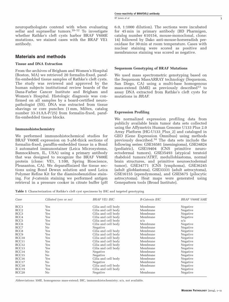
Table 1 Characterization of Rathke’s cleft cyst specimens by IHC and targeted genotyping
Case Ciliated (yes or no) BRAF VE1 IHC B-Catenin IHC BRAF V600E hME
RCC1 Yes Cilia and cell body Membrane NegativeRCC2 Yes Cilia and cell body Membrane NegativeRCC3 Yes Cilia and cell body Membrane NegativeRCC4 Yes Cilia and cell body Membrane NegativeRCC5 Yes Cilia and cell body n/a n/aRCC6 Yes Cilia and cell body Membrane NegativeRCC7 No Negative Membrane NegativeRCC8 Yes Cilia and cell body Membrane NegativeRCC9 Yes Cilia and cell body Membrane NegativeRCC10 Yes Cilia and cell body Membrane NegativeRCC11 Yes Cilia and cell body Membrane NegativeRCC12 Yes Cilia and cell body Membrane NegativeRCC13 Yes Cilia and cell body Membrane NegativeRCC14 No Negative Membrane NegativeRCC15 No Negative Membrane NegativeRCC16 Yes Cilia and cell body Membrane NegativeRCC17 No Negative Membrane NegativeRCC18 Yes Cilia and cell body Membrane NegativeRCC19 Yes Cilia and cell body n/a NegativeRCC20 No Negative Membrane Negative
Abbreviations: hME, homogenous mass-extend; IHC, immunohistochemistry; n/a, not available.
Modern Pathology (2014), 1–11
Cross-reactivity of BRAF(VE1) antibody
RT Jones et al 3

Figure 2 Immunohistochemistry analysis of ciliated epithelium. Images of hematoxylin and eosin (H&E)-stained sections and images ofimmunohistochemistry for BRAF V600E (using the BRAF VE1 antibody) of fallopian tube, bronchioles, and sinonasal respiratoryepithelium. Scale bar, 20mm.
Modern Pathology (2014), 1–11
Cross-reactivity of BRAF(VE1) antibody
4 RT Jones et al

Indirect ELISA for Cilia Peptides
Polysterene enzyme immunoassay/radio immunoas-say microplates (Costar, catalog number 9018) werecoated and kept overnight at 4 1C with 100 ml ofvarious concentrations of peptides (1000, 500, 250,125, 62.5, 31.3, 15.6, 7.8, 3.9, 2.0, and 1.0 ng/mlcorresponding to peptide amounts per well of 100,50, 25, 12.5, 6.3, 3.1, 1.6, 0.8, 0.4, 0.2, and 0.1 ng,respectively); see Figure 6 for details of peptidesequence. Wells were blocked with 0.5% bovineserum albumin in Tris-buffered saline with Tweenfor 1 h at room temperature. A dilution of 1:5000 ofanti-BRAF antibody (Spring Bioscience, catalognumber E19290, clone VE1) was used. The platewas incubated with this primary antibody for 90 minat room temperature and then washed three timeswith Tris-buffered saline with Tween. A 1:5000 dilutionof goat-anti-mouse–horseradish peroxidase (HþL) sec-ondary antibody was then added. Incubation with thesecondary antibody was for 1h at room temperature.
The wells were washed four times with Tris-bufferedsaline with Tween. Development was with 3,30,5,50-Tetramethylbenzidine substrate and the reactionwas stopped with 2 N H2SO4 after 2 min. Plateswere then read at 450 nm in a spectrophotometer.
Results
VE1 Antibody Staining of the Cilia of Rathke’s CleftCysts in the Absence of BRAF V600E Mutations
Similar to the staining pattern seen in papillarycraniopharyngioma, b-catenin was localized to themembrane of the cyst lining cells of Rathke’s cleftcysts (Figure 1 and Table 1) supporting an absence ofactivating mutations in CTNNB1. To investigatewhether Rathke’s cleft cysts harbor BRAF V600Emutations, we stained 20 Rathke’s cleft cysts withthe BRAF VE1 antibody (Table 1). As previouslyreported, we saw strong staining of pituitary cells
Table 2 BRAF VE1 staining of various human tissues with ciliated cells and sperm
Gender Age
Lung specimen BRAF VE1 IHC; bronchial airwaysLG1 F 46 PositiveLG2 M 67 PositiveLG3 M 80 PositiveLG4 F 71 PositiveLG5 M 88 PositiveLG6 M 53 PositiveLG7 F 57 PositiveLG8 F 60 Positive
Fallopian tube specimen BRAF VE1 IHC; ciliaFT1 F 75 Positive, focal ciliatedFT2 F 37 PositiveFT3 F 40 PositiveFT4 F 51 PositiveFT5 F 65 PositiveFT6 F 62 Positive
Nasal epithelium specimen BRAF VE1 IHC; ciliaNE1 F 59 PositiveNE2 M 60 Positive
Testis specimen BRAF VE1 IHC; spermTS1 M 23 PositiveTS2 M 23 PositiveTS3 M 50 Positive
Gender Age
Ependymoma specimen BRAF VE1 IHC; dots EMA, dotsEP1 M 53 Rare PositiveEP2 F 51 Rare PositiveEP3 M 48 Negative RareEP4 F 49 Positive PositiveEP5 F 43 Positive PositiveEP6 M 47 Negative PositiveEP7 F 47 Positive PositiveEP8 F 43 Rare FocalEP9 M 58 Focal FocalEP10 F 55 Positive PositiveEP11 F 52 Positive PositiveEP12 F 63 Focal Focal
Abbreviations: EMA, epithelial membrane antigen; F, female; IHC, immunohistochemistry; M, male.
Modern Pathology (2014), 1–11
Cross-reactivity of BRAF(VE1) antibody
RT Jones et al 5
that were incidentally resected along with thecysts.15 Although cells that do not carry BRAFV600E mutations should show no reactivity withthis antibody, we found strong staining in theepithelial cells of Rathke’s cleft cysts (Figure 1).Although the tumor cells of papillary craniophar-yngioma have a diffuse nuclear and cytoplasmicstaining pattern (Figure 1), we saw intense stainingof the cilia of the cyst lining cells and a more modestlevel of staining of the cell bodies of these epithelialcells (Figure 1). In regions of these cysts that had
undergone squamous metaplasia, we found strongstaining of the cilia of residual ciliated cells and ofthe cytoplasm of these ciliated cells, as well as of thecytoplasm of neighboring squamous cells that werenot overly ciliated (Supplementary Figure S1). Infive of the 20 Rathke’s cleft cyst cases, the cellslacked cilia entirely and no staining of the epithe-lium was seen whether there was squamous meta-plasia present or attenuated/cuboidal cells (Table 1).
We performed Sequenom-SNP genotyping basedtargeted sequencing (hME) using DNA extracted
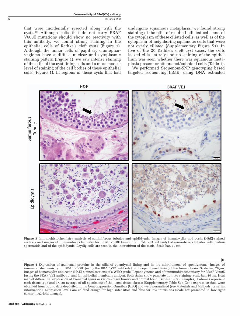
Figure 3 Immunohistochemistry analysis of seminiferous tubules and epididymis. Images of hematoxylin and eosin (H&E)-stainedsections and images of immunohistochemistry for BRAF V600E (using the BRAF VE1 antibody) of seminiferous tubules with maturespermatids and of the epididymis. Leydig cells are seen in the interstitium of the testis. Scale bar, 10mm.
Figure 4 Expression of axonemal proteins in the cilia of ependymal lining and in the microlumens of ependymoma. Images ofimmunohistochemistry for BRAF V600E (using the BRAF VE1 antibody) of the ependymal lining of the human brain. Scale bar, 20mm.Images of hematoxylin and eosin (H&E)-stained sections of a WHO grade II ependymoma and of immunohistochemistry for BRAF V600E(using the BRAF VE1 antibody) and for epithelial membrane antigen. Both stains show punctate dot-like staining. Scale bar, 10mm. Heatmap of differential expression of axonemal genes in various brain tumors and normal brain tissues (n¼350 samples). Columns representeach tissue type and are an average of all specimens of the listed tissue classes (Supplementary Table S1). Gene expression data wereobtained from public data deposited in the Gene Expression Omnibus (GEO) and were normalized (see Materials and Methods for seriesinformation). Expression levels are colored orange for high intensities and blue for low intensities (scale bar presented in low rightcorner; log2-fold change).
Modern Pathology (2014), 1–11
Cross-reactivity of BRAF(VE1) antibody
6 RT Jones et al

from 19 of the 20 Rathke’s cleft cysts (Table 1), toinvestigate whether these cysts harbored geneticmutations that would encode the BRAF V600E
protein.9,13 This method has the ability todetect mutations at an allelic fraction of o5%.Despite the strong staining with the BRAF VE1
Modern Pathology (2014), 1–11
Cross-reactivity of BRAF(VE1) antibody
RT Jones et al 7

Table
3B
LA
ST
pro
tein
–p
rote
inh
om
olo
gy
searc
hof
the
hu
man
pro
teom
eu
sin
gth
ese
qu
en
ceof
the
pep
tid
eim
mu
nogen
(FG
LA
TE
KS
RW
SG
)th
at
had
been
use
dto
gen
erate
the
BR
AF
VE
1an
tibod
y
Desc
rip
tion
Max
score
Tota
lsc
ore
Qu
ery
cover
E-v
alu
eId
en
tity
Access
ion
RA
Fp
roto
-on
cogen
ese
rin
e/t
hre
on
ine-p
rote
inkin
ase
35
35
100%
0.0
01
92%
NP
_002871.1
Seri
ne/t
hre
on
ine-p
rote
inkin
ase
A-R
af
32.5
32.5
100%
0.0
08
83%
NP
_001243125.1
a-K
eto
glu
tara
te-d
ep
en
den
td
ioxygen
ase
alk
Bh
om
olo
g7
26.5
26.5
58%
0.5
9100%
NP
_115682.1
Dyn
ein
hea
vy
chain
2,
axon
emal
(DN
AH
224
24
75%
478%
NP
_065928.2
Zin
cfi
nger
pro
tein
518A
23.1
23.1
58%
7.4
86%
NP
_001265455.1
Dyn
ein
hea
vy
chain
12,
axon
emal
(DN
AH
12)
23.1
35.2
75%
7.4
78%
NP
_001278590.1
Dyn
ein
hea
vy
chain
7,
axon
emal
(DN
AH
7)
23.1
23.1
83%
7.4
70%
NP
_061720.2
BT
B/P
OZ
dom
ain
-con
tain
ing
pro
tein
KC
TD
422.3
22.3
75%
14
80%
NP
_940686.2
An
ion
exch
an
ge
pro
tein
322.3
22.3
75%
14
80%
NP
_005061.2
Dyn
ein
hea
vy
chain
5,
axon
emal
(DN
AH
5)
22.3
49.8
75%
14
78%
NP
_001360.1
Olf
acto
ryre
cep
tor
9Q
221.4
21.4
50%
26
83%
NP
_001005283.1
Arm
ad
illo
rep
eat-
con
tain
ing
pro
tein
421.4
21.4
58%
26
86%
NP
_060546.2
Pla
tele
t-acti
vati
ng
facto
racety
lhyd
rola
seIB
subu
nit
-b21
21
41%
36
100%
NP
_001171675.1
RA
B6-i
nte
racti
ng
golg
in21
21
41%
36
100%
NP
_001139511.1
hig
h-a
ffin
ity
cG
MP
-sp
ecif
ic3’,
5’-
cycli
cp
hosp
hod
iest
era
se9A
21
21
50%
37
100%
NP
_001001576.1
Majo
rfa
cil
itato
rsu
perf
am
ily
dom
ain
-con
tain
ing
pro
tein
8(H
om
osa
pie
ns)
21
21
66%
37
64%
NP
_689991.1
MY
EL
OP
ER
OX
IDA
SE
pre
cu
rsor
(H.
sap
ien
s)21
21
83%
37
64%
NP
_000241.1
Colo
recta
lm
uta
nt
can
cer
pro
tein
isofo
rm2
(H.
sap
ien
s)21
21
66%
37
75%
NP
_002378.1
PR
ED
ICT
ED
:colo
recta
lm
uta
nt
can
cer
pro
tein
isofo
rmX
1(H
.sa
pie
ns)
21
21
66%
37
75%
XP
_005272048.1
PR
ED
ICT
ED
:colo
recta
lm
uta
nt
can
cer
pro
tein
isofo
rmX
2(H
.sa
pie
ns)
21
21
66%
37
75%
XP
_006714680.1
Colo
recta
lm
uta
nt
can
cer
pro
tein
isofo
rm1
(H.
sap
ien
s)21
21
66%
37
75%
NP
_001078846.1
F-b
ox
on
lyp
rote
in46
(H.
sap
ien
s)20.6
35.6
91%
51
67%
NP
_001073938.1
NA
CH
T,
LR
R,
an
dP
YD
dom
ain
scon
tain
ing
pro
tein
7is
ofo
rm2
(H.
sap
ien
s)20.6
20.6
58%
51
86%
NP
_996611.2
PR
ED
ICT
ED
:N
AC
HT
,L
RR
,an
dP
YD
dom
ain
scon
tain
ing
pro
tein
7is
ofo
rmX
4(H
.sa
pie
ns)
20.6
20.6
58%
51
86%
XP
_006723140.1
NA
CH
T,
LR
R,
an
dP
YD
dom
ain
scon
tain
ing
pro
tein
7is
ofo
rm1
(H.
sap
ien
s)20.6
20.6
58%
51
86%
NP
_631915.2
PR
ED
ICT
ED
:cod
an
in-1
isofo
rmX
3(H
.sa
pie
ns)
20.6
20.6
50%
51
100%
XP
_005254235.1
PR
ED
ICT
ED
:N
AC
HT
,L
RR
,an
dP
YD
dom
ain
s-con
tain
ing
pro
tein
7is
ofo
rmX
1(H
.sa
pie
ns)
20.6
20.6
58%
51
86%
XP
_006726370.1
NA
CH
T,
LR
R,
an
dP
YD
dom
ain
scon
tain
ing
pro
tein
7is
ofo
rm3
(H.
sap
ien
s)20.6
20.6
58%
51
86%
NP
_001120727.1
NA
CH
T,
LR
R,
an
dP
YD
dom
ain
scon
tain
ing
pro
tein
2is
ofo
rm3
(H.
sap
ien
s)20.6
20.6
58%
51
86%
NP
_001167554.1
NA
CH
T,
LR
R,
an
dP
YD
dom
ain
scon
tain
ing
pro
tein
2is
ofo
rm2
(H.
sap
ien
s)20.6
20.6
58%
51
86%
NP
_001167553.1
PR
ED
ICT
ED
:in
tegri
nalp
ha-1
0is
ofo
rmX
11
(H.
sap
ien
s)20.6
20.6
58%
51
86%
XP
_006711659.1
NA
CH
T,
LR
R,
an
dP
YD
dom
ain
scon
tain
ing
pro
tein
2is
ofo
rm1
(H.
sap
ien
s)20.6
20.6
58%
51
86%
NP
_060322.1
PR
ED
ICT
ED
:N
AC
HT
,L
RR
,an
dP
YD
dom
ain
s-con
tain
ing
pro
tein
7is
ofo
rmX
120.6
20.6
58%
51
86%
XP
_006723137.1
PR
ED
ICT
ED
:cod
an
in-1
isofo
rmX
2(H
.sa
pie
ns)
20.6
20.6
50%
51
100%
XP
_005254234.1
PR
ED
ICT
ED
:in
tegri
na-
10
isofo
rmX
5(H
.sa
pie
ns)
20.6
20.6
58%
51
86%
XP
_005277493.1
PR
ED
ICT
ED
:in
tegri
na-
10
isofo
rmX
4(H
.sa
pie
ns)
20.6
20.6
58%
51
86%
XP
_005277492.1
PR
ED
ICT
ED
:in
tegri
nalp
ha-1
0is
ofo
rmX
3(H
.sa
pie
ns)
20.6
20.6
58%
51
86%
XP
_005277491.1
PR
ED
ICT
ED
:in
tegri
na-
10
isofo
rmX
2(H
.sa
pie
ns)
20.6
20.6
58%
51
86%
XP
_005277490.1
Inte
gri
na-
10
pre
cu
rsor
(H.
sap
ien
s)20.6
20.6
58%
51
86%
NP
_003628.2
PR
ED
ICT
ED
:in
tegri
na-
10
isofo
rmX
1(H
.sa
pie
ns)
20.6
20.6
58%
51
86%
XP
_005277489.1
Cod
an
in-1
(H.
sap
ien
s)20.6
20.6
50%
51
100%
NP
_612486.2
PR
ED
ICT
ED
:cod
an
in-1
isofo
rmX
1(H
.sa
pie
ns)
20.6
20.6
50%
51
100%
XP
_005254233.1
HE
AT
rep
eat-
con
tain
ing
pro
tein
5A
(H.
sap
ien
s)20.6
49
66%
51
86%
NP
_056288.2
PR
ED
ICT
ED
:p
eri
cen
trin
isofo
rmX
6(H
.sa
pie
ns)
20.6
61.1
75%
51
86%
XP
_005261186.1
PR
ED
ICT
ED
:p
eri
cen
trin
isofo
rmX
5(H
.sa
pie
ns)
20.6
61.1
75%
51
86%
XP
_005261185.1
PR
ED
ICT
ED
:p
eri
cen
trin
isofo
rmX
4(H
.sa
pie
ns)
20.6
61.1
75%
51
86%
XP
_005261184.1
PR
ED
ICT
ED
:p
eri
cen
trin
isofo
rmX
3(H
.sa
pie
ns)
20.6
61.1
75%
51
86%
XP
_005261183.1
Peri
cen
trin
(H.
sap
ien
s)20.6
61.1
75%
51
86%
NP
_006022.3
PR
ED
ICT
ED
:p
eri
cen
trin
isofo
rmX
2(H
.sa
pie
ns)
20.6
61.1
75%
51
86%
XP
_005261182.1
PR
ED
ICT
ED
:p
eri
cen
trin
isofo
rmX
1(H
.sa
pie
ns)
20.6
61.1
75%
51
86%
XP
_005261181.1
PR
ED
ICT
ED
:d
yn
ein
heavy
ch
ain
9,
axon
em
al
isofo
rmX
1(H
.sa
pie
ns)
20.6
39
75%
51
67%
XP
_006721528.1
dyn
ein
heavy
ch
ain
9,
axon
em
al
isofo
rm2
(H.
sap
ien
s)20.6
52.4
75%
51
67%
NP
_001363.2
PR
ED
ICT
ED
:p
roli
ne-r
ich
mem
bra
ne
an
ch
or
1is
ofo
rmX
1(H
.sa
pie
ns)
20.2
20.2
66%
71
75%
XP
_006720107.1
Pro
lin
e-r
ich
mem
bra
ne
an
ch
or
1p
recu
rsor
(H.
sap
ien
s)20.2
20.2
66%
71
75%
NP
_821092.1
PR
ED
ICT
ED
:W
Dre
peat-
con
tain
ing
pro
tein
87-l
ike
isofo
rmX
4(H
.sa
pie
ns)
20.2
106
83%
72
86%
XP
_005274478.1
PR
ED
ICT
ED
:W
Dre
peat-
con
tain
ing
pro
tein
87-l
ike
isofo
rmX
3(H
.sa
pie
ns)
20.2
131
83%
72
86%
XP
_005274477.1
Un
ch
ara
cte
rized
pro
tein
loc100132994
(H.
sap
ien
s)20.2
20.2
41%
72
100%
NP
_001138611.1
PR
ED
ICT
ED
:W
Dre
peat-
con
tain
ing
pro
tein
87-l
ike
(H.
sap
ien
s)20.2
143
83%
72
86%
XP
_006726595.1
PR
ED
ICT
ED
:W
Dre
peat-
con
tain
ing
pro
tein
87-l
ike
isofo
rmX
1(H
.sa
pie
ns)
20.2
143
83%
72
86%
XP
_005274475.1
pro
tein
FA
M124A
isofo
rm1
(H.
sap
ien
s)20.2
20.2
50%
72
83%
NP
_659456.3
Jun
cto
ph
ilin
-4(H
om
osa
pie
ns)
20.2
20.2
75%
72
70%
NP
_115828.2
Abbre
via
tion
:K
CT
D4,
pota
ssiu
mch
an
nel
tetr
am
eri
zati
on
dom
ain
con
tain
ing
4.
Modern Pathology (2014), 1–11
Cross-reactivity of BRAF(VE1) antibody
8 RT Jones et al

antibody, we did not detect the genetic mutationencoding BRAF V600E in any of the Rathke’s cleftcysts (Table 1).
VE1 Antibody Staining of the Cilia of Various Tissues
We next used the VE1 antibody to stain a series ofhuman tissues that have ciliated cells (Figure 2). Wenoted strong staining of the cilia from fallopian tube(n¼ 6), lung bronchial airways (n¼ 8), and naso-pharyngeal airways (n¼ 2) (Table 2). The cell bodiesof the ciliated cells had weaker staining. We alsonoted very strong staining of the cilia of a mucoceleof the paranasal sinuses (Supplementary Figure S2).As cilia and flagella have a similar protein composi-tion and organization, we investigated whether theflagella of mature spermatozoa would also bestained by the VE1 antibody (testis and epididymisn¼ 3; Table 2). Indeed, we observed that the VE1antibody strongly stained the flagella of maturesperm in the seminiferous tubules and in theepididymis (Figure 3), as well as the cilia of theepididymis (Figure 3). In addition, we noted strongnuclear staining of Leydig cells in the interstitium ofthe testis, suggesting the presence of other cross-reactive epitopes in those cells.
Ependymomas Express Genes that Encode AxonemalProteins and the Microlumen of Ependymomas areStained by the VE1 Antibody
Next, we stained autopsy and neurosurgical resec-tion specimens of adult and pediatric brain thatcontained ependymal lining (n¼ 3) (Figure 4). Onceagain, we observed strong staining of the cilia. Weanalyzed publicly available expression profilingdata from a panel of human brain tumors(Supplementary Table S1). Of the 19 ependymomain this panel, about half (10 of 19 cases) demon-strated moderate expression of genes encodingaxonemal proteins, thereby supporting that thesetumor cells continue to express the proteins thatcomprise cilia. Background levels were seen for theother ependymoma samples. In a heat map, we showthat genes that encode axonemal proteins are well-expressed in ependymoma compared with a range ofother brain tumors, including astrocytomas, atypicalteratoid rhabdoid tumors, and medulloblastomaamong others (Figure 4). Nearly half of 12 randomlyselected ependymoma formalin-fixed, paraffin-embedded samples (5 of 12; Table 2) from ourtissue archives showed punctate staining corre-sponding to the cilia containing microlumens ofependymoma (Figure 4). During clinical diagnosis,punctate staining with epithelial membrane antigenis used to support the diagnosis of ependymoma. Inall cases, the punctate staining for epithelialmembrane antigen was, however, much strongerthan the punctate dots observed with VE1 staining(Figure 4).
The VE1 Antibody Recognizes Epitopes in AxonemalDyneins
We next used the BLAST (Basic Local AlignmentSearch Tool)16 to perform a standard protein–proteinhomology search of the human proteome using thesequence of the peptide immunogen (FGLA-TEKSRWSG) that had been used to generate theBRAF VE1 antibody (results in Table 3). This peptidedemonstrated strong similarity to regions of multipleaxonemal dynein heavy chain proteins, includingDNAH2, DNAH5, DNAH7, and DNAH12 (Figure 5and Table 3). In an ELISA assay, we tested the abilityof the VE1 antibody to recognize peptides containingthe region of interest of wild-type (BRAF_wt) andV600E mutant BRAF (BRAF_mut), as well as homo-logous regions of several axonemal dyneins (Figure 6).The VE1 antibody robustly recognized the peptidederived from the V600E mutant of BRAF andrecognition was minimal for the wild-type BRAFsequence (Figure 6). As predicted by the sequencehomology and the strong staining of cilia in tissuesections, peptides with wild-type sequences from thethree axonemal dynein heavy chain proteins that wetested (DNAH2, DNAH7, and DNAH12) were allrecognized by the VE1 antibody. The relative affinityfor the peptide derived from DNAH12 (DNAH12_wt)was very similar to that of peptide from the V600Emutant of BRAF (BRAF_mut). Recognition of theDNAH7-derived peptide (DNAH7_wt) was robust butless than that for the DNAH12-derived peptide.Binding of the VE1 antibody to the DNAH2-derivedpeptide was detected, but was the weakest of thethree axonemal derived peptides tested. When theglutamates that align with the E600 residue in mutantBRAF were changed to valines in the axonemalderived peptides (DNAH2_mut, DNAH7_mut, andDNAH12_mut), recognition by the VE1 antibody wasreduced to background levels, similar to the level ofrecognition of the wild-type BRAF peptide (Figure 6).
Discussion
Mutation-specific antibodies are valuable tools inclinical practice. An understanding of the limita-tions of these tools, particularly their cross-reactiv-ity with unintended proteins, is however ofsignificant importance so that the appropriate inter-pretation of staining results can be rendered. In thiswork we show that the VE1 antibody that wasgenerated to recognize the BRAF V600E mutantprotein also recognizes epitopes in multiple axone-mal dynein heavy chain proteins and that therecognition is highly dependent on a single gluta-mate residue in these axonemal proteins. In mosttissues, these proteins are predominantly restrictedto the cilia lining the luminal surface of epithelialcells so the cross-reactivity should likely notconfound diagnosis. However, moderate staining ofthe cytoplasm of ciliated cells is also seen in many
Modern Pathology (2014), 1–11
Cross-reactivity of BRAF(VE1) antibody
RT Jones et al 9

cases and could therefore be a diagnostic pitfallparticularly as the antibody gains increasingly wideracceptance and increased use. In addition, smallbiopsies, particularly those that have crush artifactor poorly oriented cells can also pose diagnosticdifficulties. Strong staining of entrapped normalpituitary, as previously described15 and seen in mostof the Rathke’s cleft cyst specimens in this study,further adds to the complexity of analysis.Moreover, in tumors like ependymoma where ciliaare found in microlumens and staining is thereforepresent in the cytoplasm, it is useful to recognizethat this dot-like pattern of staining does notindicate the presence of an oncogenic mutation inBRAF and is indeed providing similar diagnosticinformation as is garnered with epithelial membraneantigen staining. The BRAF V600E antibody has animportant role in the evaluation of brain tumorssuch as pleomorphic xanthoastrocytoma andganglioglioma, which frequently harbor mutationsin BRAF;1,13,17,18 thus, understanding the sources ofcross-reactivity of this antibody is of importantclinical value.
A recent study has demonstrated that the VE1antibody also stains a number of endocrine tissues,including the pituitary and the adrenal gland.19
There is preferential staining of discrete cell typeswith the strongest staining observed in adrenocorti-cotropic hormone (ACTH)-producing cells of theadenohypophysis and in cells comprising the innersegment of the zona fasciculata and the zonareticularis of the adrenal cortex. Accordingly,ACTH-expressing pituitary adenomas are stainedwith the VE1 antibody, yet these tumors do not
demonstrate detectable BRAF mutations. UsingBLAST we see sequence similarities with proteinsbesides axonemal dynein heavy chains, some ofwhich might be responsible for the cross-reactivitywith endocrine tissues—such as a-ketoglutarate-dependent dioxygenase alkB homolog 7, eukaryotictranslation initiation factor 2-a kinase 4, polo-likekinase-1d, potassium channel tetramerization do-main containing 4, solute carrier family 4, anionexchanger, member 3 and armadillo repeat-contain-ing protein 4.
In other tumors that harbor genetic changes ofhigh clinical importance such as the EGFR L858Rmutations in lung cancer20 and NRAS Q61Rmutations in melanoma, mutation-specific anti-bodies can facilitate screening clinical cases.21,22
Antibodies that recognize other clinically relevantmutations such as the AKT1 E17K mutation that isfound in breast cancer23 and in meningioma24 couldalso be of great use. During the use and validation
Figure 5 BLAST (Basic Local Alignment Search Tool) homologysearch of the human proteome using the sequence of the BRAFVE1 peptide immunogen (FGLATEKSRWSG). The regions ofstrongest identity between axonemal dynein heavy chain proteinsand the BRAF V600E mutant epitope are shown.
Figure 6 Enzyme-linked immunosorbent assay (ELISA) using theBRAF VE1 antibody and peptides from BRAF and axonemaldyneins. The peptides used in this experiment are displayed.ELISA details are explained in the Materials and methods section.
Modern Pathology (2014), 1–11
Cross-reactivity of BRAF(VE1) antibody
10 RT Jones et al

of such antibodies, attention for potential cross-reactivity by sequence homology searches couldhelp identify potential cross-reactive proteins andcorresponding tissues in advance.
Acknowledgments
We thank Terri Woo for assistance with immuno-histochemistry, and Marian Slaney and SebastianValentin for assistance with histology. We thankPrathapan Thiru (Bioinformatics and ResearchComputing, Whitehead Institute for BiomedicalResearch) and Parker Merrill for assistance withbioinformatics analyses. This work was supportedby the Jared Branfman Sunflowers for Life Fund forPediatric Brain and Spinal Cancer Research, the VFoundation (SS) and by a grant from King AbdulazizCity for Science and Technology (KACST), SaudiArabia (MAA).
Disclosure/conflict of interest
Sandro Santagata is a consultant and co-founder ofBayesian Diagnostics. All other authors declare noconflict of interests.
References
1 Capper D, Berghoff AS, Magerle M, et al. Immuno-histochemical testing of BRAF V600E status in 1,120tumor tissue samples of patients with brain metastases.Acta Neuropathol 2012;123:223–233.
2 Capper D, Preusser M, Habel A, et al. Assessment ofBRAF V600E mutation status by immunohisto-chemistry with a mutation-specific monoclonal anti-body. Acta Neuropathol 2011;122:11–19.
3 Kato Y, Jin G, Kuan CT, et al. A monoclonal antibodyIMab-1 specifically recognizes IDH1R132H, the mostcommon glioma-derived mutation. Biochem BiophysRes Commun 2009;390:547–551.
4 Capper D, Zentgraf H, Balss J, et al. Monoclonalantibody specific for IDH1 R132H mutation. ActaNeuropathol 2009;118:599–601.
5 Preusser M, Capper D, Ilhan-Mutlu A, et al. Brainmetastases: pathobiology and emerging targeted thera-pies. Acta Neuropathol 2012;123:205–222.
6 Preusser M, Capper D, Hartmann C, et al. testing indiagnostic neuropathology: review and practicalguideline article invited by the Euro-CNS researchcommittee. Clin Neuropathol 2011;30:217–230.
7 Chan JK, Ip YT, Cheuk W. The utility of immuno-histochemistry for providing genetic information ontumors. Int J Surg Pathol 2013;21:455–475.
8 Santagata S, Eberlin LS, Norton I, et al. Intraoperativemass spectrometry mapping of an onco-metabolite toguide brain tumor surgery. Proc Natl Acad Sci USA2014;111:11121–11126.
9 Brastianos PK, Taylor-Weiner A, Manley PE, et al.Exome sequencing identifies BRAF mutations in papil-lary craniopharyngiomas. Nat Genet 2014;46:161–165.
10 Voelker JL, Campbell RL, Muller J. Clinical, radio-graphic, and pathological features of symptomaticRathke’s cleft cysts. J Neurosurg 1991;74:535–544.
11 Shin JL, Asa SL, Woodhouse LJ, et al. Cystic lesions ofthe pituitary: clinicopathological features distinguish-ing craniopharyngioma, Rathke’s cleft cyst, and ara-chnoid cyst. J Clin Endocrinol Metab 1999;84:3972–3982.
12 Buslei R, Nolde M, Hofmann B, et al. Commonmutations of beta-catenin in adamantinomatous cra-niopharyngiomas but not in other tumours originatingfrom the sellar region. Acta Neuropathol 2005;109:589–597.
13 MacConaill LE, Campbell CD, Kehoe SM, et al. Profil-ing critical cancer gene mutations in clinical tumorsamples. PLoS One 2009;4:e7887.
14 Thiru P, Kern DM, McKinley KL, et al. Kinetochoregenes are coordinately up-regulated in human tumorsas part of a FoxM1-related cell division program. MolBiol Cell 2014;25:1983–1994.
15 Mordes DA, Lynch K, Campbell S, et al. VE1 antibodyimmunoreactivity in normal anterior pituitary andadrenal cortex without detectable BRAF V600E muta-tions. Am J Clin Pathol 2014;141:811–815.
16 Altschul SF, Gish W, Miller W, et al. Basic localalignment search tool. J Mol Biol 1990;215:403–410.
17 Dias-Santagata D, Lam Q, Vernovsky K, et al. BRAFV600E mutations are common in pleomorphicxanthoastrocytoma: diagnostic and therapeutic impli-cations. PLoS One 2011;6:e17948.
18 Lee EQ, Ruland S, LeBoeuf NR, et al. Successfultreatment of a progressive BRAF V600E-mutatedanaplastic pleomorphic xanthoastrocytoma withvemurafenib monotherapy. J Clin Oncol advanceonline publication, 4 August 2014; doi: 10.1200/jco.2013.51.1766 (e-pub ahead of print).
19 Arisawa M, Makino T, McCann SM, et al. Effect ofestrogen on the response of thyroid stimulatinghormone to substance P in rats. Endocrinol Jpn1989;36:899–903.
20 Mitsudomi T, Kosaka T, Endoh H, et al. Mutations ofthe epidermal growth factor receptor gene predictprolonged survival after gefitinib treatment in patientswith non-small-cell lung cancer with postoperativerecurrence. J Clin Oncol 2005;23:2513–2520.
21 Yu J, Kane S, Wu J, et al. Mutation-specific antibodiesfor the detection of EGFR mutations in non-small-celllung cancer. Clin Cancer Res 2009;15:3023–3028.
22 Massi D, Simi L, Sensi E, et al. Immunohistochemistryis highly sensitive and specific for the detectionof NRASQ61R mutation in melanoma. Mod Patholadvance online publication, 24 October 2014; doi:10.1038/modpathol.2014.137 (e-pub ahead of print).
23 Carpten JD, Faber AL, Horn C, et al. A transformingmutation in the pleckstrin homology domain of AKT1in cancer. Nature 2007;448:439–444.
24 Brastianos PK, Horowitz PM, Santagata S, et al.Genomic sequencing of meningiomas identifies onco-genic SMO and AKT1 mutations. Nat Genet 2013;45:285–289.
Supplementary Information accompanies the paper on Modern Pathology website (http://www.nature.com/modpathol)
Modern Pathology (2014), 1–11
Cross-reactivity of BRAF(VE1) antibody
RT Jones et al 11
Copyright © 2022 FDOKUMEN