PIK3CA -related overgrowth spectrum (PROS): Diagnostic and testing eligibility criteria,...
9
CONFERENCE REPORT PIK3CA-Related Overgrowth Spectrum (PROS): Diagnostic and Testing Eligibility Criteria, Differential Diagnosis, and Evaluation Kim M. Keppler-Noreuil, 1 Jonathan J. Rios, 2,3 Victoria E.R. Parker, 4 Robert K. Semple, 4 Marjorie J. Lindhurst, 1 Julie C. Sapp, 1 Ahmad Alomari, 5 Marybeth Ezaki, 3 William Dobyns, 6 and Leslie G. Biesecker 1 1 National Human Genome Research Institute, National Institutes of Health, Bethesda, Maryland 2 Sarah M. and Charles E. Seay Center for Musculoskeletal Research, Texas Scottish Rite Hospital for Children, Department of Pediatrics, Eugene McDermott Center for Human Growth and Development, UT Southwestern Medical Center, Dallas, Texas 3 Department of Orthopaedic Surgery, UT Southwestern Medical Center, Dallas, Texas 4 Institute of Metabolic Science, University of Cambridge Metabolic Research Laboratories, Cambridge, Massachusetts 5 Division of Vascular and Interventional Radiology, Boston Children’s Hospital and Harvard Medical School, Boston, Massachusetts 6 University of Washington, Seattle, Washington Manuscript Received: 11 August 2014; Manuscript Accepted: 30 September 2014 Somatic activating mutations in the phosphatidylinositol-3-ki- nase/AKT/mTOR pathway underlie heterogeneous segmental overgrowth phenotypes. Because of the extreme differences among patients, we sought to characterize the phenotypic spectrum associated with different genotypes and mutation burdens, in- cluding a better understanding of associated complications and natural history. Historically, the clinical diagnoses in patients with PIK3CA activating mutations have included Fibroadipose hyper- plasia or Overgrowth (FAO), Hemihyperplasia Multiple Lipoma- tosis (HHML), Congenital Lipomatous Overgrowth, Vascular Malformations, Epidermal Nevi, Scoliosis/Skeletal and Spinal (CLOVES) syndrome, macrodactyly, Fibroadipose Infiltrating Lipomatosis, and the related megalencephaly syndromes, Mega- lencephaly-Capillary Malformation (MCAP or M-CM) and Dys- plastic Megalencephaly (DMEG). A workshop was convened at the National Institutes of Health (NIH) to discuss and develop a consensus document regarding diagnosis and treatment of patients with PIK3CA-associated somatic overgrowth disorders. Participants in the workshop included a group of researchers from several institutions who have been studying these disorders and have published their findings, as well as representatives from patient-advocacy and support groups. The umbrella term of “PIK3CA-Related Overgrowth Spectrum (PROS)” was agreed upon to encompass both the known and emerging clinical entities associated with somatic PIK3CA mutations including, macro- dactyly, FAO, HHML, CLOVES, and related megalencephaly conditions. Key clinical diagnostic features and criteria for testing were proposed, and testing approaches summarized. Preliminary recommendations for a uniform approach to assessment of over- growth and molecular diagnostic testing were determined. Future areas to address include the surgical management of overgrowth tissue and vascular anomalies, the optimal approach to thrombosis risk, and the testing of potential pharmacologic therapies. Ó 2014 The Authors. American Journal of Medical Genetics Part A published by Wiley Periodicals, Inc. Key words: somatic mosaicism; PIK3CA gene; fibroadipose overgrowth; segmental overgrowth; macrodactyly; CLOVES syndrome; PIK3CA-Related Overgrowth Spectrum (PROS) How to Cite this Article: Keppler-Noreuil KM, Rios JJ, Parker VER, Semple RK, Lindhurst MJ, Sapp JC, Alomari A, Ezaki M, Dobyns W, Biesecker LG. 2015. PIK3CA-related overgrowth spectrum (PROS): Diagnostic and testing eligibility criteria, differential diagnosis, and evaluation. Am J Med Genet Part A 167A:287–295. The copyright line for this article was changed on 20 May 2016 after original online publication. This is an open access article under the terms of the Creative Commons Attribution License, which permits use, distribution and reproduction in any medium, provided the original work is properly cited. Conflict of interest: None Grant sponsors: National Human Genome Research Institute; UK: Well- come Trust, Clinical Research Training Fellowship, UK national Institute for Health Research (NIHR) Cambridge Biomedical Research Centre. Correspondence to: Kim M. Keppler-Noreuil, MD, National Human Genome Research Institute/NIH, 49 Convent, 4A83, Bethesda, MD 20892. E-mail: [email protected] Article first published online in Wiley Online Library (wileyonlinelibrary.com): 31 December 2014 DOI 10.1002/ajmg.a.36836 Ó 2014 The Authors. American Journal of Medical Genetics Part A published by Wiley Periodicals, Inc. 287
-
Upload
ittehuacan -
Category
Documents
-
view
3 -
download
0
Transcript of PIK3CA -related overgrowth spectrum (PROS): Diagnostic and testing eligibility criteria,...

�
CONFERENCE REPORT
PIK3CA-Related Overgrowth Spectrum (PROS):Diagnostic and Testing Eligibility Criteria,Differential Diagnosis, and Evaluation
Kim M. Keppler-Noreuil,1 Jonathan J. Rios,2,3 Victoria E.R. Parker,4 Robert K. Semple,4Marjorie J. Lindhurst,1 Julie C. Sapp,1 Ahmad Alomari,5 Marybeth Ezaki,3 William Dobyns,6
and Leslie G. Biesecker11National Human Genome Research Institute, National Institutes of Health, Bethesda, Maryland2Sarah M. and Charles E. Seay Center for Musculoskeletal Research, Texas Scottish Rite Hospital for Children, Department of Pediatrics,
Eugene McDermott Center for Human Growth and Development, UT Southwestern Medical Center, Dallas, Texas3Department of Orthopaedic Surgery, UT Southwestern Medical Center, Dallas, Texas4Institute of Metabolic Science, University of Cambridge Metabolic Research Laboratories, Cambridge, Massachusetts5Division of Vascular and Interventional Radiology, Boston Children’s Hospital and Harvard Medical School, Boston, Massachusetts6University of Washington, Seattle, Washington
Manuscript Received: 11 August 2014; Manuscript Accepted: 30 September 2
014How to Cite this Article:Keppler-Noreuil KM, Rios JJ, Parker VER,
Semple RK, Lindhurst MJ, Sapp JC, Alomari
A, Ezaki M, Dobyns W, Biesecker LG. 2015.
PIK3CA-related overgrowth spectrum
(PROS): Diagnostic and testing eligibility
criteria, differential diagnosis, and evaluation.
Am J Med Genet Part A 167A:287–295.
The copyright line for this article was changed on 20 May 2016 after
original online publication.
This is an open access article under the terms of the Creative Commons
Attribution License, which permits use, distribution and reproduction in
any medium, provided the original work is properly cited.
Conflict of interest: None
Grant sponsors: National Human Genome Research Institute; UK: Well-
come Trust, Clinical Research Training Fellowship, UK national Institute for
Health Research (NIHR) Cambridge Biomedical Research Centre.�Correspondence to:
Kim M. Keppler-Noreuil, MD, National Human Genome Research
Institute/NIH, 49 Convent, 4A83, Bethesda, MD 20892.
E-mail: [email protected]
Article first published online in Wiley Online Library
(wileyonlinelibrary.com): 31 December 2014
DOI 10.1002/ajmg.a.36836
Somatic activating mutations in the phosphatidylinositol-3-ki-
nase/AKT/mTOR pathway underlie heterogeneous segmental
overgrowthphenotypes.Becauseoftheextremedifferencesamong
patients, we sought to characterize the phenotypic spectrum
associated with different genotypes and mutation burdens, in-
cluding a better understanding of associated complications and
naturalhistory.Historically, the clinical diagnoses inpatientswith
PIK3CA activating mutations have included Fibroadipose hyper-
plasia or Overgrowth (FAO), Hemihyperplasia Multiple Lipoma-
tosis (HHML), Congenital Lipomatous Overgrowth, Vascular
Malformations, Epidermal Nevi, Scoliosis/Skeletal and Spinal
(CLOVES) syndrome, macrodactyly, Fibroadipose Infiltrating
Lipomatosis, and the related megalencephaly syndromes, Mega-
lencephaly-Capillary Malformation (MCAP or M-CM) and Dys-
plasticMegalencephaly (DMEG).Aworkshopwas convened at the
National Institutes of Health (NIH) to discuss and develop a
consensus document regarding diagnosis and treatment of
patients with PIK3CA-associated somatic overgrowth disorders.
Participants in theworkshop includeda groupof researchers from
several institutions who have been studying these disorders and
have published their findings, as well as representatives from
patient-advocacy and support groups. The umbrella term of
“PIK3CA-Related Overgrowth Spectrum (PROS)” was agreed
upon to encompass both the known and emerging clinical entities
associated with somatic PIK3CA mutations including, macro-
dactyly, FAO, HHML, CLOVES, and related megalencephaly
conditions. Key clinical diagnostic features and criteria for testing
were proposed, and testing approaches summarized. Preliminary
recommendations for a uniform approach to assessment of over-
growthand moleculardiagnostic testingweredetermined.Future
areas to address include the surgical management of overgrowth
tissueandvascularanomalies, theoptimalapproach tothrombosis
risk, and the testing of potential pharmacologic therapies.
2014 The Authors. American Journal of Medical Genetics Part
� 2014 The Authors. American Journal of Medical Genetics Part A published by
Wiley Periodicals, Inc.
Key words: somatic mosaicism; PIK3CA gene; fibroadipose
overgrowth; segmental overgrowth; macrodactyly; CLOVES
syndrome; PIK3CA-Related Overgrowth Spectrum (PROS)
A published by Wiley Periodicals, Inc. 287

288 AMERICAN JOURNAL OF MEDICAL GENETICS PART A
INTRODUCTION
Over the past 15 years, substantial efforts have been invested in the
clinical delineation of mosaic or segmental overgrowth disorders
[Cohen et al., 2002]. The entity of macrodactyly and macrodystro-
phia lipomatosa has long been established in the clinical literature
[Ho et al., 2007; Rios et al., 2012]. There has been confusion and
controversy regarding the diagnosis of Proteus syndrome and
PTEN-related overgrowth conditions [Biesecker et al., 1999;
Eng, 2001, 2003]. Biesecker et al. [1998] described a segmental
overgrowth condition termed hemihyperplasia with multiple lip-
omatosis (HHML). Sapp et al. [2007] delineated an apparently
distinct recognizable entity called CLOVE syndrome (Congenital
Lipomatous Overgrowth, Vascular Malformations, and Epidermal
Nevi) as a distinct disorder fromProteus syndrome. Lindhurst et al.
[2011] determined that the molecular defect in Proteus syndrome
was a somatic mosaic gain-of-function mutation in AKT1, and
Hussain et al. [2011] linked AKT2 activating mutations to over-
growth and hypoglycemia. In 2012, five papers were published
linking the PIK3CA, PIKR2, AKT3, and other genes to somatic
overgrowth and brain anomalies in humans [Lee et al., 2012;
Lindhurst et al., 2012; Poduri et al., 2012; Rios et al., 2012; Riviere
et al., 2012; Kurek et al., 2012] (Fig. 1).
The identification of PIK3CA somatic mutations was con-
firmed in patients with distinct, but partially overlapping clinical
findings [Keppler-Noreuil et al., 2014], which have included
Fibroadipose hyperplasia or Overgrowth (FAO) [Lindhurst
et al., 2012], HHML [Biesecker et al., 1998], CLOVES syndrome
FIG. 1. PI3K-AKT Pathway and associated clinical overgrowth disorders.
[Sapp et al., 2007; Alomari, 2009; Kurek et al., 2012], macro-
dactyly and muscle hemihypertrophy [Rios et al., 2012], the
related megalencephaly syndromes, Megalencephaly-Capillary
Malformation (MCAP) [Riviere et al., 2012], and hemimegalen-
cephaly [Lee et al., 2012], skin disorders including benign lichen-
oid keratosis (BLK) [Groesser et al., 2012], and epidermal nevi
(EN) and seborrheic keratosis (SK) [Hafner et al., 2007], and
Fibroadipose Infiltrating Lipomatosis [Maclellan et al., 2014]
(Fig. 2). The distinct clinical focus of the investigators has led
to ascertainment bias in the published literature, complicating a
comprehensive assessment of genotype-phenotype correlation
analysis. We set out to reconcile our current understanding of
these phenotypes to provide a more clear understanding of the
range in degree of severity of these mosaic disorders and
their relationship to particular mutations within PIK3CA, and
the level and anatomical distribution of mosaicism within this
gene. It was our objective to resolve these apparently conflicting
and overlapping designations to provide clarity in research
endeavors and to give clinicians useful designations for manage-
ment, prognostication, and the design of clinical trials of targeted
therapies.
DISCUSSION
Goals and ObjectivesA two-day workshop was convened on the NIH campus in
Bethesda, Maryland, on September 11 and 12, 2013 to discuss
emerging clinical and molecular information on the phenotypes
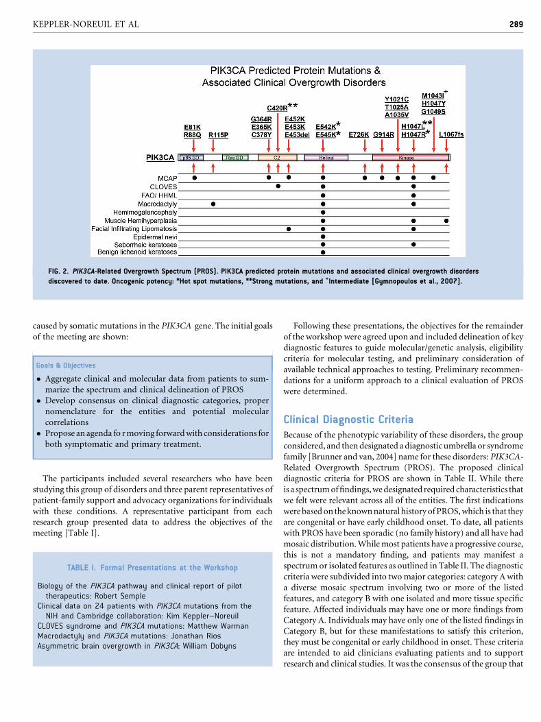
FIG. 2. PIK3CA-Related Overgrowth Spectrum (PROS). PIK3CA predicted protein mutations and associated clinical overgrowth disorders
discovered to date. Oncogenic potency: *Hot spot mutations, **Strong mutations, and +Intermediate [Gymnopoulos et al., 2007].
KEPPLER-NOREUIL ET AL 289
caused by somatic mutations in the PIK3CA gene. The initial goals
of the meeting are shown:
Goals & Objectives
� Aggregate clinical and molecular data from patients to sum-
marize the spectrum and clinical delineation of PROS
� Develop consensus on clinical diagnostic categories, proper
nomenclature for the entities and potential molecular
correlations
� Propose an agenda fo rmoving forwardwith considerations for
both symptomatic and primary treatment.
The participants included several researchers who have been
studying this group of disorders and three parent representatives of
patient-family support and advocacy organizations for individuals
with these conditions. A representative participant from each
research group presented data to address the objectives of the
meeting [Table I].
TABLE I. Formal Presentations at the Workshop
Biology of the PIK3CA pathway and clinical report of pilottherapeutics: Robert Semple
Clinical data on 24 patients with PIK3CA mutations from theNIH and Cambridge collaboration: Kim Keppler–Noreuil
CLOVES syndrome and PIK3CA mutations: Matthew WarmanMacrodactyly and PIK3CA mutations: Jonathan RiosAsymmetric brain overgrowth in PIK3CA: William Dobyns
Following these presentations, the objectives for the remainder
of the workshop were agreed upon and included delineation of key
diagnostic features to guide molecular/genetic analysis, eligibility
criteria for molecular testing, and preliminary consideration of
available technical approaches to testing. Preliminary recommen-
dations for a uniform approach to a clinical evaluation of PROS
were determined.
Clinical Diagnostic CriteriaBecause of the phenotypic variability of these disorders, the group
considered, and thendesignated adiagnostic umbrella or syndrome
family [Brunner and van, 2004] name for these disorders: PIK3CA-
Related Overgrowth Spectrum (PROS). The proposed clinical
diagnostic criteria for PROS are shown in Table II. While there
is a spectrumoffindings,wedesignated required characteristics that
we felt were relevant across all of the entities. The first indications
were basedon the knownnatural history ofPROS,which is that they
are congenital or have early childhood onset. To date, all patients
with PROS have been sporadic (no family history) and all have had
mosaic distribution.Whilemost patients have a progressive course,
this is not a mandatory finding, and patients may manifest a
spectrum or isolated features as outlined in Table II. The diagnostic
criteria were subdivided into twomajor categories: category A with
a diverse mosaic spectrum involving two or more of the listed
features, and category B with one isolated and more tissue specific
feature. Affected individuals may have one or more findings from
Category A. Individuals may have only one of the listed findings in
Category B, but for these manifestations to satisfy this criterion,
they must be congenital or early childhood in onset. These criteria
are intended to aid clinicians evaluating patients and to support
research and clinical studies. It was the consensus of the group that

TABLE II. Clinical Diagnostic Criteria for PIK3CA-RelatedOvergrowth Spectrum (PROS)
Required: Presence of somatic PIK3CA mutation*
Congenital or Early Childhood Onset
Overgrowth Sporadic and Mosaic (Other terms: Patchy, Irregular)
Features as described in either A or B
A. Spectrum (two or more features)**
1. Overgrowth: Adipose, Muscle, Nerve, Skeletal
2. Vascular Malformations: Capillary, Venous, Arteriovenous Malformation,
Lymphatic
3. Epidermal Nevus
B. Isolated features
1. Large Isolated Lymphatic Malformation
2. Isolated Macrodactyly*** OR Overgrown Splayed Feet/ Hands,
Overgrown Limbs
3. Truncal Adipose Overgrowth
4. Hemimegalencephaly (bilateral)/ Dysplastic Megalencephaly/ Focal
Cortical Dysplasia 21
5. Epidermal nevus2
6. Seborrheic Keratoses2
7. Benign Lichenoid Keratoses3
Abbreviations: þ present; � absent; HC hydrocephalus; ID intellectual disability.*If no mutation identified, then consider as presumptive PROS.**Typically Progressive. Can manifest as: Scoliosis (Kyphosis), Limb overgrowth, CNS (HC,Cerebellar tonsillar ectopia, Chiari, Megalencephaly, Mega corpus callosum, Regional lipomatousundergrowth with overgrowth, Infiltrating lipomatosis, Wilms tumor/ovarian cystadenoma.***Other terms: macrodystrophia lipomatosa, macrodactylia fibrolipomatosis and gigantism.11Dobyns WB, 2014 (unpublished data); 2Hafner et al. [2007]; 3Groesser et al. [2012].
290 AMERICAN JOURNAL OF MEDICAL GENETICS PART A
the criteria should be simple, yet as comprehensive as possible given
the current state of knowledge. The group acknowledged that these
criteria may require revision as the gene mutations and clinical
findings are further characterized.
Spectrum of PIK3CA-Related OvergrowthThe PIK3CA-Related Overgrowth Spectrum (PROS) encompasses
all the unique clinical entities, but highlights the continuum and
overlap between the diagnoses, as illustrated in theVenndiagram in
Figure 3. We anticipate that PROS will replace the previous
component clinical entities caused by somatic PIK3CAmutations,
similar to, for e.g., the use of the term“22q11.2 deletion syndrome”,
which encompasses and has replaced the previous clinical syn-
dromes of DiGeorge syndrome, Velocardiofacial syndrome,
Shphrinzten syndrome, Cayler cardiofacial syndrome, and Con-
otruncal anomaly face syndrome. In PROS, there is varying severity
in the clinical presentation of the patients, and some appear to have
tissue-specific distribution, while others are more pleiotropic.
Macrodactyly (type Imacrodactyly or lipofibromatous hamartoma
of nerve), also known as macrodystrophia lipomatosa, was charac-
terized by fibrofatty tissue enlargement and bony overgrowth,
typically within a ‘nerve territory’ with enlargement in circumfer-
ence and length of the peripheral nerve [Ho et al., 2007; Rios
et al., 2012].There is also a subset ofpatientswithmacrodactylywho
also have muscular hemihyperplasia. The original descriptions of
HHML, FAO, and macrodactyly have significant overlap and are
difficult to discriminate fromone another. TheHHMLdesignation
was originally described as moderate abnormalities of asymmetry
and overgrowth with multiple subcutaneous lipomata, and the
hemihyperplasia may be static or mildly progressive [Biesecker
et al., 1998]. The major manifestation of FAO was segmental and
progressive overgrowth of subcutaneous and visceral fibroadipose
tissue, sometimes associatedwith skeletal andmuscular overgrowth
[Lindhurst et al., 2012]. Recently, patients with facial infiltrating
lipomatosis, characterized by hemifacial soft-tissue and skeletal
overgrowth, precocious dental development, macrodontia, hemi-
macroglossia, andmucosal neuromaswere diagnosedwithPIK3CA
activating mutations [Maclellan et al., 2014]. There is also signifi-
cant overlap of HHML and FAO findings with some cases of
CLOVES syndrome. As described above, CLOVES syndrome was
characterized by congenital lipomatous overgrowth, vascular mal-
formations, EN, and skeletal/scoliosis and spinal abnormalities
[Sapp et al., 2007; Alomari, 2009]. The MCAP syndrome was
characterized by a core set of brain features including megalence-
phaly, ventriculomegaly that may progress to hydrocephalus, cere-
bellar tonsillar ectopia that may progress to Chiari malformation,
and cortical brain abnormalities (specifically polymicrogyria or
PMG). There are similarities in clinical findings to those seen in
MPPH. The MCAP syndrome can be distinguished from MPPH
syndrome based on somatic features typically recognized at birth,
such as cutaneous vascular malformations, especially capillary
malformations of the face and cutismarmorata; cutaneous syndac-
tyly and postaxial polydactyly or polysyndactyly; connective tissue
dysplasia; and focal or segmental body overgrowth [Riviere
et al., 2012; Mirzaa et al., 2013]. It is clear that while there may
be some unique features for each diagnosis, there exists consider-
able overlap between PROS. In addition, with further PIK3CA
testing, the spectrum may widen to include other distinct but
related phenotypes, such as Klippel-Trenaunay Syndrome (KTS)
[Kurek et al., 2012].
Differential DiagnosisVarious syndromes considered in the differential diagnosis of PROS
have overlapping clinical findings, such as hemihyperplasia, over-
growth, vascular anomalies, skin abnormalities, tumors, scoliosis, and
others.However, these other overgrowth syndromes have similar, but
more distinguishing features and other identifiable genetic etiologies.
They includeProteus syndrome(PS) [Biesecker et al., 1999;Lindhurst
et al., 2011], PTENHamartoma Tumor Syndrome (PHTS) and Type
II segmental Cowden syndrome [Nelen et al., 1996; Eng, 2000, 2001],
Neurofibromatosis, type 1 (NF1) [Friedman, 1998; Ferner et al., 2007;
Williams et al., 2009], and EpidermalNevus syndrome [Laura, 2013].
For several of the participating research groups, the original referring
diagnosis for many of their patients was described as PS, NF1, and
KTS; however, the patients’ findings did notmeet published diagnos-
tic criteria for these disorders. In particular, for example, none of the
patients had connective tissue nevi characteristic of Proteus syn-
drome. The KTS includes overgrowth and vascular malformations,
which are seen in the PROS, and there is somedebate about the extent
of the diagnostic clinical findings. Indeed, PIK3CA mutations were
identified in some individuals with a KTS-like phenotype [Kurek
et al., 2012].

Figure
3.PhenotypicSpectrum
ofPR
OS:disordershave
overlappingclinicalfeatures,somewithtissue-specific,localized
effects,somewithpleiotropicand
moresevere
manifestations;Abbreviations:FAO/HHML,Fibroadipose
Overgrowth/Hem
ihyperplasia-MultipleLipomatosis;ILM,Isolated
LargeLymphatic
Malform
ation;CLOVES,Congenital
LipomatousOvergrowth,Vascular
Malform
ations,EpidermalNevi,Scoliosis/Skeletal
andSpinal;EN
,EpidermalNevi;SK,
SeborrheicKeratoses;BLK,BenignLichenoidKeratoses;MCAP,
Megalencephaly-Capillary
Malform
ation;HMEG
,Hem
imegalencephaly;
DMEG
,Dysplastic
Megalencephaly
KEPPLER-NOREUIL ET AL 291

TABLE III. Testing Eligibility Criteria for Somatic PIK3CAMutations
PIK3CA mutation analysis should be performed,
If a patient has one or more of:
Key Features:Congenital Spectrum (A)* or Congenital Stand Alone (B)*
Combined Vascular Malformation – Large Capillary,Lymphatic, or Venous malformation
Congenital Musculoskeletal OvergrowthPatterning defect** (e.g., Polydactyly, Sandal gap,
Syndactyly)Congenital CNS (PMG/MEG/HC/Chiari/Syrinx)Congenital Epidermal Nevus (EN)Congenital soft doughy skin or joint hypermobility
þ/� Functional features: Hydronephrosis/Hydroureter, UrinaryIncontinence, Hematuria, Constipation, GastrointestinalBleeding, Intractable Epilepsy, Seizures, Intellectual Disability,Autism, Hypoglycemia.
Abbreviations: CNS, Central Nervous System; HC, hydrocephalus; MEG, megalencephaly; PMG,polymicrogyria.*Refer to these specific findings in Table II.**Upper (UE) and Lower Extremity (LE) findings may include, UE: broad spade-like hands withulnar deviation of the digits, symmetrical overgrowth of 1 or more digits that does not usuallyfollow a nerve territory-oriented pattern, laxity of collateral ligaments, furrowed palms and soles.LE: Overgrowth of the feet, which presents as a large “sandal” gap between great and second toes,large bulbous toes, lipomatous masses on both dorsal and plantar surfaces, broad forefoot withwide gaps between the metatarsal heads; dislocated knees, leg length discrepancy, and patellarchondromalacia [Bloom and Upton, 2013].
292 AMERICAN JOURNAL OF MEDICAL GENETICS PART A
Testing Eligibility CriteriaWe have developed guidelines that clinicians can use in deciding
which patients with overgrowth should be tested for a PIK3CA
mutation. This determination is non-trivial as testing generally
requires a biopsy. An exception to this is the testing for MCAP,
which may be diagnosed by testing of a blood or saliva sample.
Testing eligibility criteria (Table III) are based upon the clinical
criteria described in Table II, but were less strict to include a wider
range of conditions.While it is important to recognize and describe
pre-existing disease entities, key criteria were described to prompt
clinicians to obtain testing for non-descript and potentially as-yet
undiagnosed syndromeswith features described for PROS. Accom-
panying these diagnostic features, functional abnormalities may
also prompt consideration of testing. Thesemay include genitouri-
nary abnormalities such as urinary incontinence, hydronephrosis,
hydroureter, gastrointestinal abnormalities such as constipation,
gastrointestinal bleeding, and neurologic abnormalities including,
seizures, autism, and intellectual disability.
Diagnostic genetic testing for the PROS poses several challenges.
A key challenge is to determine which tissue has the greatest
likelihood of having a detectable mutation. This is related to
technical challenges with genetic testing, particular the sensitivity
and specificity of available genetic testing technique(s). Biopsies
from some PROS patients had low levels of mosaicism (<5%).
Determining the optimal tissue for biopsy is sometimes difficult
because it depends on gross visual assessment of overgrown or
affected regions. In considering the diagnosis of mosaic disorders in
general, detection of the causativemutationdependsnot only on the
particular effectsof themutation (degreeofovergrowth), but alsoon
its load and distribution in the tissue. Identification of a low-level
somatic mutation may therefore require new analytic methods.
In the experience of the workshop participants using current
techniques, molecular genetic testing for the diagnosis of PROS
requires clinically affected tissue samples, preferably freshly
obtained from dermal biopsy overlying an affected area or from
a surgical procedure of the overgrown tissue. This is because
PIK3CA mutations can be detected in affected tissues or cultured
cells at greatly varying levels [Keppler-Noreuil et al., 2014]. We
agreed that testing of blood or DNA isolated from blood is not
recommended based upon current techniques, as PIK3CA muta-
tions have not been identified in the blood by any of the laboratories
of the participating groups, except in 2 of 24 patients with MCAP,
who had an apparent de novo germline mutation in PIK3CA
[Riviere et al., 2012]. Testing can be performed using formalin-
fixed paraffin-embedded (FFPE) tissue, but with varying degrees of
success, and there is a need to ensure rigorous handling of the
samples because of risk of contamination from the microtome.
PIK3CA mutation detection in other tissues or fluids is currently
being evaluated on a research basis including saliva, urine, lymph
fluid, hair, and skin scrapings. Mutation analysis of saliva samples
has not detected any mutations, except in many patients with
MCAP and one reported patient with overgrowth of the salivary
glands (unpublished data).
The types of assays and their respective sensitivities were de-
scribed and are shown in Table IV. The choice of method for
somatic mutation detection in PIK3CA depends on several factors.
Several manufacturers have developed PCR-based assays for detec-
tionof specificPIK3CAmutations commonly found in cancers, and
many of these mutations have also been found in patients with
PROS aswell. The data to date suggest thatwhile there are a number
of mutational hotspots (e.g., PIK3CA codons 542, 545, and 1047)
there are a substantial number of rare mutations (Fig. 2). The hot
spot mutations have significantly elevated biological and biochem-
ical activities (gain-of-function) above the rare mutations; these
mutants may induce higher numbers of transformed cell foci
suggesting more rapid cell proliferation or greater oncogenic
potency. The other mutations may be grouped by oncogenic
potency into strong, intermediate, and weak [Gymnopoulos
et al., 2007]. The use of custom RFLP assays or digital droplet
PCR are typically mutation-specific and pose a tradeoff of ease of
use versus breadth of the test. Other assays are available for
screening multiple sites with a single reaction either by pooling
primer pairs or by using a multi-well format. These tests sacrifice
sensitivity for the ability to assay multiple sites. In order to detect
new or very rare mutations, sequencing entire exons is necessary.
Sanger sequencing can be used for this only if the mutation level is
relatively high (�20%). Lower levels of mosaicism can be detected
using this method, but usually only at positions where multiple
patients have varying levels of themutation and sequence traces can
be extensively compared to distinguish background peaks from low
levels of a variant. Targeted capture of the entire PIK3CA coding
region followed by next generation sequencing at very deep cover-
agemay be better suited for somaticmutation detection, as it allows
for detection of very low levels of mosaicism throughout the gene.
Optimization of these assays is an active area of research.

TABLE IV. Current Assays for Testing of Somatic PIK3CA Mutations
Method
Instrumentation
needed
Maximum number of mutations
assayed per test
Mutation
specific
Detection
Level(%)
custom RFLP fluorescent sequencer 1 þ 0.5–1
digital droplet PCR digital PCR system 1 þ .01– .001
snapshot assay fluorescent sequencer 10 þ 5–10
qBiomarker array qPCR instrument 93 þ 1
MassARRAY system MALDI-TOF mass spectrometer 200 þ 5
Sanger sequencing fluorescent sequencer NA – 5–20
next generation sequencing next gen sequencer NA – 1–10*
*Level of detection is dependent on depth of coverage at the mutant site; the lower the level of mutation, the greater the read depth needs to be for detection
KEPPLER-NOREUIL ET AL 293
It is important to interpret the result based upon the particular
assay and the tissue sampled. The finding of a mosaic mutation can
be very useful to establish a diagnosis of a PROS disorder; however,
there is a poor correlation of the mutation level (in either tissues or
cultured cells) to either the quality (nature) of themanifestations or
the overall severity of themanifestations in the patient. Importantly,
failure to detect a PIK3CA mutation does not necessarily exclude a
clinical diagnosis of PROS in individualsmeeting the clinical criteria
described earlier, as this may be due to limitations in detecting
mosaicism from sub-optimal tissue biopsies. We hypothesize that
the overall lack of correlation of severity tomutation burden or lack
of a genetic diagnosis emanates from the clinical sampling con-
straints. The ability to sample appropriate tissues is limited both by
patient considerations and surgical constraints.
TABLE V. Clinical Imaging Studies
Whole body MRI scan – if there is truncal overgrowth or involvement
(spine curvature)
For infants 6-12 months (earlier age if symptomatic): contrast-
enhanced study is preferable
For older patient: as a baseline study with contrast (contrast may
not be required depending on experience)
Cranial MRI scan – if there is facial or neurologic involvement
Plain radiographs of affected areas
Spine series – if there is curve on exam
Spinal canal ultrasonography as baseline in neonates and young
infants with truncal involvement for tethered cord and
lipomeningomyelocele
Renal ultrasonography at baseline, then every 3 months until age 8
years
Future DirectionsWe discussed the importance of developing a collaborative and
uniform phenotyping and clinical evaluation approach with the
objectives of 1) compiling data on natural history and the extent of
the associated complications of PROS, and 2) to provide useful
outcome measures for use in therapeutic drug trials. The results of
these evaluations may form the basis for assisting clinicians’ man-
agement of these patients, providing recommendations for surveil-
lance for complications of these disorders. There have been reviews
of the more commonly associated complications of this group of
disorders, which forms a basis for the recommendations of clinical
imaging [Alomari et al., 2010; Alomari et al., 2011; Kurek
et al., 2012; Keppler-Noreuil et al., 2014]. We agreed that these
evaluations (shown in Table V) should be based on the character-
istic component clinical findings in the spectrum, including: so-
matic overgrowth (limb, trunk-spine, craniofacial), CNS
overgrowth or dysplasia (brain and spine), vascular anomalies
(in particular thrombosis), dermatologic, genitourinary, gastroin-
testinal, tumorigenesis, development, and endocrine/metabolism
involvement. These current recommendations for tumor surveil-
lance are based upon a reported Wilms tumor in a patient with
CLOVES syndrome [Kurek et al., 2012] and of nephrogenic rests
(a premalignant tumor) in one of the patients reported by Keppler-
Noreuil et al. [2014]. Although the evidence is not sufficient to
demonstrate high risk, it would be prudent to consider serial
abdominal ultrasounds every 3–4 months until age 8 years
in patients with a somatic PIK3CA mutation similar to the rec-
ommendations in isolated hemihyperplasia and Beckwith–
Wiedemann syndrome. In addition, because of the finding of spinal
root and major nerve neurofibromas, as well as vascular and
lipomatous lesions involving the spine, neurological monitoring,
and spinal MRI scan should be considered in patients with truncal
involvement [Alomari et al., 2011; Keppler-Noreuil et al., 2014].
Finally, a reported risk of pulmonary embolism in patients with
CLOVES syndrome having thoracic and central phlebectasia
[Alomari et al., 2010] and in two separate patients with spinal
thrombosis and neonatal cerebral infarcts [Keppler-Noreuil
et al., 2014] suggest that it is important to be aware of the possible
associated thrombosis risk in this group of patients. It is known that
the related disorder, Proteus syndrome also has an increased risk of
thrombosis, and consideration of anticoagulant prophylaxis is
recommended in patients undergoing surgery or other procedures
that may predispose to deep venous thrombosis or pulmonary
embolism. These patients should be monitored for other potential
associated complications, including vascular malformations and
skeletal and spinal abnormalities. Future longitudinal studies of
larger cohorts are needed to further our understanding of the
extent of the findings and natural history of PROS. A consortium
or registry to aggregate clinical and molecular data may facilitate
future studies of genotype–phenotype correlations and improve

294 AMERICAN JOURNAL OF MEDICAL GENETICS PART A
treatment recommendations. The family support groups expressed
interest in participating and playing a role in the governance of
this registry.
The workshop participants are at once gratified by the progress
that has been made to date and humbled by the challenges that lie
ahead.With new found genetic understandingof PROS, sorting out
diagnostic labels is relatively straightforward compared to the
imperative to comprehensivelyunderstand the variable expressivity
and natural history of patients with PROS. We are optimistic that
the diagnostic criteria propose herein will reduce confusion among
clinical professionals and patients and stimulate the research com-
munity to think broadly and comprehensively about these disor-
ders. Most importantly, it is our primary objective to develop
targeted effective treatments for patients with these disorders.
We are extremely optimistic that this can be accomplished now
that the door on the pathophysiology of these remarkable disorders
has been opened.
ACKNOWLEDGMENTS
The authors are grateful to the patients and their families who
participated in this research study.We also thank the CLOVES and
MCAP support group representatives (Kristen Davis, Christy Col-
lins, and, Adrienne Blankenship). We thank the participants of the
workshop including Dr.MatthewWarman, Department of Ortho-
paedics, Boston Children’s Hospital and Harvard Medical School.
The NCATS Office of Rare Disease Research and the Intramural
Research Program of the National Human Genome Research
Institute provided financial support for this workshop. The ideas
and opinions expressed in this paper are those of the authors only
and do not necessarily represent any position or policy of the
National Institutes of Health or any other institution organization
to which any of the authors are affiliated. LGB and MJL declare
receipt of royalties from Genentech, and LGB declares receipt of
royalties from Amgen and an honorarium from Wiley-Blackwell.
RKS and VERP are supported by the Wellcome Trust (Senior
Research Fellowship inClinical Science 098498/Z/12/Z andClinical
Research Training Fellowship 097721/Z/11/Z, respectively), by the
UK National Institute for Health Research (NIHR) Cambridge
Biomedical Research Centre, and by the UK Medical Research
Council Centre for Obesity and Related Metabolic Diseases. VERP
is supported by the Sackler Fund. LGB, KK-N, MJL and JCS are
supported by the Intramural Research Program of the National
Human Genome Research Institute. JJR is supported by the Na-
tional Center for Advancing Translational Sciences of the NIH
under award number UL1TR001105.
REFERENCES
Alomari AI. 2009. Characterization of a distinct syndrome that associatescomplex truncal overgrowth, vascular, and acral anomalies: A descriptivestudy of 18 cases of CLOVES syndrome. Clin Dysmorphol 18:1–7.
Alomari AI, Burrows PE, Lee EY, Hedequist DJ, Mulliken JB, Fishman SJ.2010. CLOVES syndrome with thoracic and central phlebectasia:Increased risk of pulmonary embolism. J Thorac Cardiovasc Surg.140:459–466.
Alomari AI, Chaudry G, Rodesch G, Burrows PE, Mulliken JB, Smith ER,Fishman SJ, Orbach DB. 2011. Complex Spinal-Paraspinal Fast-FlowLesions inCLOVESSyndrome:Analysis ofClinical and ImagingFindingsin 6 Patients. AJNR 32:1812–1817.
Alomari AI, Spencer SA, Arnold RW, Chaudry G, Kasser JR, Burrows PE,Govender P, Padua HM, Dillon B, Upton J, Taghinia AH, Fishman SJ,Mulliiken JB, Fevurly RD, Greene AK, Landrigan-Ossar M, Paltiel HJ,Trenor CC, Kozakewich HP. 2014. Fibro-adipose vascular anomaly:clinical-radiologic-pathologic features of a newly delineated disorderof the extremity. J Pediatri Orthop 34:109–117.
Biesecker LG, Peters KF, Darling TN, Choyke P, Hill S, Schimke N,CunninghamM,Meltzer P, CohenMM Jr. 1998. Clinical differentiationbetweenProteus syndromeandhemihyperplasia: descriptionof adistinctform of hemihyperplasia. Am J Med Genet 79:311–319.
BieseckerLG,HappleR,Mulliken JB,WeksbergR,GrahamJM,ViljoenDL,Cohen MM. 1999. Proteus syndrome: diagnostic criteria, differentialdiagnosis, and patient evaluation. Am J Med Genet 84:389–395.
Bloom J, Upton J III. 2013. CLOVES syndrome. J Hand Surg Am 38:2508–2512.
Brunner HG, van Driel MA. 2004. From syndrome families to functionalgenomics. Nat Rev Genet 5:545–551.
CohenMM,NeriG,WeksbergR. 2002.Overgrowth syndromes.NewYork:Oxford University Press 1–7.
Dobyns WB. 2014 (unpublished data)
Eng C. 2000. Will the real Cowden syndrome please stand up: Reviseddiagnostic criteria. J Med Genet 37:828–830.
EngC. 2001. PTENHamartomaTumor Syndrome (PHTS) [Updated 2014Jan 23]. In: Pagon RA, AdamMP, Ardinger HH, et al., editors. GeneRe-views1 [Internet]. Seattle (WA): University of Washington, 1993–2014.Available from: http://www.ncbi.nlm.nih.gov/books/NBK1488/
EngC. 2003. PTEN:One gene,many syndromes. HumMutat. 22:183–198.
Ferner RE, Huson SM, Thomas N, Moss C, Willshaw H, Evans DG,UpadhyayaM,TowersR,GleesonM, SteigerC,KirbyA. 2007.Guidelinesfor the diagnosis and management of individuals with Neurofibromato-sis 1 (NF1). J Med Genet. 44:81–88.
Friedman JM. 1998. Neurofibromatosis 1. [Updated 2012 May 3]. In:Pagon RA, Adam MP, Ardinger HH, et al., editors. GeneReviews1
[Internet]. Seattle (WA): University of Washington, 1993–2014. Avail-able from: http://www.ncbi.nlm.nih.gov/books/NBK1109/
Groesser L, Herschberger E, Landthaler M, Hafner C. 2012. FGFR3,PIK3CA andRASmutations in benign lichenoid keratosis. Br J Dermatol166:784–788.
Gymnopoulos M, Elsliger M-A, Vogt PK. 2007. Rare cancer-specificmutation in PIK3CA show gain of function. PNAS 104:5569–5574.
Hafner C, Lopez-Knowles E, Luis NM, Toll A, Baselga E,Fernandez-Casado A, Hernandez S, Ribe A, Mentzel T, Stoehr R,Hofstaedter F, Landthaler M, Vogt T, Pujol RM, Hartmann A, RealFX. 2007. Oncogenic PIK3CA mutations occur in epidermal nevi andseborrheic keratoses with a characteristic mutation pattern. PNAS104:13450–13454.
Ho CA, Herring JA, Ezaki M. 2007. Long-term follow-up of progressivemacrodystrophia lipomatosa. A report of two cases. J. Bone Joint Surg.Am. 89:1097–1110.
Hussain K, Challis B, Rocha N, Payne F, Minic M, Thompson A, Daly A,Scott C, Harris J, Smillie BJ, Savage DB, Ramaswami U, De Lonlay P,O’Rahilly S, Barroso I, SempleRK. 2011.An activatingmutation ofAKT2and human hypoglycemia. Science. 334:474.
Keppler-Noreuil KM, Sapp JC, Lindhurst MJ, Parker VE, Blumhorst C,Darling T, Tosi LL, Huson SM, Whitehouse RW, Jakkula E, Grant I,

KEPPLER-NOREUIL ET AL 295
BalasubramanianM,Chandler KE, Fraser JL,GucevZ, CrowYJ, BrennanLM, Clark R, Sellars EA, Pena LD, Krishnamurty V, Shuen A, BravermanN, Cunningham ML, Sutton VR, Tasic V, Graham JM Jr, Geer J Jr,Henderson A, Semple RK, Biesecker LG. 2014. Clinical delineation andnatural history of the PIK3CA-related overgrowth spectrum. Am J MedGenet 164:1713–1733.
Kurek KC, Luks VL, Ayturk UM, Alomari AI, Fishman SJ, Spencer SA,Mulliken JB, BowenME, Yamamoto GL, Kozakewich HP,WarmanML.2012. Somatic mosaic activating mutations in PIK3CA cause CLOVESsyndrome. Am J Hum Genet 90:1108–1115.
Laura FS. 2013. Epidermal Nevus Syndrome. Handb Clin Neurol 111:349–368.
Lee JH, Huynh M, Silhavy JL, Kim S, Dixon-Salazar T, Heiberg A, Scott E,Bafna V, Hill KJ, Collazo A, Funari V, Russ C, Gabriel SB, Mathern GW,Gleeson JG. 2012. De novo somatic mutations in components of thePI3K-AKT3-mTOR pathway cause hemimegalencephaly. Nat Genet44:941–945.
Lindhurst MJ, Sapp JC, Teer JK, Johnston JJ, Finn EM, Peters K, Turner J,Cannons JL, BickD, Blakemore L, Blumhorst C, BrockmannK, Calder P,ChermanN,DeardorffMA, EvermanDB, Golas G, Greenstein RM,KatoBM,Keppler-Noreuil KM,Kuznetsov SA,MiyamotoRT,NewmanK,NgD, O’Brien K, Rothenberg S, Schwartzentruber DJ, Singhal V, TiraboscoR,Upton J,Wientroub S, Zackai EH,HoagK,Whitewood-Neal T, RobeyPG, Schwartzberg PL, Darling TN, Tosi LL, Mullikin JC, Biesecker LG.2011. Amosaic activating mutation in AKT1 associated with the Proteussyndrome. New Engl J Med 365:611–619.
LindhurstMJ, Parker VER, Payne F, Sapp JC, Rudge S,Harris J,WitdowskiAM, Zhang Q, Groeneveld MP, Scott CE, Daly A, Huson SM, Tosi LL,CunninghamML, Darling TN, Geer J, Gucev Z, Sutton VR, Tziotzios C,DixonAK,Halliwell T,O’Rahilly SO, SavageDB,WakelamMJO,BarrosoI, Biesecker LG, Semple RK. 2012.MosaicOvergrowthwith FibroadiposeHyperplasia is Caused by Somatic Activating Mutations in PIK3CA. NatGenet 44:928–933.
Lindhurst MJ, Wang J, Bloomhardt HM, Witkowski AM, Singh LN, BickDP,GambelloMJ, Powell CM, LeeCCR,DarlingTN,Biesecker LG. 2014.AKT1 GeneMutation Levels Are Correlated with the Type of Dermato-logic Lesions in Patients with Proteus Syndrome. J Invest Dermatol134:543–546.
MaclellanRA, LuksVL,ViveroMP,Mulliken JB, ZurakowskiD, PadwaBL,Warman ML, Greene AK, Kurek KC. 2014. PIK3CA Activating Muta-tions in Facial Infiltrating Lipomatosis. Plast Reconstr Surg 133:12e–19e.
Mirzaa GM, Riviere JB, Dobyns WB. 2013. Megalencephaly Syndromesand Activating Mutations in PI3K-AKT Pathway. Am J Med Genet163:122–130.
Nelen MR, Padberg GW, Peeters EA, Lin AY, van den Helm B, Frants RR,Coulon V, Goldstein AM, van ReenMM, Easton DF, Eeles RA, HodgsenS, Mulvihill JJ, Murday VA, Tucker MA, Mariman EC, Starink TM,Ponder BA, Ropers HH, Kremer H, Longy M, Eng C. 1996. Localizationof the gene for Cowden disease to chromosome 10q 22–23. Nat Genet13:114–116.
Poduri A, Evrony GD, Cai X, Elhosary PC, Beroukhim R, Lehtinen MK,Hills LB, Heinzen EL, Hill A, Hill RS, Barry BJ, Bourgeois BF, Riviello JJ,Barkovich AJ, Black PM, Ligon KL, Walsh CA. 2012. Somatic activationof AKT3 causes hemispheric developmental brain malformations. Neu-ron 74:41–48.
Rios JJ, Paria N, Burns DK, Israel BA, Cornelia R,Wise CA, Ezaki M. 2012.Somatic gain-of-function mutations in PIK3CA in patients withmacro-dactyly. Hum Mol Genet. 22:444–451.
Riviere JB,Mirzaa GM, O’Roak BJ, BeddaouiM, Alcantara D, Conway RL,St-Onge J, Schwartzentruber JA, Gripp KW, Nikkel SM, Worthylake T,Sullivan CT,Ward TR, Butler HE, Kramer NA, Albrecht B, Armour CM,Armstrong L, CaluseriuO,CytrynbaumC,Drolet BA, Innes AM, LauzonJL, Lin AE, Mancini GM,MeschinoWS, Reggin JD, Saggar AK, Lerman-Sagie T, Uyanik G, Weksberg R, Zirn B, Beaulieu CL, Finding of, RareDisease, Genes (FORGE), Canada Consortium,Majewski J, Bulman DE,O’DriscollM, Shendure J,GrahamJMJr,BoycottKM,DobynsWB.2012.De novo germline and postzygotic mutations in AKT3, PIK3R2 andPIK3CA cause a spectrum of related megalencephaly syndromes. NatGenet 44:934–940.
Sapp JC, Turner JT, van deKamp JM, vanKijk FS, Lowry RB, Biesecker LG.2007. Newly delineated syndrome of congenital lipomatous overgrowth,vascular malformations, and epidermal nevi (CLOVE syndrome) inseven patients. Am J Med Genet A 143A:2944–2958.
WilliamsVC, Lucas J, BabcockMA,GutmannDH,Korf B,Maria BL. 2009.Neurofibromatosis type 1 revisited. Pediatrics 123:124–133.