
Computational Design of Peptide Ligands for Ochratoxin A
17
Toxins 2013, 5, 1202-1218; doi:10.3390/toxins5061202 toxins ISSN 2072-6651 www.mdpi.com/journal/toxins Article Computational Design of Peptide Ligands for Ochratoxin A Meike Heurich, Zeynep Altintas and Ibtisam E. Tothill * Cranfield Health, Cranfield University, Cranfield, Bedfordshire MK43 0AL, England, UK; E-Mails: [email protected] (M.H.); [email protected] (Z.A.) * Author to whom correspondence should be addressed; E-Mail: [email protected]; Tel.: +44-7500-766-487; Fax: +44-1234-758-380. Received: 2 May 2013; in revised form: 13 June 2013 / Accepted: 13 June 2013 / Published: 21 June 2013 Abstract: In this paper, we describe a peptide library designed by computational modelling and the selection of two peptide sequences showing affinity towards the mycotoxin, ochratoxin A (OTA). A virtual library of 20 natural amino acids was used as building blocks to design a short peptide library against ochratoxin A template using the de novo design program, LeapFrog, and the dynamic modelling software, FlexiDock. Peptide sequences were ranked according to calculated binding scores in their capacity to bind to ochratoxin A. Two high scoring peptides with the sequences N'-Cys-Ser-Ile-Val- Glu-Asp-Gly-Lys-C' (octapeptide) and N'-Gly-Pro-Ala-Gly-Ile-Asp-Gly-Pro-Ala-Gly-Ile- Arg-Cys-C' (13-mer) were selected for synthesis from the resulting database. These synthesized peptides were characterized using a microtitre plate-based binding assay and a surface plasmon resonance biosensor (Biacore 3000). The binding assay confirmed that both de novo designed peptides did bind to ochratoxin A in vitro. SPR analysis confirmed that the peptides bind to ochratoxin A, with calculated K D values of ~15.7 μM (13-mer) and ~11.8 μM (octamer). The affinity of the peptides corresponds well with the molecular modelling results, as the 13-mer peptide affinity is about 1.3-times weaker than the octapeptide; this is in accordance with the binding energy values modelled by FlexiDock. This work illustrates the potential of using computational modelling to design a peptide sequence that exhibits in vitro binding affinity for a small molecular weight toxin. Keywords: ochratoxin A; mycotoxins; peptide; computational modelling; surface plasmon resonance; biosensor OPEN ACCESS
Transcript of Computational Design of Peptide Ligands for Ochratoxin A

Toxins 2013, 5, 1202-1218; doi:10.3390/toxins5061202
toxinsISSN 2072-6651
www.mdpi.com/journal/toxins
Article
Computational Design of Peptide Ligands for Ochratoxin A
Meike Heurich, Zeynep Altintas and Ibtisam E. Tothill *
Cranfield Health, Cranfield University, Cranfield, Bedfordshire MK43 0AL, England, UK;
E-Mails: [email protected] (M.H.); [email protected] (Z.A.)
* Author to whom correspondence should be addressed; E-Mail: [email protected];
Tel.: +44-7500-766-487; Fax: +44-1234-758-380.
Received: 2 May 2013; in revised form: 13 June 2013 / Accepted: 13 June 2013 /
Published: 21 June 2013
Abstract: In this paper, we describe a peptide library designed by computational
modelling and the selection of two peptide sequences showing affinity towards the
mycotoxin, ochratoxin A (OTA). A virtual library of 20 natural amino acids was used as
building blocks to design a short peptide library against ochratoxin A template using the
de novo design program, LeapFrog, and the dynamic modelling software, FlexiDock.
Peptide sequences were ranked according to calculated binding scores in their capacity to
bind to ochratoxin A. Two high scoring peptides with the sequences N'-Cys-Ser-Ile-Val-
Glu-Asp-Gly-Lys-C' (octapeptide) and N'-Gly-Pro-Ala-Gly-Ile-Asp-Gly-Pro-Ala-Gly-Ile-
Arg-Cys-C' (13-mer) were selected for synthesis from the resulting database. These
synthesized peptides were characterized using a microtitre plate-based binding assay and a
surface plasmon resonance biosensor (Biacore 3000). The binding assay confirmed that
both de novo designed peptides did bind to ochratoxin A in vitro. SPR analysis confirmed
that the peptides bind to ochratoxin A, with calculated KD values of ~15.7 μM (13-mer)
and ~11.8 μM (octamer). The affinity of the peptides corresponds well with the molecular
modelling results, as the 13-mer peptide affinity is about 1.3-times weaker than the
octapeptide; this is in accordance with the binding energy values modelled by FlexiDock.
This work illustrates the potential of using computational modelling to design a peptide
sequence that exhibits in vitro binding affinity for a small molecular weight toxin.
Keywords: ochratoxin A; mycotoxins; peptide; computational modelling; surface plasmon
resonance; biosensor
OPEN ACCESS

Toxins 2013, 5
1203
1. Introduction
The control of common food contaminants, like mycotoxins, is creating a strong demand for
analytical methods that permit their rapid and sensitive detection at established regulatory limits.
Mycotoxins are toxic secondary metabolites produced by various fungi (Aspergillus, Penicillium and
Fusarium) in a wide variety of foods, such as maize, coffee beans, cocoa or soy beans, as well as meat,
milk and grapes, but also beverages, like coffee, beer, grape juice or wine [1–5]. Occurrence and
growth of mycotoxins depends on both environmental and food manufacturing conditions [6,7]. The
diverse chemical structures of mycotoxins and their differing physical properties can exhibit a wide
array of biological effects on mammalian systems, e.g., genotoxic, teratogenic, mutagenic,
embryogenic or estrogenic [8], and some show immunosuppressive activity [9,10]. The mycotoxin,
ochratoxin A, has been considered by the International Agency for Research on Cancer to be possibly
carcinogenic (group 2B) for humans [11–16]. There is a growing need to monitor ochratoxin A in food
and beverages according to the EU maximum permitted level of 2.0 μg L−1 of ochratoxin A [17].
Analytical methods for the determination of ochratoxin A in wine are generally based on thin-layer
chromatography (TLC) or high-performance liquid chromatography (HPLC) [18–20]. However, these
techniques either suffer from inadequate sensitivity, due to the lack of a sensitive universal detector for
mycotoxins [21], or are expensive and time-consuming [5,22]. Although chromatography-based
methods are sufficiently sensitive and accurate, immunoassay-based test kits are a good alternative
method for high-throughput analysis, where antibodies are used as the recognition elements, providing
the specificity and sensitivity required for low level toxin detection. Antibody against ochratoxin A has
been successfully developed and employed in immunoassays [23–25] and an immunoaffinity-based
electrochemical sensor [26,27]. While these methods are attractive, immunoassays are not re-usable,
have a limited storage time and, in some cases, antibodies may show cross-reactivity with other
compound in the food matrix. Whereas the affinity sensor development in our previous work focused
on optimization of detection sensitivity with regards to the sensor platform using a disposable
CM-modified gold electrode and antibodies as the sensing molecule [26], there is still a need for a
sensitive, specific and bio-stable sensing receptor for ochratoxin A, which can be easily and
cost-effectively produced as a viable alternative for antibodies.
Replacing natural biomolecules with artificial receptors or biomimics has become an attractive area
of research in recent years [28,29]. The advantages of using these molecules are that they are robust,
more bio-stable and cost-effective to produce and can be modified easily to aid immobilization and to
add labels for detection. Molecular recognition by peptides is known for a number of biochemical
processes, such as signal transduction, metabolism, cell growth and immune defence, and is based on
several non-covalent interactions (e.g., H-bridge-bonding, salt-bridges, hydrophobic and van der Waals
interactions) [30]. Peptide receptors have many advantages over antibodies in that specific peptides
can be obtained for virtually any target, even those that are toxic or have low immunogenicity.
Peptides can be chemically synthesized, offering a wide variety of targeted modifications, such as
fluorescent or affinity tags. Due to their small molecular weight, they are more stable in a wide range
of buffer solutions and less prone to activity loss [31,32].
Recent literature described a combinatorial approach to select peptides that have been specifically
synthesized to target specific molecules, including the mycotoxins, ochratoxin A and

Toxins 2013, 5
1204
aflatoxin B [33–35]. Combinatorial peptide libraries, which consist of up to a million synthetic
peptides, are complex enough to offer unique binding sites that can be screened for peptide receptors
with improved selectivity for a specific target molecule. In contrast to combinatorial chemistry, which
is based on the screening of a large amount of compounds that were randomly synthesized, the
structure-based receptor design is built on the known target structure. Computational modelling is a
technique of representing molecular structures numerically and simulating their behaviour with
quantum equations and classical physics. Computational modelling programs, such as SYBYL [36],
allow researchers to generate and present molecular data, including geometries (bond lengths, bond
angles, torsion angles), energies (e.g., heat of formation, activation energy), electronic properties
(moments, charges, ionization potential, electron affinity), spectroscopic properties (vibrational modes,
chemical shifts) and bulk properties (volumes, surface areas, diffusion, viscosity) [37]. Molecular
modelling software and searching algorithms are traditionally applied in drug design [38]. This
technique has been successfully employed in our lab to gain a selection of synthetic receptors based on
molecular imprinting technology (MIP) that were able to interact specifically with the ochratoxin A
template for use in solid-phase extraction chromatography [39,40].
Here, we present the application of computational modelling for a peptide ligand designed for
ochratoxin A binding. Using molecular modelling software for the development and screening of
peptide sequence libraries around a known small molecular weight target structure is a novel approach.
Computationally-derived peptides were synthesized, and the in vitro binding interaction with
ochratoxin A was investigated using solid-phase binding assays and binding affinity determined by a
surface plasmon resonance biosensor (SPR, Biacore 3000).
2. Results and Discussion
2.1. Computational Modelling
Modelling the binding of ochratoxin A interaction with individual amino acid monomers was
performed to determine the binding score of each interaction (Figure 1). The monomers included in the
virtual library screening for ochratoxin A were all natural amino acids. The results for each amino acid
are compared on the basis of their binding scores, resulting in a table ranking them according to their
binding scores, expressed as a binding energy value in kcal/mol. Descending binding energy values of
the amino acids are proportional to the ascending binding score, i.e., the most likely binding interaction
with ochratoxin A.
Table 1 depicts the binding energies for amino acid monomers interacting with the ochratoxin A
template. The top five scoring amino acids interacting with ochratoxin A are phenylalanine
(F) > proline (P) > valine (V) > isoleucine (I) > leucine (L), all containing apolar side chains and
exhibiting hydrophobic characteristics. The highest binding score was seen with phenylalanine, which
contains a water-insoluble aromatic ring, which is interesting, since the ochratoxin A structure contains
a L-β-phenylalanine moiety also (shown in Figure 1A).

Toxins 2013, 5
1205
Figure 1. (A) Minimized and annealed structure of the ochratoxin A template (shown as
stick and ball); (B) The generation of new peptide compounds begins with a pool of
potential monomers (amino acids) and a virtual cavity (shown as a box) in which to place
them with the target molecule, i.e., ochratoxin A. This virtual screening process is called
“electrostatic screening”. The ochratoxin A template is shown in purple interacting with
random amino acid monomers (stick and ball) was screened using the LeapFrog
design tool.
Table 1. Binding energies for amino acid monomers modelled with ochratoxin A (shown
in descending order from the top down).
Amino acid Polarity Binding energy (kcal mol−1)
Phenylalanine (Phe) Apolar −33.52 Proline (Pro) Apolar −32.10 Valine (Val) Apolar −30.93
Isoleucine (Ile) Apolar −30.37 Leucine (Leu) Apolar −28.94 Cysteine (Cys) Polar (uncharged) −28.67 Tyrosine (Tyr) Polar (uncharged) −27.29
Methionine (Met) Apolar −26.33 Threonine (Thr) Polar (uncharged) −25.55
Tryptophan (Trp) Apolar −22.71 Alanine (Ala) Apolar −21.87
Glutamate (Glu) Polar (negatively charged) −20.63 Aspartate (Asp) Polar (negatively charged) −19.86
Asparagine (Asn) Polar (uncharged) −13.27 Lysine (Lys) Polar (positively charged) −11.72
Histidine (His) Polar (positively charged) −10.06 Glutamine (Gln) Polar (uncharged) −6.43 Arginine (Arg) Polar (positively charged) −5.65
Serine (Ser) Polar (uncharged) −5.23 Glycine (Gly) Polar (uncharged) −1.89
Toxins 2013, 5
1206
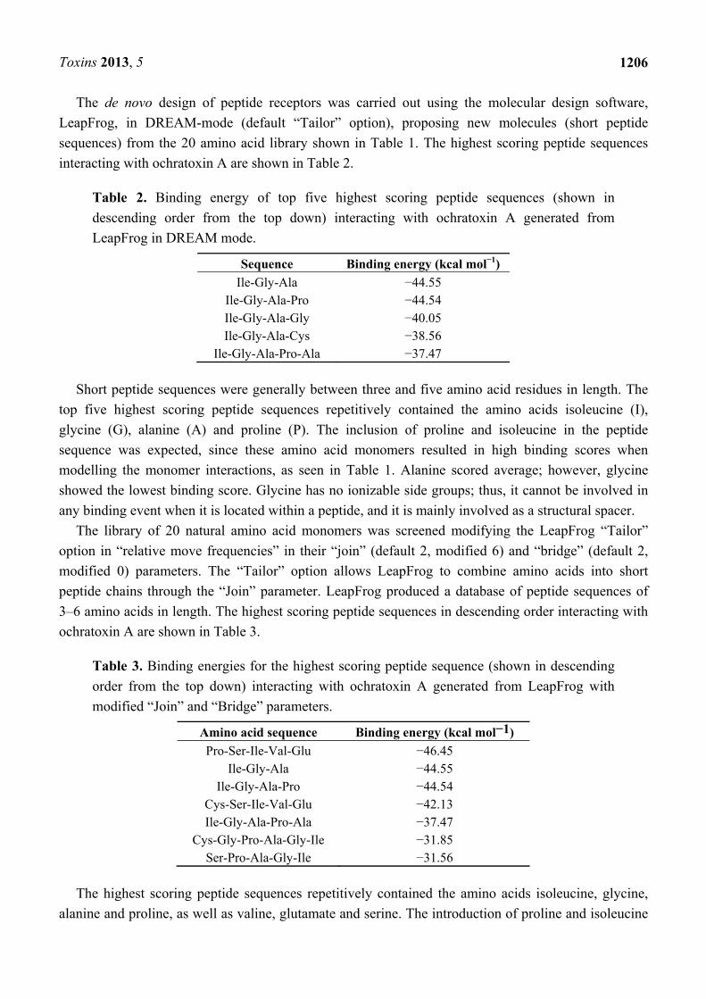
The de novo design of peptide receptors was carried out using the molecular design software,
LeapFrog, in DREAM-mode (default “Tailor” option), proposing new molecules (short peptide
sequences) from the 20 amino acid library shown in Table 1. The highest scoring peptide sequences
interacting with ochratoxin A are shown in Table 2.
Table 2. Binding energy of top five highest scoring peptide sequences (shown in
descending order from the top down) interacting with ochratoxin A generated from
LeapFrog in DREAM mode.
Sequence Binding energy (kcal mol−1)
Ile-Gly-Ala −44.55 Ile-Gly-Ala-Pro −44.54 Ile-Gly-Ala-Gly −40.05 Ile-Gly-Ala-Cys −38.56
Ile-Gly-Ala-Pro-Ala −37.47
Short peptide sequences were generally between three and five amino acid residues in length. The
top five highest scoring peptide sequences repetitively contained the amino acids isoleucine (I),
glycine (G), alanine (A) and proline (P). The inclusion of proline and isoleucine in the peptide
sequence was expected, since these amino acid monomers resulted in high binding scores when
modelling the monomer interactions, as seen in Table 1. Alanine scored average; however, glycine
showed the lowest binding score. Glycine has no ionizable side groups; thus, it cannot be involved in
any binding event when it is located within a peptide, and it is mainly involved as a structural spacer.
The library of 20 natural amino acid monomers was screened modifying the LeapFrog “Tailor”
option in “relative move frequencies” in their “join” (default 2, modified 6) and “bridge” (default 2,
modified 0) parameters. The “Tailor” option allows LeapFrog to combine amino acids into short
peptide chains through the “Join” parameter. LeapFrog produced a database of peptide sequences of
3–6 amino acids in length. The highest scoring peptide sequences in descending order interacting with
ochratoxin A are shown in Table 3.
Table 3. Binding energies for the highest scoring peptide sequence (shown in descending
order from the top down) interacting with ochratoxin A generated from LeapFrog with
modified “Join” and “Bridge” parameters.
Amino acid sequence Binding energy (kcal mol−1)
Pro-Ser-Ile-Val-Glu −46.45 Ile-Gly-Ala −44.55
Ile-Gly-Ala-Pro −44.54 Cys-Ser-Ile-Val-Glu −42.13 Ile-Gly-Ala-Pro-Ala −37.47
Cys-Gly-Pro-Ala-Gly-Ile −31.85 Ser-Pro-Ala-Gly-Ile −31.56
The highest scoring peptide sequences repetitively contained the amino acids isoleucine, glycine,
alanine and proline, as well as valine, glutamate and serine. The introduction of proline and isoleucine

Toxins 2013, 5
1207
in the peptide sequences was expected, since these amino acid monomers resulted in high binding
scores, previously as monomers and in short sequences (Tables 1 and 2). Valine is another high
scoring amino acid when modelling the monomer interaction, whereas glutamate scored as average as
alanine and serine as low as glycine. Interestingly, the majority of these amino acids are apolar, which
indicates the involvement of hydrophobic interactions; however, this also means that these peptides are
difficult to dissolve in aqueous solution.
To enhance the affinity of the peptide interaction with ochratoxin A, as well as improve the
solubility of the final peptide to be synthesized, the peptide sequences obtained from the LeapFrog
database (Table 3) were manually modified. Short peptide sequences were first dimerized to enhance
affinity and, due to the nonpolar nature of the ochratoxin A ligand, charged or polar amino acids were
carefully chosen for incorporation into the receptor to provide a second representative range of polar,
charged and hydrophobic monomers. The molecular dynamics of these peptide receptors with
ochratoxin A was modelled using FlexiDock, which calculates the binding interaction assuming the
high flexibility of the peptide around its template ochratoxin A. The resulting database of high scoring
peptide sequences contained a mixture of unmodified and modified (dimerized, charged) peptide
sequences, as shown in Table 4.
Table 4. List of 11 high scoring peptides obtained with FlexiDock shown in descending
order from the top down.
Peptide sequence Binding energy (kcal mol−1)
Gly-Pro-Ser-Ile-Val-Glu-Cys −17.24 Pro-Ser-Ile-Val-Glu-Pro-Ser-Ile-Val-Glu-Cys −16.72 Ser-Pro-Ala-Gly-Ile −16.07 Cys-Ser-Ile-Val-Glu-Asp-Gly-Lys −14.90 Cys-Gln-Ile-Val-Glu-Pro-Gln-Ile-Val-Glu −14.63 Cys-Phe-Asp-Pro-Ala-Gly-Ile-Lys −14.25 Cys-Phe-Asp-Ala-Pro-Ala-Gly-Ile-Lys −13.08 Pro-Ser-Ile-Val-Glu −12.48 Gly-Pro-Ala-Gly-Ile-Asp-Gly-Pro-Ala-Gly-Ile-Arg-Cys −11.81 Gly-Ser-Pro-Ala-Gly-Ile-Gly −11.78 Cys-Gly-Pro-Ala-Gly-Ile −8.72
The resulting binding energy values obtained by FlexiDock are not identical to the Tripos force
field and the LeapFrog application and need to be seen independently of earlier simulations, because
different force field terms were used and a site-point matching score was included in the FlexiDock
calculation. The FlexiDock database was used to compare the binding energies for modified and
unmodified sequences. Notably, the binding energy values for both modified and unmodified peptides
were similar, implying that the modifications with charged amino acids did not decrease the binding
affinity of the peptide to ochratoxin A; however, dimerization did not seem to have a significant effect
on binding energy values either.
From this database, two high scoring sequences were chosen for further analysis, a 13-peptide with
the sequence (N'-Gly-Pro-Ala-Gly-Ile-Asp-Gly-Pro-Ala-Gly-Ile-Arg-Cys-C') (Figure 2A), which is a
dimer derived from the basic sequence (Pro-Ala-Gly-Ile) listed in Table 2 as the second best score.

Toxins 2013, 5
1208
This dimer is separated by negatively charged aspartate and glycine to allow for flexibility, as well as
C'-terminal charged arginine for solubility and the N'-terminal cysteine tag for immobilization
purposes. The second sequence is an octapeptide (N'-Cys-Ser-Ile-Val-Glu-Asp-Gly-Lys-C'), as seen in
Figure 2B, which has been derived from the top score sequence, Ser-Ile-Val-Glu (Table 3). This
sequence was modified by introducing an N'-terminal cysteine and a C'-terminal negatively charged
aspartate, as well as glycine for flexibility.
Figure 2. Final result of de novo designed peptide sequences shown interacting with
ochratoxin A. The [N'-Gly-Pro-Ala-Gly-Ile-Asp-Gly-Pro-Ala-Gly-Ile-Arg-Cys-C'] peptide
(A, left) and the [N'-Cys-Ser-Ile-Val-Glu-Asp-Gly-Lys-C'] peptide (B, right) sequence are
seen as space-filled, ochratoxin A as stick and ball structures. Ochratoxin A mainly
interacts with the peptide backbone of the N-terminal end of the 13-peptide (B) and the
central region of the Octamer peptide backbone.
A BC’
C’ N’
N’
13-mer Octapeptide
Ochratoxin A
Ochratoxin A
Both selected peptide sequences have the amino acid isoleucine in common, which showed a very
high binding score when interacting with ochratoxin A (Table 1). Isoleucine has no ionizable groups
and, therefore, cannot be taking part in hydrogen bonding. Since isoleucine is very hydrophobic, it is
likely that it attracts molecules, such as ochratoxin A, through hydrophobic interactions.
Hydrophobicity is one of the major forces in ligand recognition. As the hydrophobic side-chains of an
amino acid sequence and a hydrophobic ligand come together, there is a favourable increase in the
entropy of the system as the solvent (water) molecules, which were previously in an ordered shell
around the exposed hydrophobic surface, become disordered, as well as an energetic contribution as
unfavourable apolar-polar interactions are replaced with more favourable homotypic interactions [41–43].
It seems the binding interaction of ochratoxin A with the designed peptides is established by
electrostatic/hydrophobic interactions rather than hydrogen bonding.
The two peptide sequences were then synthesized and mass spectroscopy was applied to obtain the
molecular weights of the peptides. The Medical Research Council (MRC, UK) submitted molecular
weights for the octapeptide at 834 g mol−1 and for the 13-mer peptide, 1183 g mol−1. Both peptides
were water soluble.

Toxins 2013, 5
1209
2.2. Binding Assay of Ochratoxin A-HRP to the 13-mer and Octapeptide
Solid phase binding assay provides a platform to test the binding capacity of the designed peptides
to ochratoxin A in vitro. Each peptide sequence was immobilized to the surface of a functionally
modified (R2-NH) microtitre plate via either amine coupling using N-hydroxysuccinimide
(NHS)/N-(3-dimethylaminopropyl)-N'-ethylcarbodiimide (EDC) coupling chemistry or thiol coupling
using a heterobifunctional crosslinker (N-Succinimidyl 3-(2-pyridyldithio) propionate (SPDP))
employing the terminal cysteine residues of the peptide sequences to allow for site-directed surface
attachment. Enzyme-labelled ochratoxin A-HRP was added in a concentration-dependent manner to
the immobilized peptides, and the absorbance signal (blank subtracted) was compared for both
peptides and immobilization techniques, as shown in Figure 3.
The absorbance signal confirmed that ochratoxin A-HRP binds to both amine- and thiol-coupled
octapeptide (Cys-Ser-Ile-Val-Glu-Asp-Gly-Leu) (Figure 3A). However, much higher binding signals
were obtained with the peptide immobilized using thiol chemistry. The 13-mer peptide
(Gly-Pro-Ala-Gly-Ile-Asp-Gly-Pro-Ala-Gly-Ile-Arg-Cys) showed good binding capacity to ochratoxin
A-HRP when thiol coupled, which was decreased when immobilized using amine coupling chemistry.
The results suggest that both peptides bind ochratoxin A (Figure 3B).
Figure 3. Capacity of the ochratoxin A-HRP to bind to the octapeptide and 13-mer peptide.
The interaction between serial dilutions of HRP-conjugated ochratoxin A and octapeptide
(A) and 13-mer peptide (B) immobilized to functionalized microtitre plates via amine
coupling (●) and thiol coupling (■) is expressed as absorbance at A 450 nm versus
ochratoxin A-HRP concentration (µM). Error bars illustrate the mean and standard
deviation of multiple experiments.
(A)

Toxins 2013, 5
1210
Figure 3. Cont.
(B)
2.3. SPR Analysis to Determine Binding Affinity
To determine affinities for the peptide binding interaction, the peptides were immobilized on two
adjacent flow cells onto a CM5 Biacore chip to a level of 251 RU (octapeptide) and 227 RU (13-mer
peptide). This was in order to study the binding interaction between the peptides and ochratoxin A
using a Biacore 3000. A commercial ochratoxin A-BSA conjugate was used as the analyte to obtain a
significant signal response for a binding event. Initially, one saturating concentration of ochratoxin
A-BSA conjugate (100 mg L−1) was injected over each immobilized peptide ligand to confirm binding
(Figure 4).
Figure 4. Sensorgrams displaying binding curves of 100 mg L−1 (0.15 μM) ochratoxin
A-BSA (reference subtracted) to immobilised peptides, (a) 13-mer peptide
GPAGIDGPAGIRC (blue) and (b) octapeptide CSIVEDGL (red); and 100 mg L−1
BSA alone (negative control) binding to (c) 13-mer peptide (dark grey) and
(d) octapeptide (light grey).
-30
-20
-10
0
10
20
30
40
-200 -100 0 100 200 300 400 500 600 700 800
T im e [sec]
Res
po
ns
e [R
U]
-30
-20
-10
0
10
20
30
40
-200 -100 0 100 200 300 400 500 600 700 800
T im e [sec]
Res
po
ns
e [R
U]
0
0.2
0.4
0.6
0.8
1
1.2
OD (450nm)
Ochratoxin A-HRP (μM)
NHS/EDC Amine coupling SPDP Thiol coupling
(a)
(b)
(c)
(d)

Toxins 2013, 5
1211
Injection of BSA alone showed no binding, confirming the binding interaction to be ochratoxin
A-dependent. To establish the affinity of the interaction, decreasing ochratoxin A-BSA concentrations
were injected onto the immobilized peptides surfaces (Figure 5).
Figure 5. Sensorgrams showing the binding interaction of immobilized (A) octamer
peptide CSIVEDGL and (B) 13-mer peptide GPAGIDGPAGIRC with decreasing
ochratoxin A-BSA analyte concentration (from top to bottom: 100, 1, 0.1, 0.01 mg L−1;
equivalent to 15pM–0.15 μM. BSA reference binding is shown in grey.
0
10
20
30
40
-200 0 200 400 600 800 1000
Res
po
nse
[R
U]
Time [sec]
A
0
10
20
30
40
-200 0 200 400 600 800 1000
Res
po
nse
[R
U]
Time [sec]
0
10
20
30
40
-200 0 200 400 600 800 1000
Res
po
nse
[R
U]
Time [sec]
A
0
5
10
15
20
25
30
35
40
45
-200 0 200 400 600 800 1000
Res
po
nse
[R
U]
Time [sec]
B
0
5
10
15
20
25
30
35
40
45
-200 0 200 400 600 800 1000
Res
po
nse
[R
U]
Time [sec]
0
5
10
15
20
25
30
35
40
45
-200 0 200 400 600 800 1000
Res
po
nse
[R
U]
Time [sec]
B
Figure 4 shows that both the 13-mer and octapeptide bind the ochratoxin A-BSA conjugate and
show no non-specific binding to the unconjugated BSA used as the reference, which was subtracted
from the signal curves. Immobilization methods, especially via amine coupling, can reduce the activity
of the immobilized molecule and, thus, have reduced binding capacity, which can be substantially
different from the theoretical binding capacity. This is a common and known problem in binding
interaction analysis. However, binding curves on the remaining active peptide molecules are still valid
and can be used to determine whether a binding interaction is taking place, especially when compared
to a reference surface (which is the case here) and also determine the binding affinity of an interaction.
The sensorgrams show fast on and off rates for both peptides with ochratoxin A interaction, as the
baseline was reached almost immediately after the end of ochratoxin A-BSA injection, indicating weak
affinity interactions. Those can be advantageous, as there is no need for a regeneration step, and hence,
the analysis can be performed in an isocratic buffer environment, which enhances the stability of the
peptides and improves the life-time of the receptor surface.
Figure 5 illustrates the concentration-dependent binding interaction of ochratoxin A-BSA to the
octamer peptide (Figure 5A) and the 13-mer peptide (Figure 5B). Both peptides show similar binding
curves and response units relative to ochratoxin-BSA concentration.
Due to the 1:3 to 1:6 molecular distribution of 3–6 mol ochratoxin A bound per mol BSA
(manufacturer statement), the binding interaction does not necessarily follow 1:1 binding interaction
stoichiometry, as all 3–6 ochratoxin A molecules can theoretically partake in a binding event. The
Biacore evaluation software does not allow affinity calculations for multiple analytes; thus, we chose
the standard 1:1 Langmuir binding model to calculate an estimated affinity for the interaction.

Toxins 2013, 5
1212
The 13-mer peptide exhibited an estimated binding strength to ochratoxin A with a KD of 15.7 μM,
and the octapeptide peptide resulted in a similar KD of 11.8 μM, which is categorized as weak binding
affinity. In contrast, a commercial ochratoxin A-specific antibody was tested and showed relatively
high affinity and slow off rates, indicated by a KD of 3 nM (data not shown) when immobilized and
subjected to the same ochratoxin A-BSA concentrations and conditions. The affinity of the artificial
peptide receptors corresponds well with the binding energy values derived from modelling the
interaction using FlexiDock (Table 4). The 13-mer peptide has about 1.3-times weaker binding affinity
to ochratoxin A than the octapeptide, which is mimicked in the 1.3-times higher binding energy value
calculated by FlexiDock (13-mer: −11.81 kcal/mol; Octamer: −14.9 kcal/mol). In conclusion, the
results were similar to those achieved in the previous assay format. A competitive assay with free
ochratoxin A and ochratoxin A-BSA on the SPR sensor immobilized peptides will need to be
conducted to further study the binding interaction of the peptides to ochratoxin A.
In comparison, the affinity of ochratoxin A binding non-covalently to its natural ligand (human)
albumin shows both ionic and hydrophobic forces, [44] and it has been shown that high affinity
binding sites can give KD ≈ 19 nM, while weak affinity sites show KD ≈ 1 µM [45]. The high affinity
binding site matches the same affinity range (nM) of a commercial ochratoxin A antibody for
ochratoxin-BSA, whereas the weak affinity binding site falls within the same affinity (μM) range as
the computationally designed peptides reported in this work. Other peptide receptors for mycotoxins
selected from combinatorial peptide libraries also showed μM affinities within the same range, such as
an ochratoxin A-specific peptide selected from a combinatorial peptide-phage display library with the
sequence N'-Ser-Asn-Leu-His-Pro-Lys-C', resulting in KD ≈ 2.9 μM. Furthermore, Tozzi et al. created
a combinatorial library with the lead tetrapeptide, Leu-Leu-Ala-Arg-NH2, with binding constants
KD ≈ 8.3 μM and 3.4 μM for aflatoxins B1 and B2, respectively [35]. This further shows that the
affinity of our computationally designed peptides matches the same range of those selected from
combinatorial libraries, validating computational library design and screening as a good alternative to
in vitro techniques. We further believe that these peptides, after careful cross-interaction studies and
further optimization and characterization, could be employed as a diagnostic tool for ochratoxin A.
3. Experimental Section
3.1. Materials and Reagents
The ochratoxin A-BSA conjugate, N-Succinimidyl 3-(2-pyridyldithio) propionate (SPDP) and
cysteine were purchased from Sigma Aldrich Ltd., UK. Acetate buffer and glycine were from Fluka
(Sigma-Aldrich Ltd., Dorset, UK). The CM5 sensor chips, HEPES buffered saline (HBS-EP), 1 M
ethanolamine-HCl, EDC (N-(3-dimethylaminopropyl)-N'-ethylcarbodiimide) and NHS
(N-hydroxysuccinimide), as well as the BIAcore 3000™ used for the analysis were from BIAcore
(Uppsala, Sweden). The ready-made 1-Step™ Ultra TMB (3,3',5,5'-tetramentylbenzidine) solution was
from Pierce (Pierce Inc., 3747 N. Meridian Road, P.O. Box 117, Rockford, IL 61105, USA).
Nunc™CovaLink™ NH-microtiter plates were from NunBrand, Denmark. Peptide sequences have
been synthesized and verified by mass spectroscopy by the Medical Research Council (MRC) at
Imperial College, London.

Toxins 2013, 5
1213
3.2. Computational Modelling
The workstation employed to simulate receptor-ligand interactions was a Silicon Graphics Octane
running the IRIX 6.6 operating system. It was configured with two 195 MHz reduced instruction set
processors, 712 MB memory and a 12 GB fixed drive. This system was used to execute the software
package, SYBYL 6.9/7.0 (Tripos Inc., St. Louis, MO, USA). Computational design was performed as
follows. A structural model of the ochratoxin A template was drawn according to SYBYL tutorial
“small molecule sketching”. The structure was refined using molecular mechanics by applying energy
minimization with the MAXIMIN2 command. A simulated annealing process was then applied to
obtain conformational energies lower than the minimum of energy found by energy minimization to
ensure a conformation that is as close to nature as possible (Figure 1A). Annealing conditions were
fixed as 700–200 K for 1000 fs each and run for 1000 cycles [39]. The TRIPOS force field was applied
for energy calculations. The dielectric constant corresponded water conditions, and the termination
used was “Gradient” with a cut-off value of 0.001 kcal mol−1 at a maximum of 1000 iterations.
Structures were charged using the Gasteiger-Hückel charges. For peptide receptor design, a molecular
library containing 20 natural amino acids was used as monomers. The LeapFrog genetic algorithm was
applied to screen each functional monomer of an amino acid library for its possible interaction with the
ochratoxin A template (Figure 1B). LeapFrog is an algorithm that allows for evaluations of new ligand
structures on the basis of calculating binding scores expressed as an energy value in kcal/mol. This is
calculated using electrostatic screening by trying repeatedly distinct amino acid monomers (one each
time) in different positions of the ochratoxin A template and then either keeping or discarding the
results depending on the calculated binding score of the total contributions from steric, electrostatic
and hydrogen bonding interactions (Tripos Association, St. Louis, MO, USA). LeapFrog was applied
in DREAM mode and activated for different length of runs (100,000 to 1,000,000 iterations). Amino
acid monomers were modified by adding “active hydrogens” for localizing the site of interaction,
which facilitates peptide bond formation. The input data was set on “peptide” mode; this allows the
receptor to be built by placing amino acid monomers and linking them together to produce the peptide
sequence. The binding scores were saved in a database and evaluated mainly on their empirical
binding energy. Binding energy, as calculated in LeapFrog, has three major components: steric and
electrostatic enthalpies of binding process calculated using the Tripos force field, cavity desolvation
energy and ligand desolvation energy. The peptide sequences giving the highest binding score (lowest
binding energy value) were selected as candidates for further screening using the ligand-receptor
docking module, FlexiDock (Tripos Association, St. Louis, MO, USA).This is a software tool that
calculates the binding interaction assuming the high flexibility of the peptide receptor around its
template (i.e., ochratoxin A). The docking simulation was performed using high scoring peptide
sequences obtained from LeapFrog, as well as manually modified sequences. The latter was done
under the consideration that some structures selected as high scoring by LeapFrog are synthetically
difficult to produce. Thus, charged amino acids were added to the structure to influence overall
molecular hydrophilicity with the prospect of producing a water soluble peptide. The final peptide
design from FlexiDock contained a LeapFrog-derived template sequence, which was modified in terms
of solubility and ease of immobilization in future binding assays (by addition of a cysteine residue).

Toxins 2013, 5
1214
3.3. Solid Phase Binding Assay
To test the initial binding of the peptides with ochratoxin A, the peptides were immobilized to a
(NH)-functionally modified polystyrene microtitre plate via: (1) amine-coupling using a NHS/EDC
linker and (2) thiol-coupling using SPDP, a hetero-bifunctional cross-linker reagent with amine and
sulfhydryl groups.
Amine coupling was performed as follows: 100 mg L−1 of each peptide solution in 10 mM
carbonate buffer pH 9.6 was incubated on the NH-wells for 30 min, then 200 mM EDC and 50 mM
NHS (1:1 v/v) were added and incubated for one hour. The plates were washed using PBST and
blocked using 0.1 M ethanolamine for 30 min. Thiol coupling was performed via the immobilization of
1.5 mg mL−1 SPDP in phosphate buffer (PBS) pH 7.4 incubated for 30 min at room temperature. The
peptides were added at 100 mg L−1 in a phosphate buffer pH 8.0 and incubated for another two hours
at room temperature. The plates were washed using PBST and blocked for one hour using 1 g L−1
cysteine in 1 M NaCl and 0.1 M sodium acetate, pH 4.0.
The binding of ochratoxin A-HRP conjugate in phosphate buffer pH 7.4 was performed at
decreasing serial dilutions and incubated with the immobilized peptides for 1.5 h. The zero reference
was determined with ochratoxin-HRP alone on 0.1 M ethanolamine blocked wells without
immobilized peptide. All incubations were performed at room temperature. Detection was performed,
after washing the microtiter plates using PBST, with the chromogenic HRP-substrate TMB in
ready-made solution containing hydrogen peroxide. The absorbance was read after 20 min at 450 nm.
3.4. SPR Testing for Binding Interaction
The binding interaction analysis of the peptide receptor with ochratoxin A was carried out on a
CM5 (carboxymethylated dextran) sensor chip at 25 °C. HBS-EP (0.01 M HEPES pH 7.4, 0.15 M
NaCl, 3 mM EDTA, 0.005% Surfactant P20) was used as the running and dilution buffer. Peptides
(100 mg L−1 in 10 mM acetate buffer, pH 4.5) were immobilised using 0.4 M
N-(3-dimethylaminopropyl)-N'-ethylcarbodiimide (EDC) and 0.1 M N-hydroxysuccinimide (NHS),
applying amine coupling chemistry. In brief, a 1:1 mixture of NHS and EDC was injected as
5 μL min−1 flow rate for 7 min, followed by injection of 75 µL peptide ligand (5 µL min−1) for
15 min and blocking of residual binding sites with 1 M Ethanolamine, pH 8.3 (35 µL). To establish
binding kinetics, various concentrations of ochratoxin A-BSA conjugate (0.01–100 mg L−1, 60 µL in
HBS-EP pH 7.4) were injected at 5 µL min−1 over each immobilised peptide surface. BSA alone was
injected as non-specific analyte control. Dissociation was monitored during 10 min without
dissociating agents (flowing only running buffer). The kinetic parameters of the binding reactions were
determned using BIAevaluation 3.2 software [46].
4. Conclusions
In this study, the application of computational modelling was employed to design a peptide ligand
that binds to Ochratoxin A. A synthetic peptide ligand for ochratoxin A was designed using an
innovative computational approach. The initial argument of the approach to simulate nature’s
evolution by de novo designing a peptide ligand for a small molecular weight toxin was investigated

Toxins 2013, 5
1215
using binding interaction analysis. The binding interaction of both peptides to ochratoxin A was
confirmed in in vitro binding assays, and the affinity of the interaction was established using Biacore
(SPR) analysis. Both peptide ligands showed weak affinity, indicated by fast on and off rates, and no
need for surface regeneration. The 13-mer showed faster on and off rates when immobilized. The
sensorgrams correlate well with the in silico data obtained with both LeapFrog and FlexiDock
simulations. The results have shown the clear advantages of designing a peptide ligand for ochratoxin
A in silico, which is a time-efficient, cost-effective production without exposure to toxic materials or
the use of biological or animal resources. Using this approach, several peptides for ochratoxin A were
successfully designed and synthesized, and their binding capacities were confirmed in vitro. It is also
anticipated that the peptide ligands can be used in binding assays and affinity sensors. Further work
will need to be conducted to characterise the affinity interaction between the peptides further and
conduct a cross-interaction study.
Acknowledgments
We gratefully acknowledge the European Union 6th framework integrated GOODFOOD project for
funding this work.
Conflict of Interest
The authors declare that there are no conflicts of interest.
References
1. Adams, M.; Moss, M. Food Microbiology; The Royal Society of Chemistry: Cambridge, UK,
1995; pp. 192–203.
2. Makun, H.; Gbodi, T.; Akanya, O.; Salako, E.; Ogbadu, G. Fungi and some mycotoxins
contaminating rice (Oryza sativa) in Niger state, Nigeria. Afr. J. Biotechnol. 2010, 6, 99–108.
3. Sulyok, M.; Krska, R.; Schuhmacher, R. Application of an LC-MS/MS based multi-mycotoxin
method for the semi-quantitative determination of mycotoxins occurring in different types of food
infected by moulds. Food Chem. 2010, 119, 408–416.
4. Wild, C.P.; Gong, Y.Y. Mycotoxins and human disease: A largely ignored global health issue.
Carcinogenesis 2010, 31, 71–82.
5. Bhat, R.; Rai, R.V.; Karim, A. Mycotoxins in food and feed: Present status and future concerns.
Compr. Rev. Food Sci. Food Saf. 2010, 9, 57–81.
6. Bhatnagar, D.; Brown, R.; Ehrlich, K.; Cleveland, T.E. Mycotoxins contaminating cereal grain
crops: Their occurrence and toxicity. Appl. Mycol. Biotechnol. 2002, 2, 171–196.
7. Adjou, E.S.; Dahouenon-Ahoussi, E.; Soumanou, M.M. Investigations on the mycoflora and
processing effects on the nutritional quality of peanut (Arachis hypogea L. var. TS 32-1).
J. Microbiol. Biotechnol. Food Sci. 2012, 2, 1025–1039.
8. Storari, M.; Dennert, F.G.; Bigler, L.; Gessler, C.; Broggini, G.A. Isolation of mycotoxins
producing black aspergilli in herbal teas available on the Swiss market. Food Control 2012, 26,
157–161.

Toxins 2013, 5
1216
9. Borbély, M.; Sipos, P.; Pelles, F.; Győri, Z. Mycotoxin contamination in cereals. J. Agronom.
Proc. Technol. 2010, 16, 96–98.
10. Rolle-Kampczyk, U.; Müller, A.; Diez, U.; Rehwagen, M.; Schwenke, A.; Metzner, G.;
Herbarth, O. Mycotoxins in house dust—An underestimated problem? Mycotoxin Res. 2000, 16,
100–104.
11. Hibi, D.; Suzuki, Y.; Ishii, Y.; Jin, M.; Watanabe, M.; Sugita-Konishi, Y.; Yanai, T.; Nohmi, T.;
Nishikawa, A.; Umemura, T. Site-specific in vivo mutagenicity in the kidney of gpt delta rats
given a carcinogenic dose of ochratoxin A. Toxicol. Sci. 2011, 122, 406–414.
12. Stoev, S.D. Studies on carcinogenic and toxic effects of ochratoxin A in chicks. Toxins 2010, 2,
649–664.
13. Czakai, K.; Müller, K.; Mosesso, P.; Pepe, G.; Schulze, M.; Gohla, A.; Patnaik, D.; Dekant, W.;
Higgins, J.M.; Mally, A. Perturbation of mitosis through inhibition of histone acetyltransferases:
The key to ochratoxin a toxicity and carcinogenicity? Toxicol. Sci. 2011, 122, 317–329.
14. Reddy, L.; Bhoola, K. Ochratoxins—Food contaminants: Impact on human health. Toxins 2010,
2, 771–779.
15. Pfohl-Leszkowicz, A.; Manderville, R. Review on Ochratoxin A: An overview on toxicity and
carcinogenicity in animals and humans. Mol. Nutr. Food Res. 2007, 51, 61–99.
16. Pfohl-Leszkowicz, A.; Manderville, R.A. An update on direct genotoxicity as molecular
mechanism of ochratoxin A carcinogenicity. Chem. Res. Toxicol. 2012, 25, 252–262.
17. European Commission. Commission regulation (EC) No. 123/2005 of 26 January 2005 amending
regulation (EC) No. 466/2001 as regards ochratoxin A. Off. J. Eur. Union. 2005, L25, 3–5.
18. Duarte, S.; Bento, J.; Pena, A.; Lino, C.; Delerue-Matos, C.; Oliva-Teles, T.; Morais, S.;
Correia, M.; Oliveira, M.; Alves, M. Monitoring of ochratoxin A exposure of the Portuguese
population through a nationwide urine survey—Winter 2007. Sci. Total Environ. 2010, 408,
1195–1198.
19. Rubert, J.; Sebastià, N.; Soriano, J.; Soler, C.; Mañes, J. One-year monitoring of aflatoxins and
ochratoxin A in tiger-nuts and their beverages. Food Chem. 2011, 127, 822–826.
20. Esti, M.; Benucci, I.; Liburdi, K.; Acciaro, G. Monitoring of ochratoxin A fate during alcoholic
fermentation of wine-must. Food Control 2012, 27, 53–56.
21. Sauceda-Friebe, J.C.; Karsunke, X.Y.; Vazac, S.; Biselli, S.; Niessner, R.; Knopp, D. Regenerable
immuno-biochip for screening ochratoxin A in green coffee extract using an automated
microarray chip reader with chemiluminescence detection. Anal. Chim. Acta 2011, 689, 234–242.
22. Chen, J.; Fang, Z.; Liu, J.; Zeng, L. A simple and rapid biosensor for ochratoxin A based on a
structure-switching signaling aptamer. Food Control 2012, 25, 555–560.
23. Zamfir, L.-G.; Geana, I.; Bourigua, S.; Rotariu, L.; Bala, C.; Errachid, A.; Jaffrezic-Renault, N.
Highly sensitive label-free immunosensor for ochratoxin A based on functionalized magnetic
nanoparticles and EIS/SPR detection. Sens. Actuators B 2011, 159, 178–184.
24. Yu, F.-Y.; Vdovenko, M.M.; Wang, J.-J.; Sakharov, I.Y. Comparison of enzyme-linked
immunosorbent assays with chemiluminescent and colorimetric detection for the determination of
ochratoxin A in food. J. Agric. Food Chem. 2011, 59, 809–813.

Toxins 2013, 5
1217
25. Barthelmebs, L.; Jonca, J.; Hayat, A.; Prieto-Simon, B.; Marty, J.-L. Enzyme-linked aptamer
assays (ELAAs), based on a competition format for a rapid and sensitive detection of ochratoxin
A in wine. Food Control 2011, 22, 737–743.
26. Heurich, M.; Kadir, M.K.A.; Tothill, I.E. An electrochemical sensor based on carboxymethylated
dextran modified gold surface for ochratoxin A analysis. Sens. Actuators B 2011, 156, 162–168.
27. Bonel, L.; Vidal, J.C.; Duato, P.; Castillo, J.R. An electrochemical competitive biosensor for
ochratoxin A based on a DNA biotinylated aptamer. Biosens. Bioelectron. 2011, 26, 3254–3259.
28. Tothill, I.E. Biosensors and nanomaterials and their application for mycotoxin determination.
World Mycotoxin J. 2011, 4, 361–374.
29. Mirsky, V.M.; Yatsimirsky, A. Artificial Receptors for Chemical Sensors; Wiley-VCH: Berlin,
Germany, 2010.
30. Kuchelmeister, H.Y.; Schmuck, C. Molecular Recognition of Oligopeptides and Protein Surfaces.
In Designing Receptors for the Next Generation of Biosensors; Springer: Berlin, Germany, 2013;
pp. 67–84.
31. Van Regenmortel, M. Synthetic peptides versus natural antigens in immunoassays. Ann. Biol.
Clin. 1993, 51, 39–41.
32. Tothill, I.E. Peptides as Molecular Receptors. In Recognition Receptors in Biosensors; Zourob,
M., Ed.; Springer: Berlin, Germany, 2010; pp. 249–274.
33. Giraudi, G.; Anfossi, L.; Baggiani, C.; Giovannoli, C.; Tozzi, C. Solid-phase extraction of
ochratoxin A from wine based on a binding hexapeptide prepared by combinatorial synthesis. J.
Chromatogr. A 2007, 1175, 174–180.
34. Tozzi, C.; Anfossi, L.; Giraudi, G.; Giovannoli, C.; Baggiani, C.; Vanni, A. Chromatographic
characterisation of an estrogen-binding affinity column containing tetrapeptides selected by a
combinatorial-binding approach. J. Chromatogr. A 2002, 966, 71–79.
35. Tozzi, C.; Anfossi, L.; Baggiani, C.; Giovannoli, C.; Giraudi, G. A combinatorial approach to
obtain affinity media with binding properties towards the aflatoxins. Anal. Bioanal. Chem. 2003,
375, 994–999.
36. Clark, M.; Cramer, R.D.; Van Opdenbosch, N. Validation of the general purpose Tripos 5.2 force
field. J. Comput. Chem. 2004, 10, 982–1012.
37. Richon, A.B. An introduction to molecular modeling. Mathematech 1994, 1, 83.
38. Böhm, H.-J. The computer program LUDI: A new method for the de novo design of enzyme
inhibitors. J. Comput. Aided Mol. Des. 1992, 6, 61–78.
39. Chianella, I.; Lotierzo, M.; Piletsky, S.A.; Tothill, I.E.; Chen, B.; Karim, K.; Turner, A.P.
Rational design of a polymer specific for microcystin-LR using a computational approach.
Anal. Chem. 2002, 74, 1288–1293.
40. Subrahmanyam, S.; Piletsky, S.A.; Piletska, E.V.; Chen, B.; Karim, K.; Turner, A.P.
“Bite-and-Switch” approach using computationally designed molecularly imprinted polymers for
sensing of creatinine. Biosens. Bioelectron. 2001, 16, 631–637.
41. Chothia, C.; Janin, J. Principles of protein-protein recognition. Nature 1975, 256, 705.
42. Payne, A.; Glen, R.C. Molecular recognition using a binary genetic search algorithm. J. Mol.
Graph. 1993, 11, 74–91.

Toxins 2013, 5
1218
43. Keskin, O.; Gursoy, A.; Ma, B.; Nussinov, R. Principles of protein-protein interactions: What are
the preferred ways for proteins to interact? Chem. Rev. 2008, 108, 1225.
44. Chu, F.S. Interaction of ochratoxin A with bovine serum albumin. Arch. Biochem. Biophys. 1971,
147, 359–366.
45. Il’ichev, Y.V.; Perry, J.L.; Simon, J.D. Interaction of ochratoxin A with human serum albumin.
Preferential binding of the dianion and pH effects. J. Phys. Chem. B 2002, 106, 452–459.
46. Karlsson, R.; Fält, A. Experimental design for kinetic analysis of protein-protein interactions with
surface plasmon resonance biosensors. J. Immunol. Methods 1997, 200, 121–133.
© 2013 by the authors; licensee MDPI, Basel, Switzerland. This article is an open access article
distributed under the terms and conditions of the Creative Commons Attribution license
(http://creativecommons.org/licenses/by/3.0/).


![Noninvasive Molecular Imaging of MYC mRNA Expression in Human Breast Cancer Xenografts with a [ 99m Tc]Peptide−Peptide Nucleic Acid−Peptide Chimera](https://static.fdokumen.com/doc/165x107/63214cddbc33ec48b20e4a4a/noninvasive-molecular-imaging-of-myc-mrna-expression-in-human-breast-cancer-xenografts.jpg)


