Comprehensive qPCR profiling of gene expression in single neuronal cells
10
© 2011 Nature America, Inc. All rights reserved. PROTOCOL 118 | VOL.7 NO.1 | 2012 | NATURE PROTOCOLS INTRODUCTION The human central nervous system (CNS) contains 10–100 billion neurons and glia, which mediate cognitive functions and regu- late behavior. CNS neurons have been segregated into hundreds of different subtypes depending on their location, connectivity, neurotransmitter identity, passive and active electrophysiological properties and molecular markers. Dysfunction of specific neuronal subtypes—and perhaps of glia—underlies neuro- psychiatric disorders such as Parkinson’s and Alzheimer’s diseases and autism spectrum disorders. The autologous de novo generation of neurons useful for transplantation-based therapy of CNS dis- orders has been a central aim of regenerative medicine and, thanks to recent advances in stem cell biology, it is fast becoming a reality. This development has the capacity to revolutionize the understand- ing and treatment of CNS diseases. Similarly to diverse neuronal types in the brain, neurons formed through cellular reprogramming are heterogeneous in the acquisi- tion of lineage specificity. Thus, a major challenge in the neuronal stem cell field is the characterization of lineage-specific repro- grammed human neuronal cells, a process that necessitates the use of an assay sensitive to the single-cell level 1 . Traditional immuno- cytochemical analysis is limited both by the availability of specific antibodies and the limited number of proteins that can be assayed per cell. In contrast, single-cell gene profiling can provide defini- tive evidence regarding the identity of generated neurons, such as their expression of specific lineage markers and neurotransmitter identities. Together with functional analysis, the molecular charac- terization of single cells is crucial for characterizing the conversion of cells from various sources into neurons. Although detection of small numbers of transcripts from single cells has been performed for over 20 years 2–13 , key limitations of these analyses have included the ability to comprehensively profile the expression of multiple genes from a single cell as well as the low throughput of compari- sons between cells. Furthermore, whole-genome single-cell gene profiling 14,15 is still in its infancy and is prohibitively expensive for the analysis of multiple cells. A number of applications that enable high-throughput qPCR (more than 384 reactions in an experiment) are in advanced development, such as emulsion-based digital PCR technologies (e.g., Quantalife’s Droplet Digital; Raindance Technologies’ Rainstorm), which could potentially enable absolute quantification of the number of transcripts in a sample, or microfluidic platforms for parallel analysis of multiple inputs 16 (e.g., Stokes Bio’s high- throughput diagnostic system). In addition, direct quantification of target transcripts can be performed by multiplexed profiling (e.g., Nanostring’s nCounter). To our knowledge, the only platform currently commercially available for comprehensive analysis of the limiting material found in single cells is Fluidigm’s Biomark high- throughput qPCR chip. Therefore, this protocol is tailored to use of the Fluidigm Biomark system for comprehensive qPCR profiling of single cells. This system uses a pressure-regulated microfluidic circuit in order to perform mixing of nanoliter volumes of samples and probes within individual chambers on the microfluidic chip. After loading and mixing is complete, thermal cycling is performed, coupled to the imaging of the chip at the end of each cycle 17 . Recently, we successfully converted human skin fibroblasts into functional neurons and addressed the challenge of identifying the subtype specificity of the newly converted neurons by using com- prehensive qPCR-based single-cell gene profiling 18 . The protocol we developed uses microfluidic qPCR chips (Fluidigm Biomark), which enable high-throughput qPCR-based parallel analysis of multiple genes from a single-cell sample. The system is compat- ible with commercially available Taqman probes, as well as experi- menter-designed primers in conjunction with DNA-binding dyes such as EvaGreen. Evagreen-based qPCR is substantially more cost- effective and maintains the capacity for enhanced flexibility and quality assurance. The method we describe is based on aspiration of single cells into a fine glass pipette followed by target-specific amplification using a mix of primers to the sequences of inter- est. Fluidigm BioMark dynamic arrays (48.48 or 96.96) are then used for high-throughput qPCR on a large number of independent samples (up to 48/96) across a large number of qPCR probes (up to 48/96), equivalent to 2,304 or 9,216 independent reactions on a single chip. Using this system, we explored the variation in neuronal gene expression patterns of individual induced neuronal (iN) cells 18 . Comprehensive qPCR profiling of gene expression in single neuronal cells Ami Citri 1,6 , Zhiping P Pang 2,3,6 , Thomas C Südhof 2,4 , Marius Wernig 5 & Robert C Malenka 1 1 Department of Psychiatry and Behavioral Sciences, Stanford University School of Medicine, Stanford, California, USA. 2 Department of Molecular and Cellular Physiology, Stanford University School of Medicine, Stanford, California, USA. 3 Child Health Institute of New Jersey, Department of Neuroscience and Cell Biology, Robert Wood Johnson Medical School, New Brunswick, New Jersey, USA. 4 Howard Hughes Medical Institute, Chevy Chase, Maryland, USA. 5 Department of Pathology, Institute for Stem Cell Biology and Regenerative Medicine, Stanford University School of Medicine, Stanford, California, USA. 6 These authors contributed equally to this work. Correspondence should be addressed to A.C. ([email protected]) or Z.P.P. ([email protected]) or R.C.M. ([email protected]). Published online 22 December 2011; doi:10.1038/nprot.2011.430 A major challenge in neuronal stem cell biology lies in characterization of lineage-specific reprogrammed human neuronal cells, a process that necessitates the use of an assay sensitive to the single-cell level. Single-cell gene profiling can provide definitive evidence regarding the conversion of one cell type into another at a high level of resolution. The protocol we describe uses Fluidigm Biomark dynamic arrays for high-throughput expression profiling from single neuronal cells, assaying up to 96 independent samples with up to 96 quantitative PCR (qPCR) probes (equivalent to 9,216 reactions) in a single experiment, which can be completed within 2–3 d. The protocol enables simple and cost-effective profiling of several hundred transcripts from a single cell, and it could have numerous utilities.
Transcript of Comprehensive qPCR profiling of gene expression in single neuronal cells

©20
11 N
atu
re A
mer
ica,
Inc.
All
rig
hts
res
erve
d.
protocol
118 | VOL.7 NO.1 | 2012 | nature protocols
IntroDuctIonThe human central nervous system (CNS) contains 10–100 billion neurons and glia, which mediate cognitive functions and regulate behavior. CNS neurons have been segregated into hundreds of different subtypes depending on their location, connectivity, neurotransmitter identity, passive and active electrophysiological properties and molecular markers. Dysfunction of specific neuronal subtypes—and perhaps of glia—underlies neuropsychiatric disorders such as Parkinson’s and Alzheimer’s diseases and autism spectrum disorders. The autologous de novo generation of neurons useful for transplantationbased therapy of CNS disorders has been a central aim of regenerative medicine and, thanks to recent advances in stem cell biology, it is fast becoming a reality. This development has the capacity to revolutionize the understanding and treatment of CNS diseases.
Similarly to diverse neuronal types in the brain, neurons formed through cellular reprogramming are heterogeneous in the acquisition of lineage specificity. Thus, a major challenge in the neuronal stem cell field is the characterization of lineagespecific reprogrammed human neuronal cells, a process that necessitates the use of an assay sensitive to the singlecell level1. Traditional immunocytochemical analysis is limited both by the availability of specific antibodies and the limited number of proteins that can be assayed per cell. In contrast, singlecell gene profiling can provide definitive evidence regarding the identity of generated neurons, such as their expression of specific lineage markers and neurotransmitter identities. Together with functional analysis, the molecular characterization of single cells is crucial for characterizing the conversion of cells from various sources into neurons. Although detection of small numbers of transcripts from single cells has been performed for over 20 years2–13, key limitations of these analyses have included the ability to comprehensively profile the expression of multiple genes from a single cell as well as the low throughput of comparisons between cells. Furthermore, wholegenome singlecell gene profiling14,15 is still in its infancy and is prohibitively expensive for the analysis of multiple cells.
A number of applications that enable highthroughput qPCR (more than 384 reactions in an experiment) are in advanced
development, such as emulsionbased digital PCR technologies (e.g., Quantalife’s Droplet Digital; Raindance Technologies’ Rainstorm), which could potentially enable absolute quantification of the number of transcripts in a sample, or microfluidic platforms for parallel analysis of multiple inputs16 (e.g., Stokes Bio’s highthroughput diagnostic system). In addition, direct quantification of target transcripts can be performed by multiplexed profiling (e.g., Nanostring’s nCounter). To our knowledge, the only platform currently commercially available for comprehensive analysis of the limiting material found in single cells is Fluidigm’s Biomark highthroughput qPCR chip. Therefore, this protocol is tailored to use of the Fluidigm Biomark system for comprehensive qPCR profiling of single cells. This system uses a pressureregulated microfluidic circuit in order to perform mixing of nanoliter volumes of samples and probes within individual chambers on the microfluidic chip. After loading and mixing is complete, thermal cycling is performed, coupled to the imaging of the chip at the end of each cycle17.
Recently, we successfully converted human skin fibroblasts into functional neurons and addressed the challenge of identifying the subtype specificity of the newly converted neurons by using comprehensive qPCRbased singlecell gene profiling18. The protocol we developed uses microfluidic qPCR chips (Fluidigm Biomark), which enable highthroughput qPCRbased parallel analysis of multiple genes from a singlecell sample. The system is compatible with commercially available Taqman probes, as well as experimenterdesigned primers in conjunction with DNAbinding dyes such as EvaGreen. Evagreenbased qPCR is substantially more costeffective and maintains the capacity for enhanced flexibility and quality assurance. The method we describe is based on aspiration of single cells into a fine glass pipette followed by targetspecific amplification using a mix of primers to the sequences of interest. Fluidigm BioMark dynamic arrays (48.48 or 96.96) are then used for highthroughput qPCR on a large number of independent samples (up to 48/96) across a large number of qPCR probes (up to 48/96), equivalent to 2,304 or 9,216 independent reactions on a single chip. Using this system, we explored the variation in neuronal gene expression patterns of individual induced neuronal (iN) cells18.
Comprehensive qPCR profiling of gene expression in single neuronal cellsAmi Citri1,6, Zhiping P Pang2,3,6, Thomas C Südhof 2,4, Marius Wernig5 & Robert C Malenka1
1Department of Psychiatry and Behavioral Sciences, Stanford University School of Medicine, Stanford, California, USA. 2Department of Molecular and Cellular Physiology, Stanford University School of Medicine, Stanford, California, USA. 3Child Health Institute of New Jersey, Department of Neuroscience and Cell Biology, Robert Wood Johnson Medical School, New Brunswick, New Jersey, USA. 4Howard Hughes Medical Institute, Chevy Chase, Maryland, USA. 5Department of Pathology, Institute for Stem Cell Biology and Regenerative Medicine, Stanford University School of Medicine, Stanford, California, USA. 6These authors contributed equally to this work. Correspondence should be addressed to A.C. ([email protected]) or Z.P.P. ([email protected]) or R.C.M. ([email protected]).
Published online 22 December 2011; doi:10.1038/nprot.2011.430
a major challenge in neuronal stem cell biology lies in characterization of lineage-specific reprogrammed human neuronal cells, a process that necessitates the use of an assay sensitive to the single-cell level. single-cell gene profiling can provide definitive evidence regarding the conversion of one cell type into another at a high level of resolution. the protocol we describe uses Fluidigm Biomark dynamic arrays for high-throughput expression profiling from single neuronal cells, assaying up to 96 independent samples with up to 96 quantitative pcr (qpcr) probes (equivalent to 9,216 reactions) in a single experiment, which can be completed within 2–3 d. the protocol enables simple and cost-effective profiling of several hundred transcripts from a single cell, and it could have numerous utilities.
©20
11 N
atu
re A
mer
ica,
Inc.
All
rig
hts
res
erve
d.
protocol
nature protocols | VOL.7 NO.1 | 2012 | 119
A protocol describing the derivation of iN cells is currently in preparation. Similar approaches have also been used recently for the investigation of the variability in the acquisition of neuronal phenotypes after miRNAinduced conversion of human
fibroblasts19, as well as for addressing the heterogeneity among human induced pluripotent stem cells20.
Applications of the methodThe protocol we describe here, using microfluidic qPCR for single cell gene profiling, enables the investigation of up to several hundred transcripts from single neuronal cells (Fig. 1). Although the version of the protocol described below is specific to cells in culture, further developments of this protocol could be applied to the profiling of single cells collected directly from tissue samples10 or after cell sorting3. For example, in the context of investigating neuronal function in brain slice preparations after standard wholecell patchclamp electrophysiology, cytosolic material containing cellular mRNA can be aspirated into the patch pipette (Fig. 2). Singlecell gene profiling, using the protocol described here, will enable correlation of gene profiling with cellular function. This approach has been used in the past for correlating gene expression with cellular function, using traditional PCR3,6,21,22, but it has been limited by the number of transcripts profiled from a single neuron. As neurons within the brain are very heterogeneous, both in their
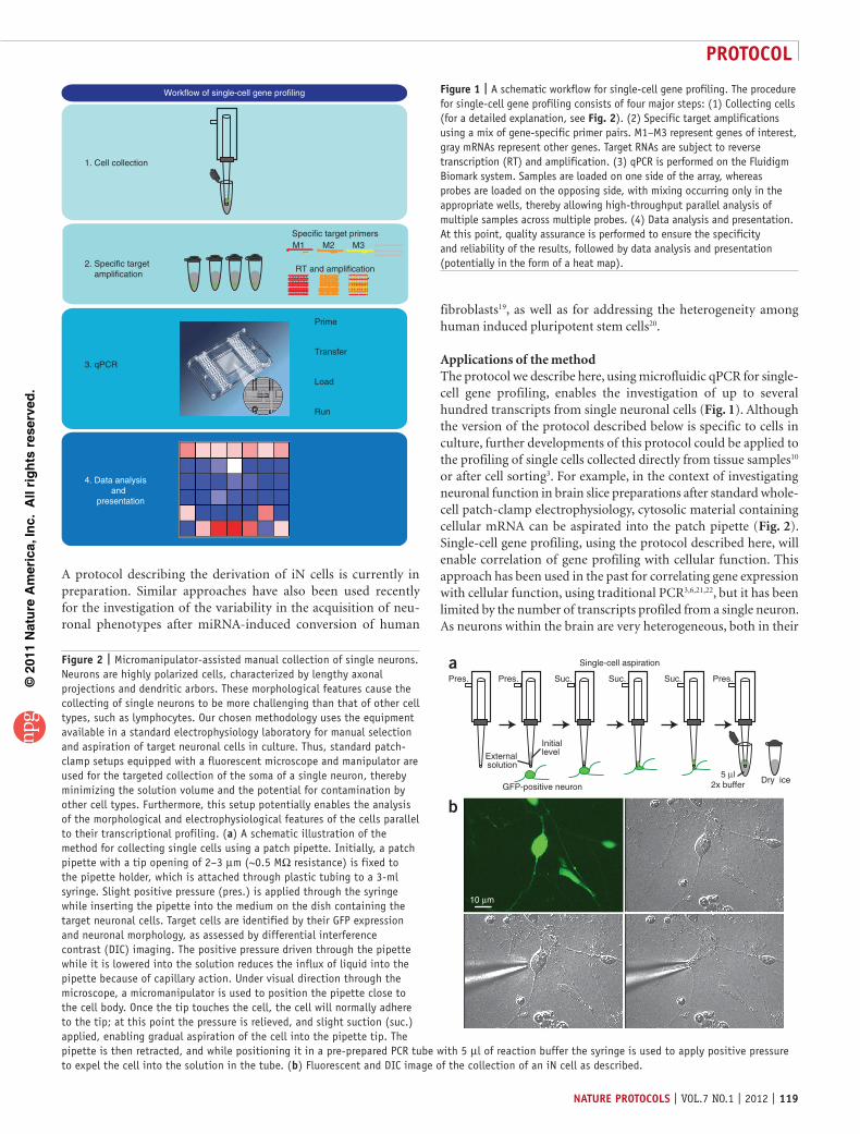
Workflow of single-cell gene profiling
1. Cell collection
3. qPCR
4. Data analysis and presentation
Prime
Transfer
Load
Run
2. Specific target amplification
Specific target primers
RT and amplification
M1 M2 M3
Figure 1 | A schematic workflow for single-cell gene profiling. The procedure for single-cell gene profiling consists of four major steps: (1) Collecting cells (for a detailed explanation, see Fig. 2). (2) Specific target amplifications using a mix of gene-specific primer pairs. M1–M3 represent genes of interest, gray mRNAs represent other genes. Target RNAs are subject to reverse transcription (RT) and amplification. (3) qPCR is performed on the Fluidigm Biomark system. Samples are loaded on one side of the array, whereas probes are loaded on the opposing side, with mixing occurring only in the appropriate wells, thereby allowing high-throughput parallel analysis of multiple samples across multiple probes. (4) Data analysis and presentation. At this point, quality assurance is performed to ensure the specificity and reliability of the results, followed by data analysis and presentation (potentially in the form of a heat map).
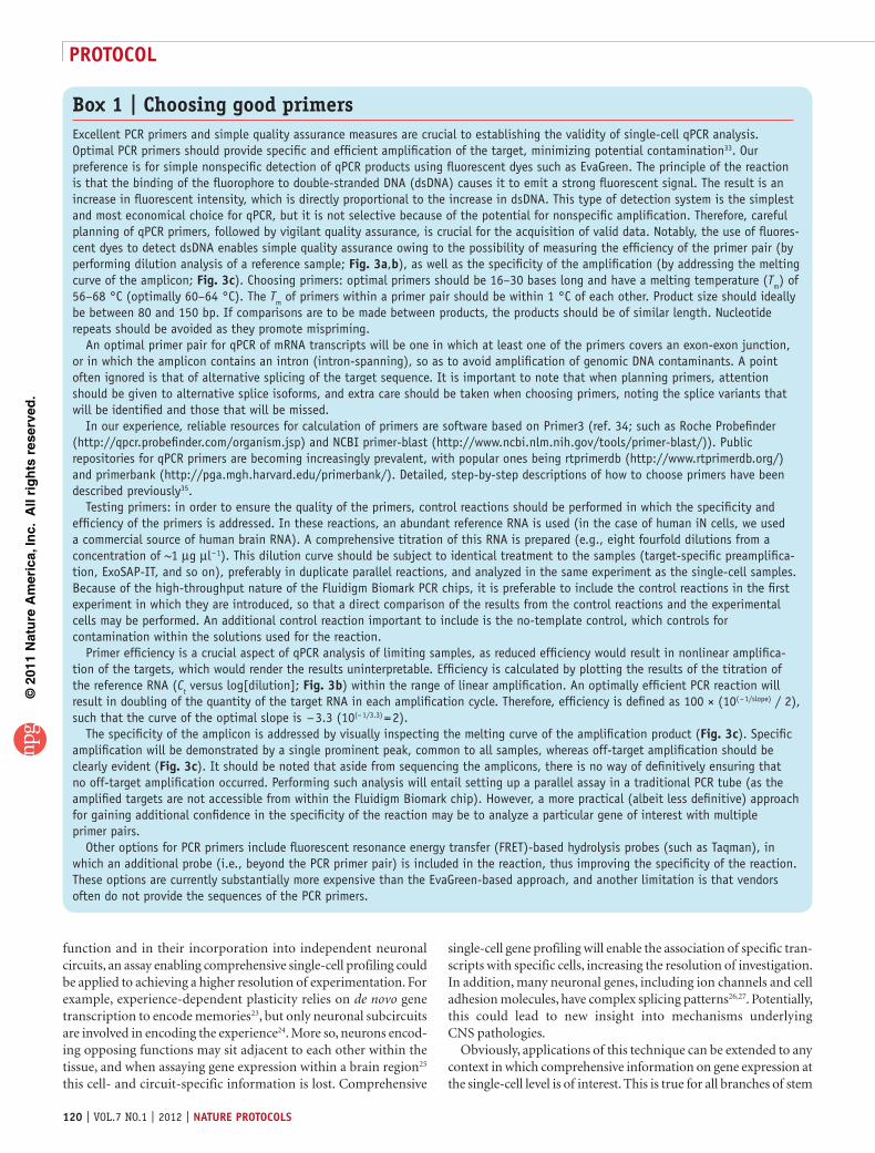
Figure 2 | Micromanipulator-assisted manual collection of single neurons. Neurons are highly polarized cells, characterized by lengthy axonal projections and dendritic arbors. These morphological features cause the collecting of single neurons to be more challenging than that of other cell types, such as lymphocytes. Our chosen methodology uses the equipment available in a standard electrophysiology laboratory for manual selection and aspiration of target neuronal cells in culture. Thus, standard patch-clamp setups equipped with a fluorescent microscope and manipulator are used for the targeted collection of the soma of a single neuron, thereby minimizing the solution volume and the potential for contamination by other cell types. Furthermore, this setup potentially enables the analysis of the morphological and electrophysiological features of the cells parallel to their transcriptional profiling. (a) A schematic illustration of the method for collecting single cells using a patch pipette. Initially, a patch pipette with a tip opening of 2–3 µm (~0.5 MΩ resistance) is fixed to the pipette holder, which is attached through plastic tubing to a 3-ml syringe. Slight positive pressure (pres.) is applied through the syringe while inserting the pipette into the medium on the dish containing the target neuronal cells. Target cells are identified by their GFP expression and neuronal morphology, as assessed by differential interference contrast (DIC) imaging. The positive pressure driven through the pipette while it is lowered into the solution reduces the influx of liquid into the pipette because of capillary action. Under visual direction through the microscope, a micromanipulator is used to position the pipette close to the cell body. Once the tip touches the cell, the cell will normally adhere to the tip; at this point the pressure is relieved, and slight suction (suc.) applied, enabling gradual aspiration of the cell into the pipette tip. The pipette is then retracted, and while positioning it in a pre-prepared PCR tube with 5 µl of reaction buffer the syringe is used to apply positive pressure to expel the cell into the solution in the tube. (b) Fluorescent and DIC image of the collection of an iN cell as described.
a
b
Single-cell aspiration
Suc.
Dry ice5 µl
2x buffer
Externalsolution
GFP-positive neuron
Pres. Pres.Pres. Suc. Suc.
Initiallevel
10 µm
©20
11 N
atu
re A
mer
ica,
Inc.
All
rig
hts
res
erve
d.
protocol
120 | VOL.7 NO.1 | 2012 | nature protocols
function and in their incorporation into independent neuronal circuits, an assay enabling comprehensive singlecell profiling could be applied to achieving a higher resolution of experimentation. For example, experiencedependent plasticity relies on de novo gene transcription to encode memories23, but only neuronal subcircuits are involved in encoding the experience24. More so, neurons encoding opposing functions may sit adjacent to each other within the tissue, and when assaying gene expression within a brain region25 this cell and circuitspecific information is lost. Comprehensive
singlecell gene profiling will enable the association of specific transcripts with specific cells, increasing the resolution of investigation. In addition, many neuronal genes, including ion channels and cell adhesion molecules, have complex splicing patterns26,27. Potentially, this could lead to new insight into mechanisms underlying CNS pathologies.
Obviously, applications of this technique can be extended to any context in which comprehensive information on gene expression at the singlecell level is of interest. This is true for all branches of stem
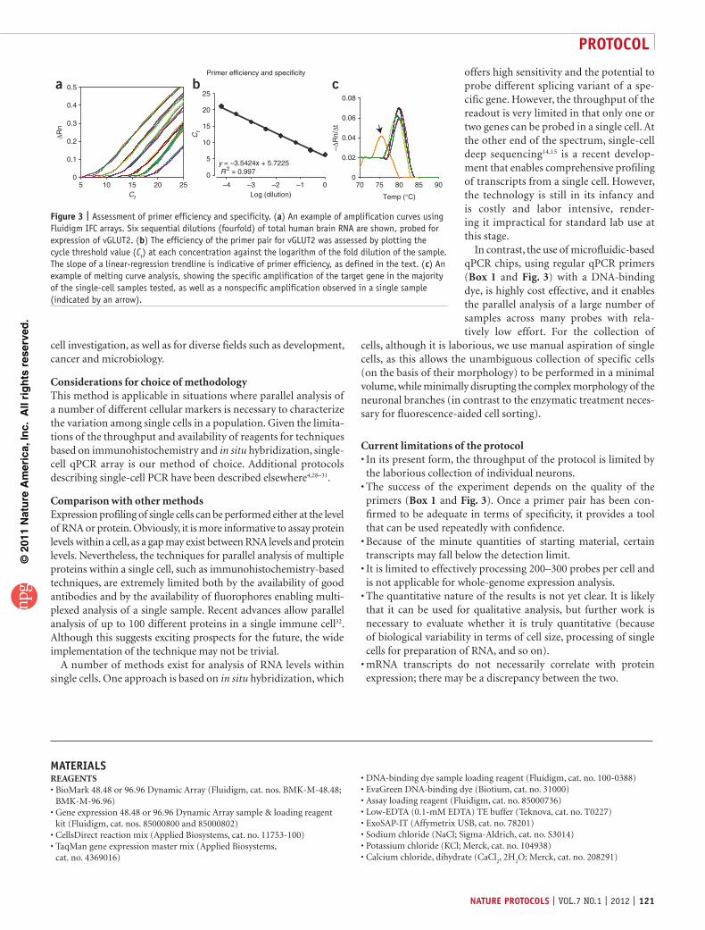
Box 1 | Choosing good primersExcellent PCR primers and simple quality assurance measures are crucial to establishing the validity of single-cell qPCR analysis. Optimal PCR primers should provide specific and efficient amplification of the target, minimizing potential contamination33. Our preference is for simple nonspecific detection of qPCR products using fluorescent dyes such as EvaGreen. The principle of the reaction is that the binding of the fluorophore to double-stranded DNA (dsDNA) causes it to emit a strong fluorescent signal. The result is an increase in fluorescent intensity, which is directly proportional to the increase in dsDNA. This type of detection system is the simplest and most economical choice for qPCR, but it is not selective because of the potential for nonspecific amplification. Therefore, careful planning of qPCR primers, followed by vigilant quality assurance, is crucial for the acquisition of valid data. Notably, the use of fluores-cent dyes to detect dsDNA enables simple quality assurance owing to the possibility of measuring the efficiency of the primer pair (by performing dilution analysis of a reference sample; Fig. 3a,b), as well as the specificity of the amplification (by addressing the melting curve of the amplicon; Fig. 3c). Choosing primers: optimal primers should be 16–30 bases long and have a melting temperature (Tm) of 56–68 °C (optimally 60–64 °C). The Tm of primers within a primer pair should be within 1 °C of each other. Product size should ideally be between 80 and 150 bp. If comparisons are to be made between products, the products should be of similar length. Nucleotide repeats should be avoided as they promote mispriming.
An optimal primer pair for qPCR of mRNA transcripts will be one in which at least one of the primers covers an exon-exon junction, or in which the amplicon contains an intron (intron-spanning), so as to avoid amplification of genomic DNA contaminants. A point often ignored is that of alternative splicing of the target sequence. It is important to note that when planning primers, attention should be given to alternative splice isoforms, and extra care should be taken when choosing primers, noting the splice variants that will be identified and those that will be missed.
In our experience, reliable resources for calculation of primers are software based on Primer3 (ref. 34; such as Roche Probefinder (http://qpcr.probefinder.com/organism.jsp) and NCBI primer-blast (http://www.ncbi.nlm.nih.gov/tools/primer-blast/)). Public repositories for qPCR primers are becoming increasingly prevalent, with popular ones being rtprimerdb (http://www.rtprimerdb.org/) and primerbank (http://pga.mgh.harvard.edu/primerbank/). Detailed, step-by-step descriptions of how to choose primers have been described previously35.
Testing primers: in order to ensure the quality of the primers, control reactions should be performed in which the specificity and efficiency of the primers is addressed. In these reactions, an abundant reference RNA is used (in the case of human iN cells, we used a commercial source of human brain RNA). A comprehensive titration of this RNA is prepared (e.g., eight fourfold dilutions from a concentration of ~1 µg µl − 1). This dilution curve should be subject to identical treatment to the samples (target-specific preamplifica-tion, ExoSAP-IT, and so on), preferably in duplicate parallel reactions, and analyzed in the same experiment as the single-cell samples. Because of the high-throughput nature of the Fluidigm Biomark PCR chips, it is preferable to include the control reactions in the first experiment in which they are introduced, so that a direct comparison of the results from the control reactions and the experimental cells may be performed. An additional control reaction important to include is the no-template control, which controls for contamination within the solutions used for the reaction.
Primer efficiency is a crucial aspect of qPCR analysis of limiting samples, as reduced efficiency would result in nonlinear amplifica-tion of the targets, which would render the results uninterpretable. Efficiency is calculated by plotting the results of the titration of the reference RNA (Ct versus log[dilution]; Fig. 3b) within the range of linear amplification. An optimally efficient PCR reaction will result in doubling of the quantity of the target RNA in each amplification cycle. Therefore, efficiency is defined as 100 × (10( − 1/slope) / 2), such that the curve of the optimal slope is − 3.3 (10(− 1/3.3) = 2).
The specificity of the amplicon is addressed by visually inspecting the melting curve of the amplification product (Fig. 3c). Specific amplification will be demonstrated by a single prominent peak, common to all samples, whereas off-target amplification should be clearly evident (Fig. 3c). It should be noted that aside from sequencing the amplicons, there is no way of definitively ensuring that no off-target amplification occurred. Performing such analysis will entail setting up a parallel assay in a traditional PCR tube (as the amplified targets are not accessible from within the Fluidigm Biomark chip). However, a more practical (albeit less definitive) approach for gaining additional confidence in the specificity of the reaction may be to analyze a particular gene of interest with multiple primer pairs.
Other options for PCR primers include fluorescent resonance energy transfer (FRET)-based hydrolysis probes (such as Taqman), in which an additional probe (i.e., beyond the PCR primer pair) is included in the reaction, thus improving the specificity of the reaction. These options are currently substantially more expensive than the EvaGreen-based approach, and another limitation is that vendors often do not provide the sequences of the PCR primers.
©20
11 N
atu
re A
mer
ica,
Inc.
All
rig
hts
res
erve
d.
protocol
nature protocols | VOL.7 NO.1 | 2012 | 121
cell investigation, as well as for diverse fields such as development, cancer and microbiology.
Considerations for choice of methodologyThis method is applicable in situations where parallel analysis of a number of different cellular markers is necessary to characterize the variation among single cells in a population. Given the limitations of the throughput and availability of reagents for techniques based on immunohistochemistry and in situ hybridization, single cell qPCR array is our method of choice. Additional protocols describing singlecell PCR have been described elsewhere4,28–31.
Comparison with other methodsExpression profiling of single cells can be performed either at the level of RNA or protein. Obviously, it is more informative to assay protein levels within a cell, as a gap may exist between RNA levels and protein levels. Nevertheless, the techniques for parallel analysis of multiple proteins within a single cell, such as immunohistochemistrybased techniques, are extremely limited both by the availability of good antibodies and by the availability of fluorophores enabling multiplexed analysis of a single sample. Recent advances allow parallel analysis of up to 100 different proteins in a single immune cell32. Although this suggests exciting prospects for the future, the wide implementation of the technique may not be trivial.
A number of methods exist for analysis of RNA levels within single cells. One approach is based on in situ hybridization, which
offers high sensitivity and the potential to probe different splicing variant of a specific gene. However, the throughput of the readout is very limited in that only one or two genes can be probed in a single cell. At the other end of the spectrum, singlecell deep sequencing14,15 is a recent development that enables comprehensive profiling of transcripts from a single cell. However, the technology is still in its infancy and is costly and labor intensive, rendering it impractical for standard lab use at this stage.
In contrast, the use of microfluidicbased qPCR chips, using regular qPCR primers (Box 1 and Fig. 3) with a DNAbinding dye, is highly cost effective, and it enables the parallel analysis of a large number of samples across many probes with relatively low effort. For the collection of
cells, although it is laborious, we use manual aspiration of single cells, as this allows the unambiguous collection of specific cells (on the basis of their morphology) to be performed in a minimal volume, while minimally disrupting the complex morphology of the neuronal branches (in contrast to the enzymatic treatment necessary for fluorescenceaided cell sorting).
Current limitations of the protocolIn its present form, the throughput of the protocol is limited by the laborious collection of individual neurons.The success of the experiment depends on the quality of the primers (Box 1 and Fig. 3). Once a primer pair has been confirmed to be adequate in terms of specificity, it provides a tool that can be used repeatedly with confidence.Because of the minute quantities of starting material, certain transcripts may fall below the detection limit.It is limited to effectively processing 200–300 probes per cell and is not applicable for wholegenome expression analysis.The quantitative nature of the results is not yet clear. It is likely that it can be used for qualitative analysis, but further work is necessary to evaluate whether it is truly quantitative (because of biological variability in terms of cell size, processing of single cells for preparation of RNA, and so on).mRNA transcripts do not necessarily correlate with protein expression; there may be a discrepancy between the two.
•
•
•
•
•
•
Primer efficiency and specificity
Temp (°C)
70 75 80 85 90
–∆R
n/∆t
0
0.02
0.04
0.06
0.08
0
5
10
15
20
25
–4 –3 –2 –1 0Log (dilution)
y = –3.5424x + 5.7225 R 2 = 0.997
Ct
5 10 15 20 250
0.1
0.2
0.3
0.4
0.5
Ct
∆Rn
a b c
Figure 3 | Assessment of primer efficiency and specificity. (a) An example of amplification curves using Fluidigm IFC arrays. Six sequential dilutions (fourfold) of total human brain RNA are shown, probed for expression of vGLUT2. (b) The efficiency of the primer pair for vGLUT2 was assessed by plotting the cycle threshold value (Ct) at each concentration against the logarithm of the fold dilution of the sample. The slope of a linear-regression trendline is indicative of primer efficiency, as defined in the text. (c) An example of melting curve analysis, showing the specific amplification of the target gene in the majority of the single-cell samples tested, as well as a nonspecific amplification observed in a single sample (indicated by an arrow).
MaterIalsREAGENTS
BioMark 48.48 or 96.96 Dynamic Array (Fluidigm, cat. nos. BMKM48.48; BMKM96.96)Gene expression 48.48 or 96.96 Dynamic Array sample & loading reagent kit (Fluidigm, cat. nos. 85000800 and 85000802)CellsDirect reaction mix (Applied Biosystems, cat. no. 11753100)TaqMan gene expression master mix (Applied Biosystems, cat. no. 4369016)
•
•
••
DNAbinding dye sample loading reagent (Fluidigm, cat. no. 1000388)EvaGreen DNAbinding dye (Biotium, cat. no. 31000)Assay loading reagent (Fluidigm, cat. no. 85000736)LowEDTA (0.1mM EDTA) TE buffer (Teknova, cat. no. T0227)ExoSAPIT (Affymetrix USB, cat. no. 78201)Sodium chloride (NaCl; SigmaAldrich, cat. no. S3014)Potassium chloride (KCl; Merck, cat. no. 104938)Calcium chloride, dihydrate (CaCl
2, 2H
2O; Merck, cat. no. 208291)
••••••••

©20
11 N
atu
re A
mer
ica,
Inc.
All
rig
hts
res
erve
d.
protocol
122 | VOL.7 NO.1 | 2012 | nature protocols
Magnesium chloride, hexahydrate (MgCl2, 6H
2O; AppliChem, cat. no. A3618)
HEPES (SigmaAldrich, cat. no. H3375)d( + )Glucose (SigmaAldrich, cat. no. G8270)Clear adhesive tape (e.g., Scotch tape, 3M)
EQUIPMENT crItIcal All equipment should be treated as potentially contaminated with RNase.
Inverted tissue culture microscope with phase contrast and epifluorescence, equipped with ×10 and ×40 objectives (Axiovert 40 CFL, Zeiss; Olympus BX51W upright microscope (Olympus); ×4 objective (Olympus XL Fluoro ×4/340 objective, 0.28 NA, Olympus)TMC 65560 antivibration table (Technical Manufacturing Corporation)Glass micropipettes (inner diameter 1.0 ± 0.05 mm; outer diameter 1.5 ± 0.05 mm; Garner Glass Company, cat. no. KG33)PC10 glass microelectrode puller (Narishige)PCR tube strips (USA Scientific)TubingSyringe (3 ml; BD Bioscience)Micromanipulator (Sutter, MPC385)Primer design software (Box 1)
••••
•
••
••••••
Genepattern software package (available from the Broad Institute; http://genepattern.broadinstitute.org)Biomark system (Fluidigm; Fluidigm integrated fluidic circuit (IFC) controller, realtime PCR analysis software)
REAGENT SETUPExtracellular solution Make up a bath solution containing 140 mM NaCl, 5 mM KCl, 2 mM CaCl
2, 2 mM MgCl
2, 10 mM HEPES and 10 mM glucose,
pH 7.4. The final osmolarity of the solution should be 310 ± 10 mOsm. crItIcal Store the solution at 4 °C for up to 1 week.Primer and sample organization Owing to the relatively high throughput nature of the protocol (analyzing up to 96 samples across 96 probes), the organization of the components is extremely important. Our preference is to use tubes of different colors for the samples and the probes, organize them consistently (e.g., strips of 8, organized on 96well plates) and label them with a simple code (e.g., numbers for probes and letters for samples).Glass pipette preparation Prepare glass pipettes using a PC10 twostage puller. Adjust the heating temperature of the two steps so that the tip of the glass pipette is close to a diameter of ~2 to 3 µm (~0.5 MΩ).
•
•
proceDurecell aspiration tIMInG 5 min per cell1| Prepare PCR strip tubes with 5 µl of 2× CellsDirect reaction mix per tube (Fig. 1). Keep the tubes on ice. crItIcal step Use RNase-free reagents and RNA protective technique throughout the protocol (clean surfaces of working area, wear gloves, avoid mouth pipetting, and use RNase-free filter tips and tubes).
2| Mount the pipette (with a tip opening between 2 and 3 µm) on a micromanipulator connected to tubing to enable transfer of pressure through the pipette.
3| Visually identify the isolated cell of choice under the microscope, use a micromanipulator to approach it with positive pressure (applied through a 3-ml syringe) to clear its surroundings and aspirate it into a patch pipette, ensuring minimal contamination by surrounding cells or cell debris (Fig. 2). crItIcal step Use minimal pressure to aspirate the cell so as to raise a minimal amount of fluid into the pipette.
4| Retract the pipette and remove the holder with syringe from the setup (maintaining the connection to the syringe). Lower the pipette tip into the solution at the bottom of the PCR tube. Apply positive pressure to eject the cell into the solution while retracting the tip of the pipette out of the solution. Freeze the cell immediately on dry ice. crItIcal step Use minimal pressure to eject the cell into the solution so as not to blow the solution out of the tube. Maintaining positive pressure while retracting the pipette is important for minimizing capillary action that could drive solution back into the tip of the pipette. crItIcal step Each micropipette can be used only once for transferring each individual cell into reaction buffer. crItIcal step It is essential to include a negative control sample for the qPCR analysis, in which no individual cell is picked; instead, transfer external solution equivalent to the carryover volume collected during the single-cell transfer into reaction buffer. The negative control will enable the detection of contamination in the subsequent steps of the procedure. pause poInt Cells can be stored for several months at − 80 °C until processing.
specific target amplification tIMInG 6 h5| Prepare primer pairs at a concentration of 20 µM for each primer. Mix the primers used for specific-target amplification (up to 100 primer pairs), bringing the final concentration of each primer to 200 nM (if you are using < 100 primer pairs, dilute accordingly with RNase-free water). Large stock solutions of this primer mix may be made and stored at − 20 °C if the same primers are to be used continuously. Note that if more than 100 genes are to be assayed, higher stock concentrations of primer pairs should be made (above 20 µM).
©20
11 N
atu
re A
mer
ica,
Inc.
All
rig
hts
res
erve
d.
protocol
nature protocols | VOL.7 NO.1 | 2012 | 123
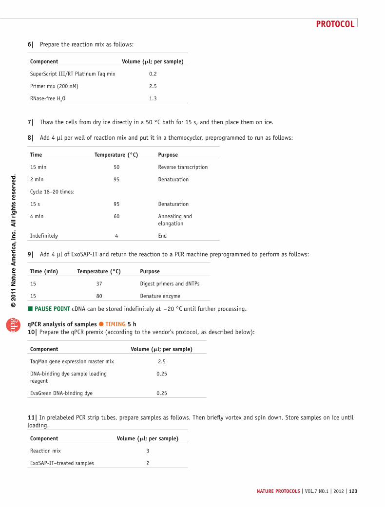
6| Prepare the reaction mix as follows:
component Volume (ml; per sample)
SuperScript III/RT Platinum Taq mix 0.2
Primer mix (200 nM) 2.5
RNase-free H2O 1.3
7| Thaw the cells from dry ice directly in a 50 °C bath for 15 s, and then place them on ice.
8| Add 4 µl per well of reaction mix and put it in a thermocycler, preprogrammed to run as follows:
time temperature (°c) purpose
15 min 50 Reverse transcription
2 min 95 Denaturation
Cycle 18–20 times:
15 s 95 Denaturation
4 min 60 Annealing and elongation
Indefinitely 4 End
9| Add 4 µl of ExoSAP-IT and return the reaction to a PCR machine preprogrammed to perform as follows:
time (min) temperature (°c) purpose
15 37 Digest primers and dNTPs
15 80 Denature enzyme
pause poInt cDNA can be stored indefinitely at − 20 °C until further processing.
qpcr analysis of samples tIMInG 5 h10| Prepare the qPCR premix (according to the vendor’s protocol, as described below):
component Volume (ml; per sample)
TaqMan gene expression master mix 2.5
DNA-binding dye sample loading reagent
0.25
EvaGreen DNA-binding dye 0.25
11| In prelabeled PCR strip tubes, prepare samples as follows. Then briefly vortex and spin down. Store samples on ice until loading.
component Volume (ml; per sample)
Reaction mix 3
ExoSAP-IT–treated samples 2
©20
11 N
atu
re A
mer
ica,
Inc.
All
rig
hts
res
erve
d.
protocol
124 | VOL.7 NO.1 | 2012 | nature protocols
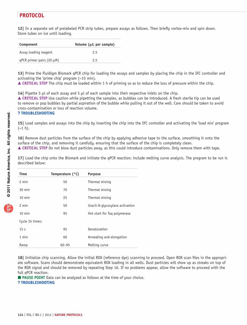
12| In a separate set of prelabeled PCR strip tubes, prepare assays as follows. Then briefly vortex-mix and spin down. Store tubes on ice until loading.
component Volume (ml; per sample)
Assay loading reagent 2.5
qPCR primer pairs (20 µM) 2.5
13| Prime the Fluidigm Biomark qPCR chip for loading the assays and samples by placing the chip in the IFC controller and activating the ‘prime chip’ program (~15 min). crItIcal step The chip must be loaded within 1 h of priming so as to reduce the loss of pressure within the chip.
14| Pipette 5 µl of each assay and 5 µl of each sample into their respective inlets on the chip. crItIcal step Use caution while pipetting the samples, as bubbles can be introduced. A fresh sterile tip can be used to remove or pop bubbles by partial aspiration of the bubble while pulling it out of the well. Care should be taken to avoid cross-contamination or loss of reaction volume.? trouBlesHootInG
15| Load samples and assays into the chip by inserting the chip into the IFC controller and activating the ‘load mix’ program (~1 h).
16| Remove dust particles from the surface of the chip by applying adhesive tape to the surface, smoothing it onto the surface of the chip, and removing it carefully, ensuring that the surface of the chip is completely clean. crItIcal step Do not blow dust particles away, as this could introduce contaminations. Only remove them with tape.
17| Load the chip onto the Biomark and initiate the qPCR reaction; include melting curve analysis. The program to be run is described below:
time temperature (°c) purpose
2 min 50 Thermal mixing
30 min 70 Thermal mixing
10 min 25 Thermal mixing
2 min 50 Uracil-N-glycosylase activation
10 min 95 Hot start for Taq polymerase
Cycle 35 times:
15 s 95 Denaturation
1 min 60 Annealing and elongation
Ramp 60–95 Melting curve
18| Initialize chip scanning. Allow the initial ROX (reference dye) scanning to proceed. Open ROX scan files in the appropri-ate software. Scans should demonstrate equivalent ROX loading in all wells. Dust particles will show up as streaks on top of the ROX signal and should be removed by repeating Step 16. If no problems appear, allow the software to proceed with the full qPCR reaction. pause poInt Data can be analyzed as follows at the time of your choice.? trouBlesHootInG
©20
11 N
atu
re A
mer
ica,
Inc.
All
rig
hts
res
erve
d.
protocol
nature protocols | VOL.7 NO.1 | 2012 | 125
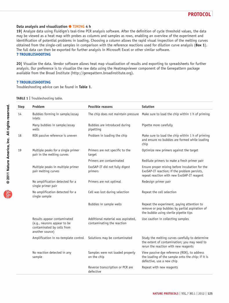
Data analysis and visualization tIMInG 4 h19| Analyze data using Fluidigm’s teal-time PCR analysis software. After the definition of cycle threshold values, the data may be viewed as a heat map with probes as columns and samples as rows, enabling an overview of the experiment and identification of potential problems in loading. Choosing a column allows the rapid visual inspection of the melting curves obtained from the single-cell samples in comparison with the reference reactions used for dilution curve analysis (Box 1). The full data can then be exported for further analysis in Microsoft Excel or other similar software.? trouBlesHootInG
20| Visualize the data. Vendor software allows heat map visualization of results and exporting to spreadsheets for further analysis. Our preference is to visualize the raw data using the Heatmapviewer component of the Genepattern package available from the Broad Institute (http://genepattern.broadinstitute.org).
? trouBlesHootInGTroubleshooting advice can be found in table 1.
taBle 1 | Troubleshooting table.
step problem possible reasons solution
14 Bubbles forming in sample/assay inlets
The chip does not maintain pressure Make sure to load the chip within 1 h of priming
Many bubbles in sample/assay wells
Bubbles are introduced during pipetting
Pipette more carefully
18 ROX passive reference is uneven Problem in loading the chip Make sure to load the chip within 1 h of priming and ensure no bubbles are formed while loading chip
19 Multiple peaks for a single primer pair in the melting curves
Primers are not specific to the target
Optimize new primers against the target
Primers are contaminated Redilute primers to make a fresh primer pair
Multiple peaks in multiple primer pair melting curves
ExoSAP-IT did not fully digest primers
Ensure proper mixing before incubation for the ExoSAP-IT reaction; if the problem persists, repeat reaction with new ExoSAP-IT reagent
No amplification detected for a single primer pair
Primers are not optimal Redesign primer pair
No amplification detected for a single sample
Cell was lost during selection Repeat the cell selection
Bubbles in sample wells Repeat the experiment, paying attention to remove or pop bubbles by partial aspiration of the bubble using sterile pipette tips
Results appear contaminated (e.g., neurons appear to be contaminated by cells from another source)
Additional material was aspirated, contaminating the reaction
Use caution in collecting samples
Amplification in no-template control Solutions may be contaminated Study the melting curves carefully to determine the extent of contamination; you may need to rerun the reaction with new reagents
No reaction detected in any sample
Samples were not loaded properly on the chip
View passive dye reference (ROX), to address the loading of the sample onto the chip; if it is defective, use a new chip
Reverse transcription or PCR are defective
Repeat with new reagents

©20
11 N
atu
re A
mer
ica,
Inc.
All
rig
hts
res
erve
d.
protocol
126 | VOL.7 NO.1 | 2012 | nature protocols
tIMInGSteps 1–4, Single-cell aspiration (day 1): 5 min per cell and 6 h for ~50 cellsSteps 5–9, Specific target amplification (day 2): 6 hSteps 10–18, qPCR analysis of samples (day 2): 5 hSteps 19 and 20, Data analysis and visualization (day 3): 4 h
antIcIpateD resultsThe Biomark system is equipped with user-friendly qPCR analysis software, in which results may be presented either as a heat map or spreadsheet, and all the key parameters of the reaction such as the threshold cycles and melting temperature are listed. Because of the high-throughput nature of the assay, we routinely include a number of internal controls, such as ACTB, GAPDH and MAP2, as well as neuron-specific reference genes, such as TUBB3, NCAM1 and SYN1, with the expectation that essentially the whole population of neuronal cells assayed will express these reference genes. The signal obtained from the internal controls enables quality assurance for multiple steps in the process, from the aspiration of single cells, through reverse transcription, specific target amplification and qPCR. Furthermore, a no-template control should be routinely included in order to identify potential sources of contamination. Identification of cellular transformation to a specific neuronal subtype is made possible by the use of neuron subtype–specific probes (Fig. 4). If the protocol is followed carefully, a success rate of close to 100% (as evaluated on the basis of the amplification of reference genes in the single cells) is expected. Although we have routinely performed two or three replicates per reaction (either loading the samples or the probes in replicates) and have essentially always observed consistent results, we recommend performing the assay in duplicate unless there are experimental limitations.
8 Ct 34 22
GAPDHTUBB3
NCAMDCX
SYN1
OLIG2GFAP
PAVLBTH
vGATGAD65
vGLUT2vGLUT1
MAP2
GAD67
HFF iN cells
Single-cell gene profiling in iN cells
Figure 4 | Single-cell gene expression profiling using Fluidigm dynamic arrays. Rows represent the evaluated genes and columns represent individual cells. Heat map (blue to red) represents the threshold Ct values as indicated. HFF, human fetal fibroblast. From Pang et al.18.
acknowleDGMents A.C. acknowledges the generous support of the AXA Research Fund. Z.P.P. is supported by the Brain and Behavior Research Foundation (National Alliance for Research on Schizophrenia and Depression (NARSAD) Young Investigator Award) and the Robert Wood Johnson Foundation. We thank N. Yang for help in experimental work. We thank S. Chavez for initial instruction in the use of the Fluidigm Biomark system, and R. Reijo Pera for access to the Fluidigm Biomark system in her lab. We also thank members of the Malenka and Südhof labs for their comments on the manuscript.
autHor contrIButIons A.C. and Z.P.P. conceived the protocol, collected the data, presented the figures and wrote the manuscript. T.C.S., M.W. and R.C.M. supervised the project and contributed to the writing.
coMpetInG FInancIal Interests The authors declare no competing financial interests.
Published online at http://www.natureprotocols.com/. Reprints and permissions information is available online at http://www.nature.com/reprints/index.html.
1. Dolmetsch, R. & Geschwind, D.H. The human brain in a dish: the promise of iPSC-derived neurons. Cell 145, 831–834 (2011).
2. Li, H.H. et al. Amplification and analysis of DNA sequences in single human sperm and diploid cells. Nature 335, 414–417 (1988).
3. Cauli, B. et al. Molecular and physiological diversity of cortical nonpyramidal cells. J. Neurosci. 17, 3894–3906 (1997).
4. Cauli, B. & Lambolez, B. in Unravelling Single Cell Genomics 81–92 (The Royal Society of Chemistry, 2010).
5. Koirala, S. & Corfas, G. Identification of novel glial genes by single-cell transcriptional profiling of Bergmann glial cells from mouse cerebellum. PLoS One 5, e9198 (2011).
6. Lambolez, B. et al. AMPA receptor subunits expressed by single Purkinje cells. Neuron 9, 247–258 (1992).
7. Surmeier, D.J. et al. Dopamine receptor subtypes colocalize in rat striatonigral neurons. Proc. Natl. Acad. Sci. USA 89, 10178–10182 (1992).
8. Tietjen, I., Rihel, J. & Dulac, C.G. Single-cell transcriptional profiles and spatial patterning of the mammalian olfactory epithelium. Int. J. Dev. Biol. 49, 201–207 (2005).
9. Tietjen, I. et al. Single-cell transcriptional analysis of neuronal progenitors. Neuron 38, 161–175 (2003).
10. Mackler, S.A., Brooks, B.P. & Eberwine, J.H. Stimulus-induced coordinate changes in mRNA abundance in single postsynaptic hippocampal CA1 neurons. Neuron 9, 539–548 (1992).
11. Mackler, S.A. & Eberwine, J.H. Diversity of glutamate receptor subunit mRNA expression within live hippocampal CA1 neurons. Mol. Pharmacol. 44, 308–315 (1993).
12. Ginsberg, S.D. et al. Single-cell gene expression analysis: implications for neurodegenerative and neuropsychiatric disorders. Neurochem. Res. 29, 1053–1064 (2004).
13. Sucher, N.J. & Deitcher, D.L. PCR and patch-clamp analysis of single neurons. Neuron 14, 1095–1100 (1995).
©20
11 N
atu
re A
mer
ica,
Inc.
All
rig
hts
res
erve
d.
protocol
nature protocols | VOL.7 NO.1 | 2012 | 127
14. Lao, K.Q. et al. mRNA-sequencing whole transcriptome analysis of a single cell on the SOLiD system. J. Biomol. Tech. 20, 266–271 (2009).
15. Tang, F. et al. mRNA-Seq whole-transcriptome analysis of a single cell. Nat. Methods 6, 377–382 (2009).
16. White, A.K. et al. High-throughput microfluidic single-cell RT-qPCR. Proc. Natl. Acad. Sci. 108, 13999–14004 (2011).
17. Spurgeon, S.L., Jones, R.C. & Ramakrishnan, R. High throughput gene expression measurement with real time PCR in a microfluidic dynamic array. PLoS One 3, e1662 (2008).
18. Pang, Z.P. et al. Induction of human neuronal cells by defined transcription factors. Nature 476, 220–223 (2011).
19. Yoo, A.S. et al. MicroRNA-mediated conversion of human fibroblasts to neurons. Nature 476, 228–231 (2011).
20. Narsinh, K.H. et al. Single cell transcriptional profiling reveals heterogeneity of human induced pluripotent stem cells. J. Clin. Invest. 121, 1217–1221 (2011).
21. Liss, B. & Roeper, J. Correlating function and gene expression of individual basal ganglia neurons. Trends Neurosci. 27, 475–481 (2004).
22. Weng, J.Y., Lin, Y.C. & Lien, C.C. Cell type-specific expression of acid-sensing ion channels in hippocampal interneurons. J. Neurosci. 30, 6548–6558 (2010).
23. McClung, C.A. & Nestler, E.J. Neuroplasticity mediated by altered gene expression. Neuropsychopharmacology 33, 3–17 (2008).
24. Lammel, S., Ion, D.I., Roeper, J. & Malenka, R.C. Projection-specific modulation of dopamine neuron synapses by aversive and rewarding stimuli. Neuron 70, 855–862 (2011).
25. Lobo, M.K. et al. Cell type-specific loss of BDNF signaling mimics optogenetic control of cocaine reward. Science 330, 385–390 (2010).
26. Allen, S.E., Darnell, R.B. & Lipscombe, D. The neuronal splicing factor Nova controls alternative splicing in N-type and P-type CaV2 calcium channels. Channels (Austin) 4, 483–489 (2010).
27. Bharadwaj, R. & Kolodkin, A.L. Descrambling Dscam diversity. Cell 125, 421–424 (2006).
28. Hodne, K., Haug, T.M. & Weltzien, F.A. Single-cell qPCR on dispersed primary pituitary cells—an optimized protocol. BMC Mol. Biol. 11, 82 (2010).
29. Morris, J., Singh, J.M. & Eberwine, J.H. Transcriptome analysis of single cells. J. Vis. Exp. published online, doi:10.3791/2634 (2011).
30. Esumi, S., Kaneko, R., Kawamura, Y. & Yagi, T. Split single-cell RT-PCR analysis of Purkinje cells. Nat. Protoc. 1, 2143–2151 (2006).
31. Li, Y. et al. An improved one-tube RT-PCR protocol for analyzing single-cell gene expression in individual mammalian cells. Anal. Bioanal. Chem. 397, 1853–1859 (2010).
32. Bendall, S.C. et al. Single-cell mass cytometry of differential immune and drug responses across a human hematopoietic continuum. Science 332, 687–696 (2011).
33. Bustin, S.A. et al. The MIQE guidelines: minimum information for publication of quantitative real-time PCR experiments. Clin. Chem. 55, 611–622 (2009).
34. Rozen, S. & Skaletsky, H. Primer3 on the WWW for general users and for biologist programmers. Methods Mol. Biol. 132, 365–386 (2000).
35. Thornton, B. & Basu, C. Real-time PCR (qPCR) primer design using free online software. Biochem. Mol. Biol. Educ. 39, 145–154 (2011).