Complex formation between the NS3 serine-type proteinase of the hepatitis C virus and NS4A and its...
10
JOURNAL OF VIROLOGY, Dec. 1995, p. 7519–7528 Vol. 69, No. 12 0022-538X/95/$04.0010 Copyright q 1995, American Society for Microbiology Complex Formation between the NS3 Serine-Type Proteinase of the Hepatitis C Virus and NS4A and Its Importance for Polyprotein Maturation RALF BARTENSCHLAGER, 1 * VOLKER LOHMANN, 1 TREVOR WILKINSON, 2 AND JAN OLIVER KOCH 1 Institute for Virology, Johannes-Gutenberg University Mainz, 55131 Mainz, Germany, 1 and Roche Products Ltd., Welwyn Garden City, Hertfordshire AL7 3AY, England Received 30 May 1995/Accepted 12 September 1995 Processing of the hepatitis C virus polyprotein is mediated by host cell signalases and at least two virally encoded proteinases. Of these, the serine-type proteinase encompassing the amino-terminal one-third of NS3 is responsible for cleavage at the four sites carboxy terminal of NS3. The activity of this proteinase is modulated by NS4A, a 54-amino-acid polyprotein cleavage product essential for processing at the NS3/4A, NS4A/4B, and NS4B/5A sites and enhancing cleavage efficiency between NS5A and NS5B. Using the vaccinia virus-T7 hybrid system to express hepatitis C virus polypeptides in BHK-21 cells, we studied the role of NS4A in proteinase activation. We found that the NS3 proteinase and NS4A form a stable complex when expressed as a single polyprotein or as separate molecules. Results from deletion mapping show that the minimal NS4A domain required for proteinase activation is located in the center of NS4A between amino acids 1675 and 1686 of the polyprotein. Amino acid substitutions within this domain destabilizing the NS3-NS4A complex also impair trans cleavage at the NS4A-dependent sites. Similarly, deletion of amino-terminal NS3 sequences impairs complex formation as well as cleavage at the NS4B/5A site but not at the NS4A-independent NS5A/5B site. These results suggest that a stable NS3-NS4A interaction is important for cleavage at the NS4A-dependent sites and that amino-terminal NS3 sequences and the central NS4A domain are directly involved in complex formation. Hepatitis C viruses (HCV) were identified as the main agents of the parentally transmitted form of non-A, non-B hepatitis (for a recent review, see reference 7). HCV cause the vast majority of transfusion-associated cases of hepatitis and account for a significant proportion of community-acquired hepatitis cases worldwide. Most, if not all, infections with HCV become chronic and result in various clinical outcomes includ- ing acute hepatitis, chronic hepatitis, liver cirrhosis, or the establishment of an asymptomatic carrier state which may per- sist for life. Furthermore, the increased incidence of hepato- cellular carcinoma in non-A, non-B hepatitis patients suggests an important role of HCV in the process of hepatocarcinogen- esis (7, 35). Sequence comparisons between various HCV cDNAs iso- lated during the past few years indicate the existence of several genotypes which can diverge by as much as 50% at the amino acid level (reference 36 and references cited therein). They are now grouped as the separate genus hepatitis C virus together with the two other genera Flavivirus and Pestivirus in the family Flaviviridae. These viruses have in common a virion with a lipid envelope and a single-stranded nonsegmented RNA of posi- tive polarity ranging in size between 9.4 (HCV) and 12.5 (pes- tivirus) kb. The RNA contains a single long open reading frame which in the case of HCV is expressed as a polyprotein of about 3,010 amino acids and which is flanked by 59 and 39 nontranslated regions. Given the lack of a convenient tissue culture system to propagate HCV, cell-free translation and cell culture expression studies were employed to analyze polypro- tein maturation. At least 10 cleavage products are generated from the polyprotein precursor and they are arranged in the order NH 2 -core-E1-E2-p7-NS2-NS3-NS4A-NS4B-NS5A-NS5B- COOH (18, 19, 21, 26, 30, 34, 39). Core, a basic RNA-binding protein, is assumed to be the major constituent of the viral nucleocapsid (reference 32 and references cited therein), E1 and E2 are most likely virion envelope glycoproteins (refer- ence 9 and references cited therein), and NS2 to NS5B are nonstructural proteins (most of them probably involved in RNA replication). Host cell signal peptidase located in the lumen of the endoplasmic reticulum is responsible for matu- ration of the structural proteins (19, 26, 30). At least two virally encoded proteinases are involved in processing of the NS pro- teins. A zinc-dependent metalloproteinase encompassing the NS2 region and the amino-terminal one-third of NS3 (the NS2-3 proteinase) is required for cleavage between NS2 and NS3 (16, 20, 22). Processing at the NS3/4A, NS4A/4B, NS4B/ 5A, and NS5A/5B sites is mediated by a serine-type proteinase which is also encoded in the amino-terminal one-third of NS3 but functioning independently from the NS2-3 proteinase (4, 10, 17, 20, 29, 39). We and others have recently shown that NS4A is an important NS3 serine-type proteinase cofactor required for cleavage at the NS3/4A, NS4A/4B, and NS4B/5A sites and enhancing cleavage efficiency between NS5A and NS5B (5, 12, 27, 38). The requirement for a proteinase cofactor is reminiscent of the situation of the closely related flaviviruses such as yellow fever virus and dengue virus. Processing of their NS proteins and maturation of nucleocapsid protein are mediated essen- tially by a proteinase heterodimer composed of the NS3 pro- teinase and the NS2B cofactor (reference 1 and references cited therein). Since mutations in NS2B affecting complex for- mation also impair proteolytic processing by NS3, formation of * Corresponding author. Mailing address: Institute for Virology, Johannes-Gutenberg University Mainz, Obere Zahlbacher Strasse 67, 55131 Mainz, Germany. Phone: 49-6131-173134. Fax: 49-6131-392359. 7519 on July 4, 2015 by guest http://jvi.asm.org/ Downloaded from
-
Upload
independent -
Category
Documents
-
view
4 -
download
0
Transcript of Complex formation between the NS3 serine-type proteinase of the hepatitis C virus and NS4A and its...
JOURNAL OF VIROLOGY, Dec. 1995, p. 7519–7528 Vol. 69, No. 120022-538X/95/$04.0010Copyright q 1995, American Society for Microbiology
Complex Formation between the NS3 Serine-Type Proteinaseof the Hepatitis C Virus and NS4A and Its Importance
for Polyprotein MaturationRALF BARTENSCHLAGER,1* VOLKER LOHMANN,1 TREVOR WILKINSON,2
AND JAN OLIVER KOCH1
Institute for Virology, Johannes-Gutenberg University Mainz, 55131 Mainz, Germany,1 andRoche Products Ltd., Welwyn Garden City, Hertfordshire AL7 3AY, England
Received 30 May 1995/Accepted 12 September 1995
Processing of the hepatitis C virus polyprotein is mediated by host cell signalases and at least two virallyencoded proteinases. Of these, the serine-type proteinase encompassing the amino-terminal one-third of NS3is responsible for cleavage at the four sites carboxy terminal of NS3. The activity of this proteinase ismodulated by NS4A, a 54-amino-acid polyprotein cleavage product essential for processing at the NS3/4A,NS4A/4B, and NS4B/5A sites and enhancing cleavage efficiency between NS5A and NS5B. Using the vacciniavirus-T7 hybrid system to express hepatitis C virus polypeptides in BHK-21 cells, we studied the role of NS4Ain proteinase activation. We found that the NS3 proteinase and NS4A form a stable complex when expressedas a single polyprotein or as separate molecules. Results from deletion mapping show that the minimal NS4Adomain required for proteinase activation is located in the center of NS4A between amino acids 1675 and 1686of the polyprotein. Amino acid substitutions within this domain destabilizing the NS3-NS4A complex alsoimpair trans cleavage at the NS4A-dependent sites. Similarly, deletion of amino-terminal NS3 sequencesimpairs complex formation as well as cleavage at the NS4B/5A site but not at the NS4A-independent NS5A/5Bsite. These results suggest that a stable NS3-NS4A interaction is important for cleavage at the NS4A-dependentsites and that amino-terminal NS3 sequences and the central NS4A domain are directly involved in complexformation.
Hepatitis C viruses (HCV) were identified as the mainagents of the parentally transmitted form of non-A, non-Bhepatitis (for a recent review, see reference 7). HCV cause thevast majority of transfusion-associated cases of hepatitis andaccount for a significant proportion of community-acquiredhepatitis cases worldwide. Most, if not all, infections with HCVbecome chronic and result in various clinical outcomes includ-ing acute hepatitis, chronic hepatitis, liver cirrhosis, or theestablishment of an asymptomatic carrier state which may per-sist for life. Furthermore, the increased incidence of hepato-cellular carcinoma in non-A, non-B hepatitis patients suggestsan important role of HCV in the process of hepatocarcinogen-esis (7, 35).Sequence comparisons between various HCV cDNAs iso-
lated during the past few years indicate the existence of severalgenotypes which can diverge by as much as 50% at the aminoacid level (reference 36 and references cited therein). They arenow grouped as the separate genus hepatitis C virus togetherwith the two other genera Flavivirus and Pestivirus in the familyFlaviviridae. These viruses have in common a virion with a lipidenvelope and a single-stranded nonsegmented RNA of posi-tive polarity ranging in size between 9.4 (HCV) and 12.5 (pes-tivirus) kb. The RNA contains a single long open readingframe which in the case of HCV is expressed as a polyproteinof about 3,010 amino acids and which is flanked by 59 and 39nontranslated regions. Given the lack of a convenient tissueculture system to propagate HCV, cell-free translation and cellculture expression studies were employed to analyze polypro-
tein maturation. At least 10 cleavage products are generatedfrom the polyprotein precursor and they are arranged in theorder NH2-core-E1-E2-p7-NS2-NS3-NS4A-NS4B-NS5A-NS5B-COOH (18, 19, 21, 26, 30, 34, 39). Core, a basic RNA-bindingprotein, is assumed to be the major constituent of the viralnucleocapsid (reference 32 and references cited therein), E1and E2 are most likely virion envelope glycoproteins (refer-ence 9 and references cited therein), and NS2 to NS5B arenonstructural proteins (most of them probably involved inRNA replication). Host cell signal peptidase located in thelumen of the endoplasmic reticulum is responsible for matu-ration of the structural proteins (19, 26, 30). At least two virallyencoded proteinases are involved in processing of the NS pro-teins. A zinc-dependent metalloproteinase encompassing theNS2 region and the amino-terminal one-third of NS3 (theNS2-3 proteinase) is required for cleavage between NS2 andNS3 (16, 20, 22). Processing at the NS3/4A, NS4A/4B, NS4B/5A, and NS5A/5B sites is mediated by a serine-type proteinasewhich is also encoded in the amino-terminal one-third of NS3but functioning independently from the NS2-3 proteinase (4,10, 17, 20, 29, 39). We and others have recently shown thatNS4A is an important NS3 serine-type proteinase cofactorrequired for cleavage at the NS3/4A, NS4A/4B, and NS4B/5Asites and enhancing cleavage efficiency between NS5A andNS5B (5, 12, 27, 38).The requirement for a proteinase cofactor is reminiscent of
the situation of the closely related flaviviruses such as yellowfever virus and dengue virus. Processing of their NS proteinsand maturation of nucleocapsid protein are mediated essen-tially by a proteinase heterodimer composed of the NS3 pro-teinase and the NS2B cofactor (reference 1 and referencescited therein). Since mutations in NS2B affecting complex for-mation also impair proteolytic processing by NS3, formation of
* Corresponding author. Mailing address: Institute for Virology,Johannes-Gutenberg University Mainz, Obere Zahlbacher Strasse 67,55131 Mainz, Germany. Phone: 49-6131-173134. Fax: 49-6131-392359.
7519
on July 4, 2015 by guesthttp://jvi.asm
.org/D
ownloaded from
a stable physical interaction between NS2B and NS3 appearsto be an essential prerequisite for NS polyprotein maturation(2, 6).In this study we tested the possibility of complex formation
between the HCV NS3 proteinase and the NS4A cofactor.Using the vaccinia virus-T7 hybrid system, we could demon-strate a stable interaction between the NS3 proteinase domainand NS4A. Destabilization of the complex by NS3 or NS4Adeletions or amino acid substitutions reduced cleavage effi-ciency at the NS3/4A, NS4A/4B, and NS4B/5A sites, suggestingthat NS3-NS4A interaction is an essential prerequisite for pro-teinase activation.
MATERIALS AND METHODSCells and viruses. Cell monolayers of the BHK-21 cell line were grown in
Dulbecco’s modified minimal essential medium (DMEM) supplemented with 2mM L-glutamine, nonessential amino acids, 100 U of penicillin, 100 mg of strep-tomycin, and 10% fetal calf serum. The recombinant vaccinia virus vTF7-3expressing the RNA polymerase of bacteriophage T7 (15) was obtained from B.Moss (National Institutes of Health, Bethesda, Md.). Stocks of recombinantvaccinia viruses were grown in human TK2 143 cell monolayers, and titers ofinfectious progeny were determined by plaque assay with the same cell line.Plasmid constructions. Standard recombinant DNA techniques were used for
construction of the expression plasmids (31). Synthetic oligonucleotides wereused to introduce initiation and termination codons by PCR methodology, usingVent polymerase according to the instructions of the manufacturer (New En-gland Biolabs, Schwalbach/Taunus, Germany) and a program of 15 cycles. In allcases, a 59 NcoI site and a 39 SpeI site were engineered for insertion into pTM1-2.Transferred PCR fragments were verified by DNA sequence analysis. For allplasmids, the numbers refer to the first and last amino acids of the expressedHCV protein (Fig. 1).To simplify the insertion of HCV fragments, the multiple cloning site of the
basic vector pTM1 (11; provided by B. Moss) was modified by restriction withSalI, filling in the 59 protruding ends by using the Klenow enzyme (New EnglandBiolabs), and religation. The resulting vector pTM1-1 was digested with BamHI,the 59 protruding ends were filled in, and the plasmid was cut with StuI andreligated. The resulting vector pTM1-2, in which the PstI, XhoI, and StuI restric-tion sites of the multiple cloning site were deleted while the BamHI site wasrestored, was used for all subsequent cloning steps. For tagging with the hem-agglutinin (HA) epitope, two complementary oligonucleotides were inserted intothe NcoI-SpeI-digested pTM1-2. All HCV fragments introduced into the uniqueNcoI site of this vector (pHA) downstream of the epitope sequence were ex-pressed as fusion proteins carrying at their amino termini the sequence MYPYDVPDYAPM (bold letters indicate the HA epitope).The basic plasmid pTM1007-3011 directing the expression of a polyprotein
from NS3 (plus 20 amino acids derived from the carboxy terminus of NS2; Fig.1) up to the stop codon of NS5B was obtained by insertion of an NcoI-SalIfragment and a SalI-SpeI fragment derived from pATA1007-3011 (5) intopTM1-2. To obtain plasmid pTM1007-3011/3A, directing the expression of theanalogous polyprotein but with a substitution of the proteinase active-site serineresidue by an alanine, an NcoI-SalI fragment and a SalI-SpeI fragment, derivedfrom pATA1007-3011/S3A (5), were inserted into pTM1-2 via NcoI and SpeI.pTM809-3011 was constructed by insertion of an NcoI-SalI fragment isolatedfrom pATA809-3011 and a SalI-SpeI fragment isolated from a pATA derivativecontaining the complete HCV open reading frame plus 76 nucleotides of the59 nontranslated region into pTM1-2. Construction of pTM1007-3011/4P wasdone by insertion of a SalI-BspEI PCR fragment, carrying the NS4A substitution,and a BspEI-SpeI fragment derived from pTM1007-3011 into pTM1007-3011,which had been digested with SalI and SpeI. Plasmid pTM1712-3011 was de-rived from pTM1007-3011 by substitution of the 3,351-bp NcoI-EcoRI fragmentby the 1,225-bp NcoI-EcoRI PCR fragment. pTM1007-1711 was derived frompTM1007-3011 by substituting the 4,083-bp NsiI-SpeI fragment by the 188-bpNsiI-SpeI PCR fragment. pTM1659-1711 was obtained by insertion of the 162-bpNcoI-SpeI PCR fragment into pTM1-2. Plasmids carrying various 59-terminalNS3 fragments (pTM1027-1238, pTM1034-1238, pTM1041-1238, pTM1049-1238, and pTM1065-1238) were obtained by insertion of NcoI-XbaI PCR frag-ments into the NcoI-SpeI-digested pTM1-2. Plasmids directing the expression ofvarious NS4A fragments carrying at their amino termini the HA epitope(pHA1659-1711, pHA1659-1692, pHA1659-1688, pHA1659-1686, pHA1659-1679, pHA1675-1711, and pHA1683-1711) were obtained by insertion of NcoI-SpeI PCR fragments into the NcoI-SpeI-digested pHA. Mutations of NS4A weregenerated by PCR by using synthetic oligonucleotides introducing the desirednucleotide exchange and carrying the BspEI site of our isolate. The resulting93-bp NcoI-BspEI PCR fragment was inserted into the NcoI-BspEI-digestedpTM1659-1711.Antisera. Antisera directed against NS3 (amino acids 1007 to 1246), NS3 and
NS4 (named 3/4; amino acids 1616 to 1738), NS5A (amino acids 2101 to 2231),and NS5B (amino acids 2419 to 2622) have been described (4, 5). The NS4A-
specific antiserum was obtained by immunization of rabbits with a MalE-NS4Afusion protein (amino acids 1658 to 1711 of the HCV polyprotein). Antiserumdirected against NS4A and NS4B (amino acids 1618 to 1960; kindly provided byL. Tomei [Instituto di Richerche di Biologia Molecolare, Rome, Italy]) has beendescribed previously (12). The antiserum directed against the HA epitope waspurchased from Hiss Diagnostics (Freiburg, Germany).Transient expression with the vaccinia virus-T7 hybrid system. A total of 3.5
3 105 cells seeded in 35-mm-diameter dishes 24 h before the experiment wereinfected with vTF7-3 at a multiplicity of infection of 5 to 10 for 1 h at roomtemperature. After removal of the inoculum, cells were incubated in DMEMcontaining fetal calf serum and glutamine for 30 min at 378C and then transfectedby using 3 mg of purified plasmid DNA and 10 ml of lipofectamine (Life Tech-nologies, Eggenstein, Germany) corresponding to 20 mg of liposomes in a totalvolume of 1 ml of Optimem medium (Life Technologies) according to theinstructions of the manufacturer. When cells were transfected with two or threedifferent plasmids, the amount of each plasmid was reduced to 1.5 mg. After 3 hat 378C, cells were washed with prewarmed DMEM lacking methionine andincubated for 4 h in DMEM supplemented with 2% dialyzed fetal calf serum, 2mM glutamine, 2.5 mM methionine, and protein labeling mixture (50 mCi/ml)(Translabel; ICN Biomedicals, Meckenheim, Germany).Cell lysis, immunoprecipitation, and protein gel electrophoresis. For immu-
noprecipitation under nondenaturing conditions, cell monolayers were washedwith phosphate-buffered saline and lysed with NPB (50 mM Tris-Cl [pH 7.5], 150mM NaCl, 1% Nonidet P-40, 1% sodium deoxycholate, 0.1% sodium dodecylsulfate [SDS]) supplemented with 1 mM phenylmethylsulfonyl fluoride and 0.001trypsin inhibitor unit per ml of aprotinin (Sigma, Deisenhofen, Germany). Celllysates were cleared by centrifugation at 13,800 3 g for 15 min at 48C, andportions of each lysate were used for immunoprecipitation with polyclonal rabbitantisera preadsorbed to protein A-Sepharose (Sigma) as described recently (5).Immunoprecipitations under denaturing conditions were performed as previ-ously described (4). For the experiment shown in Fig. 9, samples were preparedin the same way but with a modified NPB buffer containing the same componentsbut with only 0.5% sodium deoxycholate and 0.05% SDS. The best separation ofNS4A was obtained by Tricine-SDS-polyacrylamide gel electrophoresis (PAGE).Because using this gel system, we could not obtain sufficient separation of HCVproteins with higher molecular weights from background in most cases, theseproteins were analyzed by SDS-PAGE. Gels were treated for fluorography withsodium salicylate and exposed at 2708C with Biomax X-ray films (Sigma).
RESULTS
Complex formation between NS3 and NS4A. Numerousstudies have shown that the amino-terminal domain of NS3encodes a serine-type proteinase required for cleavage at allfour sites carboxy terminal of NS3 (Fig. 1). Although enzymat-ically active on its own, the 54-amino-acid NS4A is required asa cofactor to modulate proteinase activity (5, 12, 27, 38). Cleav-ages at the NS3/4A, NS4A/4B, and NS4B/5A sites strictly de-pend on NS4A, whereas processing between NS5A and NS5Balso occurs in its absence, but at least for some substrates,cleavage efficiency at this site is increased by NS4A. AlthoughNS3 proteinase modulation is now well documented, so far themechanism by which NS4A exerts this effect is not known.By making an analogy to flavivirus NS2B/3 proteinase, we
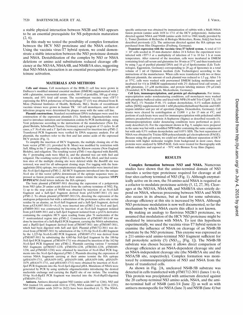
assumed that modulation of the HCV NS3 proteinase might bemediated by interaction with NS4A. To test this hypothesisexperimentally, we used the vaccinia virus-T7 hybrid system toexamine the influence of NS4A on cleavage of an NS4B-5Bsubstrate by the NS3 proteinase. This enzyme was expressed asa 211-amino-acid amino-terminal NS3 fragment sufficient forfull proteolytic activity (5) (NS3211 [Fig. 1]). The NS4B-5Bsubstrate was chosen because it allows direct comparison ofcleavage efficiencies at an NS4A-dependent cleavage site andan NS4A-independent cleavage site (the NS4B/5A site and theNS5A/5B site, respectively). Complex formation was moni-tored by coimmunoprecipitation of NS3 and NS4A from thelysate of transfected cells.As shown in Fig. 2A, uncleaved NS4B-5B substrate was
detected in cells transfected with pTM1712-3011 (lanes 1 to 4).This protein was precipitated with antiserum directed againstthe 20 carboxy-terminal NS3 amino acids, NS4A, and the ami-no-terminal half of NS4B (anti-3/4 [lane 2]) as well as withantisera monospecific for NS5A (lane 3) and NS5B (lane 4) but
7520 BARTENSCHLAGER ET AL. J. VIROL.
on July 4, 2015 by guesthttp://jvi.asm
.org/D
ownloaded from

not with the antiserum directed against the NS3 proteinasedomain (lane 1). Mature NS5B was generated from thispolyprotein in cells cotransfected with the proteinase domain(lane 8), whereas NS4B and NS5A were found exclusively asuncleaved NS4B-5A intermediate together with significantamounts of uncleaved precursor (lane 7). As shown by theproduction of mature NS5A, cleavage at the NS4B/5A junctionwas found in cells expressing the proteinase domain and NS4A(lane 11), demonstrating the essential role of NS4A for pro-cessing at this site (we were not able to detect NS4B underthese conditions, but detection of NS5A was taken to indicatecleavage at the NS4B/5A junction). Furthermore, the absenceof uncleaved precursor also indicates an increased cleavageefficiency between NS5A and NS5B.A first hint as to how NS4A might modulate the NS3 pro-
teinase was given by the coprecipitation shown in Fig. 2A, lanes
9 and 10. When NS3 was isolated by immunoprecipitationunder nondenaturing conditions using the NS3-specific anti-serum, both NS3 and NS4A were detected, although NS4Aalone was not precipitated with this antiserum (see below). Onthe other hand, NS3 was always coprecipitated with NS4A byusing the NS3/4-specific antiserum which did not directly reactwith the NS3 proteinase domain (compare lane 6 with lane 10in Fig. 2A), demonstrating formation of a detergent-stablecomplex.To analyze whether this complex also forms when NS3 and
NS4A were expressed as a single NS polyprotein precursor andto identify possible interactions of NS3 or NS4A with other NSproteins, cells were transfected with a construct directing theexpression of an NS2-5B polyprotein (pTM809-3011). To dif-ferentiate between coprecipitation and possible cross-reactiv-ity, HCV-specific proteins were isolated from half of the celllysate by immunoprecipitation under nondenaturing condi-tions and from the other half after denaturation. As shown inFig. 2B, both NS4A and the NS4AB processing intermediatewere coprecipitated under native conditions with NS3 by usingthe NS3-specific antiserum (lane 6), whereas this complex wasdestroyed when samples were denatured prior to immunopre-cipitation (lane 1). Conversely, NS3 was coprecipitated withthe NS4A-specific antiserum (lane 8) but was not detected byimmunoprecipitation of the denatured sample (lane 3). Theseresults demonstrate that the NS3 proteinase can form a com-plex both with NS4A and with the NS4AB intermediate, show-ing that cleavage at the NS4A/4B site is not essential for thisinteraction. Apart from the NS3-NS4A complex, no other co-precipitation was found under these conditions.To determine the stability of the NS3/4A complex, a pulse-
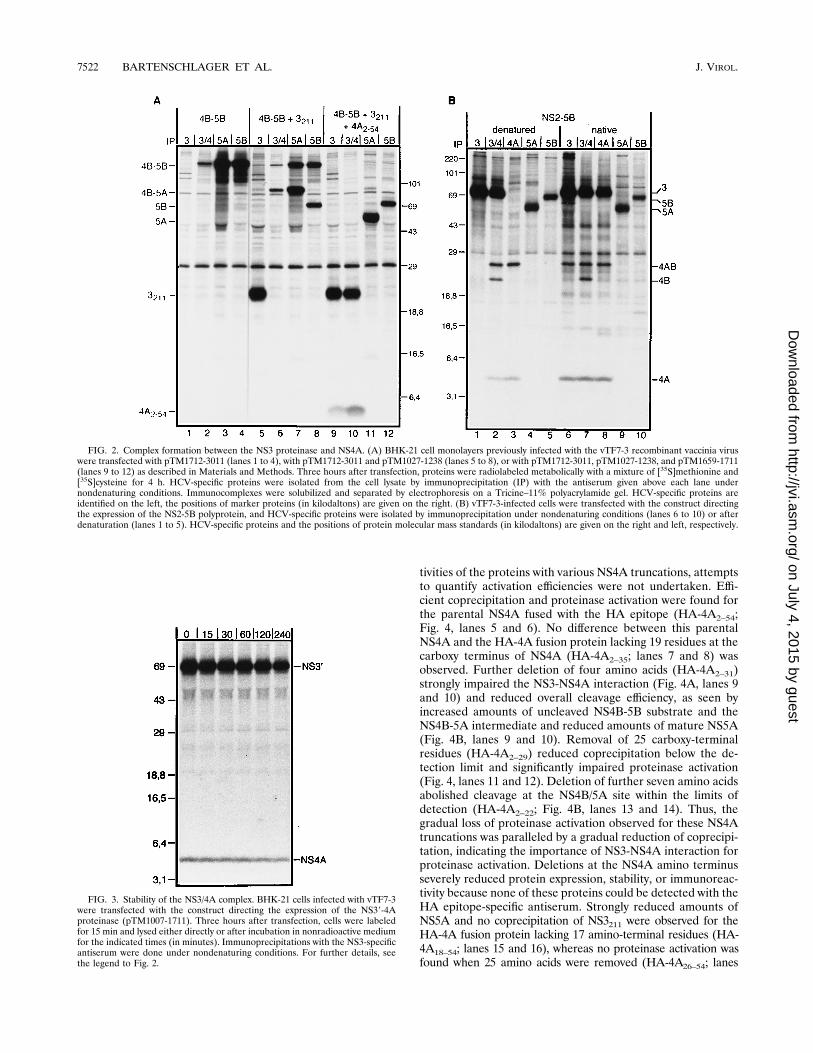
chase experiment was performed with an NS39-4A proteinase(pTM1007-1711). As shown in Fig. 3, the relative proportionsof the two proteins did not change over time. Steady-statelabeling of cells transfected with the same construct yieldedsimilar results (data not shown), demonstrating that NS3 andNS4A form a stable complex. Complex formation was alsodetected when NS4A was expressed as part of an NS4A-5Bsubstrate and released by trans cleavage at the NS4A/4B site(data not shown). Furthermore, coprecipitation was found af-ter translation of this construct in a rabbit reticulocyte lysate orafter expression in Escherichia coli (data not shown). Enzy-matic activity was not required for interaction, because NS4Acould be efficiently coprecipitated with an inactive NS3 mutantin which the active-site serine residue was substituted by analanine (data not shown).Structural and functional characterizations of NS4A. To
map the minimal NS4A domain required for interaction withNS3 and proteinase activation, a series of NS4A proteins con-taining amino- and carboxy-terminal deletions were con-structed and tested for the activation function and coprecipi-tation. In the initial set of experiments, we found that most ofthese truncated proteins did not react with our NS4A-specificantiserum. Therefore, they were fused at their amino terminiwith the HA epitope (Fig. 1 and Materials and Methods) andisolated by immunoprecipitation under nondenaturing condi-tions with a polyclonal rabbit antiserum specifically recognizingthe HA portion. Cells were transfected with constructs direct-ing the expression of the NS4B-5B substrate (pTM1712-3011),the NS3 proteinase domain (pTM1027-1238), and one of theHA-4A constructs. As controls, cells expressing only the sub-strate and proteinase (Fig. 4, lanes 1 and 2) or the substrateand NS4A (lanes 3 and 4) were analyzed in parallel. Proteinaseactivation was monitored by comparing the relative propor-tions of the NS4B-5A intermediate and NS5A. Given the dif-ferent expression levels, protein stabilities, and immunoreac-
FIG. 1. HCV genome structure and expression constructs. (A) A schematicrepresentation of the HCV polyprotein with the structural proteins encoded inthe amino-terminal quarter followed by the nonstructural proteins is shown ontop. The 59 and 39 nontranslated regions are indicated by the thin black lines. Adetailed view of the nonstructural proteins NS2 to NS5B (2 to 5B in figure)(amino acids 809 to 3011 of the polyprotein of our isolate) including the cleavagesites for the NS2-3 proteinase ( ) and the NS3 proteinase (s) is drawn below.The numbers above the arrows refer to amino acids at the P1 positions of thescissile bonds. (B) Summary of the HCV expression constructs used in this study.The numbers at the sides of the lines refer to the first and last amino acids of thepolyprotein of our isolate. Amino acids at the amino or carboxy termini ofexpressed proteins derived from the multiple cloning site are given in single-letter code. The polypeptide designation is given to the left of the correspondingexpression construct. Subscript numbers refer to the first and last amino acids ofthe particular NS protein. The HA epitope tag is indicated as a black box (forsequence, see Materials and Methods).
➝
VOL. 69, 1995 HCV NS3 ACTIVATION BY INTERACTION WITH NS4A 7521
on July 4, 2015 by guesthttp://jvi.asm
.org/D
ownloaded from
tivities of the proteins with various NS4A truncations, attemptsto quantify activation efficiencies were not undertaken. Effi-cient coprecipitation and proteinase activation were found forthe parental NS4A fused with the HA epitope (HA-4A2–54;Fig. 4, lanes 5 and 6). No difference between this parentalNS4A and the HA-4A fusion protein lacking 19 residues at thecarboxy terminus of NS4A (HA-4A2–35; lanes 7 and 8) wasobserved. Further deletion of four amino acids (HA-4A2–31)strongly impaired the NS3-NS4A interaction (Fig. 4A, lanes 9and 10) and reduced overall cleavage efficiency, as seen byincreased amounts of uncleaved NS4B-5B substrate and theNS4B-5A intermediate and reduced amounts of mature NS5A(Fig. 4B, lanes 9 and 10). Removal of 25 carboxy-terminalresidues (HA-4A2–29) reduced coprecipitation below the de-tection limit and significantly impaired proteinase activation(Fig. 4, lanes 11 and 12). Deletion of further seven amino acidsabolished cleavage at the NS4B/5A site within the limits ofdetection (HA-4A2–22; Fig. 4B, lanes 13 and 14). Thus, thegradual loss of proteinase activation observed for these NS4Atruncations was paralleled by a gradual reduction of coprecipi-tation, indicating the importance of NS3-NS4A interaction forproteinase activation. Deletions at the NS4A amino terminusseverely reduced protein expression, stability, or immunoreac-tivity because none of these proteins could be detected with theHA epitope-specific antiserum. Strongly reduced amounts ofNS5A and no coprecipitation of NS3211 were observed for theHA-4A fusion protein lacking 17 amino-terminal residues (HA-4A18–54; lanes 15 and 16), whereas no proteinase activation wasfound when 25 amino acids were removed (HA-4A26–54; lanes
FIG. 2. Complex formation between the NS3 proteinase and NS4A. (A) BHK-21 cell monolayers previously infected with the vTF7-3 recombinant vaccinia viruswere transfected with pTM1712-3011 (lanes 1 to 4), with pTM1712-3011 and pTM1027-1238 (lanes 5 to 8), or with pTM1712-3011, pTM1027-1238, and pTM1659-1711(lanes 9 to 12) as described in Materials and Methods. Three hours after transfection, proteins were radiolabeled metabolically with a mixture of [35S]methionine and[35S]cysteine for 4 h. HCV-specific proteins were isolated from the cell lysate by immunoprecipitation (IP) with the antiserum given above each lane undernondenaturing conditions. Immunocomplexes were solubilized and separated by electrophoresis on a Tricine–11% polyacrylamide gel. HCV-specific proteins areidentified on the left, the positions of marker proteins (in kilodaltons) are given on the right. (B) vTF7-3-infected cells were transfected with the construct directingthe expression of the NS2-5B polyprotein, and HCV-specific proteins were isolated by immunoprecipitation under nondenaturing conditions (lanes 6 to 10) or afterdenaturation (lanes 1 to 5). HCV-specific proteins and the positions of protein molecular mass standards (in kilodaltons) are given on the right and left, respectively.
FIG. 3. Stability of the NS3/4A complex. BHK-21 cells infected with vTF7-3were transfected with the construct directing the expression of the NS39-4Aproteinase (pTM1007-1711). Three hours after transfection, cells were labeledfor 15 min and lysed either directly or after incubation in nonradioactive mediumfor the indicated times (in minutes). Immunoprecipitations with the NS3-specificantiserum were done under nondenaturing conditions. For further details, seethe legend to Fig. 2.
7522 BARTENSCHLAGER ET AL. J. VIROL.
on July 4, 2015 by guesthttp://jvi.asm
.org/D
ownloaded from
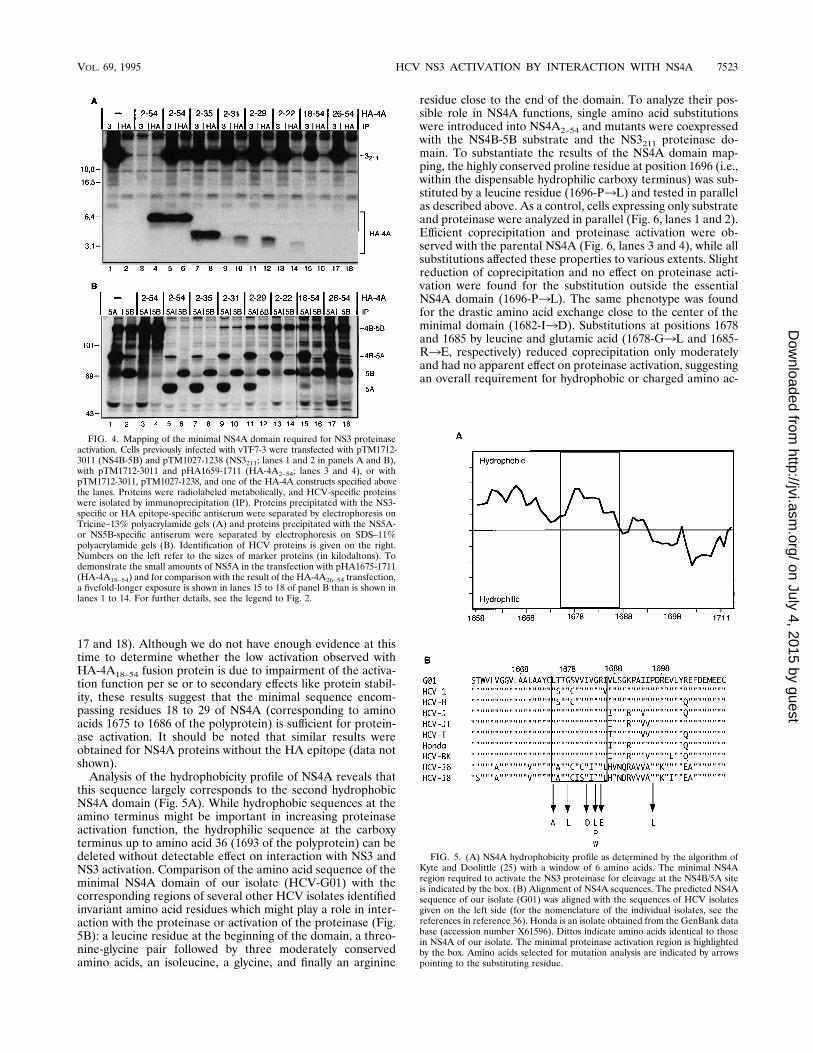
17 and 18). Although we do not have enough evidence at thistime to determine whether the low activation observed withHA-4A18–54 fusion protein is due to impairment of the activa-tion function per se or to secondary effects like protein stabil-ity, these results suggest that the minimal sequence encom-passing residues 18 to 29 of NS4A (corresponding to aminoacids 1675 to 1686 of the polyprotein) is sufficient for protein-ase activation. It should be noted that similar results wereobtained for NS4A proteins without the HA epitope (data notshown).Analysis of the hydrophobicity profile of NS4A reveals that
this sequence largely corresponds to the second hydrophobicNS4A domain (Fig. 5A). While hydrophobic sequences at theamino terminus might be important in increasing proteinaseactivation function, the hydrophilic sequence at the carboxyterminus up to amino acid 36 (1693 of the polyprotein) can bedeleted without detectable effect on interaction with NS3 andNS3 activation. Comparison of the amino acid sequence of theminimal NS4A domain of our isolate (HCV-G01) with thecorresponding regions of several other HCV isolates identifiedinvariant amino acid residues which might play a role in inter-action with the proteinase or activation of the proteinase (Fig.5B): a leucine residue at the beginning of the domain, a threo-nine-glycine pair followed by three moderately conservedamino acids, an isoleucine, a glycine, and finally an arginine
residue close to the end of the domain. To analyze their pos-sible role in NS4A functions, single amino acid substitutionswere introduced into NS4A2–54 and mutants were coexpressedwith the NS4B-5B substrate and the NS3211 proteinase do-main. To substantiate the results of the NS4A domain map-ping, the highly conserved proline residue at position 1696 (i.e.,within the dispensable hydrophilic carboxy terminus) was sub-stituted by a leucine residue (1696-P3L) and tested in parallelas described above. As a control, cells expressing only substrateand proteinase were analyzed in parallel (Fig. 6, lanes 1 and 2).Efficient coprecipitation and proteinase activation were ob-served with the parental NS4A (Fig. 6, lanes 3 and 4), while allsubstitutions affected these properties to various extents. Slightreduction of coprecipitation and no effect on proteinase acti-vation were found for the substitution outside the essentialNS4A domain (1696-P3L). The same phenotype was foundfor the drastic amino acid exchange close to the center of theminimal domain (1682-I3D). Substitutions at positions 1678and 1685 by leucine and glutamic acid (1678-G3L and 1685-R3E, respectively) reduced coprecipitation only moderatelyand had no apparent effect on proteinase activation, suggestingan overall requirement for hydrophobic or charged amino ac-
FIG. 4. Mapping of the minimal NS4A domain required for NS3 proteinaseactivation. Cells previously infected with vTF7-3 were transfected with pTM1712-3011 (NS4B-5B) and pTM1027-1238 (NS3211; lanes 1 and 2 in panels A and B),with pTM1712-3011 and pHA1659-1711 (HA-4A2–54; lanes 3 and 4), or withpTM1712-3011, pTM1027-1238, and one of the HA-4A constructs specified abovethe lanes. Proteins were radiolabeled metabolically, and HCV-specific proteinswere isolated by immunoprecipitation (IP). Proteins precipitated with the NS3-specific or HA epitope-specific antiserum were separated by electrophoresis onTricine–13% polyacrylamide gels (A) and proteins precipitated with the NS5A-or NS5B-specific antiserum were separated by electrophoresis on SDS–11%polyacrylamide gels (B). Identification of HCV proteins is given on the right.Numbers on the left refer to the sizes of marker proteins (in kilodaltons). Todemonstrate the small amounts of NS5A in the transfection with pHA1675-1711(HA-4A18–54) and for comparison with the result of the HA-4A26–54 transfection,a fivefold-longer exposure is shown in lanes 15 to 18 of panel B than is shown inlanes 1 to 14. For further details, see the legend to Fig. 2.
FIG. 5. (A) NS4A hydrophobicity profile as determined by the algorithm ofKyte and Doolittle (25) with a window of 6 amino acids. The minimal NS4Aregion required to activate the NS3 proteinase for cleavage at the NS4B/5A siteis indicated by the box. (B) Alignment of NS4A sequences. The predicted NS4Asequence of our isolate (G01) was aligned with the sequences of HCV isolatesgiven on the left side (for the nomenclature of the individual isolates, see thereferences in reference 36). Honda is an isolate obtained from the GenBank database (accession number X61596). Dittos indicate amino acids identical to thosein NS4A of our isolate. The minimal proteinase activation region is highlightedby the box. Amino acids selected for mutation analysis are indicated by arrowspointing to the substituting residue.
VOL. 69, 1995 HCV NS3 ACTIVATION BY INTERACTION WITH NS4A 7523
on July 4, 2015 by guesthttp://jvi.asm
.org/D
ownloaded from

ids at these positions. No coprecipitation and reduced protein-ase activation were found for the leucine substitution at posi-tion 1675 (1675-L3A). The most drastic effect was found forthe substitutions of the glycine residue at position 1684. Fornone of these mutants was coprecipitation detected. While forthe substitutions by leucine or tryptophan, small amounts ofNS5A were found, no NS5A was detected for the substitutionby proline, suggesting that steric constraints are the primaryreason for loss of NS4A functions. In summary, the overallcorrelation between impairment of complex formation andreduction of cleavage at the NS4B/5A site shows that interac-tion between NS3 and NS4A is important for NS3 proteinaseactivation.To analyze the influence of NS4A mutations on complex
formation and NS3-mediated cleavage at the other sites, thesubstitution of glycine by proline at position 1684, which hadthe most marked effect, was introduced into an NS39-5Bpolyprotein. This protein was expressed either alone or to-gether with NS4A, and HCV-specific proteins were isolatedunder nondenaturing conditions as described above. As a con-trol, cells expressing an NS39-5B polyprotein with an unalteredNS4A were analyzed in parallel. As shown in Fig. 7, matureNS5B and small amounts of NS3 but no NS4A, NS4B, orNS5A were found in cells expressing the NS39-5B polyproteinwith the defective NS4A (lanes 5 to 8). Instead, a series ofprocessing intermediates corresponding to NS39-5A, NS4AB-5A, NS4B-5A, and NS39-4A were observed, indicating ineffi-cient cleavage at the NS4A-dependent sites. Very smallamounts of NS3 were coprecipitated with the NS4-specific
antiserum consistent with the defect described above for thisNS4A mutant (lane 6). When NS4A was coexpressed, all ma-ture cleavage products as well as increased amounts of NS3were detected (lanes 9 to 12) and NS3 was coprecipitated withNS4A (lane 10). Thus, destabilization of the complex by anNS4A mutation impaired proteolytic cleavage at all NS4A-dependent sites and could be restored by NS4A provided intrans.Amino-terminal NS3 sequences are important for complex
formation and NS4A-mediated activation.We and others haverecently shown that amino-terminal NS3 deletions greatly re-duce cleavage at the NS4B/5A site without significantly affect-ing processing between NS5A and NS5B (5, 13). Since NS4Ais absolutely required for efficient cleavage between NS4B andNS5A but not for processing between NS5A and NS5B, amino-terminal NS3 sequences presented prime candidates for inter-action with NS4A. To test this hypothesis, cells were trans-fected with constructs directing the expression of the NS4B-5Bsubstrate, NS4A2–54, and the proteinase domain either with anunaltered or truncated amino terminus. For optimal separa-tion in the low- and high-molecular-weight range, half of eachsample was analyzed by Tricine-SDS-PAGE and SDS-PAGEin parallel (Fig. 8A and B, respectively). As a control, cellsexpressing only the substrate (Fig. 8, lanes 1 to 4), substrateplus NS4A2–54 (lanes 5 to 8), or substrate plus the parentalproteinase (lanes 9 to 12) were examined in parallel. As shownin Fig. 8B (lanes 11 and 12) in the absence of NS4A, cleavageof the substrate occurred only at the NS5A/5B site. NS5A andthe NS3-NS4A complex were clearly detected in cells express-ing in addition NS4A (Fig. 8B, lane 15, and Fig. 8A, lanes 13and 14, respectively). Removal of seven amino acids from theamino terminus of NS3 significantly destabilized the complex,as seen by the reduced amount of NS3 coprecipitated with theNS4A-specific antiserum and the small amounts of NS4A co-precipitated with the NS3-specific antiserum (Fig. 8A, lanes 17
FIG. 6. Mutation analysis of amino acid residues within NS4A conserved be-tween various HCV genotypes. Cells infected with vTF7-3 were transfected withpTM1712-3011 (NS4B-5B), pTM1027-1238 (NS3211), and a construct directingthe expression of a mutated or parental (wild-type [wt]) NS4A. HCV-specificproteins isolated by immunoprecipitation (IP) with the NS3- or NS4A-specificantiserum were separated by Tricine-SDS-PAGE (A). Proteins recovered withthe NS5A- or NS5B-specific antiserum were separated by SDS-PAGE (B). Re-sults of immunoprecipitations from cells expressing substrate and proteinase areshown in lanes 1 and 2 of each panel. For further details, see the legend toFig. 4.
FIG. 7. The proline substitution in NS4A affects cleavage at the NS4A-dependent sites. Infected cells were transfected with pTM1007-3011/4P alone(lanes 5 to 8) or together with NS4A2–54 (lanes 9 to 12). Proteins were isolatedunder nondenaturing conditions by immunoprecipitation (IP) with the antiserumgiven above each lane and analyzed by Tricine-SDS-PAGE. As a control, pro-teins isolated from cells transfected with pTM1007-3011 were analyzed in par-allel (NS39-5B; lanes 1 to 4). For further details, see the legend to Fig. 2.
7524 BARTENSCHLAGER ET AL. J. VIROL.
on July 4, 2015 by guesthttp://jvi.asm
.org/D
ownloaded from

and 18). Cleavage at the NS4B/5A junction apparently was notaffected (Fig. 8B, lanes 19 and 20). Slightly increased amountsof uncleaved precursor and NS4B-5A intermediate, indicatinga reduced processing efficiency, were found with the proteinaselacking 15 amino acids (Fig. 8B, lanes 23 and 24). Interactionof this truncated NS3 with NS4A was reduced below the de-tection limit (Fig. 8A, lanes 21 and 22). Proteinase activationby NS4A was no longer detectable when 22 amino acids weredeleted and only uncleaved NS4B-5A and mature NS5B werefound along with clearly increased amounts of uncleaved pre-cursor (Fig. 8B, lanes 27 and 28). However, given the smallamount of proteinase detected currently, we cannot ascertainwhether the overall reduction of proteolytic activity is dueexclusively to a reduction of the activity per se or at least tosome extent to the lower expression or stability of this protein(Fig. 8A, lane 25). Similar results were found for the NS3proteinase lacking 38 amino-terminal amino acids (Fig. 8, lanes29 to 32). Beside interaction with NS3, NS4A was also copre-cipitated with the NS4B-5B substrate by using NS5A- or NS5B-specific antiserum (Fig. 8A, lanes 7 and 8) and the substratewas coprecipitated with NS4A by using the NS4A-specific an-tiserum (Fig. 8B, lane 6). This coprecipitation was no longerdetectable in cells expressing NS3 proteins which efficientlycleaved the substrate (e.g., Fig. 8A, lanes 19 and 20) but wasfound in cells expressing truncated NS3 proteins with low sub-strate cleavage, suggesting that NS4A can also interact with theNS4B-5B substrate (Fig. 8A, lanes 27, 28, 31, and 32). Thisinteraction appears to be much weaker than the one observedwith NS3 because in cells expressing the NS4B-5B substrate,NS4A, and an enzymatically inactive NS3211 proteinase, NS4Awas coprecipitated with NS3 but not with the substrate (datanot shown).The results described so far for the NS3 deletion mutants
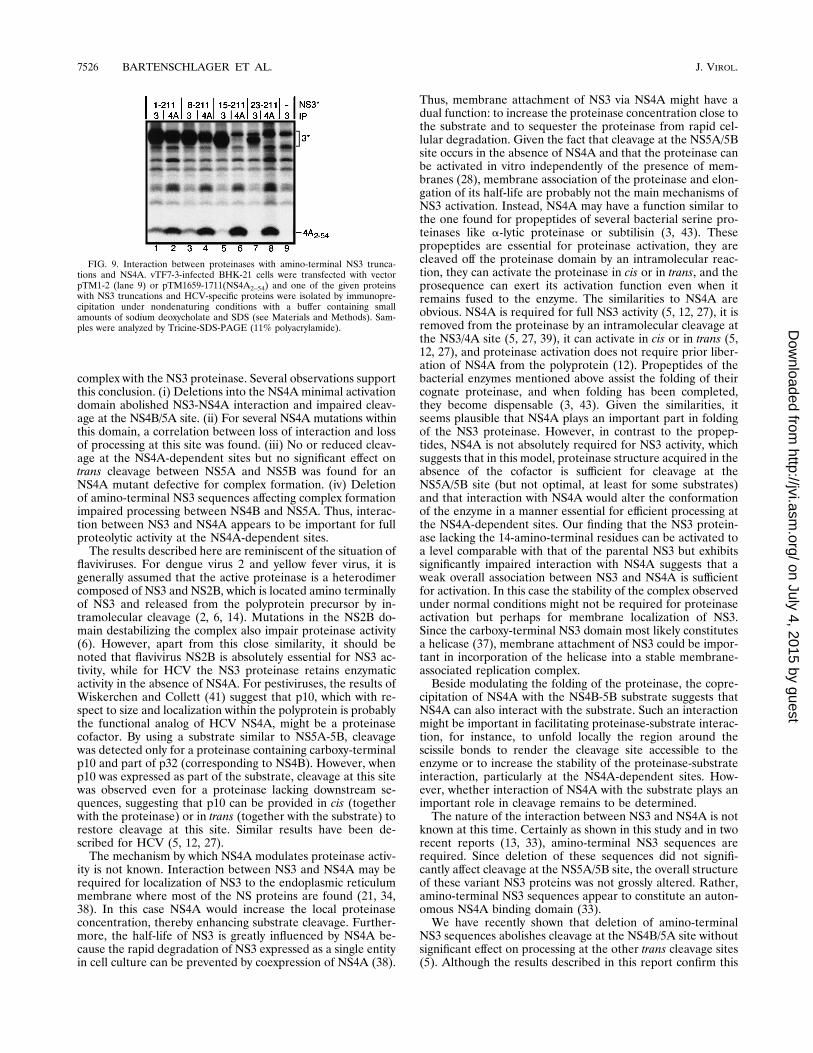
imply that proteins with amino-terminal NS3 truncations sig-nificantly impaired for interaction with NS4A can still be acti-vated (NS38–211 and NS315–211). However, since the buffer weused for cell lysis and immunoprecipitation contained 0.1%SDS, it was possible that these truncated proteinases could stillinteract with NS4A intracellularly but that these complexeswere sensitive to our lysis conditions. Since our antisera aredirected against denatured antigens, HCV-specific proteinswere not or only poorly precipitated from cell lysates preparedexclusively with nonionic detergents. Therefore, we used amodified lysis buffer containing reduced amounts of SDS forimmunoprecipitation of HCV proteins from cells expressingNS4A2–54 and either the parental NS3211 or one of the NS3proteins with amino-terminal truncations. Under these condi-tions the levels of coprecipitation of NS4A between the com-plete NS3211 proteinase and the truncated NS38–211 proteinasewere comparable (Fig. 9, lanes 1 to 4). Coprecipitation wasclearly reduced for the NS315–211 proteinase but still detectableas seen by the small amounts of NS4A in the immunoprecipi-tation with the NS3-specific antiserum (lane 5), showing thatthis truncated proteinase still retained the ability to interactwith NS4A. No coprecipitation was found for the NS323–211proteinase, consistent with the inability to activate this enzymefor cleavage at the NS4B/5A site.
DISCUSSION
Previous studies have shown that NS4A is a proteinase co-factor required for cleavage at the NS3/4A, NS4A/4B, andNS4B/5A sites and enhancing processing efficiency betweenNS5A and NS5B (5, 12, 27). In this work we analyzed themechanism of proteinase modulation by NS4A and found thatNS4A most likely exerts its effect by formation of a stable
FIG. 8. Amino-terminal sequences of NS3 are important for interaction with NS4A. BHK-21 cells previously infected with vTF7-3 were transfected withpTM1712-3011 (NS4B-5B) either alone (lanes 1 to 4) or with pTM1659-1711 (NS4A2–54; lanes 5 to 8), pTM1027-1238 (NS3211; lanes 9 to 12), or pTM1712-3011 pluspTM1659-1711 plus one of the indicated NS3 constructs (NS3*; lanes 13 to 32). Following metabolic labeling, HCV-specific proteins were isolated by immunopre-cipitation (IP) under nondenaturing conditions with the indicated antiserum. One half of the samples were separated by electrophoresis on Tricine–11% polyacrylamidegels (A), and the other half were separated on SDS–11% polyacrylamide gels (B). Identification of HCV proteins is given on the right of each gel. Only the parts ofthe gels with sufficient resolution of HCV-specific proteins are shown. For further details, see the legend to Fig. 2.
VOL. 69, 1995 HCV NS3 ACTIVATION BY INTERACTION WITH NS4A 7525
on July 4, 2015 by guesthttp://jvi.asm
.org/D
ownloaded from
complex with the NS3 proteinase. Several observations supportthis conclusion. (i) Deletions into the NS4A minimal activationdomain abolished NS3-NS4A interaction and impaired cleav-age at the NS4B/5A site. (ii) For several NS4A mutations withinthis domain, a correlation between loss of interaction and lossof processing at this site was found. (iii) No or reduced cleav-age at the NS4A-dependent sites but no significant effect ontrans cleavage between NS5A and NS5B was found for anNS4A mutant defective for complex formation. (iv) Deletionof amino-terminal NS3 sequences affecting complex formationimpaired processing between NS4B and NS5A. Thus, interac-tion between NS3 and NS4A appears to be important for fullproteolytic activity at the NS4A-dependent sites.The results described here are reminiscent of the situation of
flaviviruses. For dengue virus 2 and yellow fever virus, it isgenerally assumed that the active proteinase is a heterodimercomposed of NS3 and NS2B, which is located amino terminallyof NS3 and released from the polyprotein precursor by in-tramolecular cleavage (2, 6, 14). Mutations in the NS2B do-main destabilizing the complex also impair proteinase activity(6). However, apart from this close similarity, it should benoted that flavivirus NS2B is absolutely essential for NS3 ac-tivity, while for HCV the NS3 proteinase retains enzymaticactivity in the absence of NS4A. For pestiviruses, the results ofWiskerchen and Collett (41) suggest that p10, which with re-spect to size and localization within the polyprotein is probablythe functional analog of HCV NS4A, might be a proteinasecofactor. By using a substrate similar to NS5A-5B, cleavagewas detected only for a proteinase containing carboxy-terminalp10 and part of p32 (corresponding to NS4B). However, whenp10 was expressed as part of the substrate, cleavage at this sitewas observed even for a proteinase lacking downstream se-quences, suggesting that p10 can be provided in cis (togetherwith the proteinase) or in trans (together with the substrate) torestore cleavage at this site. Similar results have been de-scribed for HCV (5, 12, 27).The mechanism by which NS4A modulates proteinase activ-
ity is not known. Interaction between NS3 and NS4A may berequired for localization of NS3 to the endoplasmic reticulummembrane where most of the NS proteins are found (21, 34,38). In this case NS4A would increase the local proteinaseconcentration, thereby enhancing substrate cleavage. Further-more, the half-life of NS3 is greatly influenced by NS4A be-cause the rapid degradation of NS3 expressed as a single entityin cell culture can be prevented by coexpression of NS4A (38).
Thus, membrane attachment of NS3 via NS4A might have adual function: to increase the proteinase concentration close tothe substrate and to sequester the proteinase from rapid cel-lular degradation. Given the fact that cleavage at the NS5A/5Bsite occurs in the absence of NS4A and that the proteinase canbe activated in vitro independently of the presence of mem-branes (28), membrane association of the proteinase and elon-gation of its half-life are probably not the main mechanisms ofNS3 activation. Instead, NS4A may have a function similar tothe one found for propeptides of several bacterial serine pro-teinases like a-lytic proteinase or subtilisin (3, 43). Thesepropeptides are essential for proteinase activation, they arecleaved off the proteinase domain by an intramolecular reac-tion, they can activate the proteinase in cis or in trans, and theprosequence can exert its activation function even when itremains fused to the enzyme. The similarities to NS4A areobvious. NS4A is required for full NS3 activity (5, 12, 27), it isremoved from the proteinase by an intramolecular cleavage atthe NS3/4A site (5, 27, 39), it can activate in cis or in trans (5,12, 27), and proteinase activation does not require prior liber-ation of NS4A from the polyprotein (12). Propeptides of thebacterial enzymes mentioned above assist the folding of theircognate proteinase, and when folding has been completed,they become dispensable (3, 43). Given the similarities, itseems plausible that NS4A plays an important part in foldingof the NS3 proteinase. However, in contrast to the propep-tides, NS4A is not absolutely required for NS3 activity, whichsuggests that in this model, proteinase structure acquired in theabsence of the cofactor is sufficient for cleavage at theNS5A/5B site (but not optimal, at least for some substrates)and that interaction with NS4A would alter the conformationof the enzyme in a manner essential for efficient processing atthe NS4A-dependent sites. Our finding that the NS3 protein-ase lacking the 14-amino-terminal residues can be activated toa level comparable with that of the parental NS3 but exhibitssignificantly impaired interaction with NS4A suggests that aweak overall association between NS3 and NS4A is sufficientfor activation. In this case the stability of the complex observedunder normal conditions might not be required for proteinaseactivation but perhaps for membrane localization of NS3.Since the carboxy-terminal NS3 domain most likely constitutesa helicase (37), membrane attachment of NS3 could be impor-tant in incorporation of the helicase into a stable membrane-associated replication complex.Beside modulating the folding of the proteinase, the copre-
cipitation of NS4A with the NS4B-5B substrate suggests thatNS4A can also interact with the substrate. Such an interactionmight be important in facilitating proteinase-substrate interac-tion, for instance, to unfold locally the region around thescissile bonds to render the cleavage site accessible to theenzyme or to increase the stability of the proteinase-substrateinteraction, particularly at the NS4A-dependent sites. How-ever, whether interaction of NS4A with the substrate plays animportant role in cleavage remains to be determined.The nature of the interaction between NS3 and NS4A is not
known at this time. Certainly as shown in this study and in tworecent reports (13, 33), amino-terminal NS3 sequences arerequired. Since deletion of these sequences did not signifi-cantly affect cleavage at the NS5A/5B site, the overall structureof these variant NS3 proteins was not grossly altered. Rather,amino-terminal NS3 sequences appear to constitute an auton-omous NS4A binding domain (33).We have recently shown that deletion of amino-terminal
NS3 sequences abolishes cleavage at the NS4B/5A site withoutsignificant effect on processing at the other trans cleavage sites(5). Although the results described in this report confirm this
FIG. 9. Interaction between proteinases with amino-terminal NS3 trunca-tions and NS4A. vTF7-3-infected BHK-21 cells were transfected with vectorpTM1-2 (lane 9) or pTM1659-1711(NS4A2–54) and one of the given proteinswith NS3 truncations and HCV-specific proteins were isolated by immunopre-cipitation under nondenaturing conditions with a buffer containing smallamounts of sodium deoxycholate and SDS (see Materials and Methods). Sam-ples were analyzed by Tricine-SDS-PAGE (11% polyacrylamide).
7526 BARTENSCHLAGER ET AL. J. VIROL.
on July 4, 2015 by guesthttp://jvi.asm
.org/D
ownloaded from

observation by using stable recombinant vaccinia viruses ex-pressing various NS3 proteins, in the first study (5) we found astrong reduction of processing at the NS4B/5A site when only7 amino acids were deleted, while in this report by using thevaccinia virus-T7 hybrid system, 7 and even 14 residues couldbe removed without detectable effects. Because the protein-ases expressed in both systems have the same amino acidsequence, the most likely reason for this discrepancy is theassay system. Using the T7 system, Faila and coworkers foundthat cleavage at the NS4B/5A site by a proteinase lacking 15amino-terminal residues (amino acids 1042 to 1237 of thepolyprotein) was severely reduced and that cleavage at this siteby a proteinase lacking 28 residues (amino acids 1055 to 1237)was detectable only when large amounts of plasmid were trans-fected (13). In contrast, efficient cleavage at this site was ob-served for a proteinase lacking 22 amino-terminal residues(amino acids 1049 to 1215) when coexpressed under control ofthe human cytomegalovirus immediate-early promoter withNS4A and an artificial substrate composed of 30 amino acidsspanning the NS4B/5A cleavage site fused to the amino termi-nus of the E. coli dihydrofolate reductase (38). On the otherhand, the same proteinase domain fused to dihydrofolate re-ductase and coexpressed with NS4A inefficiently cleaved anNS4B-5A substrate (33). The reason for these differences isnot clear but is most likely due to differences in the expressionsystem, assay conditions, or primary sequences of the protein-ases or substrates. This issue can be clarified only with thedevelopment of an appropriate in vitro cleavage system.A minimal sequence of 12 amino acids mapping close to the
center of NS4A (amino acids 18 to 29) was found to be essen-tial for proteinase activation and, as indicated by mutationanalysis, also for complex formation. Although highly con-served in various HCV genotypes, the carboxy-terminal 19residues are not required for complex formation and protein-ase activation, suggesting that this sequence has another func-tion. Proper folding of the NS4A/4B processing site is a likelypossibility. This result is in agreement with two recent reportsmapping the NS4A region important for cofactor activity toamino acids 21 to 30 (38) or 21 to 33 (28). Furthermore, it wasshown that a peptide corresponding to amino acids 22 to 34 ofNS4A can activate the proteinase in vitro (24, 28). Althoughother possibilities exist, these results suggest a stable interac-tion between the central NS4A domain and the amino-termi-nal domain of NS3.Modulation of proteinase activity by cofactors is also found
for other viral systems. For adenovirus, an 11-amino-acid pep-tide derived from the structural protein pVI forms a disulfide-linked heterodimer with the proteinase and this interaction isessential for enzyme activation (40). For poliovirus, the 3Cproteinase is modulated by the 3D RNA-dependent RNApolymerase (23, 42). 3C is sufficient for cleavage within P2 andP3, but processing within the capsid protein precursor P1 iscatalyzed by a 3CD proteinase. An auxiliary function of 3Ddiscussed is to facilitate enzyme-substrate interaction by directinteraction of 3D with the P1 precursor (42). For Sindbis virus,a member of the alphaviruses, cleavage site preference of thensP2 proteinase is modulated by the flanking nonstructuralproteins nsP1 and nsP3, resulting in a temporal regulation ofpolyprotein processing (8).The finding that interaction of the NS3 proteinase with
NS4A is important for full proteolytic activity encourages thedevelopment of a new therapeutic concept. As shown recentlyfor yellow fever virus, mutations in NS2B blocking interactionwith the NS3 proteinase and proteinase activation are delete-rious for virus infectivity (6). By analogy, it is likely that HCVNS4A, because of its important role for polyprotein matura-
tion, is also essential for virus replication. Thus, beside thedevelopment of inhibitors targeted to the active site of theproteinase, inhibition of the NS3-NS4A interaction may pro-vide an alternative for the development of therapeutic agentsand methods urgently needed to control this insidious disease.
ACKNOWLEDGMENTS
We thank Bernie Moss for providing plasmid pTM1 and the recom-binant vaccinia virus vTF7-3 and L. Tomei for providing the NS4-specific antiserum. We are grateful to Ulrike Herian for excellenttechnical assistance and Mareike Herian for help in preparing thefigures.This work was supported by grants from the Bundesministerium fur
Forschung und Technologie (01 KI 9351) and Deutsche Forschungs-gemeinschaft (BA 1505/1-1).
REFERENCES
1. Amberg, S. M., A. Nestorowicz, D. W. McCourt, and C. M. Rice. 1994.NS2B-3 proteinase-mediated processing in the yellow fever virus structuralregion: in vitro and in vivo studies. J. Virol. 68:3794–3802.
2. Arias, C. F., F. Preugschat, and J. Strauss. 1993. Dengue virus 2 NS2B andNS3 form a stable complex that can cleave NS3 within the helicase domain.Virology 193:888–899.
3. Baker, D., J. L. Sohl, and D. A. Agard. 1992. A protein-folding reactionunder kinetic control. Nature (London) 356:263–265.
4. Bartenschlager, R., L. Ahlborn-Laake, J. Mous, and H. Jacobsen. 1993.Nonstructural protein 3 of the hepatitis C virus encodes a serine-type pro-teinase required for cleavage at the NS3/4 and NS4/5 junctions. J. Virol. 67:3835–3844.
5. Bartenschlager, R., L. Ahlborn-Laake, J. Mous, and H. Jacobsen. 1994.Kinetic and structural analyses of hepatitis C virus polyprotein processing. J.Virol. 68:5045–5055.
6. Chambers, T. J., A. Nestorowicz, S. M. Amberg, and C. M. Rice. 1993.Mutagenesis of the yellow fever virus NS2B protein: effects on proteolyticprocessing, NS2B-NS3 complex formation, and viral replication. J. Virol. 67:6797–6807.
7. Cuthbert, J. A. 1994. Hepatitis C: progress and problems. Clin. Microbiol.Rev. 7:505–532.
8. de Groot, R. J., W. R. Hardy, Y. Shirako, and J. H. Strauss. 1990. Cleavage-site preferences of Sindbis virus polyproteins containing the non-structuralproteinase. Evidence for temporal regulation of polyprotein processing invivo. EMBO J. 9:2631–2638.
9. Duboisson, J., H. H. Hsu, R. C. Cheung, H. B. Greenberg, D. G. Russell, andC. M. Rice. 1994. Formation and intracellular localization of hepatitis C virusenvelope glycoprotein complexes expressed by recombinant vaccinia andSindbis viruses. J. Virol. 68:6147–6160.
10. Eckart, M. R., M. Selby, F. Masiarz, C. Lee, K. Berger, K. Crawford, C. Kuo,G. Kuo, M. Houghton, and Q.-L. Choo. 1993. The hepatitis C virus encodesa serine proteinase involved in processing of the putative nonstructuralproteins from the viral polyprotein precursor. Biochem. Biophys. Res. Com-mun. 192:399–406.
11. Elroy-Stein, O., T. R. Fuerst, and B. Moss. 1989. Cap-independent transla-tion of mRNA conferred by encephalomyocarditis virus 59 sequence im-proves the performance of the vaccinia virus/bacteriophage T7 hybrid ex-pression system. Proc. Natl. Acad. Sci. USA 86:6126–6130.
12. Faila, C., L. Tomei, and R. de Francesco. 1994. Both NS3 and NS4A arerequired for proteolytic processing of hepatitis C virus nonstructural pro-teins. J. Virol. 68:3753–3760.
13. Faila, C., L. Tomei, and R. de Francesco. 1995. An amino-terminal domainof the hepatitis C virus NS3 protease is essential for interaction withNS4A. J. Virol. 69:1769–1777.
14. Falgout, B., R. H. Miller, and C.-J. Lai. 1993. Deletion analysis of denguetype 4 nonstructural protein NS2B: identification of a domain required forNS2B-NS3 protease activity. J. Virol. 67:2034–2042.
15. Fuerst, T. R., E. G. Niles, F. W. Studier, and B. Moss. 1986. Eukaryotictransient-expression system based on recombinant vaccinia virus that syn-thesizes bacteriophage T7 RNA polymerase. Proc. Natl. Acad. Sci. USA 83:8122–8126.
16. Grakoui, A., D. W. McCourt, C. Wychowski, S. M. Feinstone, and C. M. Rice.1993. A second hepatitis C virus-encoded proteinase. Proc. Natl. Acad. Sci.USA 90:10583–10587.
17. Grakoui, A., D. W. McCourt, C. Wychowski, S. M. Feinstone, and C. M. Rice.1993. Characterization of the hepatitis C virus-encoded serine proteinase:determination of proteinase-dependent polyprotein cleavage sites. J. Virol.67:2832–2843.
18. Grakoui, A., C. Wychowski, C. Lin, S. M. Feinstone, and C. M. Rice. 1993.Expression and identification of hepatitis C virus polyprotein cleavage prod-
VOL. 69, 1995 HCV NS3 ACTIVATION BY INTERACTION WITH NS4A 7527
on July 4, 2015 by guesthttp://jvi.asm
.org/D
ownloaded from

ucts. J. Virol. 67:1385–1395.19. Hijikata, M., N. Kato, Y. Ootsuyama, and M. Nakagawa. 1991. Gene map-
ping of the putative structural region of the hepatitis C virus genome by invitro processing analysis. Proc. Natl. Acad. Sci. USA 88:5547–5551.
20. Hijikata, M., H. Mizushima, T. Akagi, S. Mori, N. Kakiuchi, N. Kato, T.Tanaka, K. Kimura, and K. Shimotohno. 1993. Two distinct proteinaseactivities required for the processing of a putative nonstructural precursorprotein of hepatitis C virus. J. Virol. 67:4665–4675.
21. Hijikata, M., H. Mizushima, Y. Tanji, Y. Komoda, Y. Hirowatari, T. Akagi,N. Kato, K. Kimuran, and K. Shimotohno. 1993. Proteolytic processing andmembrane association of putative nonstructural proteins of hepatitis C virus.Proc. Natl. Acad. Sci. USA 90:10733–10777.
22. Hirowatari, Y., M. Hijikata, Y. Tanji, H. Nyunoya, H. Mizushima, K.Kimura, T. Tanaka, N. Kato, and K. Shimotohno. 1993. Two proteinaseactivities in HCV polypeptide expressed in insect cells using baculovirusvector. Arch. Virol. 133:349–356.
23. Jore, J., B. D. Geus, R. J. Jackson, P. H. Pouwels, and B. E. Enger-Valk.1988. Poliovirus protein 3CD is the active protease for processing of theprecursor P1 in vitro. J. Gen. Virol. 69:1627–1636.
24. Koch, J. O., and R. Bartenschlager. Unpublished results.25. Kyte, J., and R. F. Doolittle. 1982. A method for displaying the hydropathic
character of a protein. J. Mol. Biol. 157:105–132.26. Lin, C., B. D. Lindenbach, B. M. Pragai, D. W. McCourt, and C. M. Rice.
1994. Processing in the hepatitis C virus E2-NS2 region: identification of p7and two distinct E2-specific products with different C termini. J. Virol. 68:5063–5073.
27. Lin, C., B. M. Pragai, A. Grakoui, J. Xu, and C. M. Rice. 1994. Hepatitis Cvirus NS3 serine proteinase: trans-cleavage requirements and processingkinetics. J. Virol. 68:8147–8157.
28. Lin, C., J. A. Thomson, and C. M. Rice. 1995. A central region in thehepatitis C virus NS4A protein allows formation of an active NS3-NS4Aserine proteinase complex in vivo and in vitro. J. Virol. 69:4373–4380.
29. Manabe, S., I. Fuke, O. Tanishita, C. Kaji, Y. Gomi, S. Yoshida, C. Mori, A.Takamizawa, I. Yosida, and H. Okayama. 1994. Production of nonstructuralproteins of hepatitis C virus requires a putative viral protease encoded byNS3. Virology 198:636–644.
30. Mizushima, H., M. Hijikata, S.-I. Asabe, M. Hirota, K. Kimura, and K.Shimotohno. 1994. Two hepatitis C virus glycoprotein E2 products withdifferent C termini. J. Virol. 68:6215–6222.
31. Sambrook, J., E. F. Fritsch, and T. Maniatis. 1989. Molecular cloning: alaboratory manual, 2nd ed. Cold Spring Harbor Laboratory, Cold SpringHarbor, N.Y.
32. Santolini, E., G. Migliaccio, and N. La Monica. 1994. Biosynthesis and
biochemical properties of the hepatitis C virus core protein. J. Virol. 68:3631–3641.
33. Satoh, S., Y. Tanji, M. Hijikata, K. Kimura, and K. Shimotohno. 1995. TheN-terminal region of hepatitis C virus nonstructural protein 3 (NS3) isessential for stable complex formation with NS4A. J. Virol. 69:4255–4260.
34. Selby, M. J., Q.-L. Choo, K. Berger, G. Kuo, E. Glazer, M. Eckart, C. Lee, D.Chien, C. Kuo, and M. Houghton. 1993. Expression, identification and sub-cellular localization of the proteins encoded by the hepatitis C viral genome.J. Gen. Virol. 74:1103–1113.
35. Shimotohno, K. 1993. Hepatocellular carcinoma in Japan and its linkage toinfection with hepatitis C virus. Semin. Virol. 4:305–312.
36. Simmonds, P., A. Alberti, H. J. Alter, F. Bonino, D. W. Bradley, C. Brechot,J. T. Brouwer, S. W. Chan, K. Chayama, D.-S. Chen, Q.-L. Choo, M. Co-lombo, H. Cuypers, T. Date, G. M. Dusheiko, J. I. Esteban, O. Fay, S. J.Hadziyannis, J. Han, A. Hatzakis, E. C. Holmes, H. Hotta, M. Houghton, B.Irvine, M. Kohara, J. A. Kolberg, G. Kuo, J. Y. Lau, P. N. Lelie, C. Maertens,F. McOmish, T. Miyamura, M. Mizokami, A. Nomoto, A. M. Prince, H. W.Reesink, C. M. Rice, M. Roggendorf, S. W. Schalm, T. Shikata, K. Shimo-tohno, L. Stuyver, C. Trepo, A. Weiner, P. L. Yap, and M. S. Ureda. 1994. Aproposed system for the nomenclature of hepatitis C viral genotypes. Hepa-tology 19:1321–1324. (Letter.)
37. Suzich, J. A., J. K. Tamura, F. Palmer-Hill, P. Warrener, A. Grakoui, C. M.Rice, S. M. Feinstone, and M. S. Collett. 1993. Hepatitis C virus NS3 proteinpolynucleotide-stimulated nucleoside triphosphatase and comparison withrelated pestivirus and flavivirus enzymes. J. Virol. 67:6152–6158.
38. Tanji, Y., M. Hijikata, S. Satoh, T. Kaneko, and K. Shimotohno. 1995.Hepatitis C virus-encoded nonstructural protein NS4A has versatile func-tions in viral protein processing. J. Virol. 69:1575–1581.
39. Tomei, L., C. Failla, E. Santolini, R. De Francesco, and N. La Monica. 1993.NS3 is a serine protease required for processing of hepatitis C virus polypro-tein. J. Virol. 67:4017–4026.
40. Webster, A., R. T. Hay, and G. Kemp. 1993. The adenovirus protease isactivated by a virus-coded disulphide-linked peptide. Cell 72:97–104.
41. Wiskerchen, M., and M. Collett. 1991. Pestivirus gene expression: proteinp80 of bovine viral diarrhea virus is a proteinase involved in polyproteinprocessing. Virology 184:341–350.
42. Ypma-Wong, M. F., P. G. Dewalt, V. H. Johnson, J. G. Lamb, and B. L.Semler. 1988. Protein 3CD is the major poliovirus proteinase responsible forcleavage of the P1 capsid precursor. Virology 166:265–270.
43. Zhu, X., Y. Ohta, F. Jordan, and M. Inouye. 1989. Pro-sequence of subtilisincan guide refolding of denatured subtilisin in an intermolecular process.Nature (London) 339:483–484.
7528 BARTENSCHLAGER ET AL. J. VIROL.
on July 4, 2015 by guesthttp://jvi.asm
.org/D
ownloaded from