Model-based Requirements Engineering for Multifunctional ...
Upload
independentCategory
view
0download
0
Isolation, Cloning and Structural Characterisation ofBoophilin, a Multifunctional Kunitz-Type ProteinaseInhibitor from the Cattle TickSandra Macedo-Ribeiro1,2, Carla Almeida1, Barbara M. Calisto2¤, Thomas Friedrich3, Reinhard Mentele4,
Jorg Sturzebecher5, Pablo Fuentes-Prior6*, Pedro Jose Barbosa Pereira2*
1 Centro de Neurociencias e Biologia Celular (CNC), Coimbra, Portugal, 2 Instituto de Biologia Molecular e Celular (IBMC), Universidade do Porto, Porto, Portugal, 3 BASF
AG, Ludwigshafen, Germany, 4 Ludwig-Maximilian-Universitat Munchen, Munchen, Germany, 5 Zentrum fur Vaskulare Biologie und Medizin, Erfurt, Germany,
6 Cardiovascular Research Center, Consejo Superior de Investigaciones Cientıficas (CSIC)-Institut Catala de Ciencies Cardiovasculars (ICCC), Barcelona, Spain
Abstract
Inhibitors of coagulation factors from blood-feeding animals display a wide variety of structural motifs and inhibitionmechanisms. We have isolated a novel inhibitor from the cattle tick Boophilus microplus, one of the most widespreadparasites of farm animals. The inhibitor, which we have termed boophilin, has been cloned and overexpressed in Escherichiacoli. Mature boophilin is composed of two canonical Kunitz-type domains, and inhibits not only the major procoagulantenzyme, thrombin, but in addition, and by contrast to all other previously characterised natural thrombin inhibitors,significantly interferes with the proteolytic activity of other serine proteinases such as trypsin and plasmin. The crystalstructure of the bovine a-thrombin?boophilin complex, refined at 2.35 A resolution reveals a non-canonical binding modeto the proteinase. The N-terminal region of the mature inhibitor, Q16-R17-N18, binds in a parallel manner across the activesite of the proteinase, with the guanidinium group of R17 anchored in the S1 pocket, while the C-terminal Kunitz domain isnegatively charged and docks into the basic exosite I of thrombin. This binding mode resembles the previouslycharacterised thrombin inhibitor, ornithodorin which, unlike boophilin, is composed of two distorted Kunitz modules.Unexpectedly, both boophilin domains adopt markedly different orientations when compared to those of ornithodorin, inits complex with thrombin. The N-terminal boophilin domain rotates 9u and is displaced by 6 A, while the C-terminaldomain rotates almost 6u accompanied by a 3 A displacement. The reactive-site loop of the N-terminal Kunitz domain ofboophilin with its P1 residue, K31, is fully solvent exposed and could thus bind a second trypsin-like proteinase withoutsterical restraints. This finding explains the formation of a ternary thrombin?boophilin?trypsin complex, and suggests amechanism for prothrombinase inhibition in vivo.
Citation: Macedo-Ribeiro S, Almeida C, Calisto BM, Friedrich T, Mentele R, et al (2008) Isolation, Cloning and Structural Characterisation of Boophilin, aMultifunctional Kunitz-Type Proteinase Inhibitor from the Cattle Tick. PLoS ONE 3(2): e1624. doi:10.1371/journal.pone.0001624
Editor: Bostjan Kobe, University of Queensland, Australia
Received November 25, 2007; Accepted January 20, 2008; Published February 20, 2008
Copyright: � 2008 Macedo-Ribeiro et al. This is an open-access article distributed under the terms of the Creative Commons Attribution License, which permitsunrestricted use, distribution, and reproduction in any medium, provided the original author and source are credited.
Funding: This work had the financial support of grants POCTI/MGI/38108/2001 (S.M.-R.) and POCTI/BME/45559/2002 (P.J.B.P.) from Fundacao para a Ciencia e aTecnologia (Portugal), and of Plan Nacional SAF2004-00543 from Ministerio de Educacion y Ciencia (P.F.-P.). The funding bodies had no role in study design, datacollection and analysis, decision to publish or preparation of the manuscript.
Competing Interests: The authors have declared that no competing interests exist.
*E-mail: [email protected] (PF); [email protected] (PP)
¤ Current address: Instituto de Biologia Molecular de Barcelona, Parc Cientıfic de Barcelona, Barcelona, Spain
Introduction
Kunitz-type domains are common structural and functional
elements found in extracellular proteins [1], which are usually
associated with inhibition of trypsin-like serine proteinases [2].
Kunitz domains appear not only in single-domain proteins such as
the paradigmatic member of the family, bovine pancreatic trypsin
inhibitor (BPTI) [3], but also in multiple tandem repeats (e.g., the
three consecutive domains that build up the major inhibitor of the
coagulation cascade, tissue factor pathway inhibitor/TFPI) [4].
The crystal structure of the trypsin?BPTI complex [5] and
ensuing structural analysis of other proteinase?Kunitz inhibitor
pairs [6–10] provided major insights into the mechanism of serine
proteinase inhibition, as well as into our current understanding of
peptide bond cleavage by serine proteinases. In these enzyme?in-
hibitor complexes, the Kunitz domain inserts a protruding,
disulfide-stabilised loop-the reactive-site loop-into the active-site
cleft of its cognate proteinase. Essential elements of this binding
mode, nowadays termed canonical, are that (i) the reactive-site
loop adopts a substrate-like conformation by forming a short b-
strand that aligns anti-parallel to proteinase residues S214–G216,
with (ii) its P1 K/R residue occupying the S1 specificity pocket of
the enzyme [11] (The chymotrypsin(ogen)-based numbering
system is used throughout this work, as well as the Schechter
and Berger nomenclature, in which substrate/inhibitor residues
are denoted Pn, ..., P1, P19, ..., Pm9, from N- to C-terminal, where
P1-P19 is the scissile peptide bond. The corresponding proteinase
subsites that accommodate these residues are accordingly termed
Sn, ..., S1, S19, ..., Sm9.).
Hematophagous animals interfere with several mechanisms that
support homeostasis in their preys. In particular, these parasites
strictly require inhibition of blood clot formation to allow
successful feeding and digestion. Thrombin (E.C. 3.4.21.5), as
the ultimate proteinase of the coagulation cascade [12], represents
PLoS ONE | www.plosone.org 1 February 2008 | Volume 3 | Issue 2 | e1624

an attractive and perhaps obligatory target for these animals. The
proteinase possesses two characteristic surface areas enriched in
basic residues located far from the catalytic centre, termed anion-
binding exosites. Exosites I and II allow high affinity interactions
with various macromolecular substrates and receptors. For
instance, exosite I mediates selective binding of fibrinogen and
protease activated receptors (PARs), as well as the endothelial
receptor, thrombomodulin, while exosite II recognises glycosami-
noglycans such as heparin and the platelet receptor GP Ib-IX-V
(for a recent review on the role of thrombin exosites, see ref. [13]).
Previously solved crystal structures of thrombin in complex with
unrelated inhibitors derived from insects (e.g., rhodniin [14] and
triabin [15]) or leeches [16,17] have shown that exosites are also
critical for inhibitor binding.
In spite of the large number of serine proteinases targeted by
Kunitz domains, they were long considered ineffective as
thrombin inhibitors. In fact, BPTI inhibits thrombin with a Ki
value of ,100 mM, ten orders of magnitude worse than trypsin
[18,19]. The crystal structure of human a-thrombin revealed a
major reason for this observation: the reactive-site loop of a
canonically binding Kunitz inhibitor would severely clash with the
narrow active-site cleft of the proteinase, in particular with the
unique insertion Y60A–W60D [20]. Another factor that disfavours
binding of Kunitz inhibitors to thrombin is the presence of a
glutamate at position 192, a position commonly occupied by a Gln
residue. Accordingly, both the deletion of the triplet P60B–W60D
and the single E192RQ mutation notably enhance thrombin
affinity for BPTI and TFPI [21,22].
The crystal structure of the (E192Q)thrombin?BPTI complex
revealed large displacements of the insertion 60-loop, of up to 8 A
for the Ca atom of W60D [23]. This finding raised the following
speculation about the role of thrombomodulin: binding to exosite I
of the proteinase could induce repositioning of active site residues
and surrounding loops, to allow processing of a substrate that is
not cleaved by free thrombin, the anticoagulant protein C.
Although the crystal structure of the thrombin?thrombomodulin
complex did not reveal noticeable differences in the active site
region of bound thrombin, when compared to other reported
structures [24], it still remains theoretically possible that stronger
exosite-binders could allosterically elicit large distortions at the
catalytic centre.
The only thrombin inhibitor from the Kunitz family structurally
characterised to date, ornithodorin from the soft tick Ornithodorus
moubata, is comprised of two highly distorted Kunitz modules, none
of which would be able to bind a serine proteinase according to the
canonical mechanism [25]. Instead, ornithodorin docks with its C-
terminal domain into the basic exosite I of thrombin, and utilises
its N-terminal residues to bind across the active-site cleft. This N-
terminal peptide adopts a parallel conformation with regard to b-
strand S214–G216; the resulting non-substrate-like binding mode
is thus similar to that of leech-derived inhibitors hirudin [16] and
haemadin [17]. A non-canonical binding mode was also described
for tick anticoagulant peptide (TAP), an O. moubata factor Xa (FXa)
inhibitor [26].
The cattle tick Boophilus microplus (Acari: Ixodidae; recently re-
classified as Rhipicephalus microplus) is one of the most widely
distributed parasites of farm animals, causing an important
economic impact both due to blood and milk losses and leather
damage, and because it is a vector of major protozoan pathogens
[27]. Proteinase inhibitors have long been recognised in eggs and
larvae of B. microplus, and target trypsin, chymotrypsin, elastase, and
plasma kallikrein [28–33]. Interestingly, some inhibitors were shown
to prolong the activated partial thromboplastin time (APTT) for
bovine blood. At most partial sequence data has been reported for
these inhibitors, and their potential effect on blood coagulation
remains to be clarified. More recently, the antimicrobial single-
domain inhibitor, ixodidin, was isolated from the hemocytes of B.
microplus and shown to target both elastase and chymotrypsin [34],
while genomic analysis revealed the presence of additional Kunitz-
type inhibitors that are similar e.g. to TFPI [35,36].
Here we report the isolation, preliminary functional character-
isation, cDNA cloning, and heterologous expression of a novel
thrombin inhibitor from B. microplus, which we have termed
boophilin. In contrast to all previously discovered natural
thrombin inhibitors, boophilin potently inhibits additional tryp-
sin-like serine proteinases, including trypsin and plasmin. Besides,
and in line with these functional observations, its amino acid
sequence is more closely related to canonical Kunitz inhibitors
such as BPTI than to ornithodorin. These findings, in turn,
suggested that boophilin could employ the energy liberated upon
occupancy of exosite I to ‘‘loosen’’ thrombin’s 60-loop, thus
allowing insertion of its reactive-site loop in a canonical manner,
and provoked a structural investigation of its mechanism of
thrombin inhibition. However, the crystal structure of the bovine
thrombin?boophilin complex, refined at 2.35 A resolution, reveals
a non-canonical, ornithodorin-like binding mode; the reactive-site
loop of the N-terminal domain remains thus freely accessible to a
second trypsin-like proteinase. Surprisingly, all elements involved
in thrombin–inhibitor interactions occupy different relative
positions when compared to the corresponding regions in the
thrombin?ornithodorin complex.
ResultsIsolation of a thrombin inhibitor present in engorged B.microplus ticks
Approximately 150 g of engorged ticks were used for inhibitor
purification. A single peak containing thrombin inhibitory activity
was detected on ion-exchange chromatography, and further
purified by affinity chromatography on a thrombin-Sepharose
column. This two-step purification protocol yielded about 50 mg of
an essentially pure protein, as judged by PAGE analysis, which we
have termed boophilin. This inhibitor displays an apparent
molecular mass (Mr) of ,23 kDa (Supplementary Figure S1), as
estimated by SDS-PAGE under reducing conditions, which almost
doubles the value determined by mass spectrometry (13.9 kDa;
Supplementary Figure S1). The anomalous migration in SDS-
polyacrylamide gels appears to be related to the acidic character of
the protein (calculated pI 4.1; see below), which could interfere
with SDS binding. We also note that the unreduced protein
migrates faster than reduced boophilin, suggesting the presence of
a disulfide-knotted domain(s).
Boophilin was shown to double the thrombin clotting time of
human plasma at an effective concentration of about 0.1 mM
(mean of four determinations). Boophilin also showed a significant
effect on the prothrombin time (effective concentration 0.4 mM;
mean of four determinations), as well as a weaker effect on the
activated partial thromboplastin time (effective concentration
1.7 mM, mean of two determinations). Assuming that the purified
inhibitor binds bovine thrombin with tight-binding kinetics, we
determined an apparent Ki of 1.8 nM. Further, we could show
formation of a stable 1:1 thrombin?boophilin complex in solution
(Figure 1A). A free active site was required for complex formation,
as no stable proteinase?inhibitor complex could be detected upon
incubation with PPACK-inhibited thrombin (data not shown).
These findings were confirmed by surface plasmon resonance
experiments (Biacore); boophilin showed high affinity for free
human a-thrombin, but bound only weakly to PPACK-thrombin
or prethrombin-2 (not shown).
Boophilin 3D-Structure
PLoS ONE | www.plosone.org 2 February 2008 | Volume 3 | Issue 2 | e1624

Boophilin inhibits other serine proteinases in addition tothrombin
In striking contrast to other characterised thrombin inhibitors,
boophilin could block the amidolytic activity of other trypsin-like
serine proteinases, most notably trypsin and plasmin (Table 1). We
examined trypsin inhibition in more detail and determined that
boophilin inhibits this serine proteinase according to a slow-
binding mechanism, with a second order rate constant k2/Ki of
(3.0860.60)6105 M21s21 (mean of four determinations). In
addition, we corroborated formation of a stable 1:1 trypsin?boo-
philin complex in solution (Figure 1B). By contrast, the activity of
other trypsin-like serine proteinases, and in particular of FXa, was
only marginally affected (Table 1).
Boophilin is comprised of two Kunitz-type domainsEdman degradation of affinity-purified boophilin failed to reveal
a signal for any amino acid residue, suggesting that the N-terminus
of the inhibitor was blocked. However, sequences of internal
boophilin regions could be obtained form chemically or enzymat-
ically generated peptides. Comparison of these sequences against
the Swiss-Prot database revealed strong similarities to Kunitz-type
proteinase inhibitors from different species, in particular to the
prototypic member of the family, BPTI (Supplementary Table S1;
see also below). In light of the molecular mass determined with
MALDI-MS (matrix-assisted laser desorption/ionisation mass
spectrometry), this partial sequence data strongly suggested that
boophilin comprised two Kunitz-type domains (Mr<6,500 Da)
connected by a short linker.
To continue structural and functional characterisation of
boophilin, we decided to clone and express the recombinant
inhibitor. For this purpose, we constructed a cDNA library of
unfed B. microplus containing 1.86106 independent clones, with
inserts ranging from 0.5 to 4.0 kb in size. Degenerate oligonucle-
otides Lys-C11Fwd and Lys-C15Rev were designed (Supplementary
Table S2), exploiting the presence of a triplet of aromatic residues
between the second and third cysteine residue in each of the two
boophilin Kunitz-type domains. These primers were used to
amplify a single ,300-bp cDNA fragment by PCR on randomly
primed cDNA derived from poly(A)+ RNA of engorged ticks. The
size of this PCR product is in good agreement with the figure
expected for a DNA fragment coding for the sequenced boophilin
fragment (see above). A band of the same size was observed when
performing PCR reactions on the cDNA library phage stock,
confirming boophilin expression in unfed animals as well.
Screening of the cDNA library with the labelled amplified
fragment allowed identification of six independent positive clones,
which were isolated and sequenced. These independent cloning
events confirmed the presence of two boophilin variants (in the
following termed G2 and H2), as suggested by the protein
sequencing results. The complete nucleotide and deduced amino
acid sequence of variant G2 is presented in Figure 2. The DNA
sequences of the two cloned boophilin variants differ at twelve
independent positions. All of these differences arise from point
mutations and result in six amino acid exchanges in the encoded
proteins: two at positions 3 and 4 of the propeptide, and four in the
mature protein. However, these substitutions are conservative
(D66RE in the N-terminal Kunitz domain, S78RN and G82RS
in the linker region, and R110RQ in the C-terminal domain), and
are thus not expected to interfere with the inhibitory potential of
boophilin. Rather, they could reflect the adaptation to different
Figure 1. Boophilin forms stable stoichiometric complexes with free thrombin and trypsin. (A) Five-hundred nanograms of free human a-thrombin were incubated with increasing amounts of the purified inhibitor (from <140 ng, lane 2, to <2.1 mg, lane 9), and samples were resolved ina 10% polyacrylamide gel. Lanes 1 and 10 contain 500 ng thrombin and 2.1 mg boophilin, respectively. Notice formation of a single speciescorresponding to the 1:1 thrombin?boophilin complex (Thrombin?Boo). (B) Detection of equimolar trypsin?boophilin complex. Five-hundrednanograms bovine trypsin were mixed with increasing amounts of purified boophilin (from <100 ng, lane 2, to 1.2 mg, lane 9), and samples wereseparated in a 10% polyacrylamide gel. Lanes 1 and 10 contain 500 ng trypsin and 1.2 mg boophilin, respectively; cationic trypsin does not migrateinto the gel. The stoichiometric trypsin?boophilin complex is marked (Tryp?Boo).doi:10.1371/journal.pone.0001624.g001
Boophilin 3D-Structure
PLoS ONE | www.plosone.org 3 February 2008 | Volume 3 | Issue 2 | e1624

preys or the response to immunological pressure, as observed in
other blood-feeding animals.
The sequences of mature boophilin and of another related,
recently reported thrombin inhibitor, amblin [37], are compared
to the previously characterised ornithodorin and to BPTI in
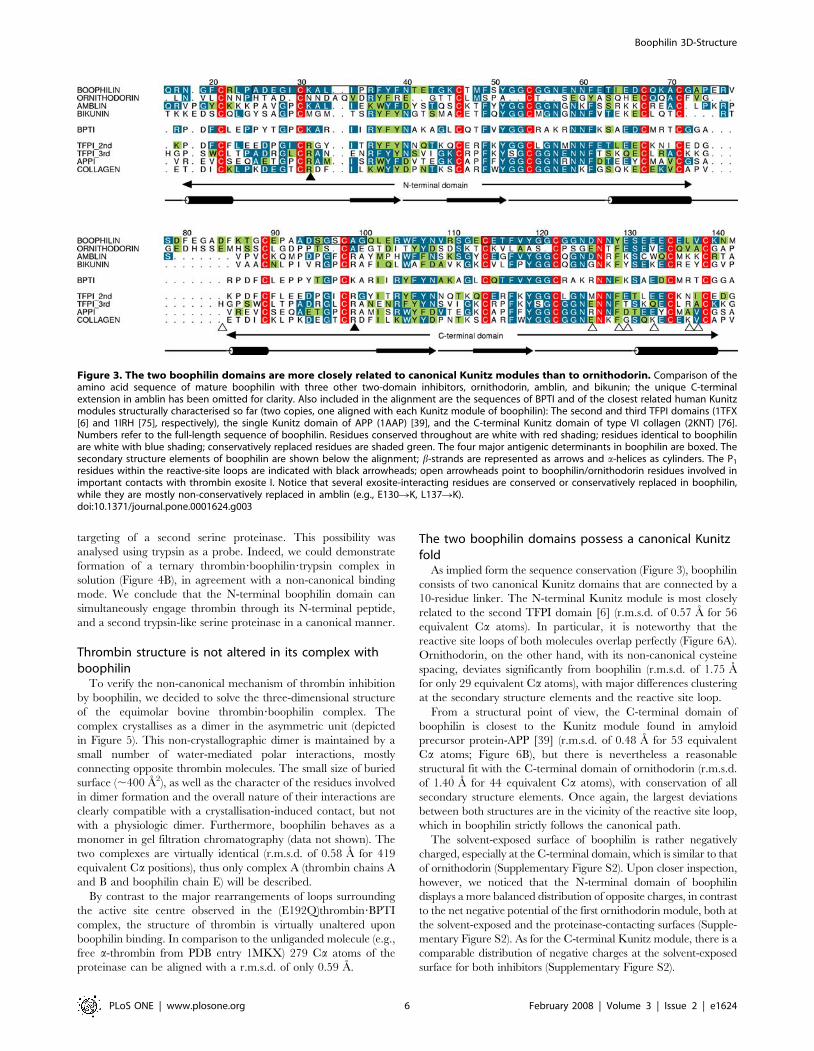
Figure 3. It is immediately apparent that both boophilin domains
are much more closely related to BPTI and other canonical
Kunitz modules than to ornithodorin. Twenty-six residues of the
N-terminal and 22 residues of its C-terminal domain are identical
to BPTI, whereas the corresponding domains in boophilin and
ornithodorin share only 12 or 13 identical residues. Furthermore,
cysteine residue spacing is exactly conserved in boophilin and
BPTI. Most relevant for serine proteinase inhibition, the sequence
of the reactive-site loop in the N-terminal domain (G28-I-C-
KQA-L-I-P35; K31 corresponds to the P1 residue) closely matches
the corresponding G12-P-C-KQA-R-I-I19 sequence in BPTI and
other Kunitz inhibitors, and the secondary binding loop (F49–
C54) is even better conserved. These sequence similarities between
boophilin and BPTI contrast with the multiple insertions and
deletions found in ornithodorin (Figure 3), which altogether hinder
its binding to serine proteinases according to the canonical
mechanism [25].
Recombinant expression of boophilin and furtherfunctional characterisation
A cDNA fragment coding for the full-length mature protein
(residues Q16 to M142, variant H2) was amplified by PCR and
subcloned into the pRBI-DsbC expression vector, as a fusion
construct with the OmpA periplasmic signal sequence (oligonu-
cleotides used for subcloning are given in Supplementary Table
S2). Escherichia coli DH5a cells were transformed with the
expression construct and used for heterologous expression of
recombinant boophilin, which was found to be correctly processed
and active in the periplasmic space of the bacteria (not shown).
The recombinant product was purified to homogeneity by anion-
exchange chromatography on Q-Sepharose FF and ResourceQ
columns, and authenticity was verified by N-terminal sequencing.
To confirm production of a correctly folded inhibitor we have
verified the inhibitory effect of recombinant boophilin on blood
coagulation (concentrations needed for doubling clotting times in the
absence of the inhibitor: thrombin time, 0.045 mM; prothrombin
time, 0.160 mM; APTT, 0.880 mM), as well as its ability to inhibit
thrombin (Ki = 3.4 nM). Next, we verified the inhibition of two other
trypsin-like serine proteinases (trypsin and plasmin), as well as a
partial effect on plasma kallikrein (80% inhibition at 2.36 mM, still
32% inhibition at 0.47 mM; compare Table 1). Finally, we have
detected a weak inhibitory effect on FVIIa (57% inhibition at
2.36 mM). In contrast, other serine proteinases (FXa, FXIIa, uPA,
sc-tPA, and tryptase) were not inhibited by boophilin. The second-
order inhibition constants against trypsin and plasmin, determined
using recombinant boophilin, are (3.8460.73)6105 M21s21 (mean
of three determinations) and (2.5960.22)6105 M21s21 (mean of
four determinations), respectively, and thus similar to the values
reported for BPTI [18,38]. All inhibitory properties of recombinant
boophilin are similar to those observed for the inhibitor isolated from
natural sources. Small differences observed are likely to be accounted
for by the presence of a mixture of boophilin isoforms, in the native
material.
By contrast, the isolated C-terminal boophilin domain, serendip-
itously obtained by cleavage of the A83–D84 peptide bond in the
interdomain linker of recombinant boophilin by an endogenous
proteinase of E. coli, showed essentially no effect on either thrombin
time, prothrombin time or partial thromboplastin time at a
concentration of 1 mM. Only at a rather high protein concentration
of 20 mM, a marginal increase (30%) in thrombin time was observed.
In addition, none of the serine proteinases tested was significantly
inhibited by the isolated boophilin domain (Table 1). Clearly,
presence of an alanine residue at the P1 position in the C-terminal
boophilin domain (Figure 3) strongly disfavours its canonical binding
to trypsin-like serine proteinases. Altogether, our results suggested
that boophilin N-terminal domain inhibited a subset of trypsin-like
serine proteinases according to the canonical mechanism, with
residue K31 occupying the S1 pocket of the bound enzyme.
Thrombin-bound boophilin retains the capability tointeract with other serine proteinases
The functional evidence presented above, combined with
structural information for thrombin?ornithodorin [25] and thrombi-
n(E192Q)?BPTI complexes [23], immediately suggested two possible
modes for boophilin binding to cognate thrombin (Figure 4A). In
both cases, the highly acidic boophilin C-terminal domain (11
aspartate/glutamate residues outbalance four basic residues) would
interact with thrombin’s positively charged exosite I. However, the
two models differ with regard to the region of the N-terminal domain
that blocks the active site of the proteinase. Binding in a canonical
manner, i.e. via reactive-site loop requires major rearrangements of
loops surrounding the active centre, and in particular of the 60-loop.
By contrast, in the ornithodorin-like conformation only the N-
terminal peptide of the inhibitor interacts with thrombin active site,
which is essentially unmodified compared to the free enzyme.
Preliminary modelling experiments indicated that both conforma-
tions could be plausibly adopted in the binary thrombin?boophilin
complex, given the considerable length of the interdomain linker.
To discriminate between these opposite binding modes, we
reasoned that the reactive-site loop of the bound inhibitor would
only be exposed in the ornithodorin-like conformation to allow
Table 1. Inhibition profile of boophilin
EnzymeInhibition of amidolytic activity (%) by
nBoophilin rBoophilinrBoophilin(C-term)
Trypsin 97 98 80
Factor Xa 10 0 0
Factor XIIa n.d. 0 0
Plasma kallikrein 66 80 0
Factor VIIa n.d. 57 0
Plasmin 87 95 25
u-PA 16 0 17
sc-tPA n.d. 4 2
Tryptase n.d. 8 8
Inhibitor samples were incubated with the nine serine proteinases listed, andthe residual activity was measured after adding proteinase-specificchromogenic substrates, as follows: Pefachrome tPA (final substrateconcentration 0.18 and 0.54 mM, respectively) to trypsin (final enzymeconcentration 90 ng/ml) or sc-tPA (1.9 mg/ml); Chromozym X (0.36 mM) to FXa(0.11 U/ml); Pefachrome FXIIa (0.18 mM) to FXIIa (1.8 mg/ml); Chromozym PK(0.36 mM) to plasma kallikrein (0.52 mg/ml); Chromozym tPA (0.73 mM) to FVIIa(2.2 mg/ml); Chromozym PL (0.18 mM) to plasmin (91 mg/ml); Pefachrome uPA(0.18 mM) to u-PA (154 U/ml); and Chromozym TH (0.18 mM) to tryptase(0.11 mg/ml). Reactions were allowed to proceed for 2–6 min, and the effect ofboophilin was estimated by setting the activity obtained without inhibitor as100%. n.d., not determined. The concentrations used for boophilin sampleswere: native (n) inhibitor, 3.53 mM; recombinant (r) inhibitor (full-length),2.36 mM; recombinant inhibitor (C-terminal domain), 15.2 mM.doi:10.1371/journal.pone.0001624.t001
Boophilin 3D-Structure
PLoS ONE | www.plosone.org 4 February 2008 | Volume 3 | Issue 2 | e1624

Figure 2. Complete nucleotide sequence of the cDNA encoding for boophilin variant G2 and predicted amino acid sequence. Thecoding region is shaded in light green, the start codon in dark green, the stop codon in red, the polyadenylation signal in orange, and the shortpoly(A) tail in yellow. The deduced amino acid sequence of pro-boophilin is shown in red below the nucleotide sequence, in one-letter code. Anorange line indicates the fragment amplified by PCR. The in-frame stop codon that precedes the start codon for pro-boophilin is shaded blue. Thededuced amino acid sequence of boophilin (142 residues) would correspond to a polypeptide with a higher molecular mass than that experimentallydetermined by MALDI-MS. However, if one considers the presence of a 15-residue pro-peptide, the resulting mature inhibitor would have atheoretical mass of 13,950 Da, in reasonable agreement with the experimentally determined value of 13,964 Da. (Notice that no putative N-glycosylation sites are present in the boophilin sequence). Further, the mature protein starts with a glutamine residue that could spontaneouslycyclise to pyrrolidone carboxylic acid (5-oxo-proline). This modification would explain the blocked N-terminus observed in native boophilin, and haspreviously been reported in other Kunitz inhibitors such as an BPTI variant from bovine lung [74]. Open triangles indicate the positions where discretedifferences exist in the amino acid sequences of the two boophilin variants.doi:10.1371/journal.pone.0001624.g002
Boophilin 3D-Structure
PLoS ONE | www.plosone.org 5 February 2008 | Volume 3 | Issue 2 | e1624
targeting of a second serine proteinase. This possibility was
analysed using trypsin as a probe. Indeed, we could demonstrate
formation of a ternary thrombin?boophilin?trypsin complex in
solution (Figure 4B), in agreement with a non-canonical binding
mode. We conclude that the N-terminal boophilin domain can
simultaneously engage thrombin through its N-terminal peptide,
and a second trypsin-like serine proteinase in a canonical manner.
Thrombin structure is not altered in its complex withboophilin
To verify the non-canonical mechanism of thrombin inhibition
by boophilin, we decided to solve the three-dimensional structure
of the equimolar bovine thrombin?boophilin complex. The
complex crystallises as a dimer in the asymmetric unit (depicted
in Figure 5). This non-crystallographic dimer is maintained by a
small number of water-mediated polar interactions, mostly
connecting opposite thrombin molecules. The small size of buried
surface (,400 A2), as well as the character of the residues involved
in dimer formation and the overall nature of their interactions are
clearly compatible with a crystallisation-induced contact, but not
with a physiologic dimer. Furthermore, boophilin behaves as a
monomer in gel filtration chromatography (data not shown). The
two complexes are virtually identical (r.m.s.d. of 0.58 A for 419
equivalent Ca positions), thus only complex A (thrombin chains A
and B and boophilin chain E) will be described.
By contrast to the major rearrangements of loops surrounding
the active site centre observed in the (E192Q)thrombin?BPTI
complex, the structure of thrombin is virtually unaltered upon
boophilin binding. In comparison to the unliganded molecule (e.g.,
free a-thrombin from PDB entry 1MKX) 279 Ca atoms of the
proteinase can be aligned with a r.m.s.d. of only 0.59 A.
The two boophilin domains possess a canonical Kunitzfold
As implied form the sequence conservation (Figure 3), boophilin
consists of two canonical Kunitz domains that are connected by a
10-residue linker. The N-terminal Kunitz module is most closely
related to the second TFPI domain [6] (r.m.s.d. of 0.57 A for 56
equivalent Ca atoms). In particular, it is noteworthy that the
reactive site loops of both molecules overlap perfectly (Figure 6A).
Ornithodorin, on the other hand, with its non-canonical cysteine
spacing, deviates significantly from boophilin (r.m.s.d. of 1.75 A
for only 29 equivalent Ca atoms), with major differences clustering
at the secondary structure elements and the reactive site loop.
From a structural point of view, the C-terminal domain of
boophilin is closest to the Kunitz module found in amyloid
precursor protein-APP [39] (r.m.s.d. of 0.48 A for 53 equivalent
Ca atoms; Figure 6B), but there is nevertheless a reasonable
structural fit with the C-terminal domain of ornithodorin (r.m.s.d.
of 1.40 A for 44 equivalent Ca atoms), with conservation of all
secondary structure elements. Once again, the largest deviations
between both structures are in the vicinity of the reactive site loop,
which in boophilin strictly follows the canonical path.
The solvent-exposed surface of boophilin is rather negatively
charged, especially at the C-terminal domain, which is similar to that
of ornithodorin (Supplementary Figure S2). Upon closer inspection,
however, we noticed that the N-terminal domain of boophilin
displays a more balanced distribution of opposite charges, in contrast
to the net negative potential of the first ornithodorin module, both at
the solvent-exposed and the proteinase-contacting surfaces (Supple-
mentary Figure S2). As for the C-terminal Kunitz module, there is a
comparable distribution of negative charges at the solvent-exposed
surface for both inhibitors (Supplementary Figure S2).
Figure 3. The two boophilin domains are more closely related to canonical Kunitz modules than to ornithodorin. Comparison of theamino acid sequence of mature boophilin with three other two-domain inhibitors, ornithodorin, amblin, and bikunin; the unique C-terminalextension in amblin has been omitted for clarity. Also included in the alignment are the sequences of BPTI and of the closest related human Kunitzmodules structurally characterised so far (two copies, one aligned with each Kunitz module of boophilin): The second and third TFPI domains (1TFX[6] and 1IRH [75], respectively), the single Kunitz domain of APP (1AAP) [39], and the C-terminal Kunitz domain of type VI collagen (2KNT) [76].Numbers refer to the full-length sequence of boophilin. Residues conserved throughout are white with red shading; residues identical to boophilinare white with blue shading; conservatively replaced residues are shaded green. The four major antigenic determinants in boophilin are boxed. Thesecondary structure elements of boophilin are shown below the alignment; b-strands are represented as arrows and a-helices as cylinders. The P1
residues within the reactive-site loops are indicated with black arrowheads; open arrowheads point to boophilin/ornithodorin residues involved inimportant contacts with thrombin exosite I. Notice that several exosite-interacting residues are conserved or conservatively replaced in boophilin,while they are mostly non-conservatively replaced in amblin (e.g., E130RK, L137RK).doi:10.1371/journal.pone.0001624.g003
Boophilin 3D-Structure
PLoS ONE | www.plosone.org 6 February 2008 | Volume 3 | Issue 2 | e1624

Significantly different arrangements of boophilin andornithodorin in their complexes with thrombin
The inter-domain linker is three residues longer in boophilin
than in ornithodorin. Nevertheless, it runs closer to the proteinase
surface in thrombin bound to the former inhibitor (Figure 7A).
This linker peptide fits tighter to thrombin’s surface features and
engages in new interactions with thrombin not seen in the
thrombin?ornithodorin complex, further contributing to inhibitor
binding. The most relevant of these are hydrophobic/aromatic
contacts of BF80 with TF34, as well as a water-mediated hydrogen
bond between the carboxylate of BE81 and the main chain
nitrogen of TY76 (Figure 7B). (We use superscripts ‘‘B’’ and ‘‘T’’
before residue names to denote boophilin and thrombin residues,
respectively).
Unexpectedly, the two Kunitz domains of boophilin not only
diverge largely in three-dimensional structure from ornithodorin,
but also occupy significantly different positions relative to the
proteinase moiety, when compared to the thrombin?ornithodorin
complex (Figure 7A). In particular, the N-terminal boophilin
moiety rotates about nine degrees and is shifted <6 A to the ‘‘left’’
in the standard orientation depicted in this figure. As for the C-
terminal module, it is in boophilin rotated by approximately six
degrees and translated by <3 A ‘‘upwards’’. These differences in
quaternary arrangement of Kunitz modules relative to the serine
proteinase moiety result in large differences when compared to the
thrombin?ornithodorin complex, as we discuss below.
Water-mediated interactions dominate at the interfacebetween thrombin exosite I and boophilin
The C-terminal domain of boophilin engages in both
hydrophobic and electrostatic interactions with thrombin’s exosite
I. In contrast to the thrombin?ornithodorin complex, however,
where direct interactions between side chains of the proteinase and
the inhibitor dominate at this interface, in the boophilin complex
the equivalent residues are mostly involved in water-mediated
interactions (Figure 7B). In addition, the guanidinium group ofTR77A donates hydrogen bonds to the hydroxyl group of BY129
and the carbonyl oxygen of BD126. Further direct hydrogen bonds
are formed between the carboxylate of BE134 and the hydroxyl
group of TY76. These polar contacts are strengthened by
hydrophobic interactions between the side chains of BL137 and
a pocket formed by proteinase residues TL65, TR67 and TI82, as
well as between BV138 and TY76.
We note that the exosite I-contacting face of boophilin,
although displaying an overall negative electrostatic potential,
harbours a smaller cluster of acidic residues than ornithodorin,
Figure 4. Thrombin-bound boophilin retains the ability to interact with other serine proteinases. (A) Schematic representation of twohypothetical thrombin?boophilin complexes. In the upper, BPTI-like mechanism, binding of the C-terminal boophilin domain to exosite I promotesextensive rearrangements of loops surrounding the active site to allow insertion of the N-terminal domain in a canonical manner. In the alternative,ornithodorin-like mechanism, exosite engagement is not associated with important modifications of the thrombin active site region, which isoccupied by the N-terminal peptide of the inhibitor in a parallel manner. (B) Demonstration of thrombin?boophilin?trypsin ternary complex formationvia native gel electrophoresis. One mg human a-thrombin?boophilin complex was incubated with increasing amounts of bovine trypsin (from<300 ng, lane 3, to <6 mg, lane 9), and samples were resolved in an 8% polyacrylamide gel. Lanes 1 and 2 contain 1 mg thrombin and 1 mgthrombin?boophilin complex, respectively; the newly formed species corresponds to the ternary complex.doi:10.1371/journal.pone.0001624.g004
Boophilin 3D-Structure
PLoS ONE | www.plosone.org 7 February 2008 | Volume 3 | Issue 2 | e1624

which are located closer to the interdomain linker. Unexpectedly,
none of the three salt bridges between glutamates in ornithodorin
and the proteinase moiety are observed in the complex with
boophilin, even though these acidic residues are conserved or
conservatively replaced. Likewise, of the five residues involved in
hydrophobic contacts in ornithodorin, one (I117) has no equivalent
in boophilin, while the structural equivalents of two others (BY129
for F103 and BE133 for V107) have an increased polar character and
are no longer involved in van-der-Waals interactions (Figure 7B).
Although the total buried area upon thrombin complexation by
boophilin is equivalent to that of the thrombin?ornithodorin complex
(,1,800 A2), the decrease in the number and strength of the
proteinase-inhibitor interactions offers an explanation for the lower
Ki determined for boophilin (see Discussion).
It is noteworthy that a good fraction of boophilin’s negative
charges cluster in the solvent exposed side of the molecule,
pointing away from the basic exosite I of thrombin (Supplemen-
tary Figure S2). This suggests that the C-terminal domain serves a
multiple role, sterically blocking substrate access, masking the
overall positive charge of the exosite, and creating a new
negatively-charged surface that can repel approaching thrombin
substrates. These combined effects would thus counteract
competitive displacement of the inhibitor by physiological
substrates, which require previous exosite I binding for processing.
The N-terminal residues of boophilin occupy the activesite cleft in a parallel manner
The experimental electron-density map (Figure 8A) unambig-
uously shows that the N-terminal residues of boophilin bind across
thrombin’s active site cleft in a parallel, hirudin-like manner [16].
In the three-dimensional structure of the thrombin?ornithodorin
complex, it was observed that the introduction of a non-natural
Figure 5. Three-dimensional structure of the a-thrombin?boo-philin complex. The crystallographic dimer present in the asymmetricunit is shown as a ribbon plot; thrombin molecules are coloured blueand green and boophilin molecules are coloured red and orange.Notice that the reactive-site loops of both inhibitor domains point awayfrom the proteinase moiety; the corresponding P1 residues, BK31 andBA99, are shown as space-filling models. Within the dimer interface,direct hydrogen bonds are formed between the side chain of BS97 andthe main-chain carbonyl of BG124 located in opposite C-terminaldomains of boophilin, and between the side chains of TD21 and TQ14A.doi:10.1371/journal.pone.0001624.g005
Figure 6. Boophilin is structurally more related to canonical Kunitz modules than to the distorted ornithodorin domains. Stereo plotsshowing the three-dimensional structure of (A) boophilin’s N-terminal domain (red) overlaid on those of the second TFPI domain, as seen in itscomplex with porcine trypsin (orange, PDB entry 1TFX) [6], and of the N-terminal ornithodorin domain (yellow, 1TOC) [25], and (B) boophilin’s C-terminal domain (red) superimposed on those of APP single Kunitz domain (cyan, 1TAW) [8], and the C-terminal ornithodorin domain (yellow, 1TOC)[25]. The N and C termini of boophilin are labelled and its disulfide bonds are shown as sticks. Notice that in spite of significant sequence similaritybetween the carboxy-terminal domain of boophilin and other Kunitz-type inhibitors (Figure 3), with strict conservation of cysteine spacing, itsputative P1 residue is nevertheless an alanine (BA99), thus precluding inhibition of trypsin-like serine proteinases.doi:10.1371/journal.pone.0001624.g006
Boophilin 3D-Structure
PLoS ONE | www.plosone.org 8 February 2008 | Volume 3 | Issue 2 | e1624

serine residue at the N-terminus of the inhibitor results in an
unfavourable chemical environment around the side chain of TE192.
By contrast, the present structure shows that this carboxylate group is
stabilised by hydrogen-bonding interactions with the side chain of
the natural N-terminal residue of mature boophilin, BQ16 (see
Figure 8B for a schematic representation of major thrombin-
boophilin interactions). Inspection of the electron density maps
suggests that the latter side chain displays a somewhat increased
flexibility, which correlates well with its above-average B-factor. The
main chain of this N-terminal residue is positioned in the S2 pocket of
thrombin, stabilised by a direct hydrogen bond between its carbonyl
oxygen and the guanidinium group of BR22.
The most relevant feature of active site interactions is the
occupancy of thrombin’s S1 subsite by the ideally suited side chain
of BR17. The guanidinium group of this boophilin residue forms
two hydrogen bonds with the carboxyl group of TD189 (3.4 and
3.6 A) at the bottom of the S1 pocket (Figure 8A). Two additional
hydrogen bonds are formed between the terminal group of BR17
and the main chain carbonyls of TG219 (direct bond) and TF227
(water-mediated). This feature also distinguishes boophilin from
ornithodorin, which lacks an equivalent residue at this position.
The following boophilin residue, BN18, interacts through its side
chain with the main chain nitrogen atom of TG219 and the
carboxyl oxygen of TE146, the latter via a solvent molecule.
Finally, BG19 is hydrogen-bonded to the carbonyl group of TG216
(Figure 8B), thus partially mimicking the interaction of a ligand
occupying the S3 pocket of the proteinase. This subsite
accommodates the side chain of BR22, which forms a direct
hydrogen bond with the hydroxyl group of TY60A. The only other
major contact between the proteinase moiety and the N-terminal
domain of boophilin is the parallel stacking of the side-chain ofBF39 with those of TP60C and TW60D.
Discussion
Tick-derived anti-hemostaticsWe report the isolation and cloning of a two-domain Kunitz
inhibitor from the cattle tick Boophilus microplus, which we have
termed boophilin. Further, we characterise boophilin’s inhibitory
mechanism by solving the three-dimensional structure of its
complex with bovine thrombin. Together with our identification of
carrapatin, a single domain Kunitz-type inhibitor in engorged ticks
(Swiss-Prot accession number P81162), as well as with previous
reports of inhibitors isolated from eggs and larvae of this parasite
[28–34,40], evidence is emerging that B. microplus utilizes a whole
array of both single- and multi-domain Kunitz inhibitors during its
whole life cycle. Recently, several salivary proteins that contain
single or tandem Kunitz repeats have also been identified in the
hard ticks Ixodes scapularis [41] and I. pacificus [42], indicating that
inhibitors from this family might play a general role as
antihemostatic factors. One of these proteins, the double-headed
inhibitor, ixolaris, was shown to inhibit the tissue factor?FVIIa
complex in a FX(a)-dependent manner by binding to exosite II in
FXa, in line with its homology to TFPI [43,44].
Figure 7. Different orientations of the Kunitz domains in thrombin complexes with boophilin and ornithodorin. (A) Stereo plotshowing a comparison of thrombin?boophilin (blue/red) and thrombin?ornithodorin (blue/yellow) complexes, after overlaying both thrombinmoieties. Notice the large displacements of the corresponding N- and C-terminal Kunitz modules relative to each other. (B) Stereo close-uphighlighting the docking of the C-terminal domain of boophilin (red) to thrombin (blue) exosite I. Water molecules are represented as orange spheresand hydrogen bonds as rows of small grey spheres. The side chains of residues involved in intermolecular contacts are shown and labelled.doi:10.1371/journal.pone.0001624.g007
Boophilin 3D-Structure
PLoS ONE | www.plosone.org 9 February 2008 | Volume 3 | Issue 2 | e1624

Recently, another two-domain Kunitz inhibitor with anti-
thrombin activity, amblin from the hard tick Amblyomma hebraeum,
has been described [37]. Notably, amblin differs from boophilin due
to (i) a much shorter interdomain linker; (ii) a relatively long, highly
basic C-terminal extension; (iii) the presence of two additional
cysteine residues located in this extension and in the C-terminal
Kunitz domain, respectively, which might form an extra disulfide
bond; and (iv) non-conservative replacements of several acidic
residues of the C-terminal domain (Figure 3). We have generated a
three-dimensional model of amblin on the basis of its closest relative
of known structure, bikunin [45]. The high pI of amblin is reflected
in extended areas of basic potential on both Kunitz domains
(Supplementary Figure S2). Thus, both strong electrostatic repulsion
and inappropriate quaternary structure would make extremely
unlikely that amblin interacts with any of the positively charged
exosites on the thrombin moiety. The lack of anti-trypsin activity by
amblin is also surprising in view of the conservation of ‘‘canonical’’
reactive-site loops, and because our homology model suggests that
the reactive-site loop of the N-terminal Kunitz domain remains
accessible to target proteinases. These observations require a
thorough investigation of amblin’s inhibitory potential.
Boophilin is a canonical Kunitz-type inhibitorInhibition of the key procoagulant enzyme, thrombin, by
canonically binding Kunitz-type inhibitors is unfavourable due to
the presence of a prominent insertion loop at position 60, and of a
glutamate at position 192. These obstacles are circumvented in the
double-headed Kunitz inhibitor from O. moubata, ornithodorin, as
only its N-terminal peptide inserts into the active-site cleft of the
proteinase [25]. A highly similar thrombin inhibitor, savignin, has
been isolated from the salivary glands of O. savignyi [46,47], and
would be expected to use the same non-canonical mechanism for
thrombin inhibition. On the other hand, the close resemblance of
boophilin to canonical Kunitz domains and the presence of an
optimal P1 residue in its N-terminal domain (K31) suggested that
boophilin could inhibit thrombin in a canonical manner, i.e. by
occupying the active-site cleft of the proteinase with the reactive-
site loop of this module. This would require a considerable
repositioning of the N-terminal Kunitz module compared to the
thrombin?ornithodorin complex, which is nevertheless possible
given that the two domains are connected by a long, flexible linker.
Canonical binding would in turn require that initial binding of the
C-terminal domain to exosite I provided enough energy to drive
insertion of the N-terminal reactive site loop past the energetic
barrier that thrombin’s 60-loop represents.
Boophilin inhibits thrombin in a non-canonical manner,despite possessing a canonical reactive-site loop
However, structural and functional evidence presented here
indicates that boophilin also adopts the non-canonical mechanism
for thrombin inhibition, in spite of possessing a regular BPTI-like
domain. We conclude that binding of the acidic C-terminal Kunitz
domains to exosite I does not induce major rearrangements of loops
surrounding thrombin’s active-site cleft. This is in line with our
previous observations of essentially unmodified active sites in the
crystal structures of thrombin?triabin [15] and thrombin?thrombo-
modulin [24] complexes. A recent crystal structure of murine
thrombin bound to an acidic peptide from PAR3 via exosite I
revealed some displacements of 60-loop residues, in particular
W60D [48]. However, analysis of the thrombin?PAR3 complex
(PDB entry 2PUX) indicates that these rearrangements are either
induced or stabilised by artifactual interactions with a neighbouring
crystal molecule. In conclusion, it would seem that interactions with
exosite I neither induce rearrangements of thrombin structure, in
particular of the 60-loop, nor are required for inhibitors or substrates
to approach the proteinase catalytic machinery. Exosite-mediated
interactions are nevertheless strictly required for positioning
unfavourable substrates (protein C, thrombin-activatable fibrinolysis
inhibitor/TAFI)/inhibitors (Kunitz) at a proper distance and
orientation to gain access to the active site of thrombin.
In spite of similar domain organisations and the presence of
highly acidic C-terminal domains, ornithodorin/savignin are
much tighter thrombin inhibitors (Ki = 1.0610212 M or
4.9610212 M) than boophilin (Ki = 1.861029 M). Further, where-
as ornithodorin and savignin do not cross-react with other serine
proteinases, boophilin is a potent inhibitor of trypsin and of the
fibrinolytic enzyme, plasmin. It is tempting to speculate that the
less efficient thrombin inhibitor, boophilin, confers an evolutionary
advantage to B. microplus by controlling opposing mechanisms of
hemostatic balance. Another interesting possibility is discussed
Figure 8. Boophilin inhibits thrombin in a non-canonical manner. (A) Stereo close-up of the thrombin active centre (blue) showing thebound tetrapeptide BQ16–BR17–BN18–BG19 of boophilin (red), along with S1A–L1–N2–V3 of ornithodorin (yellow; notice that the N-terminal residueof the latter is an artifact introduced for cloning purposes). The final electron density for the thrombin?boophilin complex, contoured at 1s, isdisplayed as a blue mesh. The catalytic triad of thrombin (TH57, TD102, TS195) is highlighted in orange and the side-chains of TD189 and TE192 arecoloured pale green and cyan, respectively. Disulfide bonds are represented as yellow sticks. (B) Schematic representation of the thrombin-boophilininteractions at the enzyme’s active site. Inhibitor residues are coloured red and hydrogen bonds are depicted as dashed lines.doi:10.1371/journal.pone.0001624.g008
Boophilin 3D-Structure
PLoS ONE | www.plosone.org 10 February 2008 | Volume 3 | Issue 2 | e1624

below. Of note, a thrombin inhibitor with an apparent Mr
,60 kDa, BmAP, has been isolated from the saliva of B. microplus
[49]. In contrast to boophilin, BmAP does not inhibit plasmin or
trypsin. Finally, a small (Mr = 1,770 Da) exosite-binding thrombin
inhibitor, microphilin, has been identified in the tick saliva [50]. It
is thus conceivable that the three inhibitors cooperate to inhibit
thrombin activity during feeding and digestion.
It is also worth mentioning that immunization of cows with
salivary gland preparations from B. microplus reduced babesiosis
[51], pointing to the immunological potential of proteins secreted
by this parasite. Of note, the sequence containing the reactive-site
loop in boophilin N-terminal domain is predicted to be the major
antigenic determinant of the protein. Given that thrombin
inhibitors are critical for hematophagous animals during feeding
and digestion, boophilin would appear as a promising candidate
for the development of recombinant vaccines.
The close relatedness of boophilin to BPTI-like Kunitz inhibitors
(Figures 3 and 6) explains its ability to potently inhibit trypsin and
plasmin. In particular the N-terminal domain possesses all the
necessary features for canonical inhibition, including a lysine side
chain (K31) ideally suited for occupying the S1 pocket of trypsin-like
serine proteinases with a serine at position 190 [7,52,53]. Further,
the I29 side chain is preferred at the P3 position in Kunitz-type
inhibitors [53]. Finally, the conserved alanine residue at position P1’
is also essential for formation of stable complexes with trypsin and
plasmin [54]. For several other serine proteinases tested, the
presence of A190 at the bottom of the S1 specificity pocket, instead
of a serine in e.g. trypsin and plasmin, increases the size of this pocket
and eliminates a potential hydrogen-bond partner for a lysine side
chain [55]. In these cases, Kunitz inhibitors with an arginine at
position P1 are strongly preferred over those with a P1 lysine [52,53],
thus explaining at least partially the poor or absent effect of boophilin
on FXa, FXIIa, uPA and t-PA. Finally, in the tryptase tetramer the
active sites of all four monomers are arranged facing an internal
pore, and are therefore inaccessible to most substrates and inhibitors
[56]. Kunitz inhibitors are too bulky to enter the tryptase channel,
explaining the inactivity of boophilin towards tryptase.
Boophilin is a bi-functional serine proteinase inhibitorAs mentioned above, boophilin possesses a rather high Ki for
thrombin inhibition when compared to the two other thrombin
inhibitors known or predicted to bind in a non-canonical manner,
ornithodorin [25] and savignin [46,47]. This observation raises the
interesting possibility that boophilin could have evolved to target not
only circulating thrombin, but to block in addition the membrane-
bound activation intermediate, meizothrombin (MzT). The crystal
structure of a MzT variant that lacks the membrane-binding
fragment 1, reveals formed exosite I and active centre, while exosite
II is covered by the kringle 2 domain of the intermediate form [57].
Therefore boophilin could dock to this form essentially as seen in the
current crystal structure (see Figure 9A for a representation of the
modelled MzT?boophilin complex).
Our current data also indicate that thrombin-bound boophilin
retains the capability to interact with additional serine proteinases,
as demonstrated by formation of a stable ternary complex with
trypsin (Figure 4), and the reactive-site loop of the N-terminal
boophilin domain would remain fully accessible in the MzT?boo-
philin complex (compare Figures 5 and 9A). These observations
immediately suggest that a proximal, membrane-bound proteinase
could easily dock to this complex. Coagulation FXa would appear
as the most attractive candidate, because of its proximity to MzT
during prothrombin activation by the prothrombinase complex
(FVa?FXa). To verify that MzT?boophilin?FXa ternary complex
formation is feasible without steric clashes, we overlaid the
structurally and functionally related second Kunitz domain of
TFPI (refer to Figure 6A) bound to trypsin [6] onto the N-terminal
boophilin domain. Next, we superposed the FXa crystallographic
model [58] onto the trypsin moiety. This revealed that the serine
proteinase moieties of FXa and meizothrombin could be cross-
linked by the N-terminal domain of a boophilin molecule
(Figure 9B). Furthermore, the N-terminal domains of both
proteinases would not interfere with ternary complex formation.
In this manner, boophilin could efficiently inhibit thrombin
generation by prothrombinase. A schematic representation of this
putative, membrane-bound complex is shown in Figure 9C. We
stress, however, that free boophilin only marginally inhibits FXa in
vitro (Table 1), and thus formation of the ternary complex would
rely on the proper orientation of both membrane-bound serine
proteinases, and would probably require additional boophilin-FVa
interactions. In support of our hypothesis, we note that both
endogenous (TFPI) [4] and tick-derived multidomain Kunitz
inhibitors ixolaris [43,44] and penthalaris [59] employ this dual
strategy for inhibiting the related FVIIa?TF?FXa complex. Of
particular note, TFPI inhibits FVIIa through its first Kunitz
domain, but only after the second Kunitz moiety has bound FXa
[4,60]. Future investigations should test the validity of this model,
or identify other targets for thrombin-bound boophilin in vivo.
Finally, we notice that the current structure has implications for
modelling procedures, in particular for macromolecular docking.
Current docking algorithms have only had limited success rates, even
when the structures of isolated modules were known [61]. Our
structure of the thrombin?boophilin complex and its comparison
with the related thrombin?ornithodorin heterodimer reveals that
caution must be exercised even when a highly homologous template
for the expected complex does exist. Indeed, these two inhibitors
share a common binding mode (blockade of basic exosite I of the
proteinase and parallel alignment of N-terminal residues across the
active-site cleft). However, the details of interacting residues (e.g.,
direct vs. water-mediated contacts at exosite I, occupancy or not of
the major S1 subsite on thrombin) vary considerably in their
complexes with the proteinase. Concomitantly, and perhaps more
striking from the viewpoint of macromolecular docking, the two
pairs of inhibitor domains adopt markedly different orientations
relative to the cognate thrombin molecule.
Materials and MethodsAnimals
Ticks were obtained from Drs. D.H. Aguirre and A.B. Gaido,
Instituto Nacional de Tecnologıa Agropecuaria, Estacion Exper-
imental Agropecuaria Salta (Argentina).
ReagentsBovine a-thrombin was isolated from fresh ox blood as described
previously [62]. Other serine proteinases were purchased from the
suppliers listed below: bovine FXa, Diagnostic Reagents Ltd. (Oxon,
England); human a-FXIIa and plasma kallikrein, Kordia Laboratory
Supplies (Leiden, Netherlands); recombinant human FVIIa (Novo-
SevenH), Novo Nordisk (Denmark); human plasmin, Behringwerke
GmbH (Marburg, Germany); recombinant tryptase, Promega
(Madison, USA); recombinant sc-tPA, Boehringer Mannheim
(Mannheim, Germany); bovine pancreatic trypsin, Serva Feinbio-
chemika (Heidelberg, Germany); and urokinase (uPA), Ribosepharm
GmbH (Haan, Germany). All chromogenic substrates used were
obtained from Pentapharm AG (Basel, Switzerland), except for
ChromozymH TH (Tos-Gly-Pro-Arg-para-nitroanilide), purchased
from Boehringer Mannheim, which also provided APTT reagent
and endoproteinases Lys-C and Asp-N. DadeH Thromboplastin-IS
was purchased from Dade Behring Marburg (Marburg, Germany).
Boophilin 3D-Structure
PLoS ONE | www.plosone.org 11 February 2008 | Volume 3 | Issue 2 | e1624

Figure 9. Predicted boophilin interactions with meizothrombin and FXa. (A) Model of the three-dimensional meizothrombin?boophilincomplex, generated by overlaying the coordinates of bovine meizothrombin (blue except for the Kringle 2 domain, in green; 1A0H) [57] onto thethrombin moiety of the current complex with boophilin. Residues BR17 and BK31 are shown as space-filling models and labelled. (B) Model of theputative MzT?boophilin?FXa ternary complex. FXa (yellow, except for domain EGF2, in olive; 1FJS) [58] was docked onto the complex displayed in (A)by juxtaposition onto a trypsin?APP complex (1TFX) [6] that had been previously overlaid onto the N-terminal domain of boophilin (red). Absence ofany steric clashes shows that formation of such a ternary complex in vivo would be feasible. The N- and C-terminal domains of boophilin (Boophilin(N) and Boophilin (C), respectively), the serine proteinase and Kringle 2 domains of meizothrombin (SP (MzT) and Kringle 2, respectively), and theserine proteinase and EGF-2 domains of factor Xa (SP (FXa) and EGF-2, respectively) are labelled. (C) Schematic representation of a possiblemechanism for boophilin inhibition of the membrane-bound prothrombinase complex. Both FXa and meizothrombin interact with the membranesurface (dark red) via their respective Gla domains (light green). The multi-domain organisation of FVa (violet) and its contacts with FXa/MzT havebeen omitted for simplicity. The N-terminal domain of boophilin (red) could bridge the catalytic domains of meizothrombin (dark green) and FXa(blue) while bound to the cofactor.doi:10.1371/journal.pone.0001624.g009
Boophilin 3D-Structure
PLoS ONE | www.plosone.org 12 February 2008 | Volume 3 | Issue 2 | e1624

All other chemicals, of the highest purity grade available, were
purchased from Merck (Darmstadt, Germany), except where noted.
Isolation of native boophilinOne-hundred fifty grams of frozen engorged ticks were
homogenised in 1 l buffer A (20 mM Tris-HCl, pH 8.0, 1 mM
CaCl2, 1 mM benzamidine). After centrifugation at 10,0006g for
10 minutes, the filtered (5-mm pore) supernatant was applied to a
Q-Sepharose (Pharmacia, Freiburg, Germany) column
(200650 mm) previously equilibrated with 20 mM Tris-HCl,
pH 8.0 (buffer B). The column was extensively washed with buffer
B and bound proteins were eluted with a linear NaCl gradient
(from 0 to 1.0 M in buffer B).
A thrombin affinity column was prepared by coupling 10 mg of
bovine a-thrombin to 2 g of CNBr-activated Sepharose 4B
(Pharmacia), according to the manufacturer’s guidelines. Ion-
exchange chromatography fractions with thrombin inhibitory
activity (determined as described below) were pooled, diluted with
two volumes of 20 mM sodium phosphate, pH 7.4 (buffer C) and
applied to the thrombin-Sepharose column previously equilibrated
in buffer C. The column was extensively washed with buffer C and
subsequently with buffer C containing 500 mM NaCl and with
double distilled water, before elution of specifically bound proteins
with 20 mM glycine-HCl, pH 2.5. The pH of the eluate was
immediately adjusted to 8.0 with 0.1 volumes 1 M Tris-HCl,
pH 8.0 before lyophilising the sample.
Activity assaysThrombin inhibitory activity was followed during boophilin
purification by measuring the residual amidolytic activity of the
proteinase towards ChromozymH TH. Bovine thrombin (900 ml,
15 nM) in assay buffer (50 mM Tris-HCl, pH 7.8, 0.1 M NaCl,
0.05% PEG 6,000) was incubated with 50 ml protein samples or
with the same volume of assay buffer for 2 minutes at 37uC.
Substrate (50 ml, 1.5 mM) was then added, and absorbance of the
reaction mixture at 405 nm was monitored for 2 minutes.
Inhibition of blood clotting by boophilinTo determine thrombin time, 100 ml citrated human plasma
were mixed with 50 ml 0.154 M NaCl or inhibitor solution and
equilibrated at 37uC. Bovine thrombin (50 ml; 5 IU/ml) was then
added and the time to clot was measured. Prothrombin time was
determined by mixing 50 ml of DadeH Thromboplastin-IS with
50 ml of either 25 mM CaCl2 or the same solution containing the
sample to be assayed. After equilibration at 37uC, 50 ml human
citrated plasma were added, and the coagulation time measured.
Activated partial thromboplastin time was determined both with
and without preincubation of the inhibitor solution (40 ml) with
50 ml citrated human plasma and 50 ml APTT reagent for
3 minutes at 37uC. Ten ml of a 125 mM CaCl2 solution were
finally added and the coagulation time was measured. Clotting
times were determined in duplicate.
Inhibition of other serine proteinasesThe amidolytic activity of each proteinase towards a specific
chromogenic substrate was assayed at room temperature, by
measuring the variation of light absorbance at 405 nm in the assay
mixture. Briefly, 200 ml of assay buffer (50 mM Tris-HCl,
pH 7.4–8.4, 154 mM NaCl) or of inhibitor were mixed with
50 ml of the enzyme solution and dispensed in 96-well microtiter
plates. After five minutes pre-incubation, 25 ml of the chromogenic
substrate solution were added and the reaction was allowed to
proceed for an enzyme-dependent period (2–6 minutes). Reactions
were stopped by adding 25 ml 50% (v/v) acetic acid. The enzymes
tested are given in the legend to Table 1, along with the
chromogenic substrates used in each case.
For determination of the second-order rate constants of trypsin/
plasmin inhibition, 200 ml of inhibitor solution were mixed with
25 ml substrate (Chromozym TH or Chromozym PL, respectively;
final concentration: 0.2 mM). The final concentration of recom-
binant boophilin was varied between 0.1 and 0.65 mM (trypsin), or
between 0.04 and 0.4 mM (plasmin). The reaction was started by
addition of 25 ml enzyme solution (final concentrations: 6.25 ng/
ml and 12.5 mg/ml, respectively), and the absorption at 405 nm
was measured over 45 minutes at 37uC. The pseudo-first order
rate of inactivation for each inhibitor concentration (kobs) was
determined from the progression curve according to the equation
[P] = A+Bt–Cexp(-kobst). Second-order rate constants were calcu-
lated from the equation k2/Ki = (1+[S0]/Km)/b, with
S0 = 0.2 mM, Km = 0.0117 mM for ChromozymH TH or
0.286 mM for ChromozymH PL; the slope b was computed from
the regression line 1/kobs versus 1/[I].
Electrophoretic mobility shift assaysFive-hundred ng human thrombin or bovine trypsin in 25 mM
Tris-HCl pH 8.0, 190 mM glycine, were mixed with increasing
concentrations of boophilin and incubated at room temperature for
15 min before adding glycerol to 5% final concentration. In other
experiments, thrombin?boophilin complexes were similarly titrated
with increasing concentrations of trypsin. Samples were resolved at
4uC in Tris-glycine-polyacrylamide gels and silver-stained.
Protein sequencingLyophilised protein samples were dissolved in 0.1 M Tris-HCl,
pH 8.0, 6 M guanidinium hydrochloride, 5% (v/v) b-mercapto-
ethanol and incubated for 16 hours at room temperature to
reduce disulfide bonds. The free thiol groups were derivatised with
5% (v/v) 4-vinylpyridine; samples were incubated for further
90 minutes at room temperature, acidified with formic acid, and
finally desalted by RP-HPLC. The S-b-pyridylethylated samples
were digested for 10 hours at 37uC with either Lys-C (in 25 mM
Tris-HCl, pH 8.5, 1 mM EDTA) or Asp-N (in 50 mM sodium
phosphate, pH 8.0). Reactions were stopped by adjusting the pH
of the samples to 2.0 with formic acid. For CNBr-cleavage, S-b-
pyridylethylated boophilin was dissolved in 70% (v/v) formic acid
containing 10% (w/v) CNBr and incubated in the dark at room
temperature for 14 h. Peptides were separated by RP-HPLC and
subjected to automated sequencing. The molecular mass of the
unmodified protein was determined by MALDI-MS.
Construction and screening of the B. microplus cDNAlibrary
Total RNA was isolated from 1 g of unfed ticks (for cDNA library
construction) or from 10 g of engorged adults (for PCR amplifica-
tion) and enriched in poly(A)+ RNA by oligo(dT)-cellulose affinity
chromatography. cDNA was synthesised from 1 mg of poly(A)+ RNA
with random hexanucleotides as primers, and a cDNA library was
constructed in l Uni-ZAP XR (Stratagene, Amsterdam, Nether-
lands), following the manufacturer’s instructions.
An internal fragment of boophilin cDNA was amplified by PCR
using degenerate oligonucleotides (Supplementary Table S2), cloned
into the BamH I site of pBluescript KS (Stratagene, Amsterdam,
Netherlands), and sequenced. A fragment of similar size was
amplified using aliquots of the cDNA library phage stock as
template, to verify the presence of boophilin cDNA in the library.
Positive clones, identified using the a-32P-dATP-labelled boophilin
Boophilin 3D-Structure
PLoS ONE | www.plosone.org 13 February 2008 | Volume 3 | Issue 2 | e1624

cDNA fragment as probe, were isolated and converted into double
stranded pBluescript phagemids by in vivo excision, and further
characterised by automated DNA sequencing of both strands.
Heterologous expression of mature boophilinMature boophilin (variant H2) was cloned into the bi-cistronic
expression vector pRBI-DsbC [63] by overlap-extension PCR.
Briefly, the OmpA signal-coding region of the vector and the
fragment of boophilin cDNA coding for the mature inhibitor were
amplified independently, using specific primers (Supplementary
Table S2). Aliquots of both PCR products were then mixed and
amplified using the previously used 59 primer for the OmpA
fragment and the 39 primer for boophilin. The amplified fragment,
coding for an OmpA signal–mature boophilin fusion protein was
cloned into the Xba I and Hind III sites of the expression plasmid.
Escherichia coli DH5a cells transformed with the expression
construct were grown in LB with 2 g/l glucose and 100 mg/l
ampicillin to an optical density of 0.7, before induction with 1 mM
IPTG (isopropyl-b-D-thiogalactopyranoside). At induction time,
2 g/l glucose, 100 mg/l ampicillin, and 5 mM N-acetyl-cysteine
were added. Cells were harvested 36 hours post-induction and
periplasmic proteins were released via cold osmotic shock. The
resulting solution was immediately applied to an 80650 mm Q-
Sepharose FF column pre-equilibrated with buffer D (20 mM Tris-
HCl, pH 8.0). The column was extensively washed with buffer D,
and bound proteins eluted with a linear salt gradient (from 0 to
0.5 M NaCl in buffer D). Fractions displaying anti-thrombin activity
were pooled, diluted with buffer D and loaded onto a ResourceQ
column (Pharmacia, Freiburg, Germany) pre-equilibrated with the
same buffer. The column was extensively washed with buffer D and
bound proteins were eluted with a linear salt gradient (0–0.4 M
NaCl in buffer D). Samples with anti-thrombin activity eluted as a
single peak. Correct processing of the recombinant inhibitor was
verified by MALDI-MS and N-terminal sequencing.
Complex preparation and crystallisationThe macromolecular complex was prepared by mixing bovine
a-thrombin with a molar excess of purified recombinant boophilin.
The mixture was incubated on ice for two hours, and the
thrombin?boophilin complex was separated from excess inhibitor
by size exclusion chromatography on a Superdex 75 10/30
column (Amersham Pharmacia), using 0.01 M MES pH 6.5,
0.1 M NaCl as the mobile phase. Fractions were collected and
analysed by SDS-PAGE. Those containing the thrombin?boophi-
lin complex were pooled and concentrated on a 10-kDa cut-off
centrifugal concentrator (Vivascience).
Single crystals of the macromolecular complex were obtained by
vapour diffusion at 279 K from 4-ml sitting drops containing equal
volumes of bovine thrombin?boophilin complex solution (7.5 mg/
ml in 0.01 M MES pH 6.5, 0.1 M NaCl) and precipitant (0.05 M
KH2PO4, 18% (w/v) PEG 8,000). Crystals belong to the
orthorhombic space group P212121, with unit cell constants
a = 92.5 A, b = 104.2 A, c = 129.1 A, and contain two complexes
per asymmetric unit (solvent content 60.6%). The crystals were
transferred to 1.56 mother liquor containing 15% glycerol as
cryoprotectant and cryo-cooled in liquid nitrogen.
Data collection, processing and refinementDiffraction data to 2.35 A were collected from a single cryo-
cooled (100 K) crystal on a MAR CCD detector at ESRF
beamline ID14-EH3. Data were processed with MOSFLM [64],
scaled with SCALA [65], and reduced with tools from the CCP4
package [65]. Data collection and refinement statistics are
summarised in Table 2.
The structure was solved by molecular replacement using AMoRe
[66] with data in the 10-4.0 A resolution range. Two proteinase
molecules were located using the coordinates of unliganded bovine
a-thrombin from PDB entry 1MKX [67] as search model. All
attempts to locate the Kunitz domains of boophilin by molecular
replacement, using truncated models of either BPTI (1BPI) [68] or
ornithodorin (1TOC) [25] were unsuccessful.
The initial electron density maps, computed after rigid body
refinement of the proteinase molecules with CNS v. 1.1 [69],
Table 2. Statistics of data collection and refinement
Crystallographic analysis
Resolution range (A) (overall/outer shell) 54.9–2.35/2.48–2.35
Space group P212121
Unit cell dimensions (A) a = 92.5 b = 104.2c = 129.2
Number of observations (total/unique) 192,392/51,554
Multiplicity (overall/outer shell) 3.7/3.7
Rmergea (overall/outer shell) 6.4/29.0
Completeness (%) (overall/outer shell) 98.0/97.4
I/s(I) (overall/outer shell) 9.5/2.6
Mathews coefficient (A3 Da21) 3.1
Solvent content (%) 60.6
Structure refinement
Rfactorb/Free Rfactor
c (%) 19.0/23.0
Nu of unique reflections (working/test set) 48,886/2,627
Water molecules 453
Ions 6 (4 PO432, 2 Na+)
Total number of atoms 7,261
Number of protein atoms 6,771
Average overall B-factor (A2) 31.2
Average protein B-factor (A2) 30.8
Average main-chain B-factor (A2) 30.5
Average side-chain B-factor (A2) 31.2
Average water B-factor (A2) 33.1
Average ion B-factor (A2) 62.4 (PO432)/32.5 (Na+)
r.m.s.d. bonded Bs (A2) 0.751
r.m.s.d. bond lengths (A) 0.008
r.m.s.d. bond angles (u) 1.137
Ramachandran plot statistics
Residues in allowed regions 717 (99.9%)
Residues in generously allowed regions 1 (0.1%)
Residues in disallowed regions 0 (0%)
Estimated coordinate error
E.s.d. from Luzzati plot (A) 0.30
DPId (A) 0.20
aRmerge = ShSi |Ihi-,Ih.|/Sh Si ,Ih., where Ihi is the observed intensity of thei-th measurement of reflection (h), including symmetry-related ones, and ,Ih.
is the mean intensity of the i observations of reflection h over allmeasurements of Ihi.
bRfactor = S||Fo|-|Fc||/S |Fo| where |Fo| and |Fc| are observed and calculatedstructure factor amplitudes, respectively.
cFree Rfactor is the cross-validation R-factor computed for a randomly chosensubset of 5% of the total number of reflections, which were not used duringrefinement.
dDiffraction-data precision indicatordoi:10.1371/journal.pone.0001624.t002
Boophilin 3D-Structure
PLoS ONE | www.plosone.org 14 February 2008 | Volume 3 | Issue 2 | e1624

displayed additional density for most of the inhibitor molecules.
Two cycles of manual rebuilding of the proteinase models on a
graphic workstation using TurboFRODO (Bio-Graphics, France),
followed by positional refinement with CNS resulted in significant
improvement of the electron density maps. This allowed manual
docking of a truncated model of BPTI close to thrombin exosite I
for each of the complexes. Further crystallographic refinement
alternated with manual model building until complete interpreta-
tion of the electron density maps. The final isotropically refined
model obtained with CNS was further refined with REFMAC_5
[70] using TLS displacement parameters.
The final model comprises residues TF1P to TR15 and TI16 toTD243 of one thrombin molecule (chains A and B, respectively)
and residues TP1N to TE14L and TI16 to TD243 of the other
proteinase moiety (chains C and D, respectively). Both boophilin
molecules (chains E and F) were modelled from BQ16 to BM142.
The side chains of TE14L (chain A), TK236 (chain B), TK1I,TK14D (chain C), TK87, TR93, TK97, TE113 (chain D), BE75,BE110 (chain E), and of BK86 (chain F) are not well defined in the
electron density maps; we have assigned an occupancy value of
zero to these side chains. The model comprises also three
phosphate and two sodium ions, 453 water molecules and an N-
acetyl-glucosamine sugar moiety attached to TN60G in chain D.
Modelling and other computational methodsSequence similarity searches were performed with the program
FastA from the Wisconsin package (version 9.0, Genetics
Computer Group, Madison, Wisconsin), and alignments were
prepared with ALSCRIPT [71]. Major antigenic peptides were
predicted with the Kolaskar & Tongaonkar method (http://bio.
dfci.harvard.edu/Tools/antigenic.pl). Models of the thrombin?
boophilin complex were built based on the co-ordinates of
(E192Q)thrombin?BPTI (1BTH) [23] and thrombin?ornithodorin
(1TOC) [25] complexes, and were subjected to energy minimiza-
tion with CNS. The amblin model was built by applying
homology-based methods as implemented in the automatic
SWISS-MODEL server (http://swissmodel.expasy.org) [72], us-
ing the structure of the Kunitz inhibitor bikunin as template [45].
Structure representations were generated with PyMOL (http://
www.pymol.org) or SETOR [73].
Accession codesThe complete nucleotide sequences of both boophilin isoforms
have been deposited with the EMBL database under accession
numbers AJ304446 and AJ304447, respectively. Coordinates for
the bovine a-thrombin?boophilin complex have been deposited
with Protein Data Bank, accession number 2ODY.
Supporting Information
Figure S1 Native boophilin is a small disulfide-linked, acidic
thrombin inhibitor. Mass spectrum of affinity-purified native
boophilin obtained using MALDI-TOF. Notice the position of the
myoglobin marker (both M+H+ and M+2H+ peaks). The inset
shows the electrophoretic analysis of purified boophilin separated
in a 15% SDS-polyacrylamide gel, stained with Coomassie
Brilliant Blue. Lane 1, non-reduced boophilin sample; lane 2,
molecular mass marker; lane 3, reduced boophilin; molecular mass
markers are given to the right.
Found at: doi:10.1371/journal.pone.0001624.s001 (0.82 MB TIF)
Figure S2 Two-domain Kunitz inhibitors of thrombin display
different surface potentials. Electrostatic surface potential (red,
negative; blue, positive) representation of (A) boophilin, (B)
ornithodorin, and (C) amblin. For image pairs in panels (A) and
(B) the left image corresponds to the exposed side of the molecule
when bound to the proteinase, and the right image results from a
180{degree sign} rotation around the y-axis, to expose the
thrombin-binding surfaces to the viewer. The model of amblin is
represented with its N-terminal domain in a similar orientation to
those of the other inhibitors. Selected topologically equivalent
residues in boophilin and ornithodorin are labelled.
Found at: doi:10.1371/journal.pone.0001624.s002 (3.19 MB TIF)
Table S1 Amino acid sequences of peptides derived from
purified native boophilin. Reduced and S-b-pyridylethylated
samples were digested with endopeptidases Lys-C or Asp-N, or
chemically hydrolysed with CNBr. The resulting fragments were
separated by RP-HPLC and major peaks were sequenced;
peptides obtained by either procedure are numbered according
to their elution order in the corresponding chromatograms.
Positions at which two different amino acid residues have been
obtained are underlined; they indicate the existence of at least two
boophilin isoforms. Overlapping peptides CNBr-Asp-N4-(Lys-C5/
Lys-C6)-Asp-N2-Lys-C15-Asp-N7 define a single sequence of 77
residues: FSYGGC4GGNENNFETIEDC5QKAC6GAPERV(N/
S)DFE(S/G)ADFKTGC1’EPAADSGSC2’AGQLERWFYNVRS-
GEC3’ETFVYGGC4’ (S/G)GN. Based on the homology to BPTI,
peptide Lys-C11 appears to correspond to the segment between the
second and the third cysteine residue of the N-terminal domain,
i.e. preceding the assembled sequence, while Asp-N8 could be
assigned to the region immediately following the C-terminal
residue in the assembled sequence.
Found at: doi:10.1371/journal.pone.0001624.s003 (0.03 MB
DOC)
Table S2 Oligonucleotides used for boophilin cloning. The
BamH I (primers Lys-C15Fwd and Lys-C15Rev), Xba I (OmpA-Fwd)
and Hind III sites (Booph-Rev) introduced for cloning purposes are
underlined. R, Y, and N, represent equimolar amounts of A and
G, C and T, and of all four bases, respectively. The triplet coding
for Q16 in primer Booph-Fwd and the termination codon in Booph-
Rev are given in bold letters.
Found at: doi:10.1371/journal.pone.0001624.s004 (0.03 MB
DOC)
Acknowledgments
We thank I. Sures and S. Gartner for help with cDNA library screening
and cloning of mature boophilin, and D. Prasa, H. Populo and M.A.
Corral-Rodriguez for help with boophilin overexpression and character-
isation. We are also indebted to C. Eckerskorn for mass spectrometric
analysis of native and recombinant boophilin samples, and to R. Gutierrez
for assistance with surface plasmon resonance experiments. We are
thankful to Robert Huber and Wolfram Bode for their support during the
initial stages of this project. We acknowledge the ESRF for provision of
synchrotron radiation facilities and thank the ESRF staff for help with data
collection at beamline ID14-EH3.
Author Contributions
Conceived and designed the experiments: PP SM PF. Performed the
experiments: PP SM CA BC RM JS PF. Analyzed the data: PP SM PF.
Contributed reagents/materials/analysis tools: PP SM TF RM JS PF.
Wrote the paper: PP SM PF.
Boophilin 3D-Structure
PLoS ONE | www.plosone.org 15 February 2008 | Volume 3 | Issue 2 | e1624

References
1. Bork P, Downing AK, Kieffer B, Campbell ID (1996) Structure and distributionof modules in extracellular proteins. Q Rev Biophys 29: 119–167.
2. Laskowski M Jr, Kato I (1980) Protein inhibitors of proteinases. Annu RevBiochem 49: 593–626.
3. Ascenzi P, Bocedi A, Bolognesi M, Spallarossa A, Coletta M, et al. (2003) The
bovine basic pancreatic trypsin inhibitor (Kunitz inhibitor): a milestone protein.Curr Protein Pept Sci 4: 231–251.
4. Broze GJ Jr (1995) Tissue factor pathway inhibitor and the revised theory ofcoagulation. Annu Rev Med 46: 103–112.
5. Huber R, Kukla D, Bode W, Schwager P, Bartels K, et al. (1974) Structure of
the complex formed by bovine trypsin and bovine pancreatic trypsin inhibitor.II. Crystallographic refinement at 1.9 A resolution. J Mol Biol 89: 73–101.
6. Burgering MJ, Orbons LP, van der Doelen A, Mulders J, Theunissen HJ, et al.(1997) The second Kunitz domain of human tissue factor pathway inhibitor:
cloning, structure determination and interaction with factor Xa. J Mol Biol 269:
395–407.
7. Helland R, Otlewski J, Sundheim O, Dadlez M, Smalas AO (1999) The crystal
structures of the complexes between bovine beta-trypsin and ten P1 variants ofBPTI. J Mol Biol 287: 923–942.
8. Scheidig AJ, Hynes TR, Pelletier LA, Wells JA, Kossiakoff AA (1997) Crystal
structures of bovine chymotrypsin and trypsin complexed to the inhibitordomain of Alzheimer’s amyloid beta-protein precursor (APPI) and basic
pancreatic trypsin inhibitor (BPTI): engineering of inhibitors with alteredspecificities. Protein Sci 6: 1806–1824.
9. Schmidt AE, Chand HS, Cascio D, Kisiel W, Bajaj SP (2005) Crystal structure
of Kunitz domain 1 (KD1) of tissue factor pathway inhibitor-2 in complex withtrypsin. Implications for KD1 specificity of inhibition. J Biol Chem 280:
27832–27838.
10. Zhang E, St. Charles R, Tulinsky A (1999) Structure of extracellular tissue factorcomplexed with factor VIIa inhibited with a BPTI mutant. J Mol Biol 285:
2089–2104.
11. Bode W, Huber R (2000) Structural basis of the endoproteinase-protein inhibitor
interaction. Biochim Biophys Acta 1477: 241–252.
12. Davie EW, Fujikawa K, Kisiel W (1991) The coagulation cascade: initiation,maintenance, and regulation. Biochemistry 30: 10363–10370.
13. Bock PE, Panizzi P, Verhamme IMA (2007) Exosites in the substrate specificityof blood coagulation reactions. J Thromb Haemost 5: 81–94.
14. van de Locht A, Lamba D, Bauer M, Huber R, Friedrich T, et al. (1995) Two
heads are better than one: crystal structure of the insect derived double domainKazal inhibitor rhodniin in complex with thrombin. EMBO J 14: 5149–5157.
15. Fuentes-Prior P, Noeske-Jungblut C, Donner P, Schleuning WD, Huber R, et al.(1997) Structure of the thrombin complex with triabin, a lipocalin-like exosite-
binding inhibitor derived from a triatomine bug. Proc Natl Acad Sci USA 94:
11845–11850.
16. Rydel TJ, Ravichandran KG, Tulinsky A, Bode W, Huber R, et al. (1990) The
structure of a complex of recombinant hirudin and human alpha-thrombin.Science 249: 277–280.
17. Richardson JL, Kroger B, Hoeffken W, Sadler JE, Pereira P, et al. (2000) Crystal
structure of the human alpha-thrombin-haemadin complex: an exosite II-binding inhibitor. EMBO J 19: 5650–5660.
18. Ascenzi P, Coletta M, Amiconi G, de Cristofaro R, Bolognesi M, et al. (1988)Binding of the bovine basic pancreatic trypsin inhibitor (Kunitz) to human
alpha-, beta- and gamma-thrombin; a kinetic and thermodynamic study.
Biochim Biophys Acta 956: 156–161.
19. Pintigny D, Dachary-Prigent J (1992) Aprotinin can inhibit the proteolytic
activity of thrombin. A fluorescence and an enzymatic study. Eur J Biochem207: 89–95.
20. Bode W, Mayr I, Baumann U, Huber R, Stone SR, et al. (1989) The refined
1.9 A crystal structure of human alpha-thrombin: interaction with D-Phe-Pro-Arg chloromethylketone and significance of the Tyr-Pro-Pro-Trp insertion
segment. EMBO J 8: 3467–3475.
21. Le Bonniec BF, Guinto ER, MacGillivray RT, Stone SR, Esmon CT (1993) The
role of thrombin’s Tyr-Pro-Pro-Trp motif in the interaction with fibrinogen,
thrombomodulin, protein C, antithrombin III, and the Kunitz inhibitors. J BiolChem 268: 19055–19061.
22. Guinto ER, Ye J, Le Bonniec BF, Esmon CT (1994) Glu192–.Gln substitutionin thrombin yields an enzyme that is effectively inhibited by bovine pancreatic
trypsin inhibitor and tissue factor pathway inhibitor. J Biol Chem 269:
18395–18400.
23. van de Locht A, Bode W, Huber R, Le Bonniec BF, Stone SR, et al. (1997) The
thrombin E192Q-BPTI complex reveals gross structural rearrangements:implications for the interaction with antithrombin and thrombomodulin.
EMBO J 16: 2977–2984.
24. Fuentes-Prior P, Iwanaga Y, Huber R, Pagila R, Rumennik G, et al. (2000)Structural basis for the anticoagulant activity of the thrombin-thrombomodulin
complex. Nature 404: 518–525.
25. van de Locht A, Stubbs MT, Bode W, Friedrich T, Bollschweiler C, et al. (1996)
The ornithodorin-thrombin crystal structure, a key to the TAP enigma? EMBO J
15: 6011–6017.
26. Wei A, Alexander RS, Duke J, Ross H, Rosenfeld SA, et al. (1998) Unexpected
binding mode of tick anticoagulant peptide complexed to bovine factor Xa. J MolBiol 283: 147–154.
27. Sonenshine DE (1991) The Biology of Ticks, Vol. 1. New York: Oxford
University Press.
28. Willadsen P, Riding GA (1980) On the biological role of a proteolytic-enzyme
inhibitor from the ectoparasitic tick Boophilus microplus. Biochem J 189: 295–303.
29. Willadsen P, McKenna RV (1983) Trypsin-chymotrypsin inhibitors from the
tick, Boophilus microplus. Aust J Exp Biol Med Sci 61: 231–238.
30. Viljoen GJ, Neitz AWH, Prozesky L, Bezuidenhout JD, Vermeulen NMJ (1985)
Purification and properties of tick egg toxic proteins. Insect Biochem 15:
475–482.
31. Vermeulen NM, Viljoen GJ, Bezuidenhout JD, Visser L, Neitz AW (1988)
Kinetic properties of toxic protease inhibitors isolated from tick eggs.
Int J Biochem 20: 621–631.
32. Tanaka AS, Andreotti R, Gomes A, Torquato RJ, Sampaio MU, et al. (1999) A
double headed serine proteinase inhibitor--human plasma kallikrein and elastase
inhibitor--from Boophilus microplus larvae. Immunopharmacology 45: 171–177.
33. Sasaki SD, Azzolini SS, Hirata IY, Andreotti R, Tanaka AS (2004) Boophilus
microplus tick larvae, a rich source of Kunitz type serine proteinase inhibitors.
Biochimie 86: 643–649.
34. Fogaca AC, Almeida IC, Eberlin MN, Tanaka AS, Bulet P, et al. (2006)
Ixodidin, a novel antimicrobial peptide from the hemocytes of the cattle tick
Boophilus microplus with inhibitory activity against serine proteinases. Peptides 27:
667–674.
35. Santos IK, Valenzuela JG, Ribeiro JM, de Castro M, Costa JN, et al. (2004)
Gene discovery in Boophilus microplus, the cattle tick: the transcriptomes of ovaries,
salivary glands, and hemocytes. Ann N Y Acad Sci 1026: 242–246.
36. Guerrero FD, Miller RJ, Rousseau ME, Sunkara S, Quackenbush J, et al. (2005)
BmiGI: a database of cDNAs expressed in Boophilus microplus, the tropical/
southern cattle tick. Insect Biochem Mol Biol 35: 585–595.
37. Lai R, Takeuchi H, Jonczy J, Rees HH, Turner PC (2004) A thrombin inhibitor
from the ixodid tick, Amblyomma hebraeum. Gene 342: 243–249.
38. Antonini E, Ascenzi P, Menegatti E, Guarneri M (1983) Multiple intermediates
in the reaction of bovine beta-trypsin with bovine pancreatic trypsin inhibitor
(Kunitz). Biopolymers 22: 363–375.
39. Hynes TR, Randal M, Kennedy LA, Eigenbrot C, Kossiakoff AA (1990) X-ray
crystal structure of the protease inhibitor domain of Alzheimer’s amyloid beta-
protein precursor. Biochemistry 29: 10018–10022.
40. Willadsen P, Riding GA (1979) Characterization of a proteolytic-enzyme
inhibitor with allergenic activity. Multiple functions of a parasite-derived protein.
Biochem J 177: 41–47.
41. Valenzuela JG, Francischetti IM, Pham VM, Garfield MK, Mather TN, et al.
(2002) Exploring the sialome of the tick Ixodes scapularis. J Exp Biol 205:
2843–2864.
42. Francischetti IMB, My Pham V, Mans BJ, Andersen JF, Mather TN, et al.
(2005) The transcriptome of the salivary glands of the female western black-
legged tick Ixodes pacificus (Acari: Ixodidae). Insect Biochem Mol Biol 35:
1142–1161.
43. Francischetti IMB, Valenzuela JG, Andersen JF, Mather TN, Ribeiro JMC
(2002) Ixolaris, a novel recombinant tissue factor pathway inhibitor (TFPI) from
the salivary gland of the tick, Ixodes scapularis: identification of factor X and factor
Xa as scaffolds for the inhibition of factor VIIa/tissue factor complex. Blood 99:
3602–3612.
44. Monteiro RQ, Rezaie AR, Ribeiro JMC, Francischetti IMB (2005) Ixolaris: a
factor Xa heparin-binding exosite inhibitor. Biochem J 387: 871–877.
45. Xu Y, Carr PD, Guss JM, Ollis DL (1998) The crystal structure of bikunin from
the inter-alpha-inhibitor complex: a serine protease inhibitor with two Kunitz
domains. J Mol Biol 276: 955–966.
46. Nienaber J, Gaspar AR, Neitz AW (1999) Savignin, a potent thrombin inhibitor
isolated from the salivary glands of the tick Ornithodoros savignyi (Acari: Argasidae).
Exp Parasitol 93: 82–91.
47. Mans BJ, Louw AI, Neitz AW (2002) Amino acid sequence and structure
modeling of savignin, a thrombin inhibitor from the tick, Ornithodoros savignyi.
Insect Biochem Mol Biol 32: 821–828.
48. Bah A, Chen Z, Bush-Pelc LA, Mathews FS, Di Cera E (2007) Crystal structures
of murine thrombin in complex with extracellular fragments of murine protease-
activated receptors PAR3 and PAR4. Proc Natl Acad Sci U S A 104:
11603–11608.
49. Horn F, dos Santos PC, Termignoni C (2000) Boophilus microplus anticoagulant
protein: an antithrombin inhibitor isolated from the cattle tick saliva. Arch
Biochem Biophys 384: 68–73.
50. Ciprandi A, de Oliveira SK, Masuda A, Horn F, Termignoni C (2006) Boophilus
microplus: its saliva contains microphilin, a small thrombin inhibitor. Exp
Parasitol 114: 40–46.
51. Jittapalapong S, Jansawan W, Barriga OO, Stich RW (2004) Reduced incidence
of Babesia bigemina infection in cattle immunized against the cattle tick, Boophilus
microplus. Ann N Y Acad Sci 1026: 312–318.
52. Czapinska H, Otlewski J (1999) Structural and energetic determinants of the S1-
site specificity in serine proteases. Eur J Biochem 260: 571–595.
53. Grzesiak A, Krokoszynska I, Krowarsch D, Buczek O, Dadlez M, et al. (2000)
Inhibition of six serine proteinases of the human coagulation system by mutants
of bovine pancreatic trypsin inhibitor. J Biol Chem 275: 33346–33352.
Boophilin 3D-Structure
PLoS ONE | www.plosone.org 16 February 2008 | Volume 3 | Issue 2 | e1624

54. Grzesiak A, Helland R, Smalas AO, Krowarsch D, Dadlez M, et al. (2000)
Substitutions at the P(1) position in BPTI strongly affect the association energywith serine proteinases. J Mol Biol 301: 205–217.
55. Sichler K, Hopfner KP, Kopetzki E, Huber R, Bode W, et al. (2002) The
influence of residue 190 in the S1 site of trypsin-like serine proteases on substrateselectivity is universally conserved. FEBS Lett 530: 220–224.
56. Pereira PJB, Bergner A, Macedo-Ribeiro S, Huber R, Matschiner G, et al.(1998) Human beta-tryptase is a ring-like tetramer with active sites facing a
central pore. Nature 392: 306–311.
57. Martin PD, Malkowski MG, Box J, Esmon CT, Edwards BF (1997) New insightsinto the regulation of the blood clotting cascade derived from the X-ray crystal
structure of bovine meizothrombin des F1 in complex with PPACK. Structure 5:1681–1693.
58. Adler M, Davey DD, Phillips GB, Kim SH, Jancarik J, et al. (2000) Preparation,characterization, and the crystal structure of the inhibitor ZK-807834 (CI-1031)
complexed with factor Xa. Biochemistry 39: 12534–12542.
59. Francischetti IM, Mather TN, Ribeiro JM (2004) Penthalaris, a novelrecombinant five-Kunitz tissue factor pathway inhibitor (TFPI) from the salivary
gland of the tick vector of Lyme disease, Ixodes scapularis. Thromb Haemost 91:886–898.
60. Girard TJ, Warren LA, Novotny WF, Likert KM, Brown SG, et al. (1989)
Functional significance of the Kunitz-type inhibitory domains of lipoprotein-associated coagulation inhibitor. Nature 338: 518–520.
61. Venclovas C, Zemla A, Fidelis K, Moult J (2003) Assessment of progress over theCASP experiments. Proteins 53 Suppl 6: 585–595.
62. Brandstetter H, Turk D, Hoeffken HW, Grosse D, Sturzebecher J, et al. (1992)Refined 2.3 A X-ray crystal structure of bovine thrombin complexes formed
with the benzamidine and arginine-based thrombin inhibitors NAPAP, 4-
TAPAP and MQPA. A starting point for improving antithrombotics. J Mol Biol226: 1085–1099.
63. Maskos K, Huber-Wunderlich M, Glockshuber R (2003) DsbA and DsbC-catalyzed oxidative folding of proteins with complex disulfide bridge patterns in
vitro and in vivo. J Mol Biol 325: 495–513.
64. Leslie AGW (1992) Recent changes to the MOSFLM package for processingfilm and image plate data. Joint CCP4+ESF-EAMCB Newsletter on Protein
Crystallography no. 26.
65. Collaborative Computational Project No. 4 (1994) The CCP4 suite: programs
for protein crystallography. Acta Crystallogr D50: 760–763.
66. Navaza J (1994) AMoRe: an automated package for molecular replacement.
Acta Crystallogr A50: 157–163.
67. Malkowski MG, Martin PD, Guzik JC, Edwards BF (1997) The co-crystal
structure of unliganded bovine alpha-thrombin and prethrombin-2: movement
of the Tyr-Pro-Pro-Trp segment and active site residues upon ligand binding.
Protein Sci 6: 1438–1448.
68. Parkin S, Rupp B, Hope H (1996) Structure of bovine pancreatic trypsin
inhibitor at 125 K definition of carboxyl-terminal residues Gly57 and Ala58.
Acta Crystallogr D52: 18–29.
69. Brunger AT, Adams PD, Clore GM, DeLano WL, Gros P, et al. (1998)
Crystallography & NMR system: A new software suite for macromolecular
structure determination. Acta Crystallogr D54: 905–921.
70. Winn MD, Isupov MN, Murshudov GN (2001) Use of TLS parameters to model
anisotropic displacements in macromolecular refinement. Acta Crystallogr D57:
122–133.
71. Barton GJ (1993) ALSCRIPT: a tool to format multiple sequence alignments.
Protein Eng 6: 37–40.
72. Schwede T, Kopp J, Guex N, Peitsch MC (2003) SWISS-MODEL: An
automated protein homology-modeling server. Nucleic Acids Res 31:
3381–3385.
73. Evans SV (1990) SETOR: Hardware lighted three-dimensional solid model
representations of macromolecules. J Mol Graphics 11: 134–138.
74. Siekmann J, Wenzel HR, Schroder W, Schutt H, Truscheit E, et al. (1987)
Pyroglutamyl-aprotinin, a new aprotinin homologue from bovine lungs--
isolation, properties, sequence analysis and characterization using 1H nuclear
magnetic resonance in solution. Biol Chem 368: 1589–1596.
75. Mine S, Yamazaki T, Miyata T, Hara S, Kato H (2002) Structural mechanism
for heparin-binding of the third Kunitz domain of human tissue factor pathway
inhibitor. Biochemistry 41: 78–85.
76. Merigeau K, Arnoux B, Perahia D, Norris K, Norris F, et al. (1998) 1.2 A
refinement of the Kunitz-type domain from the alpha3 chain of human type VI
collagen. Acta Crystallogr D54: 306–312.
Boophilin 3D-Structure
PLoS ONE | www.plosone.org 17 February 2008 | Volume 3 | Issue 2 | e1624
Copyright © 2022 FDOKUMEN