Complete nucleotide sequence of Sesbania mosaic virus: a new virus species of the genus Sobemovirus
15
Arch Virol (2001) 146: 209–223 Complete nucleotide sequence of Sesbania mosaic virus: a new virus species of the genus Sobemovirus * G. L. Lokesh, K. Gopinath, P. S. Satheshkumar, and H. S. Savithri Department of Biochemistry, Indian Institute of Science, Bangalore, India Accepted August 28, 2000 Summary. The complete nucleotide sequence of the Sesbania mosaic virus (SeMV) genomic RNA was determined by sequencing overlapping cDNA clones. The SeMV genome is 4149 nucleotides in length and encodes four potential over- lapping open reading frames (ORFs). Comparison of the nucleotide sequence and the deduced amino acid sequence of the four ORFs of SeMV with that of other sobemoviruses revealed that SeMV was closest to southern bean mosaic virus Arkansas isolate (SBMV-Ark, 73% identity). The 5 0 non-coding regions of SeMV, SBMV and southern cowpea mosaic virus (SCPMV) are nearly identical. How- ever ORF1 of SeMV which encodes for a putative movement protein of M r 18370 has only 34% identity with SBMV-Ark. ORF 2 encodes a polyprotein contain- ing the serine protease, genome linked viral protein (VPg) and RNA dependent RNA polymerase domains and shows 78% identity with SBMV-Ark. The N- terminal amino acid sequence of VPg was found to be TLPPELSIIEIP, which mapped to the region 326–337 of ORF2 product and the cleavage site between the protease domain and VPg was identified to be E 325 -T 326 . The cleavage site between VPg and RNA dependent RNA polymerase was predicted to be E 445 - T 446 based on the amino acid sequence analysis of the polyprotein from different sobemoviruses. ORF3 is nested within ORF2 in a -1 reading frame. The poten- tial ribosomal frame shift signal and the downstream stem-loop structure found in other sobemoviruses are also conserved in SeMV RNA sequence, indicating that ORF3 might be expressed via -1 frame shifting mechanism. ORF4 encodes the coat protein of SeMV, which shows 76 and 66% identity with SBMV-Ark and SCPMV, respectively. Thus the comparison of the non-coding regions and the ORFs of SeMV with other sobemoviruses clearly revealed that it is not a strain of SBMV. Phylogenetic analysis of six different sobemoviruses, including SeMV, * The nucleotide sequence reported in this paper has been submitted to Genebank sequence database and assigned the accession number AY004291.
-
Upload
independent -
Category
Documents
-
view
1 -
download
0
Transcript of Complete nucleotide sequence of Sesbania mosaic virus: a new virus species of the genus Sobemovirus
Arch Virol (2001) 146: 209–223
Complete nucleotide sequence of Sesbania mosaic virus:a new virus species of the genusSobemovirus∗
G. L. Lokesh, K. Gopinath, P. S. Satheshkumar,andH. S. Savithri
Department of Biochemistry, Indian Institute of Science, Bangalore, India
Accepted August 28, 2000
Summary. The complete nucleotide sequence of the Sesbania mosaic virus(SeMV) genomic RNA was determined by sequencing overlapping cDNA clones.The SeMV genome is 4149 nucleotides in length and encodes four potential over-lapping open reading frames (ORFs). Comparison of the nucleotide sequence andthe deduced amino acid sequence of the four ORFs of SeMV with that of othersobemoviruses revealed that SeMV was closest tosouthern bean mosaic virusArkansas isolate (SBMV-Ark, 73% identity). The 5′ non-coding regions of SeMV,SBMV andsouthern cowpea mosaic virus(SCPMV) are nearly identical. How-ever ORF1 of SeMV which encodes for a putative movement protein of Mr 18370has only 34% identity with SBMV-Ark. ORF 2 encodes a polyprotein contain-ing the serine protease, genome linked viral protein (VPg) and RNA dependentRNA polymerase domains and shows 78% identity with SBMV-Ark. The N-terminal amino acid sequence of VPg was found to be TLPPELSIIEIP, whichmapped to the region 326–337 of ORF2 product and the cleavage site betweenthe protease domain and VPg was identified to be E325-T326. The cleavage sitebetween VPg and RNA dependent RNA polymerase was predicted to be E445-T446 based on the amino acid sequence analysis of the polyprotein from differentsobemoviruses. ORF3 is nested within ORF2 in a−1 reading frame. The poten-tial ribosomal frame shift signal and the downstream stem-loop structure foundin other sobemoviruses are also conserved in SeMV RNA sequence, indicatingthat ORF3 might be expressed via−1 frame shifting mechanism. ORF4 encodesthe coat protein of SeMV, which shows 76 and 66% identity with SBMV-Ark andSCPMV, respectively. Thus the comparison of the non-coding regions and theORFs of SeMV with other sobemoviruses clearly revealed that it is not a strainof SBMV. Phylogenetic analysis of six different sobemoviruses, including SeMV,
∗The nucleotide sequence reported in this paper has been submitted to Genebank sequencedatabase and assigned the accession number AY004291.
210 G. L. Lokesh et al.
suggests that recombination event is not frequent in this group and that SeMV isa distinct member of the genus sobemovirus. The analysis also shows sobemo-viruses infecting monocotyledons and dicotyledons fall into two distinctclusters.
Introduction
Sesbania mosaic virus (SeMV), a tentative member of the sobemovirus group[25, 26], infectsSesbania grandifloraand is native to Tirupathi, Andhra Pradesh,India. SeMV genome is a single-stranded, positive sense RNA of∼ 4 kb size, thatis encapsidated in an icosahedral shell made up of 180 identical coat protein (CP)subunits of molecular weight∼ 28,000. The primary structure of SeMV CP wasdetermined by amino acid sequencing [6] and was shown to exhibit an overallidentity of 61.7% with southern cowpea mosaic virus (SCPMV) coat protein.Depending on the nature of the interactions, the chemically identical subunitsthat make up the icosahedral/spherical capsids are classified as A, B and C. TheA type subunits form pentameric clusters while B and C subunits form hexamericclusters. The three-dimensional structure of SeMV determined at 3 Å resolution[3] showed that despite the overall similarity between SeMV and SCPMV [1] inthe nature of the polypeptide fold, structural differences exist in the loops andregions close to cation binding sites. Four cation-binding sites were located in theicosahedral asymmetric unit of SeMV. Of these, the site at quasi three-fold axiswas not present in SCPMV. Based on the structure of EDTA-treated crystals ofSeMV, it was suggested that the removal of calcium at BC interface leading todisruption of the interactions between BC and CC2 interfaces might be the firststep in the disassembly of the virus [17]. However, the final three-dimensionalstructure does not provide direct information on the mechanism of assembly.Molecular details of the assembly/disassembly of the virus can be obtained by invitro expression and mutational analysis of the coat protein gene. The completegenome sequence of five distinct sobemoviruses have been determined thus far[10, 13, 15, 18, 19, 32]. A comparison of these genome sequences has revealedimportant similarities and differences in their organization and expression [28].Except forrice yellow mottle virus(RYMV), the sequences reported are all of thesobemovirus isolates from temperate regions. It would be of interest to determinethe genomic sequence of SeMV, a virus from tropical region and compare itwith other sobemoviruses to establish its taxonomic status. The availability ofthe complete genomic sequence will further enable the generation of full-lengthinfectious transcripts, which could be used to understand the mechanisms ofassembly and other important steps in the life cycle of the virus. In this paperwe report the complete nucleotide sequence of SeMV and its comparison withother sobemoviruses which clearly shows that it is a distinct member of the genussobemovirus. The nucleotide sequence and genome organization of SeMV issimilar to SBMV and other sobemoviruses that infect dicotyledons rather thanRYMV and CfMV that infect monocotyledons.
Nucleotide sequence of Sesbania mosaic virus 211
Materials and methods
Virus purification and isolation of viral RNA
Sesbania grandifloraseedlings (three-leaf stage) mechanically inoculated with SeMV showedmosaic symptoms within a week under greenhouse conditions. Infected leaves (100 g) har-vested after 15–20 days post inoculation were homogenized in 0.05 M sodium acetate buffer,pH 5.6, containing 0.02% thioglycolate (SAT, 300 ml). The homogenate was clarified with8% v/v butanol and the cell debris was removed upon centrifugation at 11,000g for 30 min.The supernatant fraction was subjected to 50% w/v ammonium sulphate precipitation, spunat 11,000g for 30 min and the pellet obtained was suspended in 10 ml of SAT buffer by stir-ring overnight. The supernatant fraction obtained after centrifugation at 11,000g for 5 minwas layered on to 10–40% preformed sucrose density gradient in SAT buffer and subjectedto centrifugation at 1,40,000g for 3 h using a Sorvall AH629 rotor. The light scattering zonewas collected, diluted and recentrifuged at 1,40,000g for 3 h to pellet the virus particles. Thefinal viral pellet was resuspended in a small volume of SAT buffer and stored at 4◦C. ViralRNA was isolated from the purified virus as described by Zimmern [33].
Polyadenylation of viral RNA
Polyadenylation of the viral RNA was carried out in vitro usingE. coli poly(A)polymerase(Amersham) in 50 mM Tris-HCl pH 7.9 containing 10 mM NaCl, 1 mM MgCl2, 1mM DTTand 0.25 mM ATP according to manufacturer’s instructions.
cDNA synthesis and cloning
SeMV RNA primed with oligonucleotides, either SeMV4DA or SeMV3A (Table 1), andpolyadenylated SeMV RNA primed with oligo (dT16) were used as templates to generate
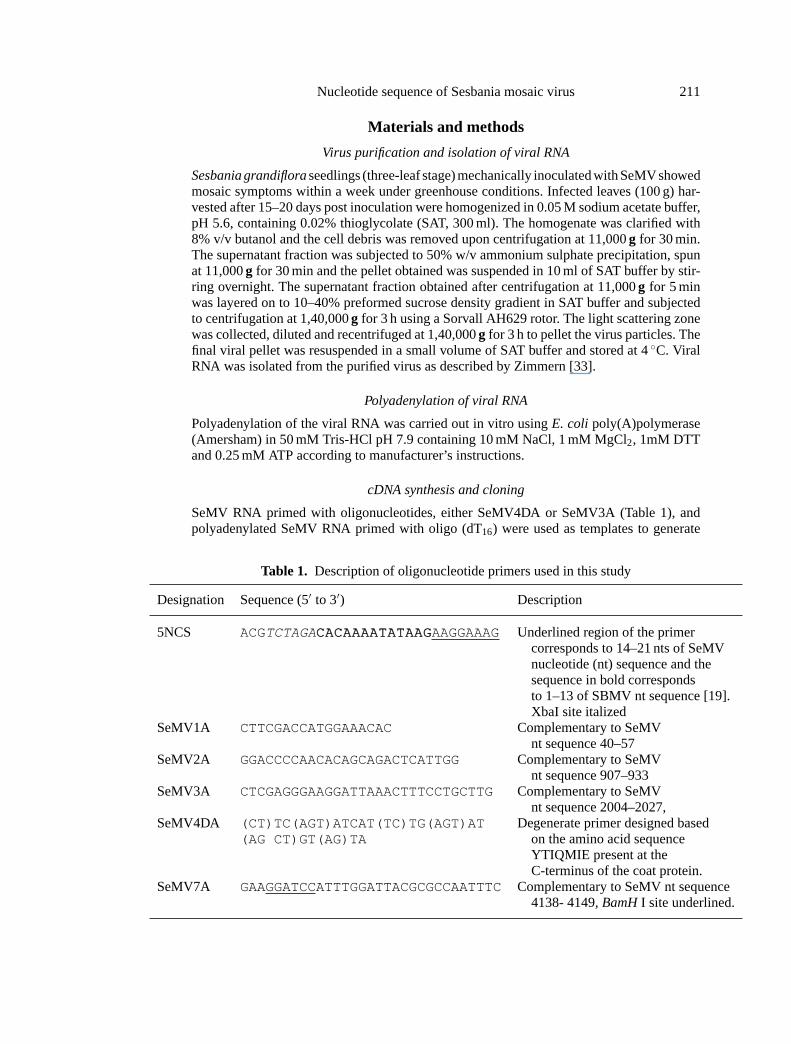
Table 1. Description of oligonucleotide primers used in this study
Designation Sequence (5′ to 3′) Description
5NCS ACGTCTAGACCAACCAAAAAAAATTAATTAAAAGGAAGGAAAG Underlined region of the primercorresponds to 14–21 nts of SeMVnucleotide (nt) sequence and thesequence in bold correspondsto 1–13 of SBMV nt sequence [19].XbaI site italized
SeMV1A CTTCGACCATGGAAACAC Complementary to SeMVnt sequence 40–57
SeMV2A GGACCCCAACACAGCAGACTCATTGG Complementary to SeMVnt sequence 907–933
SeMV3A CTCGAGGGAAGGATTAAACTTTCCTGCTTGComplementary to SeMVnt sequence 2004–2027,
SeMV4DA (CT)TC(AGT)ATCAT(TC)TG(AGT)AT Degenerate primer designed based(AG CT)GT(AG)TA on the amino acid sequence
YTIQMIE present at theC-terminus of the coat protein.
SeMV7A GAAGGATCCATTTGGATTACGCGCCAATTTCComplementary to SeMV nt sequence4138- 4149,BamHI site underlined.
212 G. L. Lokesh et al.
first strand cDNA. The first strand cDNA synthesis was carried out using superscript RT IIreverse transcriptase (Gibco-BRL). Second strand synthesis was done according to RnaseHmethod of Gubler and Hoffman [8]. The double stranded cDNA products were end-filledusing Klenow fragment of DNA polymerase I (Amersham) and size fractionated on SephacrylS-200 (Sigma) spun column. The larger fragments were ligated at the Eco RV or Sma I sites ofplasmid BlueScript II KS± (Stratagene) or pUC19, respectively. The ligation was carried outusing T4 DNA ligase (Amersham) and the ligation mix was transformed intoE. coli (DH5a,BRL) cells. The recombinant clones were screened for cDNA inserts by double digestionwith appropriate restriction enzymes.
Exonuclease III/S1 deletions
Supercoiled Plasmids for Exonuclease III (Amersham) digestion was prepared using Qiagentips (Qiagen, Inc), and ExoIII digestions were performed for 1 to 5 min according to manu-facturer’s protocol using 20 units of the enzyme permg of the DNA. The ExoIII deleted DNAwas then subjected to S1 nuclease digestion and end-filled with Klenow DNA polymerasefollowed by religation and transformation of DH5a cells. The plasmids isolated from thecolonies obtained were analyzed on the agarose gels for progressively deleted clones. Theselected deletion clones were sequenced using the appropriate M13 sequencing primers.
Sequencing of recombinant and deletion clones of SeMV
All the clones were initially sequenced with M13 sequencing primers manually by Sanger’sdideoxy chain termination method [21] using T7 Sequenase version 2.0 DNA sequencing kitand [a-32P]dATP (Amersham). Ambiguities in the sequences were resolved by resequencingusing dITP termination mixture provided in T7 sequencing kit. Some of the selected cloneswere also sequenced on ABI prism automated DNA sequencer.
RT-PCR
The first strand cDNA synthesis was performed using viral RNA and oligonucleotide primersSeMV2A or SeMV7A (Table 1) as described earlier. PCR was carried using Taq DNApolymerase (Bangalore Genei) and 1ml of the first strand cDNA mix containing 25 pmoleseach of 5NCS and SeMV2A primers in reaction volume of 25ml (Table 1). The reaction mixwas incubated at 94◦C for 5 min once, followed by 30 cycles of steps 94◦C 1 min, 55◦C1 min and 72◦C 1 min. The last cycle was followed by an additional incubation for 10 minat 72◦C. The amplification conditions with primers 5NCS and SeMV7A (Table 1) were thesame except that the chain extension step at 72◦C was increased to 4 min.
Sequence analysis of SeMV genomic RNA
SeMV genomic RNA was compiled and assembled using fragment assembly program ofWisconsin GCG package version 9.1. The nucleotide and deduced amino acid sequenceswere compared using BESTFIT, GAP and FASTA algorithms. Multiple sequence alignmentswere made using PILEUP and CLUSTALW [29] programs. Database searches were doneusing the program BLAST [2]. The phylogenetic trees were generated using DISTANCESand GROWTREE algorithms.
Results and discussion
SeMV was purified to homogeneity with a yield of 1 g/kg of the infected leaves.The RNA isolated from the purified virus migrated as a single component of Mr
Nucleotide sequence of Sesbania mosaic virus 213
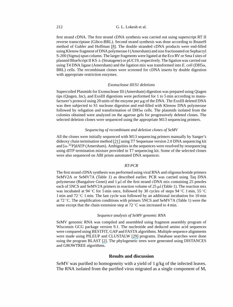
Fig. 1. Strategy used for sequencing SeMV genomic RNA. The dark line represents SeMVgenomic RNA. The 5′ and 3′ ends are marked. The closed circle at the 5′ end represents VPglinked to the 5′ end of the genome. The major clones used for the sequence determinationare shown as hatched bars. The arrows represent the direction and length of the sequence
information obtained from the primary and deletion clones
1.4 × 10⁶ when subjected to 1% agaorse gel electrophoresis (data not shown).The size of the RNA was similar to those of the other sobemoviral RNAs [9].The overall strategy used for the determination of genomic sequence of SeMV isshown in Fig. 1.
Two cDNA clones p18/2 and p24/4 identified from oligo(dT) primed cDNAlibrary released inserts of∼ 0.4 kb size. The sequence obtained from these clonesrevealed a poly(A) stretch followed by 3′ TAAACCAAATG . . . 5′. The 3′ non-coding region of SeMV thus obtained showed 68% identity with the 3′ noncodingregion of SBMV-Ark [13], suggesting that these are terminal residues at the 3′end of SeMV genome.
Ten clones, which released inserts ranging in size from 0.3 to 2.1 kb, identifiedfrom the cDNA library produced using degenerate primer SeMV4DA (Table 1),were sequenced using universal forward and reverse primers. Two of these clonespC38 and pGN134 harboring inserts of size 1.8 and 2.1 kb were selected for deter-mination of the internal sequences. These clones were subjected to ExonucleaseIII/S1 nuclease digestions from both the ends of the inserts to generate the dele-tion clones. The deletion clones were sequenced and the sequences obtained werecompiled and analyzed along with sequence obtained from p18/2 and p24/4. Thealignment of this sequence with that of SBMV-Ark [13] showed that sequence de-rived from these clones represented the nucleotides (nts) 1941–4149 (numberingaccording to SeMV, GenBank Acc. No. AY004291) at the 3′ terminus of SeMVgenomic RNA.
214 G. L. Lokesh et al.
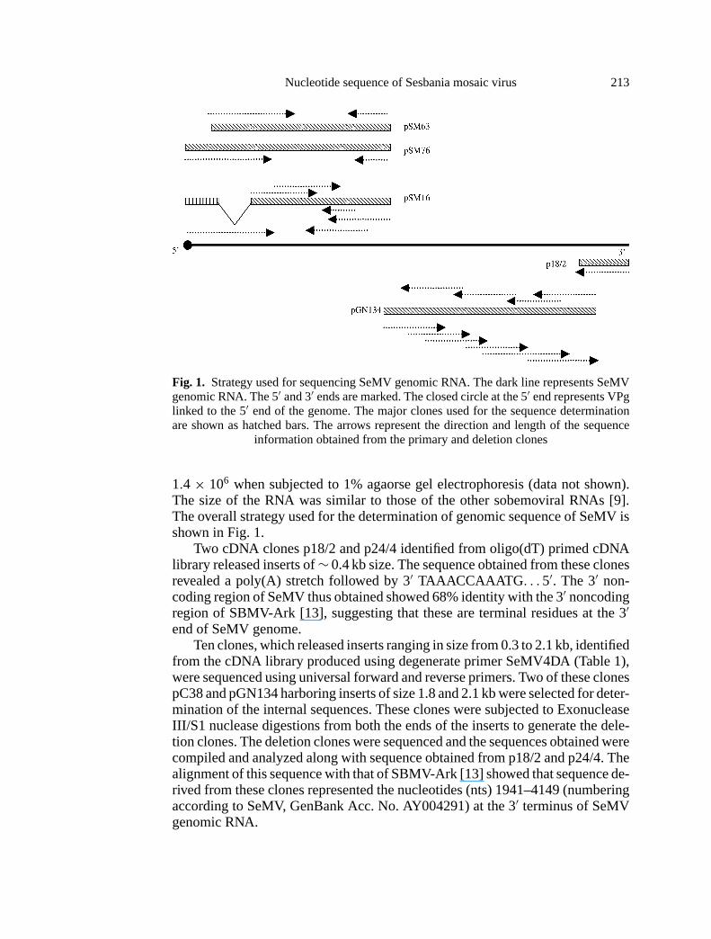
Fig. 2. Multiple sequence alignment of 5′ non-coding region of SeMV genomic RNA withthat of SBMV and SCPMV. The identities are highlighted. AUG start codons of ORF1 are
underlined
In order to determine the rest of the sequence, cDNA libraries were constructedusing oligonucleotide primer SeMV3A (Table 1) complementary to nts 2004–2027. A number of clones that represented the sequence at the 5′ end of the SeMVgenome were identified. Two clones pSM16 and pSM63 from two independentcDNA libraries were selected for ExoIII deletions. The deletion clones weresequenced and the sequence compiled from clone 63 represented nts from 14–2027 of SeMV genomic RNA. But the sequence obtained from pSM16 lacked nt231–509 indicating that the cDNA sequence represented in pSM16 could havearisen from a defective RNA that was packaged into virus particles (Fig. 1). Anumber of other clones sequenced were nearly identical in sequence with pSM63between nts 231–509. None of the clones sequenced had the terminal 5′ sequenceof the SeMV RNA. A comparison of the 5′ terminal 51 nts of pSM63 with SBMV[19] showed that it was identical from nts 14 to 64 (Fig. 2). It is possible thatthe clone pSM63 lacked 13 nucleotides from the 5′ end. Efforts to determine5′ end by 5′ RACE (Rapid Amplification of cDNA Ends) and RNA sequencingusing primer SeMV1A (Table 1) were not successful. However, RT-PCR on viralRNA template using the primer 5NCS (Table 1) which contained 5′ terminal 13nucleotides of SBMV [19] in addition to 8 nucleotides from 5′ terminal sequencederived from pSM63 and SeMV 2A or SeMV 7A (Table 1) gave products of0.9 kb and 4.2 kb respectively (data not shown). These results suggest that 5′ endSeMV genomic RNA may have the same nucleotide sequence as SBMV genomicRNA [19].
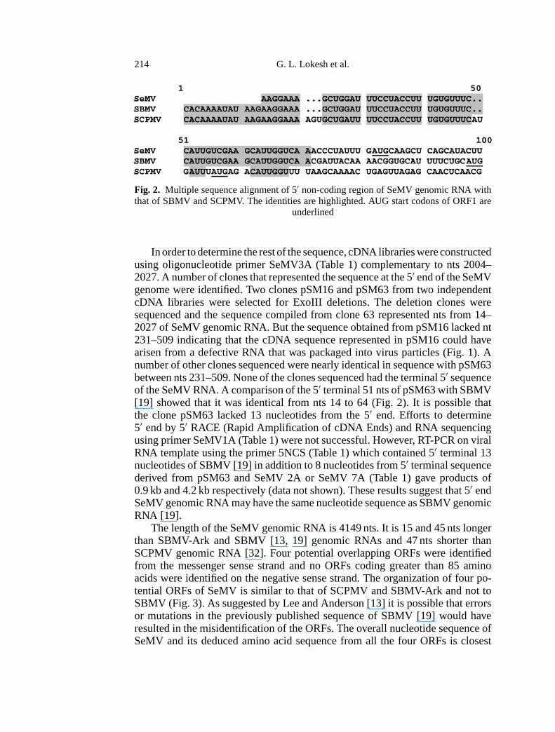
The length of the SeMV genomic RNA is 4149 nts. It is 15 and 45 nts longerthan SBMV-Ark and SBMV [13, 19] genomic RNAs and 47 nts shorter thanSCPMV genomic RNA [32]. Four potential overlapping ORFs were identifiedfrom the messenger sense strand and no ORFs coding greater than 85 aminoacids were identified on the negative sense strand. The organization of four po-tential ORFs of SeMV is similar to that of SCPMV and SBMV-Ark and not toSBMV (Fig. 3). As suggested by Lee and Anderson [13] it is possible that errorsor mutations in the previously published sequence of SBMV [19] would haveresulted in the misidentification of the ORFs. The overall nucleotide sequence ofSeMV and its deduced amino acid sequence from all the four ORFs is closest
Nucleotide sequence of Sesbania mosaic virus 215
Fig. 3. Comparison of the genome organization of SeMV with that of other sobemoviruses.The viral RNA is represented as dark line and the ORFs are represented as boxes. VPg is
shown as filled circle at the 5′ terminus
to SBMV-Ark (73% identity) followed by SCPMV (59.6%). The overall per-cent identity was much lower in Lucerne transient streak virus (LTSV), RYMVand Cocksfoot mottle virus (CfMV). These results suggested that SeMV is a newmember of the genus sobemovirus and is not a strain of SBMV. To further confirmthis a detailed analysis of non-coding regions and the deduced protein sequencesof the ORFs was carried out.
216 G. L. Lokesh et al.
Noncoding regions of SeMV RNA
The 5′ noncoding (NC) leader sequence of SeMV RNA is 76 nts long and thefirst 64 nts have 100% identity with SBMV and 76.4% with SCPMV (Fig. 2).The 5′ NC region of these viruses are relatively AT rich and may contain sites forbinding of 18S rRNA. The reason for the high degree of nucleotide identities in 5′NC region of SeMV, SBMV and SCPMV is unclear, however it is suggestive thatthese three sobemoviruses, which infect dicotyledenous plants might probablyhave a common mechanism of translation/replication.
The 3′ NC region of many plant viral genomes fold into at-RNA like structuresthat are aminoacylatable. Computational analysis of 125 nt long 3′ NC of SeMVRNA using MFOLD algorithm [34] along with those of SBMV and SCPMV didnot reveal any stem-loop structures that were common to all of them. In contrastto the 5′ NC sequence, 3′ NC of SeMV showed only 68% identity with SBMV.
Coding regions of SeMV genomic RNA
The first ORF of SeMV RNA starts from AUG codon at nucleotide 77 andends at UAG stop codon located at nucleotide position 557 (GenBank Acc. No.AY004291). This AUG is in poor context with respect to translation initiation [12]and the same holds good for the first ORF in all the sobemoviruses. In SCPMV ithas been suggested that ORF1 is expressed by 5′ end dependent ribosomal scan-ning mechanism [23]. ORF1 of SeMV can potentially encode a protein containing160 amino acid residues of Mr 18370. ORF1 in SCPMV [32] and SBMV-Ark[13] encode 21 and 17.2 kDa proteins, respectively. In vitro translation experi-ments with SCPMV had revealed products of size 21 and 25 kDa [16]. The 21 kDaproduct could correspond to that arising from ORF1, while 25 kDa product mightbe due to a translational read through of UAG stop codon of ORF1 which extendsthe protein product by 29 amino acids. This could explain for the observationof in vitro translated protein products ranging from 21–25 kDa [16] arising outof ORF1 of SCPMV. The same is probably true for SeMV, but it needs to beconfirmed by in vitro translation. Interestingly, ORF1 gene product showed only34% identity with that of SBMV-Ark and there was no significant similarity withthe corresponding proteins of other sobemoviruses. This observation further sup-ports the suggestions that SeMV is a distinct member of genus sobemovirus. Adeca-peptide with the sequence VCRECIIRAA at the C-terminus of ORF1 pro-tein (GenBank Acc. No. AY004291) is conserved among SeMV, SCPMV andSBMV-Ark, the functional significance of this motif is not known. Recent re-ports demonstrate that the ORF1 is translationally active in SCPMV, RYMV andCfMV [4, 23, 27]. The involvement of the ORF1 product in virus spread has beenrecently demonstrated by Bonneau et al. [4]. It has also been demonstrated bymutational analysis of full-length cDNA clone of SCPMV that the cell to cellmovement of the virus requires ORF1, ORF3 and CP gene products [24].
ORF2 of SeMV potentially encodes the largest protein product of the genomewith calculated Mr 105913. The ORF begins at nucleotide position 520 and ex-tends upto nucleotide 3408 resulting in a polypeptide of 962 amino acids (Gen-
Nucleotide sequence of Sesbania mosaic virus 217
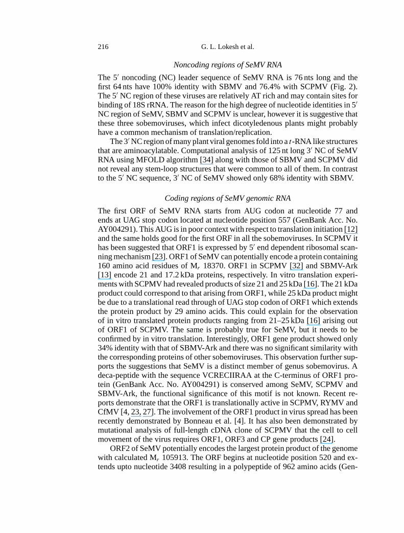
Table 2A. Percentage identities of different ORFs of sobemoviruses. The upper triangle showsidentities of ORF2 (VPg-Protease domain) and lower triangle that of RDRP domain
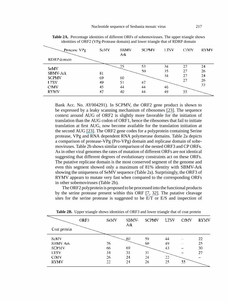
Bank Acc. No. AY004291). In SCPMV, the ORF2 gene product is shown tobe expressed by a leaky scanning mechanism of ribosomes [23]. The sequencecontext around AUG of ORF2 is slightly more favorable for the initiation oftranslation than the AUG codon of ORF1, hence the ribosomes that fail to initiatetranslation at first AUG, now become available for the translation initiation atthe second AUG [23]. The ORF2 gene codes for a polyprotein containing Serineprotease, VPg and RNA dependent RNA polymerase domains. Table 2a depictsa comparison of protease-VPg (Pro-VPg) domain and replicase domain of sobe-moviruses. Table 2b shows similar comparison of the nested ORF3 and CP ORFs.As in other viral genomes the rates of mutation of different ORFs are not identicalsuggesting that different degrees of evolutionary constraints act on these ORFs.The putative replicase domain is the most conserved segment of the genome andeven this segment showed only a maximum of 81% identity with SBMV-Arkshowing the uniqueness of SeMV sequence (Table 2a). Surprisingly, the ORF3 ofRYMV appears to mutate very fast when compared to the corresponding ORFsin other sobemoviruses (Table 2b).
The ORF2 polyprotein is proposed to be processed into the functional productsby the serine protease present within this ORF [7, 32]. The putative cleavagesites for the serine protease is suggested to be E/T or E/S and inspection of
Table 2B. Upper triangle shows identities of ORF3 and lower triangle that of coat protein
218 G. L. Lokesh et al.
SeMV ORF2 revealed several such sites. The serine protease domain of SeMVORF2 is found to have the conserved catalytic triad comprising the serine284,histidine181 and aspartate216 (GenBank Acc. No. AY004291). As observed inSBMV and SCPMV RNAs [5, 16] it is possible that the VPg is present at the 5′terminus of SeMV RNA. To confirm this, the N-terminal amino acid sequenceof VPg linked to the 5′ end of SeMV RNA was determined directly by using anautomated protein sequencer (PSQ1-Shimadzu). The sequence TLPPELSIIEIPobtained confirmed the cleavage by the protease after E325. A similar approachwas used earlier to determine the N-terminal sequence of SBMV VPg [30]. Basedon this sequence and the comparison with SBMV genomic sequence, the VPg inSeMV was mapped at region 326–445 (GenBank Acc. No. AY004291) and thecleavage site between VPg and RNA dependent RNA polymerase was predictedto be E445-T446 based on comparison with SBMV and SCPMV sequences. VPghas been shown to be essential for the infectivity of the virus [31]. It is alsodemonstrated that capped and uncapped transcripts, which are generated in vitrofrom the cloned full length cDNAs of SCPMV are∼ 5 and 28 fold less infectiousthan the native RNA isolated from the virions [24], indicating that the reduction inthe infectivity could be due to the absence of VPg at the 5′ end of these transcripts.In poliovirus it is known that VPg linked to the terminal nucleotides serves asprimer in the initiation of replication of positive and negative sense RNAs [20].However, at present the role of VPg in translation/replication in the sobemovirusesis not established.
The GDD motif characteristic of the viral RDRPs is located at the C-terminusof the putative polyprotein encoded by ORF2 (GenBank Acc. No. AY004291).The RDRP domain is the best conserved among the sobemoviruses. The eightconserved motifs defined by Koonin [11], that are characteristic of viral RDRPsare present at the C-terminal region of ORF2 of SeMV (GenBank Acc. No.AY004291).
The ORF3 of SeMV is nested within ORF2 in−1 reading frame. This ORFmay encode a protein of 144 amino acids with the calculated Mr 16251. Similarnested ORFs are observed in SCPMV, SBMV-Ark, RYMV and LTSV [3, 10, 13,18, 32]. Interestingly, the genome of CfMV does not have such a nested ORF,instead it has two overlapping ORFs 2a and 2b from which the polyprotein is pro-duced by a−1 frame shifting mechanism [14, 15]. The−1 ribosomal frameshiftsignal comprising of a heptanucleotide UUUAAAC and a putative stem-loopstructure that were originally noticed in CfMV genome [27] is also present inSeMV RNA between the nucleotide 1741–1800 upstream of the ORF3 initia-tion codon (GenBank Acc. No. AY004291). The−1 frameshift signal appearsto be common for all the sobemoviruses that are sequenced so far and is locatedupstream of the ORF3 intiation codon. Furthermore, in all these sobemovirusesthere are no stop codons between the−1 frameshift signal and the initiation codonof ORF3. Considering that the ORF3 of SeMV and other sobemoviruses to beanalogous to the ORF2b of CfMV, it has been suggested that ORF3 may be ex-pressed as a part of−1 ribosomal frameshift to yield a polypeptide of 70 kDa[27]. A 70 kDa band was indeed observed in in vitro translation experiments with
Nucleotide sequence of Sesbania mosaic virus 219
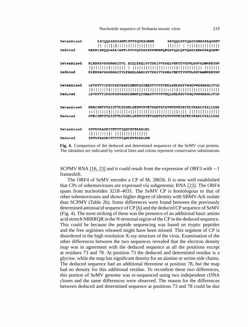
Fig. 4. Comparison of the deduced and determined sequences of the SeMV coat protein.The identities are indicated by vertical lines and colons represent conservative substitutions
SCPMV RNA [16, 23] and it could result from the expression of ORF3 with−1frameshift.
The ORF4 of SeMV encodes a CP of Mr 28656. It is now well establishedthat CPs of sobemoviruses are expressed via subgenomic RNA [23]. The ORF4spans from nucleotides 3218–4031. The SeMV CP is homologous to that ofother sobemoviruses and shows higher degree of identity with SBMV-Ark isolatethan SCPMV (Table 2b). Some differences were found between the previouslydetermined aminoacid sequence of CP [6] and the deduced CP sequence of SeMV(Fig. 4). The most striking of these was the presence of an additional basic aminoacid stretch NRRRQR in the N-terminal region of the CP in the deduced sequence.This could be because the peptide sequencing was based on tryptic peptidesand the free arginines released might have been missed. This segment of CP isdisordered in the high resolution X-ray structure of the virus. Examination of theother differences between the two sequences revealed that the electron densitymap was in agreement with the deduced sequence at all the positions exceptat residues 73 and 78. At position 73 the deduced and determined residue is aglycine, while the map has significant density for an alanine or serine side chains.The deduced sequence had an additional threonine at position 78, but the maphad no density for this additional residue. To reconfirm these two differences,this portion of SeMV genome was re-sequenced using two independent cDNAclones and the same differences were observed. The reason for the differencesbetween deduced and determined sequence at positions 73 and 78 could be due
220 G. L. Lokesh et al.
to heterogeneity in the viral preparation. It may be noted that the residue 218was reported earlier as an aspartic acid [3]. This residue was suggested to be theligand for the putative cation present at the quasi three fold axis. However, thecorrected sequence now shows that residue 226 is a proline and hence cannotbe a ligand for the putative cation. The N terminal R-domain comprising ofresidues 1–65 is rich in lysine and arginine residues that are involved in interactionwith the RNA. These are particularly not well conserved among sobemovirusesalthough the overall basic nature of the N-terminal R domain is retained. Thissuggests that the coulombic forces between the protein and nucleic acid are eithernon-specific or not very important for the assembly of the virus. The residuesin the b barrel domain (66–268) are conserved better among sobemoviruses.The motif DXXD (146-149), in which the aspartates act as ligands for Ca2+binding is common to SBMV and SeMV. It is conserved in all sobemovirusesand is also present in carmo- and tombusviruses. The exact role of the conserved
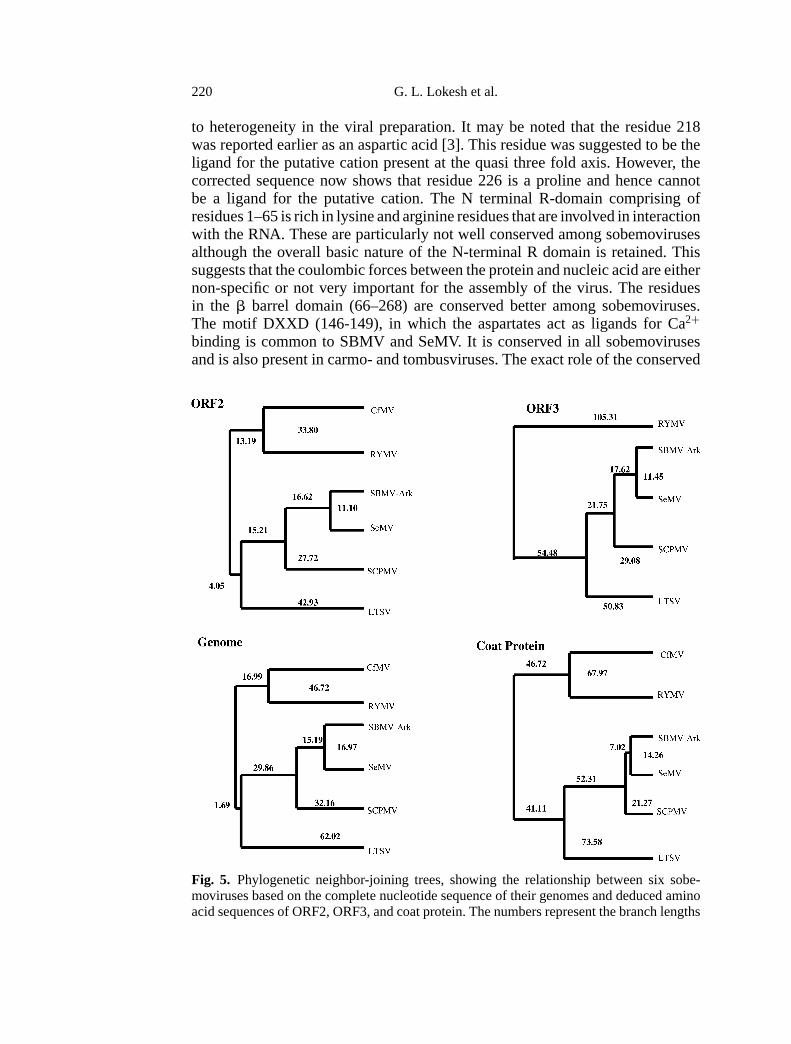
Fig. 5. Phylogenetic neighbor-joining trees, showing the relationship between six sobe-moviruses based on the complete nucleotide sequence of their genomes and deduced aminoacid sequences of ORF2, ORF3, and coat protein. The numbers represent the branch lengths
Nucleotide sequence of Sesbania mosaic virus 221
residues in the assembly of the virus can be deciphered only through mutationalanalysis.
The phylogenetic relationship of the six sobemoviruses based on their com-plete nucleotide sequence as well as the deduced amino acid sequences of ORF2,ORF3 and coat protein ORF are shown in Fig. 5. ORF1 was not used in theconstruction of the cladograms as it is highly variable among the sobemoviruses.The cladograms are nearly identical in all the cases suggesting that in the sobe-movirus group the recombination events are probably not frequent or significant.As pointed out by Sehgal [22], within the sobemovirus group SCPMV, SBMV-Ark, SeMV and LTSV (that infect the dicotyledons, Leguminosae and Fabaceae)cluster as a closely related group, while RYMV and CfMV that essentially infectmonocotyledons (Gramineae) form another related group.
The complete nucleotide sequence of SeMV and the analysis reported in thispaper shows that SeMV is a distinct member of the sobemovirus group withgenome organization similar to SBMV-Ark and SCPMV.
Acknowledgements
This work was financially supported by a grant from Department of Science and Technol-ogy, Government of India. We thank Prof M. R. N. Murthy for valuable suggestions. Theassistance of Ms. Savitha and Ms. Rekha in DNA sequencing, Mr. Michael in amino acidsequencing, Mr. Elango and Mr. Saravanan in sequence analysis is acknowledged. We thankDepartment of Biotechnology, India for providing amino acid and DNA sequencing facilitiesand Bioinformatics center at IISc. GLL is grateful to CSIR for the financial assistance.
References
1. Abad-Zaptero C, Abdel-Meguid SS, Johnson JE, Leslie AGW, Rayment I, RossmannMG, Suck D, Tsukihara T (1980) Structure of southern bean mosaic virus at 2.8 Åresolution. Nature 286: 33–39
2. Altschul SF, Gish W, Miller W, Myers EW, Lipman DJ (1990) Basic local alignmentsearch tool. J Mol Biol 215: 403–410
3. Bhuvaneswari M, Subramanya HS, Gopinath K, Savithri HS, Nayudu MV, Murthy MRN(1995) Structure of sesbania mosaic virus at 3 Å resolution. Structure 3: 1021–1030
4. Bonneau C, Brugidou C, Chen L, Beachy R, and Fauquet C (1998) Expression of riceyellow mottle P1 protein in vitro and in vivo and its involvement in virus spread. Virology244: 79–86.
5. Ghosh A, Dasgupta R, Salerno-Rife T, Rutgers T, Kaesberg P (1979), Southern beanmosaic virus has a 5′ linked protein but lacks 3′ terminal poly (A). Nucleic Acids Res 7:2137–2146
6. Gopinath K, Sundareshan S, Bhuvaneswari M, Karande A, Murthy MRN, Nayudu MV,Savithri HS (1994) Primary structure of sesbania mosaic virus coat protein: its implica-tions to the assembly and architecture of the virus. Ind J Biochem Biophys 31: 322–328
7. Gorbalenya AE, Koonin EV, Blinov VM, Donchenko AP (1988) Sobemovirus genomeappears to encode a serine protease related to cysteine proteases of picornaviruses. FEBSLett 236: 287–290
8. Gubler U, Hoffman BJ (1983) A simple and very efficient method for generating cDNAlibraries. Gene 25: 263–269
222 G. L. Lokesh et al.
9. Hull R (1988) The sobemovirus group. In: Koenig R (ed) The plant viruses, vol 3:Polyhedral virions with monopartite RNA genomes. Plenum Press, New York, pp 113–146
10. Jeffries AC, Rathjen JP, Symons RH (1995) Lucerne transient streak virus completegenome. Genbank accession number U31286
11. Koonin EV (1991) The phylogeny of RNA dependent RNA polymerases of positive-strand viruses. J Gen Virol 72: 2197–2206
12. Kozak M (1989) Scanning model for translation: An update. J Cell Biol 108:229–241
13. Lee L, Anderson EJ (1998) Nucleotide sequence of a resistance breaking mutant ofsouthern bean mosaic virus. Arch Virol 143: 2189–2201
14. Mäkinen K, Næss V, Tamm T, Truve E, Aaspõllu A, Saarma M (1995a) The putativereplicase of the cocksfoot mottle sobemovirus is translated as a part of the polyproteinby −1 ribosomal frameshift. Virology 207: 566–571
15. Mäkinen K, Tamm T, Næss V, Truve E, Puurand Ü, Munthe T, Saarma M (1995b) Char-acterization of cocksfoot mottle sobemovirus genomic RNA and sequence comparisonwith related viruses. J Gen Virol 76: 2817–2825
16. Mang KQ, Ghosh A, Kaesberg P (1982) A comparative study of the cowpea and beanstrains of southern bean mosaic virus. Virology 116: 264–274
17. Murthy MRN, Bhuvaneswari M, Subramanya HS, Gopinath K, Savithri HS (1997) Struc-ture of Sesbania mosaic virus at 3Å resolution. Biophys Chem 68: 33–42
18. Ngon a Yassi M, Ritzenthaler C, Brugidou C, Fauquet C, Beachy RN (1994) Nucleotidesequence and genome characterization of rice yellow mottle virus RNA. J Gen Virol 75:249–257
19. Othman Y, Hull R (1995) Nucleotide sequence of the bean strain of southern bean mosaicvirus. Virology 206: 287–297
20. Rueckert RR (1985) Picornaviruses and their replication. In: Fields BN (ed) Virology.Raven Press, New York, pp 705–738
21. Sanger F, Nicklen S, Coulson AR (1977) DNA sequencing with chain-terminating in-hibitors. Proc Natl Acad Sci USA 74: 5463–5467
22. Sehgal OP (1999) Sobemoviruses. In: Granoff A, Webster RG (eds) Encyclopedia ofvirology, 2nd ed. Academic Press, San Diego, pp 1674–1680.
23. Sivakumaran K, Hacker DL (1998a) The 105-kDa polyprotein of southern bean mosaicvirus is translated by scanning ribosomes. Virology 246: 34–44
24. Sivakumaran K, Fowler BC, Hacker DL (1998b) Identification of viral genes requiredfor cell-to-cell movement of southern bean mosaic virus. Virology 252: 376–386.
25. Sreenivasulu P, Nayudu MV (1982) Purification and partial characterization of sesbaniamosaic virus. Curr Sci 51: 86–87
26. Subramanya HS, Gopinath K, Nayudu MV, Savithri HS, Murthy MRN (1993) Structureof sesbania mosaic virus at 4.7 Å and partial sequence of coat protein. J Mol Biol 229:20–25
27. Tamm T, Mäkinen K, Truve E (1999) Identification of the genes encoding for the cocks-foot mottle virus proteins. Arch Virol 144: 1557–1567
28. Tamm T, Truve E (2000) Sobemoviruses. J Virol 74: 6231–624129. Thompson JD, Higgins DG, Gibson TJ (1994) CLUSTALW: improving the sensitivity of
progressive multiple sequence alignment through sequence weighting, position specificgap penalties and weight matrix choice. Nucleic Acids Res 22: 4673–4680
30. van der Wilk F, Verbeek M, Dullemans A, van den Heuvel J (1998) The genome-linkedprotein (VPg) of southern bean mosaic virus is encoded by the ORF2. Virus Genes 17:21–24
Nucleotide sequence of Sesbania mosaic virus 223
31. Veerisetty V, Sehgal OP (1980) Proteinase K-sensitive factor essential for the infectivityof southern bean mosaic virus ribonucleic acid. Phytopathology 70: 282–284
32. Wu S, Rinehart CA, Kaesberg P (1987) Sequence and organization of southern beanmosaic virus genomic RNA. Virology 161: 73–80
33. Zimmern D (1975) The 5′ end group of tobacco mosaic virus RNA is m[7]G[5′]pppGp.Nucleic Acids Res 2: 1189–1201
34. Zuker M (1989) On finding all suboptimal foldings of an RNA molecule. Science 244:48–52
Authors’ address: Dr. H. S. Savithri, Department of Biochemistry, Indian Institute ofScience, Bangalore 560 012, India.
Received April 20, 2000