Identification of a Genetic Locus Related to Antivirus Production in Pseudomonas fluorescence strain...
9
Transcript of Identification of a Genetic Locus Related to Antivirus Production in Pseudomonas fluorescence strain...

Plant Pathol. J. 25(1) : 77-85 (2009)The Plant Pathology Journal
©The Korean Society of Plant Pathology
Identification of a Genetic Locus Related to Antivirus Production in Pseudomonas fluorescence strain Gpf01 Against Cucumber mosaic virus
Saeyoull Cho1, Seon-Hwa Lee1, Sujin Park2, Kyu-Up Choi1, Junmo Cho1, Jang Hyun Hur3, Anupama Shrestha1
and Chunkeun Lim1*1Department of Bio-resource of Technology, Division of Applied Biology, College of Agriculture and Life Science, Kangwon
National University, Chuncheon 200-701, Korea 2Life Science Institute, University of Michigan, Ann Arbor, MI 48109, USA3Division of Biological Environment, College of Agriculture and Life Science, Kangwon National University, Chuncheon 200-
701, Korea
(Received on October 5, 2008; Accepted on January 6, 2009)
Pseudomonas fluorescens strain Gpf01, isolated fromginseng rhizosphere showed antiviral activity againstCucumber mosaic virus, when tested in a local host ofCMV, Chenopodium amaranticolor. Transposon mutantlibrary of Gpf01 was prepared using pGS9::Tn5 andthe mutant Gpf01-RS19 was found to loose antiviralproduction. We developed primers from the flankingregion of Tn5 and found a cosmid clone pAV1123,harboring 1.2 kb antiviral compound producing (avcf01)locus. When a sub-clone pPH9, which carried 9.3 kbregion of pAV1123, was introduced into antivirusdeficient P. fluorescens wild type strain B16, it exhibitedantiviral activity. Using Tn3-gus mutagenesis and com-plementation analysis, it was found that the genesrelated to antiviral activity production resided in a 9.3-kb HindIII-HindIII fragment of pAV1123, indicatingthat the plasmid carries an essential genes promotingantiviral activity.
Keywords : antiviral compound producing gene, Cucumbermosaic virus, Pseudomonas fluorescens
Pseudomonas species are typically gram negative, chemo-
heterotrophic, motile rods with polar flagella and are
grouped in rRNA homology group I (Palleroni et al., 1973).
Fluorescent pseudomonades are ubiquitous soil micro-
organisms and common inhabitants of the rhizosphere. The
fluorescent pseudomonads play important roles in the
environment by degrading toxic chemicals present in soils.
Certain strains colonize the root system and suppress plant
diseases by protecting seeds or roots from infection by soil-
borne fungal and bacterial plant pathogens (Défago and
Haas, 1990; O’Sullivan et al., 1992). Additionally, fluore-
scent pseudomonads produce a variety of antimicrobial
compounds such as 2,4-diacetylphloroglucinol, hydrogen
cyanide, phenazine, pyoluteorin, pyrrolnitrin etc. (Nowak-
Thompson et al., 1994; Rosales et al., 1995; Thomashaw
and Wellwe, 1988), some of which are involved in the
biological control (Keel et al., 1992; Ryu et al., 2000).
Moreover, some strains activate plant defense resulting in
systematic protection against different fungal, bacterial and
viral disease stimulating plant growth and crop yields
(Maurhofer et al., 1998). Therefore, fluorescent pseudo-
monads have been applied as biopesticides (Hosain and
Alexander, 1984) as well as to improve crop yields by
number of mechanisms (Thomashaw and Wellwe, 1988).
Viruses are one of the important plant pathogens, causing
severe losses in many economically important plants.
Among them, Cucumber mosaic virus (CMV) is a serious
threat in production of important vegetable crops all over
the world (Gooding, 1991). Cucumber mosaic virus (CMV),
belongs to the genus cucumovirus (family Bromoviridae)
and is one of the economically important virus, which
causes enormous losses by infecting more than 1000 species
of plants, shrubs and trees world-wide. It is transmitted
non-persistently into healthy plants by aphids, which
acquire the virus during their brief probes on infected hosts
or the symptomless carrier weeds in the field (Zehender,
2000). The drawback of these breeding strategies used in
agricultural crops involves, difficulty in identifying virus
resistance genes and using them in diverse cultivars while
retaining the desired quality traits of the product (Elisaveta
and Violeta, 2000). Various strategies, based on the
avoidance of sources of infection, control of vectors,
modification of cultural practices, and the use of resistant
varieties and transgenic plants have been conventionally
employed to minimize the losses caused by CMV (Klement
et al., 1996; Lampis et al., 1996). However, these strategies
have not been an effective control measure.
Several antiviral agents have been reported from different
sources, especially from higher plants, showing systemic
control ability against a wide range of viruses that infect
Mini-Review
*Corresponding author.
Phone) +82-33-250-6437, FAX) +82-33-256-8254
E-mail) [email protected]

78 Saeyoull Cho et al.
plants (Kubo et al., 1990). Raupach et al. (1996) showed the
systemic control of CMV in cucumbers and tomatoes
employing induced systemic resistance (ISR) mechanism,
using plant growth-promoting rhizobacteria (PGPR).
Similarly, induced systemic resistance to tobacco necrosis
virus (TNV) by root colonizing P. fluorescens CHA0 and
P3 strains has been reported (Maurhofer et al., 1994 and
1998). Besides, culture filtrates from Acinetobacter species
KTB3 and Pseudomonas sp. KTB61 were used to syste-
matically control some viruses containing CMV-Y in Korea
(Kim et al., 2003; Kim et al., 2004). Ipper et al. (2006) used
Serratia sp. strain Gsm01 which possesses antiviral activity
against CMV by inducing plant defense related genes and
enzymes.
Previously, Ipper et al. (2005) isolated and identified P.
fluorescence strain Gpf01 as antiviral substance producing
strain which was active against Cucumber mosaic virus
(CMV) in both, local and systemic hosts. Furthermore, we
investigated the genetic locus responsible for the antiviral
activity shown by the strain Gpf01 using transposon medi-
ated mutagenesis. The cosmid clone, pAV1123 which was
responsible for the antiviral activity was further, studied for
the locus involved in the antiviral activity by subcloning
and Tn3-gus mutagenesis for the first time in our know-
ledge.
Materials and Methods
Bacterial strains and growth condition and mainten-
ance of virus. Soil-adhered Ginseng roots, which were
collected from Hongcheon, Gangwon province, Korea, were
homogenized, serially diluted and plated onto mannitol
glutamate yeast (MGY) agar plates (Keane et al., 1970).
The agar plates were incubated at 28oC. Numerous colo-
nies, with different morphologies, were picked from the
dilution plates. Each of these colonies was assayed for the
antiviral activity using the half leaf method, as described by
Kubo et al. (1990). One colony, which showed maximum
antiviral activity, was selected and designated as Gpf01.
This colony was stored at −70oC, using nutrient broth
containing 20% glycerol, by freeze-drying in 10% skimm-
ed milk for its long-term preservation. The bacteria and
plasmids used in this study are shown in supplemental
Table 1.
CMV-Y was obtained from the virus culture collection of
the College of Forestry Science, Kangwon National
University, Chuncheon, Korea. The virus was inoculated
into Nicotiana tabacum var. Xanthi-nc, and maintained on
the same host throughout the period of this study. The
inoculums consisted of CMV-Y systematically infected
leaves ground in 0.01 M phosphate buffer [pH 7.0].
DNA techniques. Total genomic DNA of P. fluorescens
Gpf01 was isolated by modified protease-sodium dodecyl
sulfate (SDS) lyses procedure (Sambrook and Russel,
2001). Plasmid DNA of cosmid clones was also prepared
by midi-scale alkaline lysis method with some modification
(Sambrook and Russel, 2001). The plasmids were further
analyzed by cleaving total plasmids using the restriction
enzymes BamHI, EcoRI and HindIII (Promega Madison,
USA) according to the manufacturer’s instructions. Twenty
microliter of digested plasmids were used for electro-
phoresis and visualized on 0.7% of agarose gel. Standard
techniques for DNA manipulation, such as plasmid DNA
preparation, ligation, competent cell preparation, and trans-
formation were followed as described by Sambrook and
Russell (2001). Preparation of the plasmid DNA (Wizard
Minipreps; Promega) and recovery of DNA fragments from
agarose gel (Geneclean II Kit; Bio101, Rutherford, CA,
USA) were performed as described in the manufacturers,
manuals. The restriction enzymes, dNTPs, Taq DNA poly-
merase, T4 DNA ligase, and DNA marker used in this
study were supplied by Promega and Takara (Ohtsu, Japan).
Culture filtrate (CF) preparation. The strain, Gpf01, was
taken from glycerol stock and streaked onto a MGY agar
plate. A single colony was inoculated into 100 ml Muller-
Hinton (MH) broth and grown at 28oC for 48 h, with
shaking at 200 rpm. The culture supernatant was then
filtered through a 0.45 μm filter. The filtrate obtained was
used for the antiviral assay.
Antiviral bioassay. The bacteria to be tested were grown
with appropriate antibiotics into 50 ml MGY broth and
culture supernatant was obtained, which was used for
antiviral bioassay using half-leaf method on local host of
CMV, Chenopodium amaranticolor as previously descri-
bed by Kubo et al. (1990). The upper right halves of the
leaves were treated with the CF using brush and the upper
left halves were left untreated. CMV-infected fresh tobacco
leaf (0.1 g) was ground in 5 ml of phosphate buffer. After
one hour, virus preparation was inoculated onto both the
halves of the leaves by ordinary carborundum (600 mesh)
method. The plants were allowed to grow in a green house
with 12-14 h daylight and 30oC temperature. The local
lesion numbers were counted after seven days. The percent
control effect was calculated using the formula: (1-T/
C)×100, where, C is the number of local lesions on the
control half leaves and T is the number of local lesions on
treated half leaves.
Spontaneous mutation of P. fluorescens Gpf01. P. fluore-
scens Gpf01 was grown in MGY agar plate at 28oC for 16
Antiviral Activity of avcf01 From Pseudomonas fluorescence 79
h, suspended in sterile distilled water and optical density
was adjusted to 0.6 at 600 nm absorbance. The MGY agar
was prepared, autoclaved at 121 psi for 20 min, cooled to
40-50oC, and amended with filter-sterilized rifampicin to
final concentrations of 0.1, 0.8, 1.6, 10, 50 and 100 mM. 5
μl droplets of Gpf01 was spotted on MGY agar plates and
incubated at 28oC for 24 h. The strain obtained after spon-
taneous mutation was designated as Gpf01-RS.
Transposon-mediated mutagenesis of Gpf01-RS. The
suicide plasmid pGS9 in E. coli WA803 was used to
generate Tn5 insertions in the Gpf01-RS strain. The donor
strain was grown overnight on LB broth to a density of 1×
109 CFU/ml. Recipient culture was grown overnight on
MGY broth containing rifampicin at 27oC to the stationary
phase (1×10⁹ CFU/ml). Approximately 109 donor cells were
sedimented, suspended in 50 μl of dH2O, transferred to a
nitrocellulose filter on a fresh, pre-warmed LB agar plate
and incubated for 1.5 h at 37oC. Approximately 109 cells of
Gpf01-RS were sedimented in an Eppendorf tube, washed
briefly in TE buffer (10 mM Tris-HCl, 1 mM EDTA [pH
8.0]), suspended in 50 μl of dH2O, and transferred to the
filter paper containing the donor cells. The plates were
incubated for 24 h at 28oC. Transconjugants were selected
on MGY plates containing rifampicin and kanamycin at
100 and 50 μg/ml, respectively.
Two thousand transconjugant clones were picked up by
sterile toothpick method for the screening (Sambrook and
Russel, 2001). Fifty transconjugant clones were arranged
per 85×15 mm petridishes and incubated at 28oC. In order
to select Tn5 insertion transconjugant, genomic DNA of
two thousand transconjugants were amplified using Tn5-F
and Tn5-R1 primers (5' GACTCTTATACACAAGTAGCG
3' and 5' GATGCCTGCAAGCAATTCGT 3') and screened
for their antiviral activity as described before.
Inverse PCR. The PCR process used for amplification of
flanking sequences of the Tn5 insertion is outlined in
supplemental (Fig. 1). Genomic DNA was digested with
SmaI at 25oC for the minimum time necessary to achieve
complete digestion (3h). After digestion, the restriction
enzyme SmaI was inactivated at 65oC for 15 min. The
digested DNA was self ligated at a concentration of 0.3-0.5
μg/ml in the presence of 3U T4 DNA ligase (Takara Co.)
overnight at 16oC. The ligated DNA was precipitated with
ethanol, collected by centrifugation and resuspended in
sterile water to a concentration of 20 ng/ml.
The PCR was performed in a reaction containing 20 ng of
circularized DNA obtained as described above in the
presence of 50 pmol of each primer and 1.25 mM dNTPs
(Takara Co.), 5 units of Taq DNA polymerase (Takara Co.).
PCR products amplified by the SF and SR or SL primers (5'
CGCTACTTGTGTATAAGAGTC 3', 5' CGAAATGACC-
GACCAAGCGA 3', 5' GATGCCTGCAAGCAATTCGT
3') combinations were digested with SmaI. The PCR
products were electrophoresed on 0.7% agarose gel, and
purified with a Geneclean II Kit (Bio101, Rutherford, CA,
USA). Purified DNAs were ligated into pGEM-T easy
(Promega) and sequenced.
Genomic library construction, screening, and Southern
hybridization. Genomic DNA library of P. fluorescens
Gpf01 was constructed into a low copy cosmid vector
pLAFR3 and transformed into E. coli strain HB101.
Chromosomal DNA of P. fluorescens Gpf01 was partially
digested with Sau3AI generating 20-30 kb fragment. The
cosmid vector pLAFR3 was linearized with BamHI and
ligated into the Sau3AI digested fragments, packaged in
vitro with a DNA packaging kit (Boehringer Mannheim,
Edtroit, MI, USA), and transduced into E. coli HB101.
Transductants were selected on LB agar plate supplement-
ed with tetracycline (50 μg/ml). Two thousand cosmid
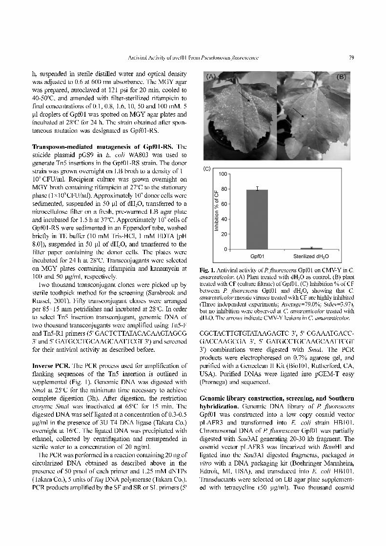
Fig. 1. Antiviral activity of P. fluorescens Gpf01 on CMV-Y in C.amaranticolor. (A) Plant treated with dH2O as control, (B) planttreated with CF (culture filtrate) of Gpf01. (C) Inhibition % of CFbetween P. fluorescens Gpf01 and dH2O, showing that C.amaranticolor mosaic viruses treated with CF are highly inhibited(Three independent experiments; Average=79.0%; Stdev=3.97),but no inhibition were observed at C. amaranticolor treated withdH2O. The arrows indicate CMV-Y lesions in C. amaranticolor.
80 Saeyoull Cho et al.
clones were picked up by sterile toothpick method
(Sambrook and Russell, 2001) for the screening. Fifty
cosmid clones were arranged per 85×15 mm petridishes
and incubated at 37oC for 12 h.
In order to screen the genomic library, plasmid DNA was
isolated from five hundred fifty cosmid clones. Each
cosmid was amplified using primer pair, 19-SL-F, 19-SR-R
(5'-CATCGGCATTCTCAACAGCCTTCAG-3', 5'-CAAA-
GCCGTTACGATGACTTCGTTG-3') and the amplified
DNA was visualized on 0.7% agarose gel. This primer pair
was able to amplify one cosmid which was designated as
pAV1123. The cosmid clones which showed avcP homo-
logous were used for Southern hybridization. Southern
hybridization was carried out as described by Sambrook
and Russell (2001). Detection was performed according to
manufacturer’s instructions (Boehringer Mannheim, Detroit,
USA).
Sub cloning of pAV1123 and complementation of mutants.
To locate the essential genes required for biosynthesis of
antiviral compound on the cosmid clone pAV1123, it was
digested with restriction enzyme HindIII and the resulting
fragments, each 14.1 kb, 1.9 kb and 9.3 kb size were sub
cloned into HindIII digested pUC19 using T4 DNA ligase
(Takara Co.), named respectively as PH14, PH2 and PH9.
Again digested fragments in pUC19 with HindIII were sub-
cloned into calf intestine alkaline phosphatase (CIAP)
(Takara Co.) treated pLAFR3, named as pPH14, pPH2 and
pPH9. The clone pAV1123, pPH14, pPH2 and pPH9 were
mobilized into a natural antiviral negative host P. fluore-
scens B16 for complementation.
The P. fluorescens Gpf01-RS (recipient strain), E. coli
pAV1123 (donor strain), and E. coli pRK2013 (helper
strain) were grown in LB broth with appropriate antibiotics
for overnight at 28oC and 37oC. The overnight cultures of
each strain were sub cultured in LB broth with appropriate
antibiotics and incubated to mid-log phase. Each strain was
washed with sterile water, mixed onto LB agar without
antibiotics and incubated at 28oC for overnight. The
bacterial cells were resuspended in sterile water, diluted and
spread on MGY containing appropriate antibiotics. One
hundred transconjugant clones were picked up by sterile
toothpick method for the screening (Sambrook and Russel,
2001). Fifty mutant clones were arranged per 85×15 mm
petridishes and incubated at 28oC. In order to select
pAV1123 insertion complementation mutant, plasmid DNA
of one hundred transconjugants were digested with restric-
tion enzyme and screened for their antiviral activity as
described before.
Tn3-gusA insertion mutagenesis of pLAFR3 clones.
Strategy of Tn3-gusA insertion mutagenesis is outlined in
(supplemental Fig. 2). The clone pAV1123, containing
genes required for antiviral activity was further mutageni-
zed with Tn3-gusA as described by Bonas et al. (1989). For
insertion mutagenesis of cosmid pAV1123 Tn3HoHo
derivative, pHoKmGus, (D. Dahlbeck and B. Staskawiz,
unpublished) was used. This plasmid harbors a promoter
less β-glucuronidase gene instead of β-galactosidase
between the inverted repeats of Tn3 which is irrelevant for
the experiments described. HB101 (pHoKmGus, pSShe)
was transformed with cosmid pAV1123 plasmid DNA.
Independent transformants (Apr, Cmr, Kmr, Tcr) were mated
with E. coli C2110 using pRK2013 as helper plasmid. The
bacterial cells were then grown in LB broth (Nal, Tc, Km)
for plasmid DNA isolation. After transformation into
DH5α and selection on LB agar (Tc, Km) the obtained
insertion derivatives were analyzed by restriction enzyme
analysis. The insertion sites of Tn3-gus in mutants were
mapped using restriction enzyme digestion analysis and
direct sequencing of the plasmid using the primer Tn3-gus
(5-CCGGTCATCTGAGACCATTAAAAGA-3), which
allows sequencing out of the Tn3-gus insertion region. The
mutagenized plasmids which carried Tn3-gus mutations
were introduced individually into the antiviral negative P.
fluorescence strain B16 by conjugation and marker ex-
change as described before.
DNA sequence analysis. A homology search was per-
formed using BLAST version 2.0, at the National Center
for Biotechnology Information website (http://www.ncbi.
nlm.nih.gov/BLAST). Assembly and analysis of the
sequencing data were performed with the software pack-
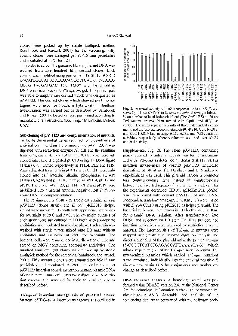
Fig. 2. Antiviral activity of Tn5 transposon mutants (P. fluore-scens Gpf01) on CMV-Y in C. amaranticolor showing inhibition% on number of local lesions/half leaf (The Gpf01-RS1 to 20 areTn5 mutant strains). Plant treated with Gpf01 and dH2O ascontrol. The graph represents results of three independent experi-ments and the Tn5 transposon mutant Gpf01-RS10, Gpf01-RS13,and Gpf01-RS19 had average 6.2%, 6.2%, and 7.8% antiviralactivities, respectively whereas other mutants had over 60.0%antiviral activity.

Antiviral Activity of avcf01 From Pseudomonas fluorescence 81
ages, vector NTITM suite 8 (Informax). GenBank data were
used for alignment of sequence of other P. fluorescens and
homology of Tn3-gus insertion region in cosmid pAV1123
with P. fluorescens Pf0-1. The DNA sequencing was
performed at Macrogen Ltd., Korea, using an automated
ABI 3730×l DNA Analyzer (Applied Biosystems).
Results and Discussion
Antiviral effects of the Gpf01 strain. The culture filtrate
prepared from Gpf01 showed high inhibitory activity
against CMV-Y (Fig. 1B). The CF treated part of the C.
amaranticolor leaves (hypersensitive host) showed 79.0%
(Stdev=3.97) inhibition of the production of local lesions
compared to the untreated part of the leaves (Fig. 1). The
control plants, treated with sterilized water, were unable to
show inhibition of CMV-Y induced lesions (Fig. 1A). The
average number of local lesion in the case of the CF treated
half leaves was much lower than those of the sterilized
water treated half leaves. When CF was used to elucidate
the control effect of the Gpf01, it was found that the plants
treated with CF showed no visible viral symptom 15dpi
(days post inoculation), and remained symptomless through-
out the study period suggesting that antiviral activity might
be due to involvement of plant defense related mechanism
or a systemic re-distribution of antiviral compounds might
show disease suppression effect on the non-treated part of
the cucumber leaves. To confirm the involvement of induc-
tion of plant defense mechanism, more exclusive experi-
mental results should provide. Kandan et al. (2002) report-
ed that application of suspension of P. fluorescens on seed,
root, leaf, and soil reduces the tomato spotted wilt virus in
tomato (TSWV) incidence under green house conditions.
Isolation of antiviral activity-deficient mutants. After
mutagenesis of P. fluorescens Gpf01 with Tn5, two thous-
ands colonies were isolated and screened for their antiviral
activity against CMV on C. amaranticolor. Insertions of
transposon Tn5 in the chromosomal DNAs of the mutants
was confirmed by amplification of approximately 3.0 kb
using Tn5-F and Tn5-R1 primers (supplemental Fig. 3).
Three mutants (Gpf01-RS10, Gpf01-RS13, and Gpf01-
RS19) showed low antiviral activity against CMV-Y on C.
amaranticolor when compared to that of wild type P.
fluorescens Gpf01 strain (Fig. 2). The Gpf01-RS10, Gpf01-
RS13, and Gpf01-RS19 mutants had 6.2%, 6.2%, and 7.8%
antiviral activities, respectively whereas other mutants had
over 60.0% antiviral activity (Fig. 2). This result suggested
that Tn5 transposon was knocked out genes associated with
antiviral agents in P. fluorescens Gpf01. The mutant Gpf01-
RS19 was choosing for further examination.
Identification of antiviral activity related locus. In order
to identify the gene which was knocked out by Tn5
transposon in the mutant Gpf01-RS19, the flanking DNA
regions of the site of the transposition was cloned and
characterized. The chromosomal DNA of mutant Gpf01-
RS19 was amplified by inverse PCR using primer pairs
(SF-SL and SF-SR). The primer pair SF and SR amplified
0.4 kb fragment on the right of Tn5 insertion region, while
the primer pair SF and SL amplified 1.5 kb on the left of
Tn5 insertion region (supplemental Fig. 4), which were
further cloned and sequenced. From BLAST search analy-
ses with the limited DNA sequence data, mutant Gpf01-
RS19 exhibited a mutation in a gene homologous to a
possible transcription regulator, AsnC family of P. fluoro-
scens strain Pf0-1 (GenBank accession no. CP000094),
showing 97.0% homology (data not shown). In this study,
the antiviral related region was termed antiviral compound
producing (avcf01) locus.
Avcf01 locus in genomic library Gpf01. The chromo-
somal DNA of P. fluorescens Gpf01 was amplified using
the primer pair (19-SL-F and 19-SR-R). This primer pair
amplified 1.2 kb fragment, sequenced and used for screen-
ing the genomic library P. fluorescens Gpf01. To identify
the avcf01 locus, five hundred fifty cosmids were screened
by PCR using this primer pair. This primer amplified 1.2 kb
fragment from only one cosmid which was designated as
pAV1123 (Fig. 3). The presence of avcf01 locus in cosmid
pAV1123 was further confirmed by a Southern blot analysis
Fig. 3. Detection of cosmid clone in Gpf01 genomic librarycontaining avcf01 locus. (A) PCR amplification of 1.2 kbfragment, a part of avcf01 locus, using 19-SL-F and 19-SR-Rprimers. (B) Detection of homologous avcf01 locus in cosmidpAV1123 of P. fluorescens Gpf01 by Southern hybridization. M:1kb DNA Ladder, lane 1: total DNA of Gpf01, lane 2: pLAFR3,Lane 3-13: various cosmid clones of Gpf01, lane 14: cosmidpAV1123.
82 Saeyoull Cho et al.
(Fig. 3). This investigation provides the first evidence that
avcf01 locus in P. fluorescens Gpf01 is required for the
antiviral activity. Mutation in this locus abolished the
antiviral activity against CMV-Y.
Complementation. The cosmid clone, E. coli pAV1123,
was mobilized into antiviral activity-deficient mutant P.
fluorescens Gpf01-RS19 in the presence of helper plasmid
E. coli pRK2013. The complementation was confirmed by
plasmid isolation from the transconjugants. The transconju-
gants were isolated and assayed for antiviral activity against
CMV-Y on C. amaranticolor. One of the transconjugants
Gpf01-RS19+pAV1123 was able to recover 74.4% antiviral
activity which was lost due to insertion of Tn5 insertion
(Fig. 4) compared to Gpf01-RS19 antiviral activity (Fig. 4).
Moreover, when cosmid clone, E. coli pAV1123 was intro-
duced into the host P. fluorescens B16, the transconjugant
exhibited 77.2% antiviral activity (Fig. 4) but, P. fluore-
scens B16 was still low (Fig. 4). When the mutant was
complemented with E. coli containing 25.3 kb cosmid
(pAV1123) of P. fluorescens in the presence of helper
plasmid, antiviral activity was recovered and reduced
incidence of local lesions of CMV-Y on the C. amaranti-
color leaves. This result also revealed that avcf01 locus is
essential for antiviral activity. Furthermore, this result
suggests that the cosmid pAV1123 might contain all genes
essential for antiviral activity against CMV-Y. The BLAST
search analysis for avcf01 locus showed 97% homology
with possible transcriptional regulator AsnC family of P.
fluorescens GPf01. It has been reported that various P.
fluorescens strains induce systematic resistance in cucumber,
tobacco and tomato against cucumber mosaic cucumovirus
(CMV), tobacco necrosis virus (TNV) and tomato spotted
wilt virus (TSWV) (Kandan et al., 2002; Maurhofer et al.,
1994; Raupach et al., 1996). It is also reported that P.
fluorescens strains induces the salicylic acid (SA) bio-
synthesis genes pchAB and phenylpropanoid metabolism
thereby improving their ability to induce different plant
viruses (Maurhofer et al., 1994; Raupach et al., 1996).
Subcloning of cosmid pAV1123 by HindIII. In order to
characterize the region responsible for the antiviral activity
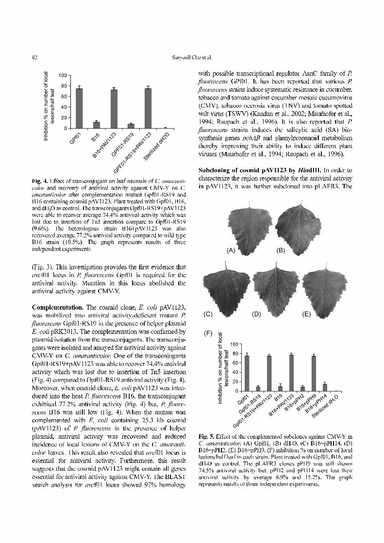
in pAV1123, it was further subcloned into pLAFR3. TheFig. 4. Effect of transconjugant on leaf necrosis of C. amaranti-
color and recovery of antiviral activity against CMV-Y on C.
amaranticolor after complementation mutant Gpf01-RS19 andB16 containing cosmid pAV1123. Plant treated with Gpf01, B16,and dH2O as control. The transconjugants Gpf01-RS19+pAV1123were able to recover average 74.4% antiviral activity which waslost due to insertion of Tn5 insertion compare to Gpf01-RS19(9.6%). The heterologous strain B16+pAV1123 was alsorecovered average 77.2% antiviral activity compared to wild typeB16 strain (10.5%). The graph represents results of threeindependent experiments.
Fig. 5. Effect of the complemented subclones against CMV-Y inC. amaranticolor. (A) Gpf01, (B) dH2O, (C) B16+pPH14, (D)B16+pPH2, (E) B16+pPH9, (F) inhibition % on number of locallesions/half leaf in each strain. Plant treated with Gpf01, B16, anddH2O as control. The pLAFR3 clones pPH9 was still shown74.5% antiviral activity but, pPH2 and pPH14 were lost theirantiviral activity by average 8.9% and 15.2%. The graphrepresents results of three independent experiments.

Antiviral Activity of avcf01 From Pseudomonas fluorescence 83
clones were named as pPH2, pPH9, pPH14 (data not
shown). When the pLAFR3 clones pPH2, pPH9, and pPH14
were complemented with B16, only pPH9 could recover
74.5% antiviral activity, whereas the other two clones,
pPH2 and pPH14 were able to show only 8.9% and 15.2%
antiviral activity (Fig. 5C, D, E, and F). This showed that
pPH9 containing 9.3 kb region of pAV1123 is responsible
for antiviral activity and the pPH9 containing 9.3 kb region
of pAV1123 by HindIII was chosen for further exami-
nation.
Tn3-gusA mutagenesis and DNA sequence analysis. The
Tn3-gus mutation in pPH9 (pAV1123) occurred randomly
at 8 locations in 8 different clones, which were named as
C1 to C8. The clones were then mobilized into B16 and
screened for antiviral activity. The complemented Tn3-gus
mutant clones into B16 were named as P1 to P8 respec-
tively. Mutants P5 to P8 lost their antiviral activity after
insertion of Tn3-gus to 10.9%, 8.0%, 16.4% and 7.8%
respectively whereas, other mutants, P1 to P4 were able to
retain this activity even after the insertion (Fig. 6). Restric-
tion enzyme maps of cosmid and sub-clones that inhibited
antiviral activity in a heterologous host, P. fluorescens B16
and Tn3-gus mutants was shown in Fig. 7. The clone C5
had mutation 50 bp upstream to that of Tn5 mutation in
Gpf01-RS19 in the transcriptional regulator, AsnC family.
The other three belong, C6 into dehydrogenase E1 compo-
Fig. 6. Effect of the Tn3-gus mutants against CMV-Y in C.amaranticolor. The Tn3-gus mutation in pPH9 occurred random-ly at 8 locations in 8 different clones, named as C1 to C8. Thecomplemented Tn3-gus mutant clones into B16, named as P1 toP8. Mutants P5 to P8 lost their antiviral activity to 10.9%, 8.0%,16.4%, and 7.8%, respectively but, mutant P1 to P4 was able toretain their antiviral activity to 68.0%, 74.3%, 70.9%, and 71.0%,respectively. The graph represents results of three independentexperiments.
Fig. 7. Restriction enzyme maps of cosmid and sub-clones that inhibited antiviral activity in a heterologous host, P. fluorescens B16 andTn3-gus mutants. The checked box (C5) is avcf01 region containing transcriptional regulator, AsnC family. The C6, C7, and C8 weredehydrogenase E1 component, aminotransferase, and hypothetical protein, respectively. Antiviral activity in heterologous host was shownin the subclone pPH9.

84 Saeyoull Cho et al.
nents, C7 into branched chain amino acid aminotransferase
II, and C8 into a hypothetical protein (Fig. 7). It is sug-
gested that the antiviral activity of P. fluorescens Gpf01 is
complex and multiple factors (At least, four different loci
might involve) inhibit CMV-Y. AsnC family in E. coli
regulates a number of metabolic genes which might be
responsible for modification enzymes, which hypothesized
to modify the antivirus component. In addition, lactate-
dehydrogenase is associated with production of antiviral
agents and the level of aminotransferase is also highly
involved in the production of antiviral component in
eukaryotic (Kudo et al., 2001; Franchetti and Grifantini
1999; Lenci et al., 2008). Although several studies explain
AsnC, dehydrogenase, and aminotransferase might play
vital role in the production of antiviral compounds in
eukaryotic, there is no exact evidence of their involvement
in the production of antiviral compound in prokaryotic so
far. Thus, further research is required to find the relation-
ship between these genes and how it affects the antivirus
production. In addition, we need to prove that P. fluorescens
Gpf01 can induce systematic resistance in other viruses
including tobacco mosaic virus (TMV), tobacco necrosis
virus (TNV), tomato spotted wilt virus (TSWV), and etc.
It is concluded that determination of antiviral activity
related to avcf01 locus in P. fluorescens Gpf01 is a novel
finding in this investigation and requires further analysis
and detailed characterization of AsnC, dehydrogenase E1
component, and aminotransferase II in this strain.
Acknowledgements
This research was supported by Agriculture and life science
research institute in the Kangwon National University.
References
Bonas, U., Stall, R. E. and Staskawicz, B. J. 1989. Genetic and
structural characterization of the avirulence gene avrBs3 from
Xanthomonas campestris pv. vesicatoria. Mol. Gen. Genet.
218:127-136.
Défago, G. and Haas, D. 1990. Pseudomonads as antagonists of
soil borne plant pathogens: modes of action and genetic analy-
sis. Soil Biochem. 6:249-291.
Elisaveta, S. and Violeta, S. 2000. Tomato lines segregation for
resistance to cucumber mosaic virus. Acta Physiologiae Plan-
tarum. 22:353-355.
Franchetti, P. and Grifantini, M. 1999. Nucleoside and non-nucle-
oside IMP dehydrogenase inhibitors as antitumor and antiviral
agents. Curr. Med. Chem. 6:599-614.
Gooding, G. V. 1991. Diseases caused by virus. In: Shew, H. D.
and Lucas, G. B. Compendium of Tobacco Diseases, APS
Press, Minnesota. 41pp.
Hosain, A. K. M. and Alexander, M. 1984. Enhancing soybean
rhizosphere colonization by Rhizobium japonicum. Appl.
Environ. Microbiol. 48:468-472.
Ipper, N. S., Kim, J. E., Koo, J. H., Hur, J. H. and Lim, C. K. 2005.
Inhibitory effects of a Korean strain Gpf01 identified as
Pseudomonas fluorescence on cucumber mosaic virus. Plant
Pathol. J. 21:262-269.
Ipper, N. S., Lee, S. H., Suk, J. K., Anupama, S., Seo, D. U., Park,
D. H., Cho, J. M., D. S., Park, Hur, J. H. and Lim, C. K. 2006.
Isolation and evaluation of an antiviral producing Serratia spp.
Strain Gsm01 against cucumber mosaic virus in Korea. Pest.
Sci. J. 10:344-350.
Kandan, A., Commare, R., Nandakumar, R., Ramlaii, M.,
Raguchander, T. and Samiyappan, R. 2002. Induction of phe-
nylpropanoid metabolism by Pseudomonas fluorescens
against tomato spotted wilt virus in tomato. Folia. Microbiol.
47:121-129.
Keane, P. J., Kerr, A. and New, P. B. 1970. Crown gall of stone
fruit. II. Identification and nomenclature of Agribacterium iso-
lates. Aust. J. Biol. Sci. 23:85-595.
Keel, C., Schnider, U., Maurhofer, M., Vousard, C., Laville, J.,
Burger, U., Wirthner, P., Hass, D. and Defago, G. 1992. Sup-
pression of root disease by Pseudomonas fluorescens CHAO:
Importance of the bacterial secondary metabolite 2,4-diacetyl
phloroglucinol. Mol. Plant-Micobe Intract. 5:4-13.
Kim, J. W., Kim, J. E., Park, B. K., Choi, O. H., Park, C. S. and
Hwang, I. G. 2003. Identification of genes for biosynthesis of
antibacterial compound from Pseudomonas fluorescens B16,
and its activity against Ralstonia solanacearum. J. Microbiol.
Biotechnol. 13:292-300.
Kim, S. K., Hwang, E. I., O, J. H., Kim, K. S., Ryu, M. H. and
Yeo, W. H. 2004. Inhibitory effects of Acinetobacter sp. KTB3
in infection of tobacco mosaic virus in tobacco plants. Plant
Pathol. J. 20:293-296.
Klement, Z., Kiraly, J. and Pozsar, I. 1996. Suppression of virus
multiplication and local lesion production in tobacco follow-
ing inoculation with a saprophytic bacterium. Acta pjyto-
pathol. Acad. Sci. Hung. 1:11-18.
Kubo, S., Ikeda, T., Imaizumi, S., Takanami, Y. and Mikami, Y.
1990. A potent plant virus inhibitor found in Mirablis jalapa
L. Ann. Phytopath. Soc. Japan. 56:481-487.
Kudo, N., Allen, M. D., Koike, H., Katsuya, Y., Suzuki, M. 2001.
Crystallization and secondary-structure determination of a
protein of the Lrp/AsnC family from a hyperthermophilic
archaeon. Acta Crystallogr D. Biol Crystallogr. 57:469-471.
Lampis, G., Deidda, D., Maullu, C., Petruzzelli, S. and Pompei, R.
1996. Karalicin, a new biologically active compound from
Pseudomonas fluorescens/putida. J. Antibiotics. 49:260-266.
Lenci, I., Piccolo, P., Francioso, S., Di Paolo, D., Galante, A.,
Angelico, M. 2008. Recurrent myocardial ischaemia during
combination antiviral therapy in a patient with chronic hepati-
tis C and normal aminotransferase levels. Dig. Liver Dis.
40:785-90.
Maurhofer, M., Hase, C., Meuwly, P., Métraux, J. P. and Défago,
G. 1994. Induction of systemic resistance of tobacco to
tobacco necrosis virus by the root-colonizing Pseudomonas
fluorescens strain CHA0: influence of the gacA gene and of
Antiviral Activity of avcf01 From Pseudomonas fluorescence 85
pyoverdine production. Phytopathol. 84:139-146.
Maurhofer, M., Reimmann, C., Schmidli-Sacherer, P., Heeb, S.,
Haas, D. and Défago, G. 1998. Salicylic acid biosynthetic
genes expressed in Pseudomonas fluorescens strain P3 improve
the induction of systemic resistance in tobacco against tobacco
necrosis virus. Phytopathol. 88:678-684.
Nowak-Thompson, B., Gould, S. J., Karus, J. and Lopper, J. E.
1994. Production of 2,4-dicetylphloroglucinol by biocontrol
agent Pseudomonas fluorescens Pf-5. Can. J. Microbiol.
40:1064-1066.
O’Sullivan, D. J. and O’Gara, F. 1992. Traits of fluorescent
Pseudomonas spp. involved in suppression of plant root
pathogens. Microbiol. Rev. 56:662-676.
Palleroni, N. J., Kunisawa, R., Contopoulou, R. and Doudoroff,
M. 1973. Nucleic acid homologies in genus Pseudomonas.
Int. J. Syst. Bacteriol. 23:333-339.
Raupach, G. S., John, L. L., Murphy, F., Tuzun, S. and Kloepper,
J. W. 1996. Induced systemic resistance in cucumber and
tomato against cucumber mosaic cucumovirus using plant
growth promoting rhizobacteria. Plant Dis. 80:891-894.
Rosales, A. M., Thomashow, L., Cook, R. J. and New, T. W. 1995.
Isolation and identification of antifungal metabolites produced
by rice-associated antagonistic Pseudomonas sp. Phytopathol.
85:1028-1032.
Ryu, J. S., Lee, S. D., Lee, Y. H., Lee, S. T., Kim, D. K., Cho, S. J,
Park, S. R., Bae, D. W., Park, K. H. and Yun, H. D. 2000.
Screening and identification of an antifungal Pseudomonas sp.
that suppresses balloon flower root rot caused by Rhusoctonia
solani. J. Microbiol. Biotechnol. 10:435-440.
Sambrook, J. and Russel, D. W. 2001. Molecular Cloning: A Lab-
oratory Manual, 3rd ed., Cold Spring Harbor Press, Cold
Spring Harbor, NY.
Thomashaw, L. S. and Wellwe, D. M. 1988. Role of a phenazine
antibiotic from Pseudomonas fluorescens in biological control
of Gaeumannomyces graminis var. tritici. J. Bacteriol.
170:3499-3508.
Zehender, G. W., Yao, C., Murphy, J. F., Sikora, E. R. and Kleoep-
per, J. W. 2000. Induction of resistance in tomato against
cucumber mosaic cucumovirus by plant growth-promoting
rhizobacteria. Biocontrol. 45:127-137.