Compartmentation of Phenolic Compounds and Phenylalanine AmmoniaLyase in Leaves of Phyllanthus...
10
Compartmentation of Phenolic Compounds and Phenylalanine Ammonia-Lyase in Leaves of Phyllanthus tenellus Roxb. and their Induction by Copper Sulphate LAURA JANE M. SANTIAGO* {, RICARDO P. LOURO { and DULCE E. DE OLIVEIRA } {Laborato ´rio de Biotecnologia, Centro de Biocieˆncias e Biotecnologia, Centro de Biocieˆncias e Biotecnologia, Universidade Estadual do Norte Fluminense, Av. Alberto Lamego 2000, Horto, Campos dos Goytacazes, Rio de Janeiro, CEP 28015-620, Brazil, {Laborato ´rio de Ultraestrutura Vegetal, Departamento de Bota ˆnica, Instituto de Biologia, Universidade Federal do Rio de Janeiro, Rio de Janeiro, Brazil and }Laborato ´rio de Gene´tica Molecular Vegetal, Departamento de Gene´tica, Instituto de Biologia, Universidade Federal do Rio de Janeiro, Rio de Janeiro, Brazil Received: 9 May 2000 Returned for revision: 9 June 2000 Accepted: 29 July 2000 The compartmentation of phenolic compounds in mature leaves of Phyllanthus tenellus and their induction by copper sulphate were analysed at histological and subcellular levels. Light and electron microscopy studies demonstrated that the vacuoles of spongy cells were the main sites of phenolic accumulation. Spraying plants with copper sulphate induced punctated lesions formed by groups of necrotic cells which accumulated brownish substances. Histochemical tests and fluorescence microscopyanalysis of the sprayed leaves indicated that the phenolic compounds increased in spongy cells within the lesions. Ultrastructural analyses showed that 3 h after elicitation, the organelles of the cells within the lesion started to collapse and the content of phenolic substances increased in the vacuole of spongy cells. Antibody against phenylalanine ammonia-lyase (PAL) from parsley cross-reacted with the crude extract of P. tenellus leaves. Two isoforms, one of 65 kD and the other of 66 kD, were identified. Immunocytochemical studies showed that PAL was synthesized in the palisade and spongy cells, mainly in the cytoplasm and chloroplasts. The phytotoxicity of Cu 2 ions induced the accumulation of PAL in sub-cellular compartments of palisade cells. PAL accumulation started to increase 3 h after elicitation and reached a maximum after 6 h, decreasing 12 h post- induction. The increase of PAL was more evident in cells within the necrotic punctated regions than in surrounding cells. Since the vacuole of palisade cells did not accumulate phenolic compounds, the in situ studies suggested that the end products of PAL synthesis play a role in palisade cell wall reinforcement or might accumulate in other tissues. The symptoms induced by copper sulphate suggest that this abiotic elicitor may be a useful tool in the understanding of the regulation of biosynthetic phenolic pathways in P. tenellus. # 2000 Annals of Botany Company Key words: Cell death, copper sulphate, heavy metal, immunolabelling, phenolic compounds, phenylalanine ammonia-lyase, Phyllanthus, transmission electron microscopy, ultrastructure. INTRODUCTION Plant secondary metabolites are formed from glucose metabolism intermediated by the shikimic, acetate and aminoacid pathways (Geissman and Crout, 1969). A range of physiological and ecological functions have been reported for these natural products, such as hormone regulation, organogenesis, plant defence against biotic and abiotic agents, chemical signalling to guide pollinators or fruit dispersers and plant–microorganism symbiosis (Curir et al., 1990; van Tunen and Mol, 1991; Vogt et al., 1994; Turlings et al., 1995). The understanding and manipulation of plant secondary metabolite biosynthesis is a major challenge in plant physiology. These substances are chemically diverse with heterogeneous distribution in dierent taxonomic groups and dierent plant organs and tissues (Gottlieb, 1982). The regulation of the enzymes involved in their synthesis is influenced by plant develop- ment as well as by external factors (van Tunen and Mol, 1991; Eckey-Kaltembach et al., 1994). Biotic and abiotic stress activate complex defence mechanisms in plant cells, altering both primary and secondary metabolism in a coordinated fashion (Marques and Brodelius, 1988; Dalkin et al., 1990). Elicitors have been employed to induce the biosynthetic pathways of important secondary metabolites in order to elucidate the mechanisms that underlie the steps of their synthesis, distribution and accumulation (Marques and Brodelius, 1988; Aertis et al., 1994; Dornenburg and Knorr, 1995). The role of abiotic stress in plant cell organization and its relation to the induction of dierent secondary metabolites has been the subject of extensive investigations (Santos et al., 1993; Lummerzhein et al., 1995). However, little is known about the influence of abiotic elicitors on specific pathways of secondary metabolite biosynthesis. Phenolic compounds are aromatic secondary metabolites formed essentially via the shikimic and/or malonic path- ways (Geissman and Crout, 1969). Modifications in their basic structure such as oxidation, hydroxylation, glyco- sylation and methylation have led to a large range of phenolic substances with dierent biological functions (Gottlieb, 1982; van Tunen and Mol, 1991; Nicholson Annals of Botany 86: 1023–1032, 2000 doi:10.1006/anbo.2000.1271, available online at http://www.idealibrary.com on 0305-7364/00/111023+10 $35.00/00 # 2000 Annals of Botany Company * For correspondence. Fax 00 55 24 7263720, e-mail laura@cbb. uenf.br
Transcript of Compartmentation of Phenolic Compounds and Phenylalanine AmmoniaLyase in Leaves of Phyllanthus...
Annals of Botany 86: 1023±1032, 2000doi:10.1006/anbo.2000.1271, available online at http://www.idealibrary.com on
Compartmentation of Phenolic Compounds and Phenylalanine Ammonia-Lyase in Leavesof Phyllanthus tenellus Roxb. and their Induction by Copper Sulphate
LAURA JANE M. SANTIAGO*{, RICARDO P. LOURO{ and DULCE E. DE OLIVEIRA}
{LaboratoÂrio de Biotecnologia, Centro de BiocieÃncias e Biotecnologia, Centro de BiocieÃncias e Biotecnologia,Universidade Estadual do Norte Fluminense, Av. Alberto Lamego 2000, Horto, Campos dos Goytacazes, Rio deJaneiro, CEP 28015-620, Brazil, {LaboratoÂrio de Ultraestrutura Vegetal, Departamento de BotaÃnica, Instituto deBiologia, Universidade Federal do Rio de Janeiro, Rio de Janeiro, Brazil and }LaboratoÂrio de GeneÂtica MolecularVegetal, Departamento de GeneÂtica, Instituto de Biologia, Universidade Federal do Rio de Janeiro, Rio de Janeiro,
Brazil
Received: 9 May 2000 Returned for revision: 9 June 2000 Accepted: 29 July 2000
1991; Eckey
0305-7364/0
* For corruenf.br
The compartmentation of phenolic compounds in mature leaves of Phyllanthus tenellus and their induction by coppersulphate were analysed at histological and subcellular levels. Light and electron microscopy studies demonstrated thatthe vacuoles of spongy cells were the main sites of phenolic accumulation. Spraying plants with copper sulphateinduced punctated lesions formed by groups of necrotic cells which accumulated brownish substances. Histochemicaltests and ¯uorescence microscopy analysis of the sprayed leaves indicated that the phenolic compounds increased inspongy cells within the lesions. Ultrastructural analyses showed that 3 h after elicitation, the organelles of the cellswithin the lesion started to collapse and the content of phenolic substances increased in the vacuole of spongy cells.Antibody against phenylalanine ammonia-lyase (PAL) from parsley cross-reacted with the crude extract of P. tenellusleaves. Two isoforms, one of 65 kD and the other of 66 kD, were identi®ed. Immunocytochemical studies showedthat PAL was synthesized in the palisade and spongy cells, mainly in the cytoplasm and chloroplasts. Thephytotoxicity of Cu2� ions induced the accumulation of PAL in sub-cellular compartments of palisade cells. PALaccumulation started to increase 3 h after elicitation and reached a maximum after 6 h, decreasing 12 h post-induction. The increase of PAL was more evident in cells within the necrotic punctated regions than in surroundingcells. Since the vacuole of palisade cells did not accumulate phenolic compounds, the in situ studies suggested that theend products of PAL synthesis play a role in palisade cell wall reinforcement or might accumulate in other tissues. Thesymptoms induced by copper sulphate suggest that this abiotic elicitor may be a useful tool in the understanding ofthe regulation of biosynthetic phenolic pathways in P. tenellus. # 2000 Annals of Botany Company
Key words: Cell death, copper sulphate, heavy metal, immunolabelling, phenolic compounds, phenylalanineammonia-lyase, Phyllanthus, transmission electron microscopy, ultrastructure.
INTRODUCTION
Plant secondary metabolites are formed from glucosemetabolism intermediated by the shikimic, acetate andaminoacid pathways (Geissman and Crout, 1969). A rangeof physiological and ecological functions have beenreported for these natural products, such as hormoneregulation, organogenesis, plant defence against biotic andabiotic agents, chemical signalling to guide pollinators orfruit dispersers and plant±microorganism symbiosis (Curiret al., 1990; van Tunen and Mol, 1991; Vogt et al., 1994;Turlings et al., 1995). The understanding and manipulationof plant secondary metabolite biosynthesis is a majorchallenge in plant physiology. These substances arechemically diverse with heterogeneous distribution indi�erent taxonomic groups and di�erent plant organs andtissues (Gottlieb, 1982). The regulation of the enzymesinvolved in their synthesis is in¯uenced by plant develop-ment as well as by external factors (van Tunen and Mol,
-Kaltembach et al., 1994).
0/111023+10 $35.00/00
espondence. Fax 00 55 24 7263720, e-mail laura@cbb.
Biotic and abiotic stress activate complex defencemechanisms in plant cells, altering both primary andsecondary metabolism in a coordinated fashion (Marquesand Brodelius, 1988; Dalkin et al., 1990). Elicitors havebeen employed to induce the biosynthetic pathways ofimportant secondary metabolites in order to elucidate themechanisms that underlie the steps of their synthesis,distribution and accumulation (Marques and Brodelius,1988; Aertis et al., 1994; Dornenburg and Knorr, 1995).The role of abiotic stress in plant cell organization and itsrelation to the induction of di�erent secondary metaboliteshas been the subject of extensive investigations (Santoset al., 1993; Lummerzhein et al., 1995). However, little isknown about the in¯uence of abiotic elicitors on speci®cpathways of secondary metabolite biosynthesis.
Phenolic compounds are aromatic secondary metabolitesformed essentially via the shikimic and/or malonic path-ways (Geissman and Crout, 1969). Modi®cations in theirbasic structure such as oxidation, hydroxylation, glyco-sylation and methylation have led to a large range ofphenolic substances with di�erent biological functions
(Gottlieb, 1982; van Tunen and Mol, 1991; Nicholson# 2000 Annals of Botany Company

ley PAL.
collected from di�erent plants.
¯uorescent compounds.
o
and Hammerschmidt, 1992). The rigidi®cation of the cellwall by lignin is both an e�ective adaptation to the terres-trial environment and a�ords protection against invasion ofphytopathogens (Gottlieb, 1982; Halhbrock and Schell,1989; Nicholson and Hammerschmidt, 1992). The acti-vation of the Rhizobium nod genes involved in root nodula-tion is mediated by ¯avonoids such as 7,4-dihydroxy¯avone(Coronado et al., 1995) and iso¯avonoids may act as signalmolecules during plant±mycorrhiza interaction (Paiva et al.,1994). The speci®c location of anthocyanins in the plantepidermal layer may protect internal cell layers against UV-B radiation (Braun and Tevini, 1993) and an increase infuranocoumarins leads to plant protection against highlevels of ozone (Eckey-Kaltenbach et al., 1994). Diverseantimicrobial phenolics such as daidzein, genistein andpsoralen are synthesized and accumulated during phyto-pathogen invasion (Graham, 1991; Nicholson and Ham-merschmidt, 1992). Also the regulation of polar auxintransport is considered to be modulated by ¯avonoids(Jacobs and Rubery, 1988).
Besides their importance in plant biology, many second-ary metabolites have been employed in human phytother-apy. Phenolic compounds synthesized by Phyllanthus L.species are potent antineoplasic agents and inhibitors of theDNA polymerase of the hepatitis B virus (Venkateswaranet al., 1987; Blumberg et al., 1989). The phenolic com-pounds of Phyllanthus emblica are inhibitors of immuno-de®ciency virus replication, with only a few side e�ects(Pettit et al., 1990; El-Mekkawy et al., 1995). Also, metha-nolic extracts of some species have been shown to be potentantiophidic and analgesic agents (Kuster et al., 1994;Santos et al., 1994; Calixto et al., 1997).
The ®rst reaction in the phenylpropanoid pathway iscatalysed by phenylalanine ammonia-lyase (PAL; EC4.3.1.5.) converting L-phenylalanine to trans-cinnamic acid(Hanson and Havir, 1981). It has been shown that de novosynthesis of PAL isoforms is induced by biotic and abioticelicitors and that the di�erential expression of PAL genesleads to the synthesis and accumulation of PAL isoformsand phenolic compounds in di�erent tissue and subcellularcompartments (Jahnen and Hahlbrock, 1988; Schmelzeret al., 1988).
Structural and immunolocalization studies of normal andstressed plants have identi®ed the main sites of synthesisand accumulation of biosynthetic enzymes (Karwatzki et al.,1993; Duke et al., 1994). Furthermore, temporal and spatialstudies into the synthesis and induction of phenoliccompounds and their biosynthetic enzymes have helped inthe understanding of the regulation and distribution of thesteps of their metabolic pathways. With this purpose inmind we used heavy metals as abiotic elicitors to inducephenolic compounds in Phyllanthus tenellus Roxb., anannual herb known in Brazilian folk medicine as ervapombinha or quebra-pedra (stone-breaker). According toPio-Correà a (1974), P. tenellus plants have been used inkidney disease as a diuretic and to dissolve renal stones.Calixto et al. (1998) also proposed that the phenoliccompounds of these plants have antinociceptive properties.Heavy metals are chemically better de®ned than biotic
1024 Santiago et al.ÐCompartmentation
elicitors, and can activate the phenylpropanoid pathway
(Kessmann et al., 1990; Parry et al., 1994). Among theelicitors tested, spraying with copper sulphate led to theformation of microlesions and the synthesis of phytoalexinsin punctated regions, a type of hypersensitive reaction (HR)symptom induced by pathogen attack.
In the present investigation, in situ studies were employedto localize, at the histological and subcellular level, the sitesof synthesis and accumulation of phenolic compounds inmature leaves of P. tenellus induced by the abiotic elicitorcopper sulphate. To elucidate further the earlier steps ofregulation of phenolic biosynthesis, compartmentation ofphenylalanine ammonia-lysase (PAL) was evaluated byimmunocytochemical assays using antibodies against pars-
f Phenolics and PAL in Phyllanthus
MATERIALS AND METHODS
Biological material
Plants of Phyllanthus tenellus were grown in a greenhouse at358C. Fifty to 60-d-old plants were sprayed with anaqueous solution of 0.5 M copper sulphate and 0.05% (v/v) Tween-20, and 1.5 cm long leaves were collected at 0, 0.5,1.5, 3, 6 and 12 h for further analysis. Control plants weretreated with 0.05% (v/v) Tween-20 in sterile distilled waterand the leaves were collected at the same time points. Formicroscopy analyses, the experiments were performed ®vetimes using about 30 sections of ®ve leaves randomly
Light and ¯uorescence microscopy
Light microscopy studies were performed on frozen20 mm thick cross-sections cut with a Micron cryostat(Walldors, Germany). To analyse phenolic compounds,samples were treated with alcoholic FeCl3 (Johansen, 1940).The leaf surface and the frozen cross-sections were analysedusing a Zeiss Axioplan epi¯uorescence microscope (GoÈ ttin-gen, Germany) equipped with a 480 nm ®lter to detect
Electron microscopy
For ultrastructural analysis, tissue was ®xed for 2 h atroom temperature, with 2.5% glutaraldehyde (v/v) and 4%paraformaldehyde (w/v) in 0.05 M sodium cacodylate bu�er(pH 7.3). Samples were post®xed for 1 h with 1% (w/v)osmium tetroxide in sodium cacodylate bu�er, dehydratedin a graded acetone series and embedded in Spurr's resin(Spurr, 1969). Ultrathin sections (80 nm) were cut with adiamond knife on a Reichert Ultracut S microtome(Vienna, Austria) and post-stained with 1% uranyl acetate(w/v) in absolute ethanol and lead citrate (Reynolds, 1963).
For the immunocytochemical assays, leaf samples were®xed with 0.2% glutaraldehyde (v/v) and 4% (w/v)paraformaldehyde in the same bu�er, dehydrated in agraded ethanol series and embedded in LRWhite resin(Polysciences, Warrington, PA, USA) at room temperature.
The in®ltrated samples were embedded in gelatine capsules,
o
and polymerized for 2 d at 378C. Ultrathin sections were
Santiago et al.ÐCompartmentation
collected on nickel grids.
(Sigma, St. Louis, MO, USA).
Antibody labellingFor immunogold labelling, nickel grids containingultrathin sections were incubated in a blocking solution of1% (w/v) bovine serum albumin (Sigma, St. Louis, MO,USA) in PBST [10 mM sodium phosphate bu�er, pH 7.2(Sigma, MO, St. Louis, USA), 500 mM NaCl and 0.1%Tween-20] for 20 min. After blocking, the samples wereincubated for 3 h with the anti-PAL antiserum from parsley(Petroselium crispum) diluted 1 : 200 in PBST. This anti-body was donated by Dr Klaus Hahlbrock (Max-Plank-Institut fuÈ r Zuchtungsforschung, KoÈ ln). The material wasthen washed in PBST for 30 min ( four times), andincubated in a solution of secondary anti-rabbit IgG anti-body conjugated with 10 nm gold particles (E.Y. Labs. Inc.,San Mateo, USA) diluted 1 : 100. Excess secondaryantibody was removed by rinsing with PBST, followed bydistilled water. After immunolabelling, sections werestained with 2% (w/v) aqueous uranyl acetate for 20 minand with lead citrate (Reynolds, 1963) for 3 min. All stepswere carried out at room temperature. For controls, theprimary antibody was omitted. Material was observed witha Zeiss 900 transmission electron microscope (Oberkochen,Germany). The number of gold particles on palisade cellswas evaluated from the 30 cells of ®ve leaves from ®ve
experiments.abundant.
Protein extraction
Frozen mature leaves were powdered in liquid N2 using amortar and pestle and suspended in extraction bu�er with0.0625 M Tris±HCl pH 6.8, 100 mM DTT and 2% SDS.After removal of cell debris by centrifugation (15 000 g,10 min, 48C) proteins were precipitated in 5 vol of acetoneat 208C for 2 h and resuspended in the same bu�er. Totalprotein content was determined with a bicinchroninic acid
protein assay kit (Sigma, St. Louis, MO, USA).Electrophoresis and Western blotting
For electrophoretic analyses, loading bu�er containing100 mM Tris-HCl pH 6.8, 4% SDS, 20% (v/v) glycerol,0.004% bromophenol blue and 200 mM DTT was added to80 mg proteins and heated at 1008C for 4 min. Proteins wereseparated by SDS/PAGE (Laemmli, 1970) with a 4%stacking gel and 12.5% running gel and electroblotted to anitrocellulose membrane in 50 mM Tris and 50 mM boricacid bu�er pH 8.8 at a constant 25 V for 16 h. Detection oftotal protein on the membrane was by Ponceau-S staining(0.2% (w/v) Ponceau-S in 3% trichloracetic acid) anddestaining with distilled water. The membrane was thenincubated in 5% powdered milk in PBS pH 7.4, washed inPBST for 10 min, and incubated for 4 h in PBSTA (PBS,0.5% (v/v) Tween-20, 1% (w/v) BSA) with the antibodyagainst PAL diluted 1 : 500. Membranes were then washedin PBST for 10 min, incubated for 2 h in PSTA with the
secondary antibody conjugated with alkaline phosphatase(Sigma, St. Louis, MO, USA) diluted 1 : 5000, and rinsedagain with PBST. The antibody-protein complex wasdeveloped with the Alkaline Phosfatase Western Kit
f Phenolics and PAL in Phyllanthus 1025
RESULTS
Leaf anatomy and ultrastructure
The leaves of Phyllanthus tenellus are glabrous anddorsiventral, formed by one epidermal layer and mesophyllconsisting of one layer of palisade cells and two layers ofspongy cells. Histochemical assays with FeCl3 on transversesections of the leaf blade demonstrated that phenolic com-pounds accumulated in isolated or in clustered, randomlydispersed spongy cells. These cells were similar in size andshape to the other cells of spongy tissue.
Ultrastructural analysis showed that palisade cells haddense cytoplasm, a small vacuole, numerous mitochondriaand chloroplasts with large starch grains, and smallplastoglobules (Fig. 1). The spongy cells develop a densecytoplasm and a large and central vacuole most of whichmay accumulate electronopaque deposits characteristic ofphenolic compounds (Figs 2±4). The chloroplasts accumu-late fewer and smaller starch grains than those observed inthe chloroplasts of palisade cells (Fig. 2). The concentrationof electronopaque substances is heterogeneous in di�erentcells. Isolated or clustered cells may not accumulate thesesubstances (Fig. 2), but phenolic compounds were observedat low (Fig. 3) or high concentration (Fig. 4) in other cells.In the latter case, the stored material had a granularappearance. Spongy cells with or without phenolic sub-stances were ultrastructurally similar. Both cells types wererandomly distributed, however the ®rst cell type was more
Changes promoted by Cu2� ions
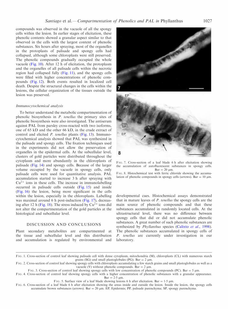
Between 1.5±2 h after spraying with copper sulphate,brown lesions appeared on the leaf surfaces. The colour ofthe lesions turned darker after 3±4 h of elicitation (Fig. 5).Light microscopy analyses of the leaf surface showed thatthe cells localized within the lesion were depressed.However, scanning and transmission electron microscopyrevealed that the wax deposits and stoma structure were notaltered (data not shown). Light microscopy of cross-sections showed that the spongy cells accumulated brown-ish substances (Fig. 6). Fluorescence microscopy of the leafsurface and transverse sections showed an accumulation ofyellow-green auto¯uorescent substances, thought to bephytoalexins, mainly in the cells in the necrotic region.The auto¯uorescent substances were observed 3±4 h afterelicitation, reaching a maximum at 12 h (Fig. 7). Histo-chemical staining of transverse sections of the leaf bladewith FeCl3 showed that these substances corresponded tophenolic compounds. The increase of these substances wasobserved only in the spongy cells within the lesion (Fig. 8).
Three hours after spraying with copper sulphate, theorganelles of the palisade cells within the lesion started
to collapse (Fig. 9). A gradual increase of phenolic
1 2
3 4
56
PG
SG
SG
SG
SG
CL
CL CL
M
MSG
SG
PG
SG
SG
PG
CL
SG
CL
PG
V
PC
EP
SP
PP
EP
1026 Santiago et al.ÐCompartmentation of Phenolics and PAL in Phyllanthus
7
of Phenolics and PAL in Phyllanthus 1027
compounds was observed in the vacuole of all the spongycells within the lesion. In earlier stages of elicitation, thesephenolic contents showed a granular aspect similar to thatobserved in the cells with the largest content of phenolicsubstances. Six hours after spraying, most of the organellesin the protoplasts of palisade and spongy cells hadcollapsed, although some chloroplasts were still preserved.The phenolic compounds gradually occupied the wholevacuole (Fig. 10). After 12 h of elicitation, the protoplasmand the organelles of all palisade cells within the necroticregion had collapsed fully (Fig. 11), and the spongy cellswere ®lled with higher concentrations of phenolic com-pounds (Fig. 12). Both events resulted in localized celldeath. Despite the structural changes in the cells within thelesions, the cellular organization of the tissues outside the
Santiago et al.ÐCompartmentation
lesion was preserved.
8
PP
EP
SP
EP
FIG. 7. Cross-section of a leaf blade 6 h after elicitation showingthe accumulation of auto¯uorescent substances in spongy cells.
Bar � 20 mm.FIG. 8. Histochemical test with ferric chloride showing the accumu-lation of phenolic compounds in spongy cells (arrows). Bar � 10 mm.
Immunocytochemical analysis
To better understand the metabolic compartmentation ofphenolic biosynthesis in P. tenellus the primary sites ofphenolic biosynthesis were also investigated. The antiserumagainst PAL from parsley cross-reacted with two isoforms,one of 65 kD and the other 66 kD, in the crude extract ofcontrol and elicited P. tenellus plants (Fig. 13). Immuno-cytochemical analysis showed that PAL was synthesized inthe palisade and spongy cells. The ®xation techniques usedin the experiments did not allow the preservation oforganelles in the epidermal cells. At the subcellular level,clusters of gold particles were distributed throughout thecytoplasm and more abundantly in the chloroplasts ofpalisade (Fig. 14) and spongy cells. Because of the largervolume occupied by the vacuole in spongy cells, onlypalisade cells were used for quantitative analysis. PALaccumulation started to increase 3 h after spraying withCu2� ions in these cells. The increase in immunolabellingoccurred in palisade cells outside (Fig. 15) and inside(Fig. 16) the lesion, being more signi®cant in the cellswithin the lesion, especially in the chloroplasts. Labellingwas maximal around 6 h post-induction (Fig. 17), decreas-ing after 12 h (Fig. 18). The stress induced by Cu2� ions didnot alter the compartmentation of the gold particles at the
histological and subcellular level.DISCUSSION AND CONCLUSIONS
Plant secondary metabolites are compartmented atthe tissue and subcellular level and this distribution
and accumulation is regulated by environmental andFIG. 1. Cross-section of control leaf showing palisade cell with dense cygrains (SG) and small plastog
FIG. 2. Cross-section of control leaf showing spongy cells with chloroplasvacuole (V) without phenolic
FIG. 3. Cross-section of control leaf showing spongy cells withFIG. 4. Cross-section of control leaf showing spongy cells with a high
Bar � 2.
FIG. 5. Surface view of a leaf blade showingFIG. 6. Cross-section of a leaf blade 6 h after elicitation showing the a
accumulate brown substances (arrows). Bar � 20 mm. EP, Epi
developmental cues. Histochemical assays demonstratedthat in mature leaves of P. tenellus the spongy cells are themain source of phenolic compounds and that thesesubstances accumulated in randomly located cells. At theultrastructural level, there was no di�erence betweenspongy cells that did or did not accumulate phenolicsubstances. A great number of such phenolic substances aresynthesized by Phyllanthus species (Calixto et al., 1998).The phenolic substances accumulated in spongy cells ofP. tenellus are currently under investigation in our
laboratory.toplasm, mitochondria (M), chloroplasts (CL) with numerous starchlobules (PG). Bar � 2 mm.ts accumulating a few starch grains and small plastoglobules as well as acompounds. Bar � 2 mm.low concentration of phenolic compounds (PC). Bar � 5 mm.er concentration of phenolic substances with a granular appearance.5 mm.lesions 6 h after elicitation. Bar � 1.5 mm.reas inside and outside the lesion. Inside the lesion, the spongy cellsdermis; PP, palisade parenchyma; SP, spongy parenchyma.

9 10
11 12
PC
PC
SP
PPEP
EP
PPSP
SGSG
PC
SG
SGSG
SG
CL
PG
MSG
SG
SGSG
SG
CL PG
M CL
SGSG
CL
PP
SP
SG
PC
SG
FIG. 9. Cross-section of a leaf blade 3 h after elicitation, showing the beginning of the collapse of organelles in palisade cells. Bar � 2 mm.FIG. 10. Cross-section of a leaf blade 6 h after elicitation, showing the collapse of the organelles in palisade and spongy cells as well as the increase
in phenolic compounds. Bar � 5 mm.FIG. 11. Cross-section of a leaf blade 12 h after elicitation, showing collapsed parenchyma cells and the highest concentration of phenolic
compounds in spongy cells. Bar � 5 mm.FIG. 12. View of organelles in totally collapsed palisade and spongy cells, and spongy cells with high concentrations of phenolic substances, 12 h
after elicitation. Bar � 5 mm.
1028 Santiago et al.ÐCompartmentation of Phenolics and PAL in Phyllanthus

kD
6665
FIG. 13. Western immunoblot analysis showing the cross-reaction ofanti-PAL parsley antibody with P. tenellus crude extract.
Santiago et al.ÐCompartmentation o
The crude extract of P. tenellus leaves cross-reacted withanti-PAL from parsley, and two isoforms of 65 and 66 kDwere identi®ed. The cytoplasm and chloroplasts ofpalisade and spongy cells were the main sites of PALsynthesis. Previous studies reported the occurrence of PALisoforms in epidermal cells of Petroselium crispumleaves (Jahnen and Hahlbrock, 1988) and in parenchy-matic cells adjacent to the xylem of Phaseolus vulgarishypocotyl (Smith et al., 1994). In the current study,the di�erent ®xation method did not preserve theP. tenellus epidermal tissue and the parenchymatic cellsof the xylem were not analysed. The synchronousaccumulation of both PAL and phenolic substances,observed in spongy cells of P. tenellus, was also observedin epidermal tissue of parsley leaves (Schmelzer et al.,1988; Neumann et al., 1991). However, this occurrencemay not be totally correlated, since in P. tenellus, palisadecells and some spongy cells accumulating PAL did notstore phenolic substances. This led to the supposition thatPAL isoforms might have been synthesized, but remainedinactive in these cells, or if they were active, that they tookpart in the synthesis of precursors which would betransported to the cell wall and/or to other cells later, asproposed for UV-irradiated oat leaves (Knooge andWeissenbock, 1986). Previous work suggested that PALisoforms localized in palisade cells play a role in thesynthesis of anthocyanins and ¯avonol glycosides inepidermal Pisum sativum cells (Hrazdina et al., 1982).However, histochemical tests on fresh tissue of P. tenellusleaves indicated that phenolic substances are restricted tospongy cells.
The phytotoxicity of copper sulphate induced thesimultaneous increase of PAL and auto¯uorescent phenoliccompounds characteristic of phytoalexins (Dixon, 1986;
Nicholson and Hammerschmidt, 1992) in spongy cells,producing necrotic lesions. It is known that tannins and¯avonoids are amongst the main phenolic substancesinduced in a plant's defence response to abiotic stress(van Tunen and Mol, 1991; Booker et al., 1996). Despitethis, high concentrations of these substances are toxic andlethal to the plant cell, leading to the occurrence ofmicrolesions (Dixon, 1986; Chessin and Zipf, 1990).
Microlesions, as well as the increase of PAL and phyto-alexins have also been shown in Arabidopsis leaves sprayedwith lead nitrate (Lummerzheim et al., 1995). However,the plant response to the phytotoxicity of heavy metalsseems to be heterogeneous since microlesions were alsoinduced in P. tenellus leaves after spraying with CdCl2 ,but not after spraying with 0.01±0.05% PbNO3 or HgCl2(data not shown). In contrast to Cu2� ions, these metalions promoted plant defoliation a few minutes afterelicitation. The di�erence in the e�ects of metal ions onplant cells remains to be explained, but it seems to berelated to their binding to di�erent membrane proteins(Woolhouse, 1983; Renwrantz et al., 1998; Zhang et al.,1999).
Both the increases of phenolic compounds andthe formation of punctated lesions in P. tenellus aresimilar to the hypersensitive response (HR) when localizedcell death occurs in response to pathogen infection(Collinge and Slusarenko, 1987; Baker and Orlandi,1995). Cytological studies have shown that cell death mayresult in necrotic or apoptotic cells during HR (Lamband Dixon, 1997).
The loss of compartmentation, also reported as one ofthe ®rst events in cell death induced by Cu2� ions (Meyerand Heath, 1988), and the synthesis of phytoalexins wererelated to the alteration in the plasma membrane(Atkinsons et al., 1990; Atkinsons, 1993). This may be aconsequence of the phytotoxicity of heavy metals, whichmay also induce oxidative stress in plants and activate theincrease of lipoxygenases involved in lipid peroxidation(Price et al., 1994; Gallego et al., 1996). The in¯uence ofCuSO4 on the generation of active oxygen species, such asH2O2 in plants has been suggested by Luna et al. (1994)and the HR is thought to be in¯uenced by H2O2production (Levine et al., 1994). The oxygenated fattyacids produced by the activity of peroxidases, phospho-lipases and lipoxygenases were considered endogenouselicitors induced during the plant defence responseassociated with phytoalexins (Kodama and Akatsuka,1991; Goodman and Novacky, 1994). This led to thesuggestion that the oxidative burst induced by bothpathogen attack and the in¯uence of metal ions mayactivate a common route during the signalling pathway ofcell death that leads to necrosis. Dypburkt et al. (1994)suggested that lower levels of active oxygen species lead toapoptosis, while severe oxidative stress may inducenecrosis.
According to our results, the palisade and spongytissues of P. tenellus are the main sites of the PAL enzyme,but phenolic compounds only accumulated in spongycells. Furthermore, it was demonstrated that Cu2� ionsinduced an increase in PAL in palisade cells while
f Phenolics and PAL in Phyllanthus 1029
phenolic compounds increased in spongy cells, giving

1514
1716
FIG. 14. Immunocytochemical analysis of PAL in the palisade cells of control leaves, showing labelling (arrows) mainly in the cytosol and in thechloroplasts. Bar � 0.5 mm.
FIG. 15. Immunolabelling of PAL in palisade cells 3 h after elicitation. The increase in labelling (arrows) outside the lesion was slight.Bar � 0.75 mm.
FIG. 16. Immunolabelling of PAL in palisade cells 3 h after elicitation. The increase in labelling (arrows) was higher inside the lesion, mostly inthe chloroplasts. Bar � 0.5 mm.
FIG. 17. Increased PAL accumulation (arrows) in chloroplasts of palisade cells inside the lesion 6 h after elicitation. Bar � 0.5 mm.
1030 Santiago et al.ÐCompartmentation of Phenolics and PAL in Phyllanthus
rise to necrotic punctate regions, leading to `HR-like'symptoms. This elicitor may be a useful tool in theunderstanding of the regulation of biosynthetic phe-nolic pathways in Phyllanthus tenellus and the diver-sity of plant defence responses against biotic and abiotic
stress.ACKNOWLEDGEMENTS
The authors are very grateful to Dr Klaus Hahlbrock, Max-Plank-Institut fuÈ r Zuchtungsforschung, KoÈ ln, Germany,for supplying anti-PAL antiserum from parsley (Petro-
selium crispum). Financial support: Conselho Nacional
Control 3 h 6 h 12 h
60
50
40
30
20
10
0
Nu
mbe
r of
gol
d pa
rtic
les
per
µm2
outside lesioninside lesion
FIG. 18. Number of colloidal gold particles per mm2 (relating to theconcentration of PAL enzyme) in palisade cells of normal and inducedleaves (n � 30) of Phyllanthus tenellus using parsley anti-PAL antibody
Santiago et al.ÐCompartmentation o
de Desenvolvimento Cientõ ®co e Tecnolo gico (CNPq),FundacË aÄ o Estadual do Norte Fluminense (FENORTE)
diluted 1 : 200. The vertical bars indicate standard deviations.
and FundacË aÄ o Universita ria Jose Bonifa cio (FUJB).
LITERATURE CITED
Aertis RJ, Gisi D, De Carolis E, De Luca V, Baumann TW. 1994.Methyl jasmonate vapor increases the developmentally controlledsynthesis of alkaloids in Catharanthus and Cinchona seedlings. ThePlant Journal 5: 635±643.
Atkinsons MM. 1993. Molecular mechanisms of plant pathogenrecognition by plants. Advances in Plant Pathology 10: 36±64.
Atkinsons MM, Kepler LD, Orlandi EW, Baker CJ. 1990. Involvementof plasma membrane Ca�� in¯ux in bacterial induction of theK�/H� and hypersensitive responses in tobacco. Plant Physiology92: 215±221.
Baker CJ, Orlandi EW. 1995. Active oxygen in plant pathogenesis.Annual Review of Phytopatology 33: 299±321.
Blumberg BS, Millman I, Venkateswaran PS, Thyagarajan SP. 1989.Hepatitis virus and hepatocellular carcinomaÐtreatment of HBVcarriers with Phyllanthus amarus. Cancer Detection and Prevention14: 195±201.
Booker FL, Antonnen S, Heagle AS. 1996. Catechin, and lignincontents of loblolly pine (Pinus taeda L.) needles after chronicexposure to ozone. New Phytologist 132: 483±492.
Braun J, Tevini M. 1993. Regulation of UV-protective pigmentsynthesis in the epidermal layer of rye seedlings (Secale cerealeL. cv. Kustro). Photochemistry and Photobiology 57: 318±323.
Calixto JB, Santos AR, Cechinel FilhoV, Yunes RA. 1998. A review ofthe plants of the genus Phyllanthus their chemistry, pharmacologyand therapeutic potential. Medicinal Research Reviews 18:225±258.
Calixto JB, Santos AR, Paulino N, Cechinel Filho V, Yunes RA. 1997.The plants of the genus Phyllanthus as a potential source of newdrugs. CieÃncia e Cultura Journal of the Brazilian Association for theAdvancement of Science 49: 422±432.
Chessin M, Zipf A. 1990. Alarm system in higher plants. BotanicalReview 56: 193±235.
Collinge DB, Slusarenko AJ. 1987. Plant gene expression in response topathogens. Plant Molecular Biology 9: 389±410.
Coronado C, Zuanazzi JAS, Sallaud C, Quirion J, Esnault R, HussonH, Kondorosi A, Ratet P. 1995. Alfafa root ¯avonoid production isnitrogen regulated. Plant Physiology 108: 533±542.
Curir P, VanSumere CF, Termini A, Barthe P, Marchesini A, Dolci M.1990. Flavonoid accumulation is correlated with adventitious rootformation in Eucalyptus gunii Hook micropropagated throughaxillary bud stimulation. Plant Physiology 92: 1148±1153.
Dalkin K, Edwards R, Edington B, Dixon RA. 1990. Stress responses inalfafa (Medicago sativa L.). I. Induction of phenylpropanoid
biosynthesis and hydrolytic enzymes in elicitor-treated cellsuspension cultures. Plant Physiology 92: 440±446.Dixon RA. 1986. The phytoalexin response: elicitation, signalling andcontrol of host gene expression. Biological Reviews of theCambridge Philosophical Society 61: 239±291.
Dornenburg H, Knorr D. 1995. Strategies for the improvement ofsecondary metabolites production in plant cell cultures. Enzymeand Microbial Technology 17: 674±684.
Duke MV, Paul RN, Elsohly HN, Sturtz G, Duke SO. 1994.Localisation of artemisin and artemisine in foliar tissues ofglanded and glandless biotypes of Artemisia annua L. InternationalJournal of Plant Science 155: 365±372.
Dypburkt JM, Ankarcrona M, Burkitt M, Sjoholm A, Strom K,Orrenius S, Nicotera P. 1994. Di�erent pro-oxidant levelsstimulate growth, trigger apoptosis or produce necrosis of insulinsecreting RINm5F cells. Journal of Biological Chemistry 269:30553±30560.
Eckey-Kaltenbach H, Ernst D, Heller W, Sandermann H Jr. 1994.Biochemical plant response to ozone. IV Cross-induction ofdefensive pathways in parsley (Petroselium crispum L.) plants.Plant Physiology 104: 67±74.
El-Mekkawy S, Meselhy RM, Kusumoto IT, Kadota S, Hattori M,Namba T. 1995. Inhibitory e�ects of egyptian folk medicines onhuman immunode®ciency virus (HIV) reverse transcriptase.Chemical and Pharmaceutical Bulletin 43: 641±648.
Gallego SM, Benavides MP, Tomaro ML. 1996. E�ect of heavy metalion excess on sun¯ower leavesÐevidence for involvement ofoxidative stress. Plant Science 121: 151±159.
Geissman TA, Crout DHG. 1969. Organic chemistry of secondarymetabolism. San Francisco, USA: Freeman, Cooper & Company.
Goodman RN, Novacky AJ. 1994. The hypersensitive reaction in plantsto pathogens. St Paul, USA: American Phytopathological SocietyPress.
Gottlieb OR. 1982. Micromolecular evolution, systematics and ecology.Berlin, Heidelberg: Springer-Verlag.
Graham TL. 1991. Flavonoid and iso¯avonoid distribution indeveloping soybean seedling tissues and in seed and root exudates.Plant Physiology 95: 594±603.
Hahlbrock K, Schell D. 1989. Physiology and molecular biology ofphenylpropanoid metabolism. Annual Review of Plant Physiologyand Plant Molecular Biology 40: 347±369.
Hanson KR, Havir EA. 1981. Phenylalanine ammonia-lyase. In:Stumpf PK, Conn EE, eds. The biochemistry of plants: a compre-hensive treatise Vol. 17. New York: Academic Press, 577±625.
Hrazdina G, Marx GA, Hoch HC. 1982. Distribution of secondaryplant metabolites and their biosynthetic enzymes in pea (Pisumsativum L.) leaves. Plant Physiology 70: 745±748.
Jacobs M, Rubery PH. 1988. Naturally occurring auxin transportregulators. Science 241: 346±349.
Jahnen W, Hahlbrock K. 1988. Di�erential regulation and tissue-speci®c distribution of enzymes of phenylpropanoid pathways indeveloping parsley seedlings. Planta 173: 453±458.
Johansen AD. 1940. Plant microtechnique. New York, London:McGraw-Hill Book Co.
Karwatzki B, Herget A, Beerhues L, Wiermann R. 1993. In situlocalization of chalcone synthase in tannin-containing plants.Phytochemistry 32: 585±590.
Kessmann H, Eduards R, Geno PW, Dixon RA. 1990. Stress responsesin alfafa (Medicago sativa L.) V. Constitutive and elicitor-inducedaccumulation of iso¯avonoid conjugates in cell suspensioncultures. Plant Physiology 94: 227±232.
Kodama WXLO, Akatsuka T. 1991. Role of oxygenated fatty acids inrice phytoalexin production. Agricultural and Biological Chemistry55: 1041±1047.
Knooge W, Weissenbock G. 1986. Tissue-distribution of secondaryphenolic biosynthesis in developing primary leaves of Avena sativaL. Planta 167: 196±205.
Kuster RM, Parente JP, Mors WB. 1994. Extrato metanoÂlico dePhyllanthus klotzschianus, como um potente antõÂdoto de veneno decobra. XIII Simpo sio de Plantas Medicinais do Brasil. Fortaleza,Ceara .
f Phenolics and PAL in Phyllanthus 1031
Laemmli UK. 1970. Cleavage of structural proteins during assembly ofthe head of bacteriophage T4. Nature 227: 680.

o
Lamb C, Dixon RA. 1997. The oxidative burst in plant diseaseresistance. Annual Review of Plant Physiology and Plant MolecularBiology 48: 251±275.
Levine A, Tenhaken R, Dixon R. 1994. H2O2 from the oxidative burstorchestrates the plant hypersensitive disease resistance response.Cell 79: 583±593.
Lummerzhein M, Sandroni M, Castresana C, de Oliveira D, Roby D,Van Montagu M. 1995. Comparative microscopy and enzymaticcharacterization of the leaf necrosis induced in Arabidopsisthaliana by lead nitrate and by Xantomonas campestris pv.campestris after lead nitrate spray. Plant, Cell and Environment18: 488±509.
Luna CM, Gonza lez CA, Trippi VS. 1994. Oxidative damage caused byexcess copper in oat leaves. Plant and Cell Physiology 35: 11±15.
Marques IA, Brodelius PE. 1988. Elicitor-induced L-tyrosine de-carboxylase from plant cell suspension cultures. Plant Physiology88: 46±51.
Meyer SLF, Heath MC. 1988. A comparision of the death induced byfungal invasion or toxic chemicals in cowpea epidermal cells. I.Cell death induced by heavy metal salts. Canadian Journal ofBotany 66: 613±623.
Neumann G, El-Ascheker A, Schwemmle B. 1991. L-Phenylalanineammonia-lyase and chalcone synthase in seedlings of Oenothera:plasmotype dependent regulation and tissue speci®c distribution.Journal of Plant Physiology 138: 263±269.
Nicholson RL, Hammerschmidt R. 1992. Phenolic compounds and theirrole in disease resistance. Annual Review of Phytopathology 30:369±389.
Paiva NL, Oommen A, Harrison MJ, Dixon RA. 1994. Regulation ofiso¯avonoid metabolism. Plant Cell Tissue and Organ Culture 38:213±220.
Parry AD, Tiller SA, Edwards R. 1994. The e�ects of heavy metals androot immersion on iso¯avonoid metabolism in alfafa (Medicagosativa L.). Plant Physiology 106: 195±202.
Pettit GR, Schaufelberger DE, Nieman RA, Dufresne C, Saenz-RenauldJA. 1990. Antineoplastic agents, 177. Isolation and structure ofPhyllanthostatin. Journal of Natural Products 53: 1406±1413.
Pio-Correà a M. 1974. DicionaÂrio das Plantas U teis do Brasil e dasExoÂticas Cultivadas Vol. 5. Rio de Janeiro, Brasil: Desenvolvi-mento Florestal, Ministe rio da Agricultura.
Price AH, Taylor A, Ripley SJ, Gri�ths A, Trewavas AJ, Knight MR.1994. Oxidative signals in tobacco increase cytosolic calcium. ThePlant Cell 6: 1301±1310.
1032 Santiago et al.ÐCompartmentation
Renwrantz L, Schmalmack W, Steenbuck M. 1998. Molecular size ofnative proteins of Mytilus serum which contains a dominant
fraction with heavy metal-binding properties. Comparative Bio-chemistry and Physiology 121: 175±180.
Reynolds ES. 1963. The use of lead citrate at high pH as an electron-opaque stain in electron microscopy. Journal of Cell Biology 17:208±212.
Santos ARS, Filho VC, Niero R, Viana AM, Moreno FN, Campos FN,Campos MM, Yunes RA, Calixto JB. 1994. Analgesic e�ects ofcallus culture extracts from selected species of Phyllanthus in mice.Journal of Pharmacy and Pharmacology 46: 755±759.
Santos I, Almeida JM, Salema R. 1993. Plants of Zea mays L.developed under enhanced UV-B radiation. I. Some ultrastruc-tural and biochemical aspects. Journal of Plant Physiology 141:450±456.
Schmelzer E, Hahnen W, Hahlbrock K. 1988. In situ localisation oflight-induced chalcone synthase mRNA, chalcone synthase,¯avonoid end products in epidermal cells of parsley leaves.Proceedings of the National Academic of Sciences, USA 85:2989±2993.
Smith CG, Rodgers MW, Zimmerlin A, Ferdinando D, Bolwell GP.1994. Tissue and subcellular immunolocalisation of enzymes oflignin synthesis in di�erentiating and wounded hypocotyl tissue ofFrench bean (Phaseolus vulgaris L.). Planta 192: 155±164.
Spurr AR. 1969. A low-viscosity epoxy resin embedding medium forelectron microscopy. Journal of Ultrastructure Research 26: 31±43.
Turlings TCJ, Loughrin JH, McCall PJ, Rose USR, Lewis WJ. 1995.How caterpillar-damaged plants protect themselves by attractingparasitic wasps. Proceedings of the National Academy of Sciences,USA 92: 4169±4174.
van Tunen AJ, Mol JNM. 1991. Control of ¯avonoid synthesis andmanipulation of ¯ower colour. In: Blackie DG, ed. Developmentalregulation of plant gene expression, Plant Biotechnology Series.New York: Chapman and Hall.
Venkateswaran PS, Millaman I, Blumberg EBS. 1987. E�ects of anextract from Phyllanthus niruri on hepatitis B and woodchuckhepatitis viruses: in vitro and in vivo studies. Proceedings of theNational Academy of Sciences, USA 84: 274±278.
Vogt T, Pollak P, Tarlyn N, Taylor LP. 1994. Pollination and wound-induced kaempferol accumulation in Petunia stigmas enhancesseed production. The Plant Cell 6: 11±23.
Woolhouse HW. 1983. Toxity and tolerance in the responses of plantsto metals. Encyclopedia of Plant Physiology. New Series 12C:245±300.
Zhang YX, Chai TY, Burkard G. 1999. Research advances of the
f Phenolics and PAL in Phyllanthus
mechanisms of heavy metal tolerance in plants. Acta BotanicaSinica 41: 453±457.