Proteomic analysis of cell suspension cultures of Boesenbergia rotunda induced by phenylalanine:...
11
ORIGINAL PAPER Proteomic analysis of cell suspension cultures of Boesenbergia rotunda induced by phenylalanine: identification of proteins involved in flavonoid and phenylpropanoid biosynthesis pathways Eng Chong Tan • Saiful Anuar Karsani • Gen Teck Foo • Sher Ming Wong • Noorsaadah Abdul Rahman • Noorzulaani Khalid • Shatrah Othman • Rohana Yusof Received: 14 February 2012 / Accepted: 2 June 2012 Ó Springer Science+Business Media B.V. 2012 Abstract Boesenbergia rotunda belongs to the Zingi- beraceae family. It is widely found throughout Southeast Asia and is commonly used as a food ingredient and in folk medicine. Extracts from this plant contain a number of important bioactive compounds such as boesenbergin, cardamonin, pinostrobin, pinocembrin, panduratin A and 4-hydroxypanduratin A. These compounds have been shown to exhibit anti-HIV protease, anti-dengue NS2B/ NS3 protease, antibacterial, antifungal, anti-inflammatory, anticancer, and antioxidant activity. Here we report the use of proteomic approaches to identify proteins that may be involved in the biosynthesis of these compounds. Protein expressions of B. rotunda suspension cultures for phenyl- alanine-treated and normal callus were compared by two- dimensional gel electrophoresis. Following image analysis, protein spots whose expressions were found to be regulated were identified using Matrix Assisted Laser Desorption- Ionization tandem mass spectrometry. In all, thirty four proteins were identified. These proteins were categorized into nine functional categories—defence mechanism, pro- tein biosynthesis, metabolism, terpenoid biosynthesis, cell division, cell organization, energy-related, signaling pro- cesses and proteins of unknown function. Eleven of the proteins involved in the phenylpropanoid biosynthetic pathway are related to the biosynthesis of cyclohexenyl chalcone derivatives. Keywords Boesenbergia rotunda Á Flavonoids Á Panduratin A Á Proteomics Á 4-Hydroxypanduratin A Introduction Boesenbergia rotunda is a small perennial plant belonging to the Zingiberaceae family. It is widely found in South East Asian countries with local names such as Chinese keys, finger root, and temu kunci. It is commonly used as a food ingredient and folk medicine to treat diseases such as aphthous ulcer, stomach discomfort, leucorrhea, dysentery, rheumatism and muscular pain. It has been shown that the primary bioactive compounds of this ginger are boesen- bergin, cardamonin, pinostrobin, pinocembrin, panduratin Electronic supplementary material The online version of this article (doi:10.1007/s11240-012-0188-8) contains supplementary material, which is available to authorized users. E. C. Tan (&) Á S. Othman Á R. Yusof (&) Department of Molecular Medicine, Faculty of Medicine, University of Malaya, 50603 Kuala Lumpur, Malaysia e-mail: [email protected] R. Yusof e-mail: [email protected] E. C. Tan Á S. A. Karsani (&) Á N. Abdul Rahman Á S. Othman Á R. Yusof Drug Design and Development Research Group, University of Malaya, 50603 Kuala Lumpur, Malaysia e-mail: [email protected] S. A. Karsani Á G. T. Foo Á S. M. Wong Á N. Khalid Faculty of Science, Institute of Biological Sciences, University of Malaya, 50603 Kuala Lumpur, Malaysia S. A. Karsani University of Malaya Centre for Proteomics Research (UMCPR), Kuala Lumpur, Malaysia G. T. Foo Á S. M. Wong Á N. Khalid Biotechnology and Bioproduct Research Cluster (UMBIO), University of Malaya, 50603 Kuala Lumpur, Malaysia N. Abdul Rahman Department of Chemistry, Faculty of Science, University of Malaya, 50603 Kuala Lumpur, Malaysia 123 Plant Cell Tiss Organ Cult DOI 10.1007/s11240-012-0188-8
-
Upload
independent -
Category
Documents
-
view
1 -
download
0
Transcript of Proteomic analysis of cell suspension cultures of Boesenbergia rotunda induced by phenylalanine:...

ORIGINAL PAPER
Proteomic analysis of cell suspension cultures of Boesenbergiarotunda induced by phenylalanine: identification of proteinsinvolved in flavonoid and phenylpropanoid biosynthesis pathways
Eng Chong Tan • Saiful Anuar Karsani • Gen Teck Foo •
Sher Ming Wong • Noorsaadah Abdul Rahman •
Noorzulaani Khalid • Shatrah Othman • Rohana Yusof
Received: 14 February 2012 / Accepted: 2 June 2012
� Springer Science+Business Media B.V. 2012
Abstract Boesenbergia rotunda belongs to the Zingi-
beraceae family. It is widely found throughout Southeast
Asia and is commonly used as a food ingredient and in
folk medicine. Extracts from this plant contain a number
of important bioactive compounds such as boesenbergin,
cardamonin, pinostrobin, pinocembrin, panduratin A and
4-hydroxypanduratin A. These compounds have been
shown to exhibit anti-HIV protease, anti-dengue NS2B/
NS3 protease, antibacterial, antifungal, anti-inflammatory,
anticancer, and antioxidant activity. Here we report the use
of proteomic approaches to identify proteins that may be
involved in the biosynthesis of these compounds. Protein
expressions of B. rotunda suspension cultures for phenyl-
alanine-treated and normal callus were compared by two-
dimensional gel electrophoresis. Following image analysis,
protein spots whose expressions were found to be regulated
were identified using Matrix Assisted Laser Desorption-
Ionization tandem mass spectrometry. In all, thirty four
proteins were identified. These proteins were categorized
into nine functional categories—defence mechanism, pro-
tein biosynthesis, metabolism, terpenoid biosynthesis, cell
division, cell organization, energy-related, signaling pro-
cesses and proteins of unknown function. Eleven of the
proteins involved in the phenylpropanoid biosynthetic
pathway are related to the biosynthesis of cyclohexenyl
chalcone derivatives.
Keywords Boesenbergia rotunda � Flavonoids �Panduratin A � Proteomics � 4-Hydroxypanduratin A
Introduction
Boesenbergia rotunda is a small perennial plant belonging
to the Zingiberaceae family. It is widely found in South
East Asian countries with local names such as Chinese
keys, finger root, and temu kunci. It is commonly used as a
food ingredient and folk medicine to treat diseases such as
aphthous ulcer, stomach discomfort, leucorrhea, dysentery,
rheumatism and muscular pain. It has been shown that the
primary bioactive compounds of this ginger are boesen-
bergin, cardamonin, pinostrobin, pinocembrin, panduratin
Electronic supplementary material The online version of thisarticle (doi:10.1007/s11240-012-0188-8) contains supplementarymaterial, which is available to authorized users.
E. C. Tan (&) � S. Othman � R. Yusof (&)
Department of Molecular Medicine, Faculty of Medicine,
University of Malaya, 50603 Kuala Lumpur, Malaysia
e-mail: [email protected]
R. Yusof
e-mail: [email protected]
E. C. Tan � S. A. Karsani (&) � N. Abdul Rahman �S. Othman � R. Yusof
Drug Design and Development Research Group,
University of Malaya, 50603 Kuala Lumpur, Malaysia
e-mail: [email protected]
S. A. Karsani � G. T. Foo � S. M. Wong � N. Khalid
Faculty of Science, Institute of Biological Sciences,
University of Malaya, 50603 Kuala Lumpur, Malaysia
S. A. Karsani
University of Malaya Centre for Proteomics Research
(UMCPR), Kuala Lumpur, Malaysia
G. T. Foo � S. M. Wong � N. Khalid
Biotechnology and Bioproduct Research Cluster (UMBIO),
University of Malaya, 50603 Kuala Lumpur, Malaysia
N. Abdul Rahman
Department of Chemistry, Faculty of Science,
University of Malaya, 50603 Kuala Lumpur, Malaysia
123
Plant Cell Tiss Organ Cult
DOI 10.1007/s11240-012-0188-8

A and 4-hydroxypanduratin (Jaipetch et al. 1982, 1983;
Trakoontivakorn et al. 2001). These compounds exhibit
HIV protease inhibition (Tewtrakul et al. 2003a, b;
Cheenpracha et al. 2006) and possess antioxidant activity
(Sohn et al. 2005; Shindo et al. 2006), antibacterial (Bha-
marapravati et al. 2006; Mahady et al. 2006), antifungal
(Phongpaichit et al. 2005; Pattaratanawadee et al. 2006;
Pompimon et al. 2009), anti-inflammatory (Tuchinda et al.
2002; Tewtrakul et al. 2009), antitumor (Kirana et al. 2003,
2007; Yun et al. 2006) and anti-parasitic activity (Saw-
angjaroen et al. 2005). Panduratin A and 4-hydroxypan-
duratin A, have also been shown to inhibit dengue NS2B/
NS3 protease (Kiat et al. 2006).
Most pharmaceutically important plant bioactive com-
pounds are flavonoids. Flavonoids are synthesized from
phenylalanine (precursor), into cinnamic acid by phenyl-
alanine ammonia lyase (PAL). PAL catalyzes the non-
oxidative deamination of phenylalanine into cinnamic
acid. Chalcone will then be produced. It is predicted that
cyclohexenyl chalcone derivatives (CCD) are produced
in the flavonoids biosynthetic pathway. However, the
enzymes that are involved in the CCD biosynthetic path-
way derived from this main pathway (flavonoids biosyn-
thetic pathway) are not known. Thus, it is the aim of this
project to identify differences in protein expression
between normal and phenylalanine-treated cell suspensions
as these changes may represent proteins that are involved
in the CCD biosynthetic pathway.
Materials and methods
Callus induction
Explants of B. rotunda were obtained from a field in
Temerloh, Pahang, Malaysia. Sprouts were surface steril-
ized by soaking in 20 % (v/v) Clorox for 20 min and a
further one min in 70 % (v/v) ethanol. This was followed
by several rinses in sterile distilled water. Meristems were
excised and placed on MS (Murashige and Skoog) media
supplemented with 1 mg l-1 2,4-D (2,4-Dichlorophe-
noxyacetic acid), 1 mg l-1 NAA (Naphthaleneacetic acid),
1 mg l-1 IAA (Indoleacetic acid) and 2 % (w/v) Phytagel
(Sigma, US) . The pH of the medium was adjusted to 5.8
with hydrochloric acid (HCl) and/or sodium hydroxide
(NaOH) prior to autoclaving at 121 �C for 20 min. All
cultures were prepared under aseptic conditions and grown
at 26 �C under 16 h light: 8 h dark photoperiod conditions
with a light intensity of 31.4 lmol m-2 s-1 provided by a
cool fluorescent lamp. Callus induced from meristem that
emerged after 3 weeks of culturing were further propa-
gated in MS agar media supplemented with 3 mg l-1 2,4-
D. Friable calli were transferred into liquid media
supplemented with 2 mg l-1 2,4-D, 1 mg l-1 NAA,
1 mg l-1 BAP (6-Benzylaminopurine), 5 g l-1 maltose,
100 mg l-1 malt extract and 1 mg l-1 D-biotin for sus-
pension cultures establishment after 1 month of propaga-
tion. The suspension cultures were then subcultured every
fortnight and sieved through a 425 lm nylon filter to
remove big callus clumps.
The culture was then induced with phenylalanine. The
determination of optimum inoculum age and phenylalanine
concentration was performed as previously described (Tan
2005). Briefly, cultures were harvested on day 10, 14 and
21, filtered and air dried. The mass of five flavonoid
compounds were then determined and compared using
HPLC. Based on these results, induction with different
concentrations of phenylalanine (0, 20 and 40 mg/l) was
then performed by mixing with liquid media. The media
were renewed in 1:4 old media to new media ratio. Again,
the mass of five flavonoid compounds were then deter-
mined and compared using HPLC. The growth of the
suspension cell culture was then measured through Settle
Cell Volume (SCV).
Total protein extraction and precipitation
Protein extraction was performed as previously described
(Park et al. 2006; Pan et al. 2010; Wang et al. 2010; Chong
et al. 2011; Pavokovic et al. 2012). Briefly, extraction
buffer was prepared by adding 160 ll of Tris–HCl (pH 9.5,
40 mM), 100 mg Polyvinyl polypyrrolidone (PVPP), 40 ll
protease inhibitor mix and 60–63 ll (approximately 1,200
units) of nuclease mix to distilled water. This solution was
made up to a final volume of 4 ml and is sufficient for 2 g
of suspension culture (Chong et al. 2011). Total protein
was extracted from fresh and frozen B. rotunda suspension
cultures. Approximately 2 g of frozen cells were ground to
fine powder in a pre-chilled mortar with a small amount of
liquid nitrogen. Extraction buffer was then added and the
mixture was ground thoroughly until it was homogenised.
500 ll of homogenate was then aliquoted into a 2 ml
microcentrifuge tube, vortexed vigorously for 1 min and
sonicated. This was repeated three times. The homogenate
was then centrifuged twice at 5,000g for 5 min and about
350–450 ll of the supernatant was recovered. Protein
extracts were concentrated by Trichloroacetic acid (TCA)
precipitation. Briefly, 37.5 % TCA (w/v) with 1 % b-
mercaptoethanol (v/v) was added to 400 ll of sample to
achieve a final volume of 2 ml. The mixture was then
incubated at -20 �C for at least 1 h. This was followed by
centrifugation at 13,000g (4 �C for 15 min). The resulting
protein pellet was washed three times in 80 % acetone
(v/v) containing 0.05 % b-mercaptoethanol (v/v) and then
air-dried in a laminar flow hood at room temperature.
The pellet was then solubilized in isoelectric focusing
Plant Cell Tiss Organ Cult
123

buffer (8 M urea, 2 % CHAPS, 0.5 % IPG buffer (GE
Healthcare), 1 M Thiourea and trace amounts of Orange G)
and incubated at room temperature for at least 30 min. The
solubilized protein was then centrifuged (10,000g at 4 �C
for 20 min) and either used immediately or stored at
-80 �C.
2-D gel analysis
Protein concentration was determined using 2-D Quant kit
(GE Healthcare) using BSA as standards. First-dimension
IEF was performed on an Ettan IPGPhor 3 IEF System (GE
Healthcare Bio-Sciences, Uppsala, Sweden) according to
the manufacturer’s recommendations on 13 cm immobiline
drystrips (pH 3–10). Briefly, drystrips were first rehydrated
for 16 h with 450 ll of rehydration buffer containing 8 M
urea, 2 % CHAPS, 0.5 % IPG Buffer (pH 3–10) and trace
amounts of bromophenol blue, and 20 lg of protein sample
from plant. The drystrips were focused for a total of
35.5 kVh. Focused IPG strips were equilibrated for 15 min
in equilibration buffer (6 M urea, 75 mM Tris–HCl pH 8.8,
29 % glycerol, 2 % SDS and trace amounts of bromo-
phenol blue) containing 1 % DTT. It was then alkylated
in the same buffer containing 2.5 % iodoacetamide for a
further 15 min. Electrophoresis of reduced and alkylated
samples were then performed on 13 cm, 10.0 % SDS-
PAGE gels on an IPGphor III electrophoresis platform (GE
Healthcare). The gels were run at 50 mA per gel until the
bromophenol blue tracking dye reached the bottom of the
gel. Following SDS-PAGE, Protein spots were visualized
using protocols described in the PlusOneTM Silver staining
kit (GE Healthcare Bio-Sciences, Uppsala, Sweden). The
complete protocol was followed for analytical gels. For
preparative gels, the protocol was modified so that glutar-
aldehyde was omitted from the sensitization step and
formaldehyde omitted from the silver reaction step. Sil-
ver-stained gels were scanned (ImageScanner III, GE
Healthcare Bio-Sciences, Uppsala, Sweden) and protein
profiles compared (Image Master Platinum software 7.0,
GE Healthcare Bio-Sciences, Uppsala, Sweden). Gel ima-
ges were analyzed using the Image Master Platinum soft-
ware 7.0 (GE Healthcare Bio-Sciences, Uppsala, Sweden).
Protein expression between the normal and phenylalanine-
treated callus was compared. A total of 4 gels were run for
each group (quadruplicates) for gels pH 3–10, and, 6 gels
for each group for gels pH 4–7. This was performed to
eliminate experimental and biological variations. To fur-
ther select against variations between individual samples,
the selection criteria for spots were very stringent. Firstly,
gels were compared within their respective groups and a
representative (virtual) gel was generated. For the genera-
tion of the virtual gel, only spots present across all gels
within the groups were considered. This was performed to
eliminate differences due to inter-individual gel variations.
The virtual gels were then compared between the different
samples to identify differentially expressed proteins. Only
spots that were found to be differentially expressed by
more than two folds were accepted for further consider-
ation. Statistical analysis of all spots that were found to
be differentially expressed was then performed using
the student’s t test. A P value \0.05 was considered as
statistically significant.
Protein identification
Protein spots were excised and in-gel digested with trypsin
(Promega) for mass spectrometric analysis according to
published protocols (Shevchenko et al. 1996; Wilm et al.
1996). Briefly, excised spots were first destained in
destaining solution (15 mM potassium ferricyanide/50 mM
sodium thiosulphate, 1:1 [v/v]). The spots were then
reduced in a solution containing 10 mM DTT/100 mM
ammonium bicarbonate for 30 min at 60 �C and alkylated
in 55 mM iodoacetamide/100 mM ammonium bicarbonate
for 20 min in the dark. The gel pieces were then washed
(39 20 min) in 50 % acetonitrile/ 100 mM ammonium
bicarbonate. This was followed by dehydration of the gel
pieces with 100 % acetonitrile and drying in a vacuum
centrifuge (SpeedVac, Thermo Scientific, Savant DNA
120). Subsequently the dried gel pieces were rehydrated
with 25 ll of 7 ng/ll trypsin (Promega trypsin gold) in
50 mM ammonium bicarbonate buffer and digested at
37 �C for 18–20 h. Tryptic peptides were then extracted
using 50 % acetonitrile for 15 min, followed by 100 %
acetonitrile for 15 min. The extracted solutions were then
pooled into a single tube and dried in a SpeedVac con-
centrator and solubilized with 10 ll of 10 % acetonitrile/
40 mM ammonium bicarbonate (Shevchenko et al. 1996;
Wilm et al. 1996).
Protein identification was essentially performed as pre-
viously described (Dahlan et al. 2011). Briefly, extracted
peptides were first desalted using ZipTip C18 (Millipore,
USA) according to protocols described by the manufac-
turer. The final elution volume following ZipTip cleanup
was 1.5 ll. The peptide samples were then mixed (1:1) with
a matrix consisting of a saturated solution of CHCA (a-
cyano-4-hydroxycinnamic acid, Sigma) prepared in 50 % 6
ACN/0.1 % TFA. Aliquots of samples (0.7 ll) were spotted
onto stainless-steel sample target plates. Peptide mass
spectra were obtained on a MALDI-TOF/TOF mass spec-
trometer (ABI 4800 plus, Applied Biosystems) in the
positive ion reflector mode. For precursor ion selection, all
fractions were measured in single MS before MS/MS was
performed. For MS/MS spectra, the peaks were calibrated
by default. The 20 most abundant precursor ions per sample
were selected for subsequent fragmentation by high energy
Plant Cell Tiss Organ Cult
123

CID. The collision energy was set to 1 keV and air was used
as the collision gas. The criterion for precursor selection
was a minimum S/N of 5. The mass accuracy was within
50 ppm for the mass measurement and within 0.1 Da for
CID experiments. The other parameters for searching were
of trypsin, 1 missed cleavage, variable modification of
carbamidomethyl and oxidation of methionine, peptide
charge of 1?, and monoisotopic. For database searches,
known contamination peaks such as keratin and autopro-
teolysis peaks for trypsin were removed before searching.
Spectra were processed and analyzed by the Global Protein
Server Explorer 3.6 software (Applied Biosystems). This
uses an internal MASCOT (Matrix Science, UK) program
for matching MS and MS/MS data against database infor-
mation. The data obtained were screened against plant dat-
abases downloaded from the Swiss-Prot/TrEMBL homepage
(http://www.expasy.ch/sprot).
Results and discussion
Effect of inoculum age and phenylalanine concentration
towards compounds production in B. rotunda
suspension cultures
The amount of flavonoids was highest in cultures harvested
on the 14th day (Table 1). The masses of pinotrobin,
alpinetin, 4-hydroxypanduratin A and panduratin A of 14th
day cultures were all three digit percent higher than those
of the 10th day culture, only pinocembrin recorded a 69 %
increase. The largest gain was alpinetin at 694 % increase
followed by panduratin A at 586 % increase. Compound
masses from 21st day cultures showed a decline of all five
compounds when compared to 14th day cultures (Table 1).
These results indicated that 14th day cultures gave the
highest yield for all the five flavonoids when no fresh
medium was used. Hence cultures were harvested on the
14th day for all subsequent investigations to ensure highest
possible yield. It was also found that the level of flavonoids
increased dose-dependently with increasing phenylalanine
concentration. Table 2 is a tabulation of the mean masses
obtained. It was apparent that supplementing standard
culture media with 40 mg/l of phenylalanine brought about
a high yield of most B. rotunda flavonoids with an increase
of between 188 to over 1,000 % increase is mass yield.
Thus, for the proteomics experiments, all cultures were
harvested at the 14th day with phenylalanine supplemen-
tation at 40 mg/l.
Proteomics analysis
A total of *1,000 (pH 3–10 2DE gels) and *2,000 (pH
4–7 gels) individual protein spots were resolved on silver
stained 2DE gels. Representative gels are shown in Fig. 1a,
b. The gels were found to be highly reproducible with little
variation in terms of spot profile and numbers. Following
image analysis, 76 proteins were found to be differently
expressed. Out of these, 34 were identified by matrix-
assisted laser desorption/ ionization tandem time of flight
(MALDI TOF/ TOF). Eleven proteins were found to be
associated with the phenylpropanoid biosynthesis pathway.
Despite the inavailability of B. rotunda genome and protein
databases, we were able to identify 45 % of the selected
protein spots. Thirty-four proteins were identified and they
were categorized into nine functional groups which were
proteins involved in plant defence mechanism, amino acids
and protein synthesis, metabolism, terpenoid biosynthesis,
cell division, cytoskeleton (cell organization), energy-
related, signaling processes and proteins of unknown
function. The identities of these proteins are shown in
Supplementary Table 1. From this point forward these
proteins will be referred to by their abbreviated names as
shown in Supplementary Table 1. Here we discuss the
proteins that were found to be differentially expressed
under the various functional categories.
Table 1 Effect of inoculum age on five selected flavanoids
Compounds Compound mass (lg ± SE) and percentage mass change based on 10-day harvest mass as standard
Harvest age in days
10 14 21
Mass Mass % Change Mass % Change
Pinostrobin 1.374 ± 0.009 6.032 ± 0.029 350.15 ± 4.04 4.149 ± 0.017 209.63 ± 2.27
Pinocembrin 0.789 ± 0.009 1.333 ± 0.021 68.95 ± 2.05 1.317 ± 0.006 66.92 ± 1.24
Alpinetin 0.221 ± 0.005 1.754 ± 0.006 693.67 ± 19.07 1.147 ± 0.010 419.00 ± 13.13
4-hydroxypanduratin A 0.430 ± 0.005 1.800 ± 0.020 318.60 ± 7.24 1.460 ± 0.009 239.53 ± 4.26
Panduratin A 0.042 ± 0.001 0.288 ± 0.023 585.71 ± 22.08 0.255 ± 0.000 507.14 ± 12.67
SE standard error
Plant Cell Tiss Organ Cult
123
Defense mechanism
Five proteins involved in plant defense mechanism were
found to be differentially expressed (identities shown in
Supplementary Table 1). NSH is a plant protein that con-
tains heme groups and thus has high binding, reversible
affinity towards oxygen (O2) and nitric oxide (NO). This
protein can be elevated when the plant is exposed to stress
conditions. NSH is found ubiquitously in the stems, leaves,
roots, seeds and flowers of plants. It is also found in
growing, differentiating seedlings (Perazzolli et al. 2004).
AP is an antioxidative agent. Genetically, it is highly
conserved among proteins of the heme peroxidase family
of enzymes which can be found in most life forms. It is
involved in oxidation-reduction processes. Under normal
or stress (biotic or abiotic) environments, plants produce a
quantity of radical oxygen species (ROS) from metabolic
processes. This can cause damage at the cellular level.
Therefore, antioxidative agents, such as ascorbate peroxi-
dase are needed to control the damage from oxidants. AP
has been reported to exhibit apoptotic suppression effect in
cells (de Pinto et al. 2006). Two forms of this protein were
found to be differentially expressed. HPosJ is categorised
in the low genetically conserved cytochrome P450 super-
family. It is involved in oxidative degradation processes
and are classified based on electron transfer from
NAD(P)H to the catalytic site of the protein (Kim et al.
2004; Sasaki et al. 2010). COM plays a vital role in the
production of feruloyl-CoA from caffeoyl-CoA by the
methylation of 3-hydroxyl group of caffeoyl-CoA, result-
ing in an end product, sinapoyl-CoA which a main com-
ponent in lignin production. The suppression of this
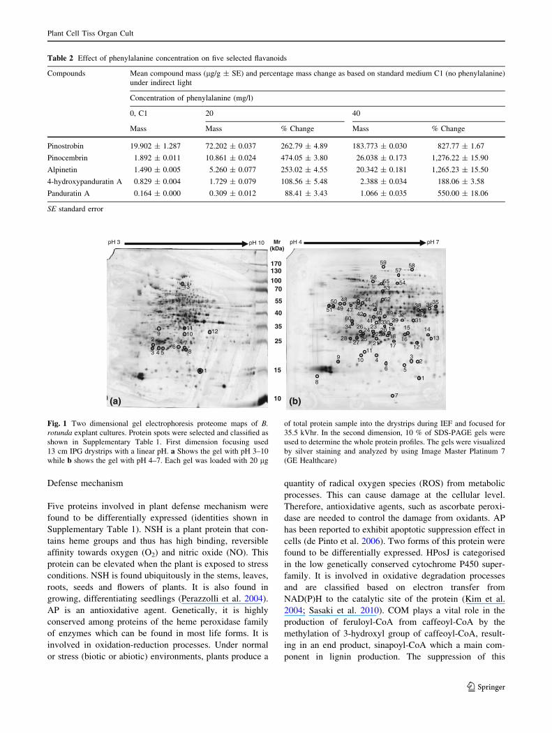
Table 2 Effect of phenylalanine concentration on five selected flavanoids
Compounds Mean compound mass (lg/g ± SE) and percentage mass change as based on standard medium C1 (no phenylalanine)
under indirect light
Concentration of phenylalanine (mg/l)
0, C1 20 40
Mass Mass % Change Mass % Change
Pinostrobin 19.902 ± 1.287 72.202 ± 0.037 262.79 ± 4.89 183.773 ± 0.030 827.77 ± 1.67
Pinocembrin 1.892 ± 0.011 10.861 ± 0.024 474.05 ± 3.80 26.038 ± 0.173 1,276.22 ± 15.90
Alpinetin 1.490 ± 0.005 5.260 ± 0.077 253.02 ± 4.55 20.342 ± 0.181 1,265.23 ± 15.50
4-hydroxypanduratin A 0.829 ± 0.004 1.729 ± 0.079 108.56 ± 5.48 2.388 ± 0.034 188.06 ± 3.58
Panduratin A 0.164 ± 0.000 0.309 ± 0.012 88.41 ± 3.43 1.066 ± 0.035 550.00 ± 18.06
SE standard error
Mr(kDa)
pH 3 pH 10
13
1110 12
1
8
7
92
3 4 56
15
10
25
55
130
40
100
35
70
170
(a)
59 5857
545556
53
5250
51
48
49 47 45
46 44
60
34
8
9
7
1
23
56
410
1112
13
1415
16
31
3536
37
38
2827
25
2426
42
43
4140 39
21
2223
322930
17
1918
2033
pH 4 pH 7
(b)
Fig. 1 Two dimensional gel electrophoresis proteome maps of B.rotunda explant cultures. Protein spots were selected and classified as
shown in Supplementary Table 1. First dimension focusing used
13 cm IPG drystrips with a linear pH. a Shows the gel with pH 3–10
while b shows the gel with pH 4–7. Each gel was loaded with 20 lg
of total protein sample into the drystrips during IEF and focused for
35.5 kVhr. In the second dimension, 10 % of SDS-PAGE gels were
used to determine the whole protein profiles. The gels were visualized
by silver staining and analyzed by using Image Master Platinum 7
(GE Healthcare)
Plant Cell Tiss Organ Cult
123
enzyme will decrease the production of lignin in plants
especially woody plants (Zhong et al. 2000). This enzyme
is also involved in the plant defense mechanism where it
triggers the production of defensive compounds, such as
phytoalexin (Yang et al. 2004). The fifth identified pro-
tein—GP—is an ROS-scavenging enzyme that catalyses
the oxidative homeostasis in plants and animals, during
biotic or abiotic stress (Faltin et al. 2010).
Amino acid and protein biosynthesis
Four proteins—PSB3, PSB6 (two different spots), MET
and PGD—were found to be regulated. Proteins under
this category play important roles in plant growth and
development. PSB3 and PSB6 (2 different forms) are the
subunits of proteosome b-type. Proteosomes are
ATP-dependent proteases that degrade intracellular miss-
folded and short-lived proteins. It is involved in cell cycle
regulation of plants and animals. Some proteosomes are
involved in the defense mechanism of plants and the
immune system in human (Santner and Estelle 2010). MET
is an enzyme involved in the biosynthesis of methionine in
animals and plants. Methionine is used in protein synthesis,
mRNA initiation process and as a regulatory molecule
(Hesse et al. 2004; Dancs et al. 2008). PGD is a multi-
conformation enzyme related to serine biosynthesis, a
process before glycolysis in plants and animals. It catalyses
the conversion of phosphoglycerate into phosphohydroxy-
pyruvate using NAD/ NADH as a cofactor (Boland and
Schubert 1983).
Plant metabolism
This group of proteins is involved in plant metabolism—
lipid metabolism, nitrogen metabolism and assimilation,
and nucleic acid metabolism. Five proteins were found to
be regulated.
Lipid metabolism
GDSL was found to be regulated. GDSL is a multifunc-
tional enzyme within the SGNH hydrolase/ esterase
superfamily. It plays a vital role in seed germination, plant
development and morphogenesis. GDSL hydrolyses phe-
nolic esters, fatty acyl-ester, lipids or fatty acids and
polysaccharides groups (Akoh et al. 2004; Clauss et al.
2008).
Nitrogen metabolism and assimilation
FNR and GS were found to be regulated. GS is a key
enzyme in nitrogen metabolism where it equalizes the
intracellular nitrogen level (Miflin and Habash 2002). This
enzyme also induces photosynthetic activity and growth
rate at low nitrogen levels (Fuentes et al. 2001). Increased
level of nitrogen in cells will also be equalized by FNR.
Nucleic metabolism-related proteins
HIS and NDK were found to be regulated. HIS is an
enzyme that regulates gene expression, specifically chro-
matin remodeling in nuclear and plant development. It is
involved in cell proliferation and differentiation, meristem
function, and organogenesis (Servet et al. 2010). NDK is a
highly conserved multiple signaling enzyme that catalyzes
the phosphorylation of nucleoside diphosphates (NDPs) to
equilibrate the amount of nucleoside triphosphate (NTPs)
for RNA, DNA and protein biosynthesis. It is also involved
in the autophosphorylation of GTP and GTPase stimulating
protein in plants. Recent studies have shown that this
enzyme is also involved in signaling processes (Hasunuma
et al. 2003; Shen et al. 2008).
Terpenoid biosynthesis
Two proteins involved in terpenoid biosynthesis—HMDPR
and DXPR—were found to be up-regulated, as shown in
Supplementary Table 1. These proteins are involved in the
non-mevalonate or 2-C-methyl-D-erythritol 4-phosphate/1-
deoxy-D-xylulose 5-phosphate (MEP/DOXP) pathway in
plant plastids, which produce isoprenoids and terpenoids,
such as, taxol. DXPR is involved in the biosynthesis of
2-C-methyl-erythritol 4-phosphate and 1-deoxy-D-xylulose
5-phosphate, while HMDPR is involved in the biosynthesis
of 1-hydroxy-2-methyl-butenyl 4-diphosphate, isopentenyl
diphosphate and dimethylallyl diphosphate (Takahashi
et al. 1998; Hasunuma et al. 2008). Furthermore, HMDPR
is currently being studied as the target in the production of
the cancer drug, taxol, from the plants of the taxus family
(Sun et al. 2009).
Cell division
Three proteins involved in cell division were found to be
regulated—STK, PP and CYC. STK is an enzyme that is
crucial in animal and plant cell division. This enzyme is
categorized under the serine/ threonine-protein kinase
superfamily which is a biological switch in microtubule
phosphorylation, metabolism, gene expression and cell
growth and division. It also functions in the defense system
by initiating the immune response in animals. Although
the proteins are highly homologous between animals and
plants, its function, particularly in plants is still poorly
understood (Hardie 1999; Reddy and Rajasekharan 2007).
CYC is a member of a large family of cyclins that is the
regulator for kinases. These proteins mediate the plant cell
Plant Cell Tiss Organ Cult
123

cycle, cell growth and differentiation, hormonal signals,
and meiosis (Cockcroft et al. 2000; d’Erfurth et al. 2010).
A predicted protein, PP was shown to be a possible tar-
geting protein from the Xklp2 (TPX2) family. These cen-
tral spindle regulator family members are kenesin-like
proteins that are localized on centrosomes and spindle pole
microtubules, before nuclear envelop breakdown and dur-
ing nuclear assembly (Vos et al. 2008; Evrard et al. 2009).
Cytoskeleton
Two different spots that were regulated were identified as
actins. Actins are involved in cytoskeleton building (cell
wall), cell division, expansion and intracellular trafficking,
all of which are important for plant morphogenesis
(Thomas et al. 2009).
Energy-related processes
Seven proteins involved in energy production were found
to be regulated as shown in Supplementary Table 1. DD
was found to be up-regulated. This enzyme functions in the
glycolysis pathway where it is located in the pyruvate-
acetyl-CoA reaction. Its’ role is in the regulation and
maintenance of the production of acetyl-CoA. Glycolysis
also involves a few enzymes which are tightly-regulated
in order to produce glyceraldehyde-3P from fructose-1,
6-diphosphate by FBP. Glyceraldehyde-3P will be further
catalyzed to produce phosphoenolpyruvate (PEP) and then
altered to pyruvate by PK. Pyruvate will then be catalyzed
by PD to form acetyl-CoA and enter the citrate cycle. ACO
in the citric cycle involves catabolism process which pro-
duces energy by catalyzing the dehydration of isocitric and
citric acid to aconitic acid, in microbs, animals and plants.
This process happens in mitocondria. Cytosolic aconitase
has the ability to regulate the iron homoestasis in microbs
and animals but not in plants (Arnaud et al. 2007). The
remaining two up-regulated energy-related proteins were
AK and ATPS. AK is one of the enzymes that cata-
lyze conversion of ATP to ADP by releasing a phosphate
group as an energy source for plants. After ADP was
produced from cellular processes, ATPS will catalyze the
Serine metabolism
1.2.4.1: PD
4.1.2.13: FBP
2.7.1.40: PK4.2.1.3: ACO
D-Glucose
Fructose-1, 6-P2
Glyceraldehyde-3P
PEP
Pyruvate
Acetyl-CoA
Citrate Isocitrate
Malate
Plastid
DXP MEP HMBPP IPPGPP
1.1.1.267:DXPR
1.17.1.2: HMDPR
Cytosol
Citrate Cycle
DMAPP
Monoterpenes
Diterpenes
Isoprene
Taxol
Monocyclic
Bicyclic
Acyclic
Myrcene4.2.3.15: myrcene/ ocimene synthase
1.8.1.4: DD(Inside citrate cycle)
Glycolysis
Fig. 2 Glucose metabolism and terpenoid biosynthesis pathway
highlighting the observed proteome changes. Proteins that changed
in expression are highlighted in grey. When phenylalanine was added,
the glycolysis pathway was regulated in the cytosol and the products
entered the plastid, going through the MEP/ DOXP pathway, for
terpenoid biosynthesis to produce monoterpenes, diterpenes and taxol.
DXP 1-deoxy-D-xylulose 5-phosphate, MEP 2-C-methyl-erythritol
4-phosphate, HMBPP 1-hydroxy-2-methyl-butenyl 4-diphosphate, IPPisopentenyl diphosphate, DMAPP dimethylallyl diphosphate, GPPgeranyl diphosphate
Plant Cell Tiss Organ Cult
123

synthesis of ATP from ADP and this was shown in the
identified protein of ATPS catalytic subunit A which was
up-regulated after the phenylalanine treatment on the cell
suspension (Beke-Somfai et al. 2010).
Signaling and hormone processes
Two signaling processes-related proteins were found to be
regulated. These proteins were GTPR and ScD. GTPR is a
Ras-related nuclear small GTP protein that is highly con-
served in animals, microbes and plants. It is a multifunc-
tional protein with roles in protein and RNA transportation,
cell cycle regulation, plant hormone regulation and sup-
presses mutation (Kim et al. 2001; Lee et al. 2007). ScD is
a member of a superfamily of oligomeric enzymes in
plants, humans and microbes. ScD has been shown to be
mostly an NAD(P)(H)-dependent dehydrogenase/ reduc-
tases. It is also a multifunctional protein involved in the
mediation of programmed cell death, plant growth hor-
mone biosynthesis or hormones conversion, and nutrient
signaling processes. These signals and hormones are
molecular switches for plant growth and development
(Cheng et al. 2002).
Proteins of unknown function
Four proteins of unknown function were also found to be
regulated (as shown in Supplementary Table 1). These
proteins were either hypothetical proteins or predicted
proteins without any known function.
Regulated proteins involved in the flavonoid
and phenylpropanoid biosynthesis pathways
In this study, B. rotunda tissue cultures were exposed to an
excess of phenylalanine. The cells utilize phenylalanine to
produce higher amounts of chalcones. This ‘‘overproduc-
tion’’ is expected to affect the expression dynamics
of various proteins including those involved in and/or
related to the flavonoid and phenylpropanoid biosynthesis
Serine metabolism
1.2.4.1: PD
4.1.2.13: FBP
2.7.1.40: PK4.2.1.3: ACO
D-Glucose
Fructose-1, 6-P2
Glyceraldehyde-3P
PEP
Pyruvate
Acetyl-CoACitrate Isocitrate
Malate
Plastid
DXP
HMBPP IPP
GPP
1.1.1.267: DXPR
1.17.1.2: HMDPR
Cytosol
Citrate Cycle
Monoterpenes
Myrcene
4.2.3.15: myrcene/ ocimene synthase
1.8.1.4: DD
Glycolysis
Caffeic acid
Excessive phenylalanine
Phenylalanine metabolism
Caffeoyl-CoA
p-Coumaric acid
p-Coumaroyl-CoA
PhenylalaninePhenylalanineenhancement
Phenylalanine biosynthesis
Cinnamic acid
Cinnamoyl-CoA
Flavonoids biosynthesis
Chalcone synthase
Pinocembrin chalcone
Naringenin chalcone Naringenin
Pinocembrin
C4H
Fig. 3 Proposed general CCD biosynthesis pathway showing pro-
teins that undergo change following induction with phenylalanine.
Proteins that changed in expression are highlighted in grey. In the
glycolysis pathway, glucose from the phenylalanine metabolism was
broken down into products that go through the MEP/DOXP pathway,
in the plastid, for terpenoid biosynthesis to produce monoterpenes,
diterpenes and taxol. Hypothetically, flavonoids biosynthesis end
product, pinocembrin chalcone, will interact with the products within
the terpenoid biosynthesis pathway, ocimene, to produce CCD. DXP1-deoxy-D-xylulose 5-phosphate, MEP 2-C-methyl-erythritol 4-phos-
phate, HMBPP 1-hydroxy-2-methyl-butenyl 4-diphosphate, IPPisopentenyl diphosphate, DMAPP dimethylallyl diphosphate,
GPP geranyl diphosphate, C4H cinnamate-4-hydroxylase
Plant Cell Tiss Organ Cult
123

pathways. Among the proteins that were found to be reg-
ulated, 11 were known to be related to the flavonoid and
phenylpropanoid biosynthesis pathways. These proteins
were the hypothetical protein OsJ_02583 (HPosJ), caf-
feoyl-CoA-O-methyltransferase (COM), fructose biphos-
phate aldolase (FBP), pyruvate kinase (PK), pyruvate
dehydrogenase (PD), dihydrolipoyl dehydrogenase (DD),
aconitate hydratase/ aconitase (ACO), 1-deoxy-D-xylulose
5-phosphate reductoisomerase (DXPR), 1-hydroxy-2-methyl-
butenyl 4-diphosphate reductase (HMDPR), ferredoxin-nitrite
reductase (FNR) and glutamine synthetase (GS). The loca-
tions of these proteins on the flavonoid and phenylpropanoid
biosynthesis pathways are shown in Figs. 2 and 3. However,
exactly how they affect the production of these compounds
remained unclear.
Acknowledgments This project is supported by MGI Grant (No:
53-02-03-1006) from the Ministry of Science, Technology and
Innovation, Malaysia. We would like to thank the Plant Biotechnol-
ogy Incubation Unit, Institute of Biological Science, Faculty of Sci-
ence, University of Malaya, for providing the plant suspension
culture. Courtesy to Proteomics lab, Medical Faculty and Faculty of
Science, University of Malaya for the professional advises.
References
Akoh CC, Lee GC, Liaw YC, Huang TH, Shaw JF (2004) GDSL
family of serine esterases/lipases. Prog Lipid Res 43:534–552.
doi:10.1016/j.plipres.2004.09.002
Arnaud N, Ravet K, Borlotti A, Touraine B, Boucherez J, Fizames C,
Briat JF, Cellier F, Gaymard F (2007) The iron-responsive
element (IRE)/iron-regulatory protein 1 (IRP1)-cytosolic acon-
itase iron-regulatory switch does not operate in plants. Biochem
J 405:523–531. doi:10.1042/BJ20061874
Beke-Somfai T, Lincoln P, Norden B (2010) Mechanical control of
ATP synthase function: activation energy difference between
tight and loose binding sites. Biochemistry 49:401–403. doi:
10.1021/bi901965c
Bhamarapravati S, Juthapruth S, Mahachai W, Mahady G (2006)
Antibacterial activity of Boesenbergia rotunda (L.) Mansf. and
Myristica fragrans Houtt. against Helicobacter pylori. Songkla-
nakarin J Sci Technol 28:157–163
Boland MJ, Schubert KR (1983) Phosphoglycerate dehydrogenase
from soybean nodules: partial purification and some kinetic
properties. Plant Physiol 71:658–661
Cheenpracha S, Karalai C, Ponglimanont C, Subhadhirasakul S,
Tewtrakul S (2006) Anti-HIV-1 protease activity of compounds
from Boesenbergia pandurata. Bioorg Med Chem 14:1710–1714.
doi:10.1016/j.bmc.2005.10.019
Cheng WH, Endo A, Zhou L, Penney J, Chen HC, Arroyo A, Leon P,
Nambara E, Asami T, Seo M, Koshiba T, Sheen J (2002) A
unique short-chain dehydrogenase/reductase in Arabidopsis
glucose signaling and abscisic acid biosynthesis and functions.
Plant Cell 14:2723–2743
Chong TE, Teck FG, Ming WS, Rahman NA, Khalid K, Karsani SA,
Othman S, Yusof R (2011) Optimization of two-dimensional gel
electrophoresis protocols for Boesenbergia rotunda in vitro
suspension culture. J Med Plants Res 5:3777–3780
Clauss K, Baumert A, Nimtz M, Milkowski C, Strack D (2008) Role
of a GDSL lipase-like protein as sinapine esterase in Brassic-
aceae. Plant J 53:802–813. doi:10.1111/j.1365-313X.2007.033
74.x
Cockcroft CE, den Boer BG, Healy JM, Murray JA (2000) Cyclin D
control of growth rate in plants. Nature 405:575–579. doi:
10.1038/35014621
Dahlan HM, Karsani SA, Rahman MA, Hamid NA, Top AG, Ngah
WZ (2011) Proteomic analysis reveals that treatment with
tocotrienols reverses the effect of H(2)O(2) exposure on
peroxiredoxin expression in human lymphocytes from young
and old individuals. J Nutr Biochem. doi:10.1016/j.jnutbio.
2011.03.018
Dancs G, Kondrak M, Banfalvi Z (2008) The effects of enhanced
methionine synthesis on amino acid and anthocyanin content of
potato tubers. BMC Plant Biol 8:65. doi:10.1186/1471-2229-
8-65
de Pinto MC, Paradiso A, Leonetti P, De Gara L (2006) Hydrogen
peroxide, nitric oxide and cytosolic ascorbate peroxidase at the
crossroad between defence and cell death. Plant J 48:784–795.
doi:10.1111/j.1365-313X.2006.02919.x
d’Erfurth I, Cromer L, Jolivet S, Girard C, Horlow C, Sun Y, To JP,
Berchowitz LE, Copenhaver GP, Mercier R (2010) The cyclin-A
CYCA1;2/TAM is required for the meiosis I to meiosis II
transition and cooperates with OSD1 for the prophase to first
meiotic division transition. PLoS Genet 6:e1000989. doi:
10.1371/journal.pgen.1000989
Evrard JL, Pieuchot L, Vos JW, Vernos I, Schmit AC (2009) Plant
TPX2 and related proteins. Plant Signal Behav 4:69–72
Faltin Z, Holland D, Velcheva M, Tsapovetsky M, Roeckel-Drevet P,
Handa AK, Abu-Abied M, Friedman-Einat M, Eshdat Y, Perl A
(2010) Glutathione peroxidase regulation of reactive oxygen
species level is crucial for in vitro plant differentiation. Plant
Cell Physiol 51:1151–1162. doi:10.1093/pcp/pcq082
Fuentes SI, Allen DJ, Ortiz-Lopez A, Hernandez G (2001) Over-
expression of cytosolic glutamine synthetase increases photo-
synthesis and growth at low nitrogen concentrations. J Exp Bot
52:1071–1081
Hardie DG (1999) Plant protein serine/threonine kinases: classifica-
tion and functions. Annu Rev Plant Physiol Plant Mol Biol
50:97–131. doi:10.1146/annurev.arplant.50.1.97
Hasunuma K, Yabe N, Yoshida Y, Ogura Y, Hamada T (2003)
Putative functions of nucleoside diphosphate kinase in plants and
fungi. J Bioenerg Biomembr 35:57–65
Hasunuma T, Takeno S, Hayashi S, Sendai M, Bamba T, Yoshimura
S, Tomizawa K, Fukusaki E, Miyake C (2008) Overexpression
of 1-deoxy-D-xylulose-5-phosphate reductoisomerase gene in
chloroplast contributes to increment of isoprenoid production.
J Biosci Bioeng 105:518–526. doi:10.1263/jbb.105.518
Hesse H, Kreft O, Maimann S, Zeh M, Hoefgen R (2004) Current
understanding of the regulation of methionine biosynthesis in
plants. J Exp Bot 55:1799–1808. doi:10.1093/jxb/erh139
Jaipetch T, Kanghae S, Pancharoen O, Patrick V, Reutrakul V,
Tuntiwachwuttikul P, White A (1982) Constituents of Boesen-bergia pandurata (syn. Kaempferia pandurata): isolation, crystal
structure and synthesis of 7-Boesenbergin A. Aust J Chem 35:
351–361
Jaipetch T, Reutrakul V, Tuntiwachwuttikul P, Santisuk T (1983)
Flavonoids in the black rhizomes of Boesenbergia pandurata.
Phytochemistry 22:625–626
Kiat TS, Pippen R, Yusof R, Ibrahim H, Khalid N, Rahman NA
(2006) Inhibitory activity of cyclohexenyl chalcone derivatives
and flavonoids of fingerroot, Boesenbergia rotunda (L.), towards
dengue-2 virus NS3 protease. Bioorg Med Chem Lett 16:3337–
3340. doi:10.1016/j.bmcl.2005.12.075
Plant Cell Tiss Organ Cult
123

Kim SH, Arnold D, Lloyd A, Roux SJ (2001) Antisense expression of
an Arabidopsis ran binding protein renders transgenic roots
hypersensitive to auxin and alters auxin-induced root growth
and development by arresting mitotic progress. Plant Cell 13:
2619–2630
Kim BG, Ko JH, Hur HG, Ahn JH (2004) Classification and
expression analysis of cytochrome P450 genes from soybean.
Agric Chem Biotechnol 47:5
Kirana C, McIntosh GH, Record IR, Jones GP (2003) Antitumor
activity of extract of Zingiber aromaticum and its bioactive
sesquiterpenoid zerumbone. Nutr Cancer 45:218–225. doi:
10.1207/S15327914NC4502_12
Kirana C, Jones GP, Record IR, McIntosh GH (2007) Anticancer
properties of panduratin A isolated from Boesenbergia pandu-rata (Zingiberaceae). J Nat Med 61:131–137
Lee Y, Roux SJ, Kim SH (2007) Biochemical characterization of a
family of proteins that stabilizes a plant Ran protein in its GTP-
bound conformation. Plant Physiol Biochem 45:515–520. doi:
10.1016/j.plaphy.2007.03.003
Mahady GB, Bhamarapravati S, Adeniyi BA, Doyle B, Locklear T,
Slover C, Pendland SL (2006) Traditional Thai medicines inhibit
Helicobacter pylori in vitro and in vivo: support for ethnomed-
ical use. Ethnobot Res Appl 4:159–165
Miflin BJ, Habash DZ (2002) The role of glutamine synthetase and
glutamate dehydrogenase in nitrogen assimilation and possibil-
ities for improvement in the nitrogen utilization of crops. J Exp
Bot 53:979–987
Pan Z, Zhu S, Guan R, Deng X (2010) Identification of 2,4-D-
responsive proteins in embryogenic callus of Valencia sweet
orange (Citrus sinensis Osbeck) following osmotic stress. Plant
Cell, Tissue Organ Cult 103:145–153. doi:10.1007/s11240-
010-9762-0
Park J-J, Yoon S-Y, Cho H, Son S, Rhee H, Park J (2006) Patterns of
protein expression upon adding sugar and elicitor to the cell
culture of Eschscholtzia californica. Plant Cell, Tissue Organ
Cult 86:257–269. doi:10.1007/s11240-006-9115-1
Pattaratanawadee E, Rachtanapun C, Wanchaitanawong P, Maha-
karnchanakul W (2006) Antimicrobial activity of spice extracts
against pathogenic and spoilage microorganisms. Kasetsart J
(Nat Sci) 40:159–165
Pavokovic D, Poljuha D, Horvatic A, Ljubesic N, Hagege D, Krsnik-
Rasol M (2012) Morphological and proteomic analyses of sugar
beet cultures and identifying putative markers for cell differen-
tiation. Plant Cell, Tissue Organ Cult 108:111–119. doi:10.1007/
s11240-011-0019-3
Perazzolli M, Dominici P, Romero-Puertas MC, Zago E, Zeier J,
Sonoda M, Lamb C, Delledonne M (2004) Arabidopsis non-
symbiotic hemoglobin AHb1 modulates nitric oxide bioactivity.
Plant Cell 16:2785–2794. doi:10.1105/tpc.104.025379
Phongpaichit S, Subhadhirasakul S, Wattanapiromsakul C (2005)
Antifungal activities of extracts from Thai medicinal plants against
opportunistic fungal pathogens associated with AIDS patients.
Mycoses 48:333–338. doi:10.1111/j.1439-0507.2005.01142.x
Pompimon W, Jomduang J, Prawat U, Mankhetkorn S (2009) Anti-
Phytopthora capsici activities and potential use as antifungal in
agriculture of Alpinia galanga Swartz, Curcuma longa Linn,
Boesenbergia pandurata Schut and Chromolaena odorata:
bioactivities guided isolation of active ingredients. Am J Agric
Biol Sci 4:83–91
Reddy MM, Rajasekharan R (2007) Serine/threonine/tyrosine protein
kinase from Arabidopsis thaliana is dependent on serine
residues for its activity. Arch Biochem Biophys 460:122–128.
doi:10.1016/j.abb.2007.01.003
Santner A, Estelle M (2010) The ubiquitin-proteasome system
regulates plant hormone signaling. Plant J 61:1029–1040. doi:
10.1111/j.1365-313X.2010.04112.x
Sasaki T, Akutsu H, Shimada H, Miura S (2010) A rice cytochrome
P450 OsCYP84A that may interact with the UV tolerance
pathway. Biosci Biotechnol Biochem 74:1045–1049
Sawangjaroen N, Subhadhirasakul S, Phongpaichit S, Siripanth C,
Jamjaroen K, Sawangjaroen K (2005) The in vitro anti-giardial
activity of extracts from plants that are used for self-medication
by AIDS patients in southern Thailand. Parasitol Res 95:17–21.
doi:10.1007/s00436-004-1264-8
Servet C, Conde e Silva N, Zhou DX (2010) Histone acetyltransferase
AtGCN5/HAG1 is a versatile regulator of developmental and
inducible gene expression in Arabidopsis. Mol Plant 3:670–677.
doi:10.1093/mp/ssq018
Shen Y, Han YJ, Kim JI, Song PS (2008) Arabidopsis nucleoside
diphosphate kinase-2 as a plant GTPase activating protein. BMB
Rep 41:645–650
Shevchenko A, Wilm M, Vorm O, Mann M (1996) Mass spectro-
metric sequencing of proteins silver-stained polyacrylamide gels.
Anal Chem 68:850–858
Shindo K, Kato M, Kinoshita A, Kobayashi A, Koike Y (2006)Analysis of antioxidant activities contained in the Boesenbergiapandurata Schult, Rhizome. Biosci Biotechnol Biochem 70:
2281–2284
Sohn JH, Han KL, Lee SH, Hwang JK (2005) Protective effects of
panduratin A against oxidative damage of tert-butylhydroperox-
ide in human HepG2 cells. Biol Pharm Bull 28:1083–1086
Sun YM, Chen M, Tang J, Liu WH, Yang CX, Yang YJ, Lan XZ,
Hsieh MH, Liao ZH (2009) The 1-hydroxy-2-methyl-butenyl
4-diphosphate reductase gene from Taxus media: Cloning,
characterization and functional identification. Afr J Biotechnol
8:8
Takahashi S, Kuzuyama T, Watanabe H, Seto H (1998) A 1-deoxy-D-
xylulose 5-phosphate reductoisomerase catalyzing the formation
of 2-C-methyl-D-erythritol 4-phosphate in an alternative nonme-
valonate pathway for terpenoid biosynthesis. Proc Natl Acad Sci
USA 95:9879–9884
Tan SK (2005) Flavonoids from Boesenbergia rotunda (L.) Mansf.:
chemistry, bioactivity and accumulation. Dissertation, Univer-
sity of Malaya
Tewtrakul S, Subhadhirasakul S, Kummee S (2003a) HIV-1 protease
inhibitory effects of medicinal plants used as self medication by
AIDS patients. Songklanakarin J Sci Technol 25:239–243
Tewtrakul S, Subhadhirasakul S, Puripattanavong J, Panphadung T
(2003b) HIV-1 protease inhibitory substances from the rhizomes
of Boesenbergia pandurata Holtt. Songklanakarin J Sci Technol
25:6
Tewtrakul S, Subhadhirasakul S, Karalai C, Ponglimanont C,
Cheenpracha S (2009) Anti-inflammatory effects of compounds
from Kaempferia parviflora and Boesenbergia pandurata. Food
Chem 115:534–538
Thomas C, Tholl S, Moes D, Dieterle M, Papuga J, Moreau F, Steinmetz
A (2009) Actin bundling in plants. Cell Motil Cytoskel 66:940–957.
doi:10.1002/cm.20389
Trakoontivakorn G, Nakahara K, Shinmoto H, Takenaka M, Onishi-
Kameyama M, Ono H, Yoshida M, Nagata T, Tsushida T (2001)
Structural analysis of a novel antimutagenic compound, 4-Hy-
droxypanduratin A, and the antimutagenic activity of flavonoids
in a Thai spice, fingerroot (Boesenbergia pandurata Schult.)
against mutagenic heterocyclic amines. J Agric Food Chem 49:
3046–3050
Tuchinda P, Reutrakul V, Claeson P, Pongprayoon U, Sematong T,
Santisuk T, Taylor WC (2002) Anti-inflammatory cyclohexenyl
chalcone derivatives in Boesenbergia pandurata. Phytochemis-
try 59:169–173
Vos JW, Pieuchot L, Evrard JL, Janski N, Bergdoll M, de Ronde D,
Perez LH, Sardon T, Vernos I, Schmit AC (2008) The plant
TPX2 protein regulates prospindle assembly before nuclear
Plant Cell Tiss Organ Cult
123

envelope breakdown. Plant Cell 20:2783–2797. doi:10.1105/tpc.
107.056796
Wang L, Pan Z-Y, Guo W-W (2010) Proteomic analysis of leaves
from a diploid cybrid produced by protoplast fusion between
Satsuma mandarin and pummelo. Plant Cell, Tissue Organ Cult
103:165–174. doi:10.1007/s11240-010-9764-y
Wilm M, Shevchenko A, Houthaeve T, Breit S, Schweigerer L, Fotsis
T, Mann M (1996) Femtomole sequencing of proteins from
polyacrylamide gels by nano-electrospray mass spectrometry.
Nature 379:466–469. doi:10.1038/379466a0
Yang Q, Trinh HX, Imai S, Ishihara A, Zhang L, Nakayashiki H,
Tosa Y, Mayama S (2004) Analysis of the involvement of
hydroxyanthranilate hydroxycinnamoyltransferase and caffeoyl-
CoA 3-O-methyltransferase in phytoalexin biosynthesis in oat.
Mol Plant Microbe Interact 17:81–89. doi:10.1094/MPMI.2004.
17.1.81
Yun JM, Kweon MH, Kwon H, Hwang JK, Mukhtar H (2006)
Induction of apoptosis and cell cycle arrest by a chalcone
panduratin A isolated from Kaempferia pandurata in androgen-
independent human prostate cancer cells PC3 and DU145.
Carcinogenesis 27:1454–1464. doi:10.1093/carcin/bgi348
Zhong R, Morrison WH III, Himmelsbach DS, Poole FL II, Ye ZH (2000)
Essential role of caffeoyl coenzyme A O-methyltransferase in lignin
biosynthesis in woody poplar plants. Plant Physiol 124:563–578
Plant Cell Tiss Organ Cult
123