Mutation at the phenylalanine hydroxylase gene (PAH) and its ...
Upload
independentCategory
view
3download
0
Arabidopsis Kelch Repeat F-Box Proteins RegulatePhenylpropanoid Biosynthesis via Controlling theTurnover of Phenylalanine Ammonia-LyaseC W OPEN
Xuebin Zhang,1 Mingyue Gou,1 and Chang-Jun Liu2
Biosciences Department, Brookhaven National Laboratory, Upton, New York 11973
Phenylalanine ammonia-lyase (PAL) catalyzes the first rate-limiting step in the phenylpropanoid pathway, which controlscarbon flux to a variety of bioactive small-molecule aromatic compounds, and to lignin, the structural component of the cellwall. PAL is regulated at both the transcriptional and translational levels. Our knowledge about the transcriptional regulationof PAL is relatively comprehensive, but our knowledge of the molecular basis of the posttranslational regulation of PALremains limited. Here, we demonstrate that the Arabidopsis thaliana Kelch repeat F-box (KFB) proteins KFB01, KFB20, andKFB50 physically interact with four PAL isozymes and mediate their proteolytic turnover via the ubiquitination-26Sproteasome pathway. The KFB genes are differentially expressed in Arabidopsis tissues and respond to developmentaland environmental cues. Up- or downregulation of their expression reciprocally affects the stability of the PAL enzymes,consequently altering the levels of phenylpropanoids. These data suggest that the KFB-mediated protein ubiquitination anddegradation regulates the proteolysis of PALs, thus posttranslationally regulating phenylpropanoid metabolism.Characterizing the KFB-mediated proteolysis of PAL enzymes may inform future strategies for manipulating the synthesisof bioactive phenolics.
INTRODUCTION
Phenylpropanoids comprise a large family of aromatic metab-olites, including the building blocks of the cell wall structuralcomponent lignin, and myriad small molecule phenolics, such ascoumarins, stilbenes, flavonoids, anthocyanins, and condensedtannins (Vogt, 2010; Fraser and Chapple, 2011), all of whichhave diverse functions in plant growth and development andplant–environment interactions (Dixon and Paiva, 1995). Manyphenolics also have antioxidant activities that can preventcancer and cardiovascular and neurodegenerative diseases andtherefore are beneficial to human health (Winkel-Shirley, 2001;Boudet, 2007; Martin, 2013). The biosynthesis of phenyl-propanoids entails a sequence of central enzyme–regulatedreactions, from which branch pathways emanate toward differ-ent aromatic end products. Multiple levels of regulation controlthese synthetic processes (Dixon and Paiva, 1995; Martin andPaz-Ares, 1997; Weisshaar and Jenkins, 1998). At the tran-scriptional level, an array of transcription factors, primarily theMYB, NAC, and WRKY domain–containing proteins, act as posi-tive or negative regulators and constitute a complex, hierarchically
organized network, modulating the transcription of thephenylpropanoid-lignin biosynthetic enzymes (Zhong and Ye,2007; Zhao and Dixon, 2011). In addition, a MYB-basic helix-loop-helix (bHLH) transcription factor-WD40 complex regulates flavonoid-anthocyanin biosynthesis (Broun, 2005). Substantial research hasexamined the transcriptional regulation of phenylpropanoid bio-synthesis, but less is known about the multifaceted regulatorymechanisms controlling phenolic biosynthesis beyond thetranscriptional level.
L-Phenylalanine ammonia-lyase (PAL; E.C.4.3.1.5) is the entrypoint enzyme directing the flow of reduced carbon to the variousbranches of phenylpropanoid metabolism. It catalyzes thenonoxidative deamination of Phe, yielding trans-cinnamic acid(Cochrane et al., 2004). PAL activity determines the flux throughthe phenylpropanoid pathway and the rate of phenylpropanoidproduction (Bate et al., 1994). PAL is a tetrameric enzyme; inmost species, its subunits are encoded by a small multigenefamily (Cramer et al., 1989; Wanner et al., 1995). Arabidopsis hasfour PAL members (Wanner et al., 1995; Raes et al., 2003): Three(PAL1, PAL2, and PAL4) exhibit a high binding affinity for Pheand are associated with both the soluble phenolic and tissue-specific lignin synthesis (Rohde et al., 2004; Huang et al., 2010).By contrast, PAL3 has much lower in vitro catalytic efficacy thanthe other three isozymes and its biological function remainsunclear (Cochrane et al., 2004).In plants, PAL activity is modulated by developmental cues
and by biotic and abiotic stresses, such as wounding, UV/bluelight irradiation, and infections by fungal pathogens (Dixon andPaiva, 1995). These stimuli affect de novo synthesis of PAL(Edwards et al., 1985) and the inactivation and/or turnover ofPAL protein (Tanaka and Uritani, 1977; Bolwell et al., 1985).Earlier studies revealed that environmental factors transiently
1 These authors contributed equally to this work.2 Address correspondence to [email protected] author responsible for distribution of materials integral to the findingspresented in this article in accordance with the policy described in theInstructions for Authors (www.plantcell.org) is: Chang-Jun Liu ([email protected]).C Some figures in this article are displayed in color online but in black andwhite in the print edition.W Online version contains Web-only data.OPENArticles can be viewed online without a subscription.www.plantcell.org/cgi/doi/10.1105/tpc.113.119644
This article is a Plant Cell Advance Online Publication. The date of its first appearance online is the official date of publication. The article has been
edited and the authors have corrected proofs, but minor changes could be made before the final version is published. Posting this version online
reduces the time to publication by several weeks.
The Plant Cell Preview, www.aspb.org ã 2013 American Society of Plant Biologists. All rights reserved. 1 of 17

increase the cellular level of PAL; after the initial increase, PALoften rapidly declines to basal or near-basal levels (Tanaka andUritani, 1977; Lawton et al., 1980; Shields et al., 1982; Jones,1984), suggesting rapid turnover of the enzyme. In addition, thehigh concentration of the biosynthetic intermediates of thepathway also causes feedback regulation, triggering the quickdecay of PAL activity (Lamb et al., 1979; Shields et al., 1982;Bubna et al., 2011). Those data imply complicated regulation ofPAL activity at posttranslational and metabolic levels. However,so far, the molecular nature of the PAL degrading system re-mains unclear.
The selective degradation of proteins largely occurs via theubiquitin-proteasome pathway. Ubiquitination-26S proteasome–controlled protein degradation acts as a powerful posttranslationalregulatory mechanism, finely tuning diverse eukaryotic cellularprocesses (Smalle and Vierstra, 2004). Ubiquitin conjugationrequires the sequential action of three enzyme complexes: theubiquitin-activating enzyme (E1), the ubiquitin-conjugating en-zyme (E2), and the ubiquitin-protein ligase (E3). Members of theSkp1-Cullin-F-box (SCF) complex class of E3 ligases generallycomprise four main subunits: SKP1, Cullin1, RBX1, and onemember of the large family of F-box proteins (del Pozo andEstelle, 2000; Lechner et al., 2006). Within this complex, cullininteracts both with SKP1 and RBX1, forming a scaffold on whichdifferent F-box proteins assemble. The F-box proteins interactselectively with the target proteins, thereby conferring specificityon the complex (del Pozo and Estelle, 2000). The Arabidopsisthaliana genome contains nearly 700 genes encoding predictedF-box proteins (Gagne et al., 2002); these fall into differentsubfamilies, according to the presence of additional protein–protein interaction domains near the C terminus. One subfamily,the Kelch motif (repeat) containing F-box (KFB) proteins, were
initially identified in Drosophila melanogaster (Bork and Doolittle,1994), and more than 270 members have been annotated fromArabidopsis, poplar (Populus trichocarpa), rice (Oryza sativa), maize(Zea mays), pine (Pinus taeda), and Physcomitrella patens (Sunet al., 2007). A few characterized KFBs mainly function in circadiancontrol or in modulating flowering time (Nelson et al., 2000; Hanet al., 2004; Somers et al., 2004; Yasuhara et al., 2004; Imaizumiet al., 2005), but their roles in regulating plant secondary metab-olism remain unclear.To gain insights into the molecular basis of the posttranslational
regulation of phenylpropanoid biosynthesis and its potentialinterplay with other biological processes, we systematicallyexplored the potential protein–protein interactions of thephenylpropanoid-lignin biosynthetic enzymes by yeast two-hybrid(Y2H) assays and tandem affinity protein purification–massspectrometry. Here, we show that three Arabidopsis KFB proteinsphysically interact with and mediate the proteolytic turnover of fourPAL isozymes, disturbing KFB expression consequently affectsthe biosynthesis of phenylpropanoids.
RESULTS
PAL Isozymes Physically Interact with KFB Proteins in Vitroand in Vivo
To explore the potential posttranslational regulation of thephenylpropanoid biosynthetic enzymes, we used the Y2H assayto identify their potential interaction partners. For this, we useda set of Arabidopsis soluble phenylpropanoid-lignin biosyntheticenzymes as the baits to screen an Arabidopsis normalized ex-pression library in yeast cells. With the Arabidopsis PAL1 and
Figure 1. Interaction of PAL Isozymes with Three KFB Proteins in Vitro.
(A) Y2H assay between pDEST-GBKT7-PAL(1/2/3/4) and pDEST-GADT7-KFB(01/20/50). Yeast grown on SD (-Leu/Trp) medium in the presence(middle) or absence of X-gal (left) and on SD (-Leu/Trp/His) medium (right).(B) Predicted F-box and Kelch repeat domains of the KFB protein.
2 of 17 The Plant Cell

PAL2 isozymes as baits, we identified interactions with threetruncated KFB prey proteins, previously named KFB01, KFB20,and KFB50 (Sun et al., 2007) (see Supplemental Figure 1 online).This observation is consistent with a recent systems biologystudy exploring the Arabidopsis interactome, wherein bothKFB01 and KFB50 were found to potentially interact with thePAL enzymes (Arabidopsis Interactome Mapping Consortium,2011). Each of the three KFB proteins contains a highly con-served F-box motif at the N terminus, which interacts with theSKP1 subunit of the E3 ligase complex (Lechner et al., 2006),and two or three putative Kelch motifs at the C terminus (Figure 1).Subsequently, we amplified the full-length cDNAs of the threeKFB proteins and four PAL isoforms and revalidated their pairwiseinteractions in Y2H assays (Figure 1). The data showed that all
four PAL isozymes interact with the three KFB proteins; how-ever, their preferences for interaction appeared slightly different(Figure 1A). In particular, PAL1 and PAL2 preferentially inter-acted with KFB01 but showed a relatively weak interaction withKFB50, whereas PAL4 displayed a strong interaction with allthree KFBs (Figure 1A).
Cytoplasmic Localization of KFB Proteins
PALs are soluble proteins that localize mostly in the cytosol;some isozymes also partially localize on the endoplasmic re-ticulum surface (Achnine et al., 2004; Bassard et al., 2012). Toexamine the localization of PAL in our system, we transientlyexpressed PAL-GFP (for green fluorescent protein) fusions in
Figure 2. BiFC Assay for the Interactions of PAL Isozymes with KFB Proteins in Planta.
(A) BiFC assay for PAL(1/2/3/4) fused with CFPc at their C termini and KFB (01/20/50) fused with nYFP at their N terminus. The full-length and thetruncated KFB(01/20/50) (DF) fusion constructs were coinfiltrated with PAL(1/2/3/4)-CFPc constructs in tobacco leaves.(B) The CFPc tag was fused at either the N or C termini of KFB50. Only CFPc fused to the N terminus of KFB50 gave a BiFC signal when coinfiltratedwith nYFP-PAL1/2 constructs.(C) The truncated F-box protein CPR30 (DF) fused with CFPc was used as the negative control.Bars = 50 mm.
KFB-Mediated Proteolysis of PAL Isozymes 3 of 17

tobacco (Nicotiana tabacum) leaves; the PAL-GFP fusions lo-calized primarily to the cytosol and also showed less extensivenetwork-like strand distribution and nuclear periphery localiza-tion (see Supplemental Figure 2A online). When the three F-boxproteins were fused with GFP either at their N- or C-terminiand transiently expressed in tobacco leaves, they also gen-erally displayed a cytosolic distribution, albeit some fluores-cence aggregates of AtKFB50-GFP fusion were evident (seeSupplemental Figure 2B online). The common cytosolic lo-calization of the full-length KFB and PAL proteins supportstheir potential interactions in vivo.
PAL Isozymes Interact with KFB Proteins in Vivo
We further examined the interactions of PAL and FKB proteins inplanta via bimolecular fluorescence complementation (BiFC),wherein the C termini of the PAL isoforms are fused with theC-terminal half of cyan fluorescent protein (CFPc), while theN termini of either the full-length or the truncated KFB proteinsare fused with the N-terminal half of yellow fluorescent protein(nYFP). Surprisingly, coexpression of PAL and the full-lengthKFB-half fluorescence protein in the leaves of tobacco failed to
reveal any complementary chimeric fluorescence signals (Figure2A). This result indicates that the interaction of a functional KFBwith PAL might attenuate the stability of PAL, leading to its rapidturnover. The F-box domain is required to mediate the in-teraction of the F-box proteins with SKP1 to form a functionalSCF-type E3 ligase (Lechner et al., 2006). Subsequently, wecreated truncated nYFP-KFB fusions, where we removed thepredicted F-box domains of KFBs and then coexpressed themwith PALs-CFPc. Coexpression of these fusions producedstrong chimeric fluorescence signals (at 515 nm) (Figure 2A),pointing to the close proximity or physical interaction of the PALisoforms with each of the truncated F-box proteins in vivo. Whenwe fused half of CFPc to the C terminus of each of the truncatedKFB proteins (rather than to their N terminus) and coexpressed itwith nYFP-PALs, there was no BiFC signal (Figure 2B). Althoughthe C-terminal fusion of KFB with a fluorophore fragment mightsterically hinder the proper assembly of the two interactingpartners, the failure of fluorescence complementation of KFB-CFPc with nYFP-PALs may also indicate that the free C terminusof the KFB proteins is required for recognition of and physicalinteraction with the PAL proteins. A previous study showed thatselective protein interaction requires the C terminus of F-boxproteins, which thus specify the ubiquitylation of the targets (delPozo and Estelle, 2000). In the BiFC assays, we included theF-box protein constitutive expresser of PR30 (CPR30) (Gouet al., 2009) as the control, but we observed no signal when thetruncated CFPc-CPR30 fusion protein, whose F-box domainalso had been removed, was coexpressed with PAL(s)-nYFP(Figure 2C). This suggests that the BiFC signals in the coexpressionof the truncated CFPc-KFB(s) and PAL(s)-nYFP reflect the specificinteraction of KFB and PAL.To further examine whether the PAL and KFB proteins form
protein complexes in planta, we conducted coimmunoprecipi-tation (co-IP) assays with transiently coexpressed PAL and thetruncated KFB harboring the hemagglutinin (HA) and cMyc tags,respectively, in tobacco leaf cells. Crude proteins were im-munoprecipitated using anti-HA antibody-conjugated agarosebeads. Although the three truncated KFBs fused with nYFP-cMycshow low, or even hardly detectable protein levels in the crude cell
Figure 3. Co-IP of HA-Tagged PAL1/2 with Myc-Tagged KFB01/20/50Proteins.
Crude lysates (Input) were immunoprecipitated with HA antibody andthen detected with HA antibody for PAL tagged with HA and CFPc andMyc antibody for the truncated KFB tagged with Myc and nYFP, re-spectively. Expected molecular mass of PAL-HA-CFPc fusion is ;100kD. KFB proteins were present at a low level or were undetectable in thecrude lysates but were enriched by immunoprecipitation. Note: In theCo-IP of KFB01, the size of nYFP-Myc-KFB01 overlapped with IgGsignal of the HA antibody.
Figure 4. Interaction of Arabidopsis KFB Proteins with ASK1 in the Y2HAssay.
Yeast cells were cotransformed with the indicated pair of expressionconstructs grown on SD (-Leu/Trp) medium (top) and SD (-Leu/Trp/His/Ade) medium (bottom). The F-box protein CPR30 was used as thepositive control. AD, pGADT7; BD, pGBKT7.[See online article for color version of this figure.]
4 of 17 The Plant Cell

lysates (Figure 3, the input), they all were coimmunoprecipitated bythe anti-HA antibody when they were coexpressed either withPAL1-HA-CFPc or PAL2-HA-CFPc. Thus, this indicates that thePAL and truncated KFB fusion proteins interact in living cells. Toconfirm that the co-IP of the half YFP- or CFP-tagged fusionproteins does not reflect the spontaneous assembly of the taggedfluorophore fragments, we included a negative control whereina truncated CPR30 (Gou et al., 2009), with its F-box domain de-leted, was fused to a YFP-cMyc tag, as was the truncated KFB.We coexpressed the control fusion with PAL1-HA-CFPc andfound that co-IP with anti-HA antibody produced no signal fornYFP-cMyc–tagged CPR30 detected with the anti-Myc antibody(see Supplemental Figure 3 online). These data confirm the spe-cific interactions of the KFB and PAL proteins in vivo.
Interestingly, when either Arabidopsis PAL1- or PAL2-HA-CFPc fusion were transiently expressed in tobacco leaves, wealso invariably obtained doublet immunoblot bands, with theupper band having the predicted size of a full-length PAL-HA-CFPc fusion protein (Figure 3; see Supplemental Figure 3 online,the input). Coexpression of PAL-HA-CFPcwith the truncatedKFBfused with nYFP-cMyc engendered an increase in the amount ofdoublets detected, particularly the upper band (Figure 3; seeSupplemental Figure 3 online, the input). We further examined theidentity of the immunoprecipitated doublet proteins by liquidchromatography–mass spectroscopy (LC-MS). We purified theenriched PAL1-HA-CFP fusion protein from the tobacco leavesby SDS-PAGE, excised the major Coomassie blue–stainingbands corresponding to the doublet immunosignals, and di-gested the proteins with trypsin for mass spectroscopy. We de-tected 443 spectra representing 89 unique peptides withcoverage of 55% of the sequence of the PAL1-HA-CFPc fusionprotein for the upper band and 373 spectra representing 81unique peptides with coverage of 52% of the PAL1 fusion for thelower band (see Supplemental Data Set 1 online). Although theincomplete coverage of the LC-MS analysis prevents us fromprecisely determining the origin of the doublet of PAL fusionprotein, one possibilitymaybe the partial degradation of theCFPctag, since the detectable C-terminal sequence of the lower bandappeared shorter than that of the upper band. Coexpression of aninactive nYFP-cMyc-KFB protein might contribute to stabilizingthe overaccumulated PAL-HA-CFPc, probably as a result of thereassembly of the half fluorescent proteins with respect to theinteraction of PAL and KFB. In addition, we also identified twounique peptides of ubiquitin in both digested samples by LC-MSanalysis (see Supplemental Data Set 1 online).
KFB Proteins Interact with SCF Complex Subunit SKP1
In the SCF-type E3 complex, SKP1 bridges between the F-boxprotein and the scaffold Cullin1, which mediates the ubiq-uitylation of the target protein (del Pozo and Estelle, 2000).Arabidopsis has more than 20 SKP1 homologs. Any given SKP1protein may interact with many F-box proteins and, conversely,the same F-box protein may bind multiple SKP1 proteins, sug-gesting the combinatorial potential to form a very large set ofSCF complexes (Risseeuw et al., 2003). To evaluate whether theKFB proteins we identified can form SCF complexes, we se-lected a well-characterized Arabidopsis SKP1 homolog, ASK1
(Zhao et al., 2003; Yasuhara et al., 2004) to examine, via the Y2Hassay, its potential interactions with the identified KFB proteins.We included the characterized F-box protein CPR30, which in-teracts with ASK1 (Gou et al., 2009) as the positive control. Wefound that KFB01 and KFB20, but not KFB50, interact withASK1 (Figure 4), suggesting that the identified KFB proteins canparticipate in the SCF complex via their direct interaction withthe ASK protein(s) and that KFB50 likely interacts with a distinctset of SKP proteins. The physical interactions of the PAL iso-zymes with KFB01, 20, and 50 and the capability of those KFBproteins to interact with SKP1, the bridging subunit of the SCFcomplex, strongly suggest that PALs, the first committed en-zyme of the phenylpropanoid pathway, might be controlled byselective degradation via SCF-mediated ubiquitylation and the26S-proteasome system.
Figure 5. KFB Proteins Attenuate PAL Stability.
(A) The relative activity of expressed PAL isozymes in the crude extractsfrom the tobacco leaves coexpressing individual PAL-GFP and KFBgenes.(B) Immunoblot detection of the stability of the PAL-GFP protein usingthe anti-GFP antibody in tobacco leaves coinfiltrated with PAL(s)-GFPand the full-length or truncated KFB gene expression constructs. Thefree GFP construct coexpressed with the full-length or truncated KFBgenes was employed as the control. Ponceau S staining served as thecontrol for the amount of protein loading.
KFB-Mediated Proteolysis of PAL Isozymes 5 of 17

The KFB Proteins Attenuate the Stability of thePAL Isozymes
To evaluate the hypothesis that KFB mediates the turnover of thePAL enzymes, we first examined whether the interaction of theKFB proteins with the PAL isozymes affects PAL activity. Weexpressed the 35S:PAL(s)-GFP expression cassette both aloneand in combination with the 35S:KFB-expressing construct ineach half of one tobacco leaf. A GFP expression cassette wasincluded as the control. The multiple half-leaves expressing PAL-GFP or GFP alone with or without KFB were collected and ho-mogenized. The crude protein extracts from the infiltrated leaveswere used formeasuringPALenzymatic activity in convertingPheto t-cinnamic acid. Coexpression of PAL isozyme-GFP fusion andthe individual KFB protein decreased PAL activity by;50 to;80%comparedwith the expression of the individual PAL isozyme (Figure5A). In agreement with a previous report (Cochrane et al., 2004), theexpressed PAL3 exhibited lower catalytic activity than did otherisoforms, and the depletion of its measurable activity by the KFBproteins was less severe (Figure 5A).
In verifying whether the reduction of PAL activity resulted fromthe degradation of PAL protein, we examined PAL-GFP stabilityby immunoblot with an anti-GFP antibody. The signals for PAL-GFP fusions (;100 kD) were reduced or absent in extracts fromleaves coexpressing PAL with individual full-length KFB protein,except for PAL3, which was less affected (Figure 5B). In con-trast, we observed no protein degradation when free GFP wascoexpressed with each KFB protein (Figure 5B), suggesting theidentified KFBs specifically target PAL isozymes. Furthermore,when we coexpressed the PAL-GFP fusion with truncated KFBproteins in which the F-box domains were removed, all the PAL
isozymes remained largely intact (Figure 5B). The changes of thePAL activity and stability in the coexpression experiments in-dicate that three Kelch repeat F-box proteins mediate the se-lective degradation of the PAL isozymes.
PAL Enzymes Are Degraded via the Ubiquitination-26SProteasome Pathway
To explore whether PAL degradation occurs via the ubiquiti-nation-26S proteasome pathway, we used a cell-free degra-dation assay to monitor PAL turnover in the presence andabsence of MG132, a specific inhibitor of the 26S proteasome(Yang et al., 2004). Transiently expressed PAL-GFP fusionproteins in tobacco cells (Figure 5) were immunoenriched withthe anti-GFP beads and then incubated with crude proteinextracts from 6-week-old wild-type Arabidopsis stems whereactive lignification occurs (Ehlting et al., 2005). We used a GFPprotein transiently expressed in tobacco leaves as the control.In the absence of MG132, the immunosignals of the PAL-GFPfusion proteins obviously declined or completely disappearedduring incubation (Figure 6). Among four PAL isoforms, the invitro turnover of PAL4 was fastest, and a substantial reductionin its immunosignal appeared within 30 min of incubation withcrude protein extract (Figure 6). Applying MG132 substantiallyblocked or retarded the breakdown of all four PAL isozymes(Figure 6). By contrast, when the enriched free GFP proteinwas incubated with crude protein extracts from Arabidopsisstems in the presence or absence of MG132, its immunosignalremained nearly unchanged throughout incubation (Figure 6).These data demonstrate that the 26S proteasome degradesPAL isozymes.
Figure 6. Cell-Free Degradation of PAL Isozymes.
Immunoprecipitation-enriched PAL(s)-GFP proteins from their transiently expressed tobacco leaves were incubated with Arabidopsis stem cell lysatesin the presence (+) or absence (2) of 0.4 mMMG132. At different times, the level of PAL(s)-GFP was monitored by immunoblot using anti-GFP antibody.The enriched free GFP was taken as the control. Ponceau S staining served as the control for the amount of protein loading.
6 of 17 The Plant Cell

PAL Isozymes Are Polyubiquitylated in Vivo
F-box protein, as a component of the SCF complex, specifiesthe targeted proteins for ubiquitylation (del Pozo and Estelle,2000). The complexity of the SCF type of E3 ligase prevented usfrom reconstituting the KFB involved ubiquitylation activityspecific for PAL isozymes in vitro. Nevertheless, we examinedthe in vivo ubiquitylation status of these isozymes by transientlyexpressing the HA-tagged PAL proteins in tobacco leaves in thepresence or absence of the MG132 proteasome inhibitor, fol-lowed by immunoprecipitation with the HA antibody. When theimmunoprecipitated PAL fusion proteins were removed from theanti-HA beads and prepared for loading on SDS-PAGE gel, wefound that the conventional harsh treatment (i.e., boiling thebead-bound PAL proteins in SDS buffer) caused nonspecificdegradation of PALs and the pull-down of the IgG unit of anti-HAantibody (see Supplemental Figure 4 online). Therefore, weprepared the immunoprecipitated PAL-HA fusion proteins byincubating them with SDS-loading buffer at 42°C; the resultinggel blots respectively were detected by anti-HA- and anti-ubiquitin antibodies. Immunoblotting revealed a few additionalanti-HA reactive bands that migrate more slowly than the predicted
PAL-HA monomers (;90 kD); by contrast, except for one faintimmunoband, probably representing the IgG of HA antibody, noother bands were observed in the mock control, indicating thatthe bands observed in the immunoprecipitated PAL(s)-HAsamples were specific for those carrying the HA epitope (Figure7A). Correspondingly, when blotted with the antiubiquitin anti-body, the immunobands largely overlapped the additional bandsdetected with the anti-HA antibody and are larger than those ofthe PAL monomer, pointing to the presence of polyubiquitinatedversions of the PAL proteins (Figure 7B). Moreover, MG132treatment of tobacco leaves enhanced the detectable ubiquiti-nated PAL-HA proteins in all PAL isozyme extracts (Figure 7).These data suggest that PAL proteins in vivo are ubiquitylatedand subject to degradation via the MG132-sensitive 26Sproteasome.
KFB Genes Are Differentially Expressed inArabidopsis Tissues
To understand the physiological functions of the three KFBs inplanta, we examined their gene expression patterns. Quantita-tive RT-PCR (qRT-PCR) analysis detected the transcripts of
Figure 7. Ubiquitylation of PAL Proteins in Vivo.
Transiently expressed PAL(s)-HA proteins were immunoprecipitation enriched from tobacco leaves preinfiltrated with buffer (2) or with buffer containing40mM MG132 (+) and then eluted from the IP beads at 42°C. The gel blots were detected by both anti-HA (A) and antiubiquitin (B) antibodies. A samplethat was immunoprecipitation enriched from the buffer-infiltrated tobacco leaf was loaded as the mock control. The PAL-HA proteins and their (poly)ubiquitylated species are indicated. The expected molecular mass for PAL-HA proteins is ;90 kD.
KFB-Mediated Proteolysis of PAL Isozymes 7 of 17

KFB01, KFB20, and KFB50 in Arabidopsis stems, rosette andcauline leaves, flowers and buds, and/or siliques but at differentlevels. As depicted in Figure 8A, KFB20 strongly expressed inrosette leaves and stems but was low or nearly undetectablein the flower and siliques. KFB50 was constitutively expressed inall the examined tissues with relatively lower expression inflowers. KFB01 exhibited overall low expression abundance, butamong all the tissues examined, it showed the highest expres-sion in flowers (Figure 8A). The transcripts of PAL1, PAL2, andPAL4 universally occurred in leaf, stem, and flower tissues withtheir predominant expression in the stem (Figure 8A); in addition,the expression of PAL1 in the flower was also high (Figure 8A).Consistent with a previous study (Rohde et al., 2004), the ex-pression level of PAL3 was low and its transcript was hardlydetected in most of the tissues we examined. In general, the
coincidence of the expression of PAL isogenes and KFB genesin the stem and flower suggests a potential functional relation-ship between them. Furthermore, we examined the expressionof the KFBs along with the development of the Arabidopsis stemand found that KFB20 showed highest expression in the de-veloping inflorescence stem where its transcript level was nearlyninefold higher than in the fully developed basal nodes (Figure8B), implying that developmental cues regulate its expression.KFB genes also are expressed in response to environmentalstimuli. When Arabidopsis seedlings were grown on mediacontaining high carbon (4% Suc), the expression levels of allthree KFB genes were reduced approximately threefold to;10-fold compared with those in the seedlings growing onmedia containing only 1% Suc (Figure 8C). Under such growthconditions, the PAL transcript exhibited a moderate induction
Figure 8. Expression Pattern of KFB01, KFB20, and KFB50 in Arabidopsis.
(A) The relative expression levels of KFB01, KFB20, and KFB50 and PAL1-4 in different tissues of Arabidopsis.(B) The relative expression levels of the KFB01, KFB20, and KFB50 genes in different nodes of the stem. The 1st represents the first basal node aboveground of ;6-week-old Arabidopsis plants.(C) The relative expression levels of the KFB01, KFB20, and KFB50 in seedlings 5 d after germination growing on one-half-strength Murashige andSkoog medium containing 1 or 4% Suc. The expression level for each gene, after normalization to that of Ubiquitin10 control gene, is expressed as therelative value to the level of PAL1 in rosette leaves that was expressed as a value of 1 in (A) to facilitate the comparison of different genes’ transcription,to its level in the first node (B), or to its level in the seedlings grown on high sugar containing medium (C). Data are the means of the three replicates withstandard deviations.
8 of 17 The Plant Cell

(on medium containing 4% Suc), whereas PAL protein level,examined by immunoblotting, showed a substantial increase(see Supplemental Figures 5A and 5B online); consequently, theseedlings growing on the higher carbon source accumulatedmore anthocyanin pigments (see Supplemental Figure 5Conline).
Disrupting the KFB Gene Enhances Biosynthesisof Phenylpropanoids
To further explore the biological functions of these KFB genes,we examined the T-DNA insertion mutant lines of KFB01,KFB20, and KFB50. RT-PCR analysis indicated that its insertionin the selected mutant lines caused null mutation of the in-dividual KFB genes (see Supplemental Figure 6 online). How-ever, those kfb single null mutant lines yielded neither adiscernible morphological phenotype nor significantly disturbedtotal PAL activity or deposition of lignin compared with wild-typeplants (Figure 9). Speculating about the functional redundancyof those identified KFB proteins in vivo, we created the doubleand triple mutant lines. Immunoblot analysis against a de-veloped anti-PAL peptide antibody revealed stronger immuno-blot signals in the high-order triple mutant lines compared withthat in the wild type and single kfb knockout mutant lines (Figure9A), implying that the simultaneous disruption of two or threeKFB genes increases the cellular level of total PAL proteins andthat KFB proteins indeed have functional redundancy in con-trolling PAL degradation. Correspondingly, the total PAL activi-ties in the multiple mutant lines had risen (Figure 9B). These datasuggest that KFB expression directly relates to the turnover ofPAL proteins in vivo. Examining the content of soluble phenolicsand lignin in the double and triple mutant lines, we founda considerable increase in the sinapoyl esters and anthocyaninsin young seedlings (Table 1); we also detected a discernible risein the content of acetyl-bromide lignin in the cell walls of maturetriple mutant plants (Figure 9C). Interestingly, the flavonol con-tent in mutant seedlings did not alter significantly compared withits level in wild-type plants, implying that additional regulatorycontrols might exist on the branch pathways of flavonol syn-thesis (Table 1).
Overexpression of KFB Impairs the Biosynthesisof Phenylpropanoids
To better assess the function of the KFBs in regulating phe-nylpropanoid biosynthesis, we constitutively expressed in-dividual KFB genes, driven by the 35S promoter, in Arabidopsis(Columbia-0). We screened the T1 generation of transgenic linesharboring individual KFB genes for potential changes of PALactivity in the inflorescence and for the lignin content in the cellwalls of their mature stems (see Supplemental Figure 7 online).Despite variation between independent transgenic lines, thetotal PAL activity in most T1 KFB-expressing plants was re-duced from 5 to 80% of that of control plants harboring anempty vector (see Supplemental Figure 5 online). In generalagreement, the content of acetyl-bromide lignin in the maturestem declined 2 to ;70% in the transgenic lines, as was evidentparticularly in the KFB20 overexpression lines (see Supplemental
Figure 7 online). In addition, in some of the transgenic lines, theanthocyanin pigmentation seen at the conjunction of the root androsette leaves and the leaf petioles of the control plants hadessentially disappeared (see Supplemental Figure 8 online).These morphological and chemical phenotypes indicate that
Figure 9. Enhancement of the Stability of PAL and the Alteration ofLignin Content in kfb Knockout Mutants.
(A) The level of endogenous PAL protein in the kfb single to triple mutantlines. The PAL proteins were monitored using a developed anti-PALpeptide antibody. The monoubiquitin immunoblot against the anti-ubiquitin antibody served for controlling the amount of protein loading.WT, the wild type.(B) The relative PAL enzyme activities in kfb mutant lines. The error barsrepresent the SD from three biological replicates.(C) The relative lignin content in inflorescent stem of kfbmutant lines. Thecontent of acetyl-bromide lignin was measured from the stem of12-week-old plants, and the level of the wild type was set as 100%. Dataare the means of five to approximately eight biological replicates withstandard deviations. *Student’s t test, P < 0.05 (in comparison with thewild type). kfb1-1, Salk_000312; kfb1-2, Salk_014388; kfb20-1,Salk_129095; kfb20-2, Salk_008497; kfb50-1, Salk_080249; double 1,kfb20-1/kfb50-1; double 2, kfb20-1/kfb1-1; double 3, kfb20-1/kfb1-2;triple, kfb20-1/kfb1-1/kfb50-1.
KFB-Mediated Proteolysis of PAL Isozymes 9 of 17

the overexpression of KFB disturbed phenylpropanoid bio-synthesis. Subsequently, we undertook more detailed analyseson the selected T2 generation of transgenic lines to validate theirinheritance of the KFB-mediated phenotypes; a few phenyl-propanoid-lignin biosynthetic mutant lines, including pal1, c4h,lac4/lac17, and tt4 were included as positive controls. qRT-PCRanalysis verified the overexpression of three KFB transgenes inthe selected transgenic lines (Figure 10). Interestingly, in someKFB transgenic lines, we observed upregulated transcription ofPAL isogenes, particularly PAL1, PAL2, and PAL3 (Figure 10).This increase in transcription probably reflects positive feedbackregulation of PAL expression at the mRNA levels in response toits protein turnover (illustrated in Figure 11). However, the in-tensity of the immunosignals of the PAL proteins was sub-stantially less in crude extracts from the KFB01, KFB20, andKFB50 overexpression lines than in the control plants (Figure11A; see Supplemental Figure 9 online); the decline in stability ingeneral coincided with the more than 50 to ;70% reduction ofPAL activity in the T2 transgenic lines (Figure 11B). The de-creased level of protein and of the activity of PALs in transgenicplants were not due to the impairment of transcription of the PALgenes, since their transcripts in most independent KFB trans-genic lines were upregulated, particularly, for the PAL1, PAL2,and PAL3 genes (Figure 10).
Disruption of PAL1 or C4H in the general phenylpropanoidpathway caused severe reduction of the accumulation of solublephenolics (Table 2). The KFB transgenic Arabidopsis leaves alsoshowed a substantial decline in the amounts of anthocyanins,flavonols, and sinapoyl esters, coincident with the alteration inthe stability and activity of PAL in those transgenic plants. Inparticular, the impairment of accumulation of flavonoids andsinapoyl malate in the KFB01 overexpression lines was moredrastic than that in pal1 mutant line deficient in single PAL iso-gene (Table 2; see Supplemental Figure 10 online). Meanwhile,the brownish pigmentation of seed coats, mainly reflecting theaccumulated proanthocyanidins (Xie et al., 2003), had largelyfaded in the mature seeds of some of KFB transgenic lines,a phenotype that was reminiscent of the classic transparenttesta mutant seeds deficient in flavonoid/anthocyanin bio-synthesis (Winkel-Shirley, 2001) (Figure 12A). The violet-redlignin staining pointed to the impairment of lignin depositioneither in the interfascicular fibers or the xylem bundles of thebasal stem of 6-week-old KFB transgenic plants (Figure 12B).Furthermore, we found a decrease of up to 24% in the contentof acetyl bromide lignin in the mature stem of selected T2
transgenic plants compared with the level in control plants(Figure 12C). These data suggest that the overexpression ofKFB01, KFB20, and KFB50 promoted the turnover of en-dogenous PAL enzymes, thus disturbing phenylpropanoidbiosynthesis.
DISCUSSION
As a major branch point between the primary and secondarymetabolic pathways in plants, PAL directs up to 30% of thefixed carbon source from the shikimate pathway to differentphenolics (Rohde et al., 2004). Its activity is exquisitely regulatedduring the developmental phases associated with the cell type–specific synthesis of lignin and the flavonoid pigments and by anarray of environmental cues associated with generating thephenylpropanoid products involved in adaptation or protection(Dixon and Paiva, 1995). Different mechanisms control the ac-tivity of this rate-limiting enzyme (Dixon and Paiva, 1995; Martinand Paz-Ares, 1997; Weisshaar and Jenkins, 1998; Vogt, 2010).For example, in many species and cell suspension cultures, PALactivity was induced transiently in response to biotic or abioticstimuli. Such transient increases of PAL activity, which oftenconstitute an early defense response leading to accumulation ofphenylpropanoid-derived phytoalexins in the cell cultures, wasevident as a consequence of a rapid stimulation of PAL mRNA
Table 1. Levels of Anthocyanins, Flavonols, and Sinapoyl Esters Accumulated in the Arabidopsis kfb Double or Triple Mutant Seedlings Five Daysafter Germination
Lines Anthocyanins (pmol/g FW) Total Flavonols (pmol/mg FW) Sinapoyl Malate (pmol/mg FW)
The wild type 75.6 6 1.9 286.3 6 15 1896.5 6 58.3kfb20-1/kfb1-1 100 6 2.3 300.2 6 23 2749.3 6 78kfb20-1/kfb1-2 99.3 6 1.9 298.1 6 6.7 2503.4 6 57.9kfb20-1/kfb50-1 110.8 6 1.5 330.6 6 18 4001.6 6 95kfb20-1/kfb1-1/kfb50-1 302.4 6 13 310.8 6 19.3 5120.55 6 109.1
Data are mean values and standard errors from three biological replicates. kfb1-1, Salk_000312; kfb1-2, Salk_014388; kfb20-1, Salk_129095; kfb20-2,Salk_008497; kfb50-1, Salk_080249. FW, fresh weight.
Figure 10. Expression of KFB Transgenes and Endogenous PAL in theT2 Generation of KFB Overexpression Arabidopsis Lines.
The expression level detected by qRT-PCR for each gene, after nor-malization to that of ubiquitin10 control gene, is expressed as a valuerelative to its level in the wild type (WT). Data are the means of the threereplicates with standard deviations. KFB1 OE lines: 3-2, 5-5; KFB20 OElines: 8-5, 8-3; KFB50 OE lines: 10-1, 11-4.
10 of 17 The Plant Cell

synthesis that engenders the de novo synthesis of enzymes(Edwards et al., 1985). The subsequent rapid decay of the in-duced activity was considered to be a posttranslational event(Tanaka and Uritani, 1977; Lamb et al., 1979; Lawton et al.,1980; Shields et al., 1982; Jones, 1984). Some researchersproposed an inactivation mechanism that reversibly convertsPAL to its inactive form (Attridge and Smith, 1973; French andSmith, 1975; Creasy, 1976), but an immunoprecipitation exper-iment suggested that the decline in PAL activity might be due toits proteolytic turnover (Tanaka and Uritani, 1977). In our study,evidence from in vitro and in planta experiments suggests thata group of ubiquitin E3 ligase F-box proteins specifically interactwith PAL isozymes and mediate their degradation through theubiquitination-26S proteasome pathway.
SCF-Mediated Polyubiquitination and Proteolysis of PAL
The SCF ubiquitin ligase complex affects a variety of de-velopmental processes, phytohormone-mediated signal trans-duction pathways, and defense responses in plants. The F-box
protein subunit imparts specificity for ubiquitinating selectedtarget proteins (del Pozo and Estelle, 2000; Lechner et al., 2006).In contrast with the growing numbers of studies on the in-volvement of ubiquitin-protein degradation pathway in regulat-ing plant growth, development, and signaling transductioncascades, information is scarce on its role in controlling theplant’s secondary metabolism, particularly phenylpropanoid-lignin biosynthesis. Several lines of evidence in the presentstudy revealed that the KFB01, KFB20, and KFB50 proteins,a subclass of F-box proteins, dominate PAL degradation. Weaffirmed the physical interaction between the individual KFBsand PAL isozymes in a series of complementary biochemicaland cellular approaches; these include the genome-wide Y2Hscreening, followed by the pairwise Y2H validation (Figure 1), thein planta BiFC assays, and the co-IP of KFBs with PAL isozymes(Figures 2 and 3). The bright cytosolic chimeric BiFC signals ofthe pairs of the truncated KFB- and PAL-half fluorescenceproteins in the living cells and the co-IP of nYFP-KFB fusionprotein by PAL(s)-CFPc coincide with our observations in theY2H assay. The observed complementary fluorescent signal inBiFC and the immunodetection of KFB in the co-IP assay are notdue to the artificial interaction of the tagged fluorophores, sincethe nonfunctional fluorophore fragments in general do not re-assemble spontaneously and the refolding and reconstitution ofa fluorescing protein only occurs at the fused partner proteinstightly interact (Kerppola, 2006, 2008; Gehl et al., 2009). Theimpossibility of an artificial interaction resulted from spontane-ous reassembly of fluorescent fragments was validated furtherby our inclusion of a similar F-box protein, CPR30, as the neg-ative control in both the BiFC and co-IP assays (Figures 2 and 3;see Supplemental Figure 3 online), where the CPR30 was tag-ged with fragment of fluorescence protein in a manner similar tothe KFBs and coexpressed with the PAL fusion proteins. Thecombination failed to generate any fluorescence emission inBiFC, and it was not pulled down by PAL fusion in the co-IP(Figures 2 and 3; see Supplemental Figure 3 online), demon-strating the viability of the adopted BiFC and co-IP systems andthe specific interaction of the identified KFBs and PAL isozymes.F-box protein dominates the substrate specificity of SCF
ubiquitin ligase, leading to ubiquitylation and degradation of theselected target proteins (del Pozo and Estelle, 2000; Lechneret al., 2006). Indeed, the interaction of PAL and KFB resulted ina discernible attenuation of PAL stability. Coexpression of KFBand PAL substantially diminished PAL concentration and activity(Figure 5). This effect on PAL stability by KFBs was evidencedfurther by manipulating KFB expression in planta (Figures 9 and11); disrupting or overexpressing the KFBs in Arabidopsis re-sulted in a reciprocal fluctuation of the endogenous level of thePAL protein, suggesting that KFBs control PAL stability in livingcells. Moreover, we found that the PAL isozymes were (poly)ubiquitylated in vivo (Figure 7). When blotted against the anti-ubiquitin antibody, a ladder of slow-mobility immunobands ofthe enriched PALs from the infiltrated tobacco leaves pointed tothe existence of polyubiquitinated PAL species in vivo (Figure 7).This slow mobility pattern of (poly)ubiquitylated protein specieswas commonly found in other studies (Shanklin et al., 1987,1989). Our detection of the polyubiquitylation of the PALproteins also corresponds with a previous study in globally
Figure 11. Attenuation of the Stability of PAL by the OveraccumulatedKFB in Planta.
(A) Quantification of the level of PAL protein based on the gel blot image,shown in Supplemental Figure 9 online by ImageJ software in the T2 KFBoverexpression lines of Arabidopsis. The amount of PAL proteins fromtwo independent T2 transgenic lines of each KFB genes was monitoredusing a developed anti-PAL peptide antibody and then normalized withthe monoubiquitin immunosignal detected against antiubiquitin antibody.(B) The relative PAL enzyme activities in KFB overexpression transgeniclines. The total PAL activity detected in the control was 10 6 0.4 pmol/mg/h. The error bars represent the SD from three biological replicates.
KFB-Mediated Proteolysis of PAL Isozymes 11 of 17

detecting ubiquitylated proteins in Arabidopsis, which predictedthat PAL is ubiquitylated (Saracco et al., 2009). The ubiqui-tylation status of PAL was further serendipitously corroboratedby LC-MS analyses (see Supplemental Data Set 1 online), al-though the abundance of the detected Ub peptides is low,probably due to the mass spectrometric approach we adoptedthat is not optimized for analyzing plant ubiquitylated species(Saracco et al., 2009). Furthermore, the PAL degradation wassensitive to MG132, the specific inhibitor of 26S proteasome(Figure 6), and the ubiquitylated PAL species appeared to bestabilized by this proteasome inhibitor (Figure 7). Collectively, allthese in vitro and in vivo data suggest PAL is degraded via theubiquitination-26S proteasome pathway, in which the identifiedKFBs are the key determinants for PAL proteolytic turnover.
Biological Implication of KFB-Mediated PAL Degradation
The ubquitination-26S mediated proteolysis has been associ-ated with two basic physiological roles protein recycling by re-moving abnormal proteins and maintaining the supply of freeamino acids during growth and starvation and cellular regulationby removing rate-limiting enzymes and/or regulatory proteins,therefore fine-tuning metabolic homeostasis, facilitating adap-tation to changing environments, and programming growth anddevelopment (Vierstra, 1996, 2009; Hellmann and Estelle, 2002).Accordingly, one envisioned role of the observed KFB-mediatedirreversible proteolysis of PAL might be as a housekeepingmechanism to remove abnormal or unnecessary PAL proteinsduring metabolism.
However, KFBs exhibited preferential expression in differenttissues, and their transcription was altered along with the de-velopment of the inflorescence stem (Figures 8A and 8B). Mostparticularly, KFB20, the one predominantly expressed in Arabi-dopsis stems and leaves, showed a higher transcript level in thedeveloping node than in basal nodes, where its vasculature wasless lignified than was the basal stem (Ehlting et al., 2005). Thus,it appears that the expression of KFB negatively coordinate thedevelopmental activation of phenylpropanoid synthesis andplant lignification.
Furthermore, environmental stimuli also modulate the ex-pression of KFB. Treating the carbon-deprived Arabidopsisseedlings with exogenous carbon sharply downregulated theexpression of KFB20 and KFB50 (Osuna et al., 2007). We veri-fied the responses of KFB to the rich carbon source or sugarsignal in present study. The expression levels of all three KFBgenes in the seedlings incubated in media containing 4% Sucdecreased threefold to ;10-fold compared with plants growingin media containing 1% Suc (Figure 8C). Conversely, PALconcentration increased substantially and so did the accumu-lation level of anthocyanins in the seedlings growing on themedia containing more sugar (see Supplemental Figure 5 on-line). This is consistent with the previous reports that the highcarbon source or the related sugar signaling induces flavonoid/anthocyanin accumulation as well as lignin deposition (Rogerset al., 2005; Solfanelli et al., 2006). The decreased expression ofKFBs in an environment rich in carbon affects the activation ofphenylpropanoid biosynthesis by mitigating the turnover of therate-limiting enzyme, PAL, which suggest that KFBs are co-ordinately regulated to modulate phenylpropanoid biosynthesis.Also, treating Arabidopsis seedlings with nitrogen upregulatedthe expression of KFB20 (Scheible et al., 2004). PAL activitylinks carbon sequestration and nitrogen assimilation; the de-amination reaction of PAL liberates an ammonium group, whichthen is assimilated, via Gln synthase, into carbon skeletons(Singh et al., 1998). Crosstalk between the nitrate level andphenylpropanoid metabolism was demonstrated in tobacco(Fritz et al., 2006), where PAL functions critically in maintainingthe proper ratio of carbon:nitrogen. The induction of KFB geneexpression by N2 might link to the exquisite control of PALstability, and thus activity, thereby regulating the nitrogen as-similation and carbon sequestration to phenylpropanoids. Thesedata corroborate a negative correlation of KFB gene expressionand phenylpropanoid accumulation.Finally, genetically manipulating the expression of KFBs (by
gene disruption or overexpression) reciprocally affects the ac-cumulation of a broad array of soluble phenolics, includingUV-protectant hydroxycinnamate esters, flavonoid/anthocyaninpigments, and the cell wall structural component, lignin (Figures
Table 2. Levels of Accumulated Anthocyanins, Flavonols, and Sinapoyl Esters in the T2 Generation of KFB Overexpression Arabidopsis Leaves
Lines Anthocyanins (pmol/g FW) Total Flavonols (pmol/mg FW) Sinapoyl Malate (pmol/mg FW)
Wild type/pMDC32 66.9 6 0.6 315 6 3 2233 6 321c4 h 4.0 6 0.12 69 6 4 79.93 6 11.26pal1 12.7 6 0.31 207 6 14 870 6 130tt4 n/a n/a 3331 6 479
lac4/17 69.6 6 0.07 245 6 36 2390 6 344KFB01-OE
#3-2 21.6 6 0.6 138 6 6 290 6 40#5-5 51.8 6 1.8 106 6 6.4 140 6 20
KFB20-OE#8-5 8.5 6 0.2 153 6 21 800 6 100#8-3 25.4 6 0.3 146 6 16 740 6 106
KFB50-OE#11-4 31.9 6 0.3 160 6 18 740 6 110#10-1 17.9 6 0.4 150 6 10 751 6 108
Data are mean values and standard deviation from three replicate analyses. n/a, undetectable; FW, fresh weight.
12 of 17 The Plant Cell

9 and 12, Tables 1 and 2). This genetic and metabolic evidenceindicates that the KFB-mediated turnover of PAL might not (ornot only) act in removing or recycling abnormal PAL proteins inthe metabolic process, but also, as a basic posttranslationalmodification mechanism, participate in programming carbonflux toward phenylpropanoid metabolites by controlling thestability of this rate-limiting enzyme. Other recently publishedwork found that the same group of KFB proteins (which theydenoted KISS ME DEADLY) also physically interact with a fewregulators of the cytokinin response, the type-B Arabidopsisresponse regulators, for degradation, thus negatively regulatingthe responses of Arabidopsis to cytokinin signals (Kim et al.,2013). The cytokinin signaling cascade has a central role inregulating cell division; it also modulates the development of theshoots and roots and stress responses (Sakakibara, 2006).F-box proteins can selectively target a set of substrates (Craigand Tyers, 1999; del Pozo and Estelle, 2000; Gagne et al., 2002;Risseeuw et al., 2003; Ho et al., 2008). The alteration ofphenylpropanoid-lignin biosynthesis, along with the disturbanceof KFB gene expression, is unlikely attributable to the changes in
Arabidopsis response regulator stability and the cytokinin re-sponse, since, for example, the reduction of the levels of phe-nolics and lignin in the KFB overexpression lines is not due tothe downregulation of PAL transcription but to the decrease inenzyme concentration (Figures 10 and 11). Considering the pos-sibility of multiple targets of the identified KFBs, this group ofproteins may act as the key regulatory linkers in planta by medi-ating the selective degradation of both catalytic enzymes andregulatory proteins, physiologically and metabolically coordinatingthe hormonal-signaling cascade and phenylpropanoid biosynthesis.It will be interesting to systematically explore the comprehensive setof the specific targets of KFBs and their potential roles in co-ordinating different biological and metabolic processes.In summary, our in vitro and in vivo biochemical and genetic
evidence demonstrated that Arabidopsis PAL isozymes aresubjected to degradation via the SCF type of E3 ubiquitin ligase-mediated ubiquitination-26S proteasome system, wherein theKelch repeat F-box proteins act as the key mediators, specifyingthe turnover of the targeted PAL isoforms. This posttranslationalmodification can substantially affect phenylpropanoid bio-synthesis and plant lignification. As many phenylpropanoids,such as anthocyanins, flavonoids, and phenolic esters, arebioactive chemicals with potential health-promoting activity toplants and humans, the identification and characterization ofthese posttranslational regulators of the rate-limiting enzymesoffer additional tools for modulating their production.
METHODS
Y2H Assay
The full-length cDNAs of Arabidopsis thaliana PAL1-4 were isolated byRT-PCR using the primers described in Supplemental Table 1 online andcloned into the bait vector pGBKT7 (Clontech). Arabidopsis-normalizedcDNA expression library in Y187 yeast cells, in which cDNAs were ligatedin the prey vector pGADT7-RecAB, was purchased from Clontech. TheY2H screen and the pairwise verification were performed according to themanufacturer’s instructions (Clontech).
Transient Expression of PAL in Tobacco Leaves and Cell-FreeDegradation Assay
The native or tagged versions of the PALs and KFB proteins were ex-pressed transiently in tobacco (Nicotiana tabacum) leaves following theprocedures of Sparkes et al. (2006).
For the cell-free degradation assay, the crude proteins were extractedfrom the PAL-GFP transient expression tobacco leaves using 50mM Tris-HCl buffer, pH 7.5, containing 150 mM NaCl, 10 mM MgCl, 1 mM EDTA,10% glycerol, 0.1%Nonidet P-40, and a 13 13 complete Roche proteaseinhibitor cocktail. The PAL(s)-GFP proteins were enriched from the to-bacco leaves via the Chromotek-GFP Trap following the manufacturer’sinstructions (Allele). The enriched PAL(s)-GFP protein was incubated at30°C with the crude proteins that were extracted from 6-week-old Arabi-dopsis stem tissues with 25 mM Tris-HCl buffer, pH 7.5, containing 10 mMNaCl, 10 mM MgCl2, 4 mM phenylmethylsulfonyl fluoride, 5 mM DTT, and10 mM ATP, and then they were sampled at the intervals stated. Theabundance of the remaining PAL(s)-GFP proteins in the samples wasdetermined by immunoblotting against the anti-GFP antibody. For treat-ments with the proteasome inhibitor, 0.4 mM MG132 was incubated withArabidopsis crude protein extracts for 20min before conducting the cell-freedegradation assay.
Figure 12. Overexpression of KFB Genes Impairs Phenylpropanoid-Lignin Synthesis.
(A) The change in seed coat pigmentation of a KFB transgenic line.(B) Phloroglucinol staining of total lignin in KFB overexpression trans-genic Arabidopsis showing one representative T2 line for each KFBtransgenes. The plants harboring an empty vector and the lignification-deficient mutant lac4 lac17 served as the controls. Bar = 300µm.(C) Total lignin content in KFB overexpression transgenic Arabidopsis.Two representative independent T2 lines for each transgene weremeasured for the acetyl bromide lignin. CWR, cell wall residue.
KFB-Mediated Proteolysis of PAL Isozymes 13 of 17

BiFC Assay
To generate the BiFC constructs, we subcloned the full-length PAL1-4genes, the full-length or truncated KFB genes, and the truncated CPR30gene into the p(MAS)DEST-GW SCYCE, p(MAS)DEST-GW SCYCE (R),pDEST-GW VYNE(R), pDEST-GW VYNE, or pDEST-GW VYNE(R) vectorsaccordingly (Gehl et al., 2009) from their entry vectors. The coexpressionwas conducted in 4- to 6-week-old tobacco leaves as described (Gehlet al., 2009). The chimeric fluorescence of the expressed fusion proteinswas detected 2 to 4 d after infiltration. Fluorescence images were cap-tured via a Leica TCS SP5 laser scan confocal microscope with excitationat 488 and 496 nm and an emission wavelength between 507 and 549 nmfor nYFP-CFPc chimeric signals and 636 and 725nm for chloroplastautofluorescence signals.
Co-IP
Tobacco leaves transiently singly expressing or coexpressing PAL(s)-HA-CFPc fusion protein (;100 kD) and the individual nYFP-Myc-KFB(DF)proteins were harvested 2 to ;4 d after infiltration. We extracted the totalsoluble proteins from homogenized tobacco leaves with a 50mMTris-HClbuffer, pH 7.5, containing 2mMEDTA, 150mMNaCl, 10% glycerol, 5 mMDTT, 0.25% Triton-X 100, and 13 complete protease inhibitor cocktail.To precipitate the antibody-antigen complexes, we mixed 20 mL of RedAnti-HA Affinity gel beads (Sigma-Aldrich) with the crude protein extractsand agitated them overnight at 4°C. The affinity beads then were pelletedby centrifugation for 1 min at 1000g and washed three times with 1 mL ofimmunoprecipitation buffer (25 mM Tris-HCl, pH 7.5, 1 mM EDTA, 150 mMNaCl, 0.15% Nonidet P-40, and 13 protease inhibitor cocktail). The protein-bound beads were mixed with 50 mL of the SDS-PAGE sample buffer andincubated at 42°C or boiled for 10 min, and 10-mL immunoprecipitatedsamples were separated by SDS-PAGE gel. Immunoblot analyses wereperformed according to theECLWestern Blotting procedure (GEHealthcare)using monoclonal anti-HA antibody, HAII clone 16B12 (Covance), ormonoclonal anti-c-Myc antibody (9E10: sc-40; Santa Cruz).
For the LC-MS analysis, the PAL1-HA-CFPc protein was im-munoprecipitated with anti-HA antibody and then developed on SDS-PAGE gel. After staining with Coomassie Brilliant Blue R 250, the visibledouble bands from the sample of PAL1-HA-CFPc and nYFP-Myc-KFB50(DF) coinfiltration were cut out. The gel slices then were reduced andalkylated sequentially with DTT and iodoacetamide. After trypsin di-gestion, the samples were subjected to LC-MS analysis.
In Vivo Ubiquitination Analysis
Tobacco leaves first were infiltrated with 35S:PAL(s)-HA cassette (inpGWB414). Three days after the first infiltration, each half leaf was pre-infiltrated with infiltration buffer with or without 40 µM MG132 and left for5 h before sampling. One gram of leaf tissues from each sample washomogenized in a protein extraction buffer as described above. To ex-amine the ubiquitylation of PAL(s)-HA, the crude protein extracts wereincubated with 20 mL of Red Anti-HA Affinity gel beads (Sigma-Aldrich)and then washed three times extensively with immunoprecipitation buffer,as described above. The protein-bound beads were mixed with 50 mL ofSDS-PAGE sample buffer and incubated at room temperature, 42, 60, or100°C for 10min, and 10-mL IPed samples were separated by SDS-PAGEgel. Both anti-HA (Covance) and antiubiquitin (Agrisera) antibodies wereused for the immunoblot.
Arabidopsis Growth Conditions and qRT-PCR Analysis
Arabidopsis seeds were surface-sterilized with 10% (v/v) bleach for10 min and washed three times with 0.1% Triton X-100 water. Sterilizedseeds were plated on a one-half-strength Murashige and Skoog me-dium containing 0.8% agar and 1% Suc. For treating with a high-carbon
source, the medium contained 4% Suc. Seeds were stratified at 4°C for2 to ;3 d and then germinated under continuous light at 22°C. Seven-day-old seedlings were transferred into soil and grown under photo-periodic cycles of 16 h of light and 8 h of dark at 22°C in a growthchamber.
For qRT-PCR analysis, the total RNAs were isolated using theRNeasy plant mini kit (Qiagen Sciences) from the tissues of rosetteleaves, cauline leaves, stems, flowers, and buds, young siliques of6-week-old plants, and the separated stem nodes from the bottom totop. cDNAs were obtained using Moloney Murine Leukemia Virus re-verse transcriptase (Promega). We treated these RNA samples withDNase I (New England Biolabs) following the manufacturer’s protocol.Reverse transcription reactions were performed by employing theMoloney Murine Leukemia Virus reverse transcriptase (Promega). Forquantitative PCR analysis, the reaction was set with SsoAdvancedSYBR Supermix (Bio-Rad) following the formalized standard proceduredescribed by Udvardi et al. (2008). The delta cycle threshold methodwas used to quantify individual gene expression, and the ArabidopsisUbiquitin10 (At4g05320) gene was used as the control for normalization.
Identifying and Generating kfb Homozygous Mutant Lines
The T-DNA insertion mutant lines of Salk_000312 and Salk_014388 forAt1g15670 (KFB01), Salk_129095 and Salk_008497 for At1g80440(KFB20), and Salk_080249 for At3g59940 (KFB50) were obtained from theABRC; the T-DNA insertions were confirmed by genomic PCR, and thetranscripts of the disrupted genes were examined by RT-PCR usingthe primers listed in the Supplemental Table 1 online. The homozygousmutant alleles deficient in the single KFB gene were crossed and the F2cross-progenies were selected by PCR; the absence of the KFB tran-scripts was confirmed by RT-PCR using RNAs extracted from F3 pop-ulations of the double or triple mutant seedlings 5 d after germination. Forsoluble phenolic extraction, 4-week-old plants were used, and for ligninmeasurement, mature stems of 12 weeks were used.
Generating KFB Transgenic Plants and Examining PAL Activityand Stability
Full-lengthKFB cDNAswere subcloned into binary vector pMDC32 (CurtisandGrossniklaus, 2003) and transferred intoArabidopsis (Columbia-0) by thefloral dip method (Clough and Bent, 1998). To measure the PAL enzymeactivity of the F-box protein transgenic plants, the crude proteins wereextracted from 4-week-old whole plants of the T2 generation lines. Theenzymatic assays were performed following the protocol described(Edwards andKessmann, 1992). To examine the protein levels of endogenousPALs in transgenic lines, we synthesized peptides C-MDPLQKPKQDRYALRand used them to raise the anti-PAL antibody (Pacific Immunology) that wethen employed in the immunoblot analyses.
Analyses of Soluble and Cell Wall Phenolics
Flavonoids and soluble phenolics were extracted from 4-week-old T2plants and analyzed by LC-MS as described by Zhang et al. (2012).Anthocyanin was extracted overnight from the seedlings of 4-week-old T2plants with 1% HCl (v/v) in 80% methanol at 4°C. The extracts werecollected. For measuring the anthocyanin content, we employed aspectroscopic method. Briefly, we determined the appropriate dilutionfactor for the samples by diluting the methanolic extracts with 25 mM KClbuffer, pH 1.0, until the absorbance of the sample at the 510 nm fell withinthe linear range of the spectrophotometer. Then, based on this factor, weprepared two dilutions of the samples, one with 25 mMKCl buffer, pH 1.0,and the other with 0.4 M sodium acetate buffer, pH 4.5. These dilutionswere equilibrated for 15 min before we measured the absorbance of eachdilution at 510 nm and at 700 nm (to correct for haze) against a blank cell
14 of 17 The Plant Cell

filled with distilled water. The absorbance of the diluted sample (A) wascalculated as follows: A = (A510 to A700) at pH 1.0 – (A510 to A700) at pH 4.5.The concentration of monomeric anthocyanin pigment in the originalsample was calculated using the following formula: anthocyanin pigment(mg/L) = (A3MW3 DF3 1000)/(e31), where MW is the molecular weightof cyandindin-3-glucoside (449.2), DF is the dilution factor, and e is themolar absorptivity of cyanindin-3-glucoside (26,900).
Lignin was analyzed histochemically using 15-µm-thick cross sectionsfrom the first basal node of the 6-week-old stems following standard pro-tocols using 1% phloroglucinol (Wiesner). The acetyl-bromide lignin wasmeasured using the mature inflorescence stems of both wild-type andtransgenic plants after 3 months of growth as described (Zhang et al., 2012).
Accession Numbers
Sequence data from this article can be found in the Arabidopsis GenomeInitiative or GenBank/EMBL databases under the following accessionnumbers: At1g15670 (KFB01), At1g80440 (KFB20), At3g59940 (KFB50),At2g37040 (PAL1), At3g53260 (PAL2), At5g04230 (PAL3), At3g10340(PAL4), and At4g05320 (Ubiquitin10).
Supplemental Data
The following materials are available in the online version of this article.
Supplemental Figure 1. Detected KFB Prey Proteins in the Initial Y2HScreening Using Arabidopsis PAL2 as the Bait.
Supplemental Figure 2. Subcellular Localization of PAL- and KFB-GFP Fusion Proteins Transiently Expressed in Tobacco EpidermalCells.
Supplemental Figure 3. Negative Control for Co-IP Experiments ofPAL–KFB Interaction.
Supplemental Figure 4. Effect of the Preparation Conditions on theStability of Immunoprecipitated PAL-HA Protein.
Supplemental Figure 5. Effect of Exogenous Suc Supply on PAL’sTranscription, Total PAL Protein Concentration, and the Content ofAccumulated Anthocyanin.
Supplemental Figure 6. Characterization of the kfb T-DNA InsertionMutant Lines.
Supplemental Figure 7. The Relative PAL Activity and Lignin Contentin the Cell Walls of the T1 Generation of the KFB01, KFB20, andKFB50 Overexpression Lines of Arabidopsis.
Supplemental Figure 8. Effect of KFB Overexpression on Anthocy-anin Pigmentation in T1 Transgenic Lines.
Supplemental Figure 9. Gel Blotting Analysis on the Level ofEndogenous PAL Protein in the KFB Overexpression TransgenicArabidopsis.
Supplemental Figure 10. UV-HPLC Profiles of Soluble Phenolics inKFB01 Overexpression Line (#3-2) Compared with the Control (Wt/pMDC32), pal1, c4h, and tt4 Mutants.
Supplemental Table 1. Primers Used in the Study.
Supplemental Data Set 1. LC-MS Analysis on the Immunoprecipi-tated PAL-HA-CFPc Doublet Proteins.
ACKNOWLEDGMENTS
We thank Lise Jouanina (Institut Jean Pierre Bourgin, France) for sharingLac4/17 double mutant seeds and John Shanklin (Brookhaven NationalLaboratory, USA) for discussing protein ubiquitylation with us. This work
is supported by the Division of Chemical Sciences, Geosciences, andBiosciences, Office of Basic Energy Sciences of the U.S. Department ofEnergy through Grant DEAC0298CH10886 (BO-147 and 157) to C.-J.L.The use of the confocal microscope in the Center for Nanosciences wassupported by the Office of Basic Energy Sciences, U.S. Department ofEnergy, under Contract DEAC02-98CH10886.
AUTHOR CONTRIBUTIONS
X.Z., M.G., and C.-J.L. designed the experiments. X.Z. performed theY2Hassays, protein subcellular localization, and cell-free assay and generatedand analyzed transgenic plants. M.G. conducted BiFC and co-IP anddetected the ubiquitylation of the PAL isozymes. X.Z. and C.-J.L. wrotethe article. All authors discussed the results and edited the article.
Received October 14, 2013; revised November 14, 2013; acceptedDecember 3, 2013; published December 20, 2013.
REFERENCES
Achnine, L., Blancaflor, E.B., Rasmussen, S., and Dixon, R.A.(2004). Colocalization of L-phenylalanine ammonia-lyase andcinnamate 4-hydroxylase for metabolic channeling in phenylpropanoidbiosynthesis. Plant Cell 16: 3098–3109.
Arabidopsis Interactome Mapping Consortium (2011). Evidence fornetwork evolution in an Arabidopsis interactome map. Science 333:601–607.
Attridge, T.H., and Smith, H. (1973). Evidence for a pool of inactivephenylalanine ammonia-lyase in Cucumis sativus seedlings.Phytochemistry 12: 1569–1574.
Bassard, J.E., Mutterer, J., Duval, F., and Werck-Reichhart, D.(2012). A novel method for monitoring the localization of cytochromesP450 and other endoplasmic reticulum membrane associated proteins:a tool for investigating the formation of metabolons. FEBS J. 279:1576–1583.
Bate, N.J., Orr, J., Ni, W., Meromi, A., Nadler-Hassar, T., Doerner,P.W., Dixon, R.A., Lamb, C.J., and Elkind, Y. (1994). Quantitativerelationship between phenylalanine ammonia-lyase levels andphenylpropanoid accumulation in transgenic tobacco identifiesa rate-determining step in natural product synthesis. Proc. Natl.Acad. Sci. USA 91: 7608–7612.
Bolwell, G.P., Bell, J.N., Cramer, C.L., Schuch, W., Lamb, C.J., andDixon, R.A. (1985). L-Phenylalanine ammonia-lyase from Phaseolusvulgaris. Characterisation and differential induction of multiple formsfrom elicitor-treated cell suspension cultures. Eur. J. Biochem. 149:411–419.
Bork, P., and Doolittle, R.F. (1994). Drosophila kelch motif is derivedfrom a common enzyme fold. J. Mol. Biol. 236: 1277–1282.
Boudet, A.M. (2007). Evolution and current status of research inphenolic compounds. Phytochemistry 68: 2722–2735.
Broun, P. (2005). Transcriptional control of flavonoid biosynthesis: Acomplex network of conserved regulators involved in multiple aspectsof differentiation in Arabidopsis. Curr. Opin. Plant Biol. 8: 272–279.
Bubna, G.A., Lima, R.B., Zanardo, D.Y., Dos Santos, W.D.,Ferrarese, Mde.L., and Ferrarese-Filho, O. (2011). Exogenouscaffeic acid inhibits the growth and enhances the lignification of theroots of soybean (Glycine max). J. Plant Physiol. 168: 1627–1633.
Clough, S.J., and Bent, A.F. (1998). Floral dip: A simplified method forAgrobacterium-mediated transformation of Arabidopsis thaliana.Plant J. 16: 735–743.
KFB-Mediated Proteolysis of PAL Isozymes 15 of 17

Cochrane, F.C., Davin, L.B., and Lewis, N.G. (2004). The Arabidopsisphenylalanine ammonia lyase gene family: Kinetic characterizationof the four PAL isoforms. Phytochemistry 65: 1557–1564.
Craig, K.L., and Tyers, M. (1999). The F-box: A new motif for ubiquitindependent proteolysis in cell cycle regulation and signal transduction.Prog. Biophys. Mol. Biol. 72: 299–328.
Cramer, C.L., Edwards, K., Dron, M., Liang, X., Dildine, S.L.,Bolwell, G.P., Dixon, R.A., Lamb, C.J., and Schuch, W. (1989).Phenylalanine ammonia-lyase gene organization and structure. PlantMol. Biol. 12: 367–383.
Creasy, L.L. (1976). Phenylalanin ammonia-lyase inacctivatingsystem in sunflower leaves. Phytochemistry 15: 673–675.
Curtis, M.D., and Grossniklaus, U. (2003). A Gateway cloning vectorset for high-throughput functional analysis of genes in planta. PlantPhysiol. 133: 462–469.
del Pozo, J.C., and Estelle, M. (2000). F-box proteins and proteindegradation: An emerging theme in cellular regulation. Plant Mol.Biol. 44: 123–128.
Dixon, R.A., and Paiva, N.L. (1995). Stress-induced phenylpropanoidmetabolism. Plant Cell 7: 1085–1097.
Edwards, K., Cramer, C.L., Bolwell, G.P., Dixon, R.A., Schuch, W.,and Lamb, C.J. (1985). Rapid transient induction of phenylalanineammonia-lyase mRNA in elicitor-treated bean cells. Proc. Natl.Acad. Sci. USA 82: 6731–6735.
Edwards, R., and Kessmann, H. (1992). Isoflavonoid phytoalexinsand their biosynthetic enzymes. In Molecular Plant Pathology: APractical Approach, S.J. Gurr, M.J. McPherson, and D.J. Bowles,eds (Oxford, UK: IRL Press), pp. 45–62.
Ehlting, J., et al. (2005). Global transcript profiling of primary stemsfrom Arabidopsis thaliana identifies candidate genes for missinglinks in lignin biosynthesis and transcriptional regulators of fiberdifferentiation. Plant J. 42: 618–640.
Fraser, C.M., and Chapple, C. (2011). The phenylpropanoid pathwayin Arabidopsis. The Arabidopsis Book 9: e0152, doi/10.1199/tab.0152.
French, C.J., and Smith, H. (1975). An inactivator of phenylalanineammonia-lyase from Gherkin hypocotyls. Phytochemistry 14: 963–966.
Fritz, C., Palacios-Rojas, N., Feil, R., and Stitt, M. (2006). Regulationof secondary metabolism by the carbon-nitrogen status in tobacco:Nitrate inhibits large sectors of phenylpropanoid metabolism. PlantJ. 46: 533–548.
Gagne, J.M., Downes, B.P., Shiu, S.H., Durski, A.M., and Vierstra,R.D. (2002). The F-box subunit of the SCF E3 complex is encodedby a diverse superfamily of genes in Arabidopsis. Proc. Natl. Acad.Sci. USA 99: 11519–11524.
Gehl, C., Waadt, R., Kudla, J., Mendel, R.R., and Hänsch, R. (2009).New Gateway vectors for high throughput analyses of protein-protein interactions by bimolecular fluorescence complementation.Mol. Plant 2: 1051–1058.
Gou, M., Su, N., Zheng, J., Huai, J., Wu, G., Zhao, J., He, J., Tang,D., Yang, S., and Wang, G. (2009). An F-box gene, CPR30,functions as a negative regulator of the defense response inArabidopsis. Plant J. 60: 757–770.
Han, L., Mason, M., Risseeuw, E.P., Crosby, W.L., and Somers,D.E. (2004). Formation of an SCF(ZTL) complex is required forproper regulation of circadian timing. Plant J. 40: 291–301.
Hellmann, H., and Estelle, M. (2002). Plant development: Regulationby protein degradation. Science 297: 793–797.
Ho, M.S., Ou, C., Chan, Y.R., Chien, C.T., and Pi, H. (2008). Theutility F-box for protein destruction. Cell. Mol. Life Sci. 65: 1977–2000.
Huang, J., Gu, M., Lai, Z., Fan, B., Shi, K., Zhou, Y.H., Yu, J.Q., andChen, Z. (2010). Functional analysis of the Arabidopsis PAL genefamily in plant growth, development, and response to environmentalstress. Plant Physiol. 153: 1526–1538.
Imaizumi, T., Schultz, T.F., Harmon, F.G., Ho, L.A., and Kay, S.A.(2005). FKF1 F-box protein mediates cyclic degradation ofa repressor of CONSTANS in Arabidopsis. Science 309: 293–297.
Jones, D.H. (1984). Phenylalanine ammonia-lyase: Regulation of itsinduction, and its role in plant development. Phytochemistry 23:1349–1359.
Kerppola, T.K. (2006). Visualization of molecular interactions byfluorescence complementation. Nat. Rev. Mol. Cell Biol. 7: 449–456.
Kerppola, T.K. (2008). Bimolecular fluorescence complementation(BiFC) analysis as a probe of protein interactions in living cells.Annu. Rev. Biophys. 37: 465–487.
Kim, H.J., Chiang, Y.H., Kieber, J.J., and Schaller, G.E. (2013). SCF(KMD) controls cytokinin signaling by regulating the degradation oftype-B response regulators. Proc. Natl. Acad. Sci. USA 110: 10028–10033.
Lamb, C.J., Merritt, T.K., and Butt, V.S. (1979). Synthesis andremoval of phenylalanine ammonia-lyase activity in illuminateddiscs of potato tuber parenchyme. Biochim. Biophys. Acta 582:196–212.
Lawton, M.A., Dixon, R.A., and Lamb, C.J. (1980). Elicitormodulation of the turnover of L-phenylalanine ammonia-lyase inFrench bean cell suspension cultures. Biochim. Biophys. Acta 633:162–175.
Lechner, E., Achard, P., Vansiri, A., Potuschak, T., and Genschik,P. (2006). F-box proteins everywhere. Curr. Opin. Plant Biol. 9: 631–638.
Martin, C. (2013). The interface between plant metabolic engineeringand human health. Curr. Opin. Biotechnol. 24: 344–353.
Martin, C., and Paz-Ares, J. (1997). MYB transcription factors inplants. Trends Genet. 13: 67–73.
Nelson, D.C., Lasswell, J., Rogg, L.E., Cohen, M.A., and Bartel, B.(2000). FKF1, a clock-controlled gene that regulates the transitionto flowering in Arabidopsis. Cell 101: 331–340.
Osuna, D., Usadel, B., Morcuende, R., Gibon, Y., Bläsing, O.E.,Höhne, M., Günter, M., Kamlage, B., Trethewey, R., Scheible, W.R., and Stitt, M. (2007). Temporal responses of transcripts, enzymeactivities and metabolites after adding sucrose to carbon-deprivedArabidopsis seedlings. Plant J. 49: 463–491.
Raes, J., Rohde, A., Christensen, J.H., Van de Peer, Y., andBoerjan, W. (2003). Genome-wide characterization of the lignificationtoolbox in Arabidopsis. Plant Physiol. 133: 1051–1071.
Risseeuw, E.P., Daskalchuk, T.E., Banks, T.W., Liu, E., Cotelesage,J., Hellmann, H., Estelle, M., Somers, D.E., and Crosby, W.L.(2003). Protein interaction analysis of SCF ubiquitin E3 ligasesubunits from Arabidopsis. Plant J. 34: 753–767.
Rogers, L.A., Dubos, C., Cullis, I.F., Surman, C., Poole, M.,Willment, J., Mansfield, S.D., and Campbell, M.M. (2005). Light,the circadian clock, and sugar perception in the control of ligninbiosynthesis. J. Exp. Bot. 56: 1651–1663.
Rohde, A., et al. (2004). Molecular phenotyping of the pal1 and pal2mutants of Arabidopsis thaliana reveals far-reaching consequenceson phenylpropanoid, amino acid, and carbohydrate metabolism.Plant Cell 16: 2749–2771.
Sakakibara, H. (2006). Cytokinins: Activity, biosynthesis, andtranslocation. Annu. Rev. Plant Biol. 57: 431–449.
Saracco, S.A., Hansson, M., Scalf, M., Walker, J.M., Smith, L.M.,and Vierstra, R.D. (2009). Tandem affinity purification and mass
16 of 17 The Plant Cell

spectrometric analysis of ubiquitylated proteins in Arabidopsis.Plant J. 59: 344–358.
Scheible, W.R., Morcuende, R., Czechowski, T., Fritz, C., Osuna,D., Palacios-Rojas, N., Schindelasch, D., Thimm, O., Udvardi,M.K., and Stitt, M. (2004). Genome-wide reprogramming of primaryand secondary metabolism, protein synthesis, cellular growthprocesses, and the regulatory infrastructure of Arabidopsis inresponse to nitrogen. Plant Physiol. 136: 2483–2499.
Shanklin, J., Jabben, M., and Vierstra, R.D. (1987). Red light-inducedformation of ubiquitin-phytochrome conjugates: Identification of possibleintermediates of phytochrome degradation. Proc. Natl. Acad. Sci. USA84: 359–363.
Shanklin, J., Jabben, M., and Vierstra, R.D. (1989). Partial purificationand peptide mapping of ubiquitin-phytochrome conjugates from oat.Biochem. 28: 6028–6034.
Shields, S.E., Wingate, V.P., and Lamb, C.J. (1982). Dual control ofphenylalanine ammonia-lyase production and removal by itsproduct cinnamic acid. Eur. J. Biochem. 123: 389–395.
Singh, S., Lewis, N.G., and Towers, G.H. (1998). Nitrogen recyclingduring phenylpropanoid metabolism in sweet potato tubers. J. PlantPhysiol. 153: 316–323.
Smalle, J., and Vierstra, R.D. (2004). The ubiquitin 26S proteasomeproteolytic pathway. Annu. Rev. Plant Biol. 55: 555–590.
Solfanelli, C., Poggi, A., Loreti, E., Alpi, A., and Perata, P. (2006).Sucrose-specific induction of the anthocyanin biosynthetic pathwayin Arabidopsis. Plant Physiol. 140: 637–646.
Somers, D.E., Kim, W.Y., and Geng, R. (2004). The F-box proteinZEITLUPE confers dosage-dependent control on the circadianclock, photomorphogenesis, and flowering time. Plant Cell 16: 769–782.
Sparkes, I.A., Runions, J., Kearns, A., and Hawes, C. (2006). Rapid,transient expression of fluorescent fusion proteins in tobacco plantsand generation of stably transformed plants. Nat. Protoc. 1: 2019–2025.
Sun, Y., Zhou, X., and Ma, H. (2007). Genome-wide analysis of Kelchrepeat containing F-box family. J. Integr. Plant Biol. 49: 940–952.
Tanaka, Y., and Uritani, I. (1977). Synthesis and turnover ofphenylalanine ammonia-lyase in root tissue of sweet potato injuredby cutting. Eur. J. Biochem. 73: 255–260.
Udvardi, M.K., Czechowski, T., and Scheible, W.R. (2008). Elevengolden rules of quantitative RT-PCR. Plant Cell 20: 1736–1737.
Vierstra, R.D. (1996). Proteolysis in plants: mechanisms and functions.Plant Mol. Biol. 32: 275–302.
Vierstra, R.D. (2009). The ubiquitin-26S proteasome system at thenexus of plant biology. Nat. Rev. Mol. Cell Biol. 10: 385–397.
Vogt, T. (2010). Phenylpropanoid biosynthesis. Mol. Plant 3: 2–20.Wanner, L.A., Li, G., Ware, D., Somssich, I.E., and Davis, K.R.
(1995). The phenylalanine ammonia-lyase gene family in Arabidopsisthaliana. Plant Mol. Biol. 27: 327–338.
Weisshaar, B., and Jenkins, G.I. (1998). Phenylpropanoid biosynthesisand its regulation. Curr. Opin. Plant Biol. 1: 251–257.
Winkel-Shirley, B. (2001). Flavonoid biosynthesis. A colorful modelfor genetics, biochemistry, cell biology, and biotechnology. PlantPhysiol. 126: 485–493.
Xie, D.-Y., Sharma, S.B., Paiva, N.L., Ferreira, D., and Dixon, R.A.(2003). Role of anthocyanidin reductase, encoded by BANYULS inplant flavonoid biosynthesis. Science 299: 396–399.
Yang, P., Fu, H., Walker, J., Papa, C.M., Smalle, J., Ju, Y.M., andVierstra, R.D. (2004). Purification of the Arabidopsis 26 S proteasome:Biochemical and molecular analyses revealed the presence of multipleisoforms. J. Biol. Chem. 279: 6401–6413.
Yasuhara, M., Mitsui, S., Hirano, H., Takanabe, R., Tokioka, Y.,Ihara, N., Komatsu, A., Seki, M., Shinozaki, K., and Kiyosue, T.(2004). Identification of ASK and clock-associated proteins asmolecular partners of LKP2 (LOV kelch protein 2) in Arabidopsis. J.Exp. Bot. 55: 2015–2027.
Zhang, K.W., Bhuiya, M.W., Pazo, J.R., Miao, Y., Kim, H., Ralph, J.,and Liu, C.-J. (2012). An engineered monolignol 4-O-methyltransferasedepresses lignin biosynthesis and confers novel metabolic capability inArabidopsis. Plant Cell 24: 3135–3152.
Zhao, D., Han, T., Risseeuw, E., Crosby, W.L., and Ma, H. (2003).Conservation and divergence of ASK1 and ASK2 gene functions duringmale meiosis in Arabidopsis thaliana. Plant Mol. Biol. 53: 163–173.
Zhao, Q., and Dixon, R.A. (2011). Transcriptional networks for ligninbiosynthesis: More complex than we thought? Trends Plant Sci. 16:227–233.
Zhong, R., and Ye, Z.H. (2007). Regulation of cell wall biosynthesis.Curr. Opin. Plant Biol. 10: 564–572.
KFB-Mediated Proteolysis of PAL Isozymes 17 of 17
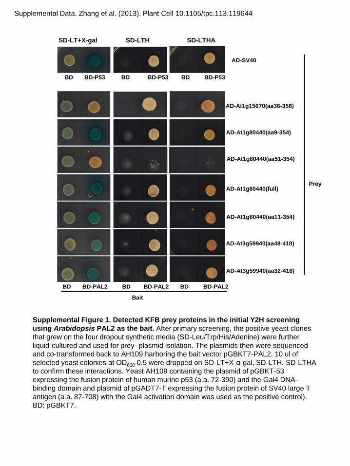
Supplemental Figure 1. Detected KFB prey proteins in the initial Y2H screening using Arabidopsis PAL2 as the bait. After primary screening, the positive yeast clones that grew on the four dropout synthetic media (SD-Leu/Trp/His/Adenine) were further liquid-cultured and used for prey- plasmid isolation. The plasmids then were sequenced and co-transformed back to AH109 harboring the bait vector pGBKT7-PAL2. 10 ul of selected yeast colonies at OD600 0.5 were dropped on SD-LT+X-α-gal, SD-LTH, SD-LTHA to confirm these interactions. Yeast AH109 containing the plasmid of pGBKT-53 expressing the fusion protein of human murine p53 (a.a. 72-390) and the Gal4 DNA-binding domain and plasmid of pGADT7-T expressing the fusion protein of SV40 large T antigen (a.a. 87-708) with the Gal4 activation domain was used as the positive control). BD: pGBKT7.
SD-LT+X-gal SD-LTH SD-LTHA
BD BD-P53 BD BD-P53 BD BD-P53
BD BD-PAL2 BD BD-PAL2 BD BD-PAL2
AD-SV40
AD-At1g15670(aa36-358)
AD-At1g80440(aa9-354)
AD-At1g80440(aa51-354)
AD-At1g80440(full)
AD-At1g80440(aa11-354)
AD-At3g59940(aa48-418)
AD-At3g59940(aa32-418)
Prey
Bait
Supplemental Data. Zhang et al. (2013). Plant Cell 10.1105/tpc.113.119644

Supplemental Figure 2. Subcellular localization of PAL (A)- and KFB-GFP (B) fusion proteins transiently expressed in tobacco epidermal cells. Bar, 40µm.
PAL1-GFP PAL2-GFP
GFP-KFB01 GFP-KFB50 GFP-KFB20
KFB01-GFP KFB20-GFP KFB50-GFP
A
B
Supplemental Data. Zhang et al. (2013). Plant Cell 10.1105/tpc.113.119644

PAL1-HA-CFPc
nYFP-Myc-KFB50(ΔF) nYFP-Myc-CPR30(ΔF)
Ab:HA Ab:Myc Ab:HA Ab:Myc
Input IP(HA)
Supplemental Figure 3. Negative control for Co-IP experiments of PAL-KFB interaction The truncated CPR30 that removes its F-box domain served as the negative control for Co-IP. Proteins from crude lysates (Input) and HA anitibody IPed samples were loaded for the immunoblot. The PAL proteins tagged with HA and CFPc were detected by the HA antibody, and the truncated F-box proteins tagged with Myc and nYFP were detected with the Myc antibody. The immuno-bands for PAL proteins are shown for each blot. Although both the truncated KFB50 and CPR30 proteins presented at low- or undetectable- levels in the crude lysates, after co-IP, the former was detected by anti-Myc antibody but the latter was not.
1 - - -
2 +
- -
3 - +
-
4 +
- +
5 +
+
-
Supplemental Data. Zhang et al. (2013). Plant Cell 10.1105/tpc.113.119644

170 130 100 70 55 40
PAL2-HA KD
- + - + - + - + RT 42 °C 60 °C 100 °C
170 130 100 70 55 40
PAL2-HA KD
- + - + - + - + RT 42 °C 60 °C 100 °C
IP(HA) Ab:HA
IP(HA) Ab:Ub
Supplemental Figure 4. Effect of the preparation conditions on the stability of immunoprecipitated PAL-HA protein Proteins extracted from tobacco leaves transiently expressed with PAL2-HA or infiltrated with buffer (Mock) were IPed with anti-HA antibody beads. 80 μl 1X SDS loading buffer were mixed with the protein-bound beads. Samples were treated for 10 minutes at room temperature (RT), 42 °C, 60 °C, or 100 °C, respectively. Equal amount (5μl) of protein was loaded to the SDS-PAGE gel. Both anti-HA antibody (A) and anti-Ubiquitin antibody (B) were used to detect PAL2-HA protein. The HA IgG bands were marked with red square and degradation products of PAL2-HA were marked with solid arrow head.
A
B
PAL2-HA
Supplemental Data. Zhang et al. (2013). Plant Cell 10.1105/tpc.113.119644

0
0.2
0.4
0.6
0.8
1
1.2
1.4
1.6
1% Suc 4% Suc
Rel
ativ
e PA
L1 e
xpre
ssio
n A
0
0.01
0.02
0.03
0.04
0.05
0.06
1% Suc 4% Suc
Anth
ocya
nin
cont
ent m
g/g
(FW
)
C
Supplemental Figure 5. Effect of exogenous sucrose supply on PAL's transcription (A), total PAL protein concentration (B), and the content of accumulated anthocyanin (C) in Arabidopsis seedlings growing on medium containing 1% or 4% sucrose. Data are the means of the three replicates with standard deviations. Ponceau S staining served as the control for the amount of protein loading (Ctrl.).
B
1% Suc 4% Suc
PAL
Ctrl.
Supplemental Data. Zhang et al. (2013). Plant Cell 10.1105/tpc.113.119644

Supplemental Figure 6. Characterization of the kfb T-DNA insertion mutant lines. RT-PCR analysis of the gene expression of KFB01, 20 and 50 in their T-DNA insertion single and triple mutant lines.
KFB01
KFB20
KFB50
Ub
Supplemental Data. Zhang et al. (2013). Plant Cell 10.1105/tpc.113.119644

Supplemental Figure 7. The relative PAL activity and lignin content in the cell walls of the T1 generation of the KFB01 (A), KFB20 (B) and KFB50 (C) over-expression lines of Arabidopsis. The data are the means of 2 or 3 experimental replicates with standard deviations. The open diamond represents PAL activity; the solid diamond represents lignin content. The amount in WT was set as 100.
0
20
40
60
80
100
120
WT
3-1
4-3
4-1
4-4
5-3
4-2
3-4
5-4
5-6
5-5
3-2
4-5
5-1
Rel
ativ
e ac
tivity
and
con
tent
(%)
A
0
20
40
60
80
100
120
WT
7-1
6-2
7-2
6-4
8-4
6-5
8-5
7-4
8-2
8-3
6-1
7-3
Rel
ativ
e ac
tivity
and
con
tent
(%)
B
0
20
40
60
80
100
120
WT
10-2
10
-3
9-1
9-3
10-5
9-
2 9-
5 11
-2
11-1
10
-1
11-4
11
-5
Rel
ativ
e ac
tivity
and
con
tent
(%)
C
KFB01 OE
KFB20 OE
KFB50 OE
Supplemental Data. Zhang et al. (2013). Plant Cell 10.1105/tpc.113.119644

Supplemental Figure 8. Effect of KFB overexpression on anthocyanin pigmentation in T1 Transgenic Lines. Left panel: The front view of the 4-week-old transgenic- and control-rosettes; Right panel: The flipside view illustrating the disappearance of anthocyanin pigment from the petioles of the KFB01 (3-2) and KFB20 (8-5) overexpression lines. Bar=5 cm
WT/pMDC32
KFB01-OE (3-2)
KFB20-OE (8-5)
Supplemental Data. Zhang et al. (2013). Plant Cell 10.1105/tpc.113.119644

Supplemental Figure 9. Gel blotting analysis on the level of endogenous PAL protein in the KFB over-expression transgenic Arabidopsis. The PAL proteins from two independent T2 transgenic lines of each KFB genes were monitored using a developed anti-PAL peptide antibody. The mono-ubiquitin immunoblot against the anti-ubiquitin antibody served for protein loading amount control.
Ab:PAL
Ab:ubi
Ctrl. 3-2 5-5 8-5 8-3 11-4 10-1
KFB01-OE KFB20-OE KFB50-OE
Supplemental Data. Zhang et al. (2013). Plant Cell 10.1105/tpc.113.119644

Supplemental Figure 10. UV-HPLC profiles of soluble phenolics in KFB01 over-expression line (#3-2), compared with the control (Wt/pMDC32), pal1, c4h and tt4 mutants. K1, kaempferol 3-O-[6''-O-(rhamnosyl) glucoside] 7-O-rhamnoside; K2, kaempferol 3 -O-glucoside 7-O-rhamnoside; K3 kaempferol 3-O-rhamnoside 7-O-rhamnoside; SM, sinapoyl malate.
WT/pMDC32
c4h
pal1
tt4
KFB01-OE (3-2)
Retention time (min)
Abso
rban
ce a
t 310
nm
(mAu
)
8 10 12 14 16 18 20 22 24 0
50
100
150
200
250
300
8 10 12 14 16 18 20 22 24 0
50
100
150
200
250
8 10 12 14 16 18 20 22 24 0
50
100
150
200
250
8 10 12 14 16 18 20 22 24 0
100
200
300
400
8 10 12 14 16 18 20 22 24 0
50
100
150
200
250
K1 K2 K3 SM
300
300
300
Supplemental Data. Zhang et al. (2013). Plant Cell 10.1105/tpc.113.119644

DOI 10.1105/tpc.113.119644; originally published online December 20, 2013;Plant Cell
Xuebin Zhang, Mingyue Gou and Chang-Jun Liuthe Turnover of Phenylalanine Ammonia-Lyase
Kelch Repeat F-Box Proteins Regulate Phenylpropanoid Biosynthesis via ControllingArabidopsis
This information is current as of January 5, 2014
Supplemental Data http://www.plantcell.org/content/suppl/2013/12/20/tpc.113.119644.DC1.html
Permissions https://www.copyright.com/ccc/openurl.do?sid=pd_hw1532298X&issn=1532298X&WT.mc_id=pd_hw1532298X
eTOCs http://www.plantcell.org/cgi/alerts/ctmain
Sign up for eTOCs at:
CiteTrack Alerts http://www.plantcell.org/cgi/alerts/ctmain
Sign up for CiteTrack Alerts at:
Subscription Information http://www.aspb.org/publications/subscriptions.cfm
is available at:Plant Physiology and The Plant CellSubscription Information for
ADVANCING THE SCIENCE OF PLANT BIOLOGY © American Society of Plant Biologists
Copyright © 2022 FDOKUMEN