Influence of Esophageal Endoscopic Submucosal Dissection ...
Upload
independentCategory
view
2download
0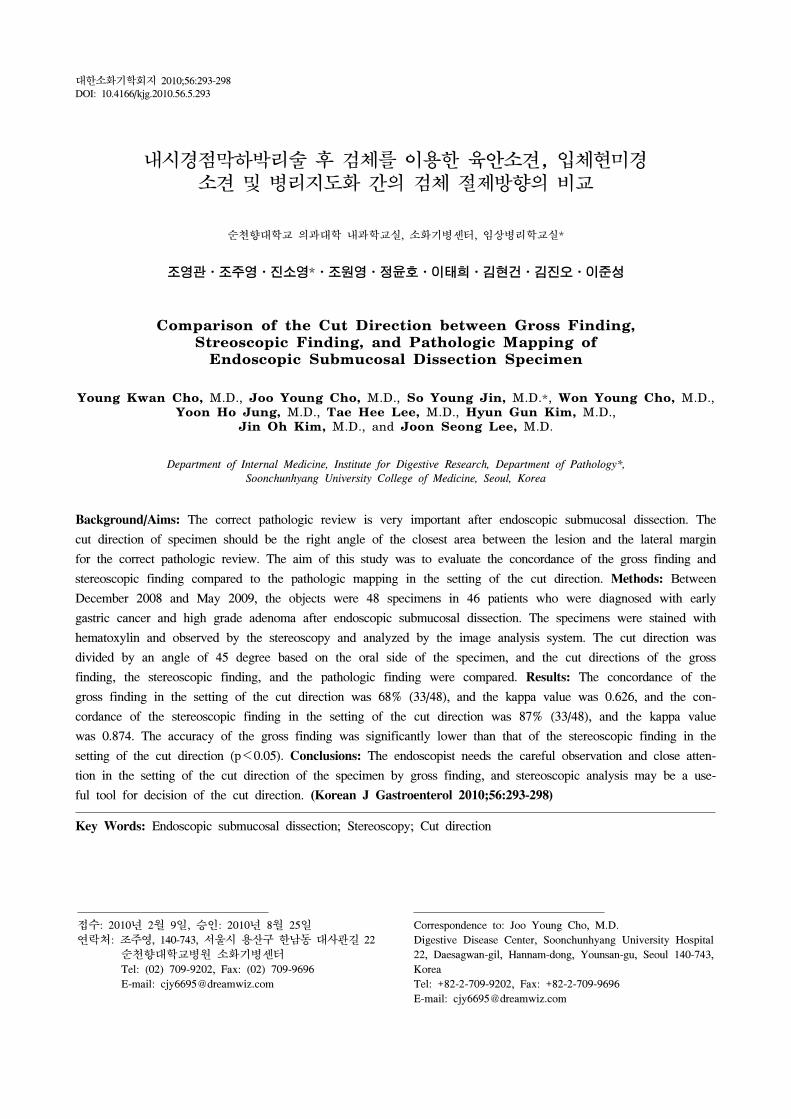
한소화기학회지 2010;56:293-298DOI: 10.4166/kjg.2010.56.5.293
수: 2010년 2월 9일, 승인: 2010년 8월 25일
연락처: 조주 , 140-743, 서울시 용산구 한남동 사 길 22
순천향 학교병원 소화기병센터
Tel: (02) 709-9202, Fax: (02) 709-9696
E-mail: [email protected]
Correspondence to: Joo Young Cho, M.D.
Digestive Disease Center, Soonchunhyang University Hospital
22, Daesagwan-gil, Hannam-dong, Younsan-gu, Seoul 140-743,
Korea
Tel: +82-2-709-9202, Fax: +82-2-709-9696
E-mail: [email protected]
내시경 막하박리술 후 검체를 이용한 육안소견, 입체 미경 소견 병리지도화 간의 검체 제방향의 비교
순천향 학교 의과 학 내과학교실, 소화기병센터, 임상병리학교실*
조 ㆍ조주 ㆍ진소 *ㆍ조원 ㆍ정윤호ㆍ이태희ㆍ김 건ㆍ김진오ㆍ이 성
Comparison of the Cut Direction between Gross Finding, Streoscopic Finding, and Pathologic Mapping of
Endoscopic Submucosal Dissection Specimen
Young Kwan Cho, M.D., Joo Young Cho, M.D., So Young Jin, M.D.*, Won Young Cho, M.D., Yoon Ho Jung, M.D., Tae Hee Lee, M.D., Hyun Gun Kim, M.D.,
Jin Oh Kim, M.D., and Joon Seong Lee, M.D.
Department of Internal Medicine, Institute for Digestive Research, Department of Pathology*, Soonchunhyang University College of Medicine, Seoul, Korea
Background/Aims: The correct pathologic review is very important after endoscopic submucosal dissection. The
cut direction of specimen should be the right angle of the closest area between the lesion and the lateral margin
for the correct pathologic review. The aim of this study was to evaluate the concordance of the gross finding and
stereoscopic finding compared to the pathologic mapping in the setting of the cut direction. Methods: Between
December 2008 and May 2009, the objects were 48 specimens in 46 patients who were diagnosed with early
gastric cancer and high grade adenoma after endoscopic submucosal dissection. The specimens were stained with
hematoxylin and observed by the stereoscopy and analyzed by the image analysis system. The cut direction was
divided by an angle of 45 degree based on the oral side of the specimen, and the cut directions of the gross
finding, the stereoscopic finding, and the pathologic finding were compared. Results: The concordance of the
gross finding in the setting of the cut direction was 68% (33/48), and the kappa value was 0.626, and the con-
cordance of the stereoscopic finding in the setting of the cut direction was 87% (33/48), and the kappa value
was 0.874. The accuracy of the gross finding was significantly lower than that of the stereoscopic finding in the
setting of the cut direction (p<0.05). Conclusions: The endoscopist needs the careful observation and close atten-
tion in the setting of the cut direction of the specimen by gross finding, and stereoscopic analysis may be a use-
ful tool for decision of the cut direction. (Korean J Gastroenterol 2010;56:293-298)
Key Words: Endoscopic submucosal dissection; Stereoscopy; Cut direction
294 한소화기학회지: 제56권 제5호, 2010
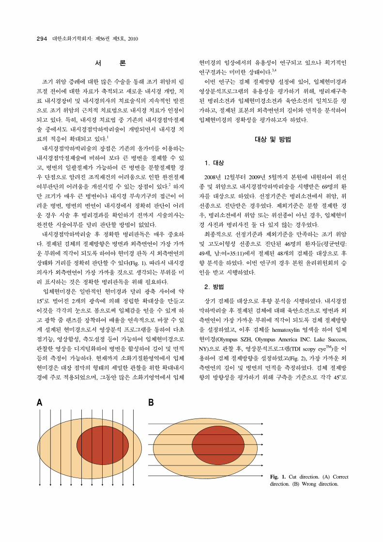
Fig. 1. Cut direction. (A) Correct
direction. (B) Wrong direction.
서 론
조기 암 증례에 한 많은 수술을 통해 조기 암의 림
이에 한 자료가 축 되고 새로운 내시경 개발, 치
료 내시경장비 내시경의사의 치료술식의 지속 인 발
으로 조기 암의 근치 치료법으로 내시경 치료가 인정이
되고 있다. 특히, 내시경 치료법 기존의 내시경 막 제
술 에서도 내시경 막하박리술이 개발되면서 내시경 치
료의 응이 확 되고 있다.1
내시경 막하박리술의 장 은 기존의 올가미를 이용하는
내시경 막 제술에 비하여 보다 큰 병변을 제할 수 있
고, 병변의 일 제가 가능하여 큰 병변을 분할 제할 경
우 단 으로 알려진 조직재건의 어려움으로 인한 완 제
여부 단의 어려움을 개선시킬 수 있는 장 이 있다.2 하지
만 크기가 매우 큰 병변이나 내시경 부속기구의 근이 어
려운 병변, 병변의 변연이 내시경에서 정확히 단이 어려
운 경우 시술 후 병리결과를 확인하기 까지 시술의사는
완 한 시술여부를 달리 단할 방법이 없었다.
내시경 막하박리술 후 정확한 병리 독은 매우 요하
다. 제된 검체의 제방향은 병변과 외측변연이 가장 가까
운 부 에 직각이 되도록 하여야 미경 독 시 외측변연의
상태와 거리를 정확히 단할 수 있다(Fig. 1). 따라서 내시경
의사가 외측변연이 가장 가까울 것으로 생각되는 부 를 미
리 표시하는 것은 정확한 병리 독을 해 필요하다.
입체 미경은 일반 인 미경과 달리 축 사이에 약
15o로 벌어진 2개의 속에 의해 정립한 확 상을 만들고
이것을 각각의 으로 으로써 입체감을 얻을 수 있게 하
고 학 즈를 장착하여 배율을 연속 으로 바꿀 수 있
게 설계된 미경으로서 상분석 로그램을 통하여 다
기능, 상합성, 측도설정 등이 가능하여 입체 미경으로
찰한 상을 디지털화하여 병변을 합성하여 길이 면
등의 측정이 가능하다. 재까지 소화기질환 역에서 입체
미경은 장 막의 형태의 세 한 찰을 한 확 내시
경에 주로 용되었으며, 그동안 많은 소화기 역에서 입체
미경의 임상에서의 유용성이 연구되고 있으나 획기 인
연구결과는 미미한 상태이다.3,4
이번 연구는 검체 제뱡향 설정에 있어, 입체 미경과
상분석 로그램의 유용성을 평가하기 해, 병리재구축
된 병리소견과 입체 미경소견과 육안소견의 일치도를 평
가하고, 제된 표본의 외측변연의 길이와 면 을 분석하여
입체 미경의 정확성을 평가하고자 하 다.
상 방법
1. 상
2008년 12월부터 2009년 5월까지 본원에 내원하여 선
종 암으로 내시경 막하박리술을 시행받은 69명의 환
자를 상으로 하 다. 선정기 은 병리소견에서 암,
선종으로 진단받은 경우 다. 제외기 은 분할 제한 경
우, 병리소견에서 암 는 선종이 아닌 경우, 입체 미
경 사진과 병리사진 둘 다 있지 않는 경우 다.
최종 으로 선정기 과 제외기 을 만족하는 조기 암
고도이형성 선종으로 진단된 46명의 환자들(평균연령:
49세, 남:여=35:11)에서 제된 48개의 검체를 상으로 후
향 분석을 하 다. 이번 연구의 경우 본원 윤리 원회의 승
인을 받고 시행하 다.
2. 방법
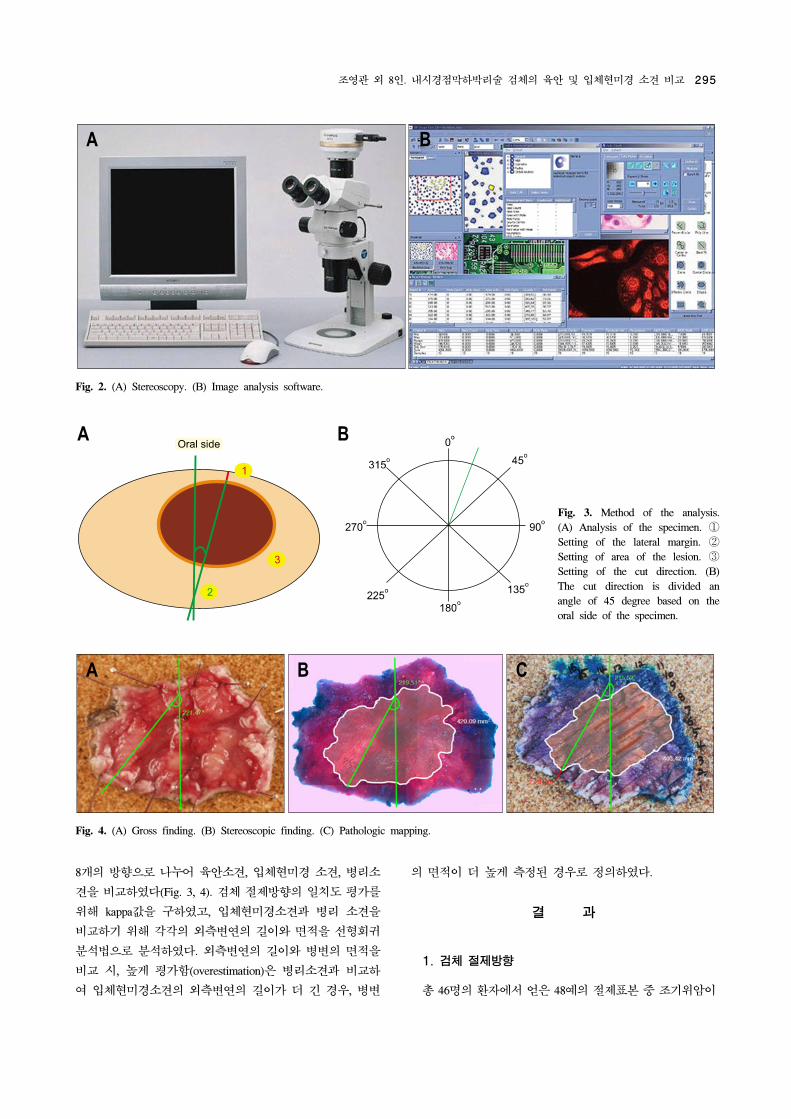
상기 검체를 상으로 후향 분석을 시행하 다. 내시경
막하박리술 후 제된 검체에 해 육안소견으로 병변과 외
측변연이 가장 가까운 부 에 직각이 되도록 검체 제방향
을 설정하 고, 이후 검체를 hematoxylin 염색을 하여 입체
미경(Olympus SZH, Olympus America INC. Lake Success,
NY)으로 찰 후, 상분석 로그램(TDI scopy eyeTM)을 이
용하여 검체 제방향을 설정하 고(Fig. 2), 가장 가까운 외
측변연의 길이 병변의 면 을 측정하 다. 검체 제방
향의 방향성을 평가하기 해 구측을 기 으로 각각 45o로
조 외 8인. 내시경 막하박리술 검체의 육안 입체 미경 소견 비교 295
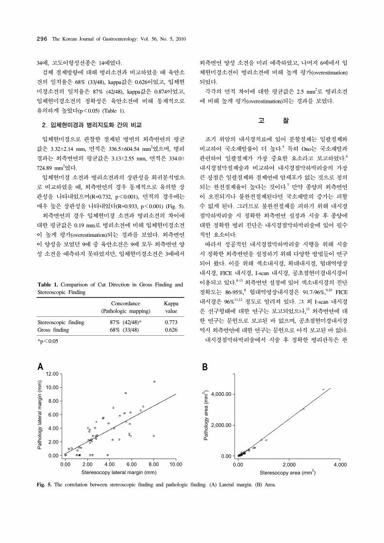
Fig. 3. Method of the analysis.
(A) Analysis of the specimen. ①
Setting of the lateral margin. ②
Setting of area of the lesion. ③
Setting of the cut direction. (B)
The cut direction is divided an
angle of 45 degree based on the
oral side of the specimen.
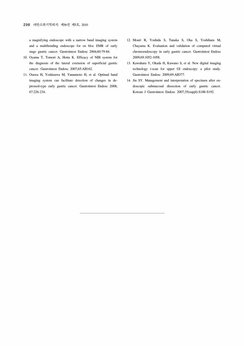
Fig. 4. (A) Gross finding. (B) Stereoscopic finding. (C) Pathologic mapping.
Fig. 2. (A) Stereoscopy. (B) Image analysis software.
8개의 방향으로 나 어 육안소견, 입체 미경 소견, 병리소
견을 비교하 다(Fig. 3, 4). 검체 제방향의 일치도 평가를
해 kappa값을 구하 고, 입체 미경소견과 병리 소견을
비교하기 해 각각의 외측변연의 길이와 면 을 선형회귀
분석법으로 분석하 다. 외측변연의 길이와 병변의 면 을
비교 시, 높게 평가함(overestimation)은 병리소견과 비교하
여 입체 미경소견의 외측변연의 길이가 더 긴 경우, 병변
의 면 이 더 높게 측정된 경우로 정의하 다.
결 과
1. 검체 제방향
총 46명의 환자에서 얻은 48 의 제표본 조기 암이
296 The Korean Journal of Gastroenterology: Vol. 56, No. 5, 2010
Table 1. Comparison of Cut Direction in Gross Finding and
Stereoscopic Finding
Concordance
(Pathologic mapping)
Kappa
value
Stereoscopic finding
Gross finding
87% (42/48)*
68% (33/48)
0.773
0.626
*p<0.05
Fig. 5. The correlation between stereoscopic finding and pathologic finding. (A) Lateral margin. (B) Area.
34 , 고도이형성선종은 14 다.
검체 제방향에 해 병리소견과 비교하 을 때 육안소
견의 일치율은 68% (33/48), kappa값은 0.626이었고, 입체
미경소견의 일치율은 87% (42/48), kappa값은 0.874이었고,
입체 미경소견의 정확성은 육안소견에 비해 통계 으로
유의하게 높았다(p<0.05) (Table 1).
2. 입체 미경과 병리지도화 간의 비교
입체 미경으로 찰한 제된 병변의 외측변연의 평균
값은 3.32±2.14 mm, 면 은 336.5±604.54 mm2 으며, 병리
결과는 외측변연의 평균값은 3.13±2.55 mm, 면 은 334.0±
724.89 mm2 다.
입체 미경 소견과 병리소견과의 상 성을 회귀분석법으
로 비교하 을 때, 외측변연의 경우 통계 으로 유의한 상
성을 나타내었으며(R=0.732, p<0.001), 면 의 경우에는
매우 높은 상 성을 나타내었다(R=0.933, p<0.001) (Fig. 5).
외측변연의 경우 입체 미경 소견과 병리소견의 차이에
한 평균값은 0.19 mm로 병리소견에 비해 입체 미경소견
이 높게 평가(overestimation)되는 결과를 보 다. 외측변연
이 양성을 보 던 9 육안소견은 9 모두 외측변연 양
성 소견을 측하지 못하 지만, 입체 미경소견은 3 에서
외측변연 양성 소견을 미리 측하 고, 나머지 6 에서 입
체 미경소견이 병리소견에 비해 높게 평가(overestimation)
되었다.
각각의 면 차이에 한 평균값은 2.5 mm2로 병리소견
에 비해 높게 평가(overestimation)되는 결과를 보 다.
고 찰
조기 암의 내시경치료에 있어 분할 제는 일 제와
비교하여 국소재발률이 더 높다.5 특히 Ono는 국소재발과
련하여 일 제가 가장 요한 요소라고 보고하 다.6
내시경 막 제술과 비교하여 내시경 막하박리술의 가장
큰 장 은 일 제와 제연에 암세포가 없는 것으로 정의
되는 완 제율이 높다는 것이다.7 만약 종양의 외측변연
이 오진되거나 불완 제된다면 국소재발의 증가는 피할
수 없게 된다. 그러므로 불완 제를 피하기 해 내시경
막하박리술 시 정확한 외측변연 설정과 시술 후 종양에
한 정확한 병리 진단은 내시경 막하박리술에 있어 필수
인 요소이다.
따라서 성공 인 내시경 막하박리술 시행을 해 시술
시 정확한 외측변연을 설정하기 해 다양한 방법들이 연구
되어 왔다. 이를 해 색소내시경, 확 내시경, 역 상
내시경, FICE 내시경, I-scan 내시경, 공 미경내시경이
이용되고 있다.8-13 외측변연 설정에 있어 색소내시경의 진단
정확도는 86-95%,8 역 상내시경은 91.7-96%,9,10 FICE
내시경은 96%11,12 정도로 알려져 있다. 그 외 I-scan 내시경
은 선구형태에 한 연구는 보고되었으나,13 외측변연에
한 연구는 문헌으로 보고된 바 없으며, 공 미경내시경
역시 외측변연에 한 연구는 문헌으로 아직 보고된 바 없다.
내시경 막하박리술에서 시술 후 정확한 병리 독은 완

Cho YK, et al. Comparison of the Gross and Stereoscopic Finding of Submucosal Dissection Specimen 297
치여부 단 추후 환자의 치료방향을 결정하는데 있어
매우 요하다. 정확한 외측변연 독을 해서 검체의
제방향이 요하며, 이를 해 검체 제방향은 병변과 외
측변연이 가장 가까운 부 에 직각이 되도록 제하여야
미경 독 시 제연의 상태와 거리를 정확히 단할 수 있
다(Fig. 4).14 하지만 부분 경우 육안으로 외측변연과 병변
이 가장 가까울 것으로 생각되는 부 를 표시하고 있으며,
이의 정확성 여부에 해서는 해서는 보고된 바 없다.
이번 연구에서 검체 제방향에 한 육안 소견과 입체
미경소견 비교 시 육안소견은 입체 미경소견에 비해 정확
성이 떨어지는 것을 확인할 수 있었다. 이는 부정확한 병리
독으로 인해 잘못된 치료로 이어질 수 있다. 이 을 보안
하기 해 측면 제연의 편 방향에 해 입체 미경을 이
용하는 것이 안이 될 수가 있지만, 실 인 여건으로는
힘들고, 한 입체 미경 소견 역시 완 히 정확하지 않다
는 제한 이 있다. 따라서 내시경 막하박리술 시행 후 내
시경의사가 육안으로 가장 가까운 측면 제연의 방향을 결
정 시 세심한 주의가 필요할 것으로 생각한다.
이번 연구에서 입체 미경소견과 병리소견은 서로 제
변연과 면 에서 통계 으로 유의한 상 성을 보여 입체
미경의 유용성을 확인할 수 있었다. 그러나 입체 미경의
제변연의 양성의 측률은 낮았고 이는 열응고, 자가용해
등에 의한 인공산물 변화가 결과에 향을 미쳤을 가능성을
생각할 수 있다.
본 연구의 제한 은 본 연구가 후향 연구라는 과 아직
조기 암, 고도이형성 선종 정상 막의 선구구조에
한 연구가 진행 이지만, 에서는 장에서처럼 명확한
선구구조 분류법이 없어 정확한 외측변연에 한 평가가 어
려웠다는 이다. 이에 해 향후 입체 미경, 확 내시경,
공 미경 내시경을 이용한 선구 구조에 한 추가
인 연구가 선행되어야 할 것으로 생각된다. 특히 외측변연
설정에 있어 공 미경 내시경은 실시간으로 조직 찰
이 용이한 장 이 있어 향후 임상 용에 한 연구가 필
요하다고 생각한다.
요 약
목 : 내시경 막하박리술 후 정확한 병리 독은 매우
요하다. 이를 해 검체 제방향은 병변과 제연이 가
장 가까운 부 에 직각이 되도록 제하여야 한다. 이번 연
구는 검체 제뱡향 설정에 있어, 병리재구축된 병리소견과
비교하여 입체 미경소견과 육안소견의 일치도를 평가하고
자 하 다. 상 방법: 2008년 12월부터 2009년 5월까지
본원에 내원하여 조기 암 고도이형성 선종으로 진단
된 46명의 환자들(평균연령: 49세, 남:여=35:11)에서 제된
48개의 표본을 상으로 하 다. 시술 후 제된 표본에
해 hematoxylin 염색을 하여 찰한 입체 미경소견을 상
분석 로그램으로 분석하 다. 구측을 기 으로 각각 45o로
8개의 방향으로 나 어 육안소견, 입체 미경 소견, 병리소
견의 제방향을 비교하 다. 결과: 제방향 설정에 있어
육안소견의 일치율은 68% (33/48), kappa값은 0.626이었고,
입체 미경소견의 일치율은 87% (42/48), kappa값은 0.874이
었다. 제방향 설정에 있어 육안소견의 정확성은 입체 미
경소견에 비해 통계 으로 유의하게 낮았다(p<0.05). 결론:
내시경의사는 가장 가까운 측면 제연의 방향을 결정 시
세심한 주의가 필요할 것으로 생각되며, 입체 미경을 이용
한 분석이 유용한 도구가 될 수 있겠다.
색인단어: 내시경 막하박리술, 입체 미경, 검체 제방향
참고문헌
1. Cho JY, Cho WY. The curren tstatus of endoscopic sub-
mucosal dissection. Korean J Gastrointest Endosc 2008;37:
317-320.
2. Gotoda T, Yanagisawa A, Sasako M, et al. Incidence of
lymph node metastasis from early gastric cancer: estimation
with a large number of cases at two large centers. Gastric
Cancer 2000;3:219-225.
3. Kudo S, Hirota S, Nakajima T, et al. Colorectal tumors and
pit pattern. J Clin Pathol 1994;47:880-885.
4. Ryozawa S, Watanabe H, Abe M, Ajioka Y, Nishikura K,
Okita K. Macroscopic and stereomicroscopic diagnosis of su-
perficial flat-type early carcinomas of the gallbladder. J Gastro-
enterol 1997;32:635-642.
5. Miyamoto S, Muto M, Hamamoto Y, et al. A new technique
for endoscopic mucosal resection with an insulated-tip elec-
trosurgical knife improves the completeness of resection of
intramucosal gastric neoplasms. Gastrointest Endosc 2002;55:
576-581.
6. Ono H. Early gastric cancer: diagnosis, pathology, treatment
techniques and treatment outcomes. Eur J Gastroenterol
Hepatol 2006;18:863-866.
7. Yokoi C, Gotoda T, Hamanaka H, Oda I. Endoscopic sub-
mucosal dissection allows curative resection of locally re-
current early gastric cancer after prior endoscopic mucosal
resection. Gastrointest Endosc 2006;64:212-218.
8. Iizuka T, Kikuchi D, Hoteya S, Yahagi N. The acetic acid +
indigocarmine method in the delineation of gastric cancer. J
Gastroenterol Hepatol 2008;23:1358-1361.
9. Sumiyama K, Kaise M, Nakayoshi T, et al. Combined use of
298 한소화기학회지: 제56권 제5호, 2010
a magnifying endoscope with a narrow band imaging system
and a multibending endoscope for en bloc EMR of early
stage gastric cancer. Gastrointest Endosc 2004;60:79-84.
10. Oyama T, Tomori A, Hotta K. Efficacy of NBI system for
the diagnosis of the lateral extension of superficial gastric
cancer. Gastrointest Endosc 2007;65:AB162.
11. Osawa H, Yoshizawa M, Yamamoto H, et al. Optimal band
imaging system can facilitate detection of changes in de-
pressed-type early gastric cancer. Gastrointest Endosc 2008;
67:226-234.
12. Mouri R, Yoshida S, Tanaka S, Oka S, Yoshihara M,
Chayama K. Evaluation and validation of computed virtual
chromoendoscopy in early gastric cancer. Gastrointest Endosc
2009;69:1052-1058.
13. Kawahara Y, Okada H, Kawano S, et al. New digital imaging
technology i-scan for upper GI endoscopy: a pilot study.
Gastrointest Endosc 2009;69:AB377.
14. Jin SY. Management and interpretation of specimen after en-
doscopic submucosal dissection of early gastric cancer.
Korean J Gastrointest Endosc 2007;35(suppl):S188-S192.
Copyright © 2022 FDOKUMEN