Comparison of higher-energy collisional dissociation and collision-induced dissociation MS/MS...
16
Proteomics Clin. Appl. 2015, 9, 453–468 453 DOI 10.1002/prca.201400113 REVIEW Capillary zone electrophoresis on-line coupled to mass spectrometry: A perspective application for clinical proteomics Martin Pejchinovski 1∗ , Dajana Hrnjez 1∗ , Adela Ramirez-Torres 1 , Vasiliki Bitsika 2 , George Mermelekas 2 , Antonia Vlahou 2,3 , Petra Z ¨ urbig 1 , Harald Mischak 1,4 , Jochen Metzger 1 and Thomas Koeck 1 1 Mosaiques Diagnostics GmbH, Hanover, Germany 2 Biotechnology Division, Biomedical Research Foundation, Academy of Athens, Athens, Greece 3 School of Biomedical and Healthcare Sciences, Plymouth University, Plymouth, UK 4 Institute of Cardiovascular and Medical Sciences, University of Glasgow, UK Received: August 18, 2014 Revised: November 21, 2014 Accepted: January 14, 2015 Clinical proteomics, a rapidly growing field, intends to use specific diagnostic pro- teomic/peptidomic markers for initial diagnosis or prognosis of the progression of various diseases. Analyses of disease-associated markers in defined biological samples can provide valuable molecular diagnostic information for these diseases. This approach relies on sensitive and highly standardized modern analytical techniques. In the recent years, one of these tech- nologies, CZE online coupled to MS (CZE-MS), has been increasingly used for the detection of peptide biomarkers (<20 kDa) in body fluids such as urine. This review presents the most relevant urinary proteomic studies addressing the application of CZE-MS in clinically relevant biomarker research between the years 2006 and 2014. Keywords: Biomarker / Capillary electrophoresis-mass spectrometry / Clinical / Proteomics Additional supporting information may be found in the online version of this article at the publisher’s web-site Correspondence: Dr. Thomas Koeck, Mosaiques Diagnostics GmbH, Mellendorferstrasse 7–9, D–30625 Hanover, Germany E-mail: [email protected] Fax: +49-511-554744-31 Abbreviations: AAV, ANCA-associated vasculitis; ADPKD, autosomal-dominant polycystic kidney disease; aGvHD, acute graft-versus-host disease; AKI, acute kidney injury; allo-HSCT, allogeneic hematopoietic stem cell transplantation; ANCA, an- tineutrophil cytoplasmic autoantibody; BBD, benign biliary dis- order; BCa, bladder cancer; CAD, coronary artery disease; CC, cholangiocarcinoma; CKD, chronic kidney disease; CVD, cardio- vascular disease; DN, diabetic nephropathy; ECM, extracellular matrix; ESRD, end-stage renal disease; HF, heart failure; IgAN, IgA nephropathy; IPP, informative peptide panel; LV, left ven- tricular; LVDD, LV diastolic dysfunction; No-OP, nonoperated individuals with UPJ obstruction; NRI, net reclassification im- provement; OP, individuals with severe UPJ obstruction; PCa, prostate cancer; PSA, prostate-specific antigen; PSC, primary sclerosing cholangitis; PUV, posterior urethral valves; RCC, renal cell carcinoma; ROC, receiver operating characteristic; SF, sin- gle factor; SVM, support vector machine; T2D, type 2 diabetes; TCMR, T-cell-mediated tubulointerstitial rejection; UPJ, uretero- pelvic junction obstruction 1 Introduction During the last decade, CZE online coupled to MS (CZE- MS) has proven its value and is now increasingly used for proteomic as well as for metabolomic analysis, in diagnosis, therapeutic treatment, and drug development [1–7]. CZE-MS as analytical tool is complementary and may provide advantages over other technologies in analyzing uri- nary proteomic biomarkers of various diseases [8–10]. The main advantage of modern proteomic tools such as CZE-MS for routine diagnostic analysis is the simultaneous detection of multiple markers [11]. CZE-MS is currently the method with the shortest route from the analytical laboratory to clin- ical application as it can be used for discovery, validation, and clinical implementation, missing out on the costly and time-consuming process of changing technology platforms with the risk of losing biomarkers [12]. Technically, CZE-MS can provide fast analysis, typically resolving 1000–4000 pep- tides per sample within approximately 45 min. Some of the ∗ These authors contributed equally to this work. Colour Online: See the article online to view Figs. 1–4 in colour. C 2015 WILEY-VCH Verlag GmbH & Co. KGaA, Weinheim www.clinical.proteomics-journal.com
Transcript of Comparison of higher-energy collisional dissociation and collision-induced dissociation MS/MS...

Proteomics Clin. Appl. 2015, 9, 453–468 453DOI 10.1002/prca.201400113
REVIEW
Capillary zone electrophoresis on-line coupled to mass
spectrometry: A perspective application for clinical
proteomics
Martin Pejchinovski1∗, Dajana Hrnjez1∗, Adela Ramirez-Torres1, Vasiliki Bitsika2,George Mermelekas2, Antonia Vlahou2,3, Petra Zurbig1, Harald Mischak1,4,Jochen Metzger1 and Thomas Koeck1
1 Mosaiques Diagnostics GmbH, Hanover, Germany2 Biotechnology Division, Biomedical Research Foundation, Academy of Athens, Athens, Greece3 School of Biomedical and Healthcare Sciences, Plymouth University, Plymouth, UK4 Institute of Cardiovascular and Medical Sciences, University of Glasgow, UK
Received: August 18, 2014Revised: November 21, 2014Accepted: January 14, 2015
Clinical proteomics, a rapidly growing field, intends to use specific diagnostic pro-teomic/peptidomic markers for initial diagnosis or prognosis of the progression of variousdiseases. Analyses of disease-associated markers in defined biological samples can providevaluable molecular diagnostic information for these diseases. This approach relies on sensitiveand highly standardized modern analytical techniques. In the recent years, one of these tech-nologies, CZE online coupled to MS (CZE-MS), has been increasingly used for the detectionof peptide biomarkers (<20 kDa) in body fluids such as urine. This review presents the mostrelevant urinary proteomic studies addressing the application of CZE-MS in clinically relevantbiomarker research between the years 2006 and 2014.
Keywords:
Biomarker / Capillary electrophoresis-mass spectrometry / Clinical / Proteomics
� Additional supporting information may be found in the online version of this article atthe publisher’s web-site
Correspondence: Dr. Thomas Koeck, Mosaiques DiagnosticsGmbH, Mellendorferstrasse 7–9, D–30625 Hanover, GermanyE-mail: [email protected]: +49-511-554744-31
Abbreviations: AAV, ANCA-associated vasculitis; ADPKD,autosomal-dominant polycystic kidney disease; aGvHD, acutegraft-versus-host disease; AKI, acute kidney injury; allo-HSCT,allogeneic hematopoietic stem cell transplantation; ANCA, an-tineutrophil cytoplasmic autoantibody; BBD, benign biliary dis-order; BCa, bladder cancer; CAD, coronary artery disease; CC,cholangiocarcinoma; CKD, chronic kidney disease; CVD, cardio-vascular disease; DN, diabetic nephropathy; ECM, extracellularmatrix; ESRD, end-stage renal disease; HF, heart failure; IgAN,IgA nephropathy; IPP, informative peptide panel; LV, left ven-tricular; LVDD, LV diastolic dysfunction; No-OP, nonoperatedindividuals with UPJ obstruction; NRI, net reclassification im-provement; OP, individuals with severe UPJ obstruction; PCa,prostate cancer; PSA, prostate-specific antigen; PSC, primarysclerosing cholangitis; PUV, posterior urethral valves; RCC, renalcell carcinoma; ROC, receiver operating characteristic; SF, sin-gle factor; SVM, support vector machine; T2D, type 2 diabetes;TCMR, T-cell-mediated tubulointerstitial rejection; UPJ, uretero-pelvic junction obstruction
1 Introduction
During the last decade, CZE online coupled to MS (CZE-MS) has proven its value and is now increasingly used forproteomic as well as for metabolomic analysis, in diagnosis,therapeutic treatment, and drug development [1–7].
CZE-MS as analytical tool is complementary and mayprovide advantages over other technologies in analyzing uri-nary proteomic biomarkers of various diseases [8–10]. Themain advantage of modern proteomic tools such as CZE-MSfor routine diagnostic analysis is the simultaneous detectionof multiple markers [11]. CZE-MS is currently the methodwith the shortest route from the analytical laboratory to clin-ical application as it can be used for discovery, validation,and clinical implementation, missing out on the costly andtime-consuming process of changing technology platformswith the risk of losing biomarkers [12]. Technically, CZE-MScan provide fast analysis, typically resolving 1000–4000 pep-tides per sample within approximately 45 min. Some of the
∗These authors contributed equally to this work.Colour Online: See the article online to view Figs. 1–4 in colour.
C© 2015 WILEY-VCH Verlag GmbH & Co. KGaA, Weinheim www.clinical.proteomics-journal.com

454 M. Pejchinovski et al. Proteomics Clin. Appl. 2015, 9, 453–468
further advantages such as robustness (toward interferingcompounds, participates, etc.) and high comparability of thedatasets established this technique as a basis for biomarkerdiscoveries [13, 14]. The raise of CZE-MS-based biomarkerdiscovery in the recent years coincided with intensification ofproteome/peptidome analysis of the human urine, which canbe collected in large quantities in noninvasive and convenientway. Thirty percent of urine proteome is of the systemic ori-gin [15]. It may therefore provide biomarkers for urogenitalas well as systemic diseases. In fact, based on the analysisby CZE-MS more than 100 000 different peptides (<20 kDa)could be detected in a total of >20 000 urine samples. At least5600 of these peptides were detected with high frequency(>30%) and can hence be considered as typical componentsof urine proteome [13]. However, alterations in peptide dis-tinction and concentration (detection limits, and/or multipledetections of the peptides) limit the number of peptides typ-ically detected in a sample to about 900–2500. The concen-tration of proteins and peptides in the urine is changing ona daily basis, depending on food and fluid intake [16]. Thesevariations can be fairly well compensated by an adjustment ofthe concentration relative to urinary creatinine levels or otherso-called “housekeeping peptides” that are typically presentin urine [17].
In this article, we aim to comprehensively review the CZE-MS-based proteomic biomedical studies performed betweenthe years 2006 and 2014, focusing on the detection of humanurinary peptide biomarkers.
2 Technical aspect of CZE-MS
CZE is a fast high-resolution separation technology based ondifferential mobility of ions through a conductive mediumin a high-voltage electric field. Mobility of the analytes is in-fluenced by their charge, the viscosity of the medium, andmolecule’s radius. Known for its compatibility with mostbuffers and analytes [12], CZE can provide stable flow and
constant voltage conditions [18]. This stability is advantageouswhen connecting to MS and establishing a robust CZE-MSsystem [11,19] that demonstrates high resolution, short anal-ysis time, and good repeatability, all of which are importantfactors for clinical application. Including preanalytical steps,CZE-ESI-TOF-MS analysis can be used for reasonably fast,stable, and reproducible analysis of body fluids in a clinicallaboratory setting (Fig. 1) [13]. Applying suitable software so-lutions to process the CE-MS raw data, including peak detec-tion, charge assignment, calibration, and database deposition[13, 14, 19, 20], allowed development of an urine proteomedatabase with currently >20 000 comparable datasets [14].While in the past direct interfacing CE with MS/MS was chal-lenging [21] and hence the correlation of basic amino acidswith migration time was often used for sequence assignment[22], recent developments have enabled sequencing using CE-MS/MS, with similar efficiency like LC-MS/MS [23].
CZE-ESI-TOF-MS-based analysis has been utilized forthe initial discovery and validation of proteomic biomarkers(Fig. 2) [24]. However, regardless of all advantages associatedwith a CZE-MS approach, it is not yet routinely used for clin-ical standard diagnostics for reasons such as the momentaryunavailability of the techniques as a ready-to-use local on-sitesolution due to, e.g., high technical demands, and the lackof approval of diagnostic disease-specific peptide/protein pat-terns by regulatory agencies [25].
3 Clinical applications for CZE-MS
Nonetheless in the recent years, a number of articles reportedon clinical CZE-MS applications in proteomics, focusingon proteomic biomarker discovery, disease diagnosis, andassessment of therapeutic treatment. Mosaiques grouphas developed CZE-MS proteomic platform and own theintellectual property rights of this technology. Therefore,most prominent examples of urinary biomarker discoveryusing CZE-MS since 2006 are given in Table 1.
Figure 1. Schematic overviewof the CZE-MS biomarkeranalysis. This figure illustratesthe workflow of CZE-MS-basedbiomarker analysis.
C© 2015 WILEY-VCH Verlag GmbH & Co. KGaA, Weinheim www.clinical.proteomics-journal.com

Proteomics Clin. Appl. 2015, 9, 453–468 455
Figure 2. Diagnostic potential of CZE-MS-based proteome analysis. Onefull urinary peptide pattern result-ing from CZE-MS-based proteomicanalysis may be classified by multi-ple disease-specific classifiers allow-ing for the diagnosis of various differ-ent pathological conditions from oneproteomic analysis. In the figure, 3Dcounterplots generated by CAD, HF,and CKD classifiers are shown. Dis-played in three dimensions: x-axis, mi-gration time in minutes; y-axis: loga-rithmic mass in Daltons; z-axis: signalintensity. CAD: cardiovascular disease.HF: heart failure with preserved ejec-tion fraction. CKD: chronic kidney dis-ease.
3.1 Kidney and urological diseases
According to the information provided by the AmericanKidney Fund, kidney diseases are the eighth leading cause ofdeaths in the United States, which might be representativefor most developed countries. It is estimated that 31 millionpeople, 10% of USA population, have chronic kidney disease(CKD) (http://www.kidneyfund.org/about-us/assets/pdfs/akf-kid–neydiseasestatistics-2012.pdf).
3.1.1 Diabetic nephropathy
Diabetic nephropathy (DN) is a progressive disease oftencomplicating long-standing diabetes and the most frequentreason for dialysis in Western countries. Standard clinicaldiagnoses is based on the presence of proteinuria as well aschanges in serum creatinine level connected to decrease inthe glomerular filtration rate [26].
In a first CZE-MS-based study, 102 DN-specific urinarypeptide biomarkers were reported in 2008 [27]. Sixty five ofthese biomarkers were selected for a DN-specific classifierthrough support vector machine (SVM) modeling to differ-entiate diabetes type 1 patients with macroalbuminuria andnormoalbuminuria. In a blinded cohort of 35 patients withdiabetes and macroalbuminuria and 35 healthy individuals,the classifier identified DN with 97% sensitivity and healthyindividuals with specificity (see Table 1). This classifier wasfurther applied to 30 patients with diabetes and microalbu-minuria. Seventeen scored positive, eight of whom showeda progression toward macroalbuminuria. The classifier was
further validated in a case–control study reported by Alkha-laf et al. in 2010 [28] in 148 patients with type 2 diabetes(T2D), of whom 64 presented with DN (macroalbuminuria >
300 mg/day). The performance of the classifier with regard tothe discrimination between type 2 diabetics with and withoutDN was confirmed with a sensitivity and specificity of 94 and91%, respectively, as well as an AUC of 0.95. In this study,the number of sequenced peptides increased to 34 of the 65peptide biomarkers. Most of them were identified as colla-gen fragments that were downregulated in the urine of DNpatients. Even though the DN classifier performed well andshares 24 peptides with the classifier CKD273, in later stud-ies, it apparently was replaced by the classifier CKD273 [29],which proved to be a more stable general diagnostic modalityfor renal diseases without sacrificing sensitivity toward DNand provided a much better depiction of pathological pro-cesses as it is based on 273 sequenced peptides.
3.1.2 CKD
CKD is characterized by a slow, progressive loss of renalfunction and glomerular filtration that may ultimately resultin end-stage renal disease (ESRD). Patients with ESRDrequire dialysis and in the end kidney transplantation. Goodet al. successfully showed that CZE-MS-based proteomicanalysis can be utilized for diagnosis of CKD [29]. Examining379 healthy controls and 230 patients with different stages ofCKD, the authors identified a urinary biomarker pattern of273 peptides and established the disease classifier CKD273through SVM modeling based on this set of peptide marker.
C© 2015 WILEY-VCH Verlag GmbH & Co. KGaA, Weinheim www.clinical.proteomics-journal.com

456 M. Pejchinovski et al. Proteomics Clin. Appl. 2015, 9, 453–468
Ta
ble
1.
Late
stre
po
rts
on
pep
tid
eb
iom
arke
rsd
isco
vere
dw
ith
CZ
E-M
Sfo
rp
rog
no
sis/
dia
gn
osi
so
fva
rio
us
dis
ease
s
Dis
ease
Bio
flu
idN
um
ber
of
bio
mar
kers
emp
loye
d
Bio
mar
ker
dis
cove
ryp
has
e(n
case
s/n
con
tro
ls)
Valid
atio
np
has
e(n
case
s/n
con
tro
ls)
Sen
siti
vity
%(t
est
set)
Sp
ecifi
city
%(t
est
set)
Ref
eren
ces
1D
iab
etic
nep
hro
pat
hy(D
N)
Uri
ne
6560
/30
35/3
597
97R
oss
ing
etal
.[27
]2
Ch
ron
icki
dn
eyd
isea
se(C
KD
)U
rin
e27
323
0/37
911
0/34
8610
0G
oo
det
al.[
29]
3A
NC
A-a
sso
ciat
edVa
scu
litis
Uri
ne
1818
/425
10/3
090
90H
aub
itz
etal
.[42
]4
IgA
nep
hro
pat
hyU
rin
e25
402/
207
22/2
790
90Ju
lien
etal
[44]
5A
uto
som
alp
oly
cyst
icki
dn
eyd
isea
se(A
DP
KD
)U
rin
e14
217
/86
24/3
588
98K
istl
eret
al.[
47]
6A
cute
kid
ney
inju
ry(A
KI)
Uri
ne
2016
/14
9/11
8982
Met
zger
etal
.[52
]7
Post
erio
ru
reth
ralv
alve
so
bst
ruct
ion
(PU
V)
Uri
ne
1213
/15
16/2
288
95K
lein
etal
.[55
]
8U
rete
rop
elvi
cju
nct
ion
ob
stru
ctio
n(U
PJ)
Uri
ne
5313
con
tro
ls/1
9N
o-O
P/1
9O
P23
/13
9480
–100
Dec
ram
eret
al.[
56]
9C
oro
nar
yar
tery
dis
ease
Uri
ne
1530
/20
47/1
298
83Z
imm
erli
etal
.[68
]U
rin
e17
15/1
426
/12
8192
von
Zu
rM
uh
len
etal
.[69
]U
rin
e23
821
2/19
671
/67
7988
Del
les
etal
.[39
]U
rin
e35
65/4
132
/15
5693
Dw
anso
net
al.[
79]
10Le
ftve
ntr
icu
lar
dys
fun
ctio
nU
rin
e85
19/1
916
/16
6994
Ku
znet
sova
etal
.[73
]11
Uri
ne
8596
/98
32/1
556
93Z
han
get
al.[
75]
12A
cute
gra
ftve
rsu
sh
ost
dis
ease
(aG
vHD
)g
rad
eIII
and
IVU
rin
e31
13/5
011
9/48
083
77W
eiss
ing
eret
al.[
84]
13A
cute
ren
alal
log
raft
reje
ctio
nU
rin
e14
16/2
328
/36
9378
Met
zger
etal
.[87
]14
Bla
dd
erca
nce
r(B
Ca)
Uri
ne
2246
/33
31/1
1/13
810
010
0T
heo
do
resc
uet
al.
[96]
15R
enal
cell
carc
ino
ma
(RC
C)
Uri
ne
8640
/68
70/6
8087
Fran
tzie
tal
.[10
6]16
Pro
stat
eca
nce
rU
rin
e12
51/3
526
491
69T
heo
do
resc
uet
al.
[111
]17
Ch
ola
ng
ioce
llula
rca
rcin
om
aU
rin
e42
14/2
742
/81
8379
Met
zger
etal
.[11
5]
C© 2015 WILEY-VCH Verlag GmbH & Co. KGaA, Weinheim www.clinical.proteomics-journal.com

Proteomics Clin. Appl. 2015, 9, 453–468 457
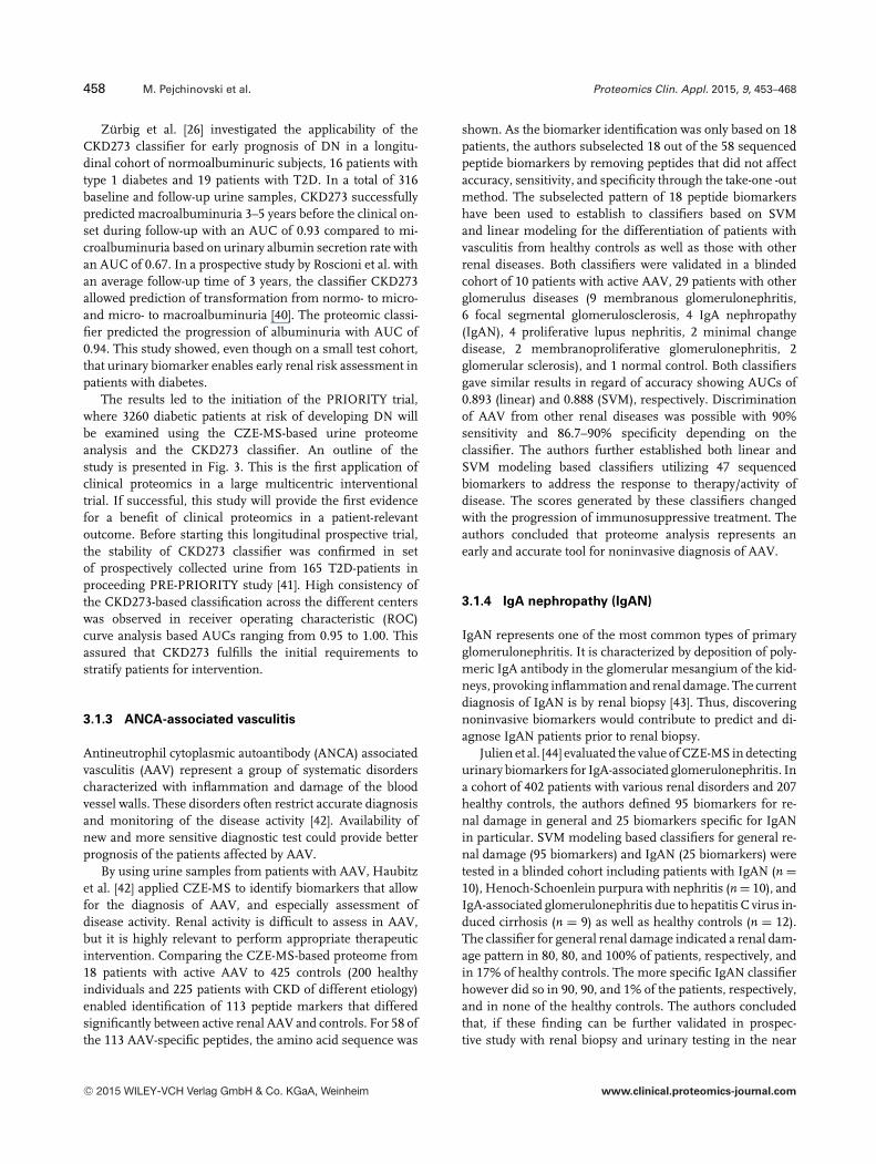
Figure 3. Design of the proteomics-driven inter-vention trial to interfere with development ofDN, PRIORITY. Underlying assumptions are that20% of normoalbuminuric patients (diabetes du-ration: 5–10 years) will show pathophysiologicalchanges indicative for early stages of DN. Tar-geted therapeutic intervention will reduce the de-velopment of microalbuminuria during a periodof 3 years in this selected cohort from 35 to 25%.To demonstrate significant benefit (a = 0.05, � =0.8), a total of 3280 patients have to be screened,among them 656 who are at risk will be random-ized. Reprinted with permission [124].
The performance of the classifier was assessed using ablinded cohort of 110 patients with various stages of CKDand 34 healthy controls, revealing a sensitivity of 85.5% andspecificity of 100% (see Table 1). Argiles et al. examined thevalue of the CKD273 in predicting ESRD or death in 53 pa-tients [30]. During follow-up of 3.6 years, none of the patientswith CKD273 score <0.55 required dialysis or died comparedto 15 patients who reached an endpoint having CKD273score >0.55. These results confirmed a prognostic value ofCKD273 classifier, unrelated to serum creatinine levels andeGRF in disease progression. The correlation of individualpeptides to either different CKD-stages (advanced-stage CKD,eGFR � 5 mL/min/1.73 m2, n = 321; moderate-stage CKDpatients, eGFR > 45 mL/min/1.73 m2, n = 1669) showedhigher association in CKD prognosis and diagnosis thanalbuminuria. Correlating peptides in both studies, regardlessof CKD-stage or CKD-progression, were either fragments ofmajor circulating proteins (beta-2-microglobulin, apolipopro-tein A-I, alpha-1-antitrypsin, serum albumin) suggestingfailure of the glomerular sieving properties, or fragmentsof extracellular matrix (ECM) (collagen type I and III)suggesting changes in intrarenal ECM turnover [31–35].
Gu et al. [36] investigated the net reclassification improve-ment and integrated discrimination improvement in assess-ing renal function when the classifier CKD273 was combinedwith classifiers for left ventricular (LV) dysfunction into asingle factor (SF) through principal component analysis. Ahigher SF classifier level correlated (p � 0.010) with worserenal function. During follow-up over 4.8 years, SF predictedprogression of CKD from stage 2 or �2 to stage �3 withsignificant net reclassification improvement and integrateddiscrimination improvement.
Nkuipou-Kenfack et al. [37] explored the potential com-bination of plasma and urine proteomics and metabolomicsto assess mild and advance CKD patients. In total, 49 pa-tients were studied: 26 patients with DN and 23 with otheretiologies. From 43 patients, follow-up data (2.8 ± 0.8 years)were available. Peptides or metabolites showing significantalternation between the two patient groups were combinedinto SVM-driven classifiers. Three classifiers were devel-oped: one plasma metabolite based (MetaboP), one urinarymetabolite based (MetaboU), and one urinary peptide based(Pept). Scores generated with all three classifiers correlatedwell with eGFR. The Pept model performed best, and noadded value could be detected by combining the proteomicsand metabolomics biomarkers into unified classifier. Thelatter is likely owed to the excellent performance of eachof these classifiers on its own, however, in a yet smallcohort.
CZE-MS-based urine proteome profile analysis combinedwith diagnostic scoring by the CKD273 classifier was alsoutilized to evaluate the effects of Irbesartan, an angiotensinreceptor blocker, in T2D patients with microalbuminuria[38]. In this study, 22 patients treated daily with eitherIrbesartan (n = 11) or placebo (n = 11) over a period of2 years were analyzed with this classifier. For patients treatedwith Irbesartan, this classification indicated an improvementof the kidney physiology depicted by the significant declinein the median of the classification factor. This effect couldnot be observed in the placebo group. Similar effects ofIrbesartan were observed in patients with coronary arterydisease (CAD) after 2-year treatment [39]. These studiesillustrated the potential of CZE-MS-based urine proteomeanalysis in monitoring therapeutic treatments.
C© 2015 WILEY-VCH Verlag GmbH & Co. KGaA, Weinheim www.clinical.proteomics-journal.com
458 M. Pejchinovski et al. Proteomics Clin. Appl. 2015, 9, 453–468
Zurbig et al. [26] investigated the applicability of theCKD273 classifier for early prognosis of DN in a longitu-dinal cohort of normoalbuminuric subjects, 16 patients withtype 1 diabetes and 19 patients with T2D. In a total of 316baseline and follow-up urine samples, CKD273 successfullypredicted macroalbuminuria 3–5 years before the clinical on-set during follow-up with an AUC of 0.93 compared to mi-croalbuminuria based on urinary albumin secretion rate withan AUC of 0.67. In a prospective study by Roscioni et al. withan average follow-up time of 3 years, the classifier CKD273allowed prediction of transformation from normo- to micro-and micro- to macroalbuminuria [40]. The proteomic classi-fier predicted the progression of albuminuria with AUC of0.94. This study showed, even though on a small test cohort,that urinary biomarker enables early renal risk assessment inpatients with diabetes.
The results led to the initiation of the PRIORITY trial,where 3260 diabetic patients at risk of developing DN willbe examined using the CZE-MS-based urine proteomeanalysis and the CKD273 classifier. An outline of thestudy is presented in Fig. 3. This is the first application ofclinical proteomics in a large multicentric interventionaltrial. If successful, this study will provide the first evidencefor a benefit of clinical proteomics in a patient-relevantoutcome. Before starting this longitudinal prospective trial,the stability of CKD273 classifier was confirmed in setof prospectively collected urine from 165 T2D-patients inproceeding PRE-PRIORITY study [41]. High consistency ofthe CKD273-based classification across the different centerswas observed in receiver operating characteristic (ROC)curve analysis based AUCs ranging from 0.95 to 1.00. Thisassured that CKD273 fulfills the initial requirements tostratify patients for intervention.
3.1.3 ANCA-associated vasculitis
Antineutrophil cytoplasmic autoantibody (ANCA) associatedvasculitis (AAV) represent a group of systematic disorderscharacterized with inflammation and damage of the bloodvessel walls. These disorders often restrict accurate diagnosisand monitoring of the disease activity [42]. Availability ofnew and more sensitive diagnostic test could provide betterprognosis of the patients affected by AAV.
By using urine samples from patients with AAV, Haubitzet al. [42] applied CZE-MS to identify biomarkers that allowfor the diagnosis of AAV, and especially assessment ofdisease activity. Renal activity is difficult to assess in AAV,but it is highly relevant to perform appropriate therapeuticintervention. Comparing the CZE-MS-based proteome from18 patients with active AAV to 425 controls (200 healthyindividuals and 225 patients with CKD of different etiology)enabled identification of 113 peptide markers that differedsignificantly between active renal AAV and controls. For 58 ofthe 113 AAV-specific peptides, the amino acid sequence was
shown. As the biomarker identification was only based on 18patients, the authors subselected 18 out of the 58 sequencedpeptide biomarkers by removing peptides that did not affectaccuracy, sensitivity, and specificity through the take-one -outmethod. The subselected pattern of 18 peptide biomarkershave been used to establish to classifiers based on SVMand linear modeling for the differentiation of patients withvasculitis from healthy controls as well as those with otherrenal diseases. Both classifiers were validated in a blindedcohort of 10 patients with active AAV, 29 patients with otherglomerulus diseases (9 membranous glomerulonephritis,6 focal segmental glomerulosclerosis, 4 IgA nephropathy(IgAN), 4 proliferative lupus nephritis, 2 minimal changedisease, 2 membranoproliferative glomerulonephritis, 2glomerular sclerosis), and 1 normal control. Both classifiersgave similar results in regard of accuracy showing AUCs of0.893 (linear) and 0.888 (SVM), respectively. Discriminationof AAV from other renal diseases was possible with 90%sensitivity and 86.7–90% specificity depending on theclassifier. The authors further established both linear andSVM modeling based classifiers utilizing 47 sequencedbiomarkers to address the response to therapy/activity ofdisease. The scores generated by these classifiers changedwith the progression of immunosuppressive treatment. Theauthors concluded that proteome analysis represents anearly and accurate tool for noninvasive diagnosis of AAV.
3.1.4 IgA nephropathy (IgAN)
IgAN represents one of the most common types of primaryglomerulonephritis. It is characterized by deposition of poly-meric IgA antibody in the glomerular mesangium of the kid-neys, provoking inflammation and renal damage. The currentdiagnosis of IgAN is by renal biopsy [43]. Thus, discoveringnoninvasive biomarkers would contribute to predict and di-agnose IgAN patients prior to renal biopsy.
Julien et al. [44] evaluated the value of CZE-MS in detectingurinary biomarkers for IgA-associated glomerulonephritis. Ina cohort of 402 patients with various renal disorders and 207healthy controls, the authors defined 95 biomarkers for re-nal damage in general and 25 biomarkers specific for IgANin particular. SVM modeling based classifiers for general re-nal damage (95 biomarkers) and IgAN (25 biomarkers) weretested in a blinded cohort including patients with IgAN (n =10), Henoch-Schoenlein purpura with nephritis (n = 10), andIgA-associated glomerulonephritis due to hepatitis C virus in-duced cirrhosis (n = 9) as well as healthy controls (n = 12).The classifier for general renal damage indicated a renal dam-age pattern in 80, 80, and 100% of patients, respectively, andin 17% of healthy controls. The more specific IgAN classifierhowever did so in 90, 90, and 1% of the patients, respectively,and in none of the healthy controls. The authors concludedthat, if these finding can be further validated in prospec-tive study with renal biopsy and urinary testing in the near
C© 2015 WILEY-VCH Verlag GmbH & Co. KGaA, Weinheim www.clinical.proteomics-journal.com

Proteomics Clin. Appl. 2015, 9, 453–468 459
future, then it will be possible to adapt CZE-MS methodologyto develop novel tests for detection of renal injuries at earlystages, assess clinical manifestation, and monitor responsesto therapy in IgA-associated renal diseases.
3.1.5 Autosomal dominant polycystic kidney disease
Autosomal dominant polycystic kidney disease (ADPKD) isthe most frequent hereditary kidney disease, affecting be-tween 1 per 400 and 1 per 1000 individuals in the generalpopulation. ADPKD is the result of mutations in PKD1 orPKD2 (85 and 15% occurrence, respectively), resulting in cystformation and loss of renal function [45,46]. In two consecu-tive studies by Kistler et al. [47,48], 38 ADPKD-specific urinarypeptide biomarkers were identified to distinguished ADPKDpatients from healthy volunteers and patients with other re-nal diseases. Based on these biomarkers, a disease classifierwas established and validated in an independent test set of 24ADPKD patients and 35 healthy subjects showing a sensitiv-ity of 87.5% and specificity of 97.5% [49]. The second studywas specifically designed to assess if the proteome analysisis able to predict the severity of ADPKD. A comparison ofthe proteomic profiles of 41 ADPKD patients and 189 healthycontrols resulted in the development of a 142 polypeptidebiomarker disease classifier, demonstrating a sensitivity of84.4% and specificity of 94.2% in an independent validationcohort of 251 ADPKD patients and 86 healthy controls [48].The majority of identified peptides were collagen fragments,which may indicate changes in ECM structural organizationduring cyst formation.
3.1.6 Acute kidney injuries
Acute kidney injury (AKI) is characterized by rapid declineof glomerular filtration and/or urine output [50, 51]. Earlyand accurate prediction is important to take interventionmeasure against its progression and life-treating complica-tions (e.g. metabolic acidosis, uremia, and death). Currently,AKI is detected by serum creatinine. However, there areseveral limitations to the usage of creatinine as diagnosticparameter, most notably since it overestimates GFR and raiserelative to kidney injuries [52]. Metzger et al. [53] performedCZE-MS-based analysis of urine samples from of 30 patients,16 of which presented with AKI. They defined a patternof 20 AKI-specific peptide biomarkers and established anSVM-based classifier. Validation of these findings wasperformed on a blinded cohort including 9 patients with AKIand 11 patients without AKI in the intensive care unit. ROCcurve analysis showed 89% sensitivity and 82% specificity forthe classification of AKI (see Table 1). In a further validation,this classifier characterized 16 patients with AKI and22 patients without AKI after allogeneic hematopoietic stemcell transplantation (allo-HSCT) as different underlying
etiology of AKI. The analysis yielded a sensitivity and speci-ficity of 94 and 82%, respectively. Basically, fragments ofcollagen type I and �1-antitrypsin were reported, which play arole in altered ECM turnover and renal ischemia/reperfusioninjuries, respectively. An increased excretion of beta-2-microglobulin was attributed to impaired megalin and cubilinexpression [54].
3.1.7 Posterior urethral valves obstruction
Posterior urethral valves (PUV) consist of a thin membraneof tissue and represent the most common cause of lower uri-nary tract obstruction in male infants. PUVs affect both theupper and lower urinary tracts, causing abnormalities suchas renal dysplasia, changes in tubular function, and changesin bladder function [55]. Klein et al. utilized CZE-MS to ana-lyze the urinary proteome of fetuses with PUV, searching forbiomarkers predicting postnatal renal function [56]. A PUVclassifier based on 12 urinary peptide biomarkers correctlypredicted postnatal renal function with 88% sensitivity and95% specificity in an independent blinded cohort of 38 PUVpatients (see Table 1).
3.1.8 Ureteropelvic junction obstruction
Ureteropelvic junction obstruction (UPJ) is the most fre-quently found cause of congenital obstructive nephropathyas a result of hydronephrosis induced by accumulation ofurine in renal pelvis or calyces [56]. Fifty-three UPJ-specificurinary peptide biomarkers were identified by Decramer et al.in neonates using CZE-MS [57, 58]. According to the degreeof hydronephrosis and the gestational age upon its detection,three groups were defined: nonoperated individuals with UPJobstruction (no-OP), individuals who might possibly undergooperation, and individuals with severe UPJ obstruction (OP).[57]. Validation in a blinded independent cohort of individu-als with OP and no-OP resulted in 94% sensitivity in regardof OP and 80% specificity in regard of no-OP (see Table 1).After 9 months, urinary prediction based on proteome pro-filing was found to be accurate for 34 of 36 patients [58].Moreover, after 15 months, the prediction was accurate for35 of 36 patients. Drube et al. highlighted that the classifierestablished by Decramer et al. was able to predict the need forsurgery in infants but not in older children with UPJ [59]. Toinvestigate the long-term consequences of UPJ, Bandin et al.studied the urinary proteome of 42 patients with UPJ obstruc-tion 5 years postoperatively or 5 years following spontaneousresolution [60]. They found no significant differences in uri-nary proteomes of patients with early surgical correction ofUPJ obstruction and age-matched controls. In contrast, uri-nary proteomes differed significantly between conservativelyfollowed patients and controls.
C© 2015 WILEY-VCH Verlag GmbH & Co. KGaA, Weinheim www.clinical.proteomics-journal.com

460 M. Pejchinovski et al. Proteomics Clin. Appl. 2015, 9, 453–468
3.2 Cardiovascular diseases
According to the European Society of Cardiology, cardio-vascular diseases (CVDs) account for over 4 million deathseach year in Europe, nearly half (47%) of all deaths (http://www.escardio.org/about/what/advocacy/EuroHeart/Pages/2012-CVD-statistics.aspx). Common manifestations of CVDsare chronic condition such as arteriosclerosis, CAD char-acterized by arteriosclerosis in the cardiac vasculature, andheart failure (HF) as well as acute event such as myocardialinfarction and stroke. CAD is the most common cause ofdeath before the age of 65 in Europe accounting for over330 000 deaths/year [61].
CVDs may affect large blood vessels of the macrocircula-tion (arteries, veins), small blood vessels of the microcircu-lation (arterioles, venule, capillaries), as well as tissue of theheart, brain, lungs, and other organs. The diagnosis of CVD isbased on various clinical and biochemical parameters. In re-cent years, the diagnostic and prognostic accuracy in regard ofchronic CVDs like HF and acute CV events have considerablyimproved, in part due to the discovery of different specific pro-teomic biomarkers such as high-sensitivity troponins and na-triuretic peptides indicative of pathological processes [62–64].Some of these molecular indicators for distinct pathophys-iological mechanisms hold the potential for early diagnosisand risk stratification. Ongoing progress in the identificationof new biomarkers for different CVDs could therefore play asignificant role in diagnosis, prognosis, prediction of recur-rences, and monitoring of therapies [65, 66]. These biomark-ers may be detectable in blood and/or urine. Urine may be anespecially valuable source for biomarkers of cardiovascularcomplications of CKD. CKD has been recognized as anindependent risk factor for CAD with a pathology that differsfrom the one found in the general population [67, 68], e.g. bya pronounced and fast-progressing arterial calcification [69]and proinflammatory, prooxidant, and procoagulant effectsof uremic toxins [70]. Up to now, CZE-MS-based biomarkerresearch for CVDs has been mostly focused on urine.
3.2.1 CAD
During recent years several studies assessing the urinary pro-teome of CAD patients by CZE-MS were published. Startingin 2008, Zimmerli et al. had shown that urinary proteomicscan identify CAD patients with high confidence and mightbe useful in monitoring the effects of therapeutic treatments[71]. Fifteen CAD-specific peptides were identified usingdiscovery phase of 30 with severe CAD, 18 patients beforeand after Ramipril treatment, and 252 healthy controls. Thesepeptides then were utilized to establish an SVM-based CADclassifier. Validation of this classifier in an independent testset of 47 patients with coronary artery bypass graft surgeryand acute coronary syndrome demonstrated a sensitivityof 98% and a specificity of 83% in 12 healthy controls,respectively. In another study by von zur Muhlen et al.
[72], patients either diagnosed CAD positive by coronaryangiography or with well-established history of unstableangina pectoris were included. In the initial discovery phase,a pattern of 17 peptides was identified in urine but not inplasma by comparing 15 patients with CAD and 14 withoutCAD. The resulting classifier identified 26 CAD patients and12 non-CAD patients in independent test set of with sensitiv-ity of 81% and specificity 92%, respectively [72]. The classifierwas further applied to a randomly selected independent setof 120 urine samples patients with malignancies and renalfailure. Eighty percent of patients with malignancies werenegative whereas fifty percent with renal failure were positiveto this CAD test. In 2010, Delles et al. [39] validated the CADspecific biomarker panel established by Zimmerli et al [71]and von zur Muhlen et al. [72]. Applied on 138 urine samplesfrom patients with and without CAD, classification by the15 biomarker classifier showed 81.4% sensitivity and 48.5%specificity while the 17 biomarker classifier showed 51.4%sensitivity and 87.9% specificity. Due to this limited perfor-mance, Delles et al. [39] used the larger number of patientsoriginating from these and other cohorts to develop a moreaccurate CAD classifier. By comparing urine samples from204 CAD patients and 382 controls, 238 CAD-specific peptidemarkers were identified and used to establish CAD238through SVM modeling. The validation was performed on 71CAD patients and 67 controls showing a sensitivity of 79%and a specificity of 88%. In all three studies, mainly peptidesbelonged to fibrillar components of the ECM, originatedespecially from collagen type I and III, which are also presentin atherosclerotic plaques [73]. In the most recent study,additional peptides derived, e.g., from �1-antitrypsin.
In 2012, von zur Muhlen et al. also discovered urinarypeptides that reflected atherosclerosis and its progression inan ApoE-deficient mouse model [74]. Interestingly, these pep-tides comprised fragments of �1-antitrypsin, EGF, collagentype I, and kidney androgen-regulated protein in line withpreviously reported studies on human subjects [29,39]. In ad-dition, the match of these urinary protein fragments with thehistological evaluation of atherosclerotic plaques indicatedthe biological relevance of the identified proteins in athero-genesis associated with CAD.
3.2.2 HF
Left ventricular (LV) HF manifests clinically either as HFwith reduced ejection fraction (HFrEF; systolic) or HF withpreserved ejection fraction HFpEF; diastolic) [75]. Earlydiagnosis is crucial but challenging and would benefit fromeasily applicable screening techniques. In 2013, Kuznetsovaet al. used CZE-MS to identify peptide biomarkers specific forsubclinical asymptomatic LV diastolic dysfunction (LVDD)by analyzing urine samples from 19 hypertensive patientswith subclinical LVDD and 19 healthy volunteers [76].This resulted in the discovery of 85 discriminating peptidebiomarkers. The classifier for this preclinical stage of HFestablished by SVM modeling was validated in a test set of
C© 2015 WILEY-VCH Verlag GmbH & Co. KGaA, Weinheim www.clinical.proteomics-journal.com

Proteomics Clin. Appl. 2015, 9, 453–468 461
16 hypertensive patients with mild to moderate symptomaticHF (New York Heart Association class II–III) and 16 healthycontrols with a sensitivity of 64% and specificity of 94%. Thepanel consisted of downregulated fragments of collagenstype I and V as well as upregulated fragments of collagen typeIII. The authors also observed reduced levels of WW domainbinding protein 11 (WBP11) pointing toward a potentiallyincreased activity of protein phosphatase-1 (PP-1), which isinvolved in calcium handling and relaxation via dephospho-rylation of phospholamban [77]. Recently, Zhang et al. [78]validated this classifier (HF1) and investigated an additionalone (HF2) based on 671 peptide biomarkers in a largepopulation study including patients with LV filling pressure,impaired LV relaxation HF patients at any stage. This resultedin development of classifier with 671 polypeptide biomarkers.Both classifiers are mainly based on up- or downregulated col-lagen fragments as biomarkers and discriminated individualswith subclinical LVDD from healthy individuals and indi-viduals with uncomplicated hypertension. By optimizationof the thresholds for HF1 and HF2, these classifiers showedsensitivity ranging from 65.6 to 93.8% toward LVDD/HFpEFand specificity from 31.1 to 66.3% in regard of controlswithout pathological pulmonary conditions. The classifiersthereby correlated with physiological tissue Doppler echocar-diography based parameters, especially early diastolic mitralannulus velocity (e′) and the E/e′-ratio (E = peak mitral inflowvelocity of the early rapid filling E-wave), which indicatesLV filling pressure. This holds great promises for clinicaldiagnostics as high E/e′ values predict cardiac mortality andrehospitalization in HF patients [79] and e′ predicts fatal andnonfatal cardiovascular events in a general population [80].
3.2.3 Stroke
Even though clinicians are excellent in assessing stroke,biomarkers supporting clinical diagnosis, identifying patientsat risk of stroke, and/or providing prognosis of outcome mayallow for risk stratification and help to decrease morbidityand mortality [81]. Dawson et al. [82] assessed the urine pro-teome of 20 patients with transient cerebral ischemic attack,10 with acute cerebral infarction and 35 with cerebrovasculardisease. They discovered two patterns of peptide biomarkersconsisting of 14 and 35 peptides, respectively. Based on thesepatterns, they established two classifiers through SVM model-ing. The 35 biomarker classifier performed better when testedin an independent blinded test cohort of 32 cases with acutestroke or transient cerebral ischemic attack and 15 controlswith cardiovascular risk, showing a sensitivity of 56%, andspecificity of 93%. While these results indicated associationof urinary proteomic biomarkers with stroke, their value indiagnosis appears moderate, so far. However, the identifiedpeptide biomarkers may help to broad our knowledge in re-gard of the pathology of acute ischemic stroke. This especiallyrefers to inter-alpha-trypsin inhibitor heavy chain H4, whichhas also been shown by other groups to be underexpressed
in acute ischemic stroke [83]. Another protein identified isFXYD-4 (CHIF), a regulator of Na-K-ATPase and thereforea key feature of the cytotoxic edema that occurs in acute is-chemic stroke [84].
Overall, based on the evidence presented, CZE-MS-basedproteomic analysis holds the potential to help in early diag-nosis of CVDs and allows new insights into the underlyingpathology. Further clinical trials and prospective studies mayextend current CVD biomarker patterns.
3.3 Transplantation-associated complications
3.3.1 Graft-versus-host disease after hematopoietic
stem cell transplantation
allo-HSCT is the most common immunotherapy to treathematological malignancies (i.e. leukemia) and certainnonautoimmune disorders (i.e. thalassemia) [85]. Althoughallo-HSCT provides rapid and potent antitumor immunity, itis associated with major complications, such as severe acutegraft-versus-host disease (aGvHD) and infections.
Currently, diagnosis of aGvHD is mainly based on clinicalfeatures such as skin lesions and tissue biopsies [86], sincea noninvasive laboratory tests is as currently not available.CZE-MS has been used to identify aGvHD-related pep-tide markers in urine of leukemic patients after HSCT.Weissinger et al. [87] compared 13 samples from patients withaGvDH of grade II or higher with 50 samples from controlsubjects. The resulting peptide biomarker pattern comprisedabout 170 GvHD-related peptides of which 31 were used inan aGvHD classifier. In fact, validation of this classifier with599 urine samples (119 aGvHD and 480 controls withoutaGvHD) collected at day +2 up to day +365 from 141 patientswith hematologic malignancies (n = 132) and hematopoieticfailure syndrome (n = 9; e.g. aplastic anemia) undergoingallo-HSCT resulted in diagnosis of aGvHD grade I or II witha sensitivity of 83.8% and specificity of 75.6%. More recently,a CZE-MS analysis was proposed to stratify patients under-going allo-HSCT for the risk for aGvHD [88]. A set of patientsamples was used, namely 37 patients with biopsy-provenaGvHD > grade II as case and: 76 time-matched samples ofpatients without aGvHD or aGvHD grade I without infectionor relapse at the time of sampling as controls. The originalaGvHD classifier was adapted to stratify patients at riskfor progression to sever aGvHD stages (grades III and IV)at least 14 days before the onset of clinical signs and todifferentiate them from non-aGvHD and aGvHD stages I–II.The aGvHD-specific classifier was further validated in 1106prospectively collected samples of 423 leukemic patients inthe range of 7–100 days after HSTC. This classifier allowedthe distinction of patients with severe aGvHD (grades IIIand IV) from those who never developed aGvHD, patientswith low or moderate aGvHD (grades I and II), and patientswith chronic GvHD>100 days after allo-HSCT with 82.4%sensitivity for aGvHD and 77.3% specificity.
C© 2015 WILEY-VCH Verlag GmbH & Co. KGaA, Weinheim www.clinical.proteomics-journal.com

462 M. Pejchinovski et al. Proteomics Clin. Appl. 2015, 9, 453–468
3.3.2 Acute T-cell-mediated tubulointerstitial kidney
allograft rejection
Reliable and timely diagnosis of acute T-cell-mediated tubu-lointerstitial rejection (TCMR) is important in the first year ofposttransplant surveillance after kidney allograft transplan-tation [89]. Currently, posttransplant surveillance of acuteallograft rejection is based on regular monitoring of serumcreatinine levels together with tissue biopsy upon signs offunctional renal impairment is part of posttransplant surveil-lance. Unfortunately, these diagnostic approaches identifyrejection only in an already advanced stage while earliersubclinical stages of renal episodes remain uncharacterized[90]. Employing CZE-MS-based urinary proteome analysis,Metzger et al. [90] developed a noninvasive test to detect bothclinical and subclinical forms of acute TCMR. The studyincluded 39 patients for biomarker discovery (16 cases withsubclinical acute TCMR and 23 nonrejection controls). Basedon the comparison of these subjects, 14 urinary peptidebiomarkers were combined in SVM modeling based classi-fier of TCMR. The performance of the classifier was tested ina validation cohort of 28 patients with TCMR (including 18subclinical and 10 clinical rejection episodes) and 36 patientswithout rejection. Major constituents of the sequenced pep-tides were collagen type I and III fragments, which implicatedalterations in the ECM as further supported by the presenceof MMP-8-positive cell in glomerular and peritubular capil-laries in the interstitum in immunohistological stainings ofareas of interstitial infiltrates. The clinical value of this clas-sifier is currently investigated in multicenter diagnose phaseIII trial (ClinicalTrials. gov identifier. NCT01315067) and asa part in a multicenter diagnostic phase III trial (EuropeanFP7- BIOMARGIN project; http://www.biomargin.eu/).
3.4 Cancerogenesis/cancer
According to the Cancer Research UK, in 2012 world-wide an estimated 8.2 million deaths were caused bycancer: 4.7 million (57%) in males and 3.5 million(43%) in females. The age-standardized mortality rateshows that there are 126 cancer deaths for every 100 000men in the world and 83 for every 100 000 women(http://www.cancerresearchuk.org/cancerinfo/cancerstats/world/mortality/). This implies the necessity for an effectivestratification of patients in the earliest stages of cancerthat can potentially being met by cancer-specific biomarkerpatterns.
3.4.1 Bladder cancer
Bladder cancer (BCa) is the most common tumor of the gen-itourinary system, leading to approximately 145 000 deathsper year worldwide [91]. Transitional cell carcinoma (TCCs)is the most common subtype of BCa in Western countries
[92]. Based on different stages and prognosis, it is categorizedinto two distinct groups: none muscle invasive cancer (pTa,pT1, pTis) and muscle invasive cancer (pT2, pT3, pT4) [93].The current standard diagnosis and surveillance procedurefor BCa is based mainly on invasive cystoscopy [94]. Recently,different reviews emphasized the need to identify biomarkersfor BCa [95–97]. Currently, two published studies appliedCZE-MS to urothelial carcinoma biomarker discovery [98,99].Theodorescu et al. established an urothelial carcinoma pat-tern consisting of 22 peptide biomarkers by comparing 46patients with urothelial carcinoma and 33 healthy subjects[99]. The diagnostic accuracy of the classifier based onthis pattern was validated in 31 patients with urothelialcarcinoma compared to 11 healthy subjects and 138 patientswith nonmalignant genitourinary diseases showing 100%sensitivity and specificity. The peptide biomarker patterncontains fibrinopeptide A, as a most prominent biomarkerlinked to pathophysiology. Fibrinopeptide A is also describedas biomarker of ovarian and gastric cancer [99]. It is formedby thrombin-catalyzed hydrolytic cleavage from fibrino-gen during blood coagulation. A connection between thecoagulant pathway and cancer incidence was describedpreviously [100]. Moreover, according to Theodorescu et al.,the level of fibrinopeptide A might be an indicator ofchemotherapeutic resistance in urothelial carcinoma [99].More recently, Schiffer et al. defined a urinary biomarkerpattern of four peptides associated with muscle-invasive BCaby comparing 71 patients with noninvasive BCa (pTis-1) and56 patients with invasive BCa (pT2–4) [98]. The classifierbased on these four biomarkers was validated in an indepen-dent test set of 90 samples from patients with noninvasiveand 40 samples with invasive tumors. In this validation trail,the classifier showed 90% sensitivity toward invasive tumorsand 52% specificity (noninvasive tumors). All four peptideswere downregulated in invasive compared to noninvasiveBCa, which were identified as fragments of collagen type I,collagen type III, membrane-associated progesterone recep-tor component 1 (PGRMC1), and uromodulin. Interestingly,PGRMC1 is reported to be involved in tumor progressionand drug binding [101–103]. Dysregulation of matrix metal-loproteinase activity and, therefore, collagen homeostasis hasalso been shown for different cancers including BCa [104].To enable implementation of these promising candidates,appropriately powered clinical trials for the specific context-of-use have been advocated [105], and recently initiated inthe TransBioBC programme (www.transbiobc.org).
3.4.2 Renal cell carcinoma
Renal cell carcinoma (RCC), the most common malignancyof the kidney, accounts for approximately 3% of adult cancers[106]. It is rapidly progressing and highly metastasizingtumor. For that reason, the mortality rate is high, resultingin over 100 000 annually deaths worldwide [107]. The prog-nosis in RCC is closely correlated with disease stage; 5-year
C© 2015 WILEY-VCH Verlag GmbH & Co. KGaA, Weinheim www.clinical.proteomics-journal.com

Proteomics Clin. Appl. 2015, 9, 453–468 463
relapse-free survival rates range from 80 to 20% for stage I andstage IV, respectively. The clinical outcome is not only influ-enced by tumor grade, but also by the presence of either histo-logical necrosis or small renal masses [108]. Thus, biomarkerssuitable for early diagnosis of RCC would be highly desirableand reduce the need for invasive biopsies. Frantzi et al. iden-tified 86 urinary peptide biomarkers that could be specificallyassociated to RCC by comparing 40 RCC patients and 68 age-matched healthy subjects [109]. The classifier based on these86 discriminatory peptides was evaluated in 30 RCC patientsand 46 nondiseased controls showing a sensitivity of 80% anda specificity of 87%. A RCC-specific differential excretion wasobserved for fibrinogen �- and �-chains, immunoglobulin Fcregions, hemoglobin subunits, Na/K-transporting ATPasesubunit �, retinitis pigmentosa GTPase regulator, VPS10domain containing receptor SorCS3, and the endothelialadhesion molecule CD99 antigen like protein 2. The clinicalutility of the RCC marker panel would be to differentiate radi-ological indeterminate lesions, to obtain further informationbefore ablative treatments, and to select patients with smallrenal masses for surveillance protocols. Additionally, peptidemarker sequence information could provide useful informa-tion on the decision on targeted pharmacologic therapy inthe setting of metastatic disease with the ability to monitorresponse.
3.4.3 Prostate cancer
Early detection of prostate cancer (PCa) is mainly basedon combinations of digital-rectal examinations, transurethralresections of the prostate, and measurements of serumprostate-specific antigen (PSA) levels. Clinical screening forPSA led to a significant increase of diagnosed cases [110–112].However, present limitations of PSA screening in discrimi-nation of benign and malignant prostatic condition resultsin 76% false positives and unnecessary prostate biopsies[113, 114]. Based on these facts, Theodorescu et al. [115]applied CZE-MS-based urinary proteome analysis on firstvoid urine samples. First the authors showed that the first10 mL of the urine void (first void) contains most of the sem-inal/prostatic fluid, a wash out from the prostatic urethra,which included the material necessary for the identificationof PCa-specific biomarkers. To evaluate the quality of firstvoid urine samples in regard of prostatic fluid content, a so-called “informative” peptide panel (IPP) was developed bycomparing first void urine from 86 patients with and withoutPCa and midstream urine of 138 male controls as referencecontrol. Further 46 female urine samples were evaluated to ex-clude any nonseminal/prostatic fluid specific peptides. Thisresulted in an SVM-based classifier derived from a pattern ofeight IPP biomarkers indicative for prostatic fluid containingfirst void urine. The IPP classifier identified 79 of 86 first voidurine samples to be “informative,” whereas 135 of 138 mid-stream urine samples were qualified to be “non-informative.”The remaining 47 (from 51) patients with biopsy-proven PCa
and 32 (from 35) controls with negative prostatic biopsy werecompared for PCa-specific biomarker discovery. This resultedin the discovery of 12 biomarkers that were used to establish aPCa-specific classifier through SVM modeling. The classifierwas then validated in “informative” urine samples from anindependent cohort of 213 subjects, 118 with PCa and 95 with-out. Patients with PCa were detected with 73% sensitivity andindividuals without PCa with 60% specificity. Adjustment ofthe observed ROC curve according to a false-negative rate ofprostate biopsy resulted in improved test characteristics. Inte-grating age and percent-free PSA to the proteomic signaturesresulted in 91% sensitivity and 61% specificity. Schiffer et al.[116] further validated this PCa classifier in an independentcohort of 184 subjects with 49 confirmed PCa cases. The PCaclassifier achieved a sensitivity of 86% and specificity of 59%.Cost-effectiveness analysis showed that CZE-MS-based urineproteomic analysis outperformed the biopsy approach as wellas PSA tests.
3.4.4 Cholangiocellular carcinoma
Cholangiocarcinoma (CC) represents a rare tumor that arisesfrom cholangiocytes of the intrahepatic and extrahepatic bil-iary tract, with incidence in United Kingdom of approximatelyone to two cases in 100 000 population [117]. The prognosis ofCC remains poor, with surgical dissection or orthotopic livertransplantation as the only curative treatment option, whichhowever only can be performed at an early tumor stage. Un-fortunately, CC is often detected at an advanced stage due tothe lack of accurate diagnostic tests. This most particularlyis true for patients with primary sclerosing cholangitis (PSC)who are at >160-fold increased risk for CC development [117].PSC is a rare cholestatic liver disease of unknown etiology,characterized by chronic inflammation, and obliterative fi-brosis of the intra- and/or extrahepatic bile ducts. In patientswith PSC, the differentiation between benign and malignantstrictures is particularly difficult even for specialists in thefield, because CC as well as chronic or acute inflammationfrequently result in similar cholangiographic findings. Afterfirst establishing CZE-MS-based bile proteomic analysis forthe detection of local changes in the biliary tract caused byCC depending on bile collection during invasive endoscopicretrograde cholangiography [118], Metzger et al. [119] estab-lished a noninvasive CZE-MS-based urine proteomic test todetect systemic changes caused by CC progression. This wasdone by comparing proteome profiles of 14 CC patients withthose of 13 patients with PSC and 14 patients with other be-nign biliary disorders (BBD) thereby identifying a CC-specificpattern composed of 42 CC-specific peptide biomarkers. TheSVM-based classifier derived from this biomarker patternwas validated in a cohort of 123 patients including 42 CCpatients, 45 PSC patients, and 36 BBD patients. It achieved83% sensitivity (CC) and 79% specificity (PSC and BBD) andwas therefore of equal diagnostic accuracy as bile proteomeanalysis (84% sensitivity, 78% specificity). Recently, a logistic
C© 2015 WILEY-VCH Verlag GmbH & Co. KGaA, Weinheim www.clinical.proteomics-journal.com

464 M. Pejchinovski et al. Proteomics Clin. Appl. 2015, 9, 453–468
Figure 4. Collagen balance. Dueto the important role of ECM inchronic disease, the differential excre-tion/regulation of different collagenfragments is shown for two majorpathological conditions—CAD andkidney disease. The direction of thebalance indicates the highest numberof peptides found in samples. Forexample, collagen type 1, a majorconstituent of CKD, was found tobe downregulated, which reflectson decreased activity of ECM andcollagenases.
regression model was established combining the classifica-tion factors of bile and urine proteome analysis. This enabledCC-diagnosis with an accuracy >90% in a set of 36 CC pa-tients, of whom 10 had concomitant PSC, 33 patients withPSC, and 18 patients with other BBD [120].
4 Summary and outlook
The studies summarized in this review illustrate that CZE-MS is a powerful platform for proteomic disease biomarkeridentification. The studies further indicate that the multi-dimensional protein/peptide biomarker patterns identifiedthrough CZE-MS-based proteomic analysis of noninvasivelyobtained urine samples carry the potential to improve clini-cal diagnostics and prognostics for many common as well asrather rare diseases. In addition, the disease-specific peptidepatterns may provide information on involved pathologicalprocesses.
Based on the published data available, CZE-MS hasallowed the reproducible analysis of >20 000 independentsamples and enabled the establishment of the by far largestcomparable proteomic dataset currently available. Also as aresult of it robustness, CZE-MS is already being applied inclinical diagnostics of several diseases, and in large multicen-tric clinical trials. The latter appears to be the most relevantcornerstone in the path toward clinical application: thedemonstration of a significant patient-relevant benefit ina randomized controlled clinical trial will likely result inthe broad application of clinical proteomics as a routinediagnostic tool.
In this respect, some of the studies reviewed even raisehope that the proteomic analysis of urine by CZE-MS mayrather sooner than later be established as a modality for rou-tine clinical diagnostics providing a more accurate earlierdiagnosis of chronic diseases like CKD and HF that posea growing public health problem. As such, one could en-vision performing urinary proteome analysis as part of theregular health check-up, and examine for significant changesindicative for a variety of diseases, as indicated in Fig. 2.
The identification of disease-indicative changes could enableearly life-style [121] and/or therapeutic interventions, therebypreventing disease, or at least significantly delaying onset.
The application of CZE-MS-based proteome analysis onurine for diagnostic and prognostic purposes appears to beespecially suitable for diseases with alterations of the ECMas part of their primary pathology due to the predominanceof collagen fragments, especially collagen type I and III,in biomarker patterns of most of the reviewed urinaryproteomic studies (Fig. 4 and Supporting InformationTable 1). It further appears that the observed differentialexcretion of distinct collagen fragments is disease specific.Besides collagen fragments, protein/peptide biomarkersidentified in urine can provide information on pathophys-iological processes during disease progression, comprising,e.g., alpha-1 antitrypsin, fibrinogen, albumin, and uro-modulin. As these peptides result from diverse proteolyticactivities, the peptides identified by CZE-MS may alsoprovide information on disease-associated alterations inproteolytic processes (protease and protease inhibitors) [122]through the statistical analysis of relative abundance ofpotential cleavage sites.
However, the reviewed studies also revealed that at thispoint in time, limitations associated with standardization andimplementation in clinical routine analysis still exist. Whileutilization of CZE-MS-based proteomic analysis for routinediagnostic purposes appears possible for certain applications,additional studies will be necessary to proof clinical validityand utility [123].
The research presented in this manuscript was supported inpart by the FP7 programmes “Markers for Sub-Clinical Car-diovascular Risk Assessment” (EU-MASCARA, HEALTH-2011278249), “Systems Biology to Identify Molecular Targets for Vas-cular Disease Treatment” (SysVasc, HEALTH-2013 603288),“Systems biology towards novel chronic kidney disease diag-nosis and treatment” (SysKID HEALTH–F2–2009–241544),HOMAGE (HEALTH-F7–305507), and “Transitional researchin Polycystic Kidney Disease” (TranCYST, EU-FP7/2007-2013,agreement no. 317246). The authors are grateful to ClemensGutzeit for help with the graphic design.
C© 2015 WILEY-VCH Verlag GmbH & Co. KGaA, Weinheim www.clinical.proteomics-journal.com

Proteomics Clin. Appl. 2015, 9, 453–468 465
The authors have declared the following potential conflict ofinterest: H. Mischak is the founder and co-owner of MosaiquesDiagnostics, who developed the CZE-MS technology for clinicalapplication. T. Koeck, J. Metzger, P. Zurbig, A. Ramırez-Torres,M. Pejchinovski, and D. Hrnjez are employees of MosaiquesDiagnostics.
5 References
[1] Mechref, Y., Analysis of glycans derived from glycoconju-gates by capillary electrophoresis-mass spectrometry. Elec-trophoresis 2011, 32, 3467–3481.
[2] Zhao, S. S., Zhong, X., Tie, C., Chen, D. D. et al., Capillaryelectrophoresis-mass spectrometry for analysis of complexsamples. Proteomics. 2012, 12, 2991–3012.
[3] Espada, A., Molina-Martin, M., Capillary electrophoresisand small molecule drug discovery: a perfect match? DrugDiscov. Today 2012, 17, 396–404.
[4] Ramautar, R., Somsen, G. W., deJong, G. J., CE-MS formetabolomics: developments and applications in the pe-riod 2010–2012. Electrophoresis 2013, 34, 86–98.
[5] Boudonck, K. J., Mitchell, M. W., Nemet, L., Keresztes, L.et al., Discovery of metabolomics biomarkers for early de-tection of nephrotoxicity. Toxicol. Pathol. 2009, 37, 280–292.
[6] Bonne, N. J., Wong, D. T., Salivary biomarker developmentusing genomic, proteomic and metabolomic approaches.Genome Med. 2012, 4, 82–94.
[7] Ito, E., Nakajima, K., Waki, H., Miseki, K. et al., Structuralcharacterization of pyridylaminated oligosaccharides de-rived from neutral glycosphingolipids by high-sensitivitycapillary electrophoresis-mass spectrometry. Anal. Chem.2013, 85, 7859–7865.
[8] Kolch, W., Mischak, H., Pitt, A. R., The molecular make-upof a tumour: proteomics in cancer research. Clin. Sci. 2005,108, 369–383.
[9] Gowda, G. A., Zhang, S., Gu, H., Asiago, V. et al.,Metabolomics-based methods for early disease diagnos-tics. Expert. Rev. Mol. Diagn. 2008, 8, 617–633.
[10] Mischak, H., Schanstra, J. P., CE-MS in biomarker discovery,validation, and clinical application. Proteomics Clin. Appl.2011, 5, 9–23.
[11] Mischak, H., Coon, J. J., Novak, J., Weissinger, E. M. et al.,Capillary electrophoresis-mass spectrometry as a powerfultool in biomarker discovery and clinical diagnosis: an up-date of recent developments. Mass Spectrom. Rev. 2009,28, 703–724.
[12] Theodorescu, D., Mischak, H., Mass spectrometry basedproteomics in urine biomarker discovery. World J. Urol.2007, 25, 435–443.
[13] Coon, J. J., Zurbig, P., Dakna, M., Dominiczak, A. F. et al., CE-MS analysis of the human urinary proteome for biomarkerdiscovery and disease diagnostics. Proteomics Clin. Appl.2008, 2, 964–973.
[14] Siwy, J., Mullen, W., Golovko, I., Franke, J., Zurbig, P.,Human urinary peptide database for multiple diseasebiomarker discovery. Proteomics Clin. Appl. 2011, 5, 367–374.
[15] Decramer, S., Gonzalez de, P. A., Breuil, B., Mischak, H. et al.,Urine in clinical proteomics. Mol. Cell. Proteomics 2008, 7,1850–1862.
[16] Mischak, H., Delles, C., Klein, J., Schanstra, J. P. et al.,Urinary proteomics based on capillary electrophoresis-coupled mass spectrometry in kidney disease: discoveryand validation of biomarkers, and clinical application. Adv.Chronic Kidney Dis. 2010, 17, 493–506.
[17] Jantos-Siwy, J., Schiffer, E., Brand, K., Schumann, G. et al.,Quantitative urinary proteome analysis for biomarker eval-uation in chronic kidney disease. J. Proteome Res. 2009, 8,268–281.
[18] Metzger, J., Luppa, P. B., Good, D. M., Mischak, H. et al.,Adapting mass spectrometry-based platforms for clinicalproteomics applications: the capillary electrophoresis cou-pled mass spectrometry paradigm. Crit Rev. Clin. Lab Sci.2009, 46, 129–152.
[19] Kolch, W., Neususs, C., Pelzing, M., Mischak, H. et al., Capil-lary electrophoresis-mass spectrometry as a powerful toolin clinical diagnosis and biomarker discovery. Mass Spec-trom. Rev. 2005, 24, 959–977.
[20] Mischak, H., Julian, B. A., Novak, J., High-resolution pro-teome/peptidome analysis of peptides and low-molecular-weight proteins in urine. Proteomics Clin. Appl. 2007, 1,792–804.
[21] Chalmers, M. J., Mackay, C. L., Hendrickson, C. L., Wittke,S. et al., Combined top-down and bottom-up mass spectro-metric approach to characterization of biomarkers for renaldisease. Anal. Chem. 2005, 77, 7163–7171.
[22] Zurbig, P., Renfrow M. B., Schiffer, E., Novak, J. et al.,Biomarker discovery by CE-MS enables sequence analy-sis via MS/MS with platform-independent separation. Elec-trophoresis 2006, 27, 2111–2125.
[23] Klein, J., Papadopoulos, T., Mischak, H., Mullen, W. et al.,Comparison of CE-MS/MS and LC-MS/MS sequencingdemonstrates significant complementarity in natural pep-tide identification in human urine. Electrophoresis 2014, 35,1060–1064.
[24] Stalmach, A., Albalat, A., Mullen, W., Mischak, H., Re-cent advances in capillary electrophoresis coupled to massspectrometry for clinical proteomic applications. Elec-trophoresis 2013, 34, 1452–1464.
[25] Desiderio, C., Rossetti, D. V., Iavarone, F., Messana, I.,Castagnola, M., Capillary electrophoresis–mass spectrom-etry: recent trends in clinical proteomics. J. Pharm. Biomed.Anal. 2010, 53, 1161–1169.
[26] Zurbig, P., Jerums, G., Hovind, P., MacIsaac, R. et al., Urinaryproteomics for early diagnosis in diabetic nephropathy. Di-abetes 2012, 61, 3304–3313.
[27] Rossing, K., Mischak, H., Dakna, M., Zurbig, P. et al., Urinaryproteomics in diabetes and CKD. J. Am. Soc. Nephrol. 2008,19, 1283–1290.
[28] Alkhalaf, A., Zurbig, P., Bakker, S. J., Bilo, H. J. et al., Multi-centric validation of proteomic biomarkers in urine specificfor diabetic nephropathy. PLoS One 2010, 5, e13421.
[29] Good, D. M., Zurbig, P., Argiles, A., Bauer, H. W. et al.,Naturally occurring human urinary peptides for use in
C© 2015 WILEY-VCH Verlag GmbH & Co. KGaA, Weinheim www.clinical.proteomics-journal.com

466 M. Pejchinovski et al. Proteomics Clin. Appl. 2015, 9, 453–468
diagnosis of chronic kidney disease. Mol. Cell. Proteomics2010, 9, 2424–2437.
[30] Argiles, A., Siwy, J., Duranton, F., Gayrard, N. et al., CKD273,a new proteomics classifier assessing CKD and its progno-sis. PLoS One 2013, 8, e62837.
[31] Candiano, G., Musante, L., Bruschi, M., Petretto, A.et al., Repetitive fragmentation products of albumin andalpha1-antitrypsin in glomerular diseases associated withnephrotic syndrome. J. Am. Soc. Nephrol. 2006, 17, 3139–3148.
[32] Prajczer, S., Heidenreich, U., Pfaller, W., Kotanko, P. et al.,Evidence for a role of uromodulin in chronic kidney dis-ease progression. Nephrol. Dial. Transplant. 2010, 25, 1896–1903.
[33] Rossing, K., Mischak, H., Rossing, P., Schanstra, J. P. et al.,The urinary proteome in diabetes and diabetes-associatedcomplications: new ways to assess disease progressionand evaluate therapy. Proteomics Clin. Appl. 2008, 2, 997–1007.
[34] Candiano, G., Musante, L., Bruschi, M., Petretto, A.et al., Repetitive fragmentation products of albumin andalpha1-antitrypsin in glomerular diseases associated withnephrotic syndrome. J. Am. Soc. Nephrol. 2006, 17, 3139–3148.
[35] Schanstra, J. P., Zurbig, P., Alkhalaf, A., Argiles, A. et al.,Diagnosis and prediction of CKD progression by assess-ment of urinary peptides. J. Am. Soc. Nephrol. 2015, PMID25589610.
[36] Gu, Y. M., Thijs, L., Liu, Y. P., Zhang, Z. et al., The urinaryproteome as correlate and predictor of renal function in apopulation study. Nephrol. Dial. Transplant. 2014, 29, 2260–2268.
[37] Nkuipou-Kenfack, E., Duranton, F., Gayrard, N., Argiles, A.et al., Assessment of metabolomic and proteomic biomark-ers in detection and prognosis of progression of renal func-tion in chronic kidney disease. PLoS One. 2014, 9, e96955.
[38] Andersen, S., Mischak, H., Zurbig, P., Parving, H. H., Ross-ing, P., Urinary proteome analysis enables assessment ofrenoprotective treatment in type 2 diabetic patients withmicroalbuminuria. BMC Nephrol. 2010, 11, 29–38.
[39] Delles, C., Schiffer, E., vonZur, M. C., Peter, K. et al., Urinaryproteomic diagnosis of coronary artery disease: identifica-tion and clinical validation in 623 individuals. J. Hypertens.2010, 28, 2316–2322.
[40] Roscioni, S. S., de, Z. D., Hellemons, M. E., Mischak, H.et al., A urinary peptide biomarker set predicts worseningof albuminuria in type 2 diabetes mellitus. Diabetologia2012, 56, 259–267.
[41] Siwy, J., Schanstra, J. P., Argiles, A., Bakker, S. J. et al.,Multicentre prospective validation of a urinary peptidome-based classifier for the diagnosis of type 2 diabeticnephropathy. Nephrol. Dial. Transplant. 2014, 29, 1563–1570.
[42] Haubitz, M., Good, D. M., Woywodt, A., Haller H. et al., Iden-tification and validation of urinary biomarkers for differen-tial diagnosis and dvaluation of therapeutic intervention inANCA associated vasculitis. Mol. Cell. Proteomics 2009, 8,2296–2307.
[43] Rodriguez-Suarez, E., Siwy, J., Zurbig, P., Mischak, H., Urineas a source for clinical proteome analysis: from discoveryto clinical application. Biochim. Biophys. Acta 2013, 1844,884–898.
[44] Julian, B. A., Wittke, S., Novak, J., Good, D. M. et al., Elec-trophoretic methods for analysis of urinary polypeptidesin IgA-associated renal diseases. Electrophoresis 2007, 28,4469–4483.
[45] Chang, M. Y., Ong, A. C., New treatments for autosomaldominant polycystic kidney disease. Br. J. Clin. Pharmacol.2013, 76, 524–535.
[46] Harris, P. C., Rossetti, S., Molecular diagnostics for autoso-mal dominant polycystic kidney disease. Nat. Rev. Nephrol.2010, 6, 197–206.
[47] Kistler, A. D., Mischak, H., Poster, D., Dakna, M. et al., Iden-tification of a unique urinary biomarker profile in patientswith autosomal dominant polycystic kidney disease. Kid-ney Int. 2009, 76, 89–96.
[48] Kistler, A. D., Serra, A. L., Siwy, J., Poster, D. et al., Urinaryproteomic biomarkers for diagnosis and risk stratificationof autosomal dominant polycystic kidney disease: a multi-centric study. PLoS One 2013, 8, e53016.
[49] Kistler, A. D., Mischak, H., Poster, D., Dakna, M. et al., Iden-tification of a unique urinary biomarker profile in patientswith autosomal dominant polycystic kidney disease. Kid-ney Int. 2009, 76, 89–96.
[50] Yohannes, S., Chawla, L. S., Evolving practices in the man-agement of acute kidney injury in the ICU (Intensive CareUnit). Clin. Nephrol. 2009, 71, 602–607.
[51] Ricci, Z., Ronco, C., Today’s approach to the critically illpatient with acute kidney injury. Blood Purif. 2009, 27, 127–134.
[52] Koyner, J. L., Vaidya, V. S., Bennett, M. R., Ma, Q. et al.,Urinary biomarkers in the clinical prognosis and early de-tection of acute kidney injury. Clin. J. Am. Soc. Nephrol.2010, 5, 2154–2165.
[53] Metzger, J., Kirsch, T., Schiffer, E., Ulger, P. et al., Urinaryexcretion of twenty peptides forms an early and accuratediagnostic pattern of acute kidney injury. Kidney Int. 2010,78, 1252–1262.
[54] Lebeau, C., Debelle, F. D., Arlt, V. M., Pozdzik, A. et al., Earlyproximal tubule injury in experimental aristolochic acidnephropathy: functional and histological studies. Nephrol.Dial. Transplant. 2005, 20, 2321–2332.
[55] Holmes, N., Harrison, M. R., Baskin, L. S., Fetal surgery forposterior urethral valves: long-term postnatal outcomes.Pediatrics 2001, 108, E7–E14.
[56] Klein, J., Lacroix, C., Caubet, C., Siwy, J. et al., Fetal urinarypeptides to predict postnatal outcome of renal disease infetuses with posterior urethral valves (PUV). Sci. Transl.Med. 2013, 5, 198ra106.
[57] Decramer, S., Wittke, S., Mischak, H., Zurbig, P. et al., Pre-dicting the clinical outcome of congenital unilateral uretero-pelvic junction obstruction in newborn by urinary proteomeanalysis. Nat. Med. 2006, 12, 398–400.
[58] Decramer, S., Bascands, J. L., Schanstra, J. P., Non-invasivemarkers of ureteropelvic junction obstruction. World J.Urol. 2007, 25, 457–465.
C© 2015 WILEY-VCH Verlag GmbH & Co. KGaA, Weinheim www.clinical.proteomics-journal.com

Proteomics Clin. Appl. 2015, 9, 453–468 467
[59] drube, J., Zurbig, P., Schiffer, E., Lau, E. et al., Urinary pro-teome analysis identifies infants but not older children re-quiring pyeloplasty. Pediatr. Nephrol. 2010, 25, 1673–1678.
[60] Bandin, F., Siwy, J., Breuil, B., Mischak, H. et al., Urinary pro-teome analysis at 5-year followup of patients with nonoper-ated ureteropelvic junction obstruction suggests ongoingkidney remodeling. J. Urol. 2012, 187, 1006–1011.
[61] Nichols, M., Townsend, N., Scarborough, P., Rayner, M.,Cardiovascular disease in Europe: epidemiological update.Eur. Heart J. 2013, 34, 3028–3034.
[62] Vasan, R. S., Biomarkers of cardiovascular disease: molec-ular basis and practical considerations. Circulation 2006,113, 2335–2362.
[63] Sherwood, M. W., Kristin, N. L., High-sensitivity troponinassays: evidence, indications, and reasonable use. J. Am.Heart Assoc. 2014, 3, e000403.
[64] Troughton, R., Michael, F. G., Januzzi, J. L., Jr., Natri-uretic peptide-guided heart failure management. Eur. HeartJ 2014, 35, 16–24.
[65] Wilson, P. W., Progressing from risk factors to omics. Circ.Cardiovasc. Genet. 2008, 1, 141–146.
[66] Balestrieri, M. L., Giovane, A., Mancini, F. P., Napoli, C., Pro-teomics and cardiovascular disease: an update. Curr. Med.Chem. 2008, 15, 555–572.
[67] Afsar, B., Turkmen, K., Covic, A., Kanbay, M., An update oncoronary artery disease and chronic kidney disease. Int. J.Nephrol. 2014, 2014, 767424.
[68] Cai, Q., Mukku, V. K., Ahmad, M., Coronary artery diseasein patients with chronic kidney disease: a clinical update.Curr. Cardiol. Rev. 2013, 9, 331–339.
[69] Stompor, T., Coronary artery calcification in chronic kidneydisease: an update. World J. Cardiol. 2014, 6, 115–129.
[70] Sallee, M., Dou, L., Cerini, C., Poitevin, S. et al., The aryl hy-drocarbon receptor-activating effect of uremic toxins fromtryptophan metabolism: a new concept to understand car-diovascular complications of chronic kidney disease. Toxins2014, 6, 934–949.
[71] Zimmerli, L. U., Schiffer, E., Zurbig, P., Kellmann, M. et al.,Urinary proteomics biomarkers in coronary artery disease.Mol. Cell. Proteomics 2008, 7, 290–298.
[72] vonZur Muhlen C., Schiffer, E., Zuerbig, P., Kellmann, M.et al., Evaluation of urine proteome pattern analysis forits potential to reflect coronary artery atherosclerosis insymptomatic patients. J. Proteome. Res. 2009, 8, 335–345.
[73] Korol, R. M., Canham, P. B., Liu, L., Viswanathan, K. et al.,Detection of altered extracellular matrix in surface layers ofunstable carotid plaque: an optical spectroscopy, birefrin-gence and microarray genetic analysis. Photochem. Photo-biol. 2011, 87, 1164–1172.
[74] vonzur Muhlen, C., Schiffer, E., Sackmann, C., Zurbig, P.et al., Urine proteome analysis reflects atherosclerotic dis-ease in an ApoE-/- mouse model and allows the discov-ery of new candidate biomarkers in mouse and humanatherosclerosis. Mol. Cell. Proteomics 2012, 11, M111.
[75] Komamura, K., Similarities and differences between thepathogenesis and pathophysiology of diastolic and systolicheart failure. Cardiol. Res. Pract. 2013, 2013, 824135.
[76] Kuznetsova, T., Mischak, H., Mullen, W., Staessen, J. A.,Urinary proteome analysis in hypertensive patients withleft ventricular diastolic dysfunction. Eur. Heart J. 2012, 33,2342–2350.
[77] Neumann, J., Altered phosphatase activity in heart failure,influence on Ca2+ movement. Basic Res. Cardiol. 2002,97(Suppl 1), I91–I95.
[78] Zhang, Z., Staessen, J. A., Thijs, L., Gu, Y. et al., Left ventric-ular diastolic function in relation to the urinary proteome: aproof-of-concept study in a general population. Int. J. Car-diol. 2014,176, 158–165.
[79] Olson, J. M., Samad, B. A., Alam, M., Prognostic value ofpulse-wave tissue Doppler parameters in patients with sys-tolic heart failure. Am. J. Cardiol. 2008, 102, 722–725.
[80] Kuznetsova, T., Thijs, L., Knez, J., Herbots, L. et al., Prognos-tic value of left ventricular diastolic dysfunction in a generalpopulation. J. Am. Heart Assoc. 2014, 3, e000789.
[81] Saenger, A. K., Christenson, R. H., Stroke biomarkers:progress and challenges for diagnosis, prognosis, differ-entiation, and treatment. Clin. Chem. 2010, 56, 21–33.
[82] Dawson, J., Walters, M., Delles, C., Mischak, H., Mullen, W.,Urinary proteomics to support diagnosis of stroke. PLoSOne 2012, 7, e35879.
[83] Kashyap, R. S., Nayak, A. R., Deshpande, P. S., Kabra, D.et al., Inter-alpha-trypsin inhibitor heavy chain 4 is a novelmarker of acute ischemic stroke. Clin. Chim. Acta 2009, 402,160–163.
[84] Feschenko, M. S., Donnet, C., Wetzel, R. K., Asinovski, N. K.et al., Phospholemman, a single-span membrane protein,is an accessory protein of Na,K-ATPase in cerebellum andchoroid plexus. J. Neurosci. 2003, 23, 2161–2169.
[85] Mohty, B., Mohty, M., Long-term complications and sideeffects after allogeneic hematopoietic stem cell transplan-tation: an update. Blood Cancer J. 2011, 1, e16.
[86] Weissinger, E. M., Mullen, W., Albalat, A., Urinary pro-teomics employing capillary electrophoresis coupled tomass spectrometry in the monitoring of patients after stemcell transplantation. Methods Mol. Biol. 2014, 1109, 293–306.
[87] Weissinger, E. M., Schiffer, E., Hertenstein, B., Ferrara, J.L. et al., Proteomic patterns predict acute graft-versus-hostdisease after allogeneic hematopoietic stem cell transplan-tation. Blood 2007, 109, 5511–5519.
[88] Weissinger, E. M., Metzger, J., Dobbelstein, C., Wolff, D.et al., Proteomic peptide profiling for preemptive diagnosisof acute graft-versus-host disease after allogeneic stem celltransplantation. Leukemia 2014, 28, 842–852.
[89] Meier-Kriesche, H. U., Schold, J. D., Srinivas, T. R., Kaplan,B., Lack of improvement in renal allograft survival despitea marked decrease in acute rejection rates over the mostrecent era. Am. J. Transplant. 2004, 4, 378–383.
[90] Metzger, J., Chatzikyrkou, C., Broecker, V., Schiffer, E. et al.,Diagnosis of subclinical and clinical acute T-cell-mediatedrejection in renal transplant patients by urinary proteomeanalysis. Proteomics Clin. Appl. 2011, 5, 322–333.
[91] Ploeg, M., Aben, K. K., Kiemeney, L. A., The present andfuture burden of urinary bladder cancer in the world. WorldJ. Urol. 2009, 27, 289–293.
C© 2015 WILEY-VCH Verlag GmbH & Co. KGaA, Weinheim www.clinical.proteomics-journal.com

468 M. Pejchinovski et al. Proteomics Clin. Appl. 2015, 9, 453–468
[92] Tyler, A., Urothelial cancers: ureter, renal pelvis, and blad-der. Semin. Oncol. Nurs. 2012, 28, 154–162.
[93] Latosisnka, A., Frantzi, M., Vlahou, A., Mischak, H., Clinicalapplications of capillary electrophoresis coupled to massspectrometry in biomarker discovery: focus on bladder can-cer. Proteomics Clin. Appl. 2013, 7, 779–793.
[94] Hollenbeck, B. K., Miller, D. C., Taub, D., Dunn, R. L. et al.,Risk factors for adverse outcomes after transurethral re-section of bladder tumors 14. Cancer 2006, 106, 1527–1535.
[95] Netto, G. J., Molecular biomarkers in urothelial carcinomaof the bladder: are we there yet? Nat. Rev. Urol. 2012, 9,41–51.
[96] Wadhwa, N., Jatawa, S. K., Tiwari, A., Non-invasive urinebased tests for the detection of bladder cancer. J. Clin.Pathol. 2012, 65, 970–975.
[97] Rink, M., Cha, E. K., Green, D., Hansen, J. et al., Biomolec-ular predictors of urothelial cancer behavior and treatmentoutcomes. Curr. Urol. Rep. 2012, 13, 122–135.
[98] Schiffer, E., Vlahou, A., Petrolekas, A., Stravodimos, K. et al.,Prediction of muscle-invasive bladder cancer using urinaryproteomics. Clin. Cancer Res. 2009, 15, 4935–4943.
[99] Theodorescu, D., Wittke, S., Ross, M. M., Walden, M. et al.,Discovery and validation of new protein biomarkers forurothelial cancer: a prospective analysis. Lancet Oncol.2006, 7, 230–240.
[100] Miller, G. J., Bauer, K. A., Howarth, D. J., Cooper, J. A. et al.,Increased incidence of neoplasia of the digestive tract inmen with persistent activation of the coagulant pathway.J. Thromb. Haemost. 2004, 2, 2107–2114.
[101] Ahmed, I. S., Chamberlain, C., Craven, R. J., S2R(Pgrmc1):the cytochrome-related sigma-2 receptor that regulateslipid and drug metabolism and hormone signaling. ExpertOpin. Drug Metab. Toxicol. 2012, 8, 361–370.
[102] Huang, Y. S., Lu, H. L., Zhang, L. J., Wu, Z., Sigma-2 recep-tor ligands and their perspectives in cancer diagnosis andtherapy. Med. Res. Rev. 2014, 34, 532–566.
[103] Roche, Y., Pasquier, D., Rambeaud, J. J., Seigneurin, D.,Duperray, A., Fibrinogen mediates bladder cancer cellmigration in an ICAM-1-dependent pathway. Thromb.Haemost. 2003, 89, 1089–1097.
[104] Szarvas, T., vom, D. F., Ergun, S., Rubben, H., Matrix metal-loproteinases and their clinical relevance in urinary bladdercancer. Nat. Rev. Urol. 2011, 8, 241–254.
[105] Vlahou, A., Back to the future in bladder cancer research.Expert Rev. Proteomics 2011, 8, 295–297.
[106] Jemal, A., Siegel, R., Xu, J., Ward, E., Cancer statistics, 2010.CA Cancer J. Clin. 2010, 60, 277–300.
[107] Siegel, R., Naishadham, D., Jemal, A., Cancer statistics,2013. CA Cancer J. Clin. 2013, 63, 11–30.
[108] Fisher, R., Gore, M., Larkin, J., Current and future systemictreatments for renal cell carcinoma. Semin. Cancer Biol.2013, 23, 38–45.
[109] Frantzi, M., Metzger, J., Banks, R. E., Husi, H. et al., Discov-ery and validation of urinary biomarkers for detection ofrenal cell carcinoma. J. Proteomics 2014, 98, 44–58.
[110] Jemal, A., Siegel, R., Ward, E., Murray, T. et al., Can-cer statistics, 2006. CA Cancer J. Clin. 2006, 56, 106–130.
[111] Schroder, F. H., Hugosson, J., Roobol, M. J., Tammela, T.L. et al., Screening and prostate-cancer mortality in a ran-domized European study. N. Engl. J. Med. 2009, 360, 1320–1328.
[112] Hugosson, J., Carlsson, S., Aus, G., Bergdahl, S. et al., Mor-tality results from the Goteborg randomised population-based prostate-cancer screening trial. Lancet Oncol. 2010,11, 725–732.
[113] Schroder, F. H., Hugosson, J., Roobol, M. J., Tammela, T.L. et al., Screening and prostate-cancer mortality in a ran-domized European study. N. Engl. J. Med. 2009, 360, 1320–1328.
[114] Mohammed, A. A., Biomarkers in prostate cancer: new eraand prospective. Med. Oncol. 2014, 31, 140–146.
[115] Theodorescu, D., Schiffer, E., Bauer, H. W., Douwes, F.et al., Discovery and validation of urinary biomarkersfor prostate cancer. Proteomics Clin. Appl. 2008, 2, 556–570.
[116] Schiffer, E., Bick, C., Grizelj, B., Pietzker, S., Schofer, W.,Urinary proteome analysis for prostate cancer diagnosis:cost-effective application in routine clinical practice in Ger-many. Int. J. Urol. 2012, 19, 118–125.
[117] Skipworth, J. R., Keane, M. G., Pereira, S. P., Update onthe management of cholangiocarcinoma. Dig. Dis. 2014,32, 570–578.
[118] Lankisch, T. O., Metzger, J., Negm, A. A., Vobetakuhl, K.et al., Bile proteomic profiles differentiate cholangiocar-cinoma from primary sclerosing cholangitis and choledo-cholithiasis. Hepatology 2011, 53, 875–884.
[119] Metzger, J., Negm, A. A., Plentz, R. R., Weismuller, T. J.et al., Urine proteomic analysis differentiates cholangio-carcinoma from primary sclerosing cholangitis and otherbenign biliary disorders. Gut 2012, 62, 122–130.
[120] Metzger, J., Mischak, H., Manns, M. P., Lankisch, T. O., Acombined bile and urine proteomic test increases diag-nostic accuracy of cholangiocarcinoma in patients with bil-iary strictures of unknown origin. Z. Gastroenterol. 2013,59, 38.
[121] Mullen, W., Gonzalez, J., Siwy, J., Franke, J. et al., A pilotstudy on the effect of short-term consumption of a polyphe-nol rich drink on biomarkers of coronary artery disease de-fined by urinary proteomics. J Agric. Food Chem. 2011, 59,12850–12857.
[122] Candiano, G., Musante, L., Bruschi, M., Petretto, A.et al., Repetitive fragmentation products of albumin andalpha1-antitrypsin in glomerular diseases associated withnephrotic syndrome. J. Am. Soc. Nephrol. 2006, 17, 3139–3148.
[123] Ioannidis, J. P., Khoury, M. J., Improving validation prac-tices in “omics” research. Science 2011, 334, 1230–1232.
[124] Mischak, H., Rossing, P., Proteomic biomarkers in diabeticnephropathy—reality or future promise? Nephrol. Dial.Transplant. 2010, 25, 2843–2845.
C© 2015 WILEY-VCH Verlag GmbH & Co. KGaA, Weinheim www.clinical.proteomics-journal.com