Comparison of different delivery systems of DNA vaccination for the induction of protection against...
7
BioMed Central Page 1 of 7 (page number not for citation purposes) Genetic Vaccines and Therapy Open Access Research Comparison of different delivery systems of DNA vaccination for the induction of protection against tuberculosis in mice and guinea pigs Lúcia de Paula 1 , Célio L Silva 2 , Daniela Carlos 1 , Camila Matias-Peres 1 , Carlos A Sorgi 1 , Edson G Soares 3 , Patrícia RM Souza 2 , Carlos RZ Bladés 2 , Fábio CS Galleti 4 , Vânia LD Bonato 2 , Eduardo DC Gonçalves 4 , Érika VG Silva 1 and Lúcia H Faccioli* 1 Address: 1 Departamento de Análises Clínicas, Toxicológicas e Bromatológicas, Faculdade de Ciências Farmacêuticas de Ribeirão Preto, Universidade de São Paulo, Av. do Café s/n, 14040-903, Ribeirão Preto, SP, Brasil, 2 NPT – Núcleo de Pesquisas em Tuberculose – Departamento de Bioquímica e Imunologia, Faculdade de Medicina de Ribeirão Preto, Universidade de São Paulo, Av. Bandeirantes, 3900, 14049-900, Ribeirão Preto, SP, Brasil, 3 Departamento de Patologia, Faculdade de Medicina de Ribeirão Preto, Universidade de São Paulo, Av. Bandeirantes, 3900, 14049-900, Ribeirão Preto, SP, Brasil and 4 Farmacore Biotecnologia Ltda, Rua dos Técnicos s/n, Campus da USP – Ribeirão Preto, SP, Brasil Email: Lúcia de Paula - [email protected]; Célio L Silva - [email protected]; Daniela Carlos - [email protected]; Camila Matias- Peres - [email protected]; Carlos A Sorgi - [email protected]; Edson G Soares - [email protected]; Patrícia RM Souza - [email protected]; Carlos RZ Bladés - [email protected]; Fábio CS Galleti - [email protected]; Vânia LD Bonato - [email protected]; Eduardo DC Gonçalves - [email protected]; Érika VG Silva - [email protected]; Lúcia H Faccioli* - [email protected] * Corresponding author Abstract The great challenges for researchers working in the field of vaccinology are optimizing DNA vaccines for use in humans or large animals and creating effective single-dose vaccines using appropriated controlled delivery systems. Plasmid DNA encoding the heat-shock protein 65 (hsp65) (DNAhsp65) has been shown to induce protective and therapeutic immune responses in a murine model of tuberculosis (TB). Despite the success of naked DNAhsp65-based vaccine to protect mice against TB, it requires multiple doses of high amounts of DNA for effective immunization. In order to optimize this DNA vaccine and simplify the vaccination schedule, we coencapsulated DNAhsp65 and the adjuvant trehalose dimycolate (TDM) into biodegradable poly (DL-lactide-co-glycolide) (PLGA) microspheres for a single dose administration. Moreover, a single-shot prime-boost vaccine formulation based on a mixture of two different PLGA microspheres, presenting faster and slower release of, respectively, DNAhsp65 and the recombinant hsp65 protein was also developed. These formulations were tested in mice as well as in guinea pigs by comparison with the efficacy and toxicity induced by the naked DNA preparation or BCG. The single-shot prime-boost formulation clearly presented good efficacy and diminished lung pathology in both mice and guinea pigs. Published: 24 January 2007 Genetic Vaccines and Therapy 2007, 5:2 doi:10.1186/1479-0556-5-2 Received: 7 June 2006 Accepted: 24 January 2007 This article is available from: http://www.gvt-journal.com/content/5/1/2 © 2007 de Paula et al; licensee BioMed Central Ltd. This is an Open Access article distributed under the terms of the Creative Commons Attribution License (http://creativecommons.org/licenses/by/2.0 ), which permits unrestricted use, distribution, and reproduction in any medium, provided the original work is properly cited.
Transcript of Comparison of different delivery systems of DNA vaccination for the induction of protection against...
BioMed CentralGenetic Vaccines and Therapy
ss
Open AcceResearchComparison of different delivery systems of DNA vaccination for the induction of protection against tuberculosis in mice and guinea pigsLúcia de Paula1, Célio L Silva2, Daniela Carlos1, Camila Matias-Peres1, Carlos A Sorgi1, Edson G Soares3, Patrícia RM Souza2, Carlos RZ Bladés2, Fábio CS Galleti4, Vânia LD Bonato2, Eduardo DC Gonçalves4, Érika VG Silva1 and Lúcia H Faccioli*1Address: 1Departamento de Análises Clínicas, Toxicológicas e Bromatológicas, Faculdade de Ciências Farmacêuticas de Ribeirão Preto, Universidade de São Paulo, Av. do Café s/n, 14040-903, Ribeirão Preto, SP, Brasil, 2NPT – Núcleo de Pesquisas em Tuberculose – Departamento de Bioquímica e Imunologia, Faculdade de Medicina de Ribeirão Preto, Universidade de São Paulo, Av. Bandeirantes, 3900, 14049-900, Ribeirão Preto, SP, Brasil, 3Departamento de Patologia, Faculdade de Medicina de Ribeirão Preto, Universidade de São Paulo, Av. Bandeirantes, 3900, 14049-900, Ribeirão Preto, SP, Brasil and 4Farmacore Biotecnologia Ltda, Rua dos Técnicos s/n, Campus da USP – Ribeirão Preto, SP, Brasil
Email: Lúcia de Paula - [email protected]; Célio L Silva - [email protected]; Daniela Carlos - [email protected]; Camila Matias-Peres - [email protected]; Carlos A Sorgi - [email protected]; Edson G Soares - [email protected]; Patrícia RM Souza - [email protected]; Carlos RZ Bladés - [email protected]; Fábio CS Galleti - [email protected]; Vânia LD Bonato - [email protected]; Eduardo DC Gonçalves - [email protected]; Érika VG Silva - [email protected]; Lúcia H Faccioli* - [email protected]
* Corresponding author
AbstractThe great challenges for researchers working in the field of vaccinology are optimizing DNAvaccines for use in humans or large animals and creating effective single-dose vaccines usingappropriated controlled delivery systems. Plasmid DNA encoding the heat-shock protein 65(hsp65) (DNAhsp65) has been shown to induce protective and therapeutic immune responses ina murine model of tuberculosis (TB). Despite the success of naked DNAhsp65-based vaccine toprotect mice against TB, it requires multiple doses of high amounts of DNA for effectiveimmunization. In order to optimize this DNA vaccine and simplify the vaccination schedule, wecoencapsulated DNAhsp65 and the adjuvant trehalose dimycolate (TDM) into biodegradable poly(DL-lactide-co-glycolide) (PLGA) microspheres for a single dose administration. Moreover, asingle-shot prime-boost vaccine formulation based on a mixture of two different PLGAmicrospheres, presenting faster and slower release of, respectively, DNAhsp65 and therecombinant hsp65 protein was also developed. These formulations were tested in mice as well asin guinea pigs by comparison with the efficacy and toxicity induced by the naked DNA preparationor BCG. The single-shot prime-boost formulation clearly presented good efficacy and diminishedlung pathology in both mice and guinea pigs.
Published: 24 January 2007
Genetic Vaccines and Therapy 2007, 5:2 doi:10.1186/1479-0556-5-2
Received: 7 June 2006Accepted: 24 January 2007
This article is available from: http://www.gvt-journal.com/content/5/1/2
© 2007 de Paula et al; licensee BioMed Central Ltd. This is an Open Access article distributed under the terms of the Creative Commons Attribution License (http://creativecommons.org/licenses/by/2.0), which permits unrestricted use, distribution, and reproduction in any medium, provided the original work is properly cited.
Page 1 of 7(page number not for citation purposes)
Genetic Vaccines and Therapy 2007, 5:2 http://www.gvt-journal.com/content/5/1/2
BackgroundTuberculosis (TB) still remains a major health problemaffecting millions of people worldwide [1]. The only TBvaccine currently available is Mycobacterium bovis BCG.However, the efficacy of BCG still remains controversial,especially against pulmonary TB in young adults, anddevelopment of a better vaccine is urgently required tocounteract the global threat of TB [2-4]. Over the past 20years, the technology of vaccine development haschanged radically. The strategy of using the pathogen itselfhas given way to creating alternate forms of antigens (suchas genes encoding specific antigens), new adjuvants andnew delivery systems, as well as employing the recentlydevised prime-boost concept [5-8]. Thus, several strategieshave been employed for generation and evaluation of newTB vaccines. Recombinant BCG strains, DNA-based vac-cines, live attenuated M. tuberculosis vaccines and subunitvaccines formulated with novel adjuvants have shownpromise in preclinical animal challenge models. The abil-ity of DNA vaccines to elicit Th1 biased CD4+ responsesand strong CTL responses make them particularly attrac-tive weapon against M. tuberculosis infection [9,10].
Experimental data collected over several years by ourgroup showed that the DNA vaccine codifying the 65 kDaheat shock protein from M. leprae (DNAhsp65) presenteda prophylactic and therapeutic effect in a murine model ofTB [11-13]. Although the prophylactic effect demon-strated initially with this vaccine was equal from live BCGvaccine in mice and the feature of this protection wereassociated with the presence of CD8+/CD44hi IFN-γ-pro-ducing/cytotoxic cells [14] there was a necessity to opti-mize the vaccine formulation in order to improve efficacyand diminish possible toxicity. The literature on plasmidDNA in vaccination suggests that four doses of nakedDNA injected intramuscularly might not be sufficient forthe generation of protective humoral and cellularimmune responses against infectious diseases in large ani-mals [15-18]. One of the pharmaceutical measures toimprove DNA vaccines has been to ameliorate the uptakeof DNA into professional antigen-presenting cells byusing DNA entrapped into polymeric particles. Biode-gradable poly (lactic-co-glycolic acid) microspheres(PLGA) represent an attractive candidate for vaccine deliv-ery [19]. Therefore, as an alternative strategy to conferlong-lasting protection against TB in mice and guineapigs, we evaluated the encapsulation of the hsp65-DNAand rhsp65 into biodegradable PLGA microspheres thathave the potential to release the antigen in a sustainedfashion. Due to its ability to induce the secretion ofcytokines in a Th1 pattern of immune response [11], tre-halose dimycolate (TDM), a glycolipid from the Mycobac-terium cell wall, was included in the formulations as anadjuvant. Moreover, a more recently devised tool for gen-erating a protective and long-lasting immune response
involves combining different vehicles carrying the sameimmunogen in heterologous prime-boost protocols [20].These prime-boost vaccination strategies consist of usingtwo different vaccines, each encoding the same antigen,administered some weeks apart. In the prevention of TB,the prime-boost strategy of combining DNA priming andboosting with BCG or recombinant proteins has beenevaluated by various authors [21,22]. Such protocols typ-ically require more than one high amount of DNA dosefor priming, followed by a booster with live vectors,thereby necessitating the use of large quantities of DNA.Taking into account these factors we also evaluated bothin mice and guinea pigs the use of a new concept in vac-cine formulation based on a mixture of two differentPLGA microspheres, presenting faster and slower releaseof, respectively, DNA encoding hsp65 and the recom-binant hsp65 protein [20]. Our aim was to achieve DNApriming and protein boosting after a single-dose vaccina-tion.
Materials and methodsAnimalsOutbred Female Hartley guinea pigs weighing 300–350 gand young adult BALB/c mice were obtained from the ani-mal facilities of the campus of Ribeirão Preto, Universi-dade de São Paulo, and were maintained under standardlaboratory conditions. Infected animals were kept in bio-hazard facilities, housed in cages within a laminar flowsafety enclosure. All experiments were approved and con-ducted in accordance with guidelines of the Animal CareCommittee of the University.
Plasmid derivationThe construction of a pVAX plasmid (Invitrogen) contain-ing the cytomegalovirus (CMV) promoter and a cDNAencoding the HSP65 gene for M. leprae (pVAX-HSP65) hasbeen previously described [23]. The vector without thehsp65 gene was used as control. DH5α Escherichia colitransformed with plasmid pVAX or the plasmid contain-ing the hsp65 gene (DNAhsp65) was cultured in LB liquidmedium (Gibco-BRL) containing kanamycin (100 μg/mL). Plasmid DNA was obtained as described in the End-oFree plasmid purification handbook (Qiagen, Ltd.,Crawley, UK). Spectrophotometric analysis using GeneQuant II apparatus (Pharmacia Biotech, Buckingham-shire, UK) revealed the 260/280 nm ratios to be ≥ 1.80.The purity of DNA preparations was confirmed on the 1%agarose gel.
Recombinant hsp65 proteinE. coli BL21 transformed with the plasmid containing themycobacterial hsp65 gene was cultured in LB mediumcontaining ampicillin (100 μg/μl). The bacterial growingwas monitored by spectrophotometry in a Shimadzu UV-1650 spectrophotometer. When the OD reached the value
Page 2 of 7(page number not for citation purposes)
Genetic Vaccines and Therapy 2007, 5:2 http://www.gvt-journal.com/content/5/1/2
of 0.6, the culture was induced with 0.1 M of IPTG (Gibco,BRL, Gaithersburg, MD, USA) and incubated at 30°Cunder agitation for 4 h. Protein purification was done aspreviously described [24].
Microspheres preparationMicrospheres were obtained by the double emulsion/sol-vent evaporation technique as previously described [25].Briefly, 30 ml dichloromethane solution containing 400mg of polymer PLGA 50:50 or PLGA 75:25 (ResomerfromBoehringer Ingelheim, Ingelheim, Germany) and 0.5 mgof TDM (Sigma, St Louis, USA) was emulsified with 0.3 mlof an inner aqueous phase containing 5 mg of DNA(DNAhsp65 or DNAv) or 1 mg of recombinant hsp65protein using a T25 Ultraturrax homogenizer (IKA –Labortechnik, Germany) to produce a primary water-in-oil emulsion. This emulsion was then mixed with 100 mlof an external aqueous phase containing 1–3 % poly vinylalcohol (Mowiol 40–88, Aldrich Chemicals, Wankee, WI,USA) as surfactant, to form a stable water-in-oil in-wateremulsion. The mixture was stirred for 6 h with a RW20IKA homogenizer for solvent evaporation. Microsphereswere collected and washed three times with sterile water,freeze-dried and stored at 4°C.
Particle diameter analysis, rate of DNA and protein encapsulation, endotoxin levels, and kinetics of DNA and protein releaseParticle diameter distribution was evaluated by laser dif-fractometry in a Shimadzu Sald 2164 apparatus (Shi-madzu, Japan). Results are expressed as median value ofdiameter distribution. Plasmid encapsulation rate wasdetermined adapted from Barman et al. [26]. Briefly, 10mg of microspheres were ressuspended in 0.2 ml of TEbuffer and 500 μl of chloroform were added to the sus-pension. The mixture was maintained under agitation for60 min. The sample was centrifuged at 14,000 rpm for 5min and the supernatant was separated for analysis. Theamount of DNA was determined as described before usingthe Gene Quant II. The protein encapsulation rate wasdetermined after addition of 0.2 ml of acetonitrile. Thesample was incubated in an ultrasound bath for 15 minallowing the complete solubilization of microspheres andit was followed by addition of water (1:1). The proteincontent was assayed by using the Comassie reagent(Pierce, Rockford, IL, USA). The protein concentrationwas determined at 600 nm using an ELISA reader (960,Metertech). The kinectics of protein release from micro-spheres was evaluated by ressuspending 30 mg of protein-loaded microspheres in 3 ml of PBS containing sodiumazide (0.05% w/v). The suspension was maintained at37°C under constant agitation at 200 rpm. In pre-estab-lished time intervals, samples of the supernatant (0.1 ml)were collected and replaced with fresh buffer. The proteinconcentration in the supernatant was determined by using
the Comassie Reagent as previously described [19]. Theendotoxin detection in formulations was made by LimulusAmebocyte Lysate test (LAL test, QCL-1000, Bio Whit-taker, CAMBREX). For this purpose microspheres wereressuspended in PBS and the suspension was maintainedin vortex until the complete homogenization.
Immunization proceduresImmunization either in mice or guinea pigs was by one ofthe following treatments, and five to ten animals wereused in each group. For naked DNA vaccination, the plas-mid DNAhsp65 was administered by intramuscular injec-tion of 100 μg DNA in saline into each quadriceps muscleon three occasions at 2-week intervals (total dose of 300μg of plasmid). Additional control animals received salineor control vector (DNAv) by using the same amount andschedule of treatments. BCG (Pasteur strain) was given asa single subcutaneous injection of about 105 live bacteriain 50 μl saline. Animals received a single-dose of an intra-muscular injection of 2.5 mg of microspheres in 50 μlsaline into each quadriceps muscle. Two microspheresformulations were evaluated: DNAhsp65/TDM-loadedPLGA 50:50 microspheres (Me-DNAhsp65/TDM) and amixture (1:1 w/w) of DNAhsp65/TDM-loaded PLGA50:50 microspheres and recombinant hsp65 protein/TDM-loaded PLGA 75:25 microspheres (Me-Prime/boost). Additional control animals received DNAvector/TDM-loaded PLGA 50:50 microspheres (Me-control).
Challenge infection of immunized animalsGuinea pigs and mice were challenged by intratrachealroute with 105 colony-forming units (CFU) of M. tubercu-losis H37Rv, 30 days after the last immunization. Animalswere killed 30 days after infection and the number of livebacteria in the lungs was determined as CFU by plating10-fold serial dilutions of homogenized tissue on Middle-brook 7H11 agar (Difco), counting colonies after 21 daysand results expressed as log10 CFU/g lung tissue.
HistologyThe upper left lobe of each animal was fixed in 10% for-malin, embedded in paraffin blocks, prepared routinely,then sectioned for light microscopy. Sections (5 μm each)were stained either with haematoxylin & eosin method.Cellular infiltrate in lung parenquima was analysed bymorphometrical measures. Results were expressed as per-cent of cellular infiltrate in lung parenchyma of animalspreviously vaccinated with different formulations andchallenged with M. tuberculosis.
Statistical analysisResults were expressed as mean (±) SD. Significance of dif-ference among groups was calculated by Student's t tests.
Page 3 of 7(page number not for citation purposes)
Genetic Vaccines and Therapy 2007, 5:2 http://www.gvt-journal.com/content/5/1/2
ResultsEncapsulation efficiency and physical characteristics of DNAhsp65/TDM-loaded microspheresPlasmid DNA was incorporated into PLGA microspheresby the double emulsion/solvent evaporation method.Encapsulation efficiency varied from 30 to 50% for DNAand around 70% for protein. Table 1 shows the amountof entrapped DNA and protein in the different formula-tions. In this work, particles were designed to have diam-eter smaller than 10 μm. Particles encapsulating DNAwere greater in diameter than microspheres encapsulatingprotein (Table 1). The association of TDM with protein orDNA in microspheres did not change the diameter orloading rate after encapsulation. Microspheres populationpresents characteristic Gaussian distribution of diameter.The microspheres formulations were also assayed fordetection of endotoxin activity using the Limulus amebo-cyte assay (LAL test). We showed in the Table 1 that endo-toxin activity in all microsphere formulations was lowerthan 0.4 EU/mg. According to the European Pharmaco-poeia the safety level for endovenous administration is 5EU/Kg/hour that corresponds to 0.1 EU per mouse (20 g)per hour.
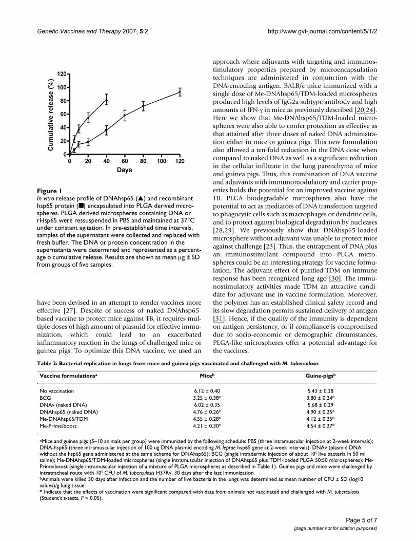
In vitro hsp65 protein, TDM and DNA release profiles from PLGA microspheresThe in vitro release profiles of recombinant hsp65 proteinentrapped into PLGA microspheres were evaluated in vitrofor over 120 days. The results showed that microspheresreleased all the encapsulated protein in a 90-day interval.Most of the protein was released after 50 days (Figure 1).The Me-DNAhsp65/TDM formulation released around80% of their DNA or TDM (not shown) load after 20 days.
Protection against M. tuberculosis replication in lungs of vaccinated animalsAs shown in Table 2, a single dose of Me-DNAhsp65/TDMformulation was able to protect mice as well as guineapigs against M. tuberculosis as efficiently as three doses ofnaked DNAhsp65. There was a significant reduction in thenumber of bacterial burden compared to control mice,
which is similar to the reduction provided by BCG in bothanimal models, which therefore can be considered asgood protection. The same level of protection as describedfor Me-DNAhsp65/TDM was observed in mice and guineapigs vaccinated with Me-Prime/boost formulation in asingle dose.
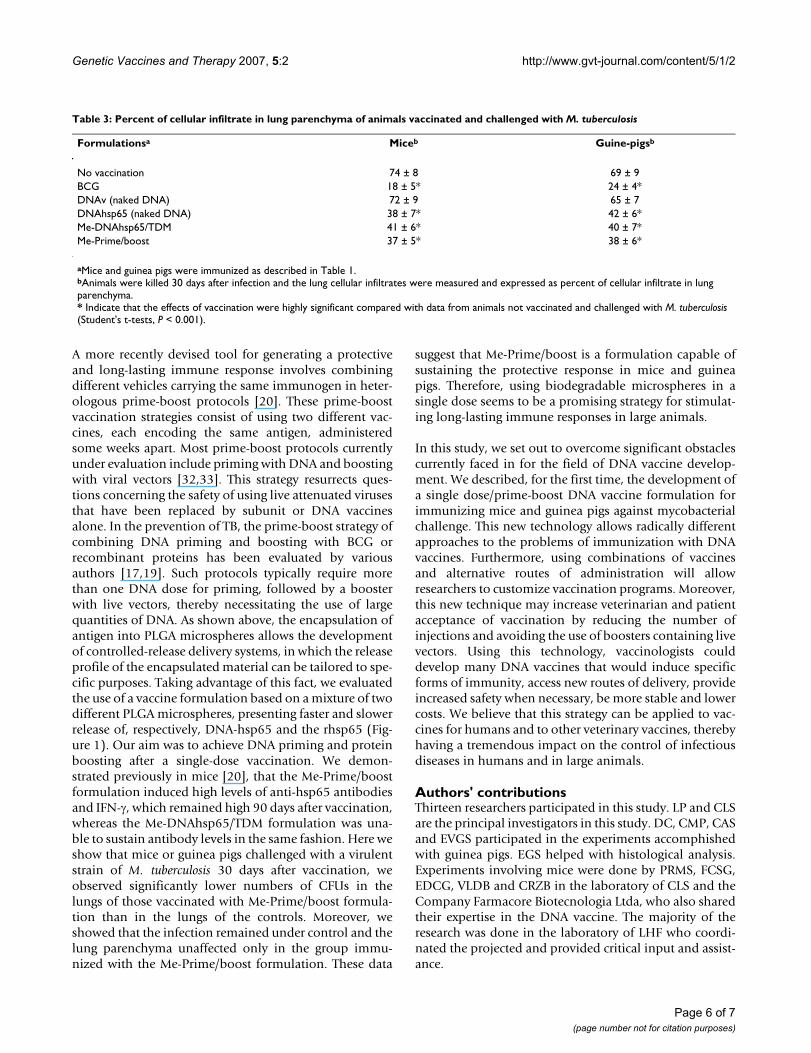
Histological analysis of lungs from mice and guinea pigs vaccinated and challenged with virulent strain of M. tuberculosisThirty days after challenge, animals were sacrificed andlungs were processed for histological analysis. Non-vacci-nated and challenged animals were used as control groupsand the results are illustrated in Table 3. We observed thatin the mice control group (no vaccinated and M. tubercu-losis-infected) around 70% of lung parenchyma was com-promised with granuloma formation widely distributed,presenting cellular infiltrate containing mainly lym-phocytes, with few macrophages and without neutrophils.Mice or guinea pigs injected with DNAhsp65 (nakedDNA) presented a very low and similar compromising oflungs. However, there were few and small granulomas inthis group with cell infiltrate composed by lymphocyte,macrophages, neutrophils and plasmocytes without ofnecrotic areas. The group vaccinated with Me-DNAhsp65/TDM formulation presented lower lung compromisingwith lymphocytes and macrophage infiltration, as well asfew neutrophilic infiltrations (Table 3). Me-Prime/boostvaccinated mice also presented lower lung compromisingwith cellular infiltration characterized by high amount ofmacrophages. In some areas tissue showed lymphocytesconcentrated around bronchus.
DiscussionThe high incidence of TB around the world and the inabil-ity of BCG to protect certain populations clearly indicatethat an improved vaccine against TB is needed. Currently,many vaccines are under development and there is adesire to simplify vaccination schedules by decreasing thenumber of doses. With this purpose, various substanceshave been added to vaccines and certain formulations
Table 1: Median values of diameter distribution, encapsulation rate, and endotoxin levels in each formulation
Formulation Composition Average diameter(μm)
Encapsulation rate(μg/mg of microspheres)
DNA – Protein
Endotoxin level(UE/mg)
Me-Control - 100% of DNAv plus TDM-loaded PLGA 50:50 microspheres
4.0 4.11 - 0.011
Me-DNAhsp65/TDM - 100% of DNAhsp65 plus TDM-loaded PLGA 50:50 microspheres
3.7 4.97 - 0.028
Me-Prime/boost A mixture of PLGA microspheres containing:- 50% of DNAhsp65 plus TDM-loaded PLGA 50:50 microspheres
3.5 4.90 - 0.030
- 50% rHsp65 protein plus TDM-loaded PLGA 75:25 microspheres
2.4 - 1.70 0.053
Page 4 of 7(page number not for citation purposes)
Genetic Vaccines and Therapy 2007, 5:2 http://www.gvt-journal.com/content/5/1/2
have been devised in an attempt to render vaccines moreeffective [27]. Despite of success of naked DNAhsp65-based vaccine to protect mice against TB, it requires mul-tiple doses of high amount of plasmid for effective immu-nization, which could lead to an exacerbatedinflammatory reaction in the lungs of challenged mice orguinea pigs. To optimize this DNA vaccine, we used an
approach where adjuvants with targeting and immunos-timulatory properties prepared by microencapsulationtechniques are administered in conjunction with theDNA-encoding antigen. BALB/c mice immunized with asingle dose of Me-DNAhsp65/TDM-loaded microspheresproduced high levels of IgG2a subtype antibody and highamounts of IFN-γ in mice as previously described [20,24].Here we show that Me-DNAhsp65/TDM-loaded micro-spheres were also able to confer protection as effective asthat attained after three doses of naked DNA administra-tion either in mice or guinea pigs. This new formulationalso allowed a ten-fold reduction in the DNA dose whencompared to naked DNA as well as a significant reductionin the cellular infiltrate in the lung parenchyma of miceand guinea pigs. Thus, this combination of DNA vaccineand adjuvants with immunomodulatory and carrier prop-erties holds the potential for an improved vaccine againstTB. PLGA biodegradable microspheres also have thepotential to act as mediators of DNA transfection targetedto phagocytic cells such as macrophages or dendritic cells,and to protect against biological degradation by nucleases[28,29]. We previously show that DNAhsp65-loadedmicrosphere without adjuvant was unable to protect miceagainst challenge [23]. Thus, the entrapment of DNA plusan immunostimulant compound into PLGA micro-spheres could be an interesting strategy for vaccine formu-lation. The adjuvant effect of purified TDM on immuneresponse has been recognized long ago [30]. The immu-nostimulatory activities made TDM an attractive candi-date for adjuvant use in vaccine formulation. Moreover,the polymer has an established clinical safety record andits slow degradation permits sustained delivery of antigen[31]. Hence, if the quality of the immunity is dependenton antigen persistency, or if compliance is compromiseddue to socio-economic or demographic circumstances,PLGA-like microspheres offer a potential advantage forthe vaccines.
In vitro release profile of DNAhsp65 (▲) and recombinant hsp65 protein (■) encapsulated into PLGA derived micro-spheresFigure 1In vitro release profile of DNAhsp65 (▲) and recombinant hsp65 protein (■) encapsulated into PLGA derived micro-spheres. PLGA derived microspheres containing DNA or rHsp65 were ressuspended in PBS and maintained at 37°C under constant agitation. In pre-established time intervals, samples of the supernatant were collected and replaced with fresh buffer. The DNA or protein concentration in the supernatants were determined and represented as a percent-age o cumulative release. Results are shown as mean μg ± SD from groups of five samples.
Table 2: Bacterial replication in lungs from mice and guinea pigs vaccinated and challenged with M. tuberculosis
Vaccine formulationsa Miceb Guine-pigsb
No vaccination 6.12 ± 0.40 5.43 ± 0.38BCG 3.25 ± 0.38* 3.80 ± 0.24*DNAv (naked DNA) 6.02 ± 0.35 5.68 ± 0.29DNAhsp65 (naked DNA) 4.76 ± 0.26* 4.90 ± 0.25*Me-DNAhsp65/TDM 4.55 ± 0.28* 4.12 ± 0.25*Me-Prime/boost 4.21 ± 0.30* 4.54 ± 0.27*
aMice and guinea pigs (5–10 animals per group) were immunized by the following schedule: PBS (three intramuscular injection at 2-week intervals); DNA-hsp65 (three intramuscular injection of 100 ug DNA plasmid encoding M. leprae hsp65 gene at 2-week intervals); DNAv (plasmid DNA without the hsp65 gene administered at the same scheme for DNAhsp65); BCG (single intradermic injection of about 105 live bacteria in 50 ml saline); Me-DNAhsp65/TDM-loaded microspheres (single intramuscular injection of DNAhsp65 plus TDM-loaded PLGA 50:50 microspheres); Me-Prime/boost (single intramuscular injection of a mixture of PLGA microspheres as described in Table 1). Guinea pigs and mice were challenged by intratracheal route with 105 CFU of M. tuberculosis H37Rv, 30 days after the last immunization.bAnimals were killed 30 days after infection and the number of live bacteria in the lungs was determined as mean number of CFU ± SD (log10 values)/g lung tissue.* Indicate that the effects of vaccination were significant compared with data from animals not vaccinated and challenged with M. tuberculosis (Student's t-tests, P < 0.05).
Page 5 of 7(page number not for citation purposes)
Genetic Vaccines and Therapy 2007, 5:2 http://www.gvt-journal.com/content/5/1/2
A more recently devised tool for generating a protectiveand long-lasting immune response involves combiningdifferent vehicles carrying the same immunogen in heter-ologous prime-boost protocols [20]. These prime-boostvaccination strategies consist of using two different vac-cines, each encoding the same antigen, administeredsome weeks apart. Most prime-boost protocols currentlyunder evaluation include priming with DNA and boostingwith viral vectors [32,33]. This strategy resurrects ques-tions concerning the safety of using live attenuated virusesthat have been replaced by subunit or DNA vaccinesalone. In the prevention of TB, the prime-boost strategy ofcombining DNA priming and boosting with BCG orrecombinant proteins has been evaluated by variousauthors [17,19]. Such protocols typically require morethan one DNA dose for priming, followed by a boosterwith live vectors, thereby necessitating the use of largequantities of DNA. As shown above, the encapsulation ofantigen into PLGA microspheres allows the developmentof controlled-release delivery systems, in which the releaseprofile of the encapsulated material can be tailored to spe-cific purposes. Taking advantage of this fact, we evaluatedthe use of a vaccine formulation based on a mixture of twodifferent PLGA microspheres, presenting faster and slowerrelease of, respectively, DNA-hsp65 and the rhsp65 (Fig-ure 1). Our aim was to achieve DNA priming and proteinboosting after a single-dose vaccination. We demon-strated previously in mice [20], that the Me-Prime/boostformulation induced high levels of anti-hsp65 antibodiesand IFN-γ, which remained high 90 days after vaccination,whereas the Me-DNAhsp65/TDM formulation was una-ble to sustain antibody levels in the same fashion. Here weshow that mice or guinea pigs challenged with a virulentstrain of M. tuberculosis 30 days after vaccination, weobserved significantly lower numbers of CFUs in thelungs of those vaccinated with Me-Prime/boost formula-tion than in the lungs of the controls. Moreover, weshowed that the infection remained under control and thelung parenchyma unaffected only in the group immu-nized with the Me-Prime/boost formulation. These data
suggest that Me-Prime/boost is a formulation capable ofsustaining the protective response in mice and guineapigs. Therefore, using biodegradable microspheres in asingle dose seems to be a promising strategy for stimulat-ing long-lasting immune responses in large animals.
In this study, we set out to overcome significant obstaclescurrently faced in for the field of DNA vaccine develop-ment. We described, for the first time, the development ofa single dose/prime-boost DNA vaccine formulation forimmunizing mice and guinea pigs against mycobacterialchallenge. This new technology allows radically differentapproaches to the problems of immunization with DNAvaccines. Furthermore, using combinations of vaccinesand alternative routes of administration will allowresearchers to customize vaccination programs. Moreover,this new technique may increase veterinarian and patientacceptance of vaccination by reducing the number ofinjections and avoiding the use of boosters containing livevectors. Using this technology, vaccinologists coulddevelop many DNA vaccines that would induce specificforms of immunity, access new routes of delivery, provideincreased safety when necessary, be more stable and lowercosts. We believe that this strategy can be applied to vac-cines for humans and to other veterinary vaccines, therebyhaving a tremendous impact on the control of infectiousdiseases in humans and in large animals.
Authors' contributionsThirteen researchers participated in this study. LP and CLSare the principal investigators in this study. DC, CMP, CASand EVGS participated in the experiments accomphishedwith guinea pigs. EGS helped with histological analysis.Experiments involving mice were done by PRMS, FCSG,EDCG, VLDB and CRZB in the laboratory of CLS and theCompany Farmacore Biotecnologia Ltda, who also sharedtheir expertise in the DNA vaccine. The majority of theresearch was done in the laboratory of LHF who coordi-nated the projected and provided critical input and assist-ance.
Table 3: Percent of cellular infiltrate in lung parenchyma of animals vaccinated and challenged with M. tuberculosis
Formulationsa Miceb Guine-pigsb
No vaccination 74 ± 8 69 ± 9BCG 18 ± 5* 24 ± 4*DNAv (naked DNA) 72 ± 9 65 ± 7DNAhsp65 (naked DNA) 38 ± 7* 42 ± 6*Me-DNAhsp65/TDM 41 ± 6* 40 ± 7*Me-Prime/boost 37 ± 5* 38 ± 6*
aMice and guinea pigs were immunized as described in Table 1.bAnimals were killed 30 days after infection and the lung cellular infiltrates were measured and expressed as percent of cellular infiltrate in lung parenchyma.* Indicate that the effects of vaccination were highly significant compared with data from animals not vaccinated and challenged with M. tuberculosis (Student's t-tests, P < 0.001).
Page 6 of 7(page number not for citation purposes)

Genetic Vaccines and Therapy 2007, 5:2 http://www.gvt-journal.com/content/5/1/2
AcknowledgementsWe thank Izaira T. Brandão and Ana S. Mason for technical assistance. Fundação de Amparo a Pesquisa do Estado de São Paulo (FAPESP), Con-selho Nacional de Desenvolvimento Científico e Tecnológico (CNPq) and Instituto do Milênio REDE TB supported this study.
References1. Kochi A: The global tuberculosis situation and the new con-
trol strategies of the World Health Organization. Tubercle1991, 72:1-6.
2. Rodrigues LC, Diwan VK, Wheeler J: Protective effect of BCGagainst TB meningitis and miliary TB: a meta-analysis. Int JEpidemiol 1993, 22:1154-1158.
3. Colditz GA, Brewer TF, Berkey CS, Wilson ME, Burdick E, FinebergHV, Mosteller F: Efficacy of BCG vaccine in the prevention oftuberculosis. Meta-analysis of the published literature. JAMA1994, 271:698-702.
4. Colditz GA, Berkey CS, Mosteller F, Brewer TF, Wilson ME, BurdickE, Fineberg HV: The efficacy of Bacillus Calmette-Guérin vac-cination of newborns and infants in the prevention of tuber-culosis: meta-analyses of the published literature. Pediatrics1995, 96:29-35.
5. Li Z, Zhang H, Fan X, Zhang Y, Huang J, Liu Q, Tjelle TE, Mathiesen I,Kjeken R, Xiong S: DNA electroporation prime and proteinboost strategy enhances humoral immunity of tuberculosisDNA vaccines in mice and non-human primates. Vaccine 2006,24:4565-4568.
6. Ferraz JC, Stavropoulos E, Yang M, Coade S, Espitia C, Lowrie DB,Colston MJ, Tascon RE: A heterologous DNA priming-Myco-bacterium bovis BCG boosting immunization strategy usingmycobacterial Hsp70, Hsp65, and Apa antigens improvesprotection against tuberculosis in mice. Infect Immun 2004,72:6945-6950.
7. Mollenkopf HJ, Groine-Triebkorn D, Andersen P, Hess J, KaufmannSH: Protective efficacy against tuberculosis of ESAT-6secreted by a live Salmonella typhimurium vaccine carrierstrain and expressed by naked DNA. Vaccine 2001,19:4028-4035.
8. Coler RN, Campos-Neto A, Ovendale P, Day FH, Fling SP, Zhu L, Ser-bina N, Flynn JL, Reed SG, Alderson MR: Vaccination with the Tcell antigen Mtb 8.4 protects against challenge with Myco-bacterium tuberculosis. J Immunol 2001, 166:6227-6235.
9. Takamura S, Matsuo K, Takebe Y, Yasutomi Y: Ag85B of mycobac-teria elicits effective CTL responses through activation ofrobust Th1 immunity as a novel adjuvant in DNA vaccine. JImmunol 2005, 175:2541-2547.
10. Denis O, Tanghe A, Palfliet K, Jurion F, van den Berg TP, VanonckelenA, Ooms J, Saman E, Ulmer JB, Content J, Huygen K: Vaccinationwith plasmid DNA encoding mycobacterial antigen 85Astimulates a CD4+ and CD8+ T-cell epitopic repertoirebroader than that stimulated by Mycobacterium tuberculosisH37Rv infection. Infect Immun 1998, 66:1527-1533.
11. Bonato VL, Goncalves ED, Soares EG, Santos Junior RR, Sartori A,Coelho-Castelo AA, Silva CL: Immune regulatory effect ofpHSP65 DNA therapy in pulmonary tuberculosis: activationof CD8+ cells, interferon-gamma recovery and reduction oflung injury. Immunology 2004, 113:130-138.
12. Lowrie DB, Tascon RE, Bonato VL, Lima VM, Faccioli LH, Stavropou-los E, Colston MJ, Hewinson RG, Moelling K, Silva CL: Therapy oftuberculosis in mice by DNA vaccination. Nature 1999,400:269-271.
13. Lowrie DB, Silva CL, Colston MJ, Ragno S, Tascon RE: Protectionagainst tuberculosis by a plasmid DNA vaccine. Vaccine 1997,15:834-838.
14. Bonato VL, Lima VM, Tascon RE, Lowrie DB, Silva CL: Identificationand characterization of protective T cells in hsp65 DNA-vac-cinated and Mycobacterium tuberculosis-infected mice. InfectImmun 1998, 66:169-175.
15. Johnson PA, Conway MA, Daly J, Nicolson C, Robertson J, Mills KH:Plasmid DNA encoding influenza virus haemagglutinininduces Th1 cells and protection against respiratory infec-tion despite its limited ability to generate antibodyresponses. J Gen Virol 2000, 81:1737-1745.
16. Noll A, Bucheler N, Bohn E, Schirmbeck R, Reimann J, Autenrieth IB:DNA immunization confers systemic, but not mucosal, pro-
tection against enteroinvasive bacteria. Eur J Immunol 1999,29:986-996.
17. Jiao X, Wang RY, Feng Z, Alter HJ, Shih JW: Modulation of cellularimmune response against hepatitis C virus nonstructuralprotein 3 by cationic liposome encapsulated DNA immuni-zation. Hepatology 2003, 37:452-460.
18. Yoshikawa T, Imazu S, Gao JQ, Hayashi K, Tsuda Y, Okada N, Tsut-sumi Y, Akashi M, Mayumi T, Nakagawa S: Non-methylated CpGmotif packaged into fusogenic liposomes enhance antigen-specific immunity in mice. Biol Pharm Bull 2006, 29:105-119.
19. Lima KM, dos Santos SA, Santos RR, Brandao IT, Rodrigues JM Jr, SilvaCL: Efficacy of DNA-hsp65 vaccination for tuberculosis varieswith method of DNA introduction in vivo. Vaccine 2003,22:49-56.
20. Ruberti M, De Melo LK, Dos Santos SA, Brandao IT, Soares EG, SilvaCL, Junior JM: Prime-boost vaccination based on DNA andprotein-loaded microspheres for tuberculosis prevention. JDrug Target 2004, 12:195-203.
21. Vordermeier HM, Rhodes SG, Dean G, Goonetilleke N, Huygen K,Hill AV, Hewinson RG, Gilbert SC: Cellular immune responsesinduced in cattle by heterologous prime-boost vaccinationusing recombinant viruses and bacille Calmette-Guerin.Immunology 2004, 112:461-470.
22. Tsenova L, Harbacheuski R, Moreira AL, Ellison E, Dalemans W,Alderson MR, Mathema B, Reed SG, Skeiky YA, Kaplan G: Evalua-tion of the Mtb72F polyprotein vaccine in a rabbit model oftuberculous meningitis. Infect Immun 2006, 74:2392-2401.
23. Lima VM, Bonato VL, Lima KM, Dos Santos SA, Dos Santos RR, Gon-calves ED, Faccioli LH, Brandao IT, Rodrigues-Junior JM, Silva CL:Role of trehalose dimycolate in recruitment of cells andmodulation of production of cytokines and NO in tuberculo-sis. Infect Immun 2001, 69:5305-5312.
24. Handley HH, Ngyuen MD, Yu DT, Gupta RS, Vaughan JH: Purifica-tion of recombinant human Hsp60: use of a GroEL-free prep-aration to assess autoimmunity in rheumatoid arthritis. JAutoimmun 1995, 8:659-673.
25. Lewis DH: Controlled release of bioactive agents from lac-tide/glycolide polymers. In Biodegradable polymers as drug deliverysystems Edited by: Chasin M, Langer R. New York, N.Y: MarcelDekker; 1990:1-43.
26. Barman SP, Lunsford L, Chambers P, Hedley ML: Two methods forquantifying DNA extracted from poly(lactide-co-glycolide)microspheres. J Control Release 2000, 69:337-344.
27. Lima KM, dos Santos SA, Rodrigues JM Jr, Silva CL: Vaccine adju-vant: it makes the difference. Vaccine 2004, 22:2374-2379.
28. Weintraub H, Cheng PF, Conrad K: Expression of transfectedDNA depends on DNA topology. Cell 1986, 46:115-122.
29. Barry ME, Pinto-Gonzalez D, Orson FM, McKenzie GJ, Petry GR,Barry MA: Role of endogenous endonucleases and tissue sitein transfection and CpG-mediated immune activation afternaked DNA injection. Hum Gene Ther 1999, 10:2461-2480.
30. Lemaire G, Tenu JP, Petit JF, Lederer E: Natural and synthetic tre-halose diesters as immunomodulators. Med Res Rev 1986,6:243-247.
31. Jiang W, Gupta RK, Deshpande MC, Schwendeman SP: Biodegrad-able poly(lactic-co-glycolic acid) microparticles for injecta-ble delivery of vaccine antigens. Adv Drug Deliv Rev 2005,57:391-410.
32. Suh YS, Park KS, Sauermann U, Franz M, Norley S, Wilfingseder D,Stoiber H, Fagrouch Z, Heeney J, Hunsmann G, Stahl-Hennig C, SungYC: Reduction of viral loads by multigenic DNA priming andadenovirus boosting in the SIVmac-macaque model. Vaccine2006, 24:1811-20.
33. Someya K, Ami Y, Nakasone T, Izumi Y, Matsuo K, Horibata S, XinKQ, Yamamoto H, Okuda K, Yamamoto N, Honda M: Induction ofpositive cellular and humoral immune responses by a prime-boost vaccine encoded with simian immunodeficiency virusgag/pol. J Immunol 2006, 176:1784-1795.
Page 7 of 7(page number not for citation purposes)
http://www.ncbi.nlm.nih.gov/entrez/query.fcgi?cmd=Retrieve&db=PubMed&dopt=Abstract&list_uids=1882440
http://www.ncbi.nlm.nih.gov/entrez/query.fcgi?cmd=Retrieve&db=PubMed&dopt=Abstract&list_uids=1882440
http://www.ncbi.nlm.nih.gov/entrez/query.fcgi?cmd=Retrieve&db=PubMed&dopt=Abstract&list_uids=8144299
http://www.ncbi.nlm.nih.gov/entrez/query.fcgi?cmd=Retrieve&db=PubMed&dopt=Abstract&list_uids=8144299
http://www.ncbi.nlm.nih.gov/entrez/query.fcgi?cmd=Retrieve&db=PubMed&dopt=Abstract&list_uids=8309034
http://www.ncbi.nlm.nih.gov/entrez/query.fcgi?cmd=Retrieve&db=PubMed&dopt=Abstract&list_uids=8309034
http://www.ncbi.nlm.nih.gov/entrez/query.fcgi?cmd=Retrieve&db=PubMed&dopt=Abstract&list_uids=7596718
http://www.ncbi.nlm.nih.gov/entrez/query.fcgi?cmd=Retrieve&db=PubMed&dopt=Abstract&list_uids=7596718
http://www.ncbi.nlm.nih.gov/entrez/query.fcgi?cmd=Retrieve&db=PubMed&dopt=Abstract&list_uids=7596718
http://www.ncbi.nlm.nih.gov/entrez/query.fcgi?cmd=Retrieve&db=PubMed&dopt=Abstract&list_uids=9529077
http://www.ncbi.nlm.nih.gov/entrez/query.fcgi?cmd=Retrieve&db=PubMed&dopt=Abstract&list_uids=9529077
http://www.ncbi.nlm.nih.gov/entrez/query.fcgi?cmd=Retrieve&db=PubMed&dopt=Abstract&list_uids=9234527
http://www.ncbi.nlm.nih.gov/entrez/query.fcgi?cmd=Retrieve&db=PubMed&dopt=Abstract&list_uids=9234527
http://www.ncbi.nlm.nih.gov/entrez/query.fcgi?cmd=Retrieve&db=PubMed&dopt=Abstract&list_uids=9423854
http://www.ncbi.nlm.nih.gov/entrez/query.fcgi?cmd=Retrieve&db=PubMed&dopt=Abstract&list_uids=8579722
http://www.ncbi.nlm.nih.gov/entrez/query.fcgi?cmd=Retrieve&db=PubMed&dopt=Abstract&list_uids=8579722
http://www.ncbi.nlm.nih.gov/entrez/query.fcgi?cmd=Retrieve&db=PubMed&dopt=Abstract&list_uids=8579722
http://www.ncbi.nlm.nih.gov/entrez/query.fcgi?cmd=Retrieve&db=PubMed&dopt=Abstract&list_uids=3459589
http://www.ncbi.nlm.nih.gov/entrez/query.fcgi?cmd=Retrieve&db=PubMed&dopt=Abstract&list_uids=3459589
http://www.ncbi.nlm.nih.gov/entrez/query.fcgi?cmd=Retrieve&db=PubMed&dopt=Abstract&list_uids=3526051