comparative study of the siddha diagnostic - EPrints@Tamil ...
148
COMPARATIVE STUDY OF THE SIDDHA DIAGNOSTIC METHODS SPECIALLY NEERKURI & NEIKURI WITH MODERN DIAGNOSTIC METHODS IN NEERIZHIVU MADHUMEHAM (DIABETES MELLITUS – TYPE 2) A DISSERTATION SUBMITTED BY Dr. Kalaimony Rabindrakumar Vidya dharshini TO THE TAMIL NADU DR. M.G.R. MEDICAL UNIVERSITY CHENNAI - 32 in partial fulfilment of the requirement for the award of the degree of DOCTOR OF MEDICINE (SIDDHA) (BRANCH V – PG. NOI NAADAL) DEPARTMENT OF NOI NAADAL GOVERNMENT SIDDHA MEDICAL COLLEGE PALAYAMKOTTAI - 627 002 OCTOBER 2018
-
Upload
khangminh22 -
Category
Documents
-
view
0 -
download
0
Transcript of comparative study of the siddha diagnostic - EPrints@Tamil ...
COMPARATIVE STUDY OF THE SIDDHA DIAGNOSTIC
METHODS SPECIALLY NEERKURI & NEIKURI WITH
MODERN DIAGNOSTIC METHODS IN NEERIZHIVU
MADHUMEHAM (DIABETES MELLITUS – TYPE 2)
A DISSERTATION SUBMITTED BY
Dr. Kalaimony Rabindrakumar Vidya dharshini
TO THE TAMIL NADU DR. M.G.R. MEDICAL UNIVERSITY
CHENNAI - 32
in partial fulfilment of the requirement
for the award of the degree of
DOCTOR OF MEDICINE (SIDDHA)
(BRANCH V – PG. NOI NAADAL)
DEPARTMENT OF NOI NAADAL
GOVERNMENT SIDDHA MEDICAL COLLEGE
PALAYAMKOTTAI - 627 002
OCTOBER 2018

i
GOVERNMENT SIDDHA MEDICAL COLLEGE AND HOSPITAL
PALAYAMKOTTAI, TIRUNELVELI - 627002,
TAMIL NADU, INDIA.
Phone: 0462-2572736/2572737/2582010 Fax: 0462 2582010
DECLARATION BY THE CANDIDATE
I hereby declare that this dissertation entitled COMPARATIVE STUDY OF THE
SIDDHA DIAGNOSTIC METHODS SPECIALLY NEERKURI & NEIKURI WITH
MODERN DIAGNOSTIC METHODS IN NEERIZHIVU MADHUMEHAM
(DIABETES MELLITUS – TYPE 2) is a bonafide and genuine research carried out
by me under the guidance and supervision of Prof. Dr. M. Krishnaveni, Professor,
Department of Noi Naadal (past) and Head, Department of Udal thathuvam and
Prof. Dr. S. Victoria, Head, Department of Noi Naadal, Government Siddha Medical
College and Hospital, Palayamkottai, Tamil Nadu, India and the dissertation has not
formed the basis for the award of any other degree.
Date:
Place: Signature of the candidate
ii
GOVERNMENT SIDDHA MEDICAL COLLEGE AND HOSPITAL
PALAYAMKOTTAI, TIRUNELVELI - 627002,
TAMIL NADU, INDIA.
Phone: 0462-2572736/2572737/2582010 Fax: 0462 2582010
CERTIFICATE
Certified that I have gone through the dissertation submitted by
Dr. Kalaimony Rabindrakumar Vidya dharshini (Reg. No. 321515002) with the tittle,
COMPARATIVE STUDY OF THE SIDDHA DIAGNOSTIC METHODS
SPECIALLY NEERKURI & NEIKURI WITH MODERN DIAGNOSTIC METHODS
IN NEERIZHIVU MADHUMEHAM (DIABETES MELLITUS – TYPE 2),
the student of final DOCTOR OF MEDICINE (SIDDHA), Department of Noi Naadal
(Branch – V) of the Government Siddha Medical College and Hospital,
Palayamkottai, Tamil Nadu, India. This dissertation does not represent or reproduce
the dissertation submitted and approved earlier.
Date:
Place: Head of the Department
iii
GOVERNMENT SIDDHA MEDICAL COLLEGE AND HOSPITAL
PALAYAMKOTTAI, TIRUNELVELI - 627002,
TAMIL NADU, INDIA.
Phone: 0462-2572736/2572737/2582010 Fax: 0462 2582010
BONAFIDE CERTIFICATE
This is to certify that the dissertation entitled COMPARATIVE STUDY OF THE
SIDDHA DIAGNOSTIC METHODS SPECIALLY NEERKURI & NEIKURI WITH
MODERN DIAGNOSTIC METHODS IN NEERIZHIVU MADHUMEHAM
(DIABETES MELLITUS – TYPE 2), is a bonafide work done by
Dr. Kalaimony Rabindrakumar Vidya dharshini (Reg. No. 321515002), Government
Siddha Medical College and Hospital, Palayamkottai, Tamil Nadu, India in partial
fulfilment of the requirement of the University rules and regulations for the award of
the degree of DOCTOR OF MEDICINE (SIDDHA), NOI NAADAL (Branch - V)
under my guidance and supervision during the academic year 2015 - 2018.
Name and Signature of the supervisor:
1. Prof. Dr. M. Krishnaveni
2. Prof. Dr. S. Victoria
Name and Signature of the Head of the Department:
Name and Signature of the Principal:

iv
ACKNOWLEDGEMENT
I express my sincere gratitude to Vice Chancellor, The Tamil Nadu DR. MGR
Medical University for conceding permission to carry out the research project.
I express my sincere thanks to Professor. Dr. (Mrs). R. Neelavathy, Principal,
Government Siddha Medical College, Palayamkottai, Tirunelveli, Tamil Nadu for
granting permission to carry out the research project.
I would like to express my heart-felt gratitude to my supervisor Professor. Dr.
(Mrs). S. Victoria, Head, Department of Noi Naadal, Government Siddha Medical
College, Palayamkottai, Tirunelveli. Her guidance and support was useful throughout
the project.
I would like to express my sincere gratefulness to my supervisor Professor. Dr. (Mrs).
M. Krishnaveni, Professor, Department of Noi Naadal (past) and Head, Department of
Udal thathuvam, Government Siddha Medical College, Palayamkottai, Tirunelveli for
generous support, patience, systemic guidance and encouragement she has provided
throughout my research and most importantly spending her valuable time with me to
complete my research successfully.
I would like to convey my grateful to Dr. R. Raja Selvi, District Siddha Medical
Officer, for granting permission to attend the Siddha Wing, Government District
Headquarters Hospital, Thoothukudi and get the patients for the study.
I wish to convey my sincere gratitude to Dr. M. Sankararama Subramanian,
Department of Noi Naadal, Government Siddha Medical College, Palayamkottai,
Tirunelveli for giving immense support during the selection of the patients.
My special thanks to Dr. A. Anbumalar, Department of Special Medicine,
Government Siddha Medical College, Palayamkottai, Tirunelveli for providing
valuable guidance and support whenever required.

v
My heart-felt thanks to Dr. S. Sri Ram, Dr. S. Sundararajan, Dr. B. Senthil Selvi and
Dr. R. Meenatchisuntharam, Department of Noi Naadal, Government Siddha Medical
College, Palayamkottai, Tirunelveli for giving support and sharing knowledge in the
selection of patients.
My grateful to Professor. Dr. A. Manoharan, Head, Department of General Medicine,
Government Siddha Medical College, Palayamkottai, Tirunelveli, for granting
permission to attend indoor patient department to select the patients, make a great
help to carry out this research.
My truthful recognitions to Dr. A. Manoraj, Community Health Medical Officer
(preventive service), Department of Ayurveda – Central Province, Pallekele,
Kundasale, Kandy, Sri Lanka for his valuable guidance all over the research and
thesis writing.
I would like to express my gratitude to my sister Kalaimony Rabindrakumar Miruna
Sudharshani, Department of Biochemistry and Molecular Biology, Faculty of
Medicine, University of Colombo, Sri Lanka for the guidance in the statistical
analysis.
I would like to thank all the technical and other staff members at the Government
Siddha Medical College, Palayamkottai, Tirunelveli and Siddha Wing, Government
District Headquarters Hospital, Thoothukudi for their continues support throughout
my research.
My heartfelt gratitude to all my colleagues for their helps and support entire period of
the study.
I take this opportunity to express the profound gratefulness from my bottom of the
heart to my beloved mother for her love and support.
Finally, I like to acknowledge every one whose names are not mentioned individually
for their support and encouragements to bring this dissertation to successful
completion.

vi
CONTENTS
Page No.
Certificates
Declaration by candidate i
Certificate by Head ii
Bonafide certificate iii
Acknowledgements iv
Table of content vi
List of tables xi
List of figures xiii
List of Abbreviations xiv
1. Introduction 1
2. Aim and objectives 3
2.1 Aim 3
2.2 Objectives 3
2.2.1 Primary objective 3
2.2.2 Secondary objectives 3
3. Literature review 4
3.1 Neerizhivu madhumeham and different names 4
3.1.1 Neerizhivu madhumeham 4
3.1.2 Other names of Neerizhivu madhumeham 4
3.2 General characters of neerizhivu 6
3.3 Types of neerizhivu 7
3.4 Causes of neerizhivu 21
3.5 Complication of neerizhivu 24
3.6 Prognosis of neerizhivu 25
3.7 Dietary regimen for neerizhivu 25
3.8 Diagnosis of Neerizhivu madhumeham according to the Siddha system 28
3.8.1 Envagaithervu 28
3.8.2 Neikuri 28
3.8.3 Neerkuri 30

vii
3.8.3.1 Colour variation of urine 31
3.8.3.2 Froth in urine 32
3.8.3.3 Odour, sediment and volume of urine 33
3.8.4 Naadi 33
3.8.4.1 The sign and symptoms for thontha naadi
(combination of two naadi) according to
Agasthiyar vaiththiya sillarai kovai 36
3.8.5 Manikkadai nool 41
3.9 Modern aspect of diabetes mellitus (DM) 42
3.9.1 General considerations 42
3.9.2 Global burden 42
3.9.3 Clinical features of Diabetes mellitus general symptoms 44
3.9.4 Pathogenesis and pathophysiology of DM 45
3.9.5 Risk factors of DM 48
3.9.6 Complications 49
3. 9.7 Diagnosis of DM 50
3.9.8 Preventing diabetes 52
4. Materials and methods 53
4.1 General procedure 53
4.1.1 Study protocol 53
4.1.2 Selection of the subjects 53
4.1.2.1 Inclusion criteria 53
4.1.2.2 Exclusion criteria 54
4.1.2.3 Criteria for withdrawal 54
4.2 Study design 54
4.3 Statistical analysis 57
4.4 Laboratory investigations 57
4.4.1 Collection of blood and blood investigations 57
4.4.2 Urine analysis – modern method 58
4.4.3 Urine analysis – Siddha method 58
4.4.3.1 Neer kuri 58
4.4.3.2 Neikuri 58
4.4.4 Naadi 58
4.4.5 Manikkadai 59

viii
4.5 Ethical clearance 59
4.6 Questionnaires 60
4.6.1 Types of questionnaire 60
5. Results 61
5.1 Diagnosis of NR 61
5.1.1 The sign and symptoms of the patients 61
5.1.2 Assessment of biochemical parameters of the blood
samples (modern aspect) of the patients attending to
the OPD and IPD at first time - GSMC, Palayamkottai,
GDHH,Thoothukudi and GSV, Tirunelveli 62
5.1.3 Assessment of biochemical parameters of the urine
samples (modern aspect) of the patients attending at baseline –
OPD and IPD at GSMC, Palayamkottai, GDHH,
Thoothukudi and GSV, Tirunelveli 63
5.1.4 Factors associating with concentration of blood sugar 64
5.1.5 Examination of urine by neerkuri (Siddha aspect) 65
5.1.6 Examination of urine by neikuri (oil drop test (Siddha
aspect) 67
5.1.6.1 Shape of the neikuri 67
5.1.6.2 Duration for testing the neikuri and the
biochemical parameters of the urine samples 68
5.1.6.3 Shape of the neikuri and the biochemical
parameters of the urine samples 70
5.1.7 Naadi 71
5.1.7.1 Correlation of neikuri with the diagnosis of naadi 71
5.1.8 Manikkadai 72
5.2 Prognosis 73
5.2.1 The changes of the common sign and symptoms observed at
baseline, after following the treatment with
Madhumega chooranam 73
5.2.2 Changes of FBS following the treatment of
Madhumega chooranam 74
5.2.3 Changes of the biochemical parameters of urine sample,
with the treatment of Madhumega chooranam 74
ix
5.2.4 Changes in the parameters of neerkuri with the treatment
of Madhumega chooranam 77
5.2.5 Changes in the neikuri with the treatment of
Madhumega chooranam 78
5.2.5.1 Changes in the shape 78
5.2.5.2 Variations in the duration of time taken to start
and disappear the scatter of neikuri 79
5.2.6 Changes in the naadi with the treatment of
Madhumega chooranam 81
5.2.7 Correlation between fasting blood sugar and naadi 82
5.2.8 Manikkadai 83
6. Discussion 84
7. Summary 89
8. Conclusion 92
9. Bibliography 93
10. Annexures
Annexure I - Consent form 98
1 Consent form - English 98
2 Consent form - Tamil 100
Annexure II - Patient information sheet 101
1 Patient information sheet - English 101
2 Patient information sheet – Tamil 104
Annexure III - Dietary regimens 107
1 Dietary regimens - Neerizhivu madhumeham
(diabetes mellitus) - English 107
2 Dietary regimens - Neerizhivu madhumeham
(diabetes mellitus) - Tamil 114
Annexure IV - Case report forms
1 Case report form I - screening 116
2 Case report form II - history 118
3 Case report form III - laboratory investigation 121
4 Case report form IV - laboratory investigation 125
(after taking anti Madhumeha drugs)
Annexure V - Ethical clearance 126
x
Annexure VI - Screening committee 127
Annexure VII - Journal publications
1 Vidya Dharshini, K., Mangalambigai, V., Krishnaveni, M.,
Muthurathinam, S., Saravanan, R. and Meenakumar, S. -
Pharmacognostical characterization of Aavarai kudineer -
A poly herbal preparation - Journal of Medicinal Plants
Studies - 2017, 5, (6): pp. 1-5. 128
2 Vidya Dharshini, K. and Neelavathy, R. - Comparative study
of locations of nadi with anatomical landmarks - A review -
World journal of pharmaceutical and medical research -
2018, 4, (4): pp. 142-144. 129
Annexure VII – Certificates
1 Research methodology & biostatics 130
2 Research methodology and public health initiative through
Siddha system of medicine 131
3 CME - Siddha maruthuva murai parisothanaigal 132

xi
LIST OF TABLES
Page No.
Table 3.1. Characters of the types of neerizhivu according to
Ramachandran (2000) 18
Table 3.2. Character of kabha neerizhivu according to Yuki
vaiththiya kaviyam 21
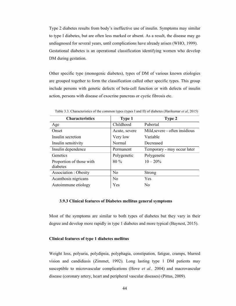
Table 3.3. Characteristics of the common types (types I and II) of
diabetes (Harikumar et al, 2015) 44
Table 5.1. The common signs and symptoms observed in patients attending
to the out and inpatient department at first time – OPD and IPD
at GSMC, Palayamkottai, GDHH, Thoothukudi and GSV,
Tirunelveli 61
Table 5.2. Assessment of the biochemical parameters of the blood
samples (modern aspect) of patients 62
Table 5.3. Biochemical parameters (modern aspect)) of the patient’s
urine samples 63
Table 5.4. Examination of neerkuri (Siddha aspect of urine examination)
in the patients at baseline 66
Table 5.5. Shape of the neikuri in urine samples and fasting blood sugar
at baseline 67
Table 5.6. Comparison of the time taken to start and disappear the scatter
of neikuri and biochemical parameters of the urine samples at
baseline 69
Table 5.7. Number of patients with different shapes of neikuri and their
biochemical properties of urine samples 70
Table 5.8. Concentrations of fasting blood sugar and naadi at base line 71
Table 5.9. Correlation of neikuri with the diagnosis of naadi at baseline 72
Table 5.10 Concentration of fasting blood sugar and manikkadai at
baseline 72
Table 5.11 Comparison of common sign and symptoms of the patient
following the treatment of Madhumega chooranam 73
Table 5.12. Changes of the biochemical parameters of the urine sample
with the treatment of Madhumega chooranam 75

xii
Table 5.13. Changes in the parameters of neerkuri with the treatment of
Madhumega chooranam 77
Table 5.14. Changes in the shape of the neikuri with the treatment of
Madhumega chooranam 78
Table 5.15. Changes in the naadi following the treatment of Madhumega chooranam 81
Table 5.16. Mean concentrations of the FBS and naadi at seventh visit 82
xiii
LIST OF FIGURES
Page No.
Figure 3.1. Trends in prevalence of diabetes, 1980 – 2014, by country
income group 43
Figure 3. 2. Physiologic and behavioural response of hyperglycemia
(Baynest, 2015) 46
Figure 3.3. Pathophysiologic of hyperglycemia and increased circulating
fatty acids in type II (Baynest, 2015) 48
Figure 4.1. Flow chart of methodology 56
Figure 5.1. The association of fasting blood sugar and urine Glucose
level 64
Figure 5.2. Morphological description (colours and froth) of neerkuri 65
Figure 5.3. Shape of the neikuri 67
Figure 5.4. Concentration of fasting blood sugar following the treatment
of Madhumega chooranam 74
Figure 5.5. Changes in the pH of the urine with the treatment of
Madhumega chooranam 76
Figure 5.6. Changes in the specific gravity of the urine with the treatment
of Madhumega chooranam 76
Figure 5.7. Variations in the time taken to start to scatter of neikuri with
the treatment of Madhumega chooranam 79
Figure 5.8. Variations in the total time taken to disappear the scatter of
neikuri with the treatment of Madhumega chooranam 80
Figure 5.9. Changes in the manikkadai of the patient following the
treatment of Madhumega chooranam 83

xiv
LIST OF ABBREVIATIONS
NR Neerizhivu Madhumeham
DM Diabetes mellitus
WHO World Health Organization
IDF International Diabetes Federation
IDDM Insulin dependent diabetes mellitus
NIDDM Non - insulin dependent diabetes mellitus
MODY Maturity onset diabetes of the young
HbA1c Glycated haemoglobin A
OPD Outpatient department
IPD Inpatient department
GSMC Government Siddha Medical College
GDHH Government District Headquarters Hospital
GSV Gopalasamudram village
FBS Fasting blood sugar
PPBS Postprandial Blood sugar
SPSS Statistical Package for the Social Science
SGOT Serum glutamic-oxaloacetic transaminase
SGPT Serum glutamic pyruvic transaminase
SD Standard deviation
n Number of subjects

1
CHAPTER 1
INTRODUCTION
Siddha system of medicine is one of the oldest systems of medicine in the world,
originated and developed by the Siddhars (Ivy and Malini, 2010). Thirumoolar is
considered as the Emperor of Siddha system of medicine and he was a Tamil Shaivite
mystic and supernatural writer. He is considered as one of the 63 nayanars and one of
the 18 Siddhars. He is the author of the famous literatures Thirumanthiram and Saiva
Siddhantam which is framed the basic principles of Siddha system (Wikipedia, 2013).
His principles on Siddha system are astonishing. Further his book Thirumoolar
vaithiyam (Karukadai 600) is one of the valuable medical book in Siddha system of
medicine. Versions of Thirumoolar are certainly suitable for the contemporary modern
world with stress and strain.
‘,Ukpagpj;jKk; thjKk;$bby;
kUtr;ryNkfk; thhpjpNghNyhLk;
cUtKk;NtwhF Kz;lTlw;fhAk;
cUfpa%isAwpQ;rpap dpf;FNk’
(Thirumoolar vaithiyam karukkadai – 600, verse 83)
The above verse expressing the aetiology, sign and symptoms of Neerizhivu
Madhumeham (NR). Increased Pittham and Vatham are the main causes for the NR.
The above verse indicates the sign and symptoms as, increase output of the urine like
a heavy rain and sweet substances pass with urine. Changes occur in the body
structure. Further arise feeling of hunger as immediate as had meals. The aetiology,
sign and symptoms of NR is similar to that of Madhumeham (Thirumoolar vaithiyam
(Karukadai 600) indicating the possibility that NR may consider as Madhumeham. In
addition NR may compare with diabetes mellitus (DM) in Allopathic system of
medicine because the sign and symptoms of DM as polyuria, weight loss and
polyphagia (Baynest, 2015) are overlapping with NR.
Diabetes mellitus is a group of metabolic diseases characterized by hyperglycemia
resulting from defects in insulin secretion, insulin action or both (Ozougwu, 2013).
2
The World Health Organization (WHO) estimated that diabetes resulted in 1.5 million
deaths in 2012, making it the 8th leading cause of death (WHO, 2013). In 2014, the
International Diabetes Federation (IDF) estimated that diabetes resulted in 4.9 million
deaths (IDF, 2014). More than 80% of diabetic deaths occur in low and middle-
income countries (Mathers and Loncar, 2006).
A number of diagnostic methods have been using in various systems of medicine
globally. Siddha system has unique assessment methods as envagaithervu (naadi,
sparism, naa, niram, mozhi, vizhi, malam and siruneer), neikuri and manikkadai to
diagnose and prognosis of the diseases (Natarajan, 2009; Shanmugavelu, 1967).
Lack of user friendly modern scientific technologies and little usage of Siddha
diagnostic methods have led to non-familiar of these diagnostic methods among the
healthcare and scientific community. A little brief studies observed in DM with siddha
diagnostic methods. Although elaborate comparative study on NR with Siddha
diagnostic methods specially with neerkuri, neikuri, naadi and manikkadai has not
been studied previously to the best of my knowledge. It is essential to give a scientific
validation to the Siddha diagnostic methods for NR to the effective reuse of in clinical
practice.
Therefore the current study was designed to test the interactions between the NR and
Siddha diagnostic methods specially neerkuri and neikuri and also compares it with
modern diagnostic methods to bring out the significance of the knowledge obtained
from the Siddha system of medicine.
3
CHAPTER 2
AIM AND OBJECTIVES
2.1 Aim
To evaluate the diagnosis and prognosis of NR using Siddha diagnostic methods.
2.2 Objectives
2.2.1 Primary objective
To study the interactions between the NR and neerkuri & neikuri and compare it
with modern diagnostic methods.
2.2.2 Secondary objectives
1. To study the correlation between NR and naadi.
2. To test the link between NR and manikadai.
3. To demonstrate the prognosis of NR using neerkuri, neikuri, naadi and
manikadai.
4. To observe the changes of neikuri after the intake of anti Neerizhivu
Madhumeha drugs.
4
CHAPTER 3
LITERATURE REVIEW
Neerizhivu madhumeham – Neerizhivu + mathumeham means excessive sweet taste
urination (Sambasivam pillai, 1998).
3.1 Neerizhivu madhumeham and different names
3.1.1 Neerizhivu madhumeham
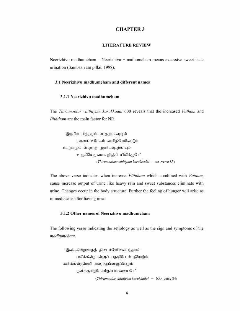
The Thirumoolar vaithiyam karukkadai 600 reveals that the increased Vatham and
Piththam are the main factor for NR.
‘,Ukpa gpj;jKk; thjKk;$bby;
kUtr;ryNkfk; thhpjpNghNyhLk;
cUtKk; NtwhF Kz;lTlw;fhAk;
cUfpNa%isAwpQ;rp apdpf;FNk’
(Thirumoolar vaithiyam karukkadai – 600>verse 83)
The above verse indicates when increase Piththam which combined with Vatham,
cause increase output of urine like heavy rain and sweet substances eliminate with
urine. Changes occur in the body structure. Further the feeling of hunger will arise as
immediate as after having meal.
3.1.2 Other names of Neerizhivu madhumeham
The following verse indicating the aetiology as well as the sign and symptoms of the
madhumeham.
‘,dpf;fpd;wthjj; jpilr;Nrhpiyae;jhd;
gdpf;fpd;wfs;Sg; gjdpNghy; ePNuhLk;
fdpf;fpd;wNkdp fiue;JntSg;NgWk;
jdpf;FkJNkfk;jg;ghkiyaNk’
(Thirumoolar vaithiyam karukkadai – 600, verse 84)

5
The verse reveals, when the combination of Vatham and Iyam, the urine will be in
sweet taste as ‘pathaneer’. The body will emaciate and change to pallor.
The above literature (Thirumoolar vaithiyam karukkadai – 600) reveals that sign and
symptoms of NR is similar with the sign and symptoms of madhumeham reveal as the
NR is nearly equal to madhumeham.
Ramachandran (2000), Sambasivam pillai (1998) and Yuki vaiththiya kaviyam (2014)
documents the sign and symptoms of the disease neerizhivu. Ramachandran (2000)
documents the sign and symptoms of neerizhivu as emaciation, pallor of the body and
eye, sweating, ache and pain, dryness of the face, hand, leg and chest, thirsty,
tiredness, fatigue, increase sleep during the day and night, loss of appetite and
excessive urination.
Sambasivam pillai (1998) documents, the meaning of neerilizhu is excessive
urination.
The Yuki vaiththiya kaviyam states the sign and symptoms of neerizhivu as,
‘ KfKq;fhe;jpneQ;ryh;e;J Kwpe;jTlYeLeLq;fp
efNkywpe;J ehtwz;L eQ;Rz;lth;Nghy;kpfr;Nrhh;e;J
gfYkputpkpuq;fpAly; ghpe;Njjl;bnkype;Jod;W
kpfNtjtdKz;lhfp Ntz;lhjjd;dk; Ntz;lhNj’
‘nka;NaNth;f;FKly;ehWk; tpopNagyfhy;kpf%b
iffhnye;JehTyh;e;J fkyKfKk;ntSntSj;J
[NahTlk;GNehFnjd;ghh; mah;e;Jfplg;ghh;fz;fhz
ngha;NajtdKz;lhFk; nghUe;jhjd;dk;nghUe;jhNj’
(Yuki vaiththiya kaviyam, verse 787 -788)
The verse 787 (first) documents burning sensation of face, dryness of chest and
mouth, tremor in the body, fatigue, increase sleep during day and night, emaciation,
ache and pain in the body and loss of appetite. The second verse indicates as sweating,
6
odour in body, excessive sleep, ache and pain in arm and leg, dryness of mouth, pallor
of face like lotus, ache and pain in the body, tiredness and loss of appetite.
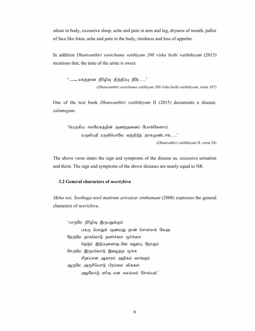
In addition Dhanvanthiri sootchuma vaithiyam 200 visha bethi vaiththiyam (2015)
mentions that, the taste of the urine is sweet.
‘ ….. kfj;jhd ePhpopT jpj;jpg;G ePNu…..’
(Dhanvanthiri sootchuma vaithiyam 200 visha bethi vaiththiyam, verse 187)
One of the text book Dhanvanthiri vaiththiyam II (2015) documents a disease,
salamegam.
‘ngUfpa ryNkfj;jpd; Fze;jidg; Ngrf;Nfsha;
kUtpaeP uUtpnghNy te;jpLe; jhfKz;lhq;…..’
(Dhanvathiri vaiththiyam II, verse 24)
The above verse states the sign and symptoms of the disease as, excessive urination
and thirst. The sign and symptoms of the above diseases are nearly equal to NR.
3.2 General characters of neerizhivu
Meha noi, Soothaga nool mattrum arivaiyar sinthamani (2008) expresses the general
characters of neerizhivu.
‘ghUNk ePhpopT ,UgJf;Fk;
gfU nghJf; FzkJ jhd; nrhy;yf; NfS
NeUNk jhfnkhL jsh;r;ir %h;r;ir
neLk; ,Lg;GisTlNd vYk;G NehFk;
NrUNk ,UknyhL ,ioj;j %r;R
rpwg;ghd Mfhuk; mjpfk; thq;Fk;
MUNk mUrpnahL gpuk;ik tpf;fy;
moNyhL vhpT kd fyf;fk; Nrhk;gy;’

7
‘Nrhk;gpNa Njfkjpy; csr;ry; NehT
rpwg;ghf cwf;fkpd;wp clYk; Njk;gp
Njk;gpNa fhw;W Vl;f mjpf Mir
jpwkhf Nky;%r;Nr thA NghFk;
Xk;gpNa NjfkJ tpswpg; NghFk;
cWk; kaf;fk; Mahrk; nkypAk; Njfk;
ntk;gpNa rpW ePUk; mjpfk; NghFk;
tpsq;Fk; ePhpoptpd; Fzk; jhd; fhNz’
(Meha noi, Soothaga nool mattrum arivaiyar sinthamani, verse 119-120)
The above stanza states excessive thirst, giddiness, back ache for long time, bony
pain, cough with difficulty in breathing, excessive intake of food, loss of taste,
hiccough, burning sensation, mind disturbance, laziness, ache and pain with tiredness,
loss of sleep, difficulty in breathing, pallor of body, loss of consciousness, fatigue,
emaciation and excessive urination as the general characters of neerizhivu.
3.3 Types of neerizhivu
Different types of neerizhivu were documented in literature. Differences noted in the
classifications of neerizhivu among the literature. Twenty types of neerizhivu were
mentioned in Meha noi, Soothaga nool mattrum arivaiyar sinthamani (2008) and Yuki
vaiththiya kaviyam (2014). Ramachandran (2000) documents twenty four types of
neerizhivu.

8
Classification I
Types and characters of neerizhivu according to Meha noi, Soothaga nool mattrum
arivaiyar sinthamani given below.
1. Mahenthira varni
‘vd;Wk; ,dp kNfe;jpu th;zp nra;if
Vw;w ryk; nea; NghNy tbAk; ghU
ed;whf Jdpjdpy; eidj;J jP apy;
eP nfhSj;jpdhy; jP vhpAk; tpsf;F NghNy
Fd;wpNa NjfkJ nkype;J NghFk;
nfhLk; jhfk; m];jpapNy R+Lz;lhFk;
Jd;whky; gjpdhop ryk; jhd; NghFk;
JlUk; kNfe;jpu th;zp FzkpjhNk’
(Meha noi, Soothaga nool mattrum arivaiyar sinthamani , verse 134)
Urine passes like ghee. If you soak a piece of cloth in that urine and fire, it will burn
like a lamp. Emaciation, severe thirst, feeling heat in bone and specially pathin naazhi
(5600g – (1 naazhi = 560g) of urine passes at a time.
2. Inthira varni
‘tpsq;fNt ,e;jpu th;zp Fzj;ij nrhy;Ntd;
tpsq;F ryk; Nfh ryk; Nghy; epwNkahFk;’
jsq;fwNt Nfh ryj;jpd; ehw;wk; fhZk;
jg;ghy; fhr;rpdhy; vhpAk; jP jhd;
msq;fwNt Mahrk; jsh;r;irAz;lhk;
moF Kfk; jhd; nkypAk; fd;dk; xl;Lk;
fsq;fkw Ntisf;F gbjhd; ehY
fhZnkd Kd;Ndhh;fs; $wpdhNu’
(Meha noi, Soothaga nool mattrum arivaiyar sinthamani, verse 136)
Colour and odour of the urine resembles as cow’s urine and burn when fire. Tiredness,
weakness, narrowness in the face, emaciation and passing 4 padi (5.2 L – (1 padi =
1.3 L) urine passes at a time

9
3. Ruththiranga varni
‘nrhy;YNk Uj;jpuhq;f th;zp Njhd;wpy;
Nrh;e;j ryk; Nfhryk; Nghy; th;zkhFk;
nty;Yk; khkpr thil kpf cz;lhFk;
tpsq;f mij fha;r;rpdhy; Njd; Nghy; thil
epy;Y ,sk; thypgDk; fpotd; Nghyhk;
epiw m];jp jhd; NtFk; gdpAz;lhFk;
my;yNt Ntis ehdhop ePU
mZFtJk; eprnkd mwpayhNk’
(Meha noi, Soothaga nool mattrum arivaiyar sinthamani, verse 138)
Urine resembles as cow’s urine. Flesh odour present in the urine and honey smell
when heat the urine. Young appearance of the body change in to old. Destruction of
bone and blood present in urine or 4 naazhi (2240 g) urine passes at a time.
4. Sooththira varni
‘jpz;zKw R+j;jpu th;zp nra;if
jpwk; Ml;bd; nfhOg;gJ Nghy; ePh; jhdhFk;
tz;zKw fha;r;rpdhy; Ml;bd; nea; Nghy;
tw;wpdhy; fUk;gpd; ePh; thrk; fhZk;
vz;zKw ypq;fj;jpy; vhpTFj;J
vOe;j Ruk; jhfk; mNuhrpak; the;jp
epz;zpakha; ehnshd;wpy; FWzp rhAk;
eP mwptha; R+j;jpudhk; th;zp jhNd’
(Meha noi, Soothaga nool mattrum arivaiyar sinthamani, verse 141)
Urine passes like goat’s fat. The urine seems as goat ghee and smell as sugar candy
juice, when reducing the volume by boil. Burning sensation and pain present in genital
region. Fever, thirst, aversion for food and vomiting present. Passing 1 kuruni (5.376
L – (1 kuruni = 5.376 L)) urine at a time.

10
5. Vishba piraban ‘rhw;W tp];g gpwgdjpd; Fzk; Vnjd;why;
rypahky; ePh; epwk; jhd; ahidapd; jd;
Nghw;W %j;jpuk; NghNy th;zkhFk;
nghUe;J kzk; mJ Nghyhk; fha;r;rp ghh;j;jhy;
khw;Wkjpy; cg;G ciwAk; xU NghNy
kdk; jsUk; cly; maUk; nkypAk; Njfk;
Njhw;Wk; xU Ntisf;F gb ehdhop
NjhifaNu ePhpwq;Fk; njhFj;J nrhy;Ny’
(Meha noi, Soothaga nool mattrum arivaiyar sinthamani, verse 144)
Colour and odour of the urine is like that of an elephant urine. Salt sedimentation is
seems when boiling the urine. Tension, tiredness and emaciation present. Passing
4 naazhi (2240 g) urine at a time.
6. Thirkentha vaahini
‘ghUkpdp Jh;nfe;j thfpdpapd; nra;if
gfUfpNwd; ghhpYs;Nshh; mwpa Ntz;b
rhUkpdp rpWePh; fw;whio rhW Nghy;
rypahkNy ehWk; Nkhh;e;J ghU
thUkij jhd; fha;r;rpdhy; gpz ehw;wk; Nghy;
thilAWk; jhjaUk; xspAk; kq;Fk;
NeUk; xU Ntis ehdhop ePU
nefpOk; Jh;nfe;j thfpdpf;nfd;Nw’
(Meha noi, Soothaga nool mattrum arivaiyar sinthamani, verse 146)
Aloe odour present in urine and the smell change in to the odour of a death body when
boiling the urine. Impotency and loss of brightness of the body presence. Four naazhi
(2240 g) urine passes at a time.
11
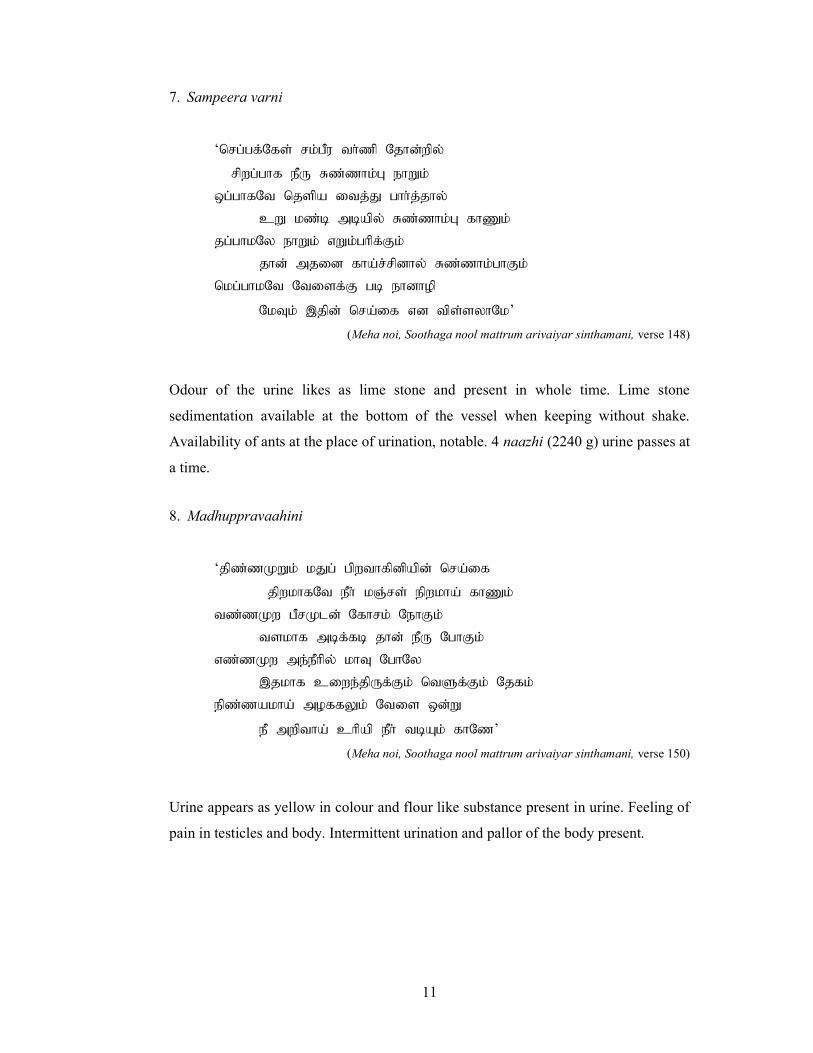
7. Sampeera varni
‘nrg;gf;Nfs; rk;gPu th;zp Njhd;wpy;
rpwg;ghf ePU Rz;zhk;G ehWk;
xg;ghfNt njspa itj;J ghh;j;jhy;
cW kz;b mbapy; Rz;zhk;G fhZk;
jg;ghkNy ehWk; vWk;ghpf;Fk;
jhd; mjid fha;r;rpdhy; Rz;zhk;ghFk;
nkg;ghkNt Ntisf;F gb ehdhop
NkTk; ,jpd; nra;if vd tps;syhNk’
(Meha noi, Soothaga nool mattrum arivaiyar sinthamani, verse 148)
Odour of the urine likes as lime stone and present in whole time. Lime stone
sedimentation available at the bottom of the vessel when keeping without shake.
Availability of ants at the place of urination, notable. 4 naazhi (2240 g) urine passes at
a time.
8. Madhuppravaahini
‘jpz;zKWk; kJg; gpwthfpdpapd; nra;if
jpwkhfNt ePh; kQ;rs; epwkha; fhZk;
tz;zKw gPrKld; Nfhrk; NehFk;
tskhf mbf;fb jhd; ePU NghFk;
vz;zKw me;ePhpy; khT NghNy
,jkhf ciwe;jpUf;Fk; ntSf;Fk; Njfk;
epz;zakha; moffYk; Ntis xd;W
eP mwptha; chpap ePh; tbAk; fhNz’
(Meha noi, Soothaga nool mattrum arivaiyar sinthamani, verse 150)
Urine appears as yellow in colour and flour like substance present in urine. Feeling of
pain in testicles and body. Intermittent urination and pallor of the body present.

12
9. Salappravaahini
‘tpl;L Nghk; ryg; gpwthfpdpapd; nra;if
tpsq;F [yk; gbf epwkhf fhZk;
njhl;LlNd gPrKk; Kjy; Nfhrk; NehFk;
jhio tpsph; rhw;wpDl epwkhk; ePU
fl;LlNd fha;r;rpdhy; rPO ehWk;
fhZNk Ntisf;F ehop ePU
kl;LlNd KfKk; thLk; Nkdp Fd;Wk;
kjpj;j m];jp jhd; fhAkpJ thwpjhNk’
(Meha noi, Soothaga nool mattrum arivaiyar sinthamani, verse 152)
Transparent urine like as glass available. The colour changes to white and foul smell
noted when heating the urine. Pain present in testicles and body. Dullness present in
face and emaciation occur. Destruction of bone also present. One naazhi (560 g) urine
passes at a time.
10. Reththa jalakkini
‘tpUk;gpaNjhh; ,uj;j [yhf;fpdpapd; thW
tpsk;GfpNwd; cyNfhh;fs; mwpa Ntz;b
eUk;G ePh; Kay; nuj;jk; NghNy fhZk;
eykhf fha;r;rpdhy; GyhYk; ehWk;
mUk;gpNa gPrKld; NfhrKk; NehFk;
mlUk; cly; nkypAk; kpf moYk; kPWk;
jUk;G nkhU Ntisf;F gbjhd; uz;L
rhANk ePuJTk; rhw;wyhNk’
(Meha noi, Soothaga nool mattrum arivaiyar sinthamani, verse 154)
Urine resembles as rabbit blood. Flashy odour presence when boil the urine. Pain
present in testicles and body. Emaciation occurs and increase piththam. Two padi (2.6
L) urine passes at a time.

13
11. Sukkila pravaahini
‘cz;Z Rf;fpy gpwthfpdpapd; nra;if
cw;WNfs; epzk; NghNy ePhpy; fhZk;
ez;zpajpy; ghilAk; fha;r;rpdhYk;
ehWNk epzthil etpyg; Nghfh
tz;zkhk; NjfkJ cyh;e;J NghFk;
tskhf Nkdp Fd;Wk; cly; js;shLk;
jz;ikaha; Ntisf;F Ke;ehop ePU
njspthf ,wq;Fk; vd;W rhw;wyhNk’
(Meha noi, Soothaga nool mattrum arivaiyar sinthamani, verse 156)
Urine like as lymph and odour of the lymph present when boiling. Emaciation, weight
loss and weakness of the body are the other symptoms. Three naazhi (1680 g) urine
passes at a time.
12. Oothaka varnan
‘NghFNk cjf th;zd; Njhd;Wk; Mdhy;
nghUe;Jk; ePh; njspe;j ryk; Mf fhZk;
ghFwNt Mahrk; kaf;fk; %h;r;ir
gz;ghfNt clYk; mah;e;J NghFk;
NtfKw Ntisf;F FWzp ePU
tpLk; vdNt ed;whf nrhd;dhh; jhNk’
(Meha noi, Soothaga nool mattrum arivaiyar sinthamani, verse 158)
Clear urine. Fatigue, faintness, unconsciousness and tiredness present. One kuruni
(5.376 L) urine passes at a time.

14
13. Malsiya varni
‘jhdKs;s ky;rpa th;zp Fzj;ijr; nrhy;Ntd;
jg;ghky; gz;bjh;fs; mwpa Ntz;b
<dKw epzk; cUfp ePhpwq;Fk;
,ij fha;r;rpdhy; kPdpd; thil cz;lhk;
Cdkw Njfk; vy;yhk; cyh;e;J NghFk;
cwthd jhjaUk; m];jp fhAk;
NkhdKw Ntisf;F %d;W ehop
NkhJnkd nghpNahh;fs; nrhd;dthNw’
(Meha noi, Soothaga nool mattrum arivaiyar sinthamani, verse 161)
Emaciation, the fat melts and pass with urine (ninam). Fish flesh odour when boiling
the urine. Dryness of the body and bones. Three naazhi (1680 g) urine passes at a
time.
14. Thoola varnan
‘jpz;zKld; Ày th;zd; jhDk;
jpwkhf ,sePh; Nghy; rykpwq;Fk;
tz;zKw ,sePhpd; thrKz;lhk;
tskhf NjfkJ nkype;J fhZk;
epz;zakha; cly; NehFk; kdf;fyf;fk;
epj;jKNk Mahrk; jsh;r;rpAz;lhk;
vz;zKwNt Ntisf;F ehop rhAk;
Vw;w Fzk; ,Jntd ,irf;fyhNk’
(Meha noi, Soothaga nool mattrum arivaiyar sinthamani, verse 163)
Colour and odour of the urine resemble as tender coconut. Emaciation, ache and pain
in the body, mind disturbance, fatigue presents daily, weakness and 1 naazhi (560 g)
urine passes at a time.

15
15. Suraari varnan
‘GfYNtd; Ruhhp th;zd; Fzj;ij ahDk;
nghUe;J fs;spd; th;zkjha; ntSj;j ePU
,fYNtd; gijNahL VFk; ,d;Dk;
,ij fha;r;rpdhy; fs;spd; thil cz;lhk;
EtYk; ,Lg;NghL nghUj;J FWf;F NghFk;
Nehpioahs; jid ntWf;Fk; jhJ Fd;Wk;
mfYk; xU Ntisf;F ehop ePh; Nghk;
mwpFtha; ,jpd; Fzk; vd;W ,ak;GthNa’
(Meha noi, Soothaga nool mattrum arivaiyar sinthamani, verse 165)
Colour of the urine seems as toddy and toddy odour present when boiling the urine.
Pain in the hip, weakness and 1 naazhi (560 g) urine passes at a time.
16. Asthi varnan
‘NghFNk m];jp th;zd; Fzk; Vnjd;why;
nghUe;J ryk; Rf;fpyj;jpd; th;zkhFk;
thFngw jhspahJ fyf;fpdhg;Nghy;
tskhfNt rpWf Áy; Nghy; ghAk;
MFNk fha;r;rpy; fl;bahfp
mJ Gife;J Jw;nfe;jthil cz;lhk;
ghfKwNt nghjpif Kdpth; nrhd;d
gz;;ghd m];jp th;zd; ghq;fpjhNk’
(Meha noi, Soothaga nool mattrum arivaiyar sinthamani, verse 167)
Colour of urine like as semen and passes like as thread. When boil, the consistency of
urine changes into solid and produce bad odour.
16
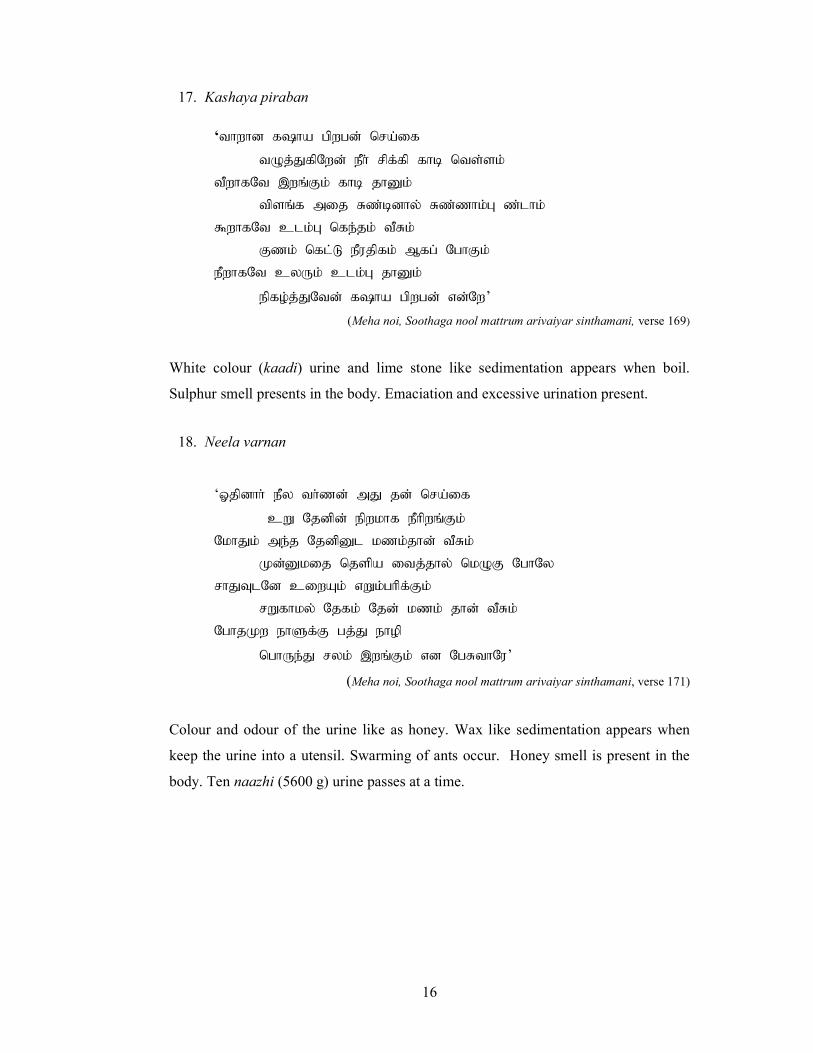
17. Kashaya piraban ‘thwhd f~ha gpwgd; nra;if
tOj;JfpNwd; ePh; rpf;fp fhb nts;sk;
tPwhfNt ,wq;Fk; fhb jhDk;
tpsq;f mij Rz;bdhy; Rz;zhk;G z;lhk;
$whfNt clk;G nfe;jk; tPRk;
Fzk; nfl;L ePujpfk; Mfg; NghFk;
ePwhfNt cyUk; clk;G jhDk;
epfo;j;JNtd; f~ha gpwgd; vd;Nw’
(Meha noi, Soothaga nool mattrum arivaiyar sinthamani, verse 169)
White colour (kaadi) urine and lime stone like sedimentation appears when boil.
Sulphur smell presents in the body. Emaciation and excessive urination present.
18. Neela varnan
‘Xjpdhh; ePy th;zd; mJ jd; nra;if
cW Njdpd; epwkhf ePhpwq;Fk;
NkhJk; me;j NjdpDl kzk;jhd; tPRk;
Kd;Dkij njspa itj;jhy; nkOF NghNy
rhJTlNd ciwAk; vWk;ghpf;Fk;
rWfhky; Njfk; Njd; kzk; jhd; tPRk;
NghjKw ehSf;F gj;J ehop
nghUe;J ryk; ,wq;Fk; vd NgRthNu’
(Meha noi, Soothaga nool mattrum arivaiyar sinthamani, verse 171)
Colour and odour of the urine like as honey. Wax like sedimentation appears when
keep the urine into a utensil. Swarming of ants occur. Honey smell is present in the
body. Ten naazhi (5600 g) urine passes at a time.

17
19. Lavanap piravaahini
‘jhdKwNt ytzg; gpwthfpdpapd;
jd;ikjid nrhy;YfpNwd; ryj;jpNy jhd;
VdKw Rz;zhk;G fhuk; Nghyhk;
,jkhf fha;r;rpdhy; fhuk; fhZk;
Cdkw vhpTlNd fLg;G R+L
cwthFk; m];jpapy; R+Lz;lhFk;
NkhdKw Ntisf;F gb ehy; ePU
NkhJnkdNt njsptha; nkhopayhNk’
(Meha noi, Soothaga nool mattrum arivaiyar sinthamani, verse 173)
Alkaline medium of urine (lime stone pH) is avilable. Burning sensation and pricking
pain occur and feeling of heat in bone is found. Four padi (5.2 L) urine passes at a
time.
20. Sukkila varnan
‘ePuhFk; Rf;fpy th;zd; Fzk; Vnjd;why;
ePh; ,iwr;rp fOtpdjpd; ryk; Nghy; fhZk;
rPuhf fha;r;rpdhy; khkprk; Nghy; ehWk;
rpwg;ghf fhZk; ,jpy; R+Lk; cz;lhk;
Nguhf ePh;j;jhiu KWf;fk; VWk;
gpyf;Nflha; jhjaUk; cly; js;shLk;
Ntuhf Ntisf;F gbjhd; %d;W
tpsq;fNt ryk; mJTk; NghFk; thNw’
(Meha noi, Soothaga nool mattrum arivaiyar sinthamani, verse 175)
Flesh wash water appearance of urine as well as fleshy smell is present when boiling
the urine. Weakness, unsteadiness available and 3 padi (3.9 L) urine passes at a time.

18
Classification II
Ramachandran, (2000) documents 24 types of neerizhivu such as,
1. Vatha neerizhivu – 3
2. Vatha piththa neerizhivu – 4
3. Piththa neerizhivu – 3
4. Piththa vatha neerizhivu – 2
5. Sileththuma neerizhivu – 4
6. Sileththuma piththa neerizhivu – 3
7. Sileththuma vatha neerizhivu – 5
The table 1 has shown the types and character of neerizhivu documented by
Ramachandran (2000).
Types of neerizhivu Characters
Smell Taste Vatha neerizhivu - 3 Type 1 Mango flower Sour
Type 2 Saffron Bitter Type 3 Kaadi Sour
Vatha piththa neerizhivu - 4 Type 1 Turmeric Bitter and sour Type 2 Oleander Five types of taste Type 3 Milk Butter Type 4 Lymph Bitter taste like sweet flag
Piththa neerizhivu - 3 Type 1 Syrup Bitter Type 2 Salt Salt Type 3 Jasmine Salty
Piththa vatha neerizhivu - 2 Type 1 Cow’s urine Astringent Type 2 Sandal Black pepper
Sileththuma neerizhivu - 4 Type 1 Screw spine Sweet Type 2 Cow dung Sweet Type 3 Lime Sweet Type 4 Blood Sweet
Sileththuma piththa neerizhivu - 3
Type 1 Bad odour Lime stone Type 2 Champak Jamun fruit Type 3 - -
Sileththuma vatha neerizhivu
All 5 types
Bad odour sour
Table. 3.1. Characters of the types of neerizhivu according to Ramachandran (2000)

19
Classification III
The text book the Yuki vaiththiya kaviyam (2014) describes 20 types of neerizhivu.
‘ce;jpapy;thjgpj;j Nrj;Jk%d;We;jhDk;
te;jpahapioAk;gj;jp kfpo;e;jplnthd;Wf;nfhd;W
te;jkpy;yhjehY kLe;jpLk; gpj;jkhWk;
je;jNrj;JkNkgj;;J jtwhkyhjpahNk’
(Yuki vaiththiya kaviyam, verse 792)
The above verse describes 20 types of neerizhivu based on Vatham, Piththam and
Kabham. Further the book documents four type of Vatha neerizhivu, six Piththa and
ten in Kabha neerizhivu.
The following verse describes the types and characters of vatha neeerizhivu
‘thjj;jpdhy; te;jehYf;Fk; tORJk;NgUq;Fzq;Nfsha;
ePjptrisAj;jkDk; ePSk;Nghrdkw;wpbDk;
Nghjg;Ngrd;dndda;nghjpe;J nghd;Nd ntz;iz tpsf;nfwpAk;
khNjkj;jpld; NfhkaKk; tUe;J ehWkd;dpaNj’
(Yuki vaiththiya kaviyam, verse 807)
‘trisg;gpukpankhd;Wf;F kfpOkpiwr;rp fOePh;
mirAnea;NghNykpjf;F khFk;trisg; gpuNkfk;
eprkhAj;jNkndDk; tifNa ePSk;ghy;Nghy; ehLnkd;W
tpirAq;fWg;Gj;Njq;fhap ypUf;FKj;jkd; Fzkd;Nw’
(Yuki vaiththiya kaviyam, verse 808)
The verse describes the types of the vatha neerizhivu as vasalai uththaman, vasalai
bramiyam, vasalai bramegam and uththaman. The literature describes the characters
of vasali uththaman as loss of appetite and the urine like as komayam. In vasalai
bramiyam, the urine appears as flesh wash water and the urine burn when contact with
fire. Urine appears as oil floating in water and milk in vasalai bramegam and
uththaman respectively.

20
The following verse describes the character of piththa neeerizhivu in.
‘cjpuKq;fhpg;GKz;lh AtUld;Gspg;GQ;rhy
rjpuJjhdope;J jiyaJRow;WQ;rhy
KjpuNtfpWfpWj;J %h;r;rpf;Fk; gpj;jkhWk;
Ngjpufd;kdJNghy NghtJtUtjhNk’
(Yuki vaiththiya kaviyam, verse 796)
The above verse describes the common characters of the Piththa neeerizhivu as the
patient prefer salt and sour taste, loss of memory, giddiness and loss of consciousness.
Even though the types of Piththa neeerizhivu is not mentioned.
In addition the following verse reveals some characters of piththa neerizhivu.
‘gpj;jj;jpdhYtUkhWf;Fk; ngUFkhidkjk;NghY
Kj;jf;fw;whioehWtJ Kjph;e;jjtohg;Nghy;ehWtJk;
Kw;wePh;gl;ltplnky;yh Kdpe;Njnahpg;G ehWtJk;
kw;wG+Ntnahpf;Fk; thff;fLj;JfopAkd;Nw’
(Yuki vaiththiya kaviyam, verse 809)
The odour of the urine in similar to that of aloe or wild spider flower or burning
smoke and the patient suffers from dysuria.
The following verse describes the types and characters of kabha neeerizhivu
‘Nrj;Jkj;jhy; Njhd;Wk;gj;Jf;FQ; nrg;gf;NfSkpjd;Ngiu
aPj;NjAkd;njrgdd;gd; ePgde;jd;ew;wPah
ePj;jRf;fpy;ytDlePSQ; rjhrptndd;NghJ
khj;Jk; tp~;ZjhDkpf kd;Dk;gj;JtifahNk’
(Yuki vaiththiya kaviyam, verse 810)
Ten types of kabha neerizhivu documented in Yuki vaiththiya kaviyam, eventhough
nine types described as uththaman, thesaban, pananthan, sukkilan, sathasivan, vishnu,
alavanan, mannan and manthiri.

21
‘cj;jkzpePh;NghypUf;f Kfe;jrjhrptd;fe;jk; Nghy;
itj;jTg;gprkpfTz;lhk; kd;DkptDEiu Nghyhk;
nkj;jtp~;Zfiuj;jkhg;Nghy; kpd;Dk;gOg;Ge;jpj;jpg;ghk;
kj;jphpnad;Nghd;G+nthpf;F khdePUk;gpuk;nghpf;Fk;’
(Yuki vaiththiya kaviyam, verse 811)
‘Rf;fpyq;fUg;gd;ghy;Nghy; Rjpj;jurKKz;lhFk;
kpf;fytzd;FjpiuePh;Nghy; kpFe;jjaph;epwkhaPnkha;f;Fk;
jf;fNrjkk;gj;Jf;Fk; jhNdFznkhd;We;jg;ghJ
xf;fKdpth;khdplh;f;F Tiuj;jhUz;ikg;gbad;Nw’
(Yuki vaiththiya kaviyam, verse 812)
The verses 811 and 812 describes the characters of urine in Kabha neerizhivu and the
table 2 has shown the characters of Kabha neerizhivu,
3.4 Causes of neerizhivu
Tri humours such as Vatham, Piththam and Kabham are the basic principal of Siddha
system of medicine, which governs the psycho-biological aspect of the body
(Natarajan, 2009). Further the author (Natarajan, 2009) documents that increases or
reduces of the tri humours causes disease. In addition, Agasthiyar vaiththiya vallathi-
600 describes (in the following verse) that increase of Vatham causes neerizhivu.
‘……%z;bLNk thjkPwpy; ePhpopNt…..’
(Agasthiyar vaiththiya vallathi-600,verse 5)
Type of Kabha neerizhivu Characters of urine Uththaman Like water Thesaban - Pananthan - Sukkilan Smell like juice of sugar cane Sathasivan Smell like sulphur Vishnu Urine shine as flour batter in water Alavanan Like horse urine or curd Mannan Excessive frothy in urine, sweat in taste, Manthiri When contact with fire the urine will burn
Table. 3.2. Character of kabha neerizhivu according to Yuki vaiththiya kaviyam

22
The text book Meha noi, Soothaga nool mattrum arivaiyar sinthamani (2008)
documents the causes, pathogenesis and the types of neerizhivu in the following
verses.
‘$whd ePhpopTk; ,UgjhFk;
nfhz;nlOe;j tuyhW $wf;NfS
thwhd ghy; nea;Ak; ,iwr;rp fs;S
tskhd kPd; Ntfhg;gz;lk; khT
CwhfNt mjpfk; jZj;j t];j;J
cwthfNt mjpfk; Grpj;jhYk;
ePwhf ngz; Nghfk; tpUk;gpr; nra;J
kpf Njfk; jbj;J mdypd; fhuzj;jhy;’
‘fhuzkha; epj;jpiu ,y;yhjjhYk;
fjpg;ghd mf;fpdpapd; ke;jj;jhYk;
rPuzpah fy; ckpAk; Grpf;fyhYk;
rpwe;jlq;fh rQ;ryj;jpd; VJthYk;
G+uzkha; %ykjpy; mdY gw;wp
nghUe;Jk; mj;jp %is nte;J nghq;fp
khuzkha; kr;irnahL nfhOg;G kw;Wk;
kq;fpNa euk;ngy;yhk; gyKk; nfl;L’
‘nfl;l cly; jdpy; cs;s ePh;fnsy;yhk;
nfWTlNd tp~ euk;G jd;dpy; Gf;fp
kl;LlNd mq;fkjpd; nfhOg;G vy;yhk;
khWkit ePuhf tbe;J ghAk;
jpl;lKld; fz; FopAk; fd;dk; xl;Lk;
NjfkJ gyk; Fiwe;J Mbg; NghFk;
nrhl;L tpOk; ePupoptpd; nra;if jd;id
nrg;GfpNwd; tpgukij Njh;e;J NfNs’
(Meha noi, Soothaga nool mattrum arivaiyar sinthamani, verse 108-110)
The above verses describe, excessive intake of milk, meat, toddy, fish, partly cooked
food and flour, increase sexual activities, loss of sleep, indigestion, taking indigestive
foods and mind disturbance are the causes for neeizhivu. Further, the above causes
promote the Piththam and the Piththam affect on the bone especially bone marrow

23
and fat in the body. Thereafter it affects the nervous system. Finally it causes
emaciation and weakness of the body and the fat excretes with the urine.
The Yuki vaiththiya kaviyam (2014) also states the causes for neerizhivu in the
following verses (verse 785, 786 and 789).
‘Nfhijah;fytpNguhq; nfhOj;jkPdpiwr;rpNahLk;
NghjNtghYnea;Ak; nghUe;jePh;kpsFrhW
MirapdhNyAz;L md;wpuhTuq;fhdhfpy;
NrhifAk;gpj;jghz;L nrhy;YePhpopTkhNk’
(Yuki vaiththiya kaviyam, verse 785)
‘kz;lye;jd;dpYs;s kdpjhpw;ngz;Zs;Nshh;f;Fk;
nfhz;lNjhh;gpuNkfe;jhd; nfhs;SKd;ndLj;j Neha;fs;
fz;Lld;if fhy; jhDq; foz;bLkz;Nzfhe;jp
cz;lePh;Rtwpf;fl;b Aile;JePhpopTthNk’
(Yuki vaiththiya kaviyam, verse 786)
‘fl;liskpFe;jpl;lhYq; fhyq;fs;jg;gpdhYk;
,l;lkhk;ghYe;nea;Ak; apujKk;Gspg;GkpQ;rpy;
tl;lkhKiyahh;jq;fs; kaf;fj;jpd;fytpahYk;
nel;biyf;Nfhiu NghNy ePhpopthFe;jhNd’
(Yuki vaiththiya kaviyam, verse 789)
‘ghpe;Njnea;Ald;ghYlNd gUj;jkPDiwr;rpaJ
,Ue;NjAz;LRfkjid apiljhd;tplhkyDgtpj;J
tUe;jhJlYk;jhd;tj;jp kd;dpAile;JePhpopthk;
jpUe;jNghfkDjpdKq; fsh;e;jnfhLikahw;nra;J’
(Yuki vaiththiya kaviyam, verse 790)
‘Nfsha;NghfkDjpdKq; fpsh;e;jnfhLikahw;nra;J
ehSQ;nrhy;ypf;FbnfLj;J ew;ngz;BUk;gjptpuij
thshh;fd;dpfw;gopj;J tuk;Gfle;Jkhwhfp
khshkhSk;tpahjpapdhy; typaNeha;fs;te;jpLNk’
(Yuki vaiththiya kaviyam, verse 791)

24
The verses describe having excessive sexual activities, having excessive workload,
avoidance of sleep after having large fish, meat, milk, ghee and decoction of pepper,
delay intake of food, excessive intake of milk, ghee, meat, sour taste, the diseases
present before affected by bramegam and rupture of cyst causes neerizhivu.
3.5 Complication of neerizhivu
Ten types of complications describes in Meha noi, Soothaga nool mattrum arivaiyar
sinthamani (2008) as,
‘fhZkpe;j ePhpopT jhd; cw;Nwhh;f;F
fjpf;fpd;w gj;J tpj mt];ij nrhy;Ntd;
G+Zfpd;w Kjy; mt];ij clk;G jhDk;
nghUj;jKw fdkhFk; gUj;J fhZk;
ez;Z rpWePh; jhd; jZf;Fk; ePh; fLf;Fk;
eykhd ,uz;lhtJ mt];ij nrhy;yf; NfS
NtZ %j;jpuk; jdpy; Rf;fpyk; fhZk;
NtWKfk; mOf;Nfwp Nkdp Fd;Wk;’
‘Fd;wpNa %d;wt];ij twSk; ehf;F
Fzkhd tha;TWk; ehyt];ij
Jd;wpNa jhfKWk; rd;dp ghjk;
JlUNk Ie;jhtJ mt];ij NfS
ntd;wp ePh; mjpfk; Nghk; jhJ Fd;Wk;
tPwhd MwhtJ mt];ij vd;dpy;
kd;wpdpNy gpujhgk; %h;r;ir NtT
kUTk; ,jd; Kwiknad nrg;gyhNk’
‘nrg;gpaNjhh; VohtJ mt];ij NfS
rpwg;ghd mNuhrpaKk; tPf;fk; cz;lhk;
xg;gpaNjhh; vl;lhtJ mt];ijahfpy;
cWk; fpue;jp gpsit cz;lhk; cwtjhf
jg;gpaNjhh; xd;gJf;F FiwAk; md;dk;
jhd; fpUkp %j;jpuj;jpy; kpFjpahFk;’
nka;g;ghd jr mt];ij Ñak; cz;lhfp
Nkyhd rlk; mopAk; cz;ikahNk’
(Meha noi, Soothaga nool mattrum arivaiyar sinthamani, verse 121-123)

25
In the first stage, the body weight increases, feeling of heaviness and dysuria occurs.
In the second stage, the sperm passes in the urine, changes occur in the appearance of
the body and the body weight decreases. Dryness of mouth and gas formation occurs
in the third and fourth stages respectively. Delirium (sanni) is present in the fifth
stage. Excessive urination, emaciation and loss of consciousness arise in the sixth
stage. At the stage of seven loss of taste and anasarca appear. Kiranthi and pilavai
appear at the stage of eight. Loss of appetite and organism appear at the stage of nine.
Finally in the tenth stage shayam (tuberculosis) ensues and the person dies.
3.6 Prognosis of neerizhivu
According to the Meha noi, Soothaga nool mattrum arivaiyar sinthamani (2008) the
following verse documents that the six types of Piththa and ten types of Kabha
neerizhivu are curable, whereas four types of Vatha neerilizhivu are incurable.
‘NfSkpdp thjkjpy; Nrh;e;J ehYk;
Nfbahd gpj;jj;jpy; MWkhFk;
thSkpdp Nrh;g;gj;NjhL vOe;j gj;Jk;
tskhfNt mitfs; ,Ugjhr;Nr
ehSkpdp thjj;jpy; ehYk; mrhj;jpak;
eykhf gpj;jj;jpy; vOe;j MWk;
R+sNt tUj;jkjhk; Nrh;g;gj;jhNy
R+o;e;j gj;Jk; rhj;jpak; vd;Wiuf;fyhNk’
(Meha noi, Soothaga nool mattrum arivaiyar sinthamani, verse 111)
3.7 Dietary regimen for neerizhivu The Meha noi, Soothaga nool mattrum arivaiyar sinthamani elucidates the foods
which can intake and avoid in the condition of neerizhivu. The following stanzas
explain the dietary regimens for neerzhivu.

26
‘nfhy;yhky; ePhpopT cs;s Ngh;f;F
$whf cgNahf gjhh;j;jk; nrhy;Ntd;
ey;yhd Nkjp ntz;nza; Nkjp NkhU
etpYk; nghd;dhq;fhzp rpWfPiu jhDk;
ty;yhd Krpl;il ,iy mtiu gpQ;R
tsh; Glyq;fhAlNd KUq;ifg; gpQ;R gj;jpuk;
my;yhj tpshq;fdpAld; Ngad; goKk;
mj;jpAld; gpQ;R rpWgaW jhNd’
‘jhdKs;s gok; NrhW ghfy; gPh;f;F
fwp Ntg;gpiyAlNd ky;ypf; fPiu
VdKs;s ney;nghhpAk; KRf;if gj;jpuk;
Vw;w vs;S mjpndz;nza; tuF jhDk;
CkwNt cSe;J cYtha; rPuk;
cwthd nfhj;jky;yp Nfhjk;NghNl
Md Ch; FUtpNahL milf;fyhDk;
mjpNdhNl fhil nts;shl;bd; ,iwr;rp cz;Z’
‘cz;Ztha; rt;thprp Jtiu rhik
cw;w fiy khdpiwr;rp cLk;G G+id
ez;Z kueha; fPhp FapYk;
eykhd jtpl;Lg; Gwh ntz;GwhTk;
rz;ZNk $it ePh; mkph;tJ khTk;
rypahky; nfhy;yhkhtpd; mz;b cz;Z
tz;zKWk; thJikapd; gUg;G nej;jyp
jpwkhd fUthL MFk; nrhy;Ny’
(Meha noi, Soothaga nool mattrum arivaiyar sinthamani, verse 129-131)
The stanzas reveal, butter, butter milk, sessile joy weed, green amaranth, common
night glory, tender beans, snake gourd, tender drum stick, wood apple, tender Indian
fig fruit, green gram, bitter gourd, king of gourd, curry leaves, coriander leaves, cold
cooked rice, madras pea pumpkin, gingelly oil, kodo millet, green gram, fenugreek,
cumin, coriander, kind of sparrow, quail bird, goat white ovis, sago, meat of art deer,
iguana, cat, switch dog, Asian palm civet, mungos, cuckoo, red turtle dove, white
dove, arrowroot flour, flour of lead wood tree seeds, white bait and dry fish are good
to intake for the condition of neerizhivu.
27
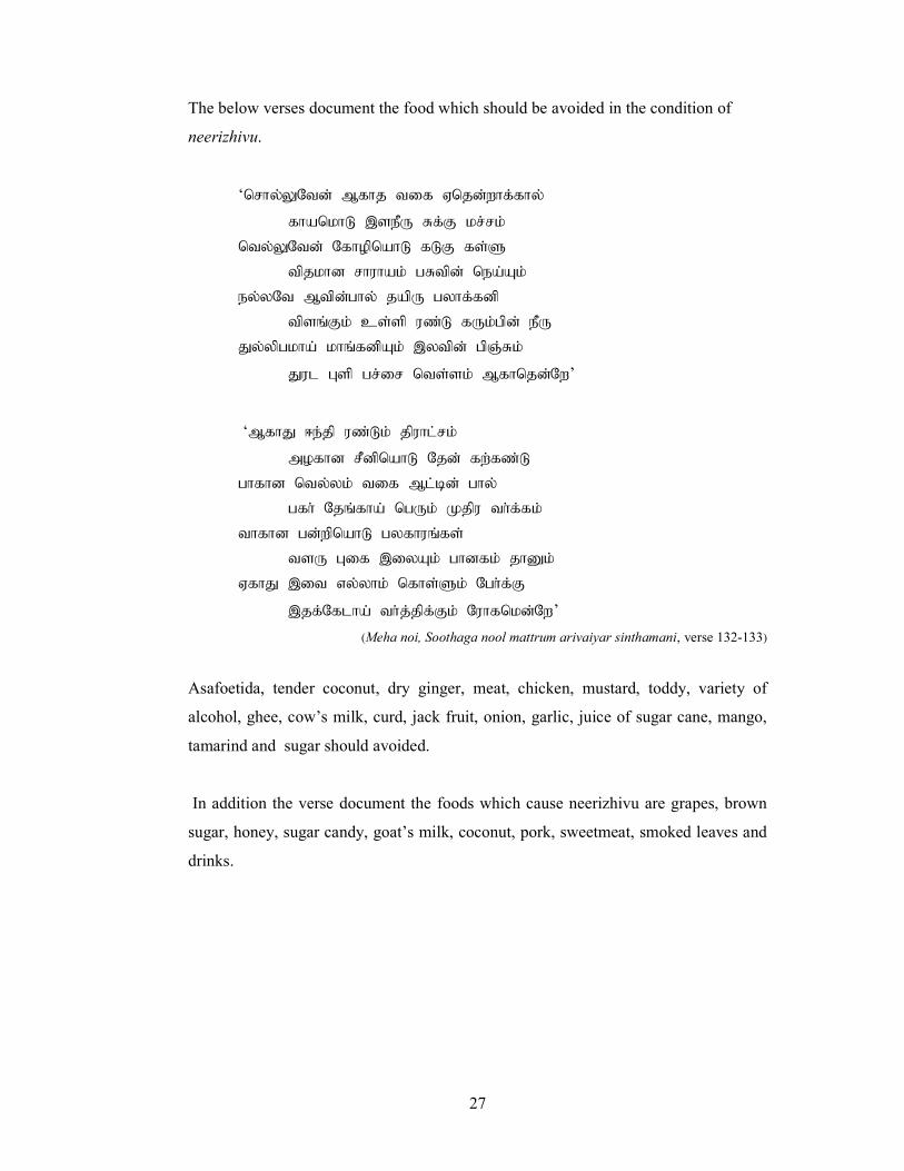
The below verses document the food which should be avoided in the condition of
neerizhivu.
‘nrhy;YNtd; Mfhj tif Vnjd;whf;fhy;
fhankhL ,sePU Rf;F kr;rk;
nty;YNtd; NfhopnahL fLF fs;S
tpjkhd rhuhak; gRtpd; nea;Ak;
ey;yNt Mtpd;ghy; japU gyhf;fdp
tpsq;Fk; cs;sp uz;L fUk;gpd; ePU
Jy;ypgkha; khq;fdpAk; ,ytpd; gpQ;Rk;
Jul Gsp gr;ir nts;sk; Mfhnjd;Nw’
‘MfhJ <e;jp uz;Lk; jpuhl;rk;
mofhd rPdpnahL Njd; fw;fz;L
ghfhd nty;yk; tif Ml;bd; ghy;
gfh; Njq;fha; ngUk; Kjpu th;f;fk;
thfhd gd;wpnahL gyfhuq;fs;
tsU Gif ,iyAk; ghdfk; jhDk;
VfhJ ,it vy;yhk; nfhs;Sk; Ngh;f;F
,jf;Nflha; th;j;jpf;Fk; Nuhfnkd;Nw’
(Meha noi, Soothaga nool mattrum arivaiyar sinthamani, verse 132-133)
Asafoetida, tender coconut, dry ginger, meat, chicken, mustard, toddy, variety of
alcohol, ghee, cow’s milk, curd, jack fruit, onion, garlic, juice of sugar cane, mango,
tamarind and sugar should avoided.
In addition the verse document the foods which cause neerizhivu are grapes, brown
sugar, honey, sugar candy, goat’s milk, coconut, pork, sweetmeat, smoked leaves and
drinks.

28
3.8 Diagnosis of Neerizhivu madhumeham according to the Siddha system
3.8.1 Envagaithervu
The Siddha system has unique assessment methods as envagaithervu (naadi, sparism,
naa, niram, mozhi, vizhi, malam and siruneer) to diagnosis the diseases (Agasthiyar
vaiththiya sillarai kovai, 2010; Natarajan, 2009; Shanmugavelu, 1967). Agasthiyar
vaiththiya sillarai kovai (2010) explains envagaithervu as,
‘juzpAs;s tpahjpjid a~;lhq;fj;jhy;
jhdwpa Ntz;LtJ NaNjnjd;dpy;
jpuzpaNjhh; ehbfz;fs; rj;jj;NjhL
Njfj;jp dJghprk; thdk;ehf;F
,uzky %j;jpukh kpitfnsl;Lk;
,jk;glNt jhd;ghh;j;Jf; Fwpg;Gf;fz;L
gudUshw; nghpNahh;fs; ghjk;Nghw;wpg;
gz;Gjtwhkw; gz;bjQ; nra;tPNu’
(Agasthiyar vaiththiya sillarai kovai, 2010)
The above stanza explains disease can be diagnosed with envagaithervu - naadi,
sparism, naa, niram, mozhi, vizhi, malam and siruneer.
3.8.2 Neikuri Neikuri is one of the methods of urine examination, based on distribution of oil drop
in urine. It is a remarkable diagnostic and prognostic parameter and well explained by
sage Theriyar and Agasthiyar (Kannusamy pillai, 1931; Shanmugavelu, 1967). In
addition the neikuri forecast the curable and incurable disease (Kannusamy pillai,
1931; Shanmugavelu, 1967). The sparding and the shape of the neikuri varied
according to the disease condition.
Literature documents the procedures to investigate the neerkuri and neikuri. Theraiyar
Neerkkuri vaiththiyam Neerkkurinool- neikuri nool moolamum uraiyum explains the
procedures to conduct the neerkuri.

29
‘mUe;Jk; MwpujKktpNuhjkjh
m/fy; myh; jyfhyT+z;lhtph;e;jow;
Fw;w mstUe;jpAwq;fpitfiw
Mbf;fyrj; jhtpNafhJnga;
xU K$h;j;jf; fiyf;Fl;gLePhp
epwf;Fwp nea;f;FwpepUkpj;jy; flNd’
‘epwf;Fwpf; Fiuj;j epUkhzePhpw;
rpwf;f ntz;nza;Nahh; rpWJspeLtpLj;
njd;Wwj;jpwe; njhypNa fhjikj;jjp
dpd;wjptiyNah newptpopawpTQ;
nrd;wJ GfYQ; nra;jpiaAzNu’
(Theraiyar Neerkkuri vaiththiyam Neerkkurinool- neikuri nool moolamum uraiyum)
The above literature explains that, during the early morning urine need to collect into
a glass utensil, on that condition that the person has ingested six taste of food on the
previous day night and good sleep. Apply a drop of oil on the surface of the urine,
within one and a half an hour after the collection of the urine. Even though, Sage
Theraiyar explains,
‘mUg;gKw;whh;f; ft;tpjptpyf;Nf’
(Theraiyar Neerkkuri vaiththiyam Neerkkurinool- neikuri nool moolamum uraiyum)
that the rules not need be followed when examining the neikuri
to a patient (Shanmugavelu, 1967).
Several literature documents different shapes of the neikuri in the condition of
neerizhivu. The Yuki vaiththiya kaviyam (2014) explains that the neerizhivu cannot be
curable when the neikuri shape in round and wreathe. In contrast Sikichcha
ratnathepam indicates the neerizhivu can be curable when the neikuri in the shape of
parts of the body, face, fish and temple. In addition the literature states that it takes a
prolong time to cure the neerilizhivu if the neikuri is in the shape of wheel. The
following verse indicates the above facts.

30
‘ifapdpnyz;izthq;fp fope;jePh;jd;dpw;Fj;j
nra;jJtl;lkhFQ; NrUe;Njhuzk;Nghy;jhDk;
[aKkpy;iyfz;lha; rhj;jpaky;yntd;W
Ja;aed;Kdpth;jhDQ; nrhy;ypaFwpg;gpjhNk’
(Yuki vaiththiya kaviyam, verse 793)
‘kd;dpa tta tq;fs; kdpjh;Nghy; kr;rq; Nfhap….
…. NuhfNk epw;f khl;lh Jiuf;FQ;rh j;jpaf; Fwpg;Ng’
(Sikichcha ratnathepam, verse 47- 52)
‘Rw;Wkr; rf;fuk;Nghy; Njhw;wpL KUtq; fz;lhy;
rj;jpa khfr; nrhd;Ndd; jhkrhj; jpae;jh dhNk’
(Sikichcha ratnathepam, verse 53)
The following verse documents the diseases can be curable if the neikuri spreads slow
and round in shape.
‘tpUj;jg;gb tKe; jhpj;Jg;gutYe;
njhpj;j nea;f;Fwpf; fpdptUj;j nkd;DyfP
uhq;fg; guty; NghdPq;F nkg;gpzpANk’
(Theraiyar Neerkkuri vaiththiyam Neerkkurinool- neikuri nool moolamum uraiyum)
3.8.3 Neerkuri
Theraiyar Neerkkuri vaiththiyam Neerkkurinool- neikuri nool moolamum uraiyum
explains the general characters to test neerkuri as,
‘te;jePh;f;fhpapil kzEiunaQ;rnyd;
iwe;jpa Ysitaiw FJKiw’
(Theraiyar Neerkkuri vaiththiyam Neerkkurinool- neikuri nool moolamum uraiyum)
Five general characters as colour, odour, froth, sediment and volume of the urine are
documented in the above verse to test neerkuri.
31
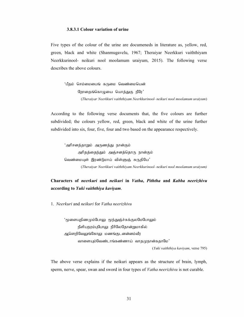
3.8.3.1 Colour variation of urine
Five types of the colour of the urine are documeneds in literature as, yellow, red,
green, black and white (Shanmugavelu, 1967; Theraiyar Neerkkuri vaiththiyam
Neerkkurinool- neikuri nool moolamum uraiyum, 2015). The following verse
describes the above colours.
‘gPjk; nrk;ikigq; fUik ntz;iknad;
Nwhijq;nfhOia nahj;JF ePNu’
(Theraiyar Neerkkuri vaiththiyam Neerkkurinool- neikuri nool moolamum uraiyum)
According to the following verse documents that, the five colours are further
subdivided; the colours yellow, red, green, black and white of the urine further
subdivided into six, four, five, four and two based on the appearance respectively.
‘mhprdj;jhWk; mUzj;J ehd;Fk;
mhpjj;ije;Jk; mQ;rdj;njhU ehd;Fk;
ntz;ikAs; ,uz;Lkha; tps;SQ; RUjpNa’
(Theraiyar Neerkkuri vaiththiyam Neerkkurinool- neikuri nool moolamum uraiyum)
Characters of neerkuri and neikuri in Vatha, Piththa and Kabha neerizhivu
according to Yuki vaiththiya kaviyam.
1. Neerkuri and neikuri for Vatha neerizhivu
‘%isAepzKk;NghY %e;JQ;r;Rf;FyNkNghYk;
ePspaeuk;GNghY ePhpNyNjhd;Wkhfpy;
MnswpNtYq;NfhY kzq;Fldd;dk;tPu
thisAk;Ntz;lhq;fz;zha; thjKehd;fjhNk’
(Yuki vaiththiya kaviyam, verse 795)
The above verse explains if the neikuri appears as the structure of brain, lymph,
sperm, nerve, spear, swan and sword in four types of Vatha neerizhivu is not curable.

32
2. Neerkuri and neikuri for Piththa neerizhivu
‘ePydPh;kQ;rzpj;J ePj;JePh;tpspj;jpUf;FQ;
rhyNtrhk;gy;jd;id fOfOntd;dPh;Nghyhk;
fhyNkapiwr;rpjd;id fOtpdePh;NghyhFk;
khy;tpopaidaph;gpj;j khdePh;f;FzkpNj’
(Yuki vaiththiya kaviyam, verse 797)
The verse indicating that, colour of the urine of piththa neerizhivu designates as
yellow, grey and fleah wash water.
3. Neerkuri and neikuri for Kabha neerizhivu
‘mUk;Gdy;$iuePU kbf;fUk;gjdpw;rhWk;
tpUk;gpajaph;ghy;ePU tpuFld;Nrj;jghFk;
jpUj;jpaNjdpw;ghFk; rpije;japt;thWNghyhk;
FUk;igNrh;Kiyey;yhNs $WQ;Nrj;Jkj;jpdNu’
(Yuki vaiththiya kaviyam, verse 798)
The above verse metions the neerkuri of the kabha neerizhivu look like the colour of
water flow from the old roof, sweet drink extracted from palmyra, curd, milk, water, a
thick consistence of boiled jaggery and honey.
3.8.3.2 Froth in urine
The verse,
‘ge;jnka;g; girapsf;fg;gLk; gUtj;
Je;jh;G+j khadpy %j;jpuj;jpw;
rk;ge;jg; gLe;jjp Eiug;GdNy’
(Theraiyar Neerkkuri vaiththiyam Neerkkurinool- neikuri nool moolamum uraiyum)
describes that when the Kabham decreases in
the body , the vayu will appear in the urine and form froth. In addition the Sikichcha

33
ratnathepam explains in the following verse that, the froth will appear in the urine if
the patient having Kabha disorders.
‘epiyf;Few; fgNk ahfpy; ePh;Eiu Nghd;wpUf;Fk;
,yFkh %j;jp uj;jp nyz;nzna tpl;Lg; ghh;f;fpy;……’
(Sikichcha ratnathepam, verse 47)
Further Therar arulichcheitha siruneerkuri sothanai explains, the froth will appear in
the kabhavatha disorders.
3.8.3.3 Odour, sediment and volume of urine
Literature explains different types of odour, sedimentations and weight of the urine
explains in various diseases conditions. (Shanmugavelu, 1967; Theraiyar Neerkkuri
vaiththiyam Neerkkurinool- neikuri nool moolamum uraiyum, 2015)
3.8.4 Naadi
Naadi is a diagnostic way to assess health status of an individual (Ivy and Malini,
2010) and a remarkable diagnostic parameter, included in envagaithervu and well
explained by Siddhars (Kalaththur kanthasami, 2012).
The following literature documents the procedures to read naadi.
‘fhpKf dbia tho;j;jpf;
ifjdpy; ehbghh;f;fpy;
ngUtpuy; yq;Fyj;jpd;
gpbj;jpl dLNt njhl;lh
nyhUtpu Nyhby; thj
Kah;eL tpuypw; gpj;je;
jpUtpuy; %d;wp Nyhby;
rpNyj;Jk ehbahNk’
(Agasthiyar vaiththiya sillarai kovai, verse 46)

34
‘Fwpahk; tyf;fuq; Ftpe;j ngUtpuy;
kwpthajd;fPo; itj;jpL%tpuy;
gpwptha; NkNywpg; gpyj;jJ thjkhk;
mwptha;eLtpuy; mkh;e;jJ gpj;jNk’
(Thirumoolar vaithiyam karukkadai – 600, verse 23)
‘gpj;j;jpd;fPNo gpuz;lJ Iakhk;
cw;Ww;Wg;ghh;f;f Xh;euk;NghLk;
gj;jpj;j%tUk; gha;fpd;wNtfj;jhy;
kj;jpj;jehsk; Nghy; toq;Fk; euk;gpj;Nj’
(Thirumoolar vaithiyam karukkadai – 600 , verse 24)
‘%d;Wtpuyhk; ngUtpuw;fPo; Kdpth;jhÇd;wp Kd;ghh;f;fj;
Njhd;Wk;thjk; eLg;gpj;je; Jiyahfr; Nrj;Jk jplj;njd;g
fhd;W kpfTk fukhaf; fLfpelf;F kitfz;lhy;
Cd;Wk; tpuiy tpl;Ltpl;L cgrhukha;’
(Venkatrajan, verse 66)
‘XJ Kjy;tpuy; thjj;jhNy
xLf;Fk; Nehnad;W mwpe;Jnfhs;S
Ngj kpy;yhky; kWtpuYk;
gpj;jj;jh nyd;W NgrptpL
Nrj kpy;yhky; %tpuyhw;
rpNyw;gdj; Js;s Neha;fnsd;Wk;
tPjg; gbapd;d jhNyte;j
tpahjp apJntd; wwpe;Jnrhy;Ny’
(Yakopu vaiththiyam 300, verse 68)
The above verses state that the procedures to examine naadi as, placed the index,
middle and ring fingers one inch below the wrist and feel the pulse. The index, middle
and ring fingers indicate the Vatham, Piththam and Kabham respectively.
The flowing verses indicating that, which hand need to use to read naadi.

35
‘kUTnka;f; Fw;wehb kjpj;jpby; tyf;if khe;jh;
mhpitah;f; fplf;ifehb aKf;fpNa Rl;lha; %l;b
tpiutpdpy; nta;athj kpfeL tpuypw; gpj;je;
jUkzp tpuypya;ae;jhndd twpe;J nrhy;Ny’
(Venkatrajan, 2014, verse 49)
‘jhndd Tyfj;Js;Ns jaq;fpa ehbghh;f;fpy;
thndd kpd;NdNfsha; tUk;Gyd; nrhy;yf;NfS
ehndDk; GUlh;f;nfy;yhk; ehbj;jhd; tyf;idahFk;
NjndD khjh;f;nfy;yhe; jplk;ngw tplf;ifrpj;Nj’
(Agasthiyar vaiththiya sillarai kovai, verse 30)
‘fz;lhNa Mlth;f;F tyf;fuK khFk;
fUTlNd ngz;gps;isf;F ,lf;fuk; vd;Nwd;
ntz;lhd ,lf;ifia tyf;if ahNy
Nkyhd tyf;ifahNy
ez;lhff; ifNja;j;J nel;b thq;fp
ehl;L% tpuYf;F tpuyz; lhik
mz;lhky; nel;lhtpl;L ghU
mirthFk; thjgpj;;j rpNyj;Jk khNk’
(Agasthiyar nool thirattu)
The above verses indicate that the right hand need to use to read naadi in male and the
left hand for female.
Ten locations indicated to read naadi in Thirumoolar vaithiyam karukkadai – 600
and the following verse indicates the locations as,
‘jhJKiwNfs; jdpj;jFjpr;re;J
XJWfhkpak; ce;jpeLkhh;G
fhJeL%f;Ff; fz;lq;fuk;GUtk;
NghJWKr;rp Gfo;gj;Jk;ghh;j;jpNl’
(Verse 54 Thirumoolar vaithiyam karukkadai – 600)
The verses state the ten locations as kuthisanthi (ankle), kamiyam (inguinal region),
unthi (abdomen), marbu (chest), kathu (ear), mooku (nose), kandam (throat),

36
karam (arm), purum (eye brow) and uchchi (frontanella). Even though Kai (upper
limb) is the common place to read naadi for all and the following verse indicating the
statement as,
‘$h;j;jplNt fd;dkJ Ropapw; whDk;
Fwpg;ghd iffspYk; kh;k];jhde; jd;dpy;
rhh;e;jplNt fZf;fhyp Dl;Gwj;jpy;
rhhpthfg; ngUtpuw;fhy; Nky jhf
Njh;e;jplNt ehbjid AgNah fpf;fj;
njspthf khe;jUf;Fr; nrg;g yhr;R
Ngh;e;jplNt rfyUf;Fq; fuj;jp dhb
Ngrpdhh; gpukKdp Ngrp dhNu’
(Thirumoolar vaithiyam karukkadai – 600, verse 54)
In disease condition, if the Vatham and the Piththam increase the naadi can be felt
between the index and middle finger, if the Vatham and the Kabham increase the
naadi can felt between index and ring finger and if the Piththam and the Kabham
increase the naadi can be felt between middle and ring finger (Vasutheva Sasththirikal
and Subramanya Sasththirikal, 2014).
3.8.4.1 The sign and symptoms for thontha naadi (combination of two
naadi) according to Agasthiyar vaiththiya sillarai kovai
Signs and symptoms of Vathapiththam
‘thjj;jpw; gpj;jkhfpy; thaJ FowpNgRk;
Ngjpj;Jf; FspUq;fhypy; tPf;fKk; ngUfTz;lhk;
jhJw;w Gj;jpjhDe; jilg;gLe; jlq;fz;khNj
NfhJw;w tapWtpk;kpf; Fwlidg; Gul;Le;jhNd’
(Agasthiyar vaiththiya sillarai kovai, 2010, verse 5; Agasthiyar munivar arulichcheitha vaiththiya
raththna surukkam 360 (moolamum uraiyum), verse 5)
The above verse states, strangely speech, chillness in leg, swelling, mentally
disturbance and abdominal discomfort occur when both the Vatham and the Piththam
increase in the body.

37
In addition another literature explains as,
‘,ire;jpLk; gpj;jk; uz;Lk;
<uiu thj Nkhby;
grpe;jpLk; tapWk; neQ;R
gwe;njwpe; jod;W nka;jhd;
trq;nfLe; jiyfp Wf;Fk;
kaq;fpL Kly;nt Jk;Gk;
Krpe;jpLk; gpj;j thj
Kiwikia mwpe;J nfhs;Ns!’
(Agasthiyar maruththuvam (Tamil maruththuva nool varisai -15) olaichchuvadith thokuppu nool, verse- 58)
The stanza states if Vatham and Piththam increase excessive appetite, burning
sensation in chest and abdomen, giddiness, faintness and feeling of heat is experienced
in the body.
Further Dhanvathiri vaiththiyam (2014) explains in following verse that, ache and
pain in the body, thirst, worries and indigestion occur, when Vatham and Piththam
increase simultaneously.
‘me;j thjq;fs; uz;L kLj;jpLk; gpj;jnkhd;We;
njhe;jpj;J elf;Fkhfpw; Nwhd;wpLq; Fzj;ijf; Nfsha;
te;Jnehe;jJjhd; tpk;kp typj;jpLk; jhfNkfQ;
rpe;jid kpfTz;lhFk; Grpg;igAQ; nrhpahjhf;Fk;
ige;njhb thjgpj;j nkd;Wjhd; gfuyhNk’
(Dhanvathiri vaiththiyam, verse 27)
38
Signs and symptoms of Piththavatham
Pain in the occipital lobe, arm and leg, emaciation, tremor, thirst, pain due to fever,
confusion and anxiety occur when Piththam and Vatham increase simultaneously and
the condition is described in the following verse,
‘gpj;jj;jpy; thjkhfpw; gplhpAq; fhYq;ifAq;
Fj;jJ NghNyah;Fq; FWfpnka; gjWk;gpd;Nd
mj;jpah AyUNkdp ahfKk; Ruj;jhy;Nehthk;
Gj;jpA kbAkpf;fg; nghWikNgha; NfhgkhNk’
(Agasthiyar vaiththiya sillarai kovai, 2010, verse 8;
Agasthiyar munivar arulichcheitha vaiththiya raththna surukkam 360 (moolamum uraiyum), verse 7)
The below verse from Dhanvathiri vaiththiyam (2014) documents that, ache and pain
in the body, burning sensation in the chest, dryness of the mouth and burning
sensation in micturition occur, when both Piththam and Vatham increase in the body.
‘vz;zpa thjnkhd;Wk; gpj;jkpuz;nlOe;jjhfpy;
Gz;nzz Tlk;GNehthk; Gifnao nahpAk; neQ;R
jpz;zkha; ehtuz;L rpWj;jePh;f; fLj;JtpOk;
mz;zyhh; ciuj;jTz;ik ahAU Nte;jNd’
(Dhanvathiri vaiththiyam, verse 30)
Signs and symptoms of Vathakabham The below verse points out the symptoms of increase Vatham and the Kabham as
pain with swelling, mental disturbance, head ache, changes in mind, pallor and the
occurrence of oedema.
‘thjj;jpy; Nrj;kkhfpy; typnahL tPf;fKz;lhk;
Ngjpj;Jj; jiyapbj;J gpzq;fpa Fzq;fs;Ntwha;j;
jPJw;w nka;ntSj;Jj; jplKld; rdQ;nry;yh
Ngjpj;J ehTNgRk; ngUfNt tPf;fKz;lhk;’
(Agasthiyar vaiththiya sillarai kovai, 2010)

39
In addition, the following verses explain the characters of increased Vatham and
Kabham together.
‘cah;e;jpLk; thjk; nuz;Lk;
xUrp Nyl;Lk jhfpy;
mah;e;jpLk; iffhy; nehe;J
mbta wijj;Jf; fhl;Lk;
jpaq;fpLe; jhJ nfl;L
Tjpuq;fSq; Fiwe;J thLk;
kaq;fpL thjh; NjhL
kUtpa ma;ae; jhNd’
(Agasthiyar maruththuvam (tamil maruththuva nool varisai -15) olaichchuvadith thokuppu nool, verse
60)
The verse states that, tiredness, pain in arm and leg, enlargement of lower abdomen
and loss of thathu and blood occur when the Vatham and the Kabham increases
together.
Further Dhanvathiri vaiththiyam (2014) explains in the following verse that, pain in
the body, numbness in the hand and foot and abdominal pain with distension
experience in body when increase Vatham and Kabham .
‘khddha; thjk; uz;LQ; rpNyj;k nkhd;nwOe;j jhfp
yhdNjhh; rhPuk; Nehthk; mq;iffhy; jpk;h;j;Jf; fhl;L
%dkhKjuj;Js;Ns T+ijA kpFe;J tpk;Kk;
Ghdyq; fz;zha; thj rpNyj;gdk; ghpe;JghNu’
(Dhanvathiri vaiththiyam, verse 32)

40
Signs and symptoms of Kabhavatham
The following verse documents, pain in the leg and occipital region and difficulty in
speech experience, when the Kabham and Vatham increase together.
‘thl;bKQ; Nrj;Jkj;jpy; te;jpL thjkhfpy;
ehl;ba fhy;fs;Nghy euk;ngyhk; typj;Jepw;Fk;
$l;ba gplhpjhDq; Fd;wNt typf;Fkhfpy;
ehl;ba tpopAnky;yhk; ehf;Ftha; FoWe;jhNd’
(Agasthiyar vaiththiya sillarai kovai, 2010, verse 10; Agasthiyar munivar arulichcheitha vaiththiya
raththna surukkam 360 (moolamum uraiyum), verse 10)
In addition the below verse describes that, disorders in abdomen, emaciation and pain
in the body occurs when increase Kabham and Vatham together.
‘ghhpj;j ma;ak; nuz;Lk;
ghpe;njhU thj Nkhby;
fwpj;J FSj;J $rpf;
Fk;gpapy; Fzf;Nf lhFk;
Nenuhj;j fhak; tw;wp
Newpjiy jpNufk; NehFQ;
rPWw;w ma;a thje;
njhe;jpg;ghe; jphpey; khNj!’
(Agasthiyar maruththuvam (tamil maruththuva nool varisai -15) olaichchuvadith thokuppu nool, verse
62)
Further Dhanvathiri vaiththiyam (2014) documents in the following verse that,
increase of appetite, the patient wish to eat a lot of sweet and burning sensation of
the body experienced, when increase Kabham and Vatham together.
‘Nrg;gpa Nrj;kk; uz;Lk; thjK nkhd;W Nrhpy;
ntg;GW fgNkypl;L ,dpg;igNa kpfTk; Ntz;Lk;
jg;gpyhg; grpAKz;lhe; jgdNk gw;wpepw;Fk;
,g;gbf; Fzq;fs; fz;lhy; rpNyj;Jk thjnkd;Nd’
(Dhanvathiri vaiththiyam,verse 29)

41
3.8.5 Manikkadai nool
Manikkadai nool is one of the diagnosis and prognosis methods of disease in the
Siddha system of medicine. The Pathinen Siddhar arulichcheitha naadi saasthiram
(2012) explaines the procedures to conduct manikkadai as measuring the
circumference of the forearm above the four finger breadth from the wrist using a
thread. There after the length of the circumference measure using the four fingers
except thumb. The sum of the finger breadth (in number) indicates the disease. The
following verse describes the procedures of measuring the Manikkadai nool.
‘kzpf;fil fhy;tpuy; js;spTz;ikaha;
kzpf;fpil faWNghl; lse;Jghh;f;ifapy;
fzpj;jpLk; tpuy;jid fz;Lnrhy;yNt
gpzpj;jpL Neha;fis gphpj;Jiuf;FNk’
(Pathinen Siddhar arulichcheitha naadi saasthiram)
Further Pathinen Siddhar arulichcheitha naadi saasthiram (2012) documents in the
following verse that, reduction of finger breadth indicates the disease status.
‘Fiue;Jtpuf;fpil apire;Jfhz;fpby;
ciue;Jntz;gpzp Tlk;gpw;rhh;e;jpLk;…..’
(Pathinen Siddhar arulichcheitha naadi saasthiram)
The following verse documents, the sum of the finger breadth is eight and a quarter,
indicates piththa disorders, fever, pramiyam, kamiyam amd disoreder of head.
‘fhl;banal;nlhL fhy;tpuw;fil
$l;bagpj;jha; FiuRunkapy;
ehl;bagpukpak; etpYq;fhkpak;
thl;barpu rpy;Neha; tUNkhuhz;by;’
(Pathinen Siddhar arulichcheitha naadi saasthiram)

42
3.9 Modern aspect of diabetes mellitus (DM)
3.9.1 General considerations
DM is a metabolic disorder characterized by the presence of chronic hyperglycaemia
accompanied by greater or lesser impairment in the metabolism of carbohydrates,
lipids and proteins (Baynest, 2015). DM is probably one of the oldest diseases known
to man. It was first reported in Egyptian manuscript about 3000 years ago (Ahmad,
2002). The World Health organization states that, diabetes is a serious, chronic disease
that occurs either when the pancreas does not produce enough insulin or when the
body cannot effectively use the insulin it produces (WHO, 2016). Prevalence of type 2
DM has been increasing steadily all over the world (Abdulfatai, 2012). Raised blood
glucose, a common effect of uncontrolled diabetes, may, over time, lead to serious
damage to the heart, blood vessels, eyes, kidneys and nerves (WHO, 2016). Diabetes
is an important public health problem and considers as one of the four priorities of non
communicable diseases (Geneva, 2011). Both the number of cases and the prevalence
of diabetes have been steadily increasing over the past few decades (WHO, 2016).
3.9.2 Global burden
Globally, an estimated 422 million adults were living with diabetes in 2014, However
108 million adults recorded as diabetes in 1980. The global prevalence of diabetes has
nearly doubled since 1980, rising from 4.7 % to 8.5 % in the adult population. It is
estimated that 439 million people would have type 2 DM by the year 2030
(Abdulfatai, 2012). Diabetes caused 1.5 million deaths in 2012. It was the eighth
leading cause of death among both sexes and the fifth leading cause of death in
women in 2012 (WHO, 2016). Higher than optimal blood glucose caused an
additional 2.2 million deaths, by increasing the risks of cardiovascular and other
diseases (WHO, 2016).
The Fig.3.1 has shown the trends in prevalence of diabetes, 1980 – 2014, by country
income group.

43
1.2.8.5.3 Classification of diabetes
The old classification is insulin dependent (IDDM) or non-insulin dependent
(NIDDM) which were proposed by WHO in 1980 and 1985 have disappeared (WHO,
1999) .The new classification system identifies four types of DM; type 1, type 2, other
specific types and gestational diabetes in 1985 (WHO, 1999).
Type 1 diabetes (Insulin dependent, juvenile or childhood onset diabetes) is
characterised by deficient insulin production in the body. People with type 1 diabetes
require daily administration of insulin to regulate the amount of glucose in their blood
(WHO, 1999).
Fig. 3.1. Trends in prevalence of diabetes, 1980 – 2014, by country income group
44
Type 2 diabetes results from body’s ineffective use of insulin. Symptoms may similar
to type I diabetes, but are often less marked or absent. As a result, the disease may go
undiagnosed for several years, until complications have already arisen (WHO, 1999).
Gestational diabetes is an operational classification identifying women who develop
DM during gestation.
Other specific type (monogenic diabetes), types of DM of various known etiologies
are grouped together to form the classification called other specific types. This group
include persons with genetic defects of beta-cell function or with defects of insulin
action, persons with disease of exocrine pancreas or cyctic fibrosis etc.
3.9.3 Clinical features of Diabetes mellitus general symptoms
Most of the symptoms are similar to both types of diabetes but they vary in their
degree and develop more rapidly in type 1 diabetes and more typical (Baynest, 2015).
Clinical features of type 1 diabetes mellitus
Weight loss, polyuria, polydipsia, polyphagia, constipation, fatigue, cramps, blurred
vision and candidiasis (Zimmet, 1992). Long lasting type 1 DM patients may
susceptible to microvascular complications (Hove et al., 2004) and macrovascular
disease (coronary artery, heart and peripheral vascular diseases) (Pittas, 2009).
Table 3.3. Characteristics of the common types (types I and II) of diabetes (Harikumar et al, 2015)
Characteristics Type 1 Type 2 Age Childhood Pubertal
Onset Acute, severe Mild,severe - often insidious
Insulin secretion Very low Variable
Insulin sensitivity Normal Decreased
Insulin dependence Permanent Temporary - may occur later
Genetics Polygenetic Polygenetic
Proportion of those with diabetes
80 % 10 – 20%
Association : Obesity No Strong
Acanthosis nigricans No Yes
Autoimmune etiology Yes No
45
Clinical features of type 2 diabetes mellitus
Most of the Type 2 patients diagnosed because of complications or incidentally. More
possibility with a high risk of large vessel atherosclerosis commonly associated with
hypertension, hyperlipidaemia and obesity. Most of the patients with type 2 diabetes
die from cardiovascular complications and end stage renal disease (Sekikawa, 1993).
3.9.4 Pathogenesis and pathophysiology of diabetes mellitus
There is a direct link between hyperglycemia, physiological and behavioural
responses. Whenever there is hyperglycemia, the brain recognises it and sends a
message through nerve impulses to pancreas and other to decrease its effect (Patidar,
2011).
Diabetes mellitus - Type 1
Type 1 DM is characterized by autoimmune destruction of insulin producing cells in
the pancreas by CD4+ and CD8+ T cells and macrophages infiltrating the islets.
Approximately 85% of patients have circulating islet cell antibodies, and the
majorities also have detectable anti-insulin antibodies before receiving insulin therapy.
The autoimmune destruction of pancreatic β cells, leads to a deficiency of insulin
secretion which results in the metabolic derangements associated with type 1 DM in
addition to the loss of insulin secretion, the function of pancreatic α – cells is also
abnormal and there is excessive secretion of glucagons in type 1 DM. Normally,
hyperglycemia leads to reduced glucagons secretion, however, in patients with type 1
DM glucagons secretion is not suppressed by hyperglycemia. The resultant
inappropriately elevated glucagons levels exacerbate the metabolic defects due to
insulin deficiency. Althougth insulin deficiency is primary defect in type 1 DM, there
is also a defect in the administration of insulin. Deficiency in insulin leads to
uncontrolled lipolysis and elevated levels of free fatty acids in the plasma, which
suppresses glucose metabolism in peripheral tissues such as skeletal muscle. This
impairs glucose utilization and insulin deficiency also decreases the expression of a
number of genes necessary for target tissues to respond normally to insulin such as
glucokinase in liver and the GLUT 4 class of glucose transporters in adipose tissue
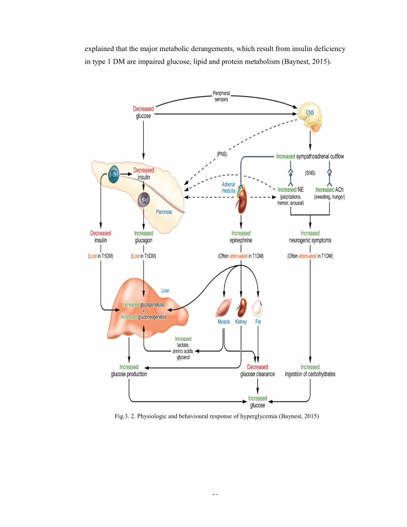
46
explained that the major metabolic derangements, which result from insulin deficiency
in type 1 DM are impaired glucose, lipid and protein metabolism (Baynest, 2015).
Fig.3. 2. Physiologic and behavioural response of hyperglycemia (Baynest, 2015)

47
Diabetes mellitus - Type 2
In type 2 DM, these mechanisms break down, with the consequence of the two main
pathological defects in type 2 diabetes that are impaired insulin secretion through a
dysfunction of the pancreatic β – cells and impaired insulin action through insulin
resistance. In situations where resistance to insulin predominates, the mass of β –
cells undergoes a transformation capable of increasing the insulin supply and
compensating for the excessive and anomalous demand. The plasma insulin
concentration usually is increased, although relative to the severity of insulin
resistance, the plasma insulin concentration is insufficiency to maintain normal
glucose homeostasis. Keeping in mind the intimate relationship between the secretion
of insulin and the sensitivity of hormone action in the complicated control of glucose
homeostasis, it is practically impossible to separate the contribution of each to the
etiopathogenesis of type 2 DM.
Insulin resistance and hyperinsulinemia eventually lead to impaired glucose tolerance.
Except for maturity onset diabetes of the young (MODY), the mode of inheritance as
an autosomal dominant trait, may result from mutations in glucokinase gene on
chromosome 7p. MODY is defined as hyperglycemia and treatable for over five years
without insulin in cases where islet cell antibodies are negative (Baynest, 2015).
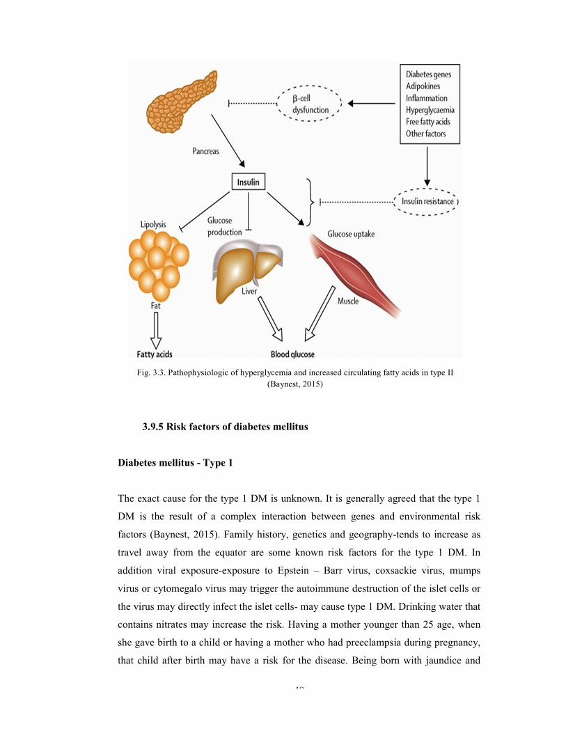
48
3.9.5 Risk factors of diabetes mellitus
Diabetes mellitus - Type 1
The exact cause for the type 1 DM is unknown. It is generally agreed that the type 1
DM is the result of a complex interaction between genes and environmental risk
factors (Baynest, 2015). Family history, genetics and geography-tends to increase as
travel away from the equator are some known risk factors for the type 1 DM. In
addition viral exposure-exposure to Epstein – Barr virus, coxsackie virus, mumps
virus or cytomegalo virus may trigger the autoimmune destruction of the islet cells or
the virus may directly infect the islet cells- may cause type 1 DM. Drinking water that
contains nitrates may increase the risk. Having a mother younger than 25 age, when
she gave birth to a child or having a mother who had preeclampsia during pregnancy,
that child after birth may have a risk for the disease. Being born with jaundice and
Fig. 3.3. Pathophysiologic of hyperglycemia and increased circulating fatty acids in type II (Baynest, 2015)
49
having a respiratory infection just after birth may be a risk for the type 1 DM
(Harikumar et al., 2015).
Diabetes mellitus – Type 2
Researchers do not fully understand why some people develop type 2 diabetes and
others do not. Some factors increase the risk of the type 2 DM. Overweight is a
primary risk factor for type 2 diabetes. In addition fat distribution especially in
abdomen, inactivity, family history and race – Blacks, Hispanics. American Indians
and Asian Americans are more prone to develop type 2 DM, age – older, especially
after age 45, probably because people tend to less exercise, lose muscle mass and gain
weight as they age and gestational diabetes may influence to cause type 2 DM
(Harikumar et al., 2015).
3.9.6 Complications
Diabetes of all types can lead to complications in many parts of the body and can
increase the overall risk of dying prematurely. When diabetes is not well managed,
complications develop that threaten health and endanger life. Acute complications are
a significant contributor to mortality. Abnormally high blood glucose can have a life
threatening impact if it triggers condition such as ketoacidosis in type 1 and 2,
hyperosmolar coma in type 2. Abnormally low blood glucose can occur in all types of
diabetes and may result in seizures or loss of consciousness. Over time diabetes can
damage the heart, blood vessels, eyes (diabetic retinopathy), kidneys (diabetic
nephropathy) and nerves (diabetic neuropathy) and increase the risk of heart disease
and stroke. Such damage can result in reduced blood flow, which combined with
nerve damage in the feet- increases the chance of foot ulcers, infection and the
eventual need for limb amputation. In pregnancy, poorly controlled diabetes increases
the risk of foetal death and other complications like congenital malformation, still
birth and maternal mortality (WHO, 2016).
Other complications are impaired growth and development, lipodystrophy,
necrobiosis, non-alcoholic fatty liver disease, infections, limited joint mobility, odema
and also it is associated with autoimmune conditions like hypothyroidism,

50
hyperthyroidism, celiac disease, vitiligo and primary adrenal insufficiency (Baynest,
2015).
3. 9.7 Diagnosis of diabetes mellitus
The identification of patient with diabetes or pre-diabetes by screening allows for
earlier intervention, with potential reductions in future complication (WHO, 2016).
1. Random plasma test
The simplest test does not require fasting before the test. If 200 or more 200
mg/dL of blood glucose it probably indicates DM but has to be reconfirmed
(Baynest, 2015).
2. Fasting plasma glucose test
Eight hours fasting hould be needed before taking the test. Blood glucose more
than 126 mg/dL on two or more tests conducted on different days confirms a
diabetes (Gillett, 2009).
3. Postprandial Blood glucose test
Measures blood glucose levels two hours after eating a meal. Postprandial
blood glucose is usually done in people who have symptoms of
hyperglycemia, or when the results of a fasting glucose test suggest possible
diabetes, but are inconclusive. Values of 200 mg/dL or more indicate diabetes
(Harikumar et al., 2014).
4. Oral glucose tolerance test
When random plasma glucose test is 160 – 200 mg/dL and the fasting plasma
test is 110 – 125 mg/dL, then this test is conducted. This blood test elevates
body’s response to glucose. This test requires fasting at least eight but not
more than sixteen hours. Fasting glucose level is determined, and then gives 75
g of glucose, 100 g for pregnant women. The blood is tested every 30 minutes
to one hour for two or three hours. This test is normal if the glucose level at
two hours is less than 140 mg/dL. A fasting level of 126 mg/dL or greater and
51
two hour glucose level of 200 mg/dL or higher confirms a diabetes diagnosis
(Baynest, 2015).
5. Glycated proteins
Proteins react spontaneously in blood with glucose to form glycated
derivatives. The extent of glycation of proteins is controlled by the
concentration of glucose in blood and by the number of reactive amino groups
present in the protein that are accessible to glucose for reaction. All proteins
with reactive sites can be measured in blood is a marker for the fluctuation of
blood glucose concentrations during a certain period. From a clinical
diagnostic point glycated proteins with a longer life time in blood are of
interest, since they reflect the exposure of these proteins to glucose for longer
periods (Baynest, 2015).
6. Glycated haemoglobin
The life span of haemoglobin in vivo is 90 -120 days. During this time
glycated haemoglobin A (HbA1c) forms, being the ketoamine compound
formed by combination of haemoglobin A and glucose. Several subfractions of
glycated haemoglobin have been isolated. Of these, glycated haemoglobin A
fraction HbA1c is most interest serving as a retrospective indicator of the
average glucose concentration even if the patient is not in a fasting state.
HbA1c is recommended as an essential indicator for the monitoring of blood
glucose control. The blood HbA1c ≥ 6.5 % is considered as diabetes (Selvin et
al., 2010).
7. Fructosamine test
Albumin is the main component of plasma proteins. As albumin also contains
free amino groups, non – enzymatic reaction with glucose in plasma occurs.
Therefore glycated albumin can similarly serve as a marker to monitor blood
glucose. Glycated albumin is usually taken to provide a retrospective measure
of average blood glucose concentration over a period of 1 to 3 weeks.
Reference interval: 205 – 285 μmol/L (Baynest, 2015).

52
8. Chemical tests
This involves testing the urine with the Benedict’s reagent. Results indicate the
person having diabetes based on the colour formation (Harikumar et al., 2014).
a) Light colour – normal
b) Parrot green colour = > 120 mg/dl
c) Dark yellow colour = > 180 mg/dl
d) Reddish brown colour = +++ >250 mg/dl
e) Brown colour = ++++ > 350 mg/dl
9. Diasticks
These are strips that are used to indicate the person having DM or not. These
strips tested with urine and based on the colour change only diagnosis the DM
(Harikumar et al., 2014).
10. Glucometers
These meters are also involving in diagnosing the DM. Within the fraction of
seconds these will give results about blood glucose levels (Harikumar et al.,
2014).
3.9.8 Preventing diabetes
Type 1 diabetes cannot be prevented with current knowledge. Effective approaches
are available to prevent type 2 diabetes and to prevent the complications and
premature death can result from all types of diabetes. It can preventable by regular
exercise, healthy diet, reduce sedentary behaviours, unhealthy foods and beverages,
avoiding smoking and controlling blood pressure (WHO, 2016).

53
CHAPER 4
MATERIALS AND METHODS
4.1 General procedure
4.1.1 Study protocol
The comparative study was carried out to assess the Siddha diagnostic methods as
neerkuri, neikuri, naadi and manikkadai with modern diagnostic methods in NR.
4.1.2 Selection of the subjects
Primary screening was done randomly at outpatient department (OPD) and inpatient
department (IPD) at Government Siddha Medical College (GSMC), Palayamkottai,
Tirunelveli, Government District Headquarters Hospital (GDHH), Thoothukudi and
Gopalasamudram village (GSV), Tirunelveli. Sixty apparently NR patients were
enrolled for the study through screening according to the inclusive and exclusive
criteria after getting their concern.
4.1.2.1 Inclusion criteria
Both sexes with age of 18 to 64 (Axel, Roland and Kerstin, 2002) were selected with
the symptoms with increased volume of urine output, immediate changes in weight
and increase appetite. Further the patients present with the urine glucose positive and
fasting blood sugar (FBS) ≥ 126 mg/dL (≥ 7 mmol/L) or Postprandial Blood sugar
(PPBS) ≥ 200 mg/dL (≥ 11.1 mmol/L) (WHO, 2006) or HbA1c ≥ 6.5 % (WHO,
2016) were consider as inclusive criteria. In addition, no history of other illness and
the subjects, who are not taking medicines also considered when the selection of the
subjects. The subjects, who satisfied the all condition described above were selected
for the study.
54
4.1.2.2 Exclusion criteria
The patient below age of 18 and above 64, FBS <126 mg/dL (7 mmol/L) or
PPBS < 200 mg/dL (<11.1 mmol/L) (WHO, 2006) or HbA1c < 6.5 % (WHO, 2016),
pregnant and lactating mothers, diabetic with other diseases and the subjects who are
on antidiabetic medication were excluded.
4.1.2.3 Criteria for withdrawal
The subjects could withdraw from the study, if the patient could not follow the
necessary instructions or could not co-operate the study or if serious condition arises,
which required urgent treatment with other drugs or therapy. The investigator
remarked the probable cause of withdrawal and provided the possible medical
treatment or referred the patients to the suitable place for further management of the
illness, if serious condition arised. In addition if the patient wishes as to withdraw
during the course of the study, the patient could withdraw from the study without
hesitation.
In the circumference of discontinuation of the study, related all data were erased and
the withdrawn patients replaced by new patients according to the inclusive and
exclusive criteria.
Case report form I (attached in annexure IV, pp No. 116) used for the selections of the
subjects. .
4.2 Study design
At the baseline visit, the patients were screened according to the case report form I
(annexure IV, pp No. 116) and selected according to inclusion and exclusion criteria
after getting the subjects consent. There after detail history was taken from the
selected subjects. Physical and systemic examination was done and the details
recorded using case report form II (annexure IV, pp No. 118). In addition the
laboratory investigations were done and the data recorded in the case report III
(annexure IV, pp No. 121).
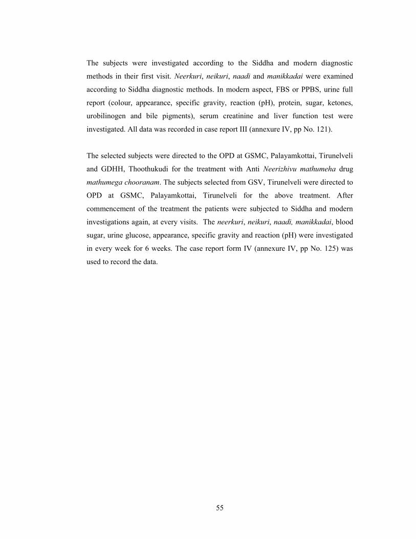
55
The subjects were investigated according to the Siddha and modern diagnostic
methods in their first visit. Neerkuri, neikuri, naadi and manikkadai were examined
according to Siddha diagnostic methods. In modern aspect, FBS or PPBS, urine full
report (colour, appearance, specific gravity, reaction (pH), protein, sugar, ketones,
urobilinogen and bile pigments), serum creatinine and liver function test were
investigated. All data was recorded in case report III (annexure IV, pp No. 121).
The selected subjects were directed to the OPD at GSMC, Palayamkottai, Tirunelveli
and GDHH, Thoothukudi for the treatment with Anti Neerizhivu mathumeha drug
mathumega chooranam. The subjects selected from GSV, Tirunelveli were directed to
OPD at GSMC, Palayamkottai, Tirunelveli for the above treatment. After
commencement of the treatment the patients were subjected to Siddha and modern
investigations again, at every visits. The neerkuri, neikuri, naadi, manikkadai, blood
sugar, urine glucose, appearance, specific gravity and reaction (pH) were investigated
in every week for 6 weeks. The case report form IV (annexure IV, pp No. 125) was
used to record the data.
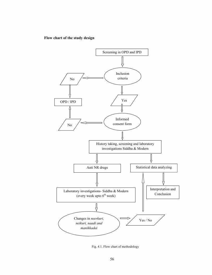
56
Flow chart of the study design
Screening in OPD and IPD
Inclusion criteria No
OPD / IPD Yes
Informed consent form No
History taking, screening and laboratory investigations Siddha & Modern
Statistical data analyzing
Interpretation and Conclusion
Anti NR drugs
Changes in neerkuri, neikuri, naadi and
manikkadai
Laboratory investigations- Siddha & Modern (every week upto 6th week)
Yes / No
Fig. 4.1. Flow chart of methodology

57
4.3 Statistical analysis
Completed questionnaires were checked before data entry. Data was entered and
analyzed on Statistical Package for the Social Science (SPSS) version 18.0 statistical
software (SPSS Inc., Chicago, IL, USA). The One- Sample Kolmogorov-Smirnov was
used to test the distribution of variables. Characteristics of patients were examined by
chi square test for categorical variables and by Student’s t - test and ANOVA for
continuous variables. Associations were tested by Pearson’s correlation coefficient.
Statistical significance was set at p< 0.05.
4.4 Laboratory investigations
Blood sugar was investigated using glucometer (one touch ultra-easy, range 20-600
mg/dL) and urinalysis reagent strip (Mission URS7010029) used for urine analysis.
In addition the serum creatinine, Serum glutamic-oxaloacetic transaminase (SGOT)
and Serum glutamic pyruvic transaminase (SGPT) tested at laboratories situated at
Palayamkottai and Thoothukudi.
4.4.1 Collection of blood and blood investigations
Blood samples (5 mL) were withdrawn from the arm of each patient under safe and
sterile condition, by a trained laboratory technician. A tourniquet was used to
increases the visibility of veins. An aliquot of blood (5 mL) was transferred to an
Ethylenediaminetetraactic acid (EDTA) coated tube and gently rotated (to avoid
hemolysis) to mix with the anticoagulant. The blood samples were analysed for serum
creatinine, SGOT and SGPT at the baseline visit.
Eight hour fasting or 2 hour post prandial blood sample was obtained by pricking the
tip of the ring finger (or suitable finger) of each patient using strile needle under safe
and sterile conditions. The blood sample was analysis for blood sugar using
glucometer (one touch ultra-easy, range 20-600 mg/dL).

58
4.4.2 Urine analysis – modern method
Urine report was assessed by analyzing specific gravity, reaction (pH), protein, sugar,
ketones, urobilinogen, bile pigments, nitrate, leucocytes and blood using urinalysis
reagent strip (Mission URS7010029) and recorded.
4.4.3 Urine analysis – Siddha method
4.4.3.1 Neer kuri
Collections of urine - clean vessels were used to collect the urine from the patients and
transfer the urine into the test utensils when conducting the test neerkuri.
Neerkuri was assessed by analyzing the colour, odour, froth and the sedimentation of
urine (Shanmugavelu, 1967), to the urine samples collected form the patients and
recorded.
4.4.3.2 Neikuri
The urine was transferred into 3.5 inch (9 cm) petri dish. A drop of gingili oil derived
from black colour sesame seeds applied on the surface of the urine, within one and a
half an hour from the collection of the urine (Shanmugavelu, 1967). The nature of
spread, shape of the spreading of oil on the surface of the urine, the time taken to start
(1st dot appear) and disappear the scatter of the neikuri were observed and recorded.
4.4.4 Naadi
The right or left elbow of the subject was griped with physicians’ left hand. Then the
fingers of the subject were snap of the knuckles (netti vanguthal) by the right fingers
of the physician. Thereafter the palm of the subject was rubbed with general force.
Then three fingers of the right hand, namely the index, middle and ring finger of the
physician gently were touched the skin over the radial artery of the subject. The index
finger was comfortably placed one inch below from the wrist crease and the other two
fingers placed next to it (the thumb should not be extended too far nor too much

59
fixed). The physician pressed and loose his three fingers simultaneously and was
concentrated every finger separately to observed Vatha, Piththa and Kabha naadi.
Left and right hand (Kai) were used to assess the naadi for female and male
respectively (Shanmugavelu, 1967).
4.4.5 Manikkadai
Manikkadai was measured as the circumference of the forearm above four finger
breath from the wrist using a thread. There after the length of the circumference
measured using the four fingers of the patient except thumb.
4.5 Ethical clearance
The ethical clearance was received from the Ethical Review Committee of GSMC,
Palayamkottai, Tirunelveli, Tamil Nadu, India (Annexure V, pp No. 126) for using
human subjects. The study procedure was explained in detail and written informed
consent was obtained from all the subjects before the commencement of the study
(Annexure I, pp No. 98). Each patient was provided with a leaflet which included the
diet chart for NR (Annexue III, pp No. 107). Results of Siddha diagnostic and
biochemical analyses were given to the patients individually. All data of the subjects
were kept confidentially.

60
4.6 Questionnaires
4.6.1 Types of questionnaire
The questionnaire case report form I was used to assess the inclusion and exclusion
criteria to selecte the patient for the study in the first vist (Annexure IV, pp No. 116).
Another general questionnaire case report form II was used to take history, physical
and systemic examination of the selected patients according to above questionnaire
(Annexure IV, pp No.118).
Laboratory investigation were recorded in case report form III (Annexure IV, pp No.
121).
Case report form IV was used to assess the prognosis (laboratory investigation) in
subsequent visits during the treatment. (Annexure IV, pp No. 125)

61
CHAPER 5
RESULTS
5.1 Diagnosis of NR
5.1.1 The sign and symptoms of the patients
The mean age of the study population was 45 ± 15 (mean ± standard deviation (SD))
years. The common sign and symptoms of the patients who have been selected for the
study has shown in the Table 5.1. High percentage of the patients had weight loss
(85 %) and excessive urination (78.8 %).
Table 5.1. The common sign and symptoms observed in patients attending to the out and in patient department at first time – OPD and IPD at GSMC, Palayamkottai, GDHH, Thoothukudi and GSV,
Tirunelveli (n=60)
Signs and symptoms n (%) Excessive urination (polyuria) 47 (78.8)
Excessive hunger (polyphagia) 26 (43.0)
Excessive thirst (polydipsia) 26 (43.0)
Tiredness/exhaustion 40 (66.6)
Changes of body weight (weight loss) 51 (85.0)
Body ache 19 (31.7)
Giddiness 23 (38.3)
Numbness/ tingling (polyneuritis) 19 (31.7)
n – Number of patients

62
5.1.2 Assessment of biochemical parameters of the blood samples (modern
aspect) of the patients attending to the OPD and IPD at first time - GSMC,
Palayamkottai, GDHH,Thoothukudi and GSV, Tirunelveli (n=60)
Table 5.2 has shown the mean FBS was 280.7 ± 145.5 mg/dL in 51 patients. Nearly
31% of the patients had higher than the mean concentration of FBS (>281.0 mg/dL) at
first visit. The mean concentration of PPBS was 274.6 ± 73.7 mg/dL in 09 patients.
Nearly 66.7% of the patients had higher than the mean concentration of PPBS (>275.0
mg/dL). None of the patient showed positive indication of serum creatinine, SGOT
and SGPT at first visit.
Table 5.2. Assessment of the biochemical parameters of the blood samples (modern aspect) of patients (n=60)
Biochemical parameter Result n %
Fasting blood sugar, mg/dL (mean ± SD) 280.7 ± 145.5
> 281.0 mg/dL 16 (31.4)
Post prandial blood sugar* (mg/dL) (mean ± SD) 274.6 ± 73.7
< 275.0 mg/dL 03 (33.3)
Serum creatinine mg/ dL Negative 60 (100)
Serum glutamic-oxaloacetic transaminase (SGOT) U/L Negative 60 (100)
Serum glutamic pyruvic transaminase (SGPT) U/L Negative 60 (100) * Data available for n = 9 patient and and n – Number of patients

63
5.1.3 Assessment of biochemical parameters of the urine samples (modern
aspect) of the patients attending at baseline – OPD and IPD at GSMC,
Palayamkottai, GDHH, Thoothukudi and GSV, Tirunelveli (n=60)
A higher percentage (81.7%) of patients’s urine samples had an acidic pH of 5.0 and
pH 6.0 was noted in 11. 7% of urine samples (Table 5.3). A high percentage (65 %) of
patients had higher specific gravity (1.030) in their urine. Increased urine glucose
(++++ or ≥ 2000 mg/dL) was noted in 15 % of patients. Urine protein was negative in
70 % of the patients. Negative indication for urine ketones, urobilinogen, bile
pigments, nitrate, leucocytes and blood were noted in all patients.
Table 5.3. Biochemical parameters (modern aspect)) of the patient’s urine samples (n=60) Biochemical parameters of the urine samples No. of samples (n) %
pH 5.0 49 81.7 6.0 7 11.7 6.5 2 3.3 7.0 2 3.3 Specific gravity 1.010 5 8.3
1.015 2 3.3 1.020 12 20 1.025 2 3.3 1.030 39 65 Urine sugar Negative 2 3.3 + (250 ± 15 mg/dL) 18 30 ++ (500 ± 30 mg/dL) 14 23.3 +++ (1000 ± 60 mg/dL) 17 28.3 ++++ (2000 ±110 mg/dL) 9 15 Urine protein
Negative 47 70 Trace (15 ± 0.15 mg/dL) 07 11.7 + (30 ± 0.3 mg/dL)
07 11.7 ++ (100 ± 1.0 mg/dL) 04 6.7 Ketones Negative 60 100

64
5.1.4 Factors associating with concentration of blood sugar
Figure 5.1 has shown that the patient diagnosed with higher urine glucose (++++ or
≥ 2000 mg/dL) had significantly (F4, 46 = 6.6111, p<0.001) higher fasting blood sugar
(446.8 ± 262.5 mg/dL; mean ±SD) than the patient diagnosed with negative urine
glucose (< 100 ± 5.0 mg/dL)
Table 5.3 continue… Biochemical parameters (modern aspect)) of the patient’s urine samples (n=60)
Biochemical parameters of the urine samples No. of samples (n) %
Urobilinogen Negative 60 100 Bile pigments Negative 60 100 Nitrates Negative 60 100 Leucocytes
Negative 60 100 Blood Negative 60 100
Fig. 5.1. The association of fasting blood sugar and urine Glucose level

65
Neither significant association nor correlation was observed between blood sugar and
urine pH and specific gravity.
5.1.5 Examination of urine by neerkuri (Siddha aspect)
Neerkuri was assessed by analysing the colour, odour, froth and the sedimentation of
the urine samples collected form the patients. Figure 5.2 has shown the colours and
froth appeared in the tested urine samples.
A B
C D
E F
Fig. 5. 2. Morphological description (colours and froth) of neerkuri; A- Seerana apakkuva neer (colour of wet straw water), B- Seeran pakkuva neer (colour of Citrus medica fruit), C- Soottai
tharakkoodiya neer (colour of Atalandia monophylla fruit), D- Athiuttina neer (colour of flame), E- Suththa seethala neer (Clear urine) and F- Froth appear in the urine

66
Table 5.4 described that the seerana apakkuva neer (wet straw water colour) was
observed in 51.7 % of patients and 11.7% of patients had suththa seethala neer (clear
urine). Odour and froth in urine noted in 13.3% and 11.7% of the patients
respectively. Sedimentation was observed in 3.3 % of the patient’s urine.
Table 5.4. Examination of neerkuri (Siddha aspect of urine examination) in the patients at baseline (n=60)
Characteristics of the urine samples No. of patients (n) (%)
Colour of the urine
Seerana apakkuva neer (colour of wet straw water)
31 51.7
Seerana pakkuva neer (colour of Citrus medica. L fruit)
12 20
Soottai tharakkoodiya neer (colour of Atalandia monophylla (Roxb.) A.DC. fruit)
08 13.3
Athiuttina neer (colour of flame)
02 3.3
Suththa seethala neer (clear urine)
07 11.7
Froth Urine samples with froth 07 11.7 Urine samples without froth 53 88.3
Sedimentation Urine samples with sedimentation 02 3.3 Urine samples without sedimentation 58 96.67
Odour Urine samples with odour Urine samples without odour
08 52
13.3 86.7

67
5.1.6 Examination of urine by neikuri (oil drop test (Siddha aspect)
5.1.6.1 Shape of the neikuri
Irregular pattern and round shape was observed in the neikuri (Figure 5.3).
The fasting blood sugar was compared with the shape of the neikuri in urine samples
tested (Table 5.5). The patients with irregular shape of neikuri in their urine samples
have shown a slightly higher concentration of fasting blood sugar (291.1 ±
154.3mg/dL) than the patients with round shape of neikuri (F1, 46 = 1.408, p=0.241).
None of the patient have developed a pearl shape in their urine samples.
A B
Table 5.5. Shape of the neikuri in urine samples and fasting blood sugar at baseline (n=51)
Shape of the neikuri No. of patients (n)
Concentration of fasting blood sugar (mg/dL)
(Mean ± SD)
Irregular 43 291.1 ± 154.3
Round 08 224. 9 ± 63.8
Fig. 5.3. Shape of the neikuri; A- Irregular shape and B- round shape

68
5.1.6.2 Duration for testing the neikuri and the biochemical parameters of
the urine samples
The time taken to start and disappear the scatter of neikuri was 161.4 ± 158.9 seconds
(2.7 ± 2.6 minutes) and 771.8 ± 483.9 seconds (12.9 ± 8.1 minutes) respectively.
The patients with low concentration of glucose (negative) in their urine have taken a
lessened time as 101.5 ± 0.7 seconds (1.7 ± 0.01 minutes) to start the scatter of neikuri
(Table 5.6). The patients with moderately high concentration of glucose (++ (500 ± 30
mg/dL) in urine, have taken more time to start the scatter of neikuri (206.7 ± 147.9
seconds). Neither significant association significantly (F1, 58 = 0.404, p=0.528) nor
correlation was observed between the level of concentration of urine glucose with the
time taken to start to scatter of neikuri. When considering the time taken to disappear
the scatter of neikuri, the time was significantly increased with the increase of glucose
levels in the urine samples.
Comparatively a lessened time was observed to start (62.5 ± 3.5 seconds) and
disappear (780.0 ± 1.2 seconds) the scatter of neikuri with the urine pH 7 (Table 5.6).
The urine samples with pH 6.5 have taken more time to start the scatter of neikuri and
the samples with pH 6 have taken a higher time to disappear the scatter of neikuri.
The urine samples with higher specific gravity (1.03) have taken more time to start the
scatter of neikuri (187.5 ± 177.6 seconds) and taken lessened time to disappear the
scatter of neikuri (639.1 ± 237.1 seconds). While, the samples with lower specific
gravity (1.01) have taken lessened time to start the scatter (64.0 ± 9.1 seconds) and
more time to disappear the scatter of neikuri (1361.2 ± 492.0 seconds) (Table 5.6).
Further the time taken to start the scatter of neikuri was increased with the increase of
specific gravity significantly (F4, 55 = 0.926, p=0.456), even though a significant (F4, 55
= 3.436, p=0.014) reduction was noted in the time of disappearing the scatter of
neikuri with the increase of specific gravity of urine samples.

69
Table 5.6. Comparison of the time taken to start and disappear the scatter of neikuri and biochemical parameters of the urine samples at baseline (n=60)
Biochemical parameters
Number of patients (n)
Time taken start to scatter of neikuri
(seconds) Mean ± SD
Time taken to disappear the scatter of neikuri (seconds)
Mean ± SD
Urine sugar level
Negative 2 101.5 ± 0.7 823.5 ± 0.7
+ 18 188.6 ± 219.2 249.8 ± 249.8
++ 14 206.7 ± 147.9 735.9 ± 291.8
+++ 17 106.5 ± 39.5 848.5 ± 489.4
++++ 9 153.3 ± 143.2 1124.1 ± 885.9
pH of urine 5 49 169.4 ± 166.7 639.0 ± 253.6
6 7 76.3 ± 22.3 1160.0 ± 528.6
6.5 2 2660.5 ± 0.7 735.9 ± 291.8
7 2 62.5 ± 3.5 780.0 ± 1.2
Specific gravity of urine 1.010 5 64.0 ± 9.1 1361.2 ± 491.9
1.015 2 101.5 ± 0.7 823.5 ± 0.7
1.020 12 129.2 ± 134.0 935.1 ± 850.2
1.025 2 148.0 ± 0.0 855.0 ± 0.0
1.030 39 187.5 ± 177.6 639.1 ± 237.1

70
5.1.6.3 Shape of the neikuri and the biochemical parameters of the urine
samples
Table 5.7 has shown, a higher percentage (86.7%) of patients had irregular shape of
neikuri. Among the patients with irregular shape of neikuri, 18 patients presented with
the urine glucose level + (250 ± 15 mg/dL) and 15 patients presented with +++ (1000
± 60 mg/dL). High specific gravity (1.030) of urine has observed in 67.3 % of urine
samples and acidic urine (pH 5) has noted in 82.7 % of urine samples in irregular
neikuri pattern of urine.
Table 5.7. Number of patients with different shapes of neikuri and their biochemical properties of urine samples (n=60)
Biochemical
parameters
No. of patients with different shape of neikuri
Irregular (n=52) Round (n=08)
n (%) n (%)
Urine sugar level
Negative 0 0 2 25
+ 18 34.6 0 0
++ 11 21.1 3 37.5
+++ 15 28.8 2 25
++++ 8 15.4 1 12.5
pH of urine
5 43 82.7 6 75
6 06 11.5 1 12.5
6.5 01 1.9 1 12.5
7 02 3.8 0 0
Specific gravity of urine
1.010 4 7.7 1 12.5
1.015 0 0 2 25
1.020 11 21.2 1 12.5
1.025 2 3.8 0 0
1.030 35 67.3 4 50
n – Number of patients

71
5.1.7 Naadi
The concentration of blood sugar was compared with the naadi of the patients (Table
5.8). The patients with Kabhavatha naadi had a significantly (F3, 47 = 3.513, p =0.022)
higher concentration of FBS (423.7 ± 289.5 mg/dL) than the patients with Vathakabha
naadi (FBS 178.8 ± 56.3 mg/dL), Piththavatha naadi (FBS 263. 3 ± 104.3 mg/dL)
and Vathapiththa naadi (FBS 268.1 ± 88.4 mg/dL). The patients with Vathakabha
naadi had comparatively lower FBS (178.8 ± 56.3 mg/dL) than the other patients.
5.1.7.1 Correlation of neikuri with the diagnosis of naadi (n=60)
The correlation of neikuri and naadi were analysed for confirm the diagnosis (Table
5.9). Among the 25 patients having Vathapiththam (diagnosed by naadi) 21 patients
(84%) were correctly classified to have Vathapiththam by the method of neikuri. All
of the Piththavatha patients (diagnosed by naadi) were correctly classified by neikuri.
Further, 75% of Kabhavatham and Vadhakabham patients were correctly
classified.This finding indicated the correlation between naadi and neikuri.
Table 5.8. Concentrations of fasting blood sugar and naadi at base line (n=51)
Naadi Number of patients FBS (mg/dL)
(mean ± SD)
Vathakabham 04 178.8 ± 56.3
Piththavatham 19 263. 3 ± 104.3
Vathapiththam 21 268.1 ± 88.4
Kabhavatham 07 423.7 ± 289.5
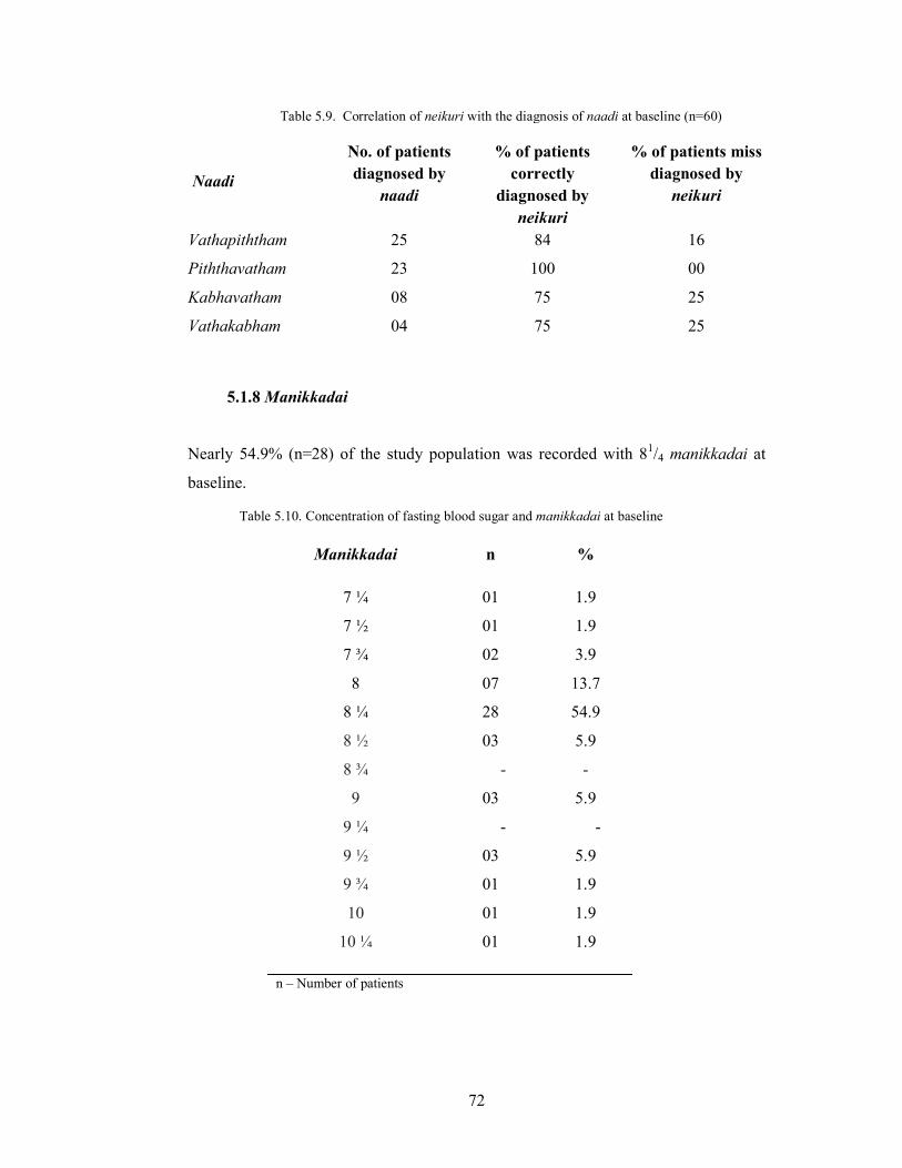
72
5.1.8 Manikkadai
Nearly 54.9% (n=28) of the study population was recorded with 81/4 manikkadai at
baseline.
Table 5.9. Correlation of neikuri with the diagnosis of naadi at baseline (n=60)
Naadi
No. of patients diagnosed by
naadi
% of patients correctly
diagnosed by neikuri
% of patients miss diagnosed by
neikuri
Vathapiththam 25 84 16
Piththavatham 23 100 00
Kabhavatham 08 75 25
Vathakabham 04 75 25
Table 5.10. Concentration of fasting blood sugar and manikkadai at baseline
Manikkadai n %
7 ¼ 01 1.9
7 ½ 01 1.9
7 ¾ 02 3.9
8 07 13.7
8 ¼ 28 54.9
8 ½ 03 5.9
8 ¾ - -
9 03 5.9
9 ¼ - -
9 ½ 03 5.9
9 ¾ 01 1.9
10 01 1.9
10 ¼ 01 1.9
n – Number of patients

73
5.2 Prognosis
5.2.1 The changes of the common sign and symptoms observed at baseline,
after the treatment with Madhumega chooranam.
The common sign and symptoms of the patients presented at baseline and end of the
study has shown in the Table 5.11. The percentage of patients having polyuria,
polyphagia, polydipsia, tiredness/exhaustion, weight loss, giddiness and
numbness/tingling reduced from the baseline to seventh visits following the treatment
of Madhumega chooranam. However, no change was observed in the visual
disturbance after the treatment of Madhumega chooranam.
Table 5.11. Comparison of common sign and symptoms of the patient following the treatment of Madhumega chooranam (n=60)
Number of patients having the signs/ symptoms (%)
Signs and symptoms
Baseline Second visit
Third visit
Fourth visit
Fifth visit
Sixth visit
Seventh visit
Excessive urination (polyuria)
78.8 58.3 53.3 41.7 25.0 16.7 13.3
Excessive hunger
(polyphagia)
43.3 33.3 26.7 20 16.7 16.7 16.7
Excessive thirst (polydipsia)
43.3 25 21.6 16.6 13.3 10.0 8.3
Tiredness/exhaustion 66.6 43.3 33.3 31.3 28.3 13.3 8.3
Changes in body weight (weight loss)
85.0 20.0 20.0 18.3 16.6 13.3 10.0
Body ache 31.7 26.7 25.0 25.0 23.3 26.7 21.6
Giddiness 38.3 26.7 20.0 11.7 8.3 5.0 5.0
Numbness/ tingling (polyneuritis)
31.7 30.0 30.0 26.7 18.3 15.0 13.3

74
5.2.2 Changes of FBS following the treatment of Madhumega chooranam
Effect of Madhumega chooranam on concentration of blood sugar was shown in
figure 5.4. The FBS was significantly (p < 0.001) reduced from the baseline (288.0 ±
162.3 mg/L) to seventh visit (167.2 ± 78.7 mg/dL) following the treatment of
Madhumega chooranam.
5.2.3 Changes of the biochemical parameters of urine sample with
the treatment of Madhumega chooranam
A higher percentage of patient’s urine samples had an acidic pH of 5.0 at baseline to
seventh visit (Table 5.12). A high percentage of patients had higher specific gravity
(1.030) in their urine samples throughout the visit. A substantial reduced was observed
in concentration of urine glucose. Approximately 67% of patients had low urine
glucose (indicated as negative in the urine strips). Similarly, the percentage of low
protein (indicated as negative) was increased from baseline (70%) to seventh visit
(91.7%).
Baseline
Secondvisit
Thirdvisit
Fourthvisit
Fifthvisit
Sixthvisit
Seventhvisit
Mean 288.0 232.2 215.8 210.3 197.7 182.4 167.2
0.0
50.0
100.0
150.0
200.0
250.0
300.0
350.0
Con
cent
rati
on o
f fa
sing
blo
od s
ugar
(m
g/L
)
Fig. 5.4. Concentration of FBS following the treatment of Madhumega chooranam

75
Table 5.12 has shown the urine glucose was reduced with the treatment of Mathumega
chooranam, whereas the patients presenced with high urine glucose was not reduced.
Presence of protein in urine was decreased at the 7th visit than baseline, with the
Table 5.12. Changes of the biochemical parameters of the urine sample with the treatment of Madhumega chooranam (n=60)
Biochemical
parameters of the urine samples
Number of patients having the signs/ symptoms
(%)
Baseline Second visit
Third visit
Fourth visit
Fifth visit
Sixth visit
Seventh visit
pH
5.0 81.7 78.3 75.0 81.7 76.7 78.3 71.7
6.0 11.7 15.0 21.7 11.7 16.7 18.3 18.3
6.5 3.3 3.3 3.3 3.3 3.3 3.3 3.3
7.0 3.3 3.3 - 3.3 3.3 - 6.7
Specific gravity
1.005 - 3.3 5.0 5.0 8.3 8.3 11.7
1.010 8.3 3.3 3.3 3.3 6.7 6.7 3.3
1.015 3.3 8.3 11.7 15.0 5.0 8.3 13.3
1.020 20.0 10.0 6.7 5.0 11.7 8.3 8.3
1.025 3.3 1.7 13.3 6.7 11.7 - 10.0
1.030 65.0 73.3 60.0 65.0 56.7 68.3 53.3
Urine sugar
Negative 3.3 26.7 23.3 23.3 45.0 56.7 66.7
+ 30.0 21.7 16.7 31.7 13.3 10.0 3.3
++ 23.3 20.0 25.0 20.0 21.7 13.3 8.3
+++ 28.3 21.7 20.0 8.3 - - 3.3
++++ 15.0 10.0 15.0 16.7 20.0 20.0 18.3
Urine protein
Negative 70.0 68.3 75.0 85.0 88.3 91.7 91.7
Trace 11.7 16.7 16.7 8.3 1.7 5.0 5.0
+ 11.7 3.3 1.7 - 3.3 - -
++ 6.7 11.7 6.7 6.7 6.7 3.3 3.3

76
Fig. 5.6. Changes in the specific gravity of the urine with the treatment of Madhumega chooranam
BaselineSecond
visitThirdvisit
Fourthvisit
Fifthvisit
Sixthvisit
Seventhvisit
Mean 5.2 5.3 5.3 5.2 5.3 5.2 5.4
5.0
5.5
6.0
pH o
f th
e ur
ine
sam
ples
BaselineSecond
visitThirdvisit
Fourthvisit
Fifthvisit
Sixthvisit
Seventhvisit
Mean 1.026 1.026 1.025 1.025 1.024 1.025 1.023
1.022
1.022
1.023
1.023
1.024
1.024
1.025
1.025
1.026
1.026
1.027
Spc
ific
gra
vity
(kg
/m³)
treatment. No changes were observed in the pH when following the treatment. From
the baseline to seventh visit the urine samples remained with an acidic pH (range 5.2-
5.4) (Figure 5.5). The specific gravity of the urine samples was fluctuated and reduced
from baseline (1.026) to seventh visit (1.023) following the treatment (Figure 5.6).
Fig. 5.5. Changes in the pH of the urine with the treatment of Madhumega chooranam

77
5.2.4 Changes in the parameters of neerkuri with the treatment of
Madhumega chooranam
Table 5.13 compares the parameters of neerkuri of urine samples from the baseline to
seventh visit with the treatment of Madhumega chooranam. There was no notable
difference observed in the colour, froth and the sedimentation of urine samples.
However, the percentage of patients having odour in their urine was reduced at
seventh visit (3.3%) when compared with baseline (13.3%)
Table 5.13. Changes in the parameters of neerkuri with the treatment of Madhumega chooranam
(n=60)
Parameters of Neerkuri Number of patients (%)
Base line
2nd visit
3rd visit
4th visit
5th visit
6th visit
7th
visit Colour of the urine Seerana apakkuva neer (colour of wet straw water )
51.7 55.0 56.6 55.0 55.0 55.0 46.6
Seerana pakkuva neer (colour of Citrus medica (Roxb.) fruit)
20.0 21.6 21.6 23.3 25.0 25.0 25.0
Soottai tharakkoodiya neer (colour of Atalandia monophylla L. fruit)
13.3 10.0 8.3 8.3 8.3 8.3 8.3
Athiuttina neer (colour of flame)
3.3 1.7 1.7 1.7 - - -
Suththa seethala neer (clear urine)
11.7 11.7 11.7 11.7 11.7 11.7 11.7
Froth Froth (+) 11.7 11.7 11.7 11.7 11.7 11.7 11.7
Froth (-) 88.3 88.3 88.3 88.3 88.3 88.3 88.3
Sedimentation Sedimentation (+) 3.3 1.7 1.7 1.7 1.7 1.7 1.7
Sedimentation (-) 96.7 98.3 98.3 98.3 98.3 98.3 98.3
Odor Odor (+) 13.3 10 8.3 8.3 5.0 3.3 3.3
Odor (-) 86.7 90.0 91.7 91.7 95.0 96.7 96.7

78
5.2.5 Changes in the neikuri with the treatment of Madhumega chooranam
5.2.5.1 Changes in the shape
Table 5.14 represents the changes in the shapes of the neikuri when taking the
treatment with the Madhumega chooranam. At baseline 52 urine samples (n=52) have
shown irregular shape and 08 urine samples (n=08) have shown round shape when
applied the oil drop. At the end of the study (after treatment with Madhumega
chooranam), 39 (75%) urine samples (out of 52 irregular shapes) changed to round,
06 (11.5%) remained irregular and 07 (13.5%) samples changed to pearl shape.
Whereas, the urine samples showed round shape (n=08) at the baseline, remained with
round shape at the end of the study. Comparatively low FBS was observed in the
patients with round shape of neikuri than the patients with irregular shape of neikuri.
Table 5.14. Changes in the shape of the neikuri with the treatment of Madhumega chooranam
Shapes of neikuri
At baseline
At 7th visit
Total Irregular
shape Round shape
pearl shape
n % n % n % n % n %
Irregular shape
52 86.7 6 11.5 39 75 07 13.5 52 86.7
Round shape 08 13.3 08 100 08 13.3
Total 60 100 06 10 47 78.3 07 11.7 60 100

79
5.2.5.2 Variations in the duration of time taken to start and disappear
the scatter of neikuri
The Figure 5.7 has shown the variations of the mean time taken to start the scatter of
neikuri from baseline to the seventh visit when taking the Madhumega chooranam. A
rapid increase was observed in the time taken to start the scatter (256 seconds) at the
4th visit and there after gradually decreased at subsequent visits.
BaselineSecond
visitThirdvisit
Fourthvisit
Fifthvisit
Sixthvisit
Seventhvisit
Mean 161.4 186.0 177.2 256.0 193.3 172.9 164.5
0.0
50.0
100.0
150.0
200.0
250.0
300.0
The
tim
e ta
ken
to s
tart
to s
catte
r of
nei
kuri
(S
econ
ds)
Fig. 5.7. Variations in the time taken to start to scatter of neikuri with the treatment of Madhumega chooranam
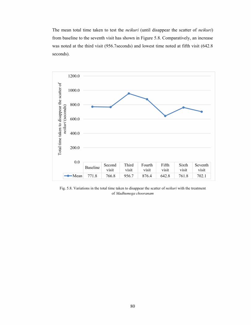
80
The mean total time taken to test the neikuri (until disappear the scatter of neikuri)
from baseline to the seventh visit has shown in Figure 5.8. Comparatively, an increase
was noted at the third visit (956.7seconds) and lowest time noted at fifth visit (642.8
seconds).
BaselineSecond
visitThirdvisit
Fourthvisit
Fifthvisit
Sixthvisit
Seventhvisit
Mean 771.8 766.8 956.7 876.4 642.8 761.8 702.1
0.0
200.0
400.0
600.0
800.0
1000.0
1200.0
Tot
al ti
me
take
n to
dis
appe
ar th
e sc
atte
r of
ne
ikur
i (se
cond
s)
Fig. 5.8. Variations in the total time taken to disappear the scatter of neikuri with the treatment of Madhumega chooranam
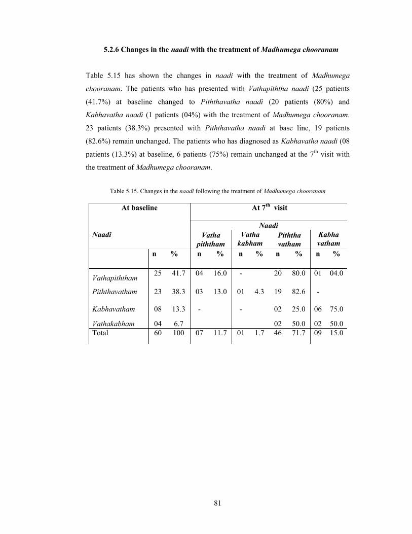
81
5.2.6 Changes in the naadi with the treatment of Madhumega chooranam
Table 5.15 has shown the changes in naadi with the treatment of Madhumega
chooranam. The patients who has presented with Vathapiththa naadi (25 patients
(41.7%) at baseline changed to Piththavatha naadi (20 patients (80%) and
Kabhavatha naadi (1 patients (04%) with the treatment of Madhumega chooranam.
23 patients (38.3%) presented with Piththavatha naadi at base line, 19 patients
(82.6%) remain unchanged. The patients who has diagnosed as Kabhavatha naadi (08
patients (13.3%) at baseline, 6 patients (75%) remain unchanged at the 7th visit with
the treatment of Madhumega chooranam.
At baseline
At 7th visit
Naadi Naadi Vatha
piththam Vatha
kabham Piththa vatham
Kabha vatham
n % n % n % n % n %
Vathapiththam 25 41.7 04 16.0 - 20 80.0 01 04.0
Piththavatham
23 38.3 03 13.0 01 4.3 19 82.6 -
Kabhavatham 08 13.3 - - 02 25.0 06 75.0
Vathakabham 04 6.7 02 50.0 02 50.0 Total 60 100 07 11.7 01 1.7 46 71.7 09 15.0
Table 5.15. Changes in the naadi following the treatment of Madhumega chooranam

82
5.2.7 Correlation between fasting blood sugar and naadi
The concentration of FBS was compared with the naadi of the patients at the seventh
visit (Table 5.16). The patients with Kabhavatha naadi had higher concentration of
FBS (361.6 ± 35.5mg/dL) than the patients with Vathapiththam naadi (FBS 128.4 ±
3.5mg/dL) and Piththavatha naadi (FBS 156.7 ± 48.2mg/dL).
Table 5.16. Mean concentrations of the FBS and naadi at seventh visit
Naadi Study population (n=58)
Mean concentration of fasting blood sugar (mg/dL)
(mean ± SD) Piththavatham 44 156.7 ± 48.2
Vathapiththam 7 128.4 ± 3.5
Kabhavatham 7 361.6 ± 35.5
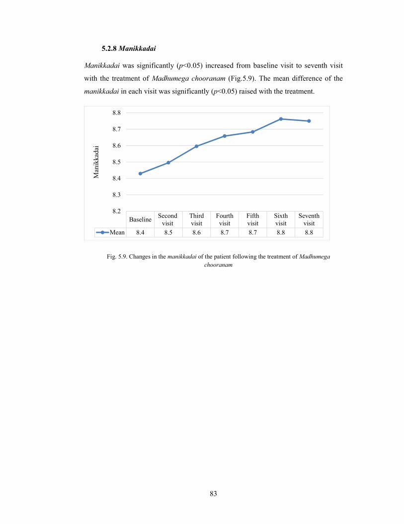
83
5.2.8 Manikkadai Manikkadai was significantly (p<0.05) increased from baseline visit to seventh visit
with the treatment of Madhumega chooranam (Fig.5.9). The mean difference of the
manikkadai in each visit was significantly (p<0.05) raised with the treatment.
BaselineSecond
visitThirdvisit
Fourthvisit
Fifthvisit
Sixthvisit
Seventhvisit
Mean 8.4 8.5 8.6 8.7 8.7 8.8 8.8
8.2
8.3
8.4
8.5
8.6
8.7
8.8
Man
ikka
dai
Fig. 5.9. Changes in the manikkadai of the patient following the treatment of Madhumega chooranam
84
CHAPTER 6
DISCUSSION
The current study documented the comparison of the Siddha diagnostic methods
specially neerkuri, neikuri, naadi and manikkadai with modern diagnostic methods in
the condition of Neerizhivu madhumeham. Specially in this study a collabration was
observed between the neikuri and naadi and this collabration followed to confirm the
neikuri. The correlation was observed between the fasting blood sugar and naadi.
Changes were documented in neerkuri, neikuri, naadi, manikkadai, concentration of
urine glucose and blood sugar between baseline and end of the study with the
treatment of Madhumega chooranam.The sign and symptoms as polyuria, polyphagia,
polydipsia, changes of body weight, tiredness, body ache, giddiness and numbness
were reduced at end of the sixth week, than the baseline with the treatment of
Madhumega chooranam.
A number of literature available for the study of comprehensive effectiveness of
traditional medicine for specific disease entities (Claudia et al., 2013). The
comparative study on Siddha diagnostic methods specially neerkuri and neikuri with
modern diagnostic methods in the condition of NR, has not been thoroughly studied
previously to the best of my knowledge. One previous study by Ekka Ranjita et al
(2013) has documented the prognostic aspect of taila bindu pariksha in prameha. In
this way, the current study is unique and innovative. The sign and symptoms of NR
were nearly related to mathumegam, neerizhivu and salamegam in Siddha system of
medicine (Thirumoolar vaithiyam karukkadai – 600, Yuki vaiththiya kaviyam and
Dhanvanthiri sootchuma vaithiyam 200 visha bethi vaiththiyam) and DM in the
system of Allopathic medicine (Habtamu, 2015). In addition, the prameha in
Ayurveda medical system was compared with DM (Ekka Ranjita et al., 2013).
The pH of the urine of majority patients was acidic (pH 5) at the baseline and end of
the sixth week (at the baseline 81.7 % and at the end of the sixth week 71.7 %). There
was no correlation observed in pH of the urine with blood sugar level. Diabetic
acidosis, uncontrolled diabetes and fasting influences the acidic urine (Compendium

85
of urine analysis, 2011). The patients were present for the study at base line (85 %)
and at the seventh visit (96.7 %) with fasting and it may be the reason for the acidic
urine. The study indicates that, the pH of the urine cannot be taken as a parameter for
confirming the NR.
There was no significant changes observed in specific gravity of urine at the base line
and at the end of the study with the treatment. No correlation observed in urine
specific gravity with blood sugar level in the current study. But, the compendium of
urine analysis documents the specific gravity of the urine increases if the glucose
concentration increase > 1000 mg/dL (>56 mmol/L) (Compendium of urine analysis,
2011). A discrepancy was noted with the study of Compendium of urine analysis and
further studies wil be needed to confirm correlation. The specific gravity of the urine
depends primarily on the amount of fluid intake by the patient, heavy sweating and
increase urine output provoked by diuretics (Compendium of urine analysis, 2011).
Excessive urination and glucose concentration in urine may be a possible reason for
the high specific gravity of urine.
Urine protein was negative for 70 % of the patients, trace amount observed at 11.7 %
and positive for 18.3 % of the patients at baseline. These findings indicate that, 81.7 %
of patients were not aroused with renal pathology at base line and 18.3 % of patients
may have silence NR for a long time. Negative urine protein level was increased at the
end of the study (91.7 %) than the baseline (70 %) and the percentage of patients
presented with urine protein gradually decreased with the treatment of Madhumega
chooram. The above finding indicates the influence of the Madhumega chooram in
renal pathology and it is an added advandage.
Correlation noted with high blood sugar level and high concentration of glucose in
urine. High concentration of glucose in urine was observed in the patients, who had
high blood sugar level. Correlation observed with urine glucose level and blood sugar
level (Morris, McGee and Kitabchi, 1981).
Significant changes was not observed at seerana apakkuva neer (wet straw water
colour) at the baseline (51.7 %) and at the end of the study (46.6 %) and no change
observed in Suththa seethala neer (transparent) at the baseline and end of the study

86
(11.7 %). These findings indicate that NR cannot be confirmed with the colour of the
urine. Even though the colour may indicate the thodam which affected the patient.
Seerana apakkuva neer, seeran pakkuva neer, soottai tharakkoodiya neer and
athiuttina neer indicate that the patient primarily affected with Piththam and suththa
seethala neer indicates that the patient primarily affected with Kabham
(Shanmugavelu, 1967). Thirumoolar vaithiyam karukkadai – 600 (Raththina nayakar
and sons, 2010) indicates that, the principal thodam which cause NR is Piththam and
Kabham. The colour of the urine may indicate the type of the NR once the NR
already diagnosed.
11.7 % of patients had froth in their urine and the readings of the neikuri and naadi of
the patients were interpreted as Kabhavatham. The sign and symptoms and blood
sugar level of the patients, who had Kabhavatha NR were not changed, even after
treatment. Availability of froth in urine indicates the Kabhavatha disease
(Shanmugavelu, 1967) and the literature further the documented, the Kabhavatha
disease cannot be curable. These finding indicated, that the disease will not be curable,
if froth present in the urine of the NR patients. The odour of the urine was reduced at
seventh visit (3.3%) when compared with baseline (13.3%). Eventhough the sediment
of the urine was not changed.
When testing the neikuri, irregular pattern and round shape of neikuri was observed in
86.7 % and 13.3 % of the patients at the baseline respectively. At the end of the
seventh visit 10 % of irregular, 78.3 % of round and 11.7 % pearl shape were
documented. Irregular, round and pearl shapes of the neikuri indicates Vatham,
Piththam and Kabham respectively (Shanmugavelu, 1967). The affected thodam
(Vathapiththa, Piththavatha, Vathakabha and Kabhavatha NR) can be identified using
the shape of the neikuri (formation of pattern) considering together with the total time
taken to test neikuri, once the NR already confirmed by other diagnostic methods. The
blood sugar level of the Vathapiththa and Piththavatha patients were less when
compare with the blood sugar of Kabhavatha patients. The text book indicates that the
Kabhavatha disorders are not curable (Shanmugavelu, 1967). Current study indicating
that, irregular shape of neikuri at baseline (86.7 %) changed to round shape (75 %) at
the end of the study with the treatment of Madhumega chooranam. In addition, blood
sugar of the NR patients with round shape of neikuri comparatively less than the blood

87
sugar of NR patients with irregular shape of neikuri. These finding indicating neikuri
can be used to test the prognosis of NR.
The Yuki vaithiya kaviyam (2002) indicates the round shape of neikuri in neerizhivu is
not curable. It may indicate the neerizhivu can not be curable, but can manage with
suitable medicines.
Clean and dry utensil need to use to test neikuri. False results was observed when
examine the neikuri without washing the utensil. It is preferable to use clean separate
vessels to collect the urine from the patients and transfer the urine into test utensil
when conducting the test. These procedures could facilitate to avoid false results.
A significant correlation was observed between naadi and neikuri in the current study
and the neikuri was confirmed together with the naadi. This correlation has not been
documented in previous studies.
75 % of Kabhavatha NR detected (by naadi) at the base line did not change at the end
of the study, whereas 80 % Vathapiththa NR was changed to Piththavatham and 82.6
% of Piththavatha NR remains as Piththavatha NR. The blood sugar of Vathapiththa
and Piththavatha NR patients reduced with the treatment of Madhumega chooram
indicating the Vathapiththa and Piththavatha NR can be managed with Madhumega
chooranam.
Pendulum movement of naadi was felt in all NR patients in the current study. Naadi
vaagadam (Sourirajan, 2014) describes the nature of the pendulum movement like as
the beat of the Veena string in the condition of Piththavatha disorders. Similar
moment was observed in the current study in the condition of NR. Further our study
documented that, the pendulum movement does not change even the blood sugar
return to normal level in the NR patients.
Four types of NR as Vathapiththam, Piththavatham, Vathakabham and Kabhavatham
were noted according to naadi in the current study. Even though Ramachandran
(2000) classified 24 types of neerizhivu under 7 catogary. In addition the current study
documented, the range of blood sugar could be detected from the naadi. Relatively
88
low blood sugar level was observed in Vathapiththa and Piththavatha NR patients
than the Kabhavatha NR patients. The current study indicates that the naadi could be
used as diagnostic as well as prognostic parameter for NR.
Manikkadai nool (circumference of wrist) is one of the diagnostic and prognostic
parameter which gives the progress of the disease by calculating the number of fingers
in decreasing or increasing order (Susila et al, 2014). Usually the length of the thread
starts with four finger breadth and ends with eleven finger breadth (Panthinen
Siddhar arulichcheitha nadi sasthiram, 2012) and the low value indicates the poor
prognosis of a disease (Susila et al, 2014). Previous studies documented the
importance of wrist circumference with respect to the DM (Capizzi, 2011; Younes et
al., 2013; Adina Mitrea, 2013). In the current study, the manikkadai nool was 8 ¼
finger breadth in 54.9% of the patients at baseline and it increased at the end of the six
week with the reduction of blood sugar level. These finding indicates the manikkadai
nool could be used as a parameter to analyse the prognosis of the NR. Panthinen
Siddhar arulichcheitha naadi sasthiram (Kanthasami Muthaliyar, 2012) documents
that 8 ¼ finger of manikkadai nool was the sign of Piththa disorders, fever, bramiyam,
kamiyam and diseases in the head. The Thirumoolar vaiththiyam (karukkadai 600)
(Raththina nayakar and sons, 2010) explains, that when the Piththam increase, the
increased Piththam combine with Vatham and it causes the NR.
The current study indicating urine glucose, pH and specific gravity may influene in
the sparding and formation of the shape of neikuri. There may a number of factors
including surface tension, molecular weight and polarity of the molecules in the urine
may influence the neikuri.
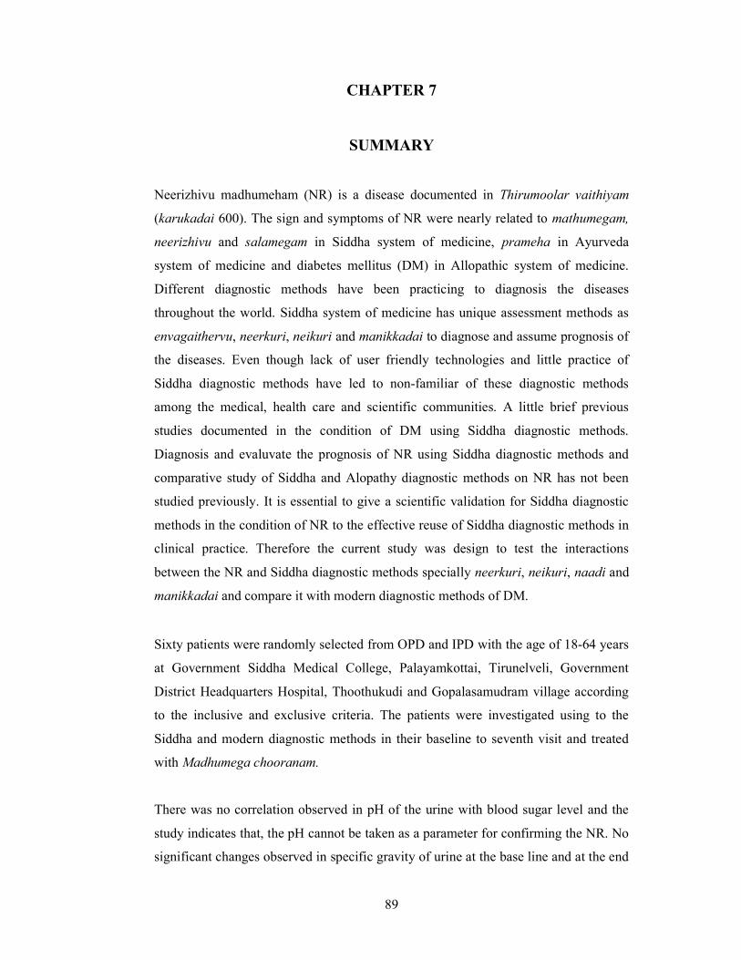
89
CHAPTER 7
SUMMARY
Neerizhivu madhumeham (NR) is a disease documented in Thirumoolar vaithiyam
(karukadai 600). The sign and symptoms of NR were nearly related to mathumegam,
neerizhivu and salamegam in Siddha system of medicine, prameha in Ayurveda
system of medicine and diabetes mellitus (DM) in Allopathic system of medicine.
Different diagnostic methods have been practicing to diagnosis the diseases
throughout the world. Siddha system of medicine has unique assessment methods as
envagaithervu, neerkuri, neikuri and manikkadai to diagnose and assume prognosis of
the diseases. Even though lack of user friendly technologies and little practice of
Siddha diagnostic methods have led to non-familiar of these diagnostic methods
among the medical, health care and scientific communities. A little brief previous
studies documented in the condition of DM using Siddha diagnostic methods.
Diagnosis and evaluvate the prognosis of NR using Siddha diagnostic methods and
comparative study of Siddha and Alopathy diagnostic methods on NR has not been
studied previously. It is essential to give a scientific validation for Siddha diagnostic
methods in the condition of NR to the effective reuse of Siddha diagnostic methods in
clinical practice. Therefore the current study was design to test the interactions
between the NR and Siddha diagnostic methods specially neerkuri, neikuri, naadi and
manikkadai and compare it with modern diagnostic methods of DM.
Sixty patients were randomly selected from OPD and IPD with the age of 18-64 years
at Government Siddha Medical College, Palayamkottai, Tirunelveli, Government
District Headquarters Hospital, Thoothukudi and Gopalasamudram village according
to the inclusive and exclusive criteria. The patients were investigated using to the
Siddha and modern diagnostic methods in their baseline to seventh visit and treated
with Madhumega chooranam.
There was no correlation observed in pH of the urine with blood sugar level and the
study indicates that, the pH cannot be taken as a parameter for confirming the NR. No
significant changes observed in specific gravity of urine at the base line and at the end

90
of the study with the treatment and no correlation observed in specific gravity with
blood sugar level. Urine protein level was decreased with the treatment of
Madhumega chooram and it is an added advandage in condition of NR. Because
generally chronic NR may affect the renal fuction and cause diabetic nephropathy.
Correlation noted with high blood sugar level and high concentration of glucose in
urine.
Current study indicated that NR cannot confirmed with the colour of the urine. Even
though the colour of the urine may indicate the type of the NR (the thodam which
affected) once the NR already diagnosed.
The study documented that the froth appear in urine of Kabhavatha NR patients and
the sign and symptoms and blood sugar level of the Kabhavatha NR patients were not
changed, even after the treatment. These indicating, that the disease will not be
manage, if froth present in the urine of the NR patients.
The affected thodam could be detectable, using the shape of the neikuri and the time
taken to test the neikuri, once the NR already confirmed by other diagnostic methods.
The blood sugar level of the Vathapiththa and Piththavatha patients were less when
compare with the blood sugar of Kabhavatha patients. In addition, blood sugar of the
NR patients with round shape of neikuri comparatively low than the blood sugar of
NR patients with irregular shape of neikuri. These finding indicating neikuri can be
used to test the prognosis of NR.
A significant correlation was observed between naadi and neikuri in the current study
and this correlation has not been documented in previous studies. Vathapiththa and
Piththavatha NR can be managed with Madhumega chooranam, eventhugh
Kabhavatha NR could not manage with Madhumega chooranam. Pendulum
movement of naadi was felt in all NR patients in the current study. Further our study
documented that, the pendulum movement does not change even the blood sugar
return to normal level in the NR patients. These finding may indicate that NR not a
curable disease.

91
Four types of naadi as Vathapiththa, Piththavatha, Vathakabha and Kabhavatha NR
patients were observed in the current study. In addition the current study documented,
the range of blood sugar could be detected from the reading of naadi and the naadi
could be used as diagnostic as well as prognostic parameter for NR. The finding
indicates the manikkadai nool could be used as a parameter to analyse the prognosis of
the NR.
Siddha diagnostic methods can be use to diagnose and assume the prognosis of NR.
Any how, further elaborated study will be needed to get insights of Siddha diagnostic
methods.

92
CHAPTER 8
CONCLUSION
The current study suggests the Siddha diagnostic method of neerkuri and neikuri help
to assess health states of Neerizhivu madhumeha patients. In addition it can be used to
findout the type of NR based on Vatha, Piththa and Kabham. Even though the naadi
may serve as a diagnostic method for Neerizhivu madhumeham, as specially pendulum
movement of naadi has observed in Neerizhivu madhumeha patients. Neerkuri,
neikuri, naadi and manikkadai may be used to assess the prognosis of the Neerizhivu
madhumeham. In conclusion, above Siddha investigative methods can be used as a
diagnostic and prognostic parameter instead of modern method, in the condition of
Neerizhivu madhumeham. However further detail studies will be needed prior to use
these methods in clinical practice.

93
BIBLIOGRAPHY
1. Abdulfatai B. Olokoba., Olusegun A.Obateru. and Lateefat B. OLokoba -
Type 2 Diabetes mellitus: A review of current trends - Oman Medical Journal -
2012, 27, (4): pp.269-273.
2. Adina Mitrea., Andreea soare., Simona Georgiana Popa., Mirela Nicoleta
Tudor., Maria Mota. and Paolo Pozzilli. - Wrist circumference: An
independent predictor of both insulin resistance and chronic kidney disease in
elderly population – Romanian Journal of Diabetes Nutrition & Metabolic
Diseases – 2013, 20, (3): pp.323 - 329.
3. Ahmed, AM - History of diabetes mellitus - Saudi Med J - 2002, 23, (4): pp.
373-378.
4. Ananthan, T., Jega Jothy Pandiyan, S., Meenatchi Sunthara Moorthy, K.,
Sellathurai, I. and Selvaraj, S. Agasthiyar vaiththiya sillaraik kovai. Chennai:
Central Council for Research in Siddha, 2010: pp. 366, 367, 370, 378.
5. Anastassios G. Pittas.- Diabetes mellitus, diagnosis and pathophysiology -
Tufts open courseware - 2009.
6. Arangarasan, S. Agasthiyar nool thirattu. Thanjavur: Saraswathy Mahal
library, 2016: pp. 62.
7. Axel R. Fugl - Meyer., Roland Melin and Kerstin S. Fugl - Meyer - Life
satisfaction in 18- to 64- year - old Swedes: In relation to gender, age, partner
and immigrant status - J Rehabil Med - 2002, 34: 239 - 246.
8. B. Raththina nayakar and sons. Agasthiyar munivar arulichcheitha vaiththiya
raththna surukkam 360 (moolamum uraiyum). Chennai: B. Raththina nayakar
and sons, 2016: pp. 3.
9. B.Raththina nayakar & sons. Thirumoolar vaithiyam (karukadai 600).
Chennai: B.Raththina nayakar & sons, 2010: pp. 5,7,10, 15.
10. Capizzi, M., Leto, G., Petrone, A., Zampetti, S., Papa, R.E., Osimani,
M., Spoletini, M., Lenzi, A., Osborn, J., Mastantuono, M., Vania, A.
and Buzzetti, R. - Wrist circumference is a clinical marker of insulin
resistance in overweight and obese children and adolescents - Circulation -
2011, 123, (16): pp. 1757-1762.

94
11. Central Council for Research in Ayurveda and Siddha. Agasthiyar vaiththiya
vallathi - 600 (moolamum uraiyum). New Delhi: Central Council for Research
in Ayurveda and Siddha, 2005, 2nd ed: pp. 3.
12. Claudia M. Witt., Andreas Michalsen., Stephanie Roll., Antonio Morandi.,
Shivnarain Gupta., Mark Rosenberg., Ludwig Kronpab., Elmar Stepelfeldt.,
Syed Hissar., Matthias Muller. and Christian Kessier - Comparative
effectiveness of a complex Ayurvedic treatment and conventional standard
care in osteoarthritis of the knee - study protocol for a ramdomized controlled
trail - Biomed central - 2013, 14, (149): pp. 1-10.
13. Division of Siddha medical books publication, Department of Indian medicine
and Homeopathy. Agasthiyar maruththuvam (tamil maruththuva nool varisai -
15) olaichchuvadith thokuppu nool. Chennai: Division of Siddha medical
books publication, Department of Indian medicine and Homeopathy, 2009: pp.
24-26.
14. Ekka Ranjita., Chadra kar Anukul. and Upadhyay, B.N. - Assessment of
prognostic aspect of Prameha (Diabetes mellitus) by Taila bindu pariksha -
International Ayurveda Medical Journal - 2013, 1, (2) : 1-4.
15. Elizabeth Selvin, Michael W. Steffes, Hong Zhu, Kunihiro Matsushita, Lynne
Wagenknecht, James Pankow, Josef Coresh. and Frederick L. Brancati. -
Glycated haemoglobin, diabetes and cardiovascular risk in nondiabetic adults -
The New England journal of medicine - 2010, 362: pp. 800-811.
16. Gillett, M.J. - International Expert Committee report on the role of the HbA1c
assay in the diagnosis of diabetes - Diabetes care - 2009, 32, (7): pp.1327 -
1334.
17. Habtamu Wondifraw Baynest. - Classification, pathophysiology, diagnosis and
management of Diabetes Mellitus - Baynes J Diabetes Metab - 2015, 6, (5):
pp. 1-9.
18. Harikumar, K., Kishor Kumar, B., Hemalatha, G.J., Bharath kumar, M. and
Steven fransis saky Lado. - A review on Diabetes Mellitus - Internal journal of
Novel trends in pharmaceutical sciences - 2015, 6, (4): pp. 201-217.
19. Hove, M.N., Kristensen, J.K., Lauritzen, T. and Bek, T. - The prevalence of
retinopathy in an unselected population of type 2 diabetes patients from Arhus
county, Denmark - Acta Opththamol Scand - 2004, 82, (4): pp. 443-448.
95
20. International Diabetes Federation (IDF). International diabetes atlas
(Retrieved). 2014, 6th ed: pp 1- 160.
21. Kalaththur kanthasami Muthaliyar. Pathinen Siddhar arulichcheitha naadi
saasthiram. Chennai: B. raththina nayakar and sons, 2012: pp. 119,120,128,
168.
22. Kannusamy pillai, S. Sikichcha ratnathepam. Chennai: B. Raththina nayakar
and sons, 1931: pp. 18, 19.
23. Mather, C.D. and Loncar, D. - Projections of global mortality and burden of
disease from 2002 to 2030 - PLOS Med -2006, 3, (11) e442.
24. Mohan, R.C. Yakopu vaiththiyam 300. Chennai: Thamarai library, 2014: pp.
27.
25. Mohan, R.C. Yuki vaiththiya kaviyam. Chennai: Thamari library, 2014: pp.
253-262.
26. Mohana raj, T. Meha noi, Soothaganool mattrum arivaiyar sinthamani.
Kanniyakumari: A.T.S.V.S. Siddha Medical college, Munchirai, Puthukkadai,
2008: pp.33, 34, 36, 37, 38, 39, 40- 53.
27. Morris, L.R., McGee, J.A. and Kitabchi, A.E. - Correlation between plasma
and urine glucose in diabetes - Ann intern med - 1981, 94, (4-1): pp. 469-471.
28. Natarajan, K. Principles of diagnosis in Siddha. Chennai: Department of
Indian Medicine and Homoeopathy, Chennai, 2009: pp. 106, 154- 159, 306-
307.
29. Ozougwu, J.C., Obimba, K.C., Belonmu, C.D. and Unakalamba, C.B. - The
Pathogenesis and pathophysiolology of type 1 and type 2 diabetes mellitus -
Journal of Physiology and Pathophysiology - 2013, 4, (4): pp. 46-57.
30. Patidar, D. Pharmacology III. Meerut: Shree Sai Prakashan, 2011, 2nd ed: pp.
113-114.
31. Peris Urbana Ivy, B. and Rani Hema Malini, R. - Modern pulsometer for
traditional Indian Medicine - International Journal of Computer Applications -
2010, 9, (3): pp. 19-23.
32. Ramachandran, S.B. Uyir kakkum Siddha maruththuvam. Chennai: Thamarai
library, 2000: pp. 360, 362, 363.
33. Ramachanthiran, S.B. Theraiyar Neerkkuri vaiththiyam Neerkkurinool -
neikuri nool moolamum uraiyum. Chennai: Thamarai library, 2015: pp 2-4, 11,
18, 19, 25, 27.

96
34. Ramachanthiran, S.P. Dhanvanthiri Sootchuma vaiththiyam 200, visha bethi
vaiththiyam. Chennai: Thamarai library, 2015: pp.67.
35. Resolution 66/2. Political declaration of the high-level meeting of the general
assembly on the prevention and control of noncommunicable diseases
(working paper series) Geneva - Harvard school of public health and world
economic forum - 2011.
36. Roche diagnostic Ltd. Compedium of urine analysis. Swizerland: Roche
diagnosis Ltd, Ct - 6343 Rotkreuz, 2011: pp 34. 36- 37. [online] Available at:
http://www.roche-diagnostics.ch/content/dam/corporate/roche-dia_ch/
documents/broschueren/professional_diagnostics/urindiagnostik/12254620001
_EN_EA_Compendium-of-urinanalysis_Broschüre_EN.pdf [Accessed 16 Mar
2018].
37. Sambasivam pillai, T.V. Tamil - English dictionary IV - part II. Chennai:
Department of Indian Medicine and Homoeopathy, 1998, 2nd ed: pp.1831 -
1833.
38. Sekikawa, A., Tominaga, M., Takahashi, K., Eguchi, H., Igarashi. M.,
Ohnuma, H., Sugiyama, K., Manaka, H., Sasaki, H. and Fukuyama, H. -
Prevalence of diabetes and impaired glucose tolerance in Funagata area, Japan
- Diabetes care - 1993, 16, (4): pp. 570-574.
39. Shanmugavelan, A. Therar arulichcheitha siruneerkkuri sothanai. Thanjavur:
Saraswathy Mahal library, 2014: pp. 10.
40. Shanmugavelu, M. Noi nadal Noi mudal nadal thirattu. Chennai: Department
of Indian Medicine and Homoeopathy, Chennai, 1967: pp. 126, 269-270.
41. Sourirajan, M. Naadi vagadam. Thanjavur: Saraswathy Mahal library, 2014:
pp. 55.
42. Susila, R., Arunadevi, R., Christian, G.J., Elansekaran, S., Ramamurthy, M.
and Logamaniyan, M. - Validation of Siddha diagnostic procedures for Madhu
piramiam with the aid of conventional diagnostic procedures - International
journal of Advanced Ayurveda, Yoga, Unani, Siddha and Homeopathy - 2015,
3, (1): pp. 197-218.
43. Vasutheva Sasththirikal, K. and Subramanya Sasththirikal, K.S. Nadi
sakkiram. Thanjavur: Director, Saraswathy Mahal Library, 2014: pp. 22.
44. Venkatrajan, S. Agasththiyar 2000 I & II. Thanjavur: Saraswathy Mahal
library, 2014: pp. 20, 21, 26.

97
45. Venkattarajan, S. Dhanvathiri vaiththiyam I. Thanjavur: Saraswathy Mahal
library, 2014: pp.10-12.
46. Venkattarajan, S. Dhanvathiri vaiththiyam II. Thanjavur: Saraswathy Mahal
library, 2014: pp. 28.
47. WHO - Definition and diagnosis of Diabetes Mellitus and intermediate
hyperglycaemia - 2006: pp. 1-46.
48. WHO (WHO/NCD/NCS/99.2) - Definition, diagnosis and classification of
Diabetes Mellitus and its complications - 1999: pp.1-59.
49. WHO Mortality database (online database). Geneva: World Health
Organization; 2011. [online] Available at: http://apps.who.int/healthinfo/
statistics /mortality/causeofdeath_ quaryaccessed [Accessed 12 Jan.2016].
50. World Health Organization - Global report on diabetes - 2016.
51. World Health Organization - The top 10 causes of death, Fact sheet No.310 -
2013.
52. www.wikipedia.com,(2013). Thirumular. [online] Available at:
https://en.wikipedia.org/wiki/ Tirumular [Accessed 16 Mar .2018].
53. Younes Jahangiri Noudeh., Farzad Hadaegh., Nasibeh Vatankhah., Amir
Abbas Momenan., Navid Saadat., Davood Khalili. and Fereidoun Azizi - Wrist
circumference as a novel predictor of Diabetes and prediabetes; results of cross
sectional and 8.8 - year follow - up studies - The Journal of clinical
endocrinology and metabolism - 2013, 98, (2): pp 777-784.
54. Zimmet, P.Z. - Kelly west lecture 1991. Challenges in diabetes epidemiology -
from West to the rest. Diabetes care - Diabetes care - 1992, 15, (2): pp. 232-
252.

98
CONSENT FORM
COMPARATIVE STUDY OF THE SIDDHA DIAGNOSTIC METHODS
SPECIALLY NEERKURI & NEIKURI WITH MODERN DIAGNOSTIC
METHODS IN NEERIZHIVU MADHUMEHAM (DIABETES MELLITUS –
TYPE 2)
CONSENT FORM
CERTIFICATE BY INVESTIGATOR
I certify that I have disclosed all details about the study in the terms easily understood
by the patient.
Date: …………………… Signature of the Investigator: ……………………
Name of Investigator: Dr. K. Vidya dharshini
CONSENT BY SUBJECT
I (Mr./Ms./Mrs) ...……………………………………………………………………
Residing at …………………………...…………………………...............................
agree and exercising my free power of choice, hereby give my consent to be included
as a subject in the study of COMPARATIVE STUDY OF THE SIDDHA
DIAGNOSTIC METHODS SPECIALLY NEERKURI & NEIKURI WITH MODERN
DIAGNOSTIC METHODS IN NEERIZHIVU MADHUMEHAM (DIABETES
MELLITUS), which is to be conducted at Government Siddha Medical College,
Palayamkottai, Tamil Nadu.
1 have been informed to my satisfaction, by the attending physician about the purpose
of the study, screening, nature of drug treatment and follow-up including the
laboratory investigations to be performed to evaluate diagnostic methods and safe
guard my body functions.
I am also aware of my right to opt out of the trial at any time during the course of the
study without having to give the reasons for doing so.
99
I am giving the consent to participate to the study with my full consciousness after
study the patient information sheet which given to me by the investigator and after full
clarification of all my doubts. Further I state that the consent is not given under any
influence or any other measures.
Name of the Subject: ………………………………………….
Signature/Thumb impression: …………………………………
Place: …………………………………………………………..
Date: ……………………………………………………………
Name of witness: ………………………………………………
Signature or Thumb impression: ………………......................
Date: …………………………………………………………..
Signature of investigating medical officer: …………..............
Date: ……………………………………………………………
Signature of Director in charge / Supervisor: …………………
Date: ……………………………………………………………
100
COMPARATIVE STUDY OF THE SIDDHA DIAGNOSTIC METHODS
SPECIALLY NEERKURI & NEIKURI WITH MODERN DIAGNOSTIC
METHODS IN NEERIZHIVU MADHUMEHAM (DIABETES MELLITUS)
Nehahspapd; xg;Gjy; gbtk;
jpUÆ jpUkjpÆ nry;tp………………………………………………………(ngau;)
Mfpa ehd; ……………………………………………………. (trpf;Fk; ,lk;)
vd; Ra epidTld; ghisaq;Nfhl;ilapy; cs;s muR rpj;jkUj;Jtf;
fy;Yhupapy; elj;jg;gLk; rpj;jkUj;Jt (ePupopT kJNkfk;) Neha; epu;za
Ma;Tf;F rk;kjk; mspg;gij njuptpj;J vOjpf; nfhLf;Fk; xg;Gjy; gbtk;.
Nkw;gb jiyg;igf; nfhz;l ePupopT kJNkf Neha; epu;zak; rk;ge;jkhd fPo;
Fwpg;gplg;gl;Ls;s kUj;Jtupdhy; Nkw;nfhs;sg;gLk; ,t; Ma;Tf;F cld;
gLtjw;F vd; Ra epidTld; KO xg;GjiyAk; njuptpj;Jf; nfhs;fpNwd;.
,e;j Ma;tpd; Nehf;fk;> Ma;Tnra;Ak; Kiw> clw; gupNrhjid> kUj;Jtk;
nra;Ak; Kiw> njhlu; fz;fhzpg;G vd;gdtw;Wld; Neha; epu;zaj;ij
kjpg;gpLtjw;fhfTk; vdJ cly; eyj;ij kjpg;gpLtjw;fhfTk; elj;jg;gLk;
Ma;T$lg; gupNrhjidfs; gw;wpa tpupthd tpsf;fk;> vd;idg; guPl;rpj;J
kUj;Jtk; nra;Ak; kUj;Jt mYtyupdhy; vdf;F njspTgLj;jg;gl;Ls;sJ.
NkYk; ,t; Ma;tpypUe;J ve;NeuKk; vt;tpj fhuzq;fSk; njuptpf;fhJ
vd;dhy; tpyf KbAk; vd;gijAk; ehd; ed;F mwpNtd;. ,t; Ma;it
Nkw;nfhs;Sk; kUj;Jtupdhy; vdf;F toq;fg;gl;l Nehahsu; jfty; jhspid
KOikahf mwpe;J nfhz;lJld; vdJ re;Njfq;fis KOikahf ,t;
Ma;it Nkw;nfhs;Sk; kUj;Jtuplk; Nfl;L mwpe;J nfhz;Nld;. mjd; gpd;du;
ahUila epu;ge;jKk; ,d;wp vd; nrhe;j tpUg;GlDk; RaepidTlDk; ,t;
Ma;Tf;fhd xg;gjiyj; njuptpj;jf; nfhs;fpd;Nwd;.
,g;gbf;F
ngau;: …………………………………………………… Kftup:…………………………………………………. ... jpfjp: ……………………………. Nehahspapd; ifnahg;gk
rhl;rpapd; ngau;: ………………………………………… jpfjp: …………………………………………………….. rhl;rpapd; ifnahg;gk; ….……………………..................
Ma;TkUj;Jtu; ifnahg;gk;………………………. jpfjp: …………………
,af;Fdu;(rpj;jh)- nghWg;G Æ Nkw;ghu;itahsu; ifnahg;gk; …………… jpfjp: ……………
101
PATIENT INFORMATION SHEET
What is the study about?
The ongoing research is to identify the suitable Siddha diagnostic method / methods to
identify the Neerizhivu Madhumeham (NR).You are invited to participate in this study
in which you will subjected to physical and systemic examination. In addition the
laboratory investigation will be done. You will be investigated according to the
Siddha diagnostic methods as urine examination Neerkuri, Neikuri, Nadi and
manikadai and modern diagnostic methods as HbA1c/ fasting blood sugar (FBS) /
post prandial blood sugar (PPBS), urine full report (UFR), serum creatinine and liver
function test (SGOT and SGPT). You will be directed for the treatment with anti
Neerizhivu Madhumeham drugs (Siddha drugs only). After commencement the
treatment, you will be subjected again for the Siddha and modern investigations every
week. Total 60 patients from this hospital will be taking a part in this study.
In the first visit to the hospital you will be subjected to brief screening and
examination with your permission. If you eligible for the study you will be informed
all the details regarding this study. After clearly understand regarding this study if you
wish, with your consent you will be included in this study and signature will be gotten
from you on a consent letter. Thereafter you will be subjected to a complete physical
and systemic examination. In addition urine examination and laboratory investigations
as described above will be done with free of charge. It will help you to assess your
disease condition to treat you and help to confirm your condition of body health.
Medicine will be given to you according to your condition of disease and advised to
come again after a week.
From the first visit onwards, you will be required to fast overnight before attending
each visit. You will be subjected for physical and systemic examination and blood and
urine samples will be taken at every visit for investigations (free of charge). At each
visit, you will be supplied with sufficient quantity of medicine according to your
clinical condition, to last until your next visit (1 week).You will be advised to come
again after a week. As this manner you will be observed for 6 weeks.
102
What you will have to do?
It is important that you follow the instructions carefully. You are advised to follow the
consumption of diet and diet chart will be given to you. We try to keep your blood
glucose level in target by make wise food choices, being physically active (exercise /
yoga) (research medical officer, involved this study will advise you). You should
follow life style modifications and food patterns as given along with information
sheet.
The study will take approximately 6 weeks to complete. After start the treatment you
are expected to visit the hospital every week to get the medicines to continue the
treatment and to get the investigations.
What happens at the end of the study?
Your clinical condition will be assumed and your drug management plane will be
decided. You will be advised to follow the decided drug management plan.
We may get an idea regarding suitable Siddha diagnostic method / methods to identify
the Neerizhivu Madhumeham.
Are there any risks?
Sterile circumstance will be maintained in the examinations and laboratory
investigation procedures. There is no possibility for risk. If any serious condition
develop which requires urgent treatment with other drugs/therapy, you will be provide
the possible medical treatment / refer you to the suitable place to manage the illness.
When you leave from the study?
Your participation in the study is entirely voluntary. If you wish you can leave from
the study at any time. If you decide to leave from the study will not affect your
medical care or relationship with your doctor. In the following circumstance as, your
blood sugar becomes very high or very low, you start on insulin or other medication
that affect blood sugar and you are taking part in any other trial your research medical
officer may decide that you should not continue in the study.
What is the cost of the study?
All the tests and investigations will be done during the study will be free of charge.

103
What you need to do, if you are decided to take part?
You will asked to sign a consent form saying that you have been given information
about the study and you voluntarily agree to take part.
It is important to follow all instructions carefully which given by your research
medical officer.

104
Nehahsu; jfty; jhs;
vd;d tifahd Ma;T Nkw;nfhs;sg;gLfpwJ? ,jd; tpguq;fs; ahit?
jw;NghJ eilngw;Wf; nfhz;bUf;Fk; Ma;thdJ nghUj;jkhd rpj;j kUj;Jt
Neha; mwpjy; Kiw %yk; ePupopT kJNkfk; vDk; Nehapid fz;lwpjy; MFk;.
cq;fs; midtiuAk; ,e;j Ma;tpy; gq;Nfw;f miof;fpd;Nwhk;. mj;Jld;
cq;fs; midtUf;Fk; cly; kw;Wk; kz;ly Nrhjidfs; elj;jg;gLk;. ,jw;F
Nkyjpfkhf Ma;T$l gupNrhjidfSf;Fk; cl;gLj;jg;gLtPu;fs;. rpj;j Neha;
mwpjy; Kiwfshd rpWePu;g; guPl;ir (ePu;f;Fwp> nea;f;Fwp)> ehb> kzpf;fil
vd;gdtw;Wld; etPd Ma;T Kiwfshd ,uj;jj;jpd; rPdp msT> rpWePu; KO
mwpf;if> gha fpwpaw;wdpd; kw;Wk; <uy; nraw;ghl;L Nrhjid vd;gdTk;
elj;jg;gLk;. KOikahd gupNrhjidfspd; gpd; ePupopT vjpu;g;G kUe;Jfs;
toq;Ftjw;fhd xOq;Ffs; Nkw;nfhs;sg;gLk;. rpfpr;ir Muk;gkhd gpd;
xt;nthU thuKk; rpj;j kUj;Jt kw;Wk; etPd gupNrhjidfs;
Nkw;nfhs;sg;gLk;. 60 kJNkf Nehahsu;fis ,e;j Ma;Tf;F cl;gLj;j
cj;Njrpf;fg;gl;Ls;sJ.
ePq;fs; itj;jparhiyf;F r%fk; jUk; Kjy; ehspy; cq;fsJ mDkjpAld;
cq;fspw;F RUf;fkhd gupNrhjid Nkw;nfhs;sg;gLk;. ePq;fs; ,t;tha;tpw;F
cl;gLj;jf; $batuhff; fhzg;gbd; cq;fspw;F ,t;tha;T njhlu;ghf
cq;fspw;Fj; njupa Ntz;ba rfy tplaq;fSk; ,t;tha;it Nkw;nfhs;Sk;
kUj;Jtuhy; cq;fspw;Fj; njspTgLj;jg;gLk;. ePq;fs; ,t;tha;T njhlu;ghd
rfy tplaq;isAk; njspthf mwpe;j gpd;du; ePq;fs; ,t;tha;tpw;F cjt
tpUk;gpdhy; cq;fspd; mDkjpAld; ,t;tha;tpy; Nru;j;Jf; nfhs;sgg;gLtPu;fs;.
mj;jld; cq;fsJ rk;kjj;jijj; njuptpw;Fk; xg;Gjy; fbjj;jpYk;
ifnaOj;jJg; ngwg;gLk;. ,jd; gpd;du; cq;fspw;F G+uzkhd cly; kw;Wk;
kz;ly gupNrhjidfs; elj;jg;gLk;. ,jw;F Nkyjpfkhf Nkw;Fwpg;gplg;gl;l
rpWePu;g; guPl;irfSk; Ma;T$l gupNrhjidfSf;Fk; ,ytrkhfr; nra;ag;gLk;.
,g;gupNrhjidfs; cq;fsJ Nehapd; jd;ikia jPu;khdpj;J kUj;Jtk; nra;a
cjTtJld; cq;fsJ cly; MNuhf;fpaj;jpidAk; mstpl cjTk;. cq;fsJ
Nehapd; jd;ikf;F Vw;g kUe;Jfs; toq;fg;gLtJld; jpUk;gTk; 1 thuj;jpd;
gpd; itj;jparhiyf;F tUif jUkhW MNyhrid toq;fg;gLk;.
cq;fsJ Kjy; tUifapd; gpd;duhd tUiffspd; NghJ Kjy; ehs; czT
cz;l gpd;du; czT vJTk; cz;zhJ (NjdPu; cl;gl) itj;jparhiyf;F
r%fkspf;f Ntz;Lk;. ePq;fs; itj;jparhiyf;F tUif jUk; xt;nthU
KiwAk; cq;fspw;F cly; kw;Wk; kz;ly gupNrhjidfs; Nkw;nfhs;sg;gLk;.
mj;Jld; cq;fsplkpUe;J rpWePu; ,uj;jk; vd;gd ngw;Wf;nfhs;sg;gl;L
Ma;T$lg; gupNrhjidfSk; ,ytrkhfr; nra;ag;gLk;. cq;fsJ xt;nthU

105
tUifapd; NghJk; cq;fsJ Neha;epiyf;F Vw;wthW 1 thu fhyj;jpw;Fg;
NghJkhd (ePq;fs; kW Kiw itj;jparhiyf;F tUk; tiu Nghjkhd) msT
kUe;Jfs; cq;fspw;F toq;fg;gLtJld; jpUk;gTk; 1 thuj;jpd; gpd;
itj;jparhiyf;F tUifjUkhW Nfl;Lf; nfhs;sg;gLtPu;fs;. ,t;thwhf 6
thuq;fs; tiu mtjhdpf;fg;gLtPu;fs;.
ePq;fs; nra;a Ntz;baJ vd;d?
kUj;Jtuhy; cq;fSf;F toq;fg;gLk; topKiwfisAk; mwpTiufisAk;
ftdkhfg; gpd;gw;w Ntz;Lk;. cz;z $baÆcz;z $lhj czTfs; gw;wp
cq;fSf;F mwptiu toq;fg;gLtJld; ,J rk;ge;jkhd czT ml;ltizAk;
cq;fspw;Fj; jug;gLk;. nghUj;jkhd czTj; Nju;Tfs;> nghUj;jkhd
clw;gapw;rp my;yJ Nahfhrdk; (Ma;it Nkw;nfhs;sk; kUj;Jtu; ,J
njhlu;ghf mwpTiu toq;Fthu;) %ykhf cq;fsJ ,uj;jj;jpd; rPdpapd;
msit Fwpg;gpl;l mstpy; NgZtjw;F Kaw;rp nra;Nthk;. cq;fspw;F
toq;fg;gLk; tho;f;if eilKiw rk;ge;jkhd mwpTiufisAk;> czTg;
gof;fk; rk;ge;jkhd mwpTiufisAk; fl;lhakhfg; gpd;gw;Wjy; Ntz;Lk;.
,e;j Ma;tpd; fhyk; mz;zsthf 6 thu fhyk; MFk;. rpfpr;ir Muk;gpj;j
gpd; gupNrhjidfs; Nkw;nfhs;tjw;fhfTk;> rpfpr;iriaj; njhlu;tjw;fhfTk;
xt;nthU thuKk; (6 thu fhyj;jpw;F) jtwhJ itj;jparhiyf;Fr; r%fkspf;f
Ntz;Lk; vd vjpu;ghu;f;fg;gLfpwJ.
Ma;tpd; Kbtpy; vd;d eilngWk; vd vjpu;ghu;f;fg;gLfpwJ?
cq;fsJ kUj;Jt epiy mDkhdk; nra;ag;gl;L kUe;J Kfhikj; jpl;lk;
jPu;khdpf;fg;gLk;. mj;Jld; ,t;tha;tpd; ,Wjpapy; ePupopT kJNkfk; vDk;
Nehapid fz;lwpa nghUj;jkhd rpj;j kUj;Jt Neha; mwpjy; Kiw my;yJ
Kiwfs; gw;wpa vz;zf; fU cUthFk; vd vjpu;ghu;f;fg;gLfpwJ.
Mgj;Jfs; VjhtJ cz;lh?
cly; kw;Wk; kz;ly Nrhjidfs; elj;jg;gLk; NghJk; Ma;T$l
gupNrhjidfspd; NghJk; Jha;ikahd #o;epiy Ngzg;gLk;. Mgj;jhd
#o;epiyfs; Vw;glf;$ba rhj;jpaq;fs; ,y;iy. VjhtJ Nkhrkhd #o;epiy
Vw;gl;L mtru rpfpr;ir Njitg;gLk; re;ju;g;gj;jpy; my;yJ Nkyjpfkhd
kUe;Jfs; Njitg;gLk; re;ju;g;gj;jpy; cq;fSf;Fj; Njitahd kUj;Jt
rpfpr;ir toq;fg;gLk; my;yJ cq;fSf;F rpfpr;ir mspf;ff;$ba nghUj;jkhd
,lj;jpw;F ePq;fs; mDg;gg;gLtPu;fs;.
106
,t; Ma;tpy; ,Ue;J ePq;fs; vg;NghJ tpyfyhk;?
,e;j Ma;tpy; cq;fs; gq;fspg;ghdJ Kw;wpYk; jd;dhu;tkhdJ. ePq;fs;
tpUk;gpdhy; ve;j Neuj;jpYk; ,e;j Ma;tpypUe;J ePq;fs; ntspNawyhk;. ePq;fs;
,t; Ma;tpypUe;J ntspNawpdhy; cq;fsJ kUj;Jt ftdpg;Ngh my;yJ
kUj;JtUldhd cq;fsJ cwNth ve;j tifapYk; ghjpg;gilahJ.
cq;fsJ FUjpapd; rPdp msT fl;Lg;ghl;L mstpy; ,y;yhJ kpf
mjpfkhfNth my;yJ kpf FiwthfNth ,Ue;jhy;> ePq;fs; ,d;Rypd; my;yJ
NtW VjhtJ rPdpapd; msitf; fl;Lg;gLj;Jk; kUe;Jfs; gad;gLj;j Ntz;ba
#o;epiy Vw;gl;lhy;> ePq;fs; Vw;fdNt VjhtJ NtW Ma;tpy; <Lgl;bUf;Fk;
re;ju;g;gq;fspy; cq;fis Ma;tpy; njhlu;j;J gad;gLj;j KbahJ vd Ma;it
Nkw;nfhs;Sk; kUj;Jtu; jPu;khdpf;fyhk;.
Ma;tpw;fhd nryT vt;tsT?
,e;j Ma;Tf;fhd fhyg;gFjpapy; Nkw;nfhs;sg;gLk; vy;yh cly; kw;Wk;
Ma;T$l gupNrhjidfSk; ,ytrkhfr; nra;ag;gLk;.
ePq;fs; ,t; Ma;tpy; gq;nfLf;f tpUk;gpdhy; vd;d nra;a Ntz;Lk;?
cq;fSf;F ,t;tha;T njhlu;ghd vy;yh tplaq;fSk;
njspTgLj;jg;gl;ljhfTk; ePq;fs; ,t;tha;tpw;F RatpUg;Gld; rk;kjpf;fpd;wPu;fs;
vd;Wk; njuptpw;Fk; xg;Gjy; gbtj;jpy; ifnaOj;J ,LkhW Nfl;fg;gLtPu;fs;.
mj;Jld; Ma;it Nkw;nfhs;Sk; kUj;Jtuhy; toq;fg;gLk; topKiwfisAk;>
MNyhridfisAk; ftdkhff; filg;gpbg;gJ mtrpakhFk;.
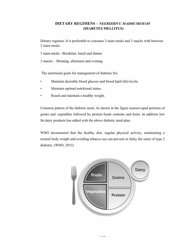
107
DIETARY REGIMENS – NEERIZHIVU MADHUMEHAM (DIABETES MELLITUS)
Dietary regimen: It is preferable to consume 3 main meals and 3 snacks with between 2 main meals.
3 main meals - Breakfast, lunch and dinner
3 snacks – Morning, afternoon and evening
The nutritional goals for management of diabetes for; • Maintain desirable blood glucose and blood lipid (fat) levels.
• Maintain optimal nutritional status.
• Reach and maintain a healthy weight. Common pattern of the diabetic meal. As shown in the figure nearest equal portions of
grains and vegetables followed by protein foods contents and fruits. In addition low
fat dairy products has added with the above diabetic meal plan.
WHO documented that the healthy diet, regular physical activity, maintaining a
normal body weight and avoiding tobacco use can prevent or delay the onset of type 2
diabetes. (WHO, 2015)

108
No. Foods Quantity Intake
Starch Each serving from this list contains:
15 g carbohydrate, 0-3 grams protein,0-1 g fat and 80 calories. In general, a single serving of starch is: • ½ cup of cooked cereal, grain, or starchy vegetable • 1/3 cup of cooked rice or pasta • 1 oz. of a bread product (such as 1 slice of whole wheat bread) • ¾ to 1 oz. of most snack foods 01 Millets
Cholam Kambu Thinai Kudiraivali, Varagu Panivaragu Samai Finger millet (ragi) Corn Bran Cereals, cooked (oats, oatmeal) Cereals (unsweetened, ready-to-eat) Rice, cooked (white or brown)
½ cup ¾ cup 1/3 cup
02 Pulses
Chickpeas Beans Peas Lentils
½ cup / 100 g / 3.5 oz. ½ cup / 90 -115 g / 3-4 oz. ½ cup / 55 g / 2 oz. ½ cup / 75 -200 g / 2.5-3.5 oz.
03 Stems Vazhaithandu ½ cup 04 Underground
stems/ Root/ Root tubers
Tuber of lotus (Thamaraikizhangu) Tuber of Kohila (Kokilakizhangu) Potato Baked with skin 3 oz. Boiled, all kinds3 oz. Yam, sweet potato, plain Snack chips (potato chips) Fat-free or baked (¾ oz.) Regular (¾ oz.)
½ cup ½ cup 1 cup ½ cup ½ cup 15-20 9-13
05 Bread and snacks Sweets, Desserts, and other carbohydrates
Bread (whole wheat, white or rye) Biscuit ( 2 ½ inches across) Cake (frosted, 2-inch square) Cookies (chocolate chip, 2 ¼ inch across) Doughnut (cake, plain, medium, about 1½ oz.)
1 oz. /1 slice 1 1 2

109
Ice cream (light or no sugar added) Jam or jelly (regular) Pudding (regular, made with reduced-fat milk) Sports drink Sugar Syrup (light, pancake type) Yogurt (frozen, fat-free) Sugar substitutes (low-calorie sweeteners)
1 1 tbsp ½ cup 1 cup (8 oz) 1 tbsp 1 tbsp 1/3 cup Free
06 Vegetables No starchy vegetable ½ cup of cooked
vegetables or vegetable juice
1 cup of raw vegetables
Broccoli Carrots Cauliflower Green beans Bean sprouts Cabbage Celery Cucumber Eggplant / Brinjal Drumstick Capsicum Mushrooms Onions Peppers (all varieties) Radishes (mullangi) Tomatoes Salad greens
½ cup Free
07 Fruits Each serving from this list contains; 15 grams carbohydrate, 0 grams fat, 0 grams protein and 60 calories. In general, a single serving of fruit is: • ½ cup of canned or fresh fruit or4 oz unsweetened fruit juice • 1 small fresh fruit (4 oz) • 2 tablespoons of dried fruit
Fruits
Apple, raw, unpeeled Banana Berries Blackberries, raw Blueberries, raw Raspberries, raw Strawberries (whole) Cherries Guava Kiwi Mango (small, 5 ½ oz.) Orange (small, 6 ½ oz.) Papaya (cubed, 8 oz.)
2 inches across/small -1 / 4 oz. Small 1 / 4 oz. ¾ cup ¾ cup 1 cup 1 ¼ cup 3 oz. / 12 number ½ cup Large 1 / 3½ oz. ½ fruit / ½ cup 1fruit ½ fruit / 1 cup

110
Pears (fresh, large, 4 oz.) Pineapple, fresh Plums, small Watermelon (cubes, 13 ½ oz.) Avocado (medium, 1 oz.) Dried fruit - Dates
½ fruit ¾ cup 2 1 slice / 1 ¼ cups 2 tbsp 3 medium
08 Milk and milk products Fat-free (skim) or low-fat (1%) milk andyogurt: Each serving from this list contains 12 grams carbohydrate, 8 grams protein,0-3 grams fat and 100 calories
Milk and milk products
Reduced-fat (1 or 2%) milk and yogurt: Milk, acidophilus milk, Lactaid Yogurt (plain, 6 oz.) (preferable) Whole milk and yogurt: Milk, buttermilk, goat’s milk Evaporated milk Yogurt (plain, 8 oz.) Plain soy milk Cheeses (with 3 grams of fat or less per oz) Cottage cheese
1 cup 1 cup 3/4 cup 1 cup ½ cup 1 cup 1 cup 1 oz. ½ cup
09 Meats Lean meats and protein sources: Each serving from this list contains 0 grams carbohydrate, 7 grams protein, 0-3 grams fat and 45 calories.
Meats
Beef (Select or Choice grades, trimmed of fat): Egg whites Fish (fresh or frozen, plain): Catfish, cod, flounder, haddock, halibut, orangeroughy, salmon, tilapia, trout, tuna) Egg Pork (lean) Poultry (with skin or fried) Sausage Processed sandwich meats (With 3 grams of fat or less per oz.)
1 oz. 2 1 oz. 1 oz. 1(limit to 3 per week) 1 oz. 1 oz. 2 oz. / 1/4 cup /1 oz. (with 4-7 grams of fat per oz.) 1 oz.

111
10 Fats In general, a single serving of fat is: • 1 teaspoon of regular margarine, vegetable oil or butter • 1 tablespoon of regular salad dressing
Nuts Almonds, cashews Peanuts Oil (olive, peanut) Margarine (lower-fat spread) Butter Cream (half and half) Cream cheese (reduced-fat) Cream cheese (regular)
3 10 1 tsp 1 tbsp 1 tsp 2 tbsp 1 ½ tbsp. 1 tbsp
11 Other Catsup (ketchup)
Pickles (medium size dill) Mustard Garlic Spices Carbonated or mineral water, Herbs (fresh or dried)
1 tbsp 1 ½ Free Free Free Free Free
tsp - tea spoon, tbsp. – table spoon

112
Foods to be chosen and foods to be limited or avoided by Diabetes Mellitus patients Foods to be chosen foods to be limited or avoided
Whole Grains Whole and multi grain breads, whole wheat and brown rice
White breads, croissants, sweet rolls, high-fat white crackers, waffles
Low-fat and multigrain crackers Short-grain and minute rice – white rice
Low-sugar, whole-grain cereals Sweetened refined cereals Oatmeal, bran, bulgur, buckwheat Commercial muffins, cakes,
doughnuts, Danish pastries, high-fat cookies
Low-fat, whole grain baked goods with added bran or oat bran
Avoid ‘white’ foods (white flour, white sugar)
Vegetables and Fruit – with lots of color Dark green leafy vegetables Parsnip, pumpkin, white potatoes
(high GI) Eat an abundance fresh/frozen vegetables
Dried fruits and fruits in heavy syrup
Unsweetened, fresh, frozen or canned fruits
Sweetened fruit juices
Milk and Alternatives Dairy products with less than 1% fat Milk products higher than 1% fat Cheese should be 10-20% MF Cheese higher than 21% MF and
creams
Meat and Alternatives Fish (canned in water, fresh, frozen), seafood
Fish with butter or breading
Skinless chicken and turkey Fried chicken, poultry with skin, wings
Lean meats with fat trimmed, wild game
Fatty marbled meats, ribs, regular ground meats, organ meats
Lean cold cuts (but watch the salt content)
High fat processed and canned meats: bacon, sausages, bologna, salami, wieners
Legumes, tofu Eggs

113
Oils, butter and nuts Olive, canola, soybean, sesame, sunflower oils (3 tspor less per day)
Hydrogenated oils, coconut and palm oils
Non-hydrogenated soft margarinesLow-calorie dressings and mayonnaise
Shortening, butter, lard, hard margarines
Light peanut butter, nuts (watch salt and calories)
Peanut butter with palm or hydrogenated fat
Unsalted seeds: flax, pumpkin,sunflower
No more than 1/4 cup nuts per day (high calories)
Defatted gravy and low-sugar condiments
Heavy gravy, cream sauces, high-sugar condiments
Cocoa powder or a small piece of dark chocolate
• Chocolate and carob
Sweets (in very small amounts) Sugar substitutes and artificial sweeteners, low sugar jams/jellies/syrups
Sugar and regular jams, jellies, syrups, candies,gelatins, gum, honey
Sugar-free candies, gelatins, gum Regular cakes, pies, cookies Low-sugar and high fiber baked goods No “white” foods (white flour, white
sugar) Low-fat and low-sugar frozen dessert Regular frozen desserts
Snack Foods Popcorn without salt, butter, or hydrogenated oils
Regular popcorn, chips, pretzels, corn chips
Choose low fat, low sugar snack foods
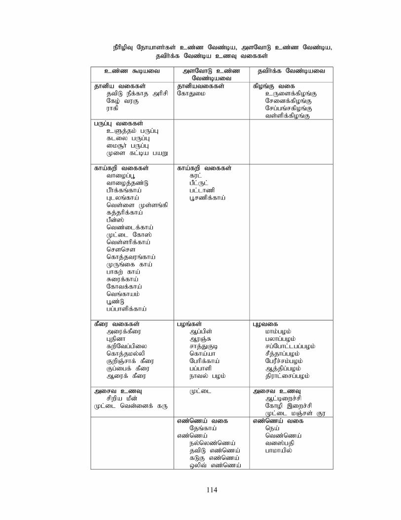
114
ePhpopT Nehahsh;fs; cz;z Ntz;ba> msNthL cz;z Ntz;ba> jtph;f;f Ntz;ba czT tiffs;
cz;z $bait msNthL cz;z Ntz;bait
jtph;f;f Ntz;bait
jhdpa tiffs; jtpL ePf;fhj mhprp Nfo; tuF uhfp
jhdpatiffs; NfhJik
fpoq;F tif cUisf;fpoq;F Nridf;fpoq;F Nrg;gq;rfpoq;F ts;spf;fpoq;F
gUg;G tiffs; cSj;jk; gUg;G fliy gUg;G ik#h; gUg;G Kis fl;ba gaW
fha;fwp tiffs; thiog;G+ thioj;jz;L gPh;f;fq;fha; Glyq;fha; nts;is Ks;sq;fp fj;jhpf;fha; gPd;]; ntz;ilf;fha; Kl;il Nfh]; nts;shpf;fha; nrsnrs nfhj;jtuq;fha; KUq;if fha; ghfw; fha; Riuf;fha; Nfhtf;fha; ntq;fhak; G+z;L gg;ghspf;fha;
fha;fwp tiffs; ful; gPl;Ul; gl;lhzp G+rzpf;fha;
fPiu tiffs; miuf;fPiu Gjpdh fwpNtg;gpiy nfhj;jky;yp FwpQ;rhf; fPiu Fg;igf; fPiu Miuf; fPiu
goq;fs; Mg;gps; MuQ;R rhj;JFb nfha;ah Nghpf;fha; gg;ghsp ehty; gok;
Gotif khk;gok; gyhg;gok; rg;Nghl;lgg;gok; rPj;jhg;gok; NguPr;rk;gok; Mj;jpg;gok; jpuhl;irg;gok;
mirt czT rpwpa kPd; Kl;il ntd;idf; fU
Kl;il mirt czT Ml;biwr;rp Nfhop ,iwr;rp Kl;il kQ;rs; Fu
vz;nza; tif Njq;fha; vz;nza; ey;nyz;nza; jtpL vz;nza; fLF vz;nza; xypt; vz;nza;
vz;nza; tif nea; ntz;nza; td];gjp ghkhapy;

115
rpw;Wz;b ru;f;fiu Nru;f;fhj gp];fl;
,dpg;G rpw;Wz;b ru;f;fiu Njd; FSf;Nfh]; [hk; nty;yk; ,dpg;G tiffs; gp];fl; tiffs; Nff; ,sePu; Fspu; ghdq;fs; (,dpg;G) kJghd tiffs; `u;ypf;]; G+];l; Nghu;fd;tpl;lh fhk;gshd;

116
CASE REPORT FORM I – SCREENING (Selection of subject)
1. Institution: Government Siddha Medical College, Palayamkottai, Tamil Nadu. 2. Name of the subject: ..……………………………………………………………… 3.Address:………………………………………………………………………………
……….………………………………………………………………………………
4. Date of Birth: Age (years): ……… 5. Gender: Male Female CRITERIA OF INCLUSION - Yes (1), No (0) 6. Age between 18 to 64 years
7. Sex: Male / Female
8. Symptoms
8.1Increase volume of urine output (polyuria – nocturnal urea)
8.2 Immediate changes in weight (weight loss / weight increase)
8.3 Increase hungry (polyphagia)
9. Urine glucose – positive
10. Blood sugar: ………….mg/dl / mmol/l
FBS (≥ 126 mg/dl or 7 mmol/l) or PPBS (≥200mg/dl or 11.1mmol/l)
HbA1C (≥6.5 % or 48 mmol / mol)
11. Patients not taking any anti Neerizhivu Madhumeham drugs
CRITERIA FOR EXCLUSION - Yes (1), No (0) 12. Age below 18 and above 64 years.
13. Blood sugar – FBS (<126 mg/dl or 7mmol/l) or PPBS (<200mg/dl or 11.1mmol/l) HbA1c(<6.5 % or 48 mmol / mol) 14. Pregnant mothers
15. Lactating mother
NO.
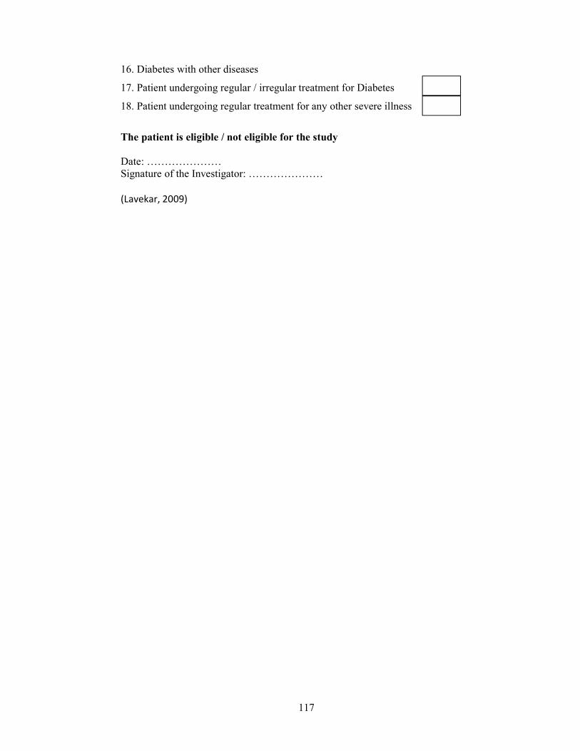
117
16. Diabetes with other diseases
17. Patient undergoing regular / irregular treatment for Diabetes
18. Patient undergoing regular treatment for any other severe illness
The patient is eligible / not eligible for the study
Date: ………………… Signature of the Investigator: ………………… (Lavekar, 2009)

118
CASE REPORT FORM II – HISTORY
1. Institution: Government Siddha Medical College, Palayamkottai, Tamil Nadu. 2. Name of the subject: ………………………………………………………………….. 3.Address:………………………………………………………………………………
………….………………………………………………………………………………
4. Date of Birth: Age (years): ……… 5. Gender: Male Female 6. Educational status:
(Illiterate 0, Read and write 1, School 2, College 3) 7. Occupation: (Unemployed 0, Desk work 1, Field work 2, Field work with physical laborer 3, Field work with intellectual 4) 8. Total Family members: ………. 9. Income per capita per month (in Rs.): …………………………..
Chief complaint with duration (if any) in days - Absent (0), Present (1) Duration 10. Polyuria (Excessive Urine) 11. Polyphagia (Excessive Hunger) 12. Polydipsia (Excessive Thirst) 13. Exhaustion/Tiredness 14. Loss / increase of body weight 15. Body ache 16. Giddiness 17. Polyneuritis (Numbness / Tingling)
NO.

119
18. Visual disturbance 19. Other
If, yes specify: _____________________________________ Personal History 20. Diet (Vegetarian 0 Non-vegetarian 1 and Lacto-vegetarian 2) 21. Appetite (Normal 0, low 1 and high 2) 22. Bowel opening (Difficult 0 and normal 1) 23. Presence of anxiety (No 0 and Yes 1) Addiction 24. Smoking (No 0 and Yes 1) If yes specify: (a) Quantity (packs): ……… (b) Total duration (in years)……………. 25. Tobacco chewing (No 0 and Yes 1) If yes specify: (a) Quantity: ………… (b) Total Duration (in years): ……………… 26. Alcohol (No 0 and Yes 1) If yes specify: (a) Quantity (in ml/day): …… (b) Total Duration (in years): ………… 27. Any other (specify): …………………………………………………………….. Family history 28. Present of family history (No 0 and Yes 1)

120
PHYSICAL EXAMINATION
29. Height (m): ……….. 30. Weight (kg): ……….. 31. BMI: …………kg/m2
32. Body status
(Underweight 0, normal 1, overweight 2 and obese 3)
33. Pulse: rate…….../min, rhythm……………………… volume…………………… character……………………………… condition of arterial wall………………
radio-femoral delay………………
34. Blood Pressure (in sitting position): ………………..mm Hg
35. Body temperature (oC): ……………
36. Respiration rate: …………. /min 37. Numbness or burning sensation in extremities (Yes 0, No 1)
37. Signs of dehydration and oedema, if any …………………………………………
SYSTEMIC EXAMINATION No abnormalities detected (NAD) (0) and abnormalities present (1)
38. CVS
If abnormal, details: ……………………………………………………………………………….. 39. CNS
If abnormal, details; ……………………………………………………………………………… 40. Digestive system If abnormal, details: …………………………………………………………………………… 41. Uro-Genital system
If abnormal, details: ……………………………………………………………………………….. 42. Respiratory system
If abnormal, details: ………………………………………………………………………………..
Date: …………………… Signature of Investigator: ………………………
(Lavekar, 2009)
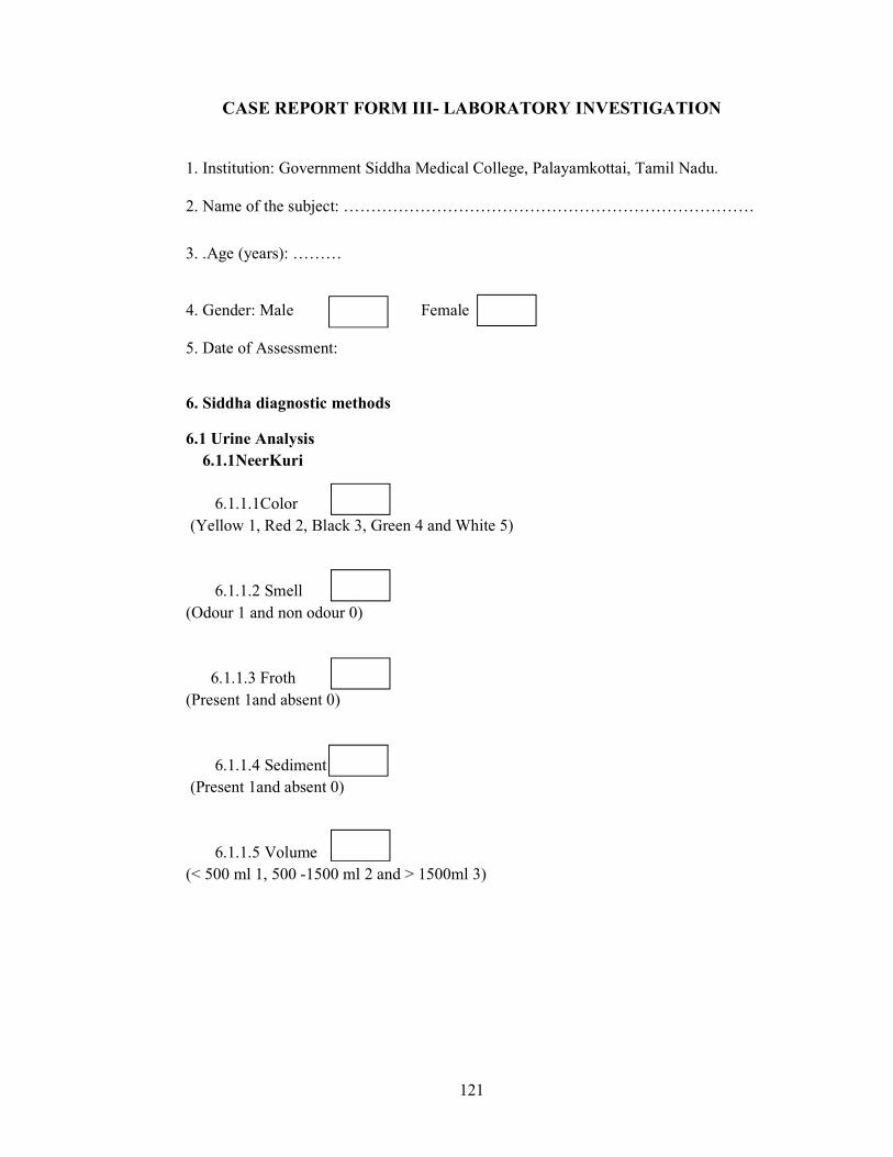
121
CASE REPORT FORM III- LABORATORY INVESTIGATION
1. Institution: Government Siddha Medical College, Palayamkottai, Tamil Nadu. 2. Name of the subject: …………………………………………………………………
3. .Age (years): ……… 4. Gender: Male Female 5. Date of Assessment:
6. Siddha diagnostic methods
6.1 Urine Analysis 6.1.1NeerKuri 6.1.1.1Color (Yellow 1, Red 2, Black 3, Green 4 and White 5)
6.1.1.2 Smell (Odour 1 and non odour 0) 6.1.1.3 Froth (Present 1and absent 0) 6.1.1.4 Sediment (Present 1and absent 0) 6.1.1.5 Volume (< 500 ml 1, 500 -1500 ml 2 and > 1500ml 3)

122
6.1.2 Neikuri 6.1.2.1 Shape Immediately immediate after 1 minute immediate after 2 minutes immediate after 3 minutes
6.1.2.2 Nature of the oil spread (Slow 0 and Quick 1) 6.1.2.3 Whether the oil directly goes inside and touches the bottom of the vessels (No 0 and Yes 1) 6.2 Nadi Analysis Type of Naadi
Right hand: ………………………………. Left hand:……………………………….
6.3Manikkadai
Finger breath of Manikkadai ……………

123
7. Modern diagnostic methods
7.1 Urine examination – UFR
1.1.1 Color (Straw color 0, Dark yellow 1, Red 2, Brick red 3)
7.1.2 Appearance (Clear 0, turbid 1)
7.1.3 Specific gravity
(1.0 - 0, 1.005 - 1, 1.010 - 2, 1.015 -3, 1.020 - 4, 1.025 - 5, 1.030 - 6) 7.1.4 Reaction (pH)
(5.0 - 0, 6.0 -1, 6.5 -2, 7.0- 3, 7.5- 4, 8.0 -5, 8.5- 6) 7.1.5 Protein - mg/dl
(Negative - 0, Trace - 1, + - 2, ++ - 3, +++ - 4)
7.1.6 Glucose – mg/dl
(Negative 0, Positive 1)
7.1.7 Ketones
(Negative 0, Positive 1) 7.1.8 Pus cells (Occasional 0, + - 1, ++ -2, +++ -3) 7.1.9 RBC
(Negative 0, Trace 1, + - 2, ++ -3, +++ -4) 7.1.10 Epithelial cells hpf
(Present 0 and absent 1)
7.1.11 Casts hpf (Present 0 and absent 1)
7.1.12 Urobilinogen
(Present 0 and absent 1)
7.1.13 Bile pigments (Present 0 and absent 1)
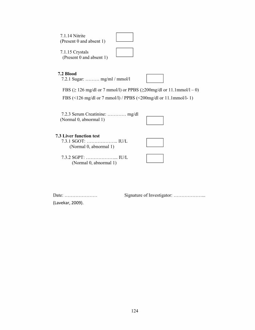
124
7.1.14 Nitrite
(Present 0 and absent 1) 7.1.15 Crystals (Present 0 and absent 1)
7.2 Blood 7.2.1 Sugar: ……… mg/ml / mmol/l FBS (≥ 126 mg/dl or 7 mmol/l) or PPBS (≥200mg/dl or 11.1mmol/l – 0)
FBS (<126 mg/dl or 7 mmol/l) / PPBS (<200mg/dl or 11.1mmol/l- 1) 7.2.3 Serum Creatinine: ………… mg/dl (Normal 0, abnormal 1) 7.3 Liver function test 7.3.1 SGOT: ……………….. IU/L (Normal 0, abnormal 1) 7.3.2 SGPT: ………………... IU/L (Normal 0, abnormal 1) Date: ………………… Signature of Investigator: ………………...
(Lavekar, 2009).

125
CASE REPORT FORM IV- LABORATORY INVESTIGATION
(After intake of anti Neerizhivu Madhumeham drugs) 1. Institution: Government Siddha Medical College, Palayamkottai, Tamil Nadu. 2. Name of the subject:
…………………………………………………………………………
3. .Age (years): ……… 4. Gender: Male Female 5. Name of the medicine: ……………………………………………………….. 6. Date of commencement of treatment: 7. Date of cessation of treatment:
NO.

126
No. of visits BT 1 2 3 4 5 6 Date of visit
Investigations Urine Analysis (Siddha system) NeerKuri Color Smell Froth Sediment Volume Neikuri
Shape-Immediately
After 1 minute
After 2 minutes
After 3 minutes
Nature of spreading
(quick or slow)
Direct way + touch the bottom
Manikkadai Nadi Urine Analysis (Modern system) Appearance Specific gravity
Reaction (pH) Glucose Blood FBS
(mg/dL or mmol/L)
PPBS (mg/dL or mmol/L)
BT – before treatment Date: ………………… Signature of Investigator: ………………..
Table 1. Investigation table. After intake of antiMadhumeha drug


~ 1 ~
Journal of Medicinal Plants Studies 2017; 5(6): 01-05
ISSN (E): 2320-3862
ISSN (P): 2394-0530
NAAS Rating 2017: 3.53
JMPS 2017; 5(6): 01-05
© 2017 JMPS
Received: 01-09-2017
Accepted: 02-10-2017
Vidya Dharshini K
Department of Noi Nadal,
Government Siddha Medical
College, Palayamkottai,
Tamil Nadu, India
Mangalambigai V
Department of Noi Nadal,
Government Siddha Medical
College, Palayamkottai,
Tamil Nadu, India
Krishnaveni M
Department of Noi Nadal,
Government Siddha Medical
College, Palayamkottai,
Tamil Nadu, India
Muthurathinam S
Govt. Drugs Testing Laboratory-
Indian Medicine, Arumbakkam,
Chennai, India
Saravanan R
Govt. Drugs Testing Laboratory-
Indian Medicine, Arumbakkam,
Chennai, India
Meenakumari S
Govt. Drugs Testing Laboratory-
Indian Medicine, Arumbakkam,
Chennai, India
Correspondence
Vidya Dharshini K
Department of Noi Nadal,
Government Siddha Medical
College, Palayamkottai,
Tamil Nadu, India
Pharmacognostical characterization of Aavarai
kudineer - A poly herbal preparation
Vidya Dharshini K, Mangalambigai V, Krishnaveni M, Muthurathinam
S, Saravanan R and Meenakumari S
Abstract Calibration of drugs and medical preparations are essential parameter in scientific scenario and it helps to
improve quality and efficacy of drugs. Aavarai Kudineer is a poly herbal Siddha drug and it has been
highly prescribing for the condition of Madhumegam (Diabetes mellitus). There are number of
formulations of Aavarai Kudineer documented in literature, whereas scientific studies has not been
reported. The current study was design to analyze Aavarai Kudineer scientifically for develop a
standardization to the formula. Precursor mixture (coarse powder) of Aavarai Kudineer and aqueous
extract of Aavarai Kudineer were tested. Pharmacognostical analysis including preliminary
phytochemical study was done. The findings of the study could be useful in diagnostic keys for
identification and preparation of aqueous extract of Aavarai Kudineer.
Keywords: Aavarai Kudineer, hypoglycemic drug, pharmacognostical study, standardization
Introduction The traditional systems of medicine have received significant popularity in all over the globe
as the drugs have curative property, less toxic and minimal side effects [1]. Siddha system of
medicine is one of the ancient traditional systems of medicine and it is believed that the Sages
laid the foundation for this system. It has great potential of medicinal resource repository goes
back to B.C 10 000–B.C 4000 [2]. Ancient Siddha text books documented 4448 variety of
diseases included Madhumegam [3].
Madhumegam or Neerilivu can be compared with diabetes mellitus [4]. It is one of the common
metabolic disorder and raised as a global problem as the morbidity of diabetes mellitus
increasing day by day [5]. The World Health Organization estimates, there will be 300 million
diabetic patients in worldwide by the year of 20256. A number of hypoglycemic herbal drugs
and herbal formulations are available in the traditional medicinal practice and they have been
widely prescribe by the physicians [5].
Aavarai Kudineer (AK) is one of such poly herbal formulation described in Siddha Materia
Medica. The ancient Siddha medical literature the Theraiyar kudineer written by the ancient
saints describes the constituents and indications of AK. AK is a common drug for diabetes
mellitus in the Siddha system of medicine and have been prescribing by the traditional medical
institutions. It is comprising of seven medicinal plants namely; leaves of Senna auriculata (L.)
Roxb. (Avirai) and Cassia fistula L. (Kondrai), Seeds of Syzygium cumini (L.) Skeels. (Naval),
wood of Salacia reticulate Wight. (Kadalazhinjil), root of Saussurea lappa Clarke. (Koshtam),
rhizome of Cyperus rotundus L. (Koraikizhangu) and bark of Terminalia arjuna (Roxb.)
Wight & Arn. (Marutham) [7].
Previous studies have shown the multi-dimensional therapeutic properties including
hypoglycemic activity of the constitutional plants of AK [5, 6]. A previous in-vivo study
confirms the anti-hyperglycemic potential of AK [8].
Differences was observed in the constituent plants and parts used to prepare the AKin
literature [4-6, 9]. A number of constituent plants of AK is replaced by another plant species; i.e.,
Salacia prinoides and Costus spiciosus were used instead of S. reticulata and S. lappa
respectively [4, 5, 9]. In addition discrepancies were noted with the parts used to prepare AK; i.e.,
whole plant of S. auriculata was used instead of leaves of S. auriculata [4], flowers and bark of
C. fistula used for leaves of C. fistula [4, 6, 9] and rhizome of C. spiciosus used for instead of S.
lappa [6].

Vidya et al. World Journal of Pharmaceutical and Medical Research
www.wjpmr.com
142
COMPARATIVE STUDY OF LOCATIONS OF NADI WITH ANATOMICAL
LANDMARKS - A REVIEW
Vidya Dharshini K.*1 and Neelavathy R.
2
1PG Scholar, Department of Noi Naadal, Government Siddha Medical College, Palayamkottai, Tamil Nadu, India.
2Principal, Government Siddha Medical College, Palayamkottai, Tamil Nadu, India.
Article Received on 12/02/2018 Article Revised on 05/03/2018 Article Accepted on 26/03/2018
INTRODUCTION
Siddha system of medicine is one of the oldest system of
medicine in the world, laid by Siddhars (Ivy and Malini,
2010). A number of diagnostic methods have been using
in various systems of medicine globally. Siddha system
has unique assessment methods as envagaithervu (nadi,
sparism, naa, niram, mozhi, vizhi, malam and siruneer)
to diagnosis the diseases (Natarajan, 2009;
Shanmugavelu, 1967). Nadi is a diagnostic way to assess
health status of an individual (Ivy and Malini, 2010) and
a remarkable diagnostic parameter, included in
envagaithervu and well explained by Siddhars
(Kalaththur kanthasami, 2012).
Tri humours such as Vatham, Pitham and Kapham are
the basic principal of Siddha system of medicine, which
governs the psycho-biological aspect of the body
(Natarajan, 2009). Increases or reduces of the tri
humours causes disease (Natarajan, 2009). In a healthy
person, the maathirai of the Nadi of Vatham will be one,
Pitham will be half and the Kapham will be quarter
(Natarajan, 2009). Derangement of this ratio indicates
the disease (Natarajan, 2009). Several locations were
indicated to the assessment of the Nadi in the Siddha text
books, to diagnosis the diseases as well as evaluate the
prognosis. User friendly modern diagnostic methods, less
number of Nadi experts and little number of publish
research documents on Nadi has led to non-familiar of
Nadi.Current study was done to locate the different Nadi
assessment places and compare the locations with
anatomical landmarks to create a strong belief regarding
old concepts.
MATERIALS AND METHODS
Literature review was done using ancient text books as
Yugimuni nadi (YN), Thirumoolar nadi nool (TNN),
Nadi vagadam (NV), Nadi sakkiram (NS) and journals.
wjpmr, 2018,4(4), 142 -144
SJIF Impact Factor: 4.639
Review Article
ISSN 2455-3301
WJPMR
WORLD JOURNAL OF PHARMACEUTICAL
AND MEDICAL RESEARCH www.wjpmr.com
*Corresponding Author: Vidya Dharshini K.
PG Scholar, Department of Noi Naadal, Government Siddha Medical College, Palayamkottai, Tamil Nadu, India.
ABSTRACT
Nadi is a remarkable diagnostic parameter in Siddha system of medicine which is included in envagaithervu and
well explained by Siddhars. User friendly modern diagnostic methods, less number of Nadi experts and little
number of publish documents has led to non-familiar of Nadi. Current study was done to locate the different Nadi
assessment places and compare the locations with anatomical landmarks. Literature review was done using text
books and journals. Various number of Nadi locations were documented in the literature. Ten locations of Nadi
assessment places were mentioned in Thirumoolar nadi nool (TNN) and Yugimuni nadi (YN) and five locations
were noted in Nadi vagadam (NV). A discrepancy was noted with the locations of Nadi among the text books.Ten
places was documented at TNN as Kuthi santhi (ankle-posterior tibial artery/dorsalis pedis artery), Kamiyam
(inguinal region-femoral artery), Unthi (epigastric region-abdominal aorta), Marbu (chest-precordial impulse),
Kaathu (ear-posterior auricular artery/superficial temporal artery), Mooku (nose-angular artery/facial artery),
Kandam (neck-common carotid artery), Karam (upper limb-axillary artery/brachial artery/ulnar artery/radial
artery/princeps pollicis artery), Puruvam (eye brow-superficial temporal artery) and Uchchi (fontanelle-cerebral
artery). Kaaladi (sole-medial and lateral plantar artery), Mulankal (Knee-popliteal artery), Mulankai (elbow-
brachial artery) and Kaikuli (armpit-axillary artery) were mentioned in YN, even though Unthi, Kaathu, Mooku
and Kamiyam not documented. Kai, Kanukkal, Kandam, Kalin peruviral (big-toe–medial plantar artery) and
Kannichchuli (fontanelle) were noted in NV. Generally upper limb was used, whereas different locations were
documented to assess Nadi. Warranting further studies needed to ensure the suitable locations to assess the Nadi in
clinical practice.
KEYWORDS: Nadi, Siddha system of medicine, anatomical landmarks.