DISSERTATION - EPrints@Tamil Nadu Dr MGR Medical ...
128
COMPARING THE EFFICIENCY OF COCONUT OIL AND PALM OIL WITH XYLENE AS A CLEARING AGENT IN CONVENTIONAL HEMATOXYLIN AND EOSIN HISTOPATHOLOGICAL STAINING PROCEDURE DISSERTATION Submitted to The Tamil Nadu Dr. M.G.R Medical University in partial fulfillment of the requirement for the degree of MASTER OF DENTAL SURGERY BRANCH - VI ORAL PATHOLOGY AND MICROBIOLOGY 2015 - 2018
-
Upload
khangminh22 -
Category
Documents
-
view
1 -
download
0
Transcript of DISSERTATION - EPrints@Tamil Nadu Dr MGR Medical ...

COMPARING THE EFFICIENCY OF COCONUT OIL
AND PALM OIL WITH XYLENE AS A CLEARING
AGENT IN CONVENTIONAL HEMATOXYLIN
AND EOSIN HISTOPATHOLOGICAL
STAINING PROCEDURE
DISSERTATION
Submitted to The Tamil Nadu Dr. M.G.R Medical University
in partial fulfillment of the requirement for the degree of
MASTER OF DENTAL SURGERY
BRANCH - VI
ORAL PATHOLOGY AND MICROBIOLOGY
2015 - 2018

CERTIFICATE
Certified that the dissertation entitled: “COMPARING THE
EFFICIENCY OF COCONUT OIL AND PALM OIL WITH XYLENE AS A
CLEARING AGENT IN CONVENTIONAL HEMATOXYLIN AND EOSIN
HISTOPATHOLOGICAL STAINING PROCEDURE” is a bonafide record of
the work done by Dr. Ashitha A S under our guidance during her post graduate
study during the period of 2015-2018 under THE TAMIL NADU DR. M.G.R
MEDICAL UNIVERSITY, CHENNAI, in partial fulfilment for the degree of
MASTER OF DENTAL SURGERY IN ORAL PATHOLOGY AND
MICROBIOLOGY, BRANCH -VI. It has not been submitted (partial or full) for
the award of any other degree or diploma.
Guide
Dr. T ISAAC JOSEPH
Professor and Head
Department of Oral Pathology and Microbiology
Sree Mookambika Institute of Dental Science
Kulasekharam, Kanya Kumari District-629161
Co-Guide
Dr. GIRISH K L
Professor


CERTIFICATE II
This is to certify that this dissertation work titled “Comparing the
Efficiency of Coconut Oil and Palm Oil with Xylene as a Clearing Agent
in Conventional Hematoxylin and Eosin Histopathological Staining
Procedure” of the candidate Dr. Ashitha AS with registration Number
2415213017for the award of MASTER OF DENTAL SURGERY in the
branch of Oral Pathology and Microbiology, [Branch- VI]. I personally
verified the urkund.com website for the purpose of plagiarism Check. I found
that the uploaded thesis file contains from introduction to conclusion pages
and result shows 4percentage of plagiarism in the dissertation.
Guide & Supervisor sign with Seal.
Date:
Place:

SREE MOOKAMBIKA INSTITUTE OF DENTAL SCIENCES,
KULASEKHARAM
ENDORSEMENT BY THE PRINCIPAL / HEAD OF THE INSTITUT ION
This is to certify that this dissertation titled “COMPARING THE
EFFICIENCY OF COCONUT OIL AND PALM OIL WITH XYLENE
AS A CLEARING AGENT IN CONVENTIONAL HEMATOXYLIN
AND EOSIN HISTOPATHOLOGICAL STAINING PROCEDURE” is a
bonafide research work done by Dr. Ashitha A S under the guidance of
Dr. T Isaac Joseph M.D.S, Professor and Head, Department of Oral Pathology
and Microbiology, Sree Mookambika Institute of Dental Sciences,
Kulasekharam.
Dr. Elizabeth Koshi MDS,
PRINCIPAL,
Sree Mookambika Institute of Dental Sciences.
V.P.M Hospital Complex,
Padanilam, Kulasekharam,
KanyaKumari District,
Tamil Nadu - 629 161

DECLARATION
I hereby declare that this dissertation titled “COMPARING THE
EFFICIENCY OF COCONUT OIL AND PALM OIL WITH XYLENE AS
A CLEARING AGENT IN CONVENTIONAL HEMATOXYLIN AND
EOSIN HISTOPATHOLOGICAL STAINING PROCEDURE” is a bonafide
record of work undertaken by me and that this thesis or a part of it has not been
presented earlier for the award of any degree, diploma, fellowship or similar title
of recognition.
Dr. Ashitha A S
MDS student,
Department of Oral Pathology and Microbiology,
Sree Mookambika Institute of Dental Sciences,
Kulasekharam, Kanya kumari District,
Tamilnadu.

ACKNOWLEDGEMENT
First and foremost, praises and thanks to ALMIGHTY , the most
beneficent and merciful, for the showers of blessings throughout my life.
With great pleasure, I take this opportunity to express my sincere
gratitude to my respected teacher and guide, Dr. T Isaac Joseph M.D.S,
Professor and Head of the Department of Oral Pathology and Microbiology
whose constant guidance, timely criticism and close supervision has enabled me
to complete my thesis work successfully. His never-ending enthusiasm will
remain as a perennial source of inspiration to my studies.
It is my distinct privilege to acknowledge my respected teacher and co-
guide Dr. Girish KL, Professor, Department of Oral Pathology and
Microbiology for his constant suggestions, good wishes and whole hearted
willingness in sharing his vast experience throughout my post graduate course.
His inspiration and untiring teaching will always be a driving force in my career.
I am happy to thank Dr. Geetha Varghese and Dr. T Prasanth, Professors,
Dr. Pradeesh Sathyan, Reader and Dr. Angelin D and Dr. Deepa A G, Senior
lecturers who always had a word of encouragement and advice. I always remain
grateful to them.
I would like to extend my deepest thanks to Dr. Velayuthan Nair, M.B.B.S,
M.S, Chairman and Dr. Rema V Nair, M.B.B.S, M.D, D.G.O, Director, Sree
Mookambika Institute of Medical Sciences for providing the lab facilities to accomplish
my dissertation work. I also extend my gratitude to Dr. Elizabeth Koshi, Principal, Sree
Mookambika Institute of Dental Sciences for the motivation and support.

I am thankful to Dr. Sarath Babu for helping me with the statistical
analysis involved in this study and to Mrs. N Ringle Kiruba for helping me with
the laboratory procedures. I am thankful to Leos Data Makers, Mr. Satheesh
& Mrs. Alphonsa for their help in carrying out all the DTP works.
I am thankful to my colleagues Dr. Ani Simila C S, Dr. Abhilasha J V,
Dr. Rajalekshmi M P and my seniors Dr. Krishna Prasad R S, Dr. Sudha Rani T,
Dr. Akhil S, Dr. Jeslin Mary S and Dr. Aldrin Jerry for their encouragement.
Special thanks goes to my beloved batch mate Dr. Vidya S whom stood
with me both at good and bad times throughout my course. I acknowledge my
colleague Dr. Swetha D whom kept me going and this work would not have been
possible without her.
I acknowledge Dr. Reshma R and Dr. Reshma Khader S for their moral
support, warm company and motivation, which drives me to give my best.
Special mention goes to Mr. Varun Nambiar and Dr. Sujith S for their
constant and unwavering support.
The people who mean the world to me, my parents Dr. A Abdul Bary
and Mrs. Shyla Bary. I am grateful to them both for being wonderful role
models to me. I am thankful to my brother Dr. Ashik Bary, my sister in law
Dr. Ashna Ashik and my grandmother Mrs. Fathima for their unfailing support.
Finally, I would like to thank my little boy Aadil A Mohammed for being my
bundle of joy and ray of hope in my life.
Dr. Ashitha A S

CONTENTS
SI No: Index Page No
1 List of Abbreviations i-ii
2 List of Tables iii
3 List of Graphs iv
4 List of Colour plates v
5 List of Annexure vi
6 Abstract vii-viii
7 Introduction 1-3
8 Aims and Objectives 4
9 Review of Literature 5-26
10 Materials and Methods 27-34
11 Results and Observations 35-44
12 Discussion 45-49
13 Summary and Conclusion 50-51
14 Bibliography ix-xv
15 Annexure

i
LIST OF ABBREVIATIONS
ATP Adenosine triphosphate
CIM Clearing and infiltration mixtures
CK Cytokeratin
CD Cluster of differentiation
DNA Deoxy ribonucleic acid
DPAS Periodic acid schiff following diastase
DWS Dish washing solution
EPA Environmental protection agency
GMS Grocott’s methanemine silver
H/E Hematoxylin and eosin
IHC Immunohistochemical staining
IUPAC International union of pure and applied chemistry
MHA Methyl lippuric acid
MTBE Methyl tert butyl ether
OSHA Occupational safety and health administration
PAS Periodic acid schiff
PCR Polymerase chain reaction
PGME Propylene glycol methyl ether
PPM Parts per million
RCRA Resource conservation and recovery act

ii
RMO Refined mineral oil
RNA Ribonucleic acid
ROS Reactive oxygen species
SBO Soluble bioorganic substances
ScCo2 Super critical carbon dioxide
STB Sulphation toluidine blue
TWA Time weighed average

iii
LIST OF TABLES
TABLE NO TITLE
Table 1 Comparison of rigidity between specimens treated with
xylene, palm oil & coconut oil
Table 2 Comparison of translucency between specimens treated with
xylene, palm oil & coconut oil
Table 3 Comparison of changes after impregnation between specimens
treated with xylene, palm oil & coconut oil
Table 4 Comparison of ease of sectioning between specimens treated
with xylene, palm oil & coconut oil
Table 5 Comparison of nuclear staining within H & E stained sections
treated with xylene, palm oil & coconut oil
Table 6 Comparison of cytoplasmic staining within H & E stained
sections treated with xylene, palm oil & coconut oil
Table 7 Comparison of clarity of staining within H & E stained
sections treated with xylene palm oil & coconut oil
Table 8 Comparison of nulear staining between H & E stained sections
treated with xylene, palm oil & coconut oil
Table 9 Comparison of cytoplasmic staining between H & E stained
sections treated with xylene, palm oil & coconut oil
Table 10 Comparison of clarity of staining between H & E stained
sections treated with xylene, palm oil & coconut oil
Table 11 Comparison of mean H & E stained sections evaluation scores
between xylene, palm oil & coconut oil

iv
LIST OF GRAPHS
Graph No TITLE
Graph 1 Comparison of rigidity between specimens treated with xylene, palm
oil & coconut oil (Group A, Group B, Group C)
Graph 2 Comparison of translucency between specimens treated with xylene,
palm oil & coconut oil (Group A, Group B, Group C).
Graph 3 Comparison of changes after impregnation between specimens
treated with xylene, palm oil & coconut oil (Group A, Group B,
Group C)
Graph 4 Comparison of ease of sectioning between specimens treated with
xylene, palm oil & coconut oil (Group A, Group B, Group C)
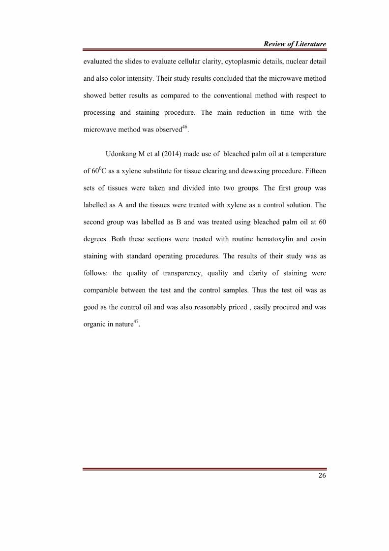
Graph 5 Comparison of nuclear staining, cytoplasmic staining and clarity
within the groups (Group A, Group B, Group C)
Graph 6 Comparison of nuclear staining, cytoplasmic staining and clarity
between the groups (Group A, Group B, Group C)

v
LIST OF COLOUR PLATES
Colour Plate No Title
CP 1 Soft tissue specimen for grossing
CP 2 Grossed specimen and labeled into C1, P1 and X1
CP 3 Tissue processing with palm oil
CP 4 Tissue processing with coconut oil
CP 5 Paraffin embedding bath
CP 6 Semiautomatic microtome
CP 7 Tissue floatation bath
CP 8 Slide warming table
CP 9 Incubator
CP 10 Hematoxylin and eosin staining reagents with palm oil
CP 11 Hematoxylin and eosin staining reagents with coconut oil
CP 12 Photomicrographs of H/E stained sections treated with xylene
(x100)
CP 13 Photomicrographs of H/E stained sections treated with palm
oil (x100)
CP 14 Photomicrographs of H/E stained sections treated with
coconut oil (x100)

vi
LIST OF ANNEXURE
Annexure No Contents
Annexure 1 Institutional Research Committee Certificate
Annexure 2 Institutional Human Ethics Committee Certificate
Annexure 3 Patient Information Sheet
English
Tamil
Malayalam
Annexure 4 Consent Form
English
Tamil
Malayalam
Annexure 5 Case sheet proforma
Annexure 6 Data entry Sheet

ABSTRACT

Abstract
vii
BACKGROUND:
The histopathological laboratory technicians are routinely exposed to
xylene during procedures like tissue processing, clearing, staining, placing a
cover slip and cleaning tissue processors. On account of the Occupational Safety
and Health Administration (OSHA) regulations, various xylene substitutes such
as limonene reagents, aliphatic hydrocarbons, vegetable oils and mineral oils
were tried in the past to avoid xylene in the laboratory. This study is designed to
establish whether the use of coconut oil and palm oil at a maintained temperature
as a clearing agent during tissue processing and as a dewaxing agent during
staining has any effect on transparency, rigidity, change after impregnation, ease
in sectioning and quality of staining such as nuclear staining, cytoplasmic
staining and clarity of staining as compared with the xylene treated counterparts.
AIMS AND OBJECTIVE:
1) To evaluate the efficiency of coconut oil and palm oil as a clearing agent
for Hematoxylin and Eosin staining procedure and compare it with
xylene.
2) To determine whether tissues cleared and dewaxed with coconut oil and
palm oil are same or superior with the xylene treated tissues.
MATERIALS AND METHODS:
A total of 30 tissue specimens were collected, fixed in 10% formalin and
sectioned into 3 equal parts and grouped as group A, B, and C. Group A tissue
specimen were taken for routine processing followed by hematoxylin and eosin
staining procedure with xylene as clearing agent, whereas group B tissue
specimens were treated with heated palm oil at a temperature maintained at 60°C

Abstract
viii
instead of xylene as a clearing agent. Similarly group C tissue specimens were
treated with heated coconut oil at a temperature maintained at 60°C instead of
xylene as a clearing agent during processing. Gross tissue specimen evaluation
like rigidity, translucency, change after impregnation and ease of sectioning were
evaluated and was compared between the groups having group A as the control.
All the specimens were processed, sectioned and stained using hematoxylin and
eosin stain and were coded and observed for evaluation of nuclear staining,
cytoplasmic staining and overall clarity of stained slide and the results were
compiled and subjected to statistical analysis.
RESULTS:
Coconut oil treated specimen showed better characteristic features than
palm oil treated specimen with respect to rigidity, translucency and change after
impregnation which was 43.33%, 63.33% and 90% respectively. Both palm oil
and coconut oil treated specimen showed similar features when compared with
that of ease of sectioning. Among 90 hematoxylin and eosin stained slides,
coconut oil treated sections showed better nuclear staining, cytoplasmic staining
and clarity of staining which was 83.33% ,93.33% and 93.33%.
CONCLUSION:
Coconut oil treated specimen showed better characteristic features than
palm oil treated specimen with respect to rigidity, translucency and change after
impregnation. Coconut oil treated sections showed better nuclear staining,
cytoplasmic staining and clarity of staining.
KEYWORDS:
Xylene, Coconut oil, Palm oil, Hematoxylin and eosin staining.

INTRODUCTION

Introduction
1
The more sophisticated, immunological and molecular biological
techniques have been introduced into pathological practice during past few
decades that helps in precise diagnosis. However, hematoxylin and eosin (H & E)
stained paraffin sections still remain the most widely used technique for routine
diagnostic work1. The main components in hematoxylin and eosin
histopathologic staining procedure are xylene and graded alcohol other than
hematoxylin and eosin. In the steps such deparaffinization, rehydration and
dehydration, xylene as well as graded alcohol are the unavoidable chemical
solutions2. A wide range of chemicals which are potentially dangerous are
employed in the pathological laboratory 3.
Xylene is an aromatic hydrocarbon which is extremely biohazardous. The
histopathological laboratory technicians are routinely exposed to xylene during
procedures like tissue processing, clearing, staining, placing a cover slip and also
while cleaning tissue processors. Xylene is exposed maximum during dewaxing
of sections.
The main effects of inhaling xylene vapours are depression of the central
nervous system with symptoms such as headache, dizziness, nausea and
vomiting. Long-term exposure may lead to irritability, insomnia, agitation,
extreme tiredness, tremors, impaired concentration and short-term memory4.
Effects to reduce the health hazards due to xylene exposure in histologic
laboratory must be made in order to make a more safer working environment by
making histopathology technicians more familial with the various health hazards,
precautions and emergency procedures with respect to xylene exposure3 .

Introduction
2
Earlier aniline oil, benzene, chloroform, dioxane and toluene were
routinely used in the histopathology laboratory. Later in 1950s xylene was made
use instead of these chemicals and it was found out that it was also a failed
alternative. Later during the mid 1970s, it was again found out that the xylene has
got neurotoxic effect 5.
The xylene free method for paraffin sections was developed and in use at
Vrinnevi hospital, Sweden since 19956. Technical grade xylene is a combination
of the 3 isomers namely, Ortho, Para and Meta. This mixture is referred to as
‘Xylol’. Studies have shown that xylene is well-absorbed by inhalation, oral and
to some extent by the dermal route7. Once entered into the body it is stored in
adipose tissue as it is soluble in it. It has a half life of 1 to 6 days in the
subcutaneous fat. Studies have shown that laboratory workers exposed for 1.5 to
18 years were described as having the equivalent of general poisoning disorders
including bone marrow toxicity and pancytopenia as caused by a wound
contaminated with xylene8.
Effects of xylene on the tissues are due to depletion of mitochondrial
enzyme adenosine triphosphate in the affected cells. Heart and kidney injuries,
some fatal blood dyscrasias, and other less dangerous problems, such as skin
erythema, drying, scaling and secondary infections are other toxic effects seen to
be associated with use of xylene5.
Occupational Safety and Health Administration (OSHA) regulations has
suggested that limonene reagents, aliphatic hydrocarbons and mineral oils were
used to avoid the usage of xylene in the histopathologic laboratory. But these

Introduction
3
xylene alternatives were proved as less effective and more costly9. Coconut oil, is
a widely used vegetable oil. It’s availability is easy and it is more popular for its
non toxic nature. Furthermore it is known to get oxidized in a very slow rate, heat
stable, and has got the highest resistance to rancidity10. Palm oil is widely
available and a safer substitute for xylene. Hence, here we attempt to check the
use of coconut oil and palm oil as a clearing agent during tissue processing and as
a dewaxing agent during hematoxylin and eosin staining procedure has got any
effect when compared with xylene processed tissue.
This study is designed to establish whether the usage of coconut oil and
palm oil at a maintained temperature as a clearing agent during tissue processing
and as a dewaxing agent during staining procedure has got any effect on
transparency, rigidity, change after impregnation, ease in sectioning and quality
of staining such as nuclear staining, cytoplasmic staining and clarity of staining
as compared with the xylene treated counterparts.

AIMS & OBJECTIVES

Aims and Objectives
4
AIM
To evaluate the efficiency of coconut oil and palm oil as a clearing agent
for hematoxylin and eosin staining procedure and compare it with xylene.
OBJECTIVE
To determine whether tissues cleared and dewaxed with coconut oil and
palm oil are same or superior with that of xylene treated tissues.

REVIEW OF LITERATURE

Xylene was first isolated and named by the French chemist
Cahours in the year 1850
Xylene (from Greek means
three isomers of dimethylbenzene, or in a
(CH3)2C6H4 with each of the
with which two methyl groups
liquids with a great industrial value. The mixture is referred to as both xylene
xylenes. Xylene chemically
distinguished by the
which specify to which
groups are attached. Carbon
of the ring carbons bonded to a methyl group, and counting towards the second
methyl group, the o-isomer
isomer is 1,3-dimethylbenzene and the
three isomers, the p-isomer is the most indu
oxidized to terephthalic acid
Review of Literature
Xylene was first isolated and named by the French chemist
in the year 1850, has been discovered as a component
means "wood"), xylol or dimethylbenzene is any one of
of dimethylbenzene, or in a combination form. It has got a
each of these three compounds possess a central
methyl groups are attached. These are colorless and flammable
great industrial value. The mixture is referred to as both xylene
chemically exists in three isomeric forms. The isomers can be
designations namely ortho (-o-), meta-(m-) and
which specify to which carbon atoms (of the benzene ring) the two
are attached. Carbon atoms around the ring are counted starting from one
of the ring carbons bonded to a methyl group, and counting towards the second
isomer has the IUPAC name of 1,2-dimethylbenzene, the
dimethylbenzene and the p-isomer is 1,4-dimethylbenzene. Of the
isomer is the most industrially used after since it can be
terephthalic acid11.
Review of Literature
5
Xylene was first isolated and named by the French chemist Auguste
, has been discovered as a component of wood tar.
is any one of
form. It has got a formula
central benzene ring
colorless and flammable
great industrial value. The mixture is referred to as both xylene or
three isomeric forms. The isomers can be
) and para(-p-),
) the two methyl
around the ring are counted starting from one
of the ring carbons bonded to a methyl group, and counting towards the second
dimethylbenzene, the m-
dimethylbenzene. Of the
strially used after since it can be

Review of Literature
6
Based on respective isomers, the physical and chemical properties of
xylene differs. The melting point for m-xylene is around 47.87 °C and for p
xylene is roughly around 13.26 °C. The boiling point for each isomer is around
140 °C. The density of each isomer is around 0.87 g/ml. It is less denser
than water. In air, xylene can be smelled at low concentrations between 0.08 to
3.6 ppm (parts per million).In water, xylene can be tasted at a level between 0.53
to 1.8 ppm4.
In laboratories xylene is used to prepare dry ice bath. It has also got its
purpose in light microscopes, the synthetic immersion oil can be removed from
the microscopic objective using xylene solvent. In histopathology laboratory the
application of xylene are, it is used as a clearing agent, to remove the paraffin
wax and in restaining of archival slides and prior to mounting.12
XYLENE AND ITS HEALTH HAZARDS
The conventional hematoxylin and eosin staining procedure is the gold
standard technique and usage of xylene as a clearing agent in H/E procedure is
valid, but its major demerits are cost containment, toxicity and polluted working
environment13. Xylene is easily absorbed by the oro-respiratory mucosal tract
following ingestion and inhalation4. The penetration of xylene through dermal
and epidermal layers are enhanced when it is present in varied physical states. In
humans, 64% of the intaken dose reaches the systemic circulation14. Xylene is
rapidly distributed to the tissues once it is absorbed. On absorption by blood, it
attaches to serum protein to form a complex. Of the total absorbed xylene, 95% is
metabolized in the liver to methyl hippuric acid (MHA) and about 70–80% of

Review of Literature
7
metabolites are removed from body as urine within a time period of 24 hours.
Since xylene is lipophilic, it is accumulated in adipose tissue. In murine studies,
adipose tissues were examined to study the half-life for the elimination of xylene,
which was estimated to be 7 hours, whereas in humans it is around 40 hours15.
As per OSHA regulations, serum evaluation of MHA, which is the major
metabolic product of xylene is mandatory among laboratory staffs and chemical
factory employees. This is an accurate estimation of atmospheric exposure of
xylene in the professional environment14.
United State Department of Health and Human services and Oak ridge
National Laboratory reviewed the toxic effect of xylene and found out that xylene
can cause health effects as a result of both acute (<14days) and chronic (>365 days)
exposure. Accidental splash of xylene in the eye can result in eye damage. The
effects can begin to occur with exposure to air levels of 100ppm. Following xylene
ingestion, throat irritation can occur at approximately 200ppm within 3-5 minutes.
Xylene exposure at 200ppm ≤ can cause lung irritation, causes chest pain and
results in shortness of breath. Liver and kidney damage are confirmed as a result of
high exposure to xylene. Extreme exposure can result in pulmonary oedema4.
Depletion of adenosine triphosphate, which is a mitochondrial enzyme are
confirmed in specifically affected cells. Renal injuries, hematologic discrepancies
which are morbid and other immediate complications such as erythema, pruritis,
exfoliation of the dermal and epidermal layers; all these changes predisposes the
skin to acquire opportunistic infections which are considered to be noxious effects
associated with the usage of xylene16.

Review of Literature
8
Xylene is capable of penetrating the garments and laboratory safety
accessories such as gloves and boots which in turn leads to blistering and
charring of skin and oedematous changes at the submicroscopic level 4. Dermal
absorption is minimal following exposure to xylene vapour. Ingestion of xylene
has proved to cause severe gastrointestinal problems in humans. Limited studies
were available for confirming and addressing the potential carcinogenic effects
of xylene.
United States Environment Protection Agency (EPA) has placed xylene
in weight of evidence group D, not classifiable as to human carcinogenicity17.
Intra uterine effects of xylene on the feotus showed that retarded bone
osteogenesis. In animals, behavioural changes has been reported in the absence
of maternal toxicity. Inhalation of xylene by conceived woman can reach the
growing foetus and it will contaminate the breast milk in lactating women4.
The studies on air contaminants by OSHA (Occupational safety and
health administration-2005) reported a condition named as “Organic Solvent
Syndrome” in which chronic exposure to xylene can cause generalized
symptoms like cephalgia, irritable frame of mind, depression, loss of sleep,
anxiety, fatigue, tremors, lack of concentration and short-term memory4.
Ogata M et al (1970) evaluated the urine of persons who are exposed to
vapours of toluene and m- or p-xylene as a test exposure to check for the
presence of hippuric acid and m- or p- methyl hippuric acid. His study has
shown that hippuric acid was excreted equivalent to 68 % of the toluene
absorbed, and m-methyl hippuric acid equivalent to 72% of the m-xylene

Review of Literature
9
absorbed. Up to hydrocarbon concentrations of 200 ppm the total quantity of
hippuric acids excreted was proportional to the total exposure 18.
Savoleinen H et al (1980) studied the effects of xylene on central
nervous system. They isolated xylene in the peri, epi and endoneurium , which
is due to the solubility of xylene through the hydrophobic layers of the neural
envelop. They concluded that the metabolites of xylene interferes with the
biochemical functions of proteins, this eventually leads to the malfunctioning of
neurons. The pathogenesis of this is as follows: the chemical interaction with
the protein layers or by the dissolution of the lipid layers are crucial for the
physiological functioning of the membrane and the transmembrane proteins. At
the biochemical level another important reaction i.e, methyl benzaldehyde
production through the microsomal enzymes have also been reported19.
In a study by Jacobson GA et al (2003), they researched the association
between the low level TWA atmospheric xylene in the aerosol exposure and
urinary MHA among humans. A statistically significant result was obtained
between environmental xylene exposure and urinary MHA measurements which
was less than 15 p.p.m . And it was an accurate and reliable measurement for
the same. Values were estimated before and after a work day and xylene
exposure was less. However, MHA was significantly positive in all workers
irrespective of their work hours. The study was followed up and the values of
the MHA concentration in post-shift urine of 1.3 gm of creatinine after
exposure to a TWA of 100 p.p.m. xylene 14.

Review of Literature
10
Hipolito RN (1980) elaborate the various symptoms seen in association
with the laboratory technicians exposed to xylene over a period of 4 to 10 years.
The symptoms included chronic headache, chest pain, leucopenia, malaise,
impaired lung functioning, electrocardiographic abnormalities, dyspnea,
cyanosis of hands, fever, and mental confusion. All these above mentioned
symptoms were reversible 20.
Taskinen H et al (1989) did a prospective cohort study in Scandinavian
countries in which he studied the exposure of six organic solvents including
xylene to evaluate any inherited defects are obvious in the foetus. The factors
which significantly increased in the ratio of miscarriage of pregnancy following
exposure of men/fathers working in close association with toluene or other
organic solvents. Finally they concluded that there is no such association
between parental exposures and congenital malformation in foetuses.21
Uchida Y et al (1993), in their study on the effects of xylene in
musculoskeletal system observed that workers exposed to xylene reported
reduced grasping power and associated reduced muscle power in extremities
more frequently than unexposed controls group. Their study conclude that this
was due to the effects on neurons by xylene, rather than a direct effect on
muscles22.
Revilla AS et al (2007) assessed in-vitro adverse of effects toluene and
xylene in hepatic tissue of murine samples for the aerobic respiration of
succinate compounds. In their study various parameters such as membrane
potential, ionic calcium release, reactive oxygen species, Adenosine tri
Review of Literature
11
phosphate levels were assessed. According to their research, mitochondrial
uncoupling through ATP depletion was the main cause for the biological
hazardous effects of toluene and xylene.23
Adrian F et al (2013) investigated the possible adverse effects of xylene
on the vestibulocochlear system in human beings. A statistically significant
results were obtained among the exposed and nonexposed participants. The
xylene exposed individuals had pure tone threshold and an auditory brain stem
response. A close association was observed between the auditory function and
high urinary output of the chief xylene metabolite i.e, MHA. Poorer test result
results were obtained in individuals with higher exposure than that with lower
exposure24.
Review of Literature
12
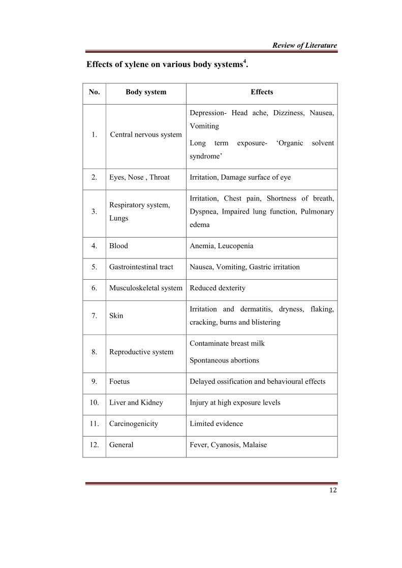
Effects of xylene on various body systems4.
No. Body system Effects
1. Central nervous system
Depression- Head ache, Dizziness, Nausea,
Vomiting
Long term exposure- ‘Organic solvent
syndrome’
2. Eyes, Nose , Throat Irritation, Damage surface of eye
3. Respiratory system,
Lungs
Irritation, Chest pain, Shortness of breath,
Dyspnea, Impaired lung function, Pulmonary
edema
4. Blood Anemia, Leucopenia
5. Gastrointestinal tract Nausea, Vomiting, Gastric irritation
6. Musculoskeletal system Reduced dexterity
7. Skin Irritation and dermatitis, dryness, flaking,
cracking, burns and blistering
8. Reproductive system Contaminate breast milk
Spontaneous abortions
9. Foetus Delayed ossification and behavioural effects
10. Liver and Kidney Injury at high exposure levels
11. Carcinogenicity Limited evidence
12. General Fever, Cyanosis, Malaise

Review of Literature
13
Xylene substitutes
Generally, there are four classes of substitutes for xylene. These include
1. Limonene reagents
2. Aliphatic hydrocarbon mixtures
3. Aromatic hydrocarbon mixtures
4. Mineral oil mixtures
Limonene reagents
These reagents are prepared by steam distillation of orange peels. Major
component is d-limonene. It is a biodegradable, noncorrosive and nonflammable
product. Since they do not contain benzene and toluene, toxicity is less. Dry
reasonably faster and leaves no residue. Because of its high vapor pressure, it
does not evaporate fast; hence, cover slipping of multiple slides can be done
easily. It produces minimal tissue shrinkage, soluble in alcohol and also in
mounting media. But it is expensive and is oily in nature, with very offensive
odor and has got degreasing effect when in contact with skin4.
Aliphatic hydrocarbon mixtures
These substitutes need longer time to produce similar changes on the
tissue as that of their aromatic group because of their aliphatic structure. Mostly
they are odorless and can be recovered by distillation. It is not easily
biodegradable. Less expensive than limonene reagents. They are non greasy and
less irritating to the skin than xylene and d-limonene-based clearing agents. It is
classified as hazardous waste due to its flammability. More expensive and less
tolerant of contamination than xylene4.
Review of Literature
14
Aromatic hydrocarbon mixtures
Not much known because they are regarded as toxic as that of xylene.
Aromatic hydrocarbon combinations with higher boiling point and lower
volatility than xylene are being manufactured4.
Mineral oil mixtures
Combinations of various mineral oils are potential substitutes to xylene.
Pure isopropanol or combined with liquid paraffin wax is an viable and
inexpensive substitute for xylene in tissue processing4. Wastage management of
mineral oil and its mixtures is conducted by mixing it with the already used
paraffin and final solid is reduced to ashes9.
The major benefits of eliminating xylene from tissue processing
techniques are:
1. Physical and mental fitness.
2. Limiting the exposure of a hazardous chemical, thereby reducing the
cumulative effects of exposure to toxic chemicals for laboratory staff.
3. Economic wastage management protocol.
Xylene-free method offers maximum safety as well as superior quality
in result, and it is cost-effective also25. Several studies have been conducted to
replace toxic agent xylene with substitutes in both clearing and dewaxing.
Bruun RB et al (1992) studied the efficiency of cooking oils such as
olive oil and coconut oil over xylene. The study was divided into two groups,
where in one group with histochemical staining method and the second group

Review of Literature
15
with immunohistochemical staining. Their study showed marginal differences
in the tissue property between the xylene processed when compared with the oil
processed specimens. All the oil processed tissue samples were found adequate
for histologic diagnosis. No differences were recorded between the above
mentioned staining techniques. However, there was no qualitative differences
between the groups, long-term reproducibility of oil-processed tissue remained
to be justified in their study26.
Buesa RJ (2000) conducted a study in which crude mineral oil products
were used instead of xylene in tissue processing. Routine tissue processing
methods were followed. The mineral oil was specially prepared as follows: an
admixture of ethanol, isopropyl alcohol, and mineral oil was maintained at a
temperature between 450C and 500C, with vacuum pressure and agitation. This
was then subjected with mineral oil and molten paraffin wax in subsequent
steps. The sample considered for this study were twelve routine conventional
histochemical stainings and twenty one immunohistochemical staining. These
groups were repeatedly tested nine times by multiple reviewers in a blinded
method. The evaluation of the results in their study showed that the tissue
processed with mineral oil were equivalent to that of the tissues which was
processessd using xylene9.
A major benefit of mineral oil is that it is organic and is biologic and
environment friendly. It also has an added advantage of simplifying the tedious
task of microtomy9.

Review of Literature
16
Peshkov MV and Busea RJ (2009) did a study and that showed mineral
oil as the better clearing agents with respect to quality of sectioning and which
helped the pathologists to draw to an accurate diagnosis using with both
automatic and manual procedures, when mineral oil and isopropanol were used at
a ratio of 5:1 and 2:1 respectively. Then it was subjected to undiluted mineral oil,
maintained at a temperature of 500C, there by making this a more safer and less
expensive substitute of xylene16.
Andre et al (1994) in their study made use clearing
and infiltration mixtures (CIMs) as a substitute for xylene. The tissue sample for
the study was obtained from liver, brain and breast samples. Four CIMs, were
taken and each was a combination of 2 parts of molten paraffin and 1 part of
xylene or mono saturated, unsaturated or saturated oil. A substitute of paraffin to
that of xylene only was considered to be the fifth regimen. The routine standard
tissue processing protocol was done for the same. Evaluations were carried out
during embedding, microtomy, and hematoxylin and eosin staining procedures.
Gross details and microscopic characteristic evaluation were done by experienced
professionels and concluded that wheather the tissue sections were appropriate
for the diagnosis. Eventhough various CIMs could be used as effective xylene
substitutes for satisfactory processing of tissues, occasionally minor errors
occurred during processing and it also had disadvantages during sectioning and
staining procedures27.
Eleanor A et al (1997) presented a cost-effective, environmentally safer
tissue processing technique that resulted in superior morphological details and

Review of Literature
17
antigenicity preservation. In their procedure, they made use of paraffin oil instead
of common xylene and limonene reagents. Paraffin oil was compatible with
existing laboratory equipment and could be used in closed tissue processing
instruments. The oil is economical because it is easily and quickly restored for
repeated usage28.
An evaluation was carried out by Falkeholm L et al (2001) in which tissue
samples from 10 archival blocks were obtained, from breast, skin and intestine
tissue. Three sections were made out of each tissue block and stained using H&E,
periodic acid-Schiff (PAS), and Van Gieson’s method. All were processed using
xylene free method and also by conventional method. Once the slides were
obtained, they were blinded and subjected to microscopic examination by nine
pathologists. All the results were computed and scored which was found to be
good in case of xylene free sections than that of xylene treated tissue sections.
The staining characteristics of H&E and PAS stains were found similar.
However, the sections stained with Van Geison were of poor quality which was
accounted as usage of a batch of degraded stains. Thus they concluded the
acceptability and application of non-xylene methods in myriad tissue staining
techniques6.
Ofusori DA et al (2009) prepared a concoction of xylene and kerosene.
This salient properties of xylene as a clearing agent was incorporated in this
mixture in order to achieve good staining and cellular features. Their study results
revealed that tissues were cleared well without any derangement in the
morphology. On microscopic examination, the tissue staining characteristics were

Review of Literature
18
approved by well qualified pathologists. The results of this study concluded that
the xylene-kerosene solution could proved to be a promising clearing agent in the
near future.29
Bleuel E et al (2012) evaluated a solvent-free processing protocol in which
fluid state of carbon dioxide was used as an intermediate. A series of staining
techniques with standard operating procedures were carried out in their study. The
clearing characteristics of this experimental solution was equivalent to that of the
routine xylene processed tissue. The gross tissue size reduction was found to be at
15% for both the solutions. They concluded that this solution was a reliable
clearing agent with appreciable tissue staining, morphology accounted to strong
antigenicity.30
Kunhua W et al (2012) prepared a concoction of 86% white oil and 14%
N-heptane which was evaluated with xylene. Murine samples and human samples
were treated with both these solutions and subjected to multiple staining
procedures. On grossing both the groups demonstrated ease of sectioning and
minimal tissue size reduction. The experimental solution used in their study
revealed good cellular structure and histology with clarity in nuclear and
cytoplasmic boundaries, when stained with hematoxylin and eosin solutions. The
authors also found statistically significant values when the xylene and xylene
substitute were stained following routine histochemical and
immunohistochemical staining procedure.31
Piniewska D et al (2012) evaluated in their study, xylene free option for
DNA extraction technique. In this study they used methyl tert-butyl ether

Review of Literature
19
(MTBE), instead of xylene, for deparaffinization of tissue sections. The samples
for their study was obtained from multiple tissues from randomly selected
necropsies. They specifically evaluated deparaffinization procedure in association
with automatic tissue staining equipment. Molecular methods were employed for
DNA extraction and were amplified using Polymerase chain reaction and were
genotyped with reference to the data base of human identification identifier. The
conclusion of their study was that no significant changes were found between the
two solutions with respect to the isolated microsatellite loci32.
Nangia R et al (2013) conducted a study to estimate the reliability of the
tissue processing method using conventional microwave for head and neck
biopsy samples by comparing two different xylene free techniques with the
conventional method, based on the clarity of nucleo -cytoplasmic differentiation
and staining of tissues processed by each method. In their study twenty oral
mucosal biopsy specimens were cut into three equal parts and each part were
processed by three processing techniques. Hematoxylin and eosin staining was
performed at the same time and grading was done by four oral pathologists. They
concluded that this methods shortens the time for tissue processing without
affecting the architecture of the cells and with increased intensity of staining but
it requires microwave tissue processor for best result.33
Aydin I et al (2013) conducted a study in which they assessed the
efficiency of a relatively newer solutions to formaldehyde and xylene on routine
tissue processing techniques. In their study five fixative solutions and four clearing
agents were used. The tissue samples obtained were scrutinized for the isolation of

Review of Literature
20
DNA and RNA using chromogenic in situ hybridization. The qualitative
assessment of DNA was conducted by polymerase chain reaction. Histochemical
and immunohistochemical (IHC) staining results were compared in their study. The
study results summarized that all the tissue sections had good quality. However,
solutions that contain glycoxal as a main component in IHC stained sections
needed to be evaluated. The clearance of signals with chromogenic in situ
hybridization were almost same and well suitable for all the tissue samples.
Formaldehyde free tissue samples were found to be appropriate for nucleic acid
fixation.34
Taneeru S et al (2013) compared the efficiency of xylene free sections
processed with limonene oil and sesame oil and compared them with
conventionally deparaffinized H&E sections. Their study revealed better results
with sesame oil than limonene oil in tissue processing35.
Premalatha BR et al (2013) did a study to assess the efficiency of refined
mineral oil (RMO) for deparaffinizing the tissues when compared with that of
xylene in routine hematoxylin and eosin staining procedure. Their study
concluded that the staining quality obtained by xylene free method, using refined
mineral oil was equally effective as the conventional method. In addition to that,
this method is safer, faster and cost effective36.
Sermadi W et al (2014) studied the reliability of coconut oil as a tissue
clearing agent to compare it with xylene. In their study two groups containing 60
samples each were processed with the same tissue being divided into two parts.
The first segment of every tissue was placed in xylene and the second segment

Review of Literature
21
was immersed in coconut oil. All processed samples were evaluated for both
grossing and microscopic features and comparison was done between the two
groups. Their study concluded that there is significant shrinkage noted in xylene
treated specimens when compared to that in coconut oil treated specimens and
both the segments of the tissues treated in different solutions showed good
staining qualities and morphologic cellular features10.
Ankle MR et al (2011), Ramulu S et al (2012) and Negi A et al (2013)
evaluated the efficiency of 1.7% dish washing solution (DWS) in
deparaffinization steps in H&E staining procedure. Among all, one section was
stained with conventional H and E and other with DWS as a dewaxing agent in
hematoxylin and eosin staining method. Slides were scored for various
parameters such as nuclear staining, cytoplasmic staining, clarity of staining,
uniformity, and crispness of staining. From the scores of these parameters the
diagnostic adequacy of the slides was assessed. The conclusion of their study was
that, a concentration of 1.7% of the dish wash solution was an efficacious
deparaffinizing agent. This also proved to be an organic solution with relatively
lesser biological effects, eco-friendly, and most importantly cost effective and a
rapidly acting agent37,13,1.
Henwood AF et al (2013) studied on special stains which was dewaxed by
domestic dishwashing detergent in hot water and compared to xylene and alcohol
dewaxing. They used multiple fungal stains used in their study were Grocott’s
Methenamine Silver (GMS), Periodic Acid Schiff’s following diastase (PAS),
and Sulphation Toluidine Blue (STB). A concentration of 2% dish wash solution

Review of Literature
22
was used for the purpose of dewaxing. Their study revealed that detergent
dewaxing is not only rapid, economical and biofriendly in contrast with xylene.
This solution also proved to be a far superior clearing agent for fungal stains such
as PAS, GMS and STB38.
Ananthaneni A et al (2014) used 95% lemon water in addition to 1.5%
dish wash solution as a dewaxing agent during conventional hematoxylin and
eosin staining technique. Their study focused on identifying the staining
characteristics of cytoplasm. This solution also deparaffinized better than that of
xylene treated specimens with minimal paraffin wax retention39.
An experimental study by Indu S et al (2014) compared the efficiency of
cedar wood oil, as a possible alternative for xylene in routine hematoxylin and
eosin staining procedures. 8% cedar wood oil and xylene were used in their
study. The standardization protocol in their study was as follows: nuclear and
cytoplasmic features, staining clarity, homogeneity of staining were assessed
based on a scoring system. Their study revealed that a comparable association
can be made between cederwood oil and xylene with respect to the cellular
features mentioned in the standardization protocol.40
Swamy SRG et al (2015) conducted a study to evaluate the clearing
efficiency and eco-friendly aspect of four naturally available oils. They are as
follows Carrot oil, Olive oil, Pine oil and Rose oil, which were compared with
the features of xylene. The methodology of their study was as follows: Formalin
fixed tissue samples were taken and were subjected through ascending grades of
alcohol. Tissue samples were immersed in each of the oils considered, and was

Review of Literature
23
incubated in a hot air oven at 60-65 degree Celsius. The evaluation process was
divided into two sub categories namely cellular architecture and quality of
staining. All these test solutions were compared with xylene as a control
solutions. The authors concluded that all the test solutions were equivalent to
xylene as a clearing agent. However, among all the oils ,pine oil was by far the
most superior test oil in both physical and clearing features. It also had far better
cellular characteristics and appreciable clarity of staining, relative to the other test
oils 5.
Inorder to achieve a complete xylene free environment, xylene has to be
eliminated from all the essential steps in histopathological staining techniques.
Various studies were also carried out for the replacement of xylene in tissue
processing as well as staining.
Lyon H et al (1995) in their experimental study included three
unbranched, saturated, aliphatic monoesters containing 12–14 carbon atoms. On
large-scale testing of these compounds, they found butyldecanoate seen to be the
closest to an ideal substitute for aromatic and aliphatic hydrocarbons in the
histology department: the section quality was almost equal to that obtained with
xylene. For dewaxing, it was used at a temperature maintained at 30–35°C41.
Maini D (1999) evaluated two short-chain aliphatic hydrocarbons and two
long chain aliphatic hydrocarbons as a xylene substitutes in their study. The H&E
stained slides were individually assessed by eight pathologists. The following
features were evaluated as follows: nuclear staining, cytoplasmic staining,
cytoplasmic clarity, hematoxylin staining, eosin staining, and overall appearance

Review of Literature
24
of the section for each clearing agent. Their study concluded that overall,
performance of the xylene substitutes studied was found to be inferior to xylene
by the evaluators42.
Temel SG et al (2005) conducted an innovative study using 1,1,1
trichloroethane in hematoxylin and eosin staining method employing a
conventional microwave. Experimental groups were processed with xylene and
1,1,1 trichloroethane and stained with hematoxylin and eosin using a microwave
oven at a temperature maintained at 180°C for 30 sec. This method reduced the
procedure time taken for the whole procedure, but also obtained a superior
staining quality when compared to those stained by the conventional method.
Their study results concluded that 1,1,1 trichloroethane can be used as a safe and
effective clearing agent in hematoxylin and eosin staining technique43.
Chen CY et al (2010) used Propylene glycol methyl ether (PGME) as
substitute for xylene in histotechnology and histochemistry applications. Tissue
specimens were fixed and cleared in either PGME or xylene, embedded in
paraffin wax, then dewaxed in either PGME or xylene. Then sections were
treated with the stains namely hematoxylin and eosin and three special stains of
the Gordon/Sweet silver staining method, PAS, and Masson's trichrome, and
immunostains including actin, CD3, CK, CK7 and CK9. Later these sections
were mounted in a resinous medium consisting of PGME. Variables such as
water tolerance, dimension change, organic solvency, and anti-fading efficacy
also were assessed in their study. Tissues treated with PGME did not undergone
shrinkage when compared to those treated with xylene. PGME treated tissues

Review of Literature
25
exhibited less organic solvency than xylene. There was no discernible change in
the colors of stains in sections processed with PGME even after storage for two
years. Their study results confirmed that PGME is a novel xylene substitute for
applications in histotechnology and histochemistry techniques44.
Stockert JC et al (2012) replaced xylene with n -heptane for paraffin
embedding and dewaxing procedures. Immediately after fixation for 30 minutes,
tissue samples were sectioned into small pieces and fixation was done in a
freshly prepared fixative for 24 hours. Specimens were washed in running tap
water for 1 day and treated with ascending grades of alcohol for 1 hour at each
concentrations. Clearing was accomplished using two changes of n -heptane for
10 minutes. Control samples were cleared in xylene and embedded using
paraffine wax. The tissues cleared in n-heptane were placed in a mixture of
paraffin and n-heptane in a ratio of 1:1 at 56 ° C for 1 hour, then embedded in
paraffin wax at 56°C for 24 hour. Sections obtained were de-waxed with n-
heptane for 15 minutes and hydrated in descending orders of alcohols. Their
study revealed excellent preservation of morphology in all sections. Staining by
hematoxylin and eosin and Masson’s trichrome was identical to that of sections
processed with xylene45.
Patil S et al (2014) evaluated and compared the diagnostic ability of
selective soft tissue specimens processed and stained by the conventional and
also by xylene free microwave method. Each specimen was cut into two halves
with one half processed and stained by the conventional method while the other
by the microwave method. After the procedure ,blinded and four observers
Review of Literature
26
evaluated the slides to evaluate cellular clarity, cytoplasmic details, nuclear detail
and also color intensity. Their study results concluded that the microwave method
showed better results as compared to the conventional method with respect to
processing and staining procedure. The main reduction in time with the
microwave method was observed46.
Udonkang M et al (2014) made use of bleached palm oil at a temperature
of 600C as a xylene substitute for tissue clearing and dewaxing procedure. Fifteen
sets of tissues were taken and divided into two groups. The first group was
labelled as A and the tissues were treated with xylene as a control solution. The
second group was labelled as B and was treated using bleached palm oil at 60
degrees. Both these sections were treated with routine hematoxylin and eosin
staining with standard operating procedures. The results of their study was as
follows: the quality of transparency, quality and clarity of staining were
comparable between the test and the control samples. Thus the test oil was as
good as the control oil and was also reasonably priced , easily procured and was
organic in nature47.

MATERIALS & METHODS

Materials and Methods
27
Study Setting:
The present study was carried out in the Department of Oral Pathology
and Microbiology, Sree Mookambika Institute of Dental Sciences, Kulasekharam
after obtaining clearance from the Institutional Research & Ethical Committee
Board.
Study period: One year
Study design: Cross sectional Comparative study.
Study subjects:
Soft tissue specimen from those patients fulfilling the inclusion and
exclusion criteria of the study are to be taken from the department of oral and
maxillofacial surgery of Sree Mookambika institute of dental sciences. All of
these tissues are to be taken during routine surgical removal of impacted third
molar. The subjects selected for the study were explained about this study and
informed consent was be taken.
Detailed description of the groups:
Group A - Tissue undergoing conventional hematoxylin and eosin
histopathological procedure with Xylene .
Group B - Tissue undergoing conventional hematoxylin and eosin
histopathological procedure replacing Xylene with palm oil.
Group-C - Tissue undergoing conventional hematoxylin and eosin
histopathological procedure replacing Xylene with coconut oil.

Materials and Methods
28
Inclusion criteria:
1. Gingival tissue.
2. Tissue specimen with size measuring not less that 1cm.
Exclusion criteria:
1. Inadequate tissue specimen.
2. Tissue specimen which are not adequately formalin fixed.
Sample size of each group:
Group A– 30
Group B – 30
Group C – 30
Total sample size of the study: 90
Sample size is calculated based on the equation: ���
(�)�
P = Percentage of any one variants in study
Q= 100 – P
d= 20% of P
Level of confidence=95%
Level of power=80%
With reference to the study conducted by Udonkang et at in 201447, the obtained
P = 80
Q = 20
d = 16
Sample size= 25,
Here 30 is included as the sample size per group for this study.

Materials and Methods
29
Equipment and Armamentarium
1. Tissue cassette
2. 10% Buffered Formalin
3. Absolute alcohol (Nice Chemical Pvt Ltd)
4. Xylene (Nice Chemical Pvt. Ltd)
5. Palm oil (Freshly extracted palm oil- Oil palm India Ltd, Kollam)
6. Coconut oil (Freshly extracted coconut oil - Gandhipuram Oil Mills,
Trivandrum)
7. Paraffin wax (Nice Chemical Pvt.Ltd)
8. L-blocks
9. Paraffin wax bath (Guna Serological water bath)
10. Rotory microtome (Spencers, Model No : 1010-SMT-006)
11. Tissue floating water bath (Yorco)
12. Slide warming table (SH Brand)
13. Staining Jar
14. Microscope slides (Labtech Medico P Ltd)
15. Slide rack (Equitron)
16. Hematoxylin (Nice Chemical Pvt.Ltd)
17. Eosin (Nice Chemical Pvt.Ltd)
18. Cover slip 25x50mm (Blue star)
19. DPX Mountant (Nice Chemical Pvt.Ltd)

Materials and Methods
30
20. Electronic Timer (Pacer)
21. Dish wash solution (Vim)
22. Paraffin wax (Himedia Laboratory Pvt. Ltd)
23. Incubator (Kemi)
24. Light microscope (Labomed, Model number Lx200)
Procedure in Detail :
Soft tissue specimen from those patients fulfilling the inclusion and
exclusion criteria of the study were taken from the department of oral and
maxillofacial surgery of sree mookambika institute of dental sciences. All of
these tissues were taken during routine surgical removal of impacted third molar.
The subjects selected for the study were explained about this study and informed
consent was taken. The sample included a total of 30 tissue specimens.
Each of 30 specimens removed during surgical procedure were fixed with
10% buffered formalin for 48 hour All the tissue specimens were cut into three
equal parts and were arranged it experimental groups namely Group A, Group B
and Group C, in which each group consisting of 30 tissue bits.
For Group A, after fixation, tissues were dehydrated through ascending
grades of alcohol (70%, 90%, 100%) for 1 hour in each change. Dehydrated
tissues were dealcoholized (cleared) by using two changes of xylene, xylene 1 for
45 minutes and xylene II for 30 minutes. The cleared tissues were then
impregnated in two changes of molten paraffin wax (wax I for 1 hour and wax II
for 1 hour 30 minutes). Embedding was carried out in molten paraffin wax using

Materials and Methods
31
plastic disposal cassettes and allowed to solidify before microtomy. Tissue blocks
were sectioned at thickness of 4 μm with a rotary microtome and sections floated
in a warm water bath and each picked in pairs on albuminized glass slides.
Before staining, the slides were dewaxed in xylene I and II for 5 minute
each, was then passed through descending grades of alcohol (100%, 90%, 70% )
and was rinsed it in water for 2 minutes. Hydrated sections were stained in
haematoxylin solution for 10 minutes and was rinsed in water for 2 minutes.
Then differentiation was done by a single dip in 1% acid alcohol and tap water
wash for 30 seconds. The sections were then counterstained in 1% eosin solution
for 1 minute, and dehydrated with 70% ,90% and 100% for 1 minute each.
Clearing of the slides using 3 changes of xylene (I, II and III) for 1 minute each
was done, air dried and mounted with DPX mountant.
For Group B, after fixation, tissues were dehydrated through ascending
grades of alcohol (70%, 90%, 100%) for 1 hour in each change. Dehydrated
tissues were dealcoholized (cleared) by using two changes of heated palm oil at
60ºC in an incubator for 1 hour each. The cleared tissues were impregnated in
two changes of molten paraffin wax (wax I for 1 hour and wax II for 1 hour 30
minutes).Embedding was done in molten paraffin wax using plastic disposal
cassettes and allowed to solidify before microtomy. Tissue blocks were sectioned
at 4 μm with a rotary microtome and sections floated in a warm water bath and
each picked in pairs on albuminized glass slides.
Before staining, the slides must be dewaxed in heated palm oil at 60ºC in
an incubator for 5 minute each, and was rinsed it in 1 change of 1.7% dish wash

Materials and Methods
32
solution prewarmed at 60°C .Then was passed it through descending grades of
alcohol (100%, 90%, 70% ) for 5 minutes each and was rinsed it in water for 2
minutes. Hydrated sections were stained in haematoxylin solution for 10 minutes
and was rinsed in water for 2 minutes. Then differentiation was done by a single
dip in 1% acid alcohol and tap water wash for 5 minutes. The sections were then
counterstained in 1% eosin solution for 2 minutes, and dehydrated with 70%
,90% and 100% for 1 minute each. Clearing of the slides using 2 changes of
preheated palm oil at 60 ºC for 1 minute each was done, air dried and mounted
with DPX mountant.
For Group C, after fixation, tissues were dehydrated through ascending
grades of alcohol (70%, 90%, 100%) for 1 hour in each change. Dehydrated
tissues must be dealcoholized (cleared) by using two changes of heated coconut
oil at 60ºC in an incubator for 1 hour each. The cleared tissues were impregnated
in two changes of molten paraffin wax (wax I for 1 hour and wax II for 1 hour 30
minutes).Embedding was done in molten paraffin wax using plastic disposal
cassettes and allowed to solidify before microtomy. Tissue blocks were sectioned
at 4 μm with a rotary microtome and sections floated in a warm water bath and
each picked in pairs on albuminized glass slides.
Before staining, the slides must be dewaxed in heated coconut oil at 60ºC
in an incubator for 5 minute each, and was rinsed it in 1 change of 1.7% dish
wash solution prewarmed at 60°C .Then was passed it through descending grades
of alcohol (100%, 90%, 70% ) for 5 minutes each and was rinsed it in water for 2
minutes. Hydrated sections were stained in haematoxylin solution for 10 minutes

Materials and Methods
33
and was rinsed in water for 2 minutes. Then differentiation was done by a single
dip in 1% acid alcohol and tap water wash for 5 minutes. The sections were then
counterstained in 1% eosin solution for 2 minutes, and dehydrated with 70%
,90% and 100% for 1 minute each. Clearing of the slides using 2 changes of
preheated coconut oil at 60 ºC for 1 minute each was done, air dried and mounted
with DPX mountant.
Interpretation of Results :
During the tissue processing, in all the three groups, macroscopic
observations based on rigidity, translucency, change after impregnation, and ease
of sectioning were noted. Microscopic examination of prepared hematoxylin and
eosin stained slides for nuclear staining, cytoplasmic staining and overall clarity
of staining in all the three groups were evaluated by an experienced pathologist
(observer I ) under 10x and 40x magnification using Light microscope.
Parameters to be studied are
I) Gross tissue specimen evaluation
a) Rigidity.
b) Translucency.
c) Change after impregnation.
d) Ease of sectioning.
II) Hematoxylin and eosin stained slide evaluation
a) Nuclear staining.
b) Cytoplasmic staining.
c) Clarity of staining.

Materials and Methods
34
Statistical Analysis :
All data were entered in a prepared data entry sheet. Data were entered in
Microsoft excel application. The results obtained was analyzed by statistical
package for SPSS 16.0 version. Annova test , Dunnet test and Chi square test
was applied to find statistical significance between the groups.

COLOR PLATES

CP
CP -2 : Grossed specimen and labeled into C1, P1 and X1
CP -1 : Soft tissue specimen for grossing
Grossed specimen and labeled into C1, P1 and X1
Colour Plates
Grossed specimen and labeled into C1, P1 and X1

CP
CP
CP -3 : Tissue processing with cocount oil
CP -4 : Tissue processing with palm oil
Colour Plates

CP -5 : Paraffin embedding bath
CP -6 : Semiautomatic microtome
Colour Plates

CP -7 : Tissue floatation bath
CP -8 : Slide warming table
CP -9 : Incubator
Colour Plates

CP -10 : Hematoxylin and eosin staining reagents with palm oil
CP -11 : Hematoxylin
Hematoxylin and eosin staining reagents with palm oil
Hematoxylin and eosin staining reagents with coconut oil
Colour Plates
Hematoxylin and eosin staining reagents with palm oil
and eosin staining reagents with coconut oil

CP -12 : Photomicrograph
Photomicrographs
Photomicrographs of H/E stained sections treated with xylene
(x100)
Photomicrographs
of H/E stained sections treated with xylene

CP -13 : Photomicrograph
Photomicrographs
Photomicrographs of H/E stained sections treated with palm oil
(x100)
Photomicrographs
of H/E stained sections treated with palm oil

CP -14 : Photomicrograph
Photomicrographs
Photomicrographs of H/E stained sections treated with coconut oil
(x100)
Photomicrographs
of H/E stained sections treated with coconut oil

RESULTS & OBSERVATIONS

Results and Observation
35
The present study was carried out in Department of Oral pathology and
Microbiology, Sree Mookambika Institute of Dental Sciences, Kulasekharam.
Soft tissue specimens from those patients undergoing third molar extraction and
fulfilling the inclusion and exclusion criteria of the study were obtained from the
department of oral and maxillofacial surgery. A total of 30 tissue specimens
were collected, fixed in 10% formalin and sectioned into 3 equal parts and
grouped as group A, B, and C.
Group A tissue specimen were taken for routine processing followed by
hematoxylin and eosin staining procedure with xylene as clearing agent, whereas
group B tissue specimens were treated with heated palm oil at a temperature
maintained at 60°C instead of xylene as a clearing agent. Similarly group C tissue
specimens were treated with heated coconut oil at a temperature maintained at
60°C instead of xylene as a clearing agent during processing. Gross tissue
specimen evaluation like rigidity, translucency, change after impregnation and
ease of sectioning were evaluated between groups based on scoring criteria given
by Wajid Sermadi et al10.
All the specimens were processed, sectioned and stained using
hematoxylin and eosin stain. After staining, all slides were coded and observed
for evaluation of nuclear staining, cytoplasmic staining and overall clarity of
stained slide and subsequent scores were given. After the evaluation of tissue
specimens and stained slides, the results were compiled and subjected to
statistical analysis. The data was expressed in number, percentage. Statistical
Package for Social Sciences (SPSS 16.0 version) was used for analysis. One way

Results and Observation
36
ANOVA (Post hoc) followed by Dunnet t-test and Chi square test was applied to
find the statistical significance between the groups. P value (p<0.05) considered
significant at 95% confidence interval.
The rigidity of the specimen was compared between the groups having
group A as the control. Group B and group C were evaluated to determine
whether they are inferior, same or superior as that of group A specimens. Among
30 specimens, which were cleared using palm oil, 33.33%, 40% and 26.67%
showed inferior, same and superior rigidity characteristics respectively. Among
30 specimens cleared using coconut oil, 23.33%, 43.33% and 33.33% showed
inferior, same and superior rigidity characteristics as that of xylene with p value
of 0.04, 0.01 and 0.03 which was statistically significant (Table 1).
The translucency of the specimen was compared between the groups having
group A as the control. Among 30 specimens, which were cleared using palm oil,
20%, 26.67% and 53.33% showed inferior, same and superior translucency
characteristics respectively. Among 30 specimens cleared using coconut oil, 20%,
16.67% and 63.33% showed inferior, same and superior translucency characteristics
as that of xylene with p value 0.04, 0.001(statistically highly significant) and 0.01
(Table 2).
The changes after impregnation of the specimen was compared between
the groups having group A as the control. Among 30 specimens, which were
cleared using palm oil, 16.67% and 83.33% showed inferior and same change
after impregnation characteristics. Among 30 specimens cleared using coconut
oil, 10% and 90% showed inferior and same change after impregnation with a p

Results and Observation
37
value of 0.04 and 0.04 which was statistically significant (Table 3). None of the
specimens among group B and group C showed superior features as than that of
xylene.
The ease of sectioning of the specimen was compared between the groups
having group A as the control. Among 30 specimens, which were cleared using
palm oil, 6.67% and 93.33% showed inferior and same ease of sectioning
characteristics. Among 30 specimens cleared using coconut oil 6.67% and
93.33% showed inferior and same ease of sectioning characteristics with a p
value 0.04 (statistically significant) and 2.78 (statistically not significant) Table
4). None of the specimens among group B and group C showed superior features
as than that of xylene.
All the specimens were processed, sectioned and stained using
hematoxylin and eosin stain. Group A specimens were subjected to routine
hematoxylin and eosin staining procedure, whereas group B and group C
specimens were cleared using palm oil and coconut oil maintained at a
temperature of 60°C respectively. The stained sections were assessed for nuclear
staining, cytoplasmic staining and over all clarity of staining.
When all the stained slides were assessed for nuclear staining, among
group A, 6.67% showed poor nuclear staining and 93.33% showed good nuclear
staining, with a p value of 0.06, which was statistically not significant. Among
group B, 30% showed poor nuclear staining, whereas 70% showed good nuclear
staining with a p value of 0.03, which was statistically significant. Among group

Results and Observation
38
C, 16.67% showed poor nuclear staining and 83.33% showed good nuclear
staining, with a p value of 0.04, which was statistically significant.
Cytoplasmic staining of all stained slides were evaluated and among
group A, 6.67% showed poor cytoplasmic staining and 93.33% showed good
cytoplasmic staining, with a p value of 0.06, which was not statistically
significant. Among group B, 16.67% showed poor cytoplasmic staining and
83.33% with good cytoplasmic staining features, with a p value of 0.04 which
was statistically significant. Among group C, 6.67% showed poor cytoplasmic
staining and 93.33% with good cytoplasmic staining, with a p value of 0.06
which was not statistically significant.
All the stained slides were assessed for clarity of staining and among
group A, 6.67% showed poor clarity of staining, whereas 93.33% showed good
clarity of staining with a p value of 0.06 which is statistically not significant.
Among group B 20% showed poor clarity of staining and 80% showed good
clarity in staining, with a p value of 0.03, which is statistically significant.
Among group C, 6.67%showed poor clarity of staining, whereas 93.33%
showed good clarity of staining, with a p value of 0.06 which is not statistically
significant.

TABLES

Tables
39
Table-1: Comparison of rigidity between specimens treated with xylene, palm
oil & coconut oil
Groups
Inferior to xylene Same as xylene Superior to xylene
Number % Number % Number %
Xylene 0 0.00 30 100.00 0 0.00
Palm oil 10* 33.33* 12* 40.00* 8* 26.67*
Coconut oil 7* 23.33* 13* 43.33* 10* 33.33*
p value 0.04 0.01 0.03
(*p<0.05 significant group-A with other groups)
Table-2: Comparison of translucency between specimens treated with xylene,
palm oil & coconut oil
Groups
Inferior to xylene Same as xylene Superior to xylene
Number % Number % Number %
Xylene 0 0.00 30 100.00 0 0.00
Palm oil 6* 20.00 8* 26.67 16* 53.33
Coconut oil
6* 20.00 5* 16.67 19* 63.33
P value 0.04 0.001 0.01
(*p<0.05 significant group-A with other groups)

Tables
40
Table-3: Comparison of changes after impregnation between specimens treated
with xylene, palm oil & coconut oil
Groups
Inferior to xylene Same as xylene Superior to xylene
Number % Number % Number %
Xylene 0 0.00 30 100.00 0 0.00
Palm oil 5* 16.67 25* 83.33 0 0.00
Coconut oil 3* 10.00 27 90.00 0 0.00
P value 0.04 0.04
(*p<0.05 significant group-A with other groups)
Table-4: Comparison of ease of sectioning between specimens treated with
xylene, palm oil & coconut oil
Groups
Inferior to xylene Same as xylene Superior to xylene
Number % Number % Number %
Xylene 0 0.00 30 100.00 0 0.00
Palm oil 2* 6.67 28 93.33 0 0.00
Coconut oil 2* 6.67 28 93.33 0 0.00
P value 0.04 2.78
(*p<0.05 significant group-A with other groups)

Tables
41
Table-5: Comparison of nuclear staining within H & E stained sections treated
with xylene, palm oil & coconut oil
Groups
Poor Good
P value Number % Number %
Xylene 2 6.67 28 93.33 0.06
Palm oil 9 30.00 21* 70.00 0.03
Coconut oil 5 16.67 25* 83.33 0.04
(*p<0.05 significant compared with the groups)
Table-6: Comparison of cytoplasmic staining within H & E stained sections
treated with xylene, palm oil & coconut oil
Groups Poor Good
P value Number % Number %
Xylene 2 6.67 28 93.33 0.06
Palm oil 5 16.67 25* 83.33 0.04
Coconut oil 2 6.67 28 93.33 0.06
(*p<0.05 significant compared with the groups)

Tables
42
Table-7: Comparison of clarity of staining within H & E stained sections
treated with xylene, palm oil & coconut oil
Groups
Poor Good
P value
Number % Number %
Xylene 2 6.67 28 93.33 0.06
Palm oil 6 20.00 24* 80.00 0.03
Coconut oil 2 6.67 28 93.33 0.06
(*p<0.05 significant compared with the groups)
Table-8: Comparison of nuclear staining between H & E stained sections
treated with xylene, palm oil & coconut oil
Groups
Poor Good
Number % Number %
Xylene 2 6.67 28 93.33
Palm oil 9* 30.00 21* 70.00
Coconut oil 5* 16.67 25* 83.33
P value 0.04 0.04
(*p<0.05 significant compared Group-A with other groups)

Tables
43
Table-9: Comparison of cytoplasmic staining between H & E stained sections
treated with xylene, palm oil & coconut oil
Groups
Poor Good
Number % Number %
Xylene 2 6.67 28 93.33
Palm oil 5* 16.67 25* 83.33
Coconut oil 2 6.67 28 93.33
P value 0.04 0.04
(*p<0.05 significant compared Group-A with other groups)
Table-10: Comparison of clarity of staining between H & E stained sections
treated with xylene, palm oil & coconut oil
Groups
Poor Good
Number % Number %
Xylene 2 6.67 28 93.33
Palm oil 6* 20.00 24* 80.00
Coconut oil 2 6.67 28 93.33
P value 0.04 0.04
(*p<0.05 significant compared Group-A with others)

Tables
44
Table-11: Comparison of mean H & E stained sections evaluation scores
between xylene, palm oil & coconut oil
Groups Nuclear staining
(MEAN±SD)
Cytoplasmic staining
(MEAN±SD)
Clarity of staining
(MEAN±SD)
Xylene 0.93±0.25 0.93±0.25 0.93±0.25
Palm oil 0.70±0.47* 0.83±0.37* 0.80±0.40*
Coconut oil 0.83±0.37*,# 0.93±0.25# 0.93±0.25#
(*p<0.05 significant compared Group-A with other groups,
#p<0.05 significant compared Group-B with other groups)

GRAPHS

Graph-1 Comparison of rigidity between specimens treated with
palm oil & coconut oil (Group A, Group B, Group C)
Graph-2 Comparison of translucency between specimens treated with
xylene, palm oil & coconut oil (Group A, Group B, Group C).
0
5
10
15
20
25
30
Group
Sam
ple
(n
)
Inferior to xylene
0
5
10
15
20
25
30
Group
Sam
ple
(n
)
Inferior to xylene
1 Comparison of rigidity between specimens treated with
& coconut oil (Group A, Group B, Group C)
2 Comparison of translucency between specimens treated with
xylene, palm oil & coconut oil (Group A, Group B, Group C).
Group-A Group-B Group-C
Groups
Inferior to xylene Same as xylene Superior to xylene
Group-A Group-B Group-C
Groups
Inferior to xylene Same as xylene Superior to xylene
Graphs
1 Comparison of rigidity between specimens treated with xylene,
& coconut oil (Group A, Group B, Group C)
2 Comparison of translucency between specimens treated with
xylene, palm oil & coconut oil (Group A, Group B, Group C).
Superior to xylene
Superior to xylene

Graph -3 Comparison of changes after impregnation between specimens
treated with xylene,
Graph-4 Comparison of ease of sectioning between specimens treated with
xylene, palm oil & coconut oil
0
5
10
15
20
25
30
Group
Sam
ple
(n
)
Inferior to xylene
0
5
10
15
20
25
30
Group
Sam
ple
(n
)
Inferior to xylene
3 Comparison of changes after impregnation between specimens
treated with xylene, palm oil & coconut oil (Group A, Group B, Group C)
4 Comparison of ease of sectioning between specimens treated with
palm oil & coconut oil (Group A, Group B, Group C)
Group-A Group-B Group-C
Groups
Inferior to xylene Same as xylene Superior to xylene
Group-A Group-B Group-C
Groups
Inferior to xylene Same as xylene Superior to xylene
Graphs
3 Comparison of changes after impregnation between specimens
palm oil & coconut oil (Group A, Group B, Group C)
4 Comparison of ease of sectioning between specimens treated with
(Group A, Group B, Group C)
Superior to xylene
Superior to xylene
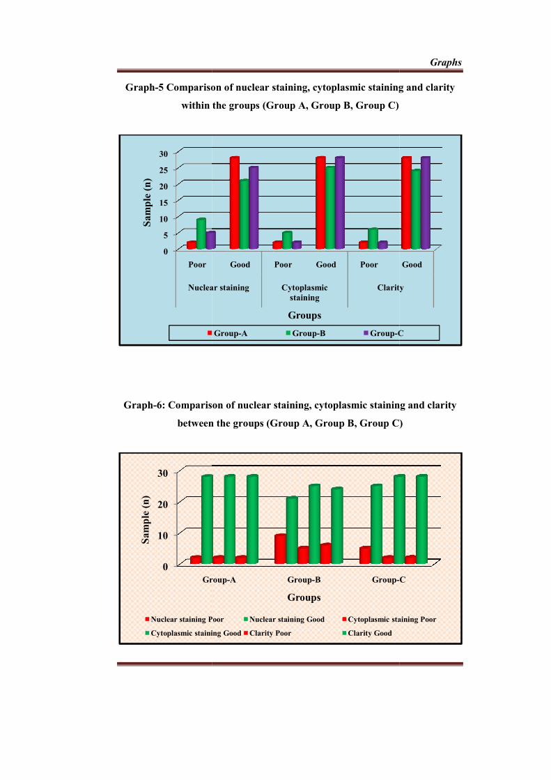
Graph-5 Comparison of nuclear staining, cytoplasmic staining and clarity
within the groups (Group A, Group B, Group C)
Graph-6: Comparison of nuclear staining, cytoplasmic staining and clarity
between the groups
0
5
10
15
20
25
30
Poor
Nuclear staining
Sam
ple
(n
)
0
10
20
30
Group
Sam
ple
(n
)
Nuclear staining Poor
Cytoplasmic staining Good
5 Comparison of nuclear staining, cytoplasmic staining and clarity
within the groups (Group A, Group B, Group C)
6: Comparison of nuclear staining, cytoplasmic staining and clarity
between the groups (Group A, Group B, Group C)
Good Poor Good Poor
Nuclear staining Cytoplasmic staining
Clarity
Groups
Group-A Group-B Group-C
Group-A Group-B Group-C
Groups
Nuclear staining Poor Nuclear staining Good Cytoplasmic staining Poor
Cytoplasmic staining Good Clarity Poor Clarity Good
Graphs
5 Comparison of nuclear staining, cytoplasmic staining and clarity
within the groups (Group A, Group B, Group C)
6: Comparison of nuclear staining, cytoplasmic staining and clarity
(Group A, Group B, Group C)
Good
Clarity
C
C
Cytoplasmic staining Poor

DISCUSSION
Discussion
45
Xylene became the clearing agent of choice when chloroform was found
as a carcinogen. A safer substitute xylene was also declared as a health hazardous
clearing agent. Under the Resource conservation and recovery act (RCRA),
xylene is considered to be one of the hazardous waste5. Later it was replaced with
safer chemicals which became a major objective of researchers and
manufacturers. The proposed substitutes included terpenes, alkanes, vegetable oil
etc16. Considering all the hazardous health effects of xylene, this study was done
in search of a much more safer alternative to xylene using palm oil and coconut
oil. These two oils are widely used as a natural flavor additives for food and also
in alternative medicine therapy.
Clearing agent is used as an intermediate solvent which is completely
miscible with ethanol and also with paraffin wax. Ideally this solvent will
displace the ethanol in the tissue, then this in turn will be displaced by molten
paraffin wax. In order to consider any solution as a clearing agent, it has to
penetrate into tissue rapidly to clear them completely. For a better penetration of
any solution, the viscosity of the solution plays a major role, that is a less viscous
solution penetrates faster when compared to that of high viscous solutions.
According to Bernoulli’s principle of fluid dynamics, viscosity of the fluid is
considered to be indirectly proportional to the temperature, which means, as the
temperature increases the viscosity of the fluid decreases and as a result
penetration of fluid increases5.
In this present study both palm oil and coconut oil were more viscous
when compared with that of xylene. Hence in order to decrease the viscosity of

Discussion
46
these oils and for an increased penetration into tissues and clear them at a faster
rate, clearing of tissue after dehydration in both processing and staining
procedures were carried out using an incubator at a temperature maintained at
60°C.
Routine paraffin wax tissue processing requires properly timed
dehydration, clearing (de-alcoholization), infiltration and embedding procedures.
On microtomy, sections were dewaxed and further dehydration was done before
staining procedure. In this study, tissue specimens were processed in parallel with
xylene, palm oil and coconut oil as a clearing agent and as s dewaxing agent.
While evaluating the rigidity of the gross tissue specimens treated with
palm oil and coconut oil, 33.33% of coconut oil treated specimen showed
superior quality than that of xylene and 43.33% showed similar features as that of
xylene treated specimens. 26.67% of palm oil treated specimen showed superior
features and 40% showed same features as that of xylene. Translucency of the
specimen was also evaluated in our study, which showed 63.33% showing better
features in coconut oil treated specimens and 53.33% in palm oil treated
specimens. 83.33% of palm oil treated tissues and 93.33% of coconut oil treated
tissues showed same features of change after impregnation as that of xylene
treated specimens. 93.33% of both palm oil and coconut oil treated specimens
showed same qualities of that of xylene while evaluation of ease of sectioning.
While comparing the nuclear staining, cytoplasmic staining and clarity of staining
of all the hematoxyline and eosin stained slides, coconut oil treated slides showed
better staining qualities ie 83.33%, 93.33% and 93.33% respectively.

Discussion
47
Rasmussen et al in 1992 made use of mixture of coconut oil and olive oil
as a clearing agent in routine hematoxylin and eosin histopathological staining
procedure. In their study, incomplete impregnation was noted and that leading to
problems while sectioning and they concluded that the mixture was ineffective as
a clearing agent48. In contrast to the observation in their study, this study found
out that coconut oil treated specimen showed better features with respect to
gross tissue evaluation, especially with changes after impregnation and ease of
sectioning thereafter. This difference could due to the presence of olive oil in
the prepared mixture, which would have adversely affected the whole
procedure there by interacting with the properties of coconut oil.
A study by Taneeru S et al in 2013 evaluated the efficiency of
limonene oil and sesame oil in terms of nuclear staining, cytoplasmic staining,
uniformity, clarity and intensity of staining35. Their study concluded that
specimens cleared with sesame oil had all the staining features which were
adequate for that of diagnosis aspect, which were consistent with that of our
study results.
Sermadi W et al in 2014 checked the efficiency of coconut oil as a
clearing agent, where in it was concluded that xylene treated tissue specimens
were more rigid (73%) than that of coconut oil treated specimens.
Translucency of the specimen was also evaluated in their study, which showed
100% better features in coconut oil treated specimens. 100% of coconut oil
treated specimen showed same features as that of xylene treated specimens
with respect to change after impregnation and ease of sectioning .In their

Discussion
48
study there was not much difference in staining quality and tissue architecture
in both coconut oil and xylene treated slides10.
Our study results are in agreement with another study by Udonkang M et
al in 2014, using bleached palm oil and their results showed 93% of palm oil
processed tissues appeared transparent after clearing as compared with xylene
processed specimens. Ease of microtomy was also evaluated and they concluded
that easy microtomy was found in 100% of xylene exposed specimen and 73.7%
of palm oil exposed specimens. Hematoxylin and eosin stained slide evaluation
was done and evaluated based on nuclear staining, cytoplasmic staining and
clarity of staining, in which 100% of all specimens showed normal nuclear and
cytoplasmic staining in both the groups47.
Indu S et al in 2014 concluded that adequate nuclear staining was
observed in 90% in cederwood oil and 93% in xylene. Also adequate cytoplasmic
staining and overall uniformity of staining were noted in 93.33% of cedarwood
oil treated slides40. All the samples in their study had appreciable results when
treated with cederwood oil as compared with that of xylene according to the
grossing and microscopic features.
Sugunakar Raju G S et al in 2015 evaluated four different oils namely
rose oil, carrot oil, pine oil and olive oil as a clearing agent instead of xylene.
Gross tissue specimen evaluation such as translucency, rigidity, shrinkage,
section cutting and cellular architecture and staining quality were evaluated for
all the oils. Pine oil was having superior characteristic features when compared
with the other three oils. Tissue morphology was well preserved in all of the

Discussion
49
tissue sections cleared by using these four different oils and a clear demarcation
was noticed in nucleus and cytoplasm. Their results concluded that over all
staining quality was equivalent with that of xylene treated slides5. Our study
results with respect to nuclear and cytoplasmic clarity in hematoxylin and eosin
stained slides were in agreement with this study.
In another study conducted by Buesa RJ in 2000, mineral oil was used in
proportion with ethanol and isopropyl alcohol as a clearing agent for tissue
specimen9. Here in this study the processed tissue with mineral oil showed
equivalent qualities when compared with that of xylene processed tissue.

SUMMARY & CONCLUSION

Summary
50
The main aim of this study was to evaluate the efficiency of coconut oil
and palm oil as a clearing agent for hematoxylin and eosin staining procedure.
A total of 30 tissue specimens were collected, fixed in 10% formalin and
sectioned into 3 equal parts and grouped as group A, B, and C. Group A tissue
specimen were taken for routine processing followed by hematoxylin and eosin
staining procedure with xylene as clearing agent, whereas group B tissue
specimens were treated with heated palm oil at a temperature maintained at 60°C
instead of xylene as a clearing agent. Similarly group C tissue specimens were
treated with heated coconut oil at a temperature maintained at 60°C instead of
xylene as a clearing agent during processing. Gross tissue specimen evaluation
like rigidity, translucency, change after impregnation and ease of sectioning were
evaluated and compared between the groups having group A as the control. All
the specimens were processed, sectioned and stained using hematoxylin and eosin
stain and were coded and observed for evaluation of nuclear staining,
cytoplasmic staining and overall clarity of stained slide.
Coconut oil treated specimen showed better characteristic features than
palm oil treated specimen with respect to rigidity, translucency and change after
impregnation which was 43.33%, 63.33% and 90% respectively. Both palm oil
and coconut oil treated specimen showed similar features when compared with
that of ease of sectioning. Among 90 hematoxylin and eosin stained slides
coconut oil treated sections showed better nuclear staining, cytoplasmic staining
and clarity of staining which was 83.33% , 93.33% and 93.33%.

Conclusion
51
Xylene is an aromatic hydrocarbon which is extremely biohazardous.
The exposure of xylene is maximum during dewaxing of sections and the
histopathological laboratory technicians are routinely exposed to xylene during
procedures. Effects to minimize the health hazards in the histopathology
laboratory should be made to create a much more safer working environment by
increasing the awareness among histopathology lab manager and assistants.
Short educational programs targeting standard operating procedures, safety
precautions and emergency management should be encouraged.
The xylene free method for paraffin sections was developed and in use
at Vrinnevi hospital, Sweden since 19956. On account of the Occupational
Safety and Health Administration regulations, various other xylene alternatives
namely limonene reagents, aliphatic hydrocarbons, vegetable oils and mineral
oils were tried in the past to avoid xylene usage in the laboratory.
This study is designed to establish whether the usage of coconut oil and
palm oil at a maintained temperature as a clearing agent during tissue
processing and as a dewaxing agent during staining procedure has any effect on
transparency, rigidity, change after impregnation, ease in sectioning and quality
of staining such as nuclear staining, cytoplasmic staining and clarity of staining
as compared with the xylene treated counterparts. Our study results suggests that
both the oils, especially coconut oil treated specimen showed better characteristic
features than palm oil treated specimen with respect to both gross specimen
evaluation and also with the quality of staining. Further studies with larger sample
size and more parameters are required to validate the better results with the usage
of biofriendly xylene alternatives.

BIBLIOGRAPHY

Bibliography
ix
1. Negi A Puri, Gupta R, Chauhan I, Nangia R, Sachdeva A. Biosafe alternative
to xylene: A comparative study. J Oral Maxillofac pathol 2013; 17(3) :363-6.
2. Bancroft JD, Gamble M. Tissue processing: Theory and practice of histologic
techniques. 6th ed. USA Churchill Livingstone: Elsevier; 2011 .P. 83-93
3. Dapson JC, Dapson RW. Clearing agents. Hazardous materials in
histopathology laboratory, regulations, risks, handling and disposal. 4th ed.
Battle Creek: Anatech; 2005. p. 65-78.
4. Kandyala R, Raghavendra S P, Ragasekharan S T. Xylene: An overview of its
health hazards and preventive measures. J Oral Max Pathol 2010;14(1) :1-5.
5. Raju GSS, Surapaneni RKN, Pavan G, Kulkarni, Thokala M R, Pavan P. Bio-
friendly alternatives for xylene - Carrot oil, olive oil, pine oil, rose oil. Journal
of clinical and diagnostic research. 2015;9(11):16-8.
6. Falkeholm L, Crawford. A Xylene-free method for histologic preparation: A
multicentre evaluation. Laboratory Investigation 2001;81(9) :1213-21
7. Rajan ST, Malathi N. Health hazards of xylene. Journal of clinical and
diagnostic research 2014;8(2) :271-4.
8. Erikson T, Amed V, Leibach S J, Bushnik F, Saxon A, Knopse W H. Acute
bone marrow toxicity and pancytopenia following exposure to lead chromate,
xylene and ethylbenzene in a degloving injury. Am J Haematol 1994;47(4)
:257-61.
9. Beusa RJ. Mineral oil: The best xylene substitute for tissue processing yet?.
The J Histotechnol 2000;23(2) :143-8.

Bibliography
x
10. Sermadi W, Prabhu S, Acharya S, Javali S B. Comparing the efficacy of
coconut oil and xylene as a clearing agent in the histopathology laboratory. J
Oral Maxillofac Pathol 2014;18(1) :49-53.
11. Fabri, Jorg Graeser, Ulrich, and Simo, Thomas A. Xylene In: Ullmann's
Encyclopedia of Industrial Chemistry. 7th edition. Weinheim: Wiley-VCH;
2000. p.2-8
12. Carson, Freida, Hladik, Christa. Histotechnology: A Self-Instructional Text.
3rd edition. American Society for Clinical Pathology Press; 2009. p.35-7.
13. Ramulu S, Koneru A, Ravikumar S, Sharma P ,Ramesh DNS V, Patil R.
Liquid dish washing soap: An excellent substitute for xylene and alcohol in
hematoxylin and eosin staining procedure. Journal of Orofacial Sciences
2012;4(1):37-42.
14. Jacobson GA, Ann SA. Biological Monitoring of Low Level Occupational
Xylene Exposure and the Role of Recent Exposure. Occup. Hyg 2003;47(4):
331–6.
15. Langman JM. Xylene: Its toxicity, measurements of exposure levels,
absorption, metabolism and clearance. Pathology 1994;26(3) :301-9.
16. Busea RJ, Peshkov MV. Histology without xylene. Annals of Diagnostic
Pathology 2009;13(4) :246-256.
17. Toxicological profile for xylene. U.S Department of Health and Human
Services, public health service, Agency for toxic substance and disease
registry. Atlanta: Georgia;1993.

Bibliography
xi
18. Ogata M, Tomokuni K, Takatsuka Y. Urinary excretion of hippuric acid and
m or p-methylhippuric acid in the urine of persons exposed to vapours of
toluene and -xylene as a test of exposure. Br J Int Med 1970;27(8) :43-50.
19. Savoleinen H, Pfaffli P. Dose-dependent neurochemical changes during short
term inhalation exposure to m-xylene. Arch Toxicol 1980;45 :117-122.
20. Hipolito R N. Xylene poisoning in laboratory workers: Case reports and
discussions. Lab Med 1980;11(3) :593-595.
21. Taskinen H, Anttila A, Lindbohm M, Sallmén M, Hemminki K. Spontaneous
abortions and congenital malformations among the wives of men
occupationally exposed to organic solvents. Scandinavian Journal of Work,
Environment & Health 1989;15(5) :345-352.
22. Uchida Y, Nakatsuka H, Ukai H, Watanabe T, Liu Y T, Huang M Y.
Symptoms and signs in workers exposed predominantly to xylene. Int Arch
Occup Environ Health 1993;64(8) :597-605.
23. Revilla A S, Pestana C R, Pardo-Andreu G L, Santos A C, Uyemura S A,
Gonzales M E, Curti C. Potential toxicity of toluene and xylene evoked by
mitochondrial uncoupling. Toxicol In Vitro 2007;21(5) :782–788.
24. Adrian F, Bradley M, Felipe C. Xylene-Induced Auditory Dysfunction in
Humans. Ear Hear 2013;34(5) :651–660.
25. Haas M. Eliminating the use of xylene in tissue processing. The Biomedical
scientist 2010;487-488.
Bibliography
xii
26. Bruun R B, Norring H K, Mellerup I, Sether G, Christensen. Vegetable oils
instead of xylene in tissue processing. Acta Pathologica, Microbiologica et
Immunologica Scandinavica 1992;100(9) :829-831.
27. Andre GG, Wenger JB, Rebolloso D, Arrington JB, Mehm WJ. Evaluation of
Clearing and Infiltration Mixtures (CIMs) As Xylene Substitutes for Tissue
Processing. J Histotechnol 1994;17(2):137–42.
28. Eleanor A, Michael HE, Matthew H. Cost effective, environmentally safe
tissue processing method with paraffin oil. Journal of Histotechnology
1997;20(2):133-137.
29. Ofusori DA, Ayoka AO, Adeeyo OA, Adewole SO. Mixture of kerosene and
xylene: a contribution to clearing agents. Int. J. Morphol 2009;27(1) : 211-
218.
30. Bleuel E, Den Dunnen WFA. Solvent free tissue processing using super-
critical CO2. Histopathology 2012;61(6) :1198-1208.
31. Kunhua W, Chuming F, Xun L. A Novel Non-Toxic Xylene Substitute (SBO)
for Histology. Afr J Trdit Complement Altern Med 2012;9(1) :43-49.
32. Piniewska D, Wojtas M, Polańska N, Stawowiak A, Konieczna-Waśkowska
M, et al. The Comparison of Paraffin Dewaxing Using Methyl Tert-Butyl
Ether and Xylene in DNA Extraction from Autopsy Specimens. J Forensic
Res 2012;3(2):175-179.
33. Nangia R, Puri A, Gupta R, Bansal S, Negi A, Mittal M. Comparison of
conventional tissue processing with microwave processing using

Bibliography
xiii
commercially available and domestic microwaves. Indian J Oral Sci
2013;4(2):64-69.
34. Aydin I, Yorukoglu K, Cingoz S, Agilkaya S. The effect of the alternative
solutions to formaldehyde and xylene on tissue processing. Indian J Pathol
Microbiol 2013;56(3) :221-230.
35. Taneeru S, Guttikonda VR, Vanajakshi CN, Korlepara R. Xylene free
method for tissue processing: a pilot study. Health Sciences 2013;2(3) :1-12.
36. Premalatha BR, Patil S, Rao RS, Indu M. Mineral oil- A Biofriendly
substitute for xylene in Deparaffinization: A Novel Method. JCDP 2013;
14(2) :281-286.
37. Ankle MR, Joshi PS. A Study to evaluate the efficacy of xylene-free
hematoxylin and eosin staining procedure as compared to the conventional
hematoxylin and eosin staining: An experimental study. J oral and
maxillofacial Pathology 2011;15(2) :161-7.
38. Henwood A F, Prasad L, Bourke V M. The application of heated detergent
dewaxing and rehydration to techniques for the demonstration of fungi: a
comparison to routine xylene-alcohol dewaxing. Journal of Histotechnology
2013;36(2) :45-50.
39. Ananthaneni A, Namala S, Guduru VS, Ramprasad VVS, Ramisetty SD,
Udayashankar U, Naik KK. Efficacy of 1.5% Dish Washing Solution and
95% Lemon Water in Substituting Perilous Xylene as a Deparaffinizing
Agent for Routine H and E Staining Procedure: A Short Study. Scientifica

Bibliography
xiv
[Internet].[cited 2017 March];707310. Available from
http://dx.doi.org/10.1155/2014/707310.
40. Indu S, Ramesh V, Indu PC, Prashad KV, Premalatha B, Ramadoss K.
Comparative efficacy of cedarwood oil and xylene in hematoxylin and eosin
staining procedures: An experimental study. J Nat Sc Biol Med
2014;5(2):284-7.
41. Lyon H, Holm I, Prent P, Balslev E. Non-hazardous organic solvents in the
paraffin-embedding technique: a rational approach. Histochemistry and Cell
Biology 1995;103(4) :263-269.
42. Maini D. A Closer Look at Xylene Substitutes to Reduce the Hazardous
Waste Stream. Technical Bulletin for Histotechnology 1999;30 :15-18.
43. Temel SG, Noyan S, Cavusoglu I, Kahveci Z. A simple and rapid microwave-
assisted hematoxylin and eosin staining method using 1,1,1 trichloroethane as
a dewaxing and a clearing agent. Biotechnic Histochemistry 2005;80(4) :123-
132.
44. Chen CY, He T, Mao XL, Friis TE, Qin RH, Jian YT. A novel xylene
substitute for histotechnology and histochemistry. Biotechnic Histochemistry
2010;85(4): 231-240.
45. Stockert JC, Lopez-Arias B, Castillo PD, Romero A, Blazquez-Castro A.
Replacing xylene with n-heptane for paraffin embedding. Biotechnic
Histochemistry 2012;87(7) :464-467.

Bibliography
xv
46. Patil S, Rao RS, Nagaraja A, Kumar SD. Comparison of conventional and
microwave histo-processing of various oral soft tissue specimens. J Dent Res
Rev 2014;1(1):3-6.
47. Udonkang M, Eluwa M, Ekanem TB, Asuquo OR, Akpantah AO. Bleached
palm oil as substitute for xylene in histology. JPCS 2014;8 :8-17.
48. Rasmussen B, Hjort K, Mellerup I, Sether G, Christensen N. Vegetable oils
instead of xylene in tissue processing. Acta Pathol Microbio Immunol
Scandinavica 1992;100(9):827-31.

ANNEXURE


CONSENT FORM PART 1 OF 2
INFORMATION FOR PARTICIPANTS OF THE STUDY
Dear Volunteers, We welcome you and thank you for your keen interest in
participation in this research project. Before you participate in this study, it is important for you to understand why this research is being carried out. This form will provide you all the relevant details of this research. It will explain the nature, the purpose, the benefits, the risks, the discomforts, the precautions and the information about how this project will be carried out. It is important that you read and understand the contents of the form carefully. This form may contain certain scientific terms and hence, if you have any doubts or if you want more information, you are free to ask the study personnel or the contact person mentioned below before you give your consent and also at any time during the entire course of the project.
1. Name of the Principal Investigator:
Dr. Ashitha.A.S, Post Graduate student Department of Oral Pathology and Microbiology, Sree Mookambika Institute of Dental Sciences, Kulasekaram, KanyaKumari District-629161
2. Name of the Guide: Dr. T. Isaac Joseph Head of the Department, Department of Oral Pathology and Microbiology,
Sree Mookambika Institute of Dental Sciences. Kulasekaram, KanyaKumari District-629161
3. Name of the Co-Guide: Dr. Girish K.L,
Professor, Department of Oral Pathology and Microbiology.
Sree Mookambika Institute of Dental Sciences. Kulasekaram, KanyaKumari District-629161 4. Institute: Sree Mookambika Institute of Dental Sciences,
V.P.M Hospital complex, Padanilam, Kulasekaram, Kanyakumari District– 629161 Tamilnadu.
5. Title of the study: “Comparing the efficiency of coconut oil and palm oil with xylene as a clearing agent in conventional Hematoxylin and Eosin histopathological staining procedure” 6. Background information: Scientific Journals

7. Aims and Objectives: To evaluate the efficiency of coconut oil and palm oil as a clearing agent
for Hematoxylin and Eosin staining procedure and compare it with xylene.
To determine whether tissues cleared and dewaxed with coconut oil and palm oil are same or superior with the xylene treated tissues.
8. Scientific justification of the study:
On account of the Occupational Safety and Health Administration (OSHA) regulations, various xylene substitutes, such as, limonene reagents, aliphatic hydrocarbons, vegetable oils and mineral oils were tried in the past to avoid xylene in the laboratory. However, these substitutes were found to be less effective and more expensive5. Coconut oil is a commonly used vegetable oil, available throughout the tropical world. It is non-toxic, heat stable, slow to oxidize and has highest resistance to rancidity6. Palm oil promises to be widely available and safe substitute for xylene. Hence, here we attempt to check the use of coconut oil and palm oil as a clearing agent during tissue processing and as a dewaxing agent during Hematoxylin and Eosin staining procedure has any effect when compared with the xylene processed tissue. 9. Procedure for the study: Soft tissue specimen from those patients fulfilling the inclusion and exclusion criteria of the study are to be taken from the department of Oral and maxillofacial surgery of Sree Mookambika institute of dental sciences. All of these tissues are to be taken during routine surgical removal of impacted third molar. The subjects selected for the study must be explained about this study and informed consent must be taken. The sample included a total of 30 tissue specimens.
Each of 30 specimens removed during surgical procedure were fixed with 10% buffered formalin for 48 hour All the tissues have to cut into three equal parts and arrange it experimental group namely Group A, Group B and Group C, in which each group consisting of 30 tissue bits.
For Group A, after fixation, tissues were dehydrated through ascending grades of alcohol (70%, 90%, 95%, absolute 1, absolute II and absolute III) for 1 hour in each change. Dehydrated tissues must be dealcoholized (cleared) by using two changes of xylene, xylene 1 for 45 minutes and xylene II for 30 minutes. The cleared tissues have to be infiltrated in two changes of molten paraffin wax (wax 1 for 1 hour and wax II for 1 hour 30 minutes). Embedding must be done in molten paraffin wax using plastic disposal cassettes and allow it to solidify before microtomy. Tissue blocks must be sectioned at 4 μm with a rotary microtome and sections floated in a warm water bath and each picked in pairs on albuminized glass slides.
Before staining, the slides must be dewaxed in xylene I and II for 5 minute each, pass it through descending grades of alcohol (absolute, 90%, 70% ) and rinse it in water for 2 minutes. Hydrated sections must be stained in Haematoxylin solution for 10 minutes and rinse in water for 2 minutes.Then differentiation must be done by a single dip in 1% acid alcohol and tap water

wash for 30 seconds. The sections must be counterstained in 1% Eosin solution for 1 minute, and dehydrate with 70% ,80% and 90% for 1 minute each. Two changes of absolute alcohol must be done. Clear the slides using 3 changes of xylene (I,II and III) for 1 minute each have to be done, air dry and mount with DPX mountant.
For Group B, after fixation, tissues were dehydrated through ascending grades of alcohol (70%, 90%, 95%, absolute 1, absolute 2 and absolute 3) for 1 hour in each change. Dehydrated tissues must be dealcoholized (cleared) by using two changes of heated palm oil at 60ºC for 1 hour each. The cleared tissues have to be infiltrated in two changes of molten paraffin wax (wax I for 1 hour and wax II for 1 hour 30 minutes).Embedding must be done in molten paraffin wax using plastic disposal cassettes and allow it to solidify before microtomy. Tissue blocks must be sectioned at 4 μm with a rotary microtome and sections floated in a warm water bath and each picked in pairs on albuminized glass slides.
Before staining, the slides must be dewaxed in heated palm oil at 60ºC for 5 minute each, pass it through descending grades of alcohol (absolute, 90%, 70% ) and rinse it in water for 2 minutes. Hydrated sections must be stained in Haematoxylin solution for 10 minutes and rinse in water for 2 minutes.Then differentiation must be done by a single dip in 1% acid alcohol and tap water wash for 30 seconds. The sections must be counterstained in 1% Eosin solution for 1 minute, and dehydrate with 70% ,80% and 90% for 1 minute each. Two changes of absolute alcohol must be done. Clear the slides using 3 changes of preheated palm oil at 60 ºC for 1 minute each have to be done, air dry and mount with DPX mountant.
For Group C, after fixation, tissues were dehydrated through ascending grades of alcohol (70%, 90%, 95%, absolute 1, absolute 2 and absolute 3) for 1 hour in each change. Dehydrated tissues must be dealcoholized (cleared) by using two changes of heated coconut oil at 60ºC for 1 hour each. The cleared tissues have to be infiltrated in two changes of molten paraffin wax (wax I for 1 hour and wax II for 1 hour 30 minutes). Embedding must be done in molten paraffin wax using plastic disposal cassettes and allow to solidify before microtomy. Tissue blocks must be sectioned at 4 μm with a rotary microtome and sections floated in a warm water bath and each picked in pairs on albuminized glass slides.
Before staining, the slides must be dewaxed in heated coconut oil at 60ºC for 5 minute each, pass it through descending grades of alcohol (absolute, 90%, 70%) and rinse it in water for 2 minutes. Hydrated sections must be stained in Haematoxylin solution for 10 minutes and rinse in water for 2 minutes. Then differentiation must be done by a single dip in 1% acid alcohol and tap water wash for 30 seconds. The sections must be counterstained in 1% Eosin solution for 1 minute, and dehydrate with 70% ,80% and 90% for 1 minute each. Two changes of absolute alcohol must be done. Clear the slides using 3 changes of heated coconut oil at 60ºC for 1 minute each have to be done, air dry and mount with DPX mountant.
During the tissue processing, in all the three groups, macroscopic observations based on rigidity, translucency, change after impregnation, gross shrinkage and ease of sectioning must be noted. Microscopic examination of

prepared Hematoxylin and Eosin stained slides for nuclear staining, cytoplasmic staining and overall clarity of staining in all the three groups must be evaluated by two experienced pathologists (observer I and observer II) under 10x and 40x magnification using Light microscope. All data are to be entered in data entry sheet prepared. Data will be entered in Microsoft excel application.
10. Expected risks for the participants:
No risk.
11. Expected benefits of research for the participants: You will not be required to pay for this test. You can enquire about the outcome of the procedures and your details.
12. Maintenance of confidentiality:
You have the right to confidentiality regarding the privacy of your medical information
(Personal details, results of physical examinations, investigations, and your medical history).
By signing this document, you will be allowing the research team investigators, other study Personnel, sponsors, institutional ethics committee and any person or agency required by law to view your data, if required.
The results of clinical tests and therapy performed as part of this research may be included in your medical record.
The information from this study, if published in scientific journals or presented at scientific meetings, will not reveal your identity.
13.Why have I been chosen to be in this study? a) Chosen because of grouping under the inclusion and exclusion criteria b) Need of good sampling size
14.How many samples will be in the study? 30 samples per group.
15. Agreement of compensation to the participants (In case of a study related injury):
Patient will be taken care in case of complication and medical treatment will be provided in the institution.
16. Anticipated prorated payment, if any, to the participant(s) of the study: No 17. Can I withdraw from the study at any time during the study period?
The participation in this research is purely voluntary and you have the right to withdraw from this study at any time during the course of the study without giving any reasons.
However, it is advisable that you talk to the research team prior to stopping information.

18. If there is any new findings/information, would I be informed? Yes 19. Expected duration of the participant’s participation in the study: 1 Year 20. Any other pertinent information: No other information 21. Whom do I contact for further information? For any study related queries, you are free to contact:
Dr. Ashitha A.S, Post Graduate student. Department of Oral Pathology and Microbiology, Sree Mookambika Institute of Dental Sciences, Kulasekaram, KanyaKumari District-629161. Mobile No: 8281401187 [email protected]
Place: Signature of Principal Investigator Date: Place: Signature of the participant Date:

PART 2 OF 2
PARTICIPANTS CONSENT FORM
The details of the study have been explained to me in writing and the
details have been fully explained to me. I am informed that the result of the study
is beneficial to me and the society. I confirm that I have understood the study and
had the opportunity to ask questions. I understand that my participation in the
study is voluntary and that I am free to withdraw at any time, without giving any
reason. I understand that there is no risk for me in participating in this study. I
have been given an information sheet giving details of the study. I fully consent
to participate in the study titled
“Comparing the efficiency of coconut oil and palm oil with xylene as a
clearing agent in conventional Hematoxylin and Eosin histopathological staining
procedure.”
Serial no/Reference no:
Name of the participant:
Address of the participant:
Contact number of the participant:
Signature/Thumb impression of the participant/ Legal guardian (if age< 18) Witnesses 1.
2.
Place:
Date:

Jl×Rp T¥Ym Tϧ 1
Bn®p TeúLtTYoLÞdLô] ®YWm
AuTôokR TeúLtTô[oLú[, CkR BWônf£«p BoYjÕPu TeúLtL Yk§ÚdÏm EeLÞdÏ
GeLÞûPV YWúYt©û]Ùm Su±ûVÙm ùR¬®jÕd ùLôs¡ú\ôm. CkR BWônf£«p LXkÕ ùLôsYRtÏ Øu×, CÕ GR]ôp úUtùLôs[lTÓ¡\Õ Guß ×¬kÕ ùLôsYÕ ªLÜm AY£VUô]Õ. CkR T¥Ym BWônf£dÏ ùRôPo×ûPV AY£VUô]Õ. CkR T¥Ym BWônf£dÏ ùRôPo×ûPV ®YWeLû[ ùR¬kÕ ùLôs[ ERÜ¡\Õ. CkR BWônf£ CRàûPV RuûU, úRûY, AàáXeLs, ATôVm, YN§«uûU, Øuù]fN¬dûL Utßm RYpLs GpXôm GqYôß úUtùLôs[lTÓ¡\Õ GuT§û] ®Y¬d¡\Õ. C§p Bn®tLô] T¥Yj§p Es[ ϱl×Lû[ T¥jÕ ×¬kÕd ùLôsYÕ ªLÜm AY£VUô]Õ. C§p £X A±®Vp ùNôtLs TVuTÓjRlTh¥Úd¡\Õ. AR]ôp HúRàm IVeLú[ô ApXÕ úUÛm HúRàm ®YWeLs ùR¬kÕ ùLôs[ úYi¥ CÚkRôúXô CkR BWônf£ ùNnTYûWúVô ApXÕ ¸úZ ùLôÓdLlTh¥ÚdÏm SToL°p VôûWúVàm, Jl×RÛdÏ ØuúTô ApXÕ CqYôWVôf£«u ØÝ LôXj§úXô GkR®RUô] RVdLØm Cu± úLhÓ ùR¬kÕ ùLô[s[Xôm.
1. ØRuûU úNôRû]Vô[o ùTVo : PôdPo. AxRô, H.Gv. ØÕ¨ûX UôQYo Yôn úSônL°u CVp× Utßm
ÖiÔ«¬«p ©¬Ü c êLôm©Lô Tp UÚjÕYf Lpí¬, ÏXúNLWm - 629 161, Lu²VôÏU¬
UôYhPm. 2. Y¯LôhÓ]o ùTVo : PôdPo. ¥. INd ú_ôNl RûXûU úTWô£¬Vo, Yôn úSônL°u CVp× Utßm
ÖiÔ«¬«p ©¬Ü c êLôm©Lô Tp UÚjÕYf Lpí¬, ÏXúNLWm - 629 161, Lu²VôÏU¬
UôYhPm. 3. ÕûQ Y¯LôhÓ]o ùTVo : PôdPo. ¡¬x úL.Gp úTWô£¬Vo Yôn úSônL°u CVp× Utßm
ÖiÔ«¬«p ©¬Ü c êLôm©Lô Tp UÚjÕYf Lpí¬, ÏXúNLWm - 629 161, Lu²VôÏU¬
UôYhPm.

4. ¨ßY]m : c êLôm©Lô Tp UÚjÕYd Lpí¬, ®.©.Gm. UÚjÕYUû] Y[ôLm,
TP¨Xm, ÏXúNLWm, Lu²VôÏU¬ UôYhPm 629 161 RªrSôÓ. 5. BWônf£«u RûXl× :
¶UPôd³u Utßm úVô£u §Ñ CVp× ¨\úYßTôh¥p úReLôn GiùQn Utßm TôU«p GiùQ«p ûNû] ùR° ùTôÚ[ôLl TuTÓj§ ARu Bt\ûX Jl©Pf ùNnÙm ùNVpØû\.
6. ©u]¦ RLYp :
A±®Vp NôokR ϱlúTÓ
7. ϱdúLôsLs:
¶UPôd³u Utßm CúVô£u ¨\ úYßTôÓ ùNVpØû\«p úReLôn GiùQn Utßm TôU«p GiùQn ùR°ùTôÚ[ôL TVuTÓj§ ARu Bt\ûX ûNàPu U§l©ÓRp.
úReLôn GiùQn Utßm TôU«p GiùQûV ûYjÕ ùNnR §Ñ ùR°YûPRp Utßm ùUÝÏ ALt\p JúW Uô§¬VôL Es[Rô ApXÕ ûNu]ôp ùNnVlThP §ÑdLs úUXôL Es[Rô GuTûR Tt± LiÓ©¥jRp.
8. Bn®u A±®Vp NôoSR ¨ìTQm:
BdÏúT`]p úN@l¥ Auh ùapj Ahª²v¥úW`u (OSHA) ®§Øû\lT¥, ûNu úTôu\ TpúYßThP Uôt\dáßLs úUô²u úNoUeLs, A@úT¥d ûahúWôLôoTuv, RôYW GiùQn Utßm RôÕ GiùQn ØRVYtû\ BnYôûX«p Øu]o ûNàdÏ T§XôL úNô§jÕ TôojR]o.
CÚl©àm CkR Uôt\dáßLs Aû]jÕm ®ûX A§LUô]RôLÜm, Ïû\kR Bt\ûX ùLôiPûYVôLÜm CÚkRÕ. úReLôn GiùQn CkR EX¡p G°§p ¡ûPdL á¥V Aû]YWôÛm TVuTÓjRTPd áÑ¥V RôYW GiGQn CÕ SfÑjRuûUVt\Õ, ùYlT ¨ûX Uô\ôRÕ, ùUÕYôL L²Vá¥VÕ Utßm ÕoSôt\jûR ùTÚU[Ü RÓdLdá¥VÕ. Tû] GiùQn A§L A[®p ¡ûPdLdá¥VRôLÜm TôÕLôlT]RôLÜm ûN]u Uôt\ôLÜm LÚRlTÓ¡\Õ.
CkR CPj§ÚkÕ SôeLs úReLôn GiùQn Utßm TôU«p GiùQûV §Ñ ùNVpØû\«u úTôÕ ùR° ùTôÚ[ôLÜm Utßm ¶UPôd³u CúVô£u ¨\úYßTôÓ ùNVpØû\«u úTôÕ ùUÝL¯dÏm ùTôÚ[ôLÜm TVuTÓj§ HúRàm TXu Es[Rô Guß ûNu §Ñ ùNnØû\ÙPu Jl©hÓ TôodÏm ØVt£ûV ùNn¡ú\ôm.
9. ùNnØû\:
úSôVô°L°u ùUu §Ñ Uô§¬ GÓdÏm ùTôÝÕ AYoL°u Esú[±V Utßm ùY°úV±V ϱl×Lû[ c êLôm©Lô Tp UÚjÕYd

Lpí¬«u Yôn Utßm ØL AßûY £¡hûN ©¬®p CÚkÕ GÓdLlTÓ¡\Õ. CkR §Ñ Uô§¬Ls Aû]jÕm YZdLUô] Tp ALtßm Øû\«u úTôÕ GÓdLlTÓ¡\Õ. CûR úRokùRÓjRRtLô] LôWQeLû[Ùm CkR Bn®û] Tt±Ùm ®[d¡ LôhPôVUôL JlTkR YôdÏêXm YôeL úYiÓm. CkR Uô§¬Ls ùUôjRm 30 §Ñ Uô§¬Lû[ ùLôiPÕ.
CkR 30 Uô§¬Ls JqùYôußm AßûY £¡hûN«u úTôÕ GÓdLlThÓ AûR 10% TôÕLôdLlThP @TôoU²p 48 U¦ úSWm ùTôÚjR úYiÓm, ©u× §ÑdLû[ êuß N¬Tô§VôL ©¬jÕ ©¬Ü A, ©¬Ü B, ©¬Ü C Guß úNôRû]l ©¬®p Y¬ûNlTÓj§, JqùYôÚ ©¬®Ûm Es[ §ÑdLû[ 30 TôLeL[ôL ©¬dL úYiÓm.
A ©¬®p, ùTôÚjÕRp, L¯kR ©\Ï, EVoRW BpLLôûX (70%, 90%, 95%, AlùNôÙh II AlùNôÙh III) ùLôiÓ JÚ U¦ úSWj§tÏ JÚ Øû\ §ÑdL°u DWlTRjûR ¿dL úYiÓm. DWTRm ¿d¡V §ÑdLû[ BpdLôp CÚkÕ ©¬dL CÚúYß YûLVô] ûNu, ûNu I-p 45 ¨ªPØm, ûNu II-p 30 ¨ªPØm ûYdL úYiÓm. JÚ ©[ôv¥d Eû\«p úUôpPu TôWô@©u ùUÝûL TVuTÓj§ T§V ûYjÕ ©u ûUdúWôúPôªdÏ Øu× §UôdL úYiÓm. §Ñ ÕiÓLû[
ÑZp CVk§WUô] ûUdúWôúPôm ûYjÕ 4m A[®p ©¬jùRÓjÕ, AûR ªRUô] ãÓ ùLôiP Ri½¬p ªRdL ®hÓ Apתu LiQô¥ ®pûX«p JqùYôu\ôL úRokùRÓjÕ ûYdL úYiÓm.
¨\úYßTôh¥tÏ Øu×, LiQô¥ ®pûXLû[ ûNu I Utßm II-p 5 ¨ªPm ÅRm ûYjÕ ùUÝûL ALt± ¸r RW BpLLôp (AlùNôÙh 90%, 70%) Uôt± ©u AûR Ri½¬p 2 ¨ªPm AXN úYiÓm. DWlTRúU±V ùRôϧLû[ ¨\úYßTôÓ ùNnÙm úTôÕ ¶UPôd³u LûWNp 10 ¨ªPm ûYjÕ ©u 4 ¨ªPm Ri½¬p AXN úYiÓm. ©u× 1% BpLôLp AªXj§p JÚ RPûY ØÝLf ùNnÕ 30 ®]ô¥Ls ÏZôn ¿¬p LÝ¡ úYßTôÓ ùNnV úYiÓm. ©\Ï ùRôϧLû[ áÓRp ¨\úYßTôÓ ùNnV 1% CúVô£u LûWNp 1 ¨ªPm ûYjÕ ©u 70%, 80%, 90% ÅRm 1 ¨ªPm DWlTRm ¿dL úYiÓm. CÚ úYß AlùNôh BpLôp ùNnV úYiÓm. êuß ®RUô] ûNu (I, II Utßm III) 1 ¨ªPm ÅRm ûYjÕ ®pûXLû[ ùR°Yôd¡, çnûUVôd¡ DPX ùU[uPuh ûYjÕ TôPm ùNnV úYiÓm.
B ©¬®p ùTôÚjÕRÛdÏ ©\Ï, EVoRW BpLLôp (70%, 90%, 95% AlùNôh 1, AlùNôh 2, AlùNôh 3)ûV 1 U¦ úSWm ûYjÕ §ÑdLû[ DWTRm ALt\ úYiÓm. ©u DWTRm ALt±V §ÑdLû[ CÚ úYß ®RUô] 600C ùLôiP ãPô] TôU«p GiùQ«p 1 U¦ úSWm ùR°YôdL úYiÓm. ùR°Yô¡V §ÑdLû[ CÚ ®RUô] úUôpPu TôWô@©u ùUÝ¡p (ùUÝÏ 1-p 1 U¦ úSWØm, ùUÝÏ II-p Ju\ûW U¦ úSWØm) Y¥LhP úYiÓm. ©[ôv¥d Eû\«p úUôpPu TôWô@©u ùUÝÏ ûYjÕ TR²ÓRp ùNnÕ ©u ûUdúWôúPôªd Øu× §PUôdL úYiÓm. §Ñ ÕiÓLû[ ÑZp CVk§WUô] ûUdúWôúPôm
êXm 4m A[®p ©¬jùRÓjÕ, AûR ªRUô] ãÓ ùLôiP Ri½¬p

ªRdL ®hÓ Apתu LiQô¥ ®pûXL°p JqùYôu\ôL úRokùRÓjÕ ûYdL úYiÓm. %
¨\ úYßTôh¥tÏ Øu×, LiQô¥ ®pûXLû[ 600C ùLôiP ãPô] TôU«p GiùQ«p 5 ¨ªPm ûYjÕ ùUÝûL ALt±, ¸rRW BpLLôÛdÏ (lùNôh 90%, 70%) ùLôiÓ ùNuß ©u 2 ¨ªPm Ri½¬p AXN úYiÓm. DWTRúU±V ùRôϧLû[ ¶UôPôd³u LûWNp 10 ¨ªPm ûYjÕ ©u 2 ¨ªPm Ri½¬p AX£ ¨\úYßTôÓ ùNnV úYiÓm. ©u× 1% BpLLôp AªXj§p JÚ RPûY ØÝLf ùNnÕ ÏZôn ¿¬p 30 ®]ô¥Ls LÝ¡ ©¬jùRÓdL úYiÓm. 1% CúVô£u LûWNp ùRôϧLû[ 1 ¨ªPm ûYjÕ áÓRp ¨\ úYßTôÓ ùNnÕ ©u 70%, 80%. 90%-p 1 ¨ªPm ûYjÕ DWTRm ALt\ úYiÓm. CÚ®RUô] AlùNôh BpLLôp ùNnV úYiÓm. êuß ®RUô] 600C ùLôiP ãPôd¡V TôU«p GiùQ«p 1 ¨ªPm ûYjÕ ®pûXLû[ ùR°Yôd¡, ÕnûUVôd¡ DPX ùU[uPuh ûYjÕ TôPm ùNnV úYiÓm.
C ©¬®p ùTôÚjÕRÛdÏ ©\Ï, EVoRW BpLLô®p (70%, 90%, 9%) AlùNôh 1C AlùNôh 2, AlùNôh 3) 1 U¦ úSWm ûYjÕ §ÑdL°p Es[ DWTRm ALt\ úYiÓm. ©u DWTRm ALt±V §ÑdLû[ CÚ®RUô] úReLôn GiùQ«p 600C 1 U¦ úSWm ûYjÕ ùR°YôdL úYiÓm. ùR°Yô¡V §ÑdLû[ CÚ®RUô] úUôpPu TôWô@©u ùUÝÏ (ùUÝÏ 1-p 1 U¦ úSWØm, ùUÝÕ 2-p Ju\ûW U¦ úSWØm) ûYjÕ Y¥LhP úYiÓm. ©[ôv¥d Eû\«p úUôpPu TôWô@©u ùUÝÏ ûYjÕ TR²ÓRp ùNnÕ ©u ûUdúWôúPôªdÏ Øu §PUôdL úYiÓm. §Ñ ÕiÓLû[ ÑZp CVk§WUô] ûUdúWôúPôm
êXm 4m A[®p ©¬jùRÓjÕ, AûR ªRUô] ãÓ ùLôiP Ri½¬p ªRdL ®hÓ Apתu LiQô¥ ®pûXL°p JqùYôu\ôL úRokùRÓjÕ ûYdL úYiÓm.
¨\úYßTôh¥tÏ Øu×, LiQô¥ ®pûXLû[ 600C ùLôiP ãPô] úReLôn GiùQ«p 5 ¨ªPm ûYjÕ ùUÝûL ALt±, ¸r RW BpLôLÛdÏ (AlùNôh 90%, 70%) ùLôiÓ ùNuß ©u Ri½¬p 2 ¨ªPm AXN úYiÓm. DWTRúU±V ùRôϧLû[ ¶UPôd°u LûWNp 10 ¨ªPm ûYjÕ ©u, 2 ¨ªPm Ri½¬p AX£ ¨\úYßTôÓ ùNnV úYiÓm. ©u× 1% BpLLôp AªXj§p JÚ RPûY ØÝLfùNnÕ ÏZôn ¿¬p 30 ®]ô¥Ls LÝ¡ ©¬jùRÓdL úYiÓm. 1% CúVô£u LûWNp ùRôϧLû[ 1 ¨ªPm ûYjÕ áÓRp ¨\úYßTôÓ ùNnÕ ©u 70%, 80%, 90%, 1 ¨ªPm ûYjÕ DWTRm ALt\ úYiÓm. CÚ ®RUô] AlùNôh BpLLôp ùNnV úYiÓm. êuß ®RUô] ãPô] úReLôn GiùQûV 600C---p 1 ¨ªPm ®pûXLû[ ûYjÕ ùR°Yôd¡, çnûUVôd¡, DPX ùU[uPuh ûYjÕ TôPm ùNnV úYiÓm.
§Ñ ùNVpØû\«u úTôÕ êuß ©¬ÜL°Ûm, úUdúWôvúLô©d Bt\ôûX ùLôiÓ ARu L¥]RuûU, FÓÚYp, TXUô]RtÏ ©\Ïs[ Uôt\m, ÑWÑÚlTô] ÑÚdLeLs G°§p ©¬jùRÓjRp B¡VYtû\ LiLô¦dL úYiÓm. ûUdúWôvúLô©d úNôRû]dÏ úRûYVô]

aUPôd³u CúVô£u ¨\úYßTôÓ ®pûXLs ûUVdLÚ ¨\úYßTôÓ, ûNhúPô©Xôvªd ¨\úYßTôÓ Utßm Aû]jÕ êuß ®RUô] ¨\úYßTôÓLû[Ùm úSôn CVp©û] LiÓ©¥dÏm YpÛ]ûW (TôoûYVô[o 1 Utßm TôoûYVô[o 2) ûYjÕ U§l©P úYiÓm. Aû]jÕ úLôhTôÓLû[Ùm ϱjÕ ûYdL úYiÓm. RLYpLs Aû]jÕm ûUdúWôNô@lh Gd^p Al°úL`²p T§V úYiÓm.
10. TeúLtTô[o G§oTôodÏm ®û[ÜLs CpûX
11. TeúLtTô[o BWônf£«p G§oTôodÏm AàáXeLs?
CkR úNôRû]dLôL TQm GÕÜm ùNÛjR úYiPôm
CkR ùNnØû\«u ®û[ÜLû[Ùm, ®[dLeLû[Ùm ¿eLs úLhÓ ùR¬kÕ ùLôs[Xôm.
12. CWL£VjRuûU LôjRp? EeL°Pm CÚkÕ úNL¬jR GkR ®TWØm CWL£VUôL
ûYdLlTÓm. CRuêXm ¡ûPdÏm ×s°®TWm UhÓm ùY°«PlTÓm Ut\T¥ R²ST¬u ùNôkR ®TWeLs ùY°«PlTPUôhPôÕ.
13. GR]ôp CkR Bn®p Sôu TeúLtL úRokùRÓdLlThúPu?
A) G]Õ Lp® ¨ßY]j§u ¨TkRû]LÞdÏ CÕ EhThPÕ. B) RôeLs CkR Bn®u úNol× Utßm ®Ól× LhPû[«u Es
AûUVl ùTßYRôp C) NêLj§tÏ ER®dÏ
14. CkR Bn®p GjRû] úTo TeúLt¡\ôoLs?
JqùYôÚ ÏìlÛm 30 úTo
15. CkR Bn®u êXm HúRàm ©u®û[ÜLs HtThPôp BWônf£Vô[o ùTôßl× HtTôWô?
Bm
16. CkR BWônf£«p TeÏùTßúYôÚdÏ Gq®R ùRôûLÙm YZeLlTÓUô?
CpûX
17. Sôu CkR BWônf£«ÚkÕ ®ÚlTlThPôp GkR LôXLhPj§Ûm ®XLXôUô? úSôVô°«u GkR JÚ LhÓlTôÓ, ¨TkRû]L°u ¸r CkR Bn®tÏ EhTÓjRlTP®pûX. AYoL°u ØÝ JjÕûZl× Utßm NmURj§u úT¬p UhÓúU TeùLÓjÕs[]o. 18. HúRàm קV ùNn§, קV LiÓ©¥l× Tt± Sôu A±®dLlTÓúY]ô? Bm 19. BWônf£«u G§oTôodLlTÓm TeÏLôX A[Ü? JÚ YÚPm
20. úYß HúRàm ùTôÚjRUô] ®TWeLs EiPô? CpûX 21. CqYôWônf£ûVl Tt±V ®YWeLû[ Vô¬Pm úLhÓ ùR¬kÕd ùLôsYÕ? PôdPo. AxRô, H.Gv. ØÕLûX UôQYo Yôn úSônL°u CVp× Utßm ÖiÔ«¬«Vp ©¬Ü c êLôm©Lô Tp UÚjÕYd Lpí¬, ÏXúNLWm - 629 161, Lu²VôÏU¬ UôYhPm. ûLúT£ : 828140-1187
ªu AgNp : [email protected] CPm : ØRuûUBWônf£Vô[¬u úR§ : ûLùVôlTm

Jl×Rp T¥Ym
RûXl× :
“¶UPôd³u Utßm úVÑ®u §Ñ CVp× ¨\úYßTôh¥p úReLôn GiùQn Utßm TôU«p GiùQ«p ûNû] ùR°
ùTôÚ[ôLl TuTÓj§ ARu Bt\ûX Jl©Pf ùNnÙm ùNVpØû\”
TeúLtTô[¬u ùTVo : ØLY¬ :
CkR BWônf£«u RLYpLs Aû]jÕm Gu²Pm ùR°YôL GÝjÕêXm ®[dLlThÓs[Õ. CkR BWônf£«u Ø¥ÜLs G]dÏ úSW¥VôL TVuTWô®hPôÛm UÚjÕYjÕû\«u Øuú]t\j§tÏ TVuTÓm GuTûR A±úYu. CqYôWônf£ûVl Tt± Sôu ùR°YôL ׬kÕd ùLôiÓsú[u. Sôu Rô]ôL ØuYkÕ C§p TeÏl ùTß¡ú\u GuTûR A±úYu. C§ÚkÕ GkR úSWØm GdLôWQØm á\ôUp YkRôÛm CkR UÚjÕYUû]«p G]dÏ ¡ûPdÏm UÚjÕY ER® Gq®Rj§Ûm Tô§dLlTPôÕ GuTûRÙm A±úYu. CqYôWônf£«u êXm YÚm Ø¥ÜLs Utßm RLYpLû[ A±®Vp Õû\«u TVuTôÓLÞdÏ (UhÓúU) ETúVôLlTÓj§d ùLôs[ NmU§d¡ú\u. G]dÏ CqYôWônf£ûVl Tt± ®¬Yô] RLYpLs APe¡V T¥Ym RWlThÓs[Õ.
TeúLtTô[¬u ûLùVôlTm -……… úR§ :
TeúLtTô[¬u ØLY¬ :
TeúLtTô[¬u ûLúT£ Gi :
Nôh£ JlTm úR§ :
ùTVo & ØLY¬ :
ØRuûU BWônf£Vô[¬u ûLùVôlTm úR§ :

SREE MOOKAMBIKA INSTITUTE OF DENTAL SCIENCES, KULASEKHARAM
(Affiliated to Tamilnadu Dr. MGR Medical University)
COMPARING THE EFFICIENCY OF COCONUT OIL AND PALM OIL WITH XYLENE AS A CLEARING AGENT IN CONVENTIONAL
HEMATOXILIN AND EOSIN STAINING PROCEDURE.
CASE PROFORMA
Name : OP No: Age/Sex : Occupation: Address : Ph no: Medical status: Provisional diagnosis: Code number for specimen: Date:

DATA SHEET
COMPARING THE EFFICIENCY OF COCONUT OIL AND PALM OIL WITH XYLENE AS A CLEARING AGENT IN
CONVENTIONAL HEMATOXYLIN AND EOSIN PROCEDURE.
Observer - I
Gross tissue specimen evaluation
Sample no: Rigidity Translucency Change after impregnation
Gross shrinkage
Ease of sectioning
SCORE 0 - Finding is inferior to xylene treated specimen SCORE 1 - Finding is similar to xylene treated specimen SCORE 2 - Finding is superior to xylene treated specimen
H/E stained slide evaluation
Sample No: Nuclear staining Cytoplasmic
staining Clarity of staining
SCORE 0 – Indistinct SCORE 1 – Distinct

Jl×Rp T¥Ym Tϧ 1
Bn®p TeúLtTYoLÞdLô] ®YWm
AuTôokR TeúLtTô[oLú[, CkR BWônf£«p BoYjÕPu TeúLtL Yk§ÚdÏm EeLÞdÏ
GeLÞûPV YWúYt©û]Ùm Su±ûVÙm ùR¬®jÕd ùLôs¡ú\ôm. CkR BWônf£«p LXkÕ ùLôsYRtÏ Øu×, CÕ GR]ôp úUtùLôs[lTÓ¡\Õ Guß ×¬kÕ ùLôsYÕ ªLÜm AY£VUô]Õ. CkR T¥Ym BWônf£dÏ ùRôPo×ûPV AY£VUô]Õ. CkR T¥Ym BWônf£dÏ ùRôPo×ûPV ®YWeLû[ ùR¬kÕ ùLôs[ ERÜ¡\Õ. CkR BWônf£ CRàûPV RuûU, úRûY, AàáXeLs, ATôVm, YN§«uûU, Øuù]fN¬dûL Utßm RYpLs GpXôm GqYôß úUtùLôs[lTÓ¡\Õ GuT§û] ®Y¬d¡\Õ. C§p Bn®tLô] T¥Yj§p Es[ ϱl×Lû[ T¥jÕ ×¬kÕd ùLôsYÕ ªLÜm AY£VUô]Õ. C§p £X A±®Vp ùNôtLs TVuTÓjRlTh¥Úd¡\Õ. AR]ôp HúRàm IVeLú[ô ApXÕ úUÛm HúRàm ®YWeLs ùR¬kÕ ùLôs[ úYi¥ CÚkRôúXô CkR BWônf£ ùNnTYûWúVô ApXÕ ¸úZ ùLôÓdLlTh¥ÚdÏm SToL°p VôûWúVàm, Jl×RÛdÏ ØuúTô ApXÕ CqYôWVôf£«u ØÝ LôXj§úXô GkR®RUô] RVdLØm Cu± úLhÓ ùR¬kÕ ùLô[s[Xôm.
1. ØRuûU úNôRû]Vô[o ùTVo : PôdPo. AxRô, H.Gv. ØÕ¨ûX UôQYo Yôn úSônL°u CVp× Utßm
ÖiÔ«¬«p ©¬Ü c êLôm©Lô Tp UÚjÕYf Lpí¬, ÏXúNLWm - 629 161, Lu²VôÏU¬
UôYhPm. 2. Y¯LôhÓ]o ùTVo : PôdPo. ¥. INd ú_ôNl RûXûU úTWô£¬Vo, Yôn úSônL°u CVp× Utßm
ÖiÔ«¬«p ©¬Ü c êLôm©Lô Tp UÚjÕYf Lpí¬, ÏXúNLWm - 629 161, Lu²VôÏU¬
UôYhPm. 3. ÕûQ Y¯LôhÓ]o ùTVo : PôdPo. ¡¬x úL.Gp úTWô£¬Vo Yôn úSônL°u CVp× Utßm
ÖiÔ«¬«p ©¬Ü c êLôm©Lô Tp UÚjÕYf Lpí¬, ÏXúNLWm - 629 161, Lu²VôÏU¬
UôYhPm.

4. ¨ßY]m : c êLôm©Lô Tp UÚjÕYd Lpí¬, ®.©.Gm. UÚjÕYUû] Y[ôLm,
TP¨Xm, ÏXúNLWm, Lu²VôÏU¬ UôYhPm 629 161 RªrSôÓ. 5. BWônf£«u RûXl× :
¶UPôd³u Utßm úVô£u §Ñ CVp× ¨\úYßTôh¥p úReLôn GiùQn Utßm TôU«p GiùQ«p ûNû] ùR° ùTôÚ[ôLl TuTÓj§ ARu Bt\ûX Jl©Pf ùNnÙm ùNVpØû\.
6. ©u]¦ RLYp :
A±®Vp NôokR ϱlúTÓ
7. ϱdúLôsLs:
¶UPôd³u Utßm CúVô£u ¨\ úYßTôÓ ùNVpØû\«p úReLôn GiùQn Utßm TôU«p GiùQn ùR°ùTôÚ[ôL TVuTÓj§ ARu Bt\ûX ûNàPu U§l©ÓRp.
úReLôn GiùQn Utßm TôU«p GiùQûV ûYjÕ ùNnR §Ñ ùR°YûPRp Utßm ùUÝÏ ALt\p JúW Uô§¬VôL Es[Rô ApXÕ ûNu]ôp ùNnVlThP §ÑdLs úUXôL Es[Rô GuTûR Tt± LiÓ©¥jRp.
8. Bn®u A±®Vp NôoSR ¨ìTQm:
BdÏúT`]p úN@l¥ Auh ùapj Ahª²v¥úW`u (OSHA) ®§Øû\lT¥, ûNu úTôu\ TpúYßThP Uôt\dáßLs úUô²u úNoUeLs, A@úT¥d ûahúWôLôoTuv, RôYW GiùQn Utßm RôÕ GiùQn ØRVYtû\ BnYôûX«p Øu]o ûNàdÏ T§XôL úNô§jÕ TôojR]o.
CÚl©àm CkR Uôt\dáßLs Aû]jÕm ®ûX A§LUô]RôLÜm, Ïû\kR Bt\ûX ùLôiPûYVôLÜm CÚkRÕ. úReLôn GiùQn CkR EX¡p G°§p ¡ûPdL á¥V Aû]YWôÛm TVuTÓjRTPd áÑ¥V RôYW GiGQn CÕ SfÑjRuûUVt\Õ, ùYlT ¨ûX Uô\ôRÕ, ùUÕYôL L²Vá¥VÕ Utßm ÕoSôt\jûR ùTÚU[Ü RÓdLdá¥VÕ. Tû] GiùQn A§L A[®p ¡ûPdLdá¥VRôLÜm TôÕLôlT]RôLÜm ûN]u Uôt\ôLÜm LÚRlTÓ¡\Õ.
CkR CPj§ÚkÕ SôeLs úReLôn GiùQn Utßm TôU«p GiùQûV §Ñ ùNVpØû\«u úTôÕ ùR° ùTôÚ[ôLÜm Utßm ¶UPôd³u CúVô£u ¨\úYßTôÓ ùNVpØû\«u úTôÕ ùUÝL¯dÏm ùTôÚ[ôLÜm TVuTÓj§ HúRàm TXu Es[Rô Guß ûNu §Ñ ùNnØû\ÙPu Jl©hÓ TôodÏm ØVt£ûV ùNn¡ú\ôm.
9. ùNnØû\:
úSôVô°L°u ùUu §Ñ Uô§¬ GÓdÏm ùTôÝÕ AYoL°u Esú[±V Utßm ùY°úV±V ϱl×Lû[ c êLôm©Lô Tp UÚjÕYd Lpí¬«u Yôn Utßm ØL AßûY £¡hûN ©¬®p CÚkÕ

GÓdLlTÓ¡\Õ. CkR §Ñ Uô§¬Ls Aû]jÕm YZdLUô] Tp ALtßm Øû\«u úTôÕ GÓdLlTÓ¡\Õ. CûR úRokùRÓjRRtLô] LôWQeLû[Ùm CkR Bn®û] Tt±Ùm ®[d¡ LôhPôVUôL JlTkR YôdÏêXm YôeL úYiÓm. CkR Uô§¬Ls ùUôjRm 30 §Ñ Uô§¬Lû[ ùLôiPÕ.
CkR 30 Uô§¬Ls JqùYôußm AßûY £¡hûN«u úTôÕ GÓdLlThÓ AûR 10% TôÕLôdLlThP @TôoU²p 48 U¦ úSWm ùTôÚjR úYiÓm, ©u× §ÑdLû[ êuß N¬Tô§VôL ©¬jÕ ©¬Ü A, ©¬Ü B, ©¬Ü C Guß úNôRû]l ©¬®p Y¬ûNlTÓj§, JqùYôÚ ©¬®Ûm Es[ §ÑdLû[ 30 TôLeL[ôL ©¬dL úYiÓm.
A ©¬®p, ùTôÚjÕRp, L¯kR ©\Ï, EVoRW BpLLôûX (70%, 90%, 95%, AlùNôÙh II AlùNôÙh III) ùLôiÓ JÚ U¦ úSWj§tÏ JÚ Øû\ §ÑdL°u DWlTRjûR ¿dL úYiÓm. DWTRm ¿d¡V §ÑdLû[ BpdLôp CÚkÕ ©¬dL CÚúYß YûLVô] ûNu, ûNu I-p 45 ¨ªPØm, ûNu II-p 30 ¨ªPØm ûYdL úYiÓm. JÚ ©[ôv¥d Eû\«p úUôpPu TôWô@©u ùUÝûL TVuTÓj§ T§V ûYjÕ ©u ûUdúWôúPôªdÏ Øu× §UôdL úYiÓm. §Ñ ÕiÓLû[
ÑZp CVk§WUô] ûUdúWôúPôm ûYjÕ 4m A[®p ©¬jùRÓjÕ,
AûR ªRUô] ãÓ ùLôiP Ri½¬p ªRdL ®hÓ Apתu LiQô¥ ®pûX«p JqùYôu\ôL úRokùRÓjÕ ûYdL úYiÓm.
¨\úYßTôh¥tÏ Øu×, LiQô¥ ®pûXLû[ ûNu I Utßm II-p 5 ¨ªPm ÅRm ûYjÕ ùUÝûL ALt± ¸r RW BpLLôp (AlùNôÙh 90%, 70%) Uôt± ©u AûR Ri½¬p 2 ¨ªPm AXN úYiÓm. DWlTRúU±V ùRôϧLû[ ¨\úYßTôÓ ùNnÙm úTôÕ ¶UPôd³u LûWNp 10 ¨ªPm ûYjÕ ©u 4 ¨ªPm Ri½¬p AXN úYiÓm. ©u× 1% BpLôLp AªXj§p JÚ RPûY ØÝLf ùNnÕ 30 ®]ô¥Ls ÏZôn ¿¬p LÝ¡ úYßTôÓ ùNnV úYiÓm. ©\Ï ùRôϧLû[ áÓRp ¨\úYßTôÓ ùNnV 1% CúVô£u LûWNp 1 ¨ªPm ûYjÕ ©u 70%, 80%, 90% ÅRm 1 ¨ªPm DWlTRm ¿dL úYiÓm. CÚ úYß AlùNôh BpLôp ùNnV úYiÓm. êuß ®RUô] ûNu (I, II Utßm III) 1 ¨ªPm ÅRm ûYjÕ ®pûXLû[ ùR°Yôd¡, çnûUVôd¡ DPX ùU[uPuh ûYjÕ TôPm ùNnV úYiÓm.
B ©¬®p ùTôÚjÕRÛdÏ ©\Ï, EVoRW BpLLôp (70%, 90%, 95% AlùNôh 1, AlùNôh 2, AlùNôh 3)ûV 1 U¦ úSWm ûYjÕ §ÑdLû[ DWTRm ALt\ úYiÓm. ©u DWTRm ALt±V §ÑdLû[ CÚ úYß ®RUô] 600C ùLôiP ãPô] TôU«p GiùQ«p 1 U¦ úSWm ùR°YôdL úYiÓm. ùR°Yô¡V §ÑdLû[ CÚ ®RUô] úUôpPu TôWô@©u ùUÝ¡p (ùUÝÏ 1-p 1 U¦ úSWØm, ùUÝÏ II-p Ju\ûW U¦ úSWØm) Y¥LhP úYiÓm. ©[ôv¥d Eû\«p úUôpPu TôWô@©u ùUÝÏ ûYjÕ TR²ÓRp ùNnÕ ©u ûUdúWôúPôªd Øu× §PUôdL úYiÓm. §Ñ ÕiÓLû[ ÑZp CVk§WUô] ûUdúWôúPôm
êXm 4m A[®p ©¬jùRÓjÕ, AûR ªRUô] ãÓ ùLôiP Ri½¬p
ªRdL ®hÓ Apתu LiQô¥ ®pûXL°p JqùYôu\ôL úRokùRÓjÕ ûYdL úYiÓm. %

¨\ úYßTôh¥tÏ Øu×, LiQô¥ ®pûXLû[ 600C ùLôiP ãPô] TôU«p GiùQ«p 5 ¨ªPm ûYjÕ ùUÝûL ALt±, ¸rRW BpLLôÛdÏ (lùNôh 90%, 70%) ùLôiÓ ùNuß ©u 2 ¨ªPm Ri½¬p AXN úYiÓm. DWTRúU±V ùRôϧLû[ ¶UôPôd³u LûWNp 10 ¨ªPm ûYjÕ ©u 2 ¨ªPm Ri½¬p AX£ ¨\úYßTôÓ ùNnV úYiÓm. ©u× 1% BpLLôp AªXj§p JÚ RPûY ØÝLf ùNnÕ ÏZôn ¿¬p 30 ®]ô¥Ls LÝ¡ ©¬jùRÓdL úYiÓm. 1% CúVô£u LûWNp ùRôϧLû[ 1 ¨ªPm ûYjÕ áÓRp ¨\ úYßTôÓ ùNnÕ ©u 70%, 80%. 90%-p 1 ¨ªPm ûYjÕ DWTRm ALt\ úYiÓm. CÚ®RUô] AlùNôh BpLLôp ùNnV úYiÓm. êuß ®RUô] 600C ùLôiP ãPôd¡V TôU«p GiùQ«p 1 ¨ªPm ûYjÕ ®pûXLû[ ùR°Yôd¡, ÕnûUVôd¡ DPX ùU[uPuh ûYjÕ TôPm ùNnV úYiÓm.
C ©¬®p ùTôÚjÕRÛdÏ ©\Ï, EVoRW BpLLô®p (70%, 90%, 9%) AlùNôh 1C AlùNôh 2, AlùNôh 3) 1 U¦ úSWm ûYjÕ §ÑdL°p Es[ DWTRm ALt\ úYiÓm. ©u DWTRm ALt±V §ÑdLû[ CÚ®RUô] úReLôn GiùQ«p 600C 1 U¦ úSWm ûYjÕ ùR°YôdL úYiÓm. ùR°Yô¡V §ÑdLû[ CÚ®RUô] úUôpPu TôWô@©u ùUÝÏ (ùUÝÏ 1-p 1 U¦ úSWØm, ùUÝÕ 2-p Ju\ûW U¦ úSWØm) ûYjÕ Y¥LhP úYiÓm. ©[ôv¥d Eû\«p úUôpPu TôWô@©u ùUÝÏ ûYjÕ TR²ÓRp ùNnÕ ©u ûUdúWôúPôªdÏ Øu §PUôdL úYiÓm. §Ñ ÕiÓLû[ ÑZp CVk§WUô] ûUdúWôúPôm
êXm 4m A[®p ©¬jùRÓjÕ, AûR ªRUô] ãÓ ùLôiP Ri½¬p
ªRdL ®hÓ Apתu LiQô¥ ®pûXL°p JqùYôu\ôL úRokùRÓjÕ ûYdL úYiÓm.
¨\úYßTôh¥tÏ Øu×, LiQô¥ ®pûXLû[ 600C ùLôiP ãPô] úReLôn GiùQ«p 5 ¨ªPm ûYjÕ ùUÝûL ALt±, ¸r RW BpLôLÛdÏ (AlùNôh 90%, 70%) ùLôiÓ ùNuß ©u Ri½¬p 2 ¨ªPm AXN úYiÓm. DWTRúU±V ùRôϧLû[ ¶UPôd°u LûWNp 10 ¨ªPm ûYjÕ ©u, 2 ¨ªPm Ri½¬p AX£ ¨\úYßTôÓ ùNnV úYiÓm. ©u× 1% BpLLôp AªXj§p JÚ RPûY ØÝLfùNnÕ ÏZôn ¿¬p 30 ®]ô¥Ls LÝ¡ ©¬jùRÓdL úYiÓm. 1% CúVô£u LûWNp ùRôϧLû[ 1 ¨ªPm ûYjÕ áÓRp ¨\úYßTôÓ ùNnÕ ©u 70%, 80%, 90%, 1 ¨ªPm ûYjÕ DWTRm ALt\ úYiÓm. CÚ ®RUô] AlùNôh BpLLôp ùNnV úYiÓm. êuß ®RUô] ãPô] úReLôn GiùQûV 600C---p 1 ¨ªPm ®pûXLû[ ûYjÕ ùR°Yôd¡, çnûUVôd¡, DPX ùU[uPuh ûYjÕ TôPm ùNnV úYiÓm.
§Ñ ùNVpØû\«u úTôÕ êuß ©¬ÜL°Ûm, úUdúWôvúLô©d Bt\ôûX ùLôiÓ ARu L¥]RuûU, FÓÚYp, TXUô]RtÏ ©\Ïs[ Uôt\m, ÑWÑÚlTô] ÑÚdLeLs G°§p ©¬jùRÓjRp B¡VYtû\ LiLô¦dL úYiÓm. ûUdúWôvúLô©d úNôRû]dÏ úRûYVô] aUPôd³u CúVô£u ¨\úYßTôÓ ®pûXLs ûUVdLÚ ¨\úYßTôÓ, ûNhúPô©Xôvªd ¨\úYßTôÓ Utßm Aû]jÕ êuß ®RUô] ¨\úYßTôÓLû[Ùm úSôn CVp©û] LiÓ©¥dÏm YpÛ]ûW (TôoûYVô[o 1 Utßm TôoûYVô[o 2) ûYjÕ U§l©P úYiÓm.

Aû]jÕ úLôhTôÓLû[Ùm ϱjÕ ûYdL úYiÓm. RLYpLs Aû]jÕm ûUdúWôNô@lh Gd^p Al°úL`²p T§V úYiÓm.
10. TeúLtTô[o G§oTôodÏm ®û[ÜLs CpûX 11. TeúLtTô[o BWônf£«p G§oTôodÏm AàáXeLs?
CkR úNôRû]dLôL TQm GÕÜm ùNÛjR úYiPôm
CkR ùNnØû\«u ®û[ÜLû[Ùm, ®[dLeLû[Ùm ¿eLs úLhÓ ùR¬kÕ ùLôs[Xôm.
12. CWL£VjRuûU LôjRp? EeL°Pm CÚkÕ úNL¬jR GkR ®TWØm CWL£VUôL
ûYdLlTÓm. CRuêXm ¡ûPdÏm ×s°®TWm UhÓm ùY°«PlTÓm Ut\T¥ R²ST¬u ùNôkR ®TWeLs ùY°«PlTPUôhPôÕ.
13. GR]ôp CkR Bn®p Sôu TeúLtL úRokùRÓdLlThúPu?
A) G]Õ Lp® ¨ßY]j§u ¨TkRû]LÞdÏ CÕ EhThPÕ. B) RôeLs CkR Bn®u úNol× Utßm ®Ól× LhPû[«u Es
AûUVl ùTßYRôp C) NêLj§tÏ ER®dÏ
14. CkR Bn®p GjRû] úTo TeúLt¡\ôoLs? JqùYôÚ ÏìlÛm 30 úTo
15. CkR Bn®u êXm HúRàm ©u®û[ÜLs HtThPôp BWônf£Vô[o ùTôßl× HtTôWô?
Bm
16. CkR BWônf£«p TeÏùTßúYôÚdÏ Gq®R ùRôûLÙm YZeLlTÓUô?
CpûX 17. Sôu CkR BWônf£«ÚkÕ ®ÚlTlThPôp GkR LôXLhPj§Ûm ®XLXôUô? úSôVô°«u GkR JÚ LhÓlTôÓ, ¨TkRû]L°u ¸r CkR Bn®tÏ EhTÓjRlTP®pûX. AYoL°u ØÝ JjÕûZl× Utßm NmURj§u úT¬p UhÓúU TeùLÓjÕs[]o. 18. HúRàm קV ùNn§, קV LiÓ©¥l× Tt± Sôu A±®dLlTÓúY]ô? Bm 19. BWônf£«u G§oTôodLlTÓm TeÏLôX A[Ü? JÚ YÚPm 20. úYß HúRàm ùTôÚjRUô] ®TWeLs EiPô? CpûX

21. CqYôWônf£ûVl Tt±V ®YWeLû[ Vô¬Pm úLhÓ ùR¬kÕd ùLôsYÕ? PôdPo. AxRô, H.Gv. ØÕLûX UôQYo Yôn úSônL°u CVp× Utßm ÖiÔ«¬«Vp ©¬Ü c êLôm©Lô Tp UÚjÕYd Lpí¬, ÏXúNLWm - 629 161, Lu²VôÏU¬ UôYhPm. ûLúT£ : 828140-1187
ªu AgNp : [email protected]
CPm : ØRuûUBWônf£Vô[¬u úR§ : ûLùVôlTm

Jl×Rp T¥Ym
RûXl× :
“¶UPôd³u Utßm úVÑ®u §Ñ CVp× ¨\úYßTôh¥p
úReLôn GiùQn Utßm TôU«p GiùQ«p ûNû] ùR°
ùTôÚ[ôLl TuTÓj§ ARu Bt\ûX Jl©Pf ùNnÙm ùNVpØû\”
TeúLtTô[¬u ùTVo : ØLY¬ :
CkR BWônf£«u RLYpLs Aû]jÕm Gu²Pm ùR°YôL GÝjÕêXm ®[dLlThÓs[Õ. CkR BWônf£«u Ø¥ÜLs G]dÏ úSW¥VôL TVuTWô®hPôÛm UÚjÕYjÕû\«u Øuú]t\j§tÏ TVuTÓm GuTûR A±úYu. CqYôWônf£ûVl Tt± Sôu ùR°YôL ׬kÕd ùLôiÓsú[u. Sôu Rô]ôL ØuYkÕ C§p TeÏl ùTß¡ú\u GuTûR A±úYu. C§ÚkÕ GkR úSWØm GdLôWQØm á\ôUp YkRôÛm CkR UÚjÕYUû]«p G]dÏ ¡ûPdÏm UÚjÕY ER® Gq®Rj§Ûm Tô§dLlTPôÕ GuTûRÙm A±úYu. CqYôWônf£«u êXm YÚm Ø¥ÜLs Utßm RLYpLû[ A±®Vp Õû\«u TVuTôÓLÞdÏ (UhÓúU) ETúVôLlTÓj§d ùLôs[ NmU§d¡ú\u. G]dÏ CqYôWônf£ûVl Tt± ®¬Yô] RLYpLs APe¡V T¥Ym RWlThÓs[Õ.
TeúLtTô[¬u ûLùVôlTm -……… úR§ :
TeúLtTô[¬u ØLY¬ :
TeúLtTô[¬u ûLúT£ Gi :
Nôh£ JlTm úR§ :
ùTVo & ØLY¬ :
ØRuûU BWônf£Vô[¬u ûLùVôlTm úR§ :


600 C
600 C
600 C
600
C
600 C
600 C