A Dissertation on - EPrints@Tamil Nadu Dr MGR Medical ...
127
A Dissertation on PROSPECTIVE RANDOMIZED TRIAL OF LONG TERM RESULTS OF INGUINAL HERNIA REPAIR USING AUTOADHESIVE MESH COMPARED TO CLASSICAL LICHTENSTEIN TECHNIQUE WITH SUTURES AND POLYPROPYLENE MESH Dissertation Submitted to THE TAMIL NADU Dr.M.G.R. MEDICAL UNIVERSITY CHENNAI- 600032 with partial fulfillment of the regulations for the award of the degree of M.S. GENERAL SURGERY (BRANCH 1) COIMBATORE MEDICAL COLLEGE, COIMBATORE MAY 2018
-
Upload
khangminh22 -
Category
Documents
-
view
1 -
download
0
Transcript of A Dissertation on - EPrints@Tamil Nadu Dr MGR Medical ...

A Dissertation on
PROSPECTIVE RANDOMIZED TRIAL OF LONG TERM RESULTS OF INGUINAL HERNIA REPAIR USING
AUTOADHESIVE MESH COMPARED TO CLASSICAL LICHTENSTEIN TECHNIQUE WITH SUTURES AND
POLYPROPYLENE MESH
Dissertation Submitted to
THE TAMIL NADU Dr.M.G.R. MEDICAL UNIVERSITY
CHENNAI- 600032
with partial fulfillment of the regulations
for the award of the degree of
M.S. GENERAL SURGERY
(BRANCH 1)
COIMBATORE MEDICAL COLLEGE,
COIMBATORE
MAY 2018


CERTIFICATE
Certified that this is the Bonafide Dissertation in ‘’ PROSPECTIVE
RANDOMIZED TRIAL OF LONG TERM RESULTS OF
INGUINAL HERNIA REPAIR USING AUTOADHESIVE MESH
COMPARED TO CLASSICAL LICHTENSTEIN TECHNIQUE
WITH SUTURES AND POLYPROPYLENE MESH’’ was a work
done by Dr.RAGUPATHY M.J. and submitted in partial fulfillment of
the requirements for the Degree of M.S.General Surgery, Branch I of
The Tamil nadu Dr.M.G.R Medical University, Chennai.
Date: Professor and Unit Chief
Department of General Surgery
Coimbatore Medical College.
Date: Professor and HOD
Department of General Surgery
Coimbatore Medical College.
Date: The DEAN
Coimbatore Medical College

DECLARATION
I Solemnly declare that the Dissertation titled ‘’ PROSPECTIVE
RANDOMIZED TRIAL OF LONG TERM RESULTS OF
INGUINAL HERNIA REPAIR USING AUTOADHESIVE MESH
COMPARED TO CLASSICAL LICHTENSTEIN TECHNIQUE
WITH SUTURES AND POLYPROPYLENE MESH’’ was done by
me at Coimbatore Medical College during the academic year July 2016 –
June 2017 under the guidance of Prof. Dr.V.Elango, M.S. this
Dissertation is submitted to the Tamilnadu Dr.M.G.R Medical University
towards the fulfillment of the requirement for the award of M.S. Degree
in General Surgery (Branch ).
PLACE: Dr.RAGUPATHY. M.J
DATE:

ACKNOWLEDGEMENT
It gives me a immense pleasure to express my deep sense of
gratitude to my unit chief and professor and head of the department of
general surgery Dr.V.Elango,M.S, for his excellent guidance and valuable
suggestions during the course of study and in preparation of this
dissertation. I also express my heartfelt thanks to my former Professor,
Prof. Dr.S.Natarajan,M.S, for his suggestions at the apt time that has
helped me in the completion of this work. Iam greatful to my assistant
professor, Dr.S.Meena,M.S;DGO, Dr.S.Muthulakshmi,M.S;DGO,
Dr.Ravi ,M.S;DLO, for their help and guidance throughout this study. I
express my gratitude to Dr.Asokan.,Dean, Coimbatore medical college
hospital for permitting me to use the clinical material for the study. I
express my thanks to my friends and all others who have helped me in the
preparation of this dissertation. Last not the least; I heartily thank all the
patients for their kind support without whom this study could ever have
been done.
PLACE: Dr. RAGUPATHY. M.J

CERTIFICATE – II
This is to certify that this dissertation work titled PROSPECTIVE
RANDOMIZED TRIAL OF LONG TERM RESULTS OF
INGUINAL HERNIA REPAIR USING AUTOADHESIVE MESH
COMPARED TO CLASSICAL LICHTENSTEIN TECHNIQUE
WITH SUTURES AND POLYPROPYLENE MESH of the candidate
DR.RAGUPATHY.M.J with registration Number 221511311 for the
award of M.S in the branch of General Surgery,I personally verified
the urkund.com website for the purpose of plagiarism Check. I found that
the uploaded thesis file contains 96 pages from introduction to conclusion
and the result shows 1% (One) percentage of plagiarism in the
dissertation.
Guide & Supervisor sign with Seal.


INDEX
SR.NO CONTENT PAGE NO.
I INTRODUCTION
II AIMS AND OBJECTIVES
III REVIEW OF LITERATURE
IV MATERIALS AND METHODS
V OBSERVATIONS AND RESULTS
VI DISCUSSION
VII SUMMARY
VIII CONCLUSION
IX BIBLIOGRAPHY
X
ANNEXURES
PROFORMA
CONSENT FORM
MASTER CHART

INTRODUCTION
Hernia is defined as a protrusion of a viscus or a part of a viscus
through an abnormal open
ing in the walls of its containing cavity. It is derived from the greek word
‘hernios’ meaning an offshoot, a budding or bulge.
INGUINAL HERNIA is the protrusion of part of the contents of
abdomen through the inguinal region of the abdominal wall. This inguinal
region is a weak part in the abdominal wall due to the presence of
inguinal canal, the deep inguinal ring and the superficial inguinal ring.
Evolution in the treatment of inguinal hernias has equalled to the
technological developments in this field. The most significant advances to
impact inguinal hernia repair have been the addition of prosthetic
materials to conventional tissue repairs.
Following introduction of mesh for hernia repair, newer measures
focus on post hernioplasty pain syndrome, quality of life and return to
normal activities. A repair that results in an asymptomatic recurrence will
not be as clinically significant as a repair that imparts a significant
amount of chronic pain, but does not lead to recurrence.

Post henioplasty pain syndrome otherwise called as mesh
inguinodynia is a new concept that has its concern following the usage of
prosthetic mesh in inguinal hernia .
Currently, inguinal hernia repair by lichtenstein’s technique is the
most popular procedure, performed by most of the surgeons across the
world. Because it is easy to learn and reproducible, it can be performed
under local anaesthesia.
However, this procedure is following by an unacceptably high rate
of chronic pain, numbness and discomfort starting from the immediate
post operativeperiod to years after surgery. Various studies report that
about 19-29% of patients have chronic inguinal pain and about 11% of
patients reveal chronic inguinal pain was perceived during work and
leisure activities. About 9-26% of patients have numbness and 11-27% of
patients report groin discomfort.
Lichtenstein technique utilizes polypropylene mesh for posterior
wall strengthening and is fixed by means of sutures. Various hypothesis
states that the possible mechanism of chronic inguinal pain is attributed to
entrapment of minor or major nerves by the sutures fixed to mesh, injury
to ligaments and muscles during dissection, mesh shrinkage and mesh
scarring.

AIM OF THE STUDY
The aim of this study is to prospectively analyze the results of
parietexself gripping mesh versus sutured mesh in lichtensteins’s inguinal
hernioplasty with regards to duration of surgery and postoperative
complications and pain.

REVIEW OF LITERATURE
During the era of 1500 bc, hernias were treated with trusses and
bandage . surgery for inguinal hernia was started during 1st century AD
.the 1st report of groin hernia classification based on anatomy of defect i.e
,inguinal vs femoral dates back to 14th century .the anatomical
descriptions of direct and indirect type of inguinal hernia was 1st reported
in 1559.
Initially celsus performed the surgery through an incision in the
neck of the scrotum. The hernia sac was dissected off, the spermatic cord
7 transected at the external ring along with orchiectomy.
In 700 A.D, mass ligature of the sac and the cord at the external
ring was recommended, with excision of sac & division of cord & distal
to the ligature as said by paul of aegina.
Tissue Repairs :
Marcy, an American surgeon and a pupil of lister, was the 1st who
begun the definitive & corrective hernia repair. He introduced aseptic
techniques in the repair. He also 1st recognized the importance of the
fascia transversalis& of the closing internal ring. After that he published
his report of 2 patients, where he utilized carbolised catgut tom suture the

ring. The failure is that this procedure neither ligated the hernia sac, nor
repaired the posterior wall of inguinal canal.
In 1881, Lucas championniere, reported the 1st case in which the
external oblique aponeurosis was slit to reveal the canal, which allowed
the dissection & ligation of sac at internal ring under direct vision.
Bassini revolutionized the surgical repair of inguinal hernia with
his novel anatomical dissection & low recurrence rates. He recognized
the 2 important cause of failure of procedures performed at that time. The
1st cause was ,non anatomical 1 layer tissue repair & the other cause was
left out larger internal opening, through which spermatic cord passes.
Bassini’s repair emphasized both on high ligation of sac at the
internal ring as well as suture reinforcement of posterior inguinal canal as
triple layered repair. He 1st performed the procedure in 1884,and
published his outcomes in 1889. The recurrence was just 5 among 250
patients. He was called the ‘Father of modern heniorraphy’.
William halsted added another layer of repair approximating
internal oblique fascia to coopers ligament. In halsted I repair, the
spermatic cord was transplanted to the subcutaneous tissue. Since the
cord is sometimes quite sensitive, in halstedII , the cord was placed under
external oblique aponeurosis.

Halsted also contributed in taking some of the tension in repair
area, byperforming the relaxing incision in anterior rectus sheath, which
is made more medially in the direction of external oblique fibres.
Lotheissen, in 1898 introduced 1st technique of coopers ligament
repair by suturing conjoint tendon to the pectineal ligament. The repairs is
particularly valuable in repairing a case bof recurrent inguinal hernia,
where the inguinal ligament is being destroyed. In 1899, A.H.Ferguson
warned surgeons to “ leave the cord alone for it is the sacred highway
along which travel vital elements indispensable to the perpetuity of our
race.”
Chester mcvay in 1940 popularized the coopers ligament repair
with addition of a relaxing incision to reduce the increased wound
tension. Mcvay , stated that , since the fascia transversalis was not
attached to the inguinal ligament and since they were in 2 different
planes, there was no anatomic reason for suturing them together.
All these techniques have shared the primary goal of reducing
long term hernia recurrence rates. Unless done in some special centres or
done by surgeons particularly interested in the subject, the recurrence rate
was reported to between 10-30%.

Shouldice of Toronto described multilayered repair, a pure tissue
repair. Recurrence rate in <1%. This is the most successful pure tissue
repair method. Suturing only the local tissues without addition of any
prosthetic material.
A hernia repair done with undue tension is doomed to failure. This
failure is not due to inherent weakness of tissues, but due to ischemic
necrosis of the tissues caused by pressure of sutures under tension. In the
early 20th century efforts have been started towards a repair that imparts
least tension to the tissues that are brought together to repair the hernia
defect.
Darn effect:
Darn repairs were 1st introduced to reduce wound tension by using
either autologous tissue or synthetic suture to bridge the gap between
fascial tissues.
Patch graft repairs:
Inspite of darning technique, then comes the patch in the form of
sheet of natural tissues, biological materials or synthetic sheets or weaves
to fill in the gap in the weakened posterior wall of the canal.

Initially silver wire filigree sheets, tantalum metal sheets were
used. However, metal fatigue causes fragmentation of these materials and
hernia recurrence.
Natural tissues such as flaps of fascia from the thigh, fasialata,
abdominal wall, sheets of skin, as reported by Mair was used. Results
proved uniformly diasappointing.
Prosthetic mesh repair:
Usher , popularized the use of modern polymer plastics in the form
of woven or knitted sheet of polyamide, polypropylene, followed by
polyester, polytetrafluroethylene sheets.
Lichtenstein and gilbert used a plastic mesh patch across the
inguinal floor , deep to or in front of repaired fascia transversalis. They
also reported the use of rolled up strip or folded piece of mesh, as a plug
in to wide internal ring , femoral hernia, recurrent inguinal hernia.
History of inguinal hernia repair is incomplete without mention
about preperitoneal repair. This approach has ancient hindus record being
used for strangulated inguinal hernia. Described in Europe in middle ages
and 16th century. This approach was started practicing towards the end of
19th century.

Preperitoneal repair:
Cheatle, introduced the modern era of transabdominal,
extraperitoneal repair. He first tried with midline incision, but later
utilized low transverse or pfannensteil incision. He peeled the peritoneum
of the abdominal wall & bladder & transected the sac and repaired the
internal ring from above. The strength of transversalis fascia is reinforced
by the addition of a prosthesis deep to it. The advantage of this procedure
is that prosthesis can be placed between hernia sac & hernia defect.
This also avoids entry in to the inguinal canal, therefore, nerves
that course through the inguinal canal are avoided and there is minimal
manipulation of spermatic cord.
This approach was strongly recommend by Nyhus. This was
further popularized by his group, and by read, between 1968 and 1979.
Read further recommended this route for the use of prosthetic material.
This concept was further endorsed by Nyhus, Mcvay and Wantz.
But this approach from above was not popularized for routine
hernia repairs.
Stoppa, particulary recommends this technique for problematic
cases in which repeated repairs of the multiple recurrent hernia has been
done, because, here the anatomy would have been disturbed and the

tissues would have been scarred and weakened by the previous
procedures.
Nyhus and condon popularized iliopubic tract repair. This
combines a preperitoneal tissue based repair with the implantation of
mesh. By suturing, transversalis fascia to cooper;s ligament, the femoral
canal is obliterated.
Around the internal ring, the leaflets of transversalis fascia are
sutured to the iliopubic tract to tighten the ring. Thus it addresses femoral,
direct &indirect inguinal hernias.
Kugel’s repair aims to maximize on preperitoneal approach, while
minimizing on the length of the skin and fascia incision.
The prolene hernia system, was constructed to take advantage of
the benefits of the anterior and preperitoneal repair, using an open
approach. The mesh consists of 2 large flaps with an intervening
connector and the overlay flap has a split to accommodate the spermatic
cord. The underlay is positioned in the preperitoneal space, while overlay
rests on the inguinal floor. The overlay flap reinforces the inguinal floor
similar to a tension free repair and the underlay portion overlaps the
direct, indirect and femoral hernia defects.

Laparoscopic repairs:
Ger , introduced laparoscopic transperitoneal closure of internal
orifice of inguinal hernia with metal clips as minimal access surgery.
Since 1990, this technique is gaining its popularity.
The advantage of this approach is that in experienced hands, it is
quick, relatively atraumatic. Bilateral hernias can be repaired in same
sitting.clinically unsuspected contralateral hernias can be identified and
repaired. Post op recovery and return to activity is rapid as the procedure
is relatively painless.
The disadvantage of this procedure is that it needs general
anaesthesia. There is violation of abdominal cavity with future risk of
adhesion and appearance of new port site hernias.
The surgical complication included small and large bowel
perforation, bladder laceration, mesh erosion into bladder, transient
testicular pain, scrotal hydrocele, pelvic osteitis, nerve entrapment
syndrome particulary lateral cutaneous nerve and genitofemoral nerve.
The predominant techniques in laproscopic procedures includes,
1. Transabdominalpreperitonel (TAPP) repair
2. Totally extraperitoneal (TEP) repair
3. Intraperitonealonlay mesh (IOPM)

Incidence
About 75% of all abdominal wall hernias are found in the groin.
Among all types of groin hernias , 95% are in the inguinal canal with the
remainder being femoral hernia defects.
Inguinal hernias are more common in men than in women and the
ratio is 10:1. Although femoral hernias are found more often in women,
the inguinal hernia is still the most common hernia in women. The overall
life time risk of developing a groin hernia is approximately 25% in males
and 2% in females. About 2/3rd of inguinal hernias are indirect.
Laparoscopic studies have reported rates of contralateral defects as high
as 22% with 28% of them to be symptomatic during short term follow-up.
After an initial peak in the infant, groin hernias are more prevalent
with advancing age. Incidence varies between 5-8% in age groups of 25-
45 years. Incidence is 20%in 45-55 years age group, about 25% in 55-65
years age group, 30% in 65-75 years age group, and is about >45% of
males at 75 years of age and older.
Groin hernia repair is associated with excellent short & long term
outcomes. Complications of the procedure exists and must be recognized.
The true measures of success for various types of hernia can be
analysed based on various studies.

Conventional repair has been withdrawn nowadays , because of
more number of recurrences. There are many randomized prospective
trials that compares incidence of recurrence between conventional repair,
tension free repair, and laparoscopic repair. Both laparoscopic and
tension free repair have been credited with much lower recurrence rate
than conventional repairs.
After the use of prosthetic mesh repair, the attention of the
surgeons has been turned from recurrence to the concept of
inguinodyniai.e; chronic significant posthernioplasty pain. The incidence
of this varies between 6 – 31%.
Koninger et al studied in 280 patients and compared chronic pain
after hernia repair.
He compared between shouldice, Lichtenstein and TAPP repair
done by familiar surgeons.
The study results shows 16% of patients in TAPP group and 38%
of patients in shouldice and 32% of patients in lichensteins group had
pain. In this 22% of patients in shouldice, 24% of patients in
lichtenstein& 15% in laparoscopic techniques experienced slight
discomfort. About 15% of patients in shouldice, 5%in Lichtenstein & 1 %
in laparoscopic repair suffered medium intensity pain. About 3% of

patients in shouldice& Lichtenstein & none in TAPP group experienced
pain.
Thus it was clear that the new concept of mesh inguinodynia exists
and this interferes to a great extent with patients life. Several large series
with systemic followup have reported pain ranging from 30-70%.
Anatomy of the inguinal region :
The surgeon must have a comprehensive understanding of the
anatomy of the groin to select and use various options of hernia repair
properly.
This anatomical knowledge lowers the incidence of recurrence and
avoids complications. These anatomic considerations must be understood
in both anterior and posterior aspects, so as to encounter different
situations.
Layers of anterior abdominal wall in inguinal region:
1. Skin
2. Subcutaneous tissue/superficial fascia
3. Deep fascia (innominate fascia/Gallaudet’s fascia)
4. External oblique aponeurosis, including inguinal ligament, lacunar,
reflected part of inguinal ligament
5. Spermatic cord

6. Internal oblique muscle, transverse abdominis, aponeurosisfalxinguinalis
and conjoint tendon
7. Anterior lamina of fascia transversalis
8. Posterior lamina of fascia transversalis
9. Preperitoneal
10. Peritoneum
The groin is made up of a complex network of muscles, ligaments
and fascia and these are interwoven in a multiplanar fashion.
Skin and subcutaneous tissue:
The skin of the abdomen has for the most part transverse langers
line. Incision along the langers line produces cosmetically acceptable
surgical scar.
The subcutaneous tissue is the superficial fascia of the anterior
abdominal wall.
It has 2 layers.
1. A superficial fatty layer called campers fascia. This continues with the
superficial fascia of the thigh
2. Deep membranous layer called scarpa’s fascia. This fuses with the deep
fascia of the thigh below the inguinal ligament.

The subcutaneous tissue includes, the superficial circumflex iliac,
superficial epigastric and external pudental vessels. If encountered during
surgery these vessels can be retracted or divided when necessary.
The deep fascia , otherwise called as innominate or gallauder’s fascia is
inconspicuous by its presence.
External oblique muscle and aponeurosis:
It takes origin from lower 8 ribs. The fibres are directed medially
and inferiorly and gets insertion by means of broad aponeurosis into the
xiphoid process, lineaalba, pubic crest, pubic tubercle and the anterior
half of iliac crest.
The inferior edge of external oblique aponeurosis forms the
inguinal ligament. It extends from pubic tubercle till anterior superior
iliac spine.
Superficial inguinal ring is a triangular defect in external oblique
aponeurosis just above and medial to pubic tubercle. It forms spermatic
fascia around spermatic cord.
Internal oblique muscle and aponeurosis:
Deep to external oblique muscle, lies the internal oblique and
aponeurosis. It originates from lumbar fascia, the anterior 2/3rd of iliac
crest, the lateral 2/3rd of inguinal ligament. The fibres are directed
superiorly, laterally and slightly inferiorly in the inguinal region and gets

inserted into lower 3 ribs and their costal cartilages, xiphoid process,
linea alba and pubic symphysis.
It forms cremasteric muscle and fascia over th spermatic cord.
Transverse abdominis muscle and fascia and aponeurosis: Deep to the internal oblique lies the transverse abdominis muscle.
It originates from lower 6 costal cartilages, the lumbar fascia, the anterior
2/3rd of iliac crest, the lateral 3rd of inguinal ligament.
Mostly the fibers are directed transversely , but slightly oblique
downward direction in the inguinal region. The fibres get inserted into
xiphoid process, the lineaalba and pubic symphysis.
The lowest tendinousfibers of transverse abdominis fuses with
similar fibers from internal oblique and forms conjoint tendon. This gets
attached medially, to linea alba, pubic crest and pectineal line and has its
insertion to xiphoid process, the linea alba and pubic symphysis.
The posterior border of external oblique is free, where as the
posterior border of internal oblique and transverse abdominis are attached
to the lumbar vertebrae by lumbar fascia.
The strength and continuity of the transverse abdominis muscle
and aponeurosis is essential for the prevention and treatment of inguinal
hernia. Transversalis fascia is the connective tissue that underlies the

abdominal wall musculature and this forms the floor of inguinal canal. It
tends to be more dense in this area, but relatively still remains thin.
Rectus abdominis:
It is a long strap muscle, arise by 2 heads from the anterior surface
of pubic symphysis and pubic crest and gets inserted into 5th,6th,7th costal
cartilages and the xiphoid process.
The rectus abdominis muscle is enclosed between the aponeurosis
of external oblique, internal oblique and transverse abdominis which
together forms rectus sheath.
Pyramidalis:
This muscle is often absent
It gets origin from the anterior surface of the pubis and gets inserted into
linea alba.
Arterial supply of anterior abdominal wall muscles:
Superior epigastric artery- terminal branch of internal thoracic
artery. Inferior epigastric artery- branch of external iliac artery.
Deep circumflex iliac artery- branch of external iliac artery Lower 2
posterior intercostal arteries- branch of descending thoracic aorta.
4 lumbar arteries- branch of abdominal aorta.

Venous drainage of anterior abdominal wall
1. Superficial veins of the anterior abdominal wall- this forms a network
around the umbilicus and diverges out of the umbilicus.
Above the umbilicus: the veins drain via, lateral thoracic vein into the
axillary vein
Below the umbilicus: the veins drain via superficial epigastric into
femoral vein.
2. Deep veins of the anterior abdominal wall follows the arteries of the same
name.
a. Superficial epigastric vein into internal thoracic vein
b. Inferior epigastric vein into external iliac vein
c. Deep circumflex iliac vein to external iliac vein
d. Posterior intercostal veins into azygos veins
e. Lumbar veins to inferior vena cava
Lymphatic drainage of anterior abdominal wall
1. Superficial lymph vessels:
above the umbilicus- anterior axillary lymph nodes.
Below the umbilicus- superficial inguinal lymph nodes

2 .deep lymph vessels:
Drains in the internal thoracic, external iliac, posterior mediastinal
,paraaortic lymph nodes.
Innervations of the anterior abdominal wall muscles:
1. External oblique, internal oblique, transverse abdominis muscle
Lower 6 thoracic nerves
Iliohypogastric nerve
Ilioinguinal nerve
2. Rectus muscle
Lower 6 thoracic nerves.
3. Pyramidalis
Subcostal nerve.
Nerves of special interest in inguinal region:
Ilioinguinal nerve:
Arise from L1 and emerges from lateral border of psoas major
muscle. Just medial to ASIS , crosses internal oblique muscle and enters
inguinal canal and exits through the superficial inguinal ring.
Nerve supply:
Skin of the upper and medial thigh.
Males: supplies penis and upper scrotum
Females: supplies mons pubis and labia majus.

Iliohypogastric nerve:
Arises from t12-l1. It pierces the deep abdominal wall and travels
between internal oblique and transversusabdominis muscle.
Nerve supply : Internal oblique and transverse abdominis muscle.
Genitofemoralnerve :
Arises from L1-L2. Emerges along the anterior aspect of psoas.
Genital branch - enters the inguinal canal, just lateral to
inferior epigastric
Vessels and travels through the superficial inguinal ring.
Nerve supply :
Males : supplies scrotum and cremaster muscle.
Females : supplies mons pubis and labium majus. Femoral branch -
courses along femoral sheath.
Nerve supply :
Supplies the skin anterior to upper part of femoral triangle.
Lateral femoral cutaneous nerve :
Arises from L2-L3.Emerges from lateral border of psoas muscle. Crosses
iliacus muscle obliquely towards AS1S and passes inferior to inguinal
ligament.

Nerve supply : Supplies lateral aspect of thigh.
FIGURE 3 : BLOOD SUPPLY AND NERVE SUPPLY OF
ANTERIOR ABDOMINAL WALL
Inguinal canal :
Extends from deep inguinal ring to superficial inguinal ring
directed downwards and medially. About 4cm length in adults.
In new born babies, the deep inguinal ring is almost superimposed
on superficial inguinal ring. So, the inguinal canal is very much shorter in
length and the obliquity is slight. As the baby grows, the deep inguinal
ring moves laterally.

Superficial inguinal ring:
It is a triangular defect in the external oblique aponeurosis, just
1.25cm above the pubictubercle. In normal persons the aperture
does not admits the tipof little finger. It is Bounded by a superomedial
and inferomedialcms, which is being joined by the crisscrossed
intercrural fibers.
Deep inguinal ring:
It is an 'u' shaped condensation of the transversalis fascia, lying
about 1.25 cm above the midpoint of inguinal ligament.
Transverse abdominisaponeuroticarch, forms the superior cms of
the deep inguinal ring. Iliopubic tract forms the inferior cms of the deep
inguinal ring. Medially, the deep inguinal ring is related to the inferior
epigastric vessels, which branches upwards from the external iliac
vessels.
Boundaries of the inguinal canal :
Anteriorly : Skin
Superficial fascia
External oblique aponeurosis
More laterally the internal oblique muscle.
Posteriorly :In its whole extent :

Fascia transversalis extraperitoneal tissue
Parietal peritoneum
In its medial 2/3 rd :
Conjoint tendon
Reflected part of inguinal ligament
In its lateral 1/3 rd :
Interfoveolar
ligament Superiorly :
Arched fibers of lower edge of internal oblique muscle,
Transverses abdominis muscle and aponeurosis.
Inferiorly :
Grooved upper surface of inguinal ligament
In its medial end : lacunar ligament.
In males, inguinal canal transmits spermatic cord,
ilioinguinalnerve and genital branch of genitofemoral nerve.
In females, it transmits, round ligament of uterus, ilioinguinal nerve and
genital branch of genitofemoral nerve.
Coverings of the spermatic cord:
• External spermatic fascia - derived from external oblique
aponeurosis.
• Cremasteric fascia - derived from internal oblique muscle.
• Internal spermatic fascia - derived from fascia transversalis.
Contents of spermatic cord :
• Vas deferens
• Arteries : Testicular artery
Cremasteric artery
Artery to vas deferens
• Veins : Pampiniform plexus of veins
Cremasteric vein
Veins to vas deferens
• Testicular lymphatics
• Sympathetic nerves (testicular plexus)
Inguinal ligament (poupart's ligament):
Thickened lower part of external oblique aponeurosis,
extending from the anterior superior iliac spine and laterally to the
superior ramus of pubis.
Fossa related to the lower anterior abdominal wall :
Above the inguinal ligament, in the posterior surface ofthe
anterior abdominal wall, there are three shallow fossa.
Midline - median umbilical ligament
(formed by obliterated urachus )
Medially - medial umbilical ligament
(formed by obliterated umbilical arteries )

Laterally - lateral umbilical ligament
(contains inferior epigastric artery)
Supravesical fossa :lies between median and medial umbilical
ligament
- Site of external supravesical hernia.
Medial inguinal fossa : lies between medial and lateral umbilical
ligament
- Site of direct inguinal hernia
Lateral inguinal fossa : lies lateral to inferior epigastric arteries
- Contains internal ring
- Site of indirect inguinal hernia.
FIG :HESSELBACH'S TRIANGLE

Hesselbach'striangle :
Boundaries :
Medially - lateral edge of rectus abdominis
Superolaterally - inferior epigastric artery
Inferolaterally - inguinal ligament
Most of the direct inguinal hernia and external supravesicalinguinal
hernia occurs through this hesselbach's triangle.
Iliopubictract6:
It is a fibrous condensation of endoluminal fascia. It is theaponeurotic
band extending from anterior inferior iliac spine to the pubic tubercle. It
contributes to the inferior border of deep inguinal ring. Also forms the
anterior margin of femoral sheath, together with transversalis fascia.
Cooper's ligament ( Pectineal ligament )6:
It is a thick strong tendinous band formed principally
bytendinous fibers of the lacunar ligament and aponeurotic fibers of
internal oblique, transversusabdominis, and pectineus muscles. It covers
the periosteum of the superior pubic ramus, the pectineal line and upper
part of the pectinate fascia. Because it works as a firm anchor for
muscular, tendinous and fascial layers of the groin, it is used in surgical
hernia repair.

Inguinal hernia :
A hernia is a protrusion of a part of a viscus or whole of
the viscus, through an abnormal opening in the wall of the cavity
which contains it.
An inguinal hernia is the protrusion of a part of the contents of the
abdomen through the inguinal region of the abdominal wall.
This may be congenital or acquired disease.
Congenital hernia:
Occurs commonly in paediatric age group, due to
impedenceof normal development.
The processusvaginalis is a diverticulum of peritoneum, that
precedes during the descent of the testis from the
intraabdominalspace in to the scrotum. Normally between 36 and
40 weeks of intrauterine life, the processusvaginalis closes. This
shuts off the peritoneal cavity from the scrotum at the level of the
internal ring. This process may continue up to first few months of
life.
Persistence of the processusvaginalis, leads to the occurrence
of indirect inguinal hernia, particularly in premature babies.

In females, similarly persistent patent canal of nuck causes
indirect inguinal hernia.
Acquired hernia :
Two important factors important for the development of
hernia are:
1. weakness of the abdominal wall musculature.
2. increased intraabdominal pressure
Causes of abdominal muscle weakness :
1. Obesity :
- excessive fat causes separation of muscle fibers and
thereby muscle weakness, leading to direct inguinal hernia.
2. Repeated pregnancy
3. Surgical incisions :
- division of ilioinguinal nerve during appendicectomy,causes
muscle weakness, leading to direct inguinal hernia.
4. Tissue biology in inguinal hernia :
- decreased ratio of collagen type I to type III, causes
decreased wound tensile strength, disaggregated collagen tracts
with decreased collagen fiber density.

5. Connective tissue destruction :
- occurs following, smoking, ageing, connective tissue Damage,
systemic illness, leads to reduction in the strength of transverse
aponeurosis and fascia.
Causes of increased intraabdominalpressure :
• Whooping cough in childrens.
• Chronic cough in tuberculosis, chronic bronchitis.
• Enlarged prostate.
• Bladder neck obstruction or uretn^al stricture.
• Straining at heavy weight lifting.
• Prolonged vomiting.
• Constipation or straining at defecation.
• Intraabdominal malignancy.
Mechanisms that prevent inguinal hernia :1.
Obliquity of the inguinal canal :
During raised intraabdominal pressure, the posterior wall of
the inguinal canal comes and gets apposed to the anterior wall.
This prevents the abdominal wall contents to come out through the
inguinal canal.

2. Shutter mechanism :
This is done by the arched fibers of the internal oblique and
transverse abdominis. During raised intraabdominal pressure, this
muscle gets contracted and this brings the arched fibers of the
internal oblique and transverse abdominis towards the floor.
In patients with higher position of this, aponeurotic arch,
during muscle contraction the arch does not reaches the inguinal
ligament. This leaves a weak area in the posterior wall of the
inguinal canal, causing direct inguinal hernia.
3. Sphincteric action at the deep inguinal ring :
At the deep inguinal ring, there is a transversalisfascial
sling and this reinforces the medial and inferior margin of the ring.
During increased intraabdominal pressure, the transverses
abdominismuscle contracts and this pulls the transversalisfascial
sling superiorly and laterally. This pulls the deep inguinal ring
superiorly and laterally and also closes the deep inguinal ring
around the cord.
4. Ball valve action of the cremastermuscle :
During raised intraabdominal pressure the cremasteric muscle
contracts and the spermatic cord is pulled up in to the canal and

plugged in to the deep inguinal ring. Strong fibers of internal
oblique in front of deep inguinal ring prevents indirect inguinal
hernia and similarly, strong conjoint tendon in front of hesselbach's
triangle prevents direct inguinal hernia.
Parts of hernia :
Hernia consists of three parts :
1. The sac
2. The contents of the sac
3. The coverings cf the sac
The sac :
It is a pouch of peritoneum that comes out through the
abdominal musculature. In childrens it is very delicate. In adults
particularly, in long standing cases it is quite thick.
It consists of four parts :
The mouth : it is the opening of the sac through which
the contents enter the sac
The neck : it is the part which passes through the
abdominal musculature and it is the most constricted part

• The body : it is the main portion of the sac
• The fundus : it is the most redundant part of the sac
The contents20" :
It is the viscus that lies within the sac
Enterocele : content is a loop of intestine.
Omentocele / epiplocele : content is omentum.
Cystocele : content is urinary bladder occurs in sliding hernia.
Richter's hernia : content is a portion of circumference of intestine.
Littre's hernia : content is meckel's diverticulum.
Amyand'shernia : content is appendix.
Maydl's hernia: when two loops of intestine is in the manner of'W\
The coverings :
It is the layers of the abdominal wall that covers the hernia sac.
Classification of hernia :
Depending on the behavioural characteristic, it is classified as:
1. Reducible hernia
2. Irreducible hernia
3. Obstructed / incarcerated hernia
4. Strangulated hernia
5. Inflamed hernia

Reducible hernia :
When a hernia reduces spontaneously, on lying down or
manually reduced by the patient or by the surgeon, it is called as
reducible hernia.
The two most important charecteristics of hernia is:
1. It is reducible
2. Impulse on coughing present.
Irreducible hernia :
When the contents of the hernia cannot be returned back to the
abdomen, it is called an irreducible hernia.
Causes of irreducibility :
1. Adhesions of the contents to each other.
2. Adhesions of the contents to the sac.
3. Adhesions between the sac
4. Narrowing of the neck of the sac due to fibrosis, in case of
continued use of truss.
5. Omentocele - difficult to reduce
6. If the content is large intestine, and if this becomes obstructed
7. Sliding hernia
8. Scrotal abdomen - when there is a massive hernia
inside the scrotum, it often becomes irreducible.

Obstructed or incarcerated hernia :
It is irreducibility + intestinal obstruction. When the hernia is
associated with intestinal obstruction due to occlusion of the
bowel it is called obstructed hernia.
If a portion of colon is the content of the sac and is
blocked with faeces, it is called incarcerated hernia.
Characteristic features of obstructed hernia :
1. Expansile cough impulse, not present.
2. Hernia is irreducible.
3. Patient doesnot complain of pain.
4. The hernia is lax and not tender.
5. Features of intestinal obstruction present.
Strangulated hernia :
It is irreducibility + intestinal obstruction + arrest of
blood supply to the contents.
Pathology:
As, the neck of the sac becomes very much
constricted,intestinal obstruction occurs initially, followed by
intestinal dilatation.

At first, the venous return gets disturbed and the intestines
becomes congested and bright red followed by oozing of serous
fluid from the intestinal wall in to the sac. Later, the arterial supply
to the intestine also gets impeded. Ecchymoses starts to appear in
the serosa. Blood starts to ooze both in to the lumen and in to the
sac. Now, the fluid within the sac becomes blood stained and the
serosa becomes dull and covered with fibrinous exudates.
Gradually, the intestine starts to loose its tone and becomes
flabby. The viability of the intestine starts diminishing and there is
translocation of bacteria from the intestinal lumen in to the sac.
This makes the fluid to be fully loaded with bacteria and toxins.
Thrombosis of the mesenteric vessels occurs.
Finally, gangrene starts to appear at the place of constriction in
theantimesenteric border and progresses to the whole of theintestine.
Characteristic features of strangulated hernia :
1. Hernia is irreducible.
2. Expansile cough impulse absent.
3. Patient complains of pain and vomiting.
4. The hernia is tense and tender.
5. Features of intestinal obstruction present.

Inflamed hernia :
When a hernia becomes inflamed, it is called inflamed hernia.
Causes of inflammation :From outside - illfitting truss From inside
- when the content is an inflamed appendix or meckel's
Characteristic features of inflamed hernia :
1. Overlying skin becomes red and edematous.
2. Hernia is painful, swollen and tender.
3. No signs of intestinal obstruction.
Anatomic classification of inguinal hernia :
1. Direct inguinal hernia
2. Indirect inguinal hernia
Rare varieties :
1. Sliding hernia
2. Interstitial hernia
3. Richter's hernia
4. Littre's hernia
5. Maydl's hernia
Direct inguinal hernia :
A direct inguinal hernia is one which protrudes outthrough
the posterior wall of the inguinal canal ie., through
hesselbach's triangle. The neck of the direct hernia lies medial to

inferior epigastricvessels. It is an acquired hernia. More common in
elderly and in persons. With poor abdominal musculature ( presence of
elongated malgaigne's Bulges).
More than 50% are bilateral. Females are not affected.
Clinical picture :
Appears as a forward bulge, spherical shaped.
Hernia is always incomplete, doesnot descends into scrotum.
Reduces spontaneously on lying down.
Three finger test : impulse felt on middle finger.
Finger invagination test : impulse felt on the pulp of finger.
Ring occlusion test : swelling appears medial to occluding
finger.
Ogilvie hernia : a type of direct hernia, which occurs through a
small rigid circular orifice in the conjoint tendon just lateral to
where it inserts with rectus sheath. It is not an acquired one.
Indirect inguinal hernia :

Syn. : oblique inguinal hernia
A indirect inguinal hernia is one, in which the
abdominalcontents enter the deep inguinal ring, then travels the
whole length of the inguinal canal and exits through the superficial
inguinal ring.
This occurs through the preformed partial or completely
patent processusvaginalis.
More common on the right side due to delay in the closure of
processusvaginalis following slower descent of the testis on right
side.
It is bilateral in 12% of cases.
Occurs both in male and female in the ratio of 20:1.
More common in younger age group.
Clinical picture :
Descends obliquely medially and downwards.
Pyriform shaped.
May extend downwards in to the scrotum, as complete one.
Three finger test : impulse felt on the index finger
Finger invagination test : impulse felt on the tip of the finger
Ring occlusion test : swelling doesnot appears medial to
occluding Finger.
"Silk glove" sign :

During finger invagination test, when the examiner's
fingerlies on the spermatic cord with the tipat the external ring, if
the patient is asked to cough or perform a valsalva
maneuver. The inguinal hernia will be felt as a silk like
sensation, against the gloved finger of the examiner.
Types of indirect inguinal hernia:
1. Bubonocele :
Here the hernia is limited to the inguinal canal as
theprocessus vaginalis is patent just up to the
superficial inguinal ring.
2. Funicular hernia :
Here the hernia is limited to just above the testis as the
processus vaginalis is closed just above the epididymis.
Testis can be palpable separately.
3. Complete or vaginal or scrotal hernia :
Here the processusvaginalis is patent throughout so that the hernial
sac is continuous with the tunica vaginalis of the testis. The
hernia descends down to the bottom of the scrotum, lying
infront and at sides of the testis. Testis can be felt posterior to the
hernia sac.

Differential diagnosis of inguinal hernia19:
In males,
1. Encysted hydrocele of the cord.
2. Vaginal hydrocele.
3. Spermatocele.
4. Varicocele.
5. Femoral hernia.
6. Diffuse lipoma of the cord.
7. Incompletely descended testis in the inguinal canal - an
inguinal hernia is often associated with this.
8. Enlarged lymph nodes.
9. Torsion of testis, Retractile testis.
10. Lymph varix or lymphangiectasis.
11. Psoas abscess, enlarged psoas bursa.
12. Femoral aneurysm.
In females,
1. Hydrocele of the canal of the nuck
2. Femoral hernia
Classification of groin hernias :
Many number of classification systems have been attempted by the
authors including Rutkow, Robbins, Nyhus, Gilbert, Schumpelick to

devise a standard one. These classification systems allows for
standardization in comparing the outcomes ot various types of hernias,
intraoperatively but not preoperatively, hence tneir clinical Significance
is very much limited up to till date. Preoperative assessment now is very
much relied upon physical examination of the patient.
Fruchaud, is the one who described the concept of
themyopectineal orifice. He described transversalis fascia, as
common site of weakness and this predisposes to a^ types
hernias. Hence the hernia treatment should be goaled at restoring the
integrity of this orifice to prevent recurrences following repair.
Nyhus classification:
This is a more detailed classification based on
1. Location of the defect
2. Size of the defect
3. Integrity of the inguinal ring and inguinal floor
This is the most widely accepted classification now, but its Sensitivity is
limited in laparoscopic cases for the assessment of distortion of the
inguinal floor and posterior wall.
Type I
Indirect hernia, internal ring normal, occursprimarily as a
congenital hernia, in infants, childrens, small adults.

Type II
Indirect hernia, internal ring enlarged without impingement on the
floor of the inguinal canal, doesnot extend in to the scrotum
Type III A : Direct hernia; size is not taken in to account
Type III B : Indirect hernia that has enlarged enough to encroach upon
the posterior inguinal wall. Includes indirect sliding, scrotal, femoral
hernia.
Type III C : Femoral hernia.
Type IV : Recurrent hernia.
Gilbert classification :
This classification is based on the intraoperative assessment of the
Internal ring.
Type I : small, indirect
Type II : moderate, indirect
Type III : large, indirect
Type IV : entire floor, direct
Type V : diverticular, direct
Type VI :pantaloon

Type VII : femoral
Treatment:
Indications of surgery:
1. In childrens, all inguinal hernias should be repaired without delay,
because of risk of complications of incarceration and
strangulation.Complication rate when operating urgently for a
strangulated hernia in a child is 20 times that of a planned procedure.
2. In adults, the risk of hernia operation is negligible, and the
recurrence rate, when a good repair has been done, is so small
that there is hardly any reason for not operating on all hernias as
soon as they are diagnosed.
3. In elderly, the patient should be operated on elective basis,
because of the associated medical problems rather than citing
these problems as cause for not operating.
4. The small, wide necked direct inguinal hernias in elderly
patients that pop out and back on coughing can be left alone
unless they show signs of growing.
Preoperative assessment:
1. To quit smoking.
2. Grossly overweight patients should be advised to reduce their
weight before surgery.
3. Cardiovascular, pulmonary, renal pathologies and conditions
such as diabetes mellitus should be looked for and controlled.
Anaesthesia:
Can be performed under local or spinal anaesthesia.
Operative techniques :
Herniotomy :
In childrens, as the pathology is persistent processusvaginalisand not
the posterior wall weakening, herniotomy is the treatment of choice.
After preliminary dissection , Sac is identified, separated from the
cord by blunt dissection to minimize trauma to the cord. Sac opened,
contents reduced, transfixed at the neck & excess sac excised.
Wound closed in layers.
Herniorrhaphy :
In adult patients with hernia, the natural barriers of hernia formation
will be failed. The muscles and fascia have lost their function and
there is weakness both at the level of internal ring and posterior
wall.
Hence doing simple herniotomy will not be sufficient and this will
land up in recurrence.
The success of a hernia repair depends on tension free closure of the
hernia defect. Previous methods of just closing the hernia defect has
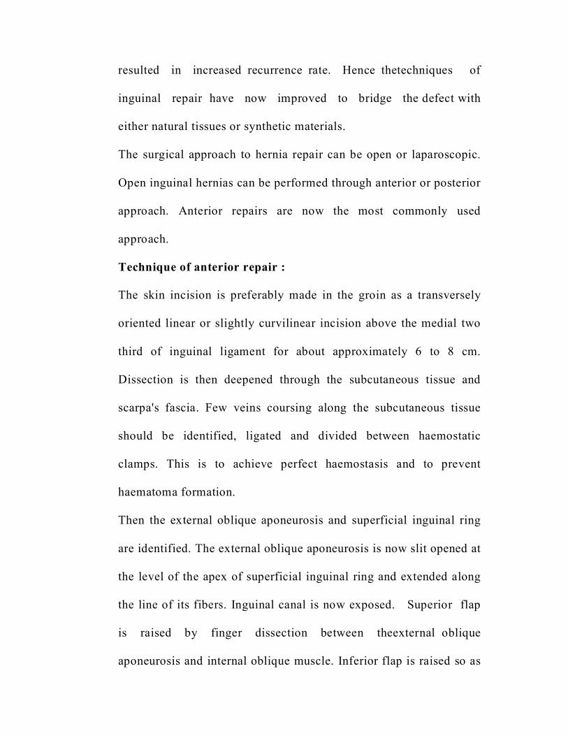
resulted in increased recurrence rate. Hence thetechniques of
inguinal repair have now improved to bridge the defect with
either natural tissues or synthetic materials.
The surgical approach to hernia repair can be open or laparoscopic.
Open inguinal hernias can be performed through anterior or posterior
approach. Anterior repairs are now the most commonly used
approach.
Technique of anterior repair :
The skin incision is preferably made in the groin as a transversely
oriented linear or slightly curvilinear incision above the medial two
third of inguinal ligament for about approximately 6 to 8 cm.
Dissection is then deepened through the subcutaneous tissue and
scarpa's fascia. Few veins coursing along the subcutaneous tissue
should be identified, ligated and divided between haemostatic
clamps. This is to achieve perfect haemostasis and to prevent
haematoma formation.
Then the external oblique aponeurosis and superficial inguinal ring
are identified. The external oblique aponeurosis is now slit opened at
the level of the apex of superficial inguinal ring and extended along
the line of its fibers. Inguinal canal is now exposed. Superior flap
is raised by finger dissection between theexternal oblique
aponeurosis and internal oblique muscle. Inferior flap is raised so as

to reveal the shelving edge of the inguinal Ligament.
Mobilization of the spermatic cord:
The spermatic cord is mobilized at the level of pubic tubercle.
This can be done both by blunt and sharp dissection. If the spermatic
cord is mobilized too laterally, this will result in loss of tissue planes
and the identification of essential structures will be difficult
resulting in injury to spermatic cord structures. This also disrupts the
floor of inguinal canal.
Now, the ilioinguinal nerve, iliohypogastric nerve, genital
branch of the genitofemoral nerve are identified and preserved.
The cremasteric muscle overlying the spermatic cord is dissected
parallel to the line of fibers and the spermatic cord is skeletonised.
The cremasteric artery and vein can be encountered now at the level
of internal ring. Damage to this structure could result in significant
amount of bleeding. Hence these vessels should be identified and
avoided or cauterizedor ligated and divided.
The skeletonizationof the cord is very much
important, because This allows good exposure and visualization
of the deepinguinal ring, the transversalis fascia, the arched fibers
of the internal oblique and transversusabdominis, and the conjoined
tendon.

Identification of the sac :
At this level, we can able to differentiate and identify indirect
hernial sac from direct hernial sac. A direct hernia can be seen
posterior to the cord as it protrudes through the weakness in the floor
of the inguinal canal. An indirect inguinal hernial sac is located deep
to the cremastermucle. After dividing the muscle longitudinally, the
sac can be seen in an anteromedial position to the cord, extending
superiorly through the internal ring.
A pantaloon hernia will present with both the direct and
indirect component in the same inguinal canal.
High ligation of the sac:
In case of indirect hernial sac, the sac is separated carefully
from the cord structures up to the level of internal ring. The sac is
opened and inspected for any visceral contents within. The sac is
now transfixed at the level of the neck at the internal ring. This
reduction of the hernia sac in to the preperitoneal space is called
high ligation of the sac. The excess sac is then excised. In caseof
larger sac, the sac is opened just close to the internal ring. The
proximal one ligated and the distal sac left undisturbed.
In case of direct hernia, as there is no true sac
reduction can be achieved during posterior wall repair.

Anterior tissue repairs :
Before the advent of prosthetic mesh repair, tissue repair was
advocated. Even now, tissue repair has an important role in the
treatment of inguinal hernia, in places where there is
contraindication for use of prosthetic mesh.
The Indications for tissue repair are :
In case of contaminated operative field ie.,
strangulatedhernia.
In concern of possible azoospermia, due to long term effect
of mesh on vas.
The various tissue repairs are :
• Bassini repair
• Shouldice repair
• Mcvay repair.
Bassinirepair14 :
This is a triple layer repair, done to restore the integrity of the
pelvic floor. This is the basic approach and once popular technique for
non anatomic repair.
Here the transverses aponeurosis, internal oblique
musculoaponeurotic arch, transversalis fascia is sutured to the inguinal
ligament, with repair of the medial end of the internal inguinal ring. This

repair has a high incidence of neurovascular injury and higher recurrence
rate.
Shouldicerepair14 :
It is a multilayered repair done under local anaesthesia. Repair of
the posterior wall ie.,transversalis fascia and tightening of the internal
ring is the basis of this tissue repair.
Here, the transversalis fascia is split opened starting from internal
ring to pubic tubercle and then the four layered repair is started. The
shouldice hospital utilizes 32 or 34 gauge stainless steel wire as a
continuous suture, but now modern surgeons are using synthetic suture
materials.
The first layer repair consists of suturing the free edge of the lower
transversalis to the posterior surface of upper transversalisfascia and
lateral part of posterior rectus sheath, in a continuous imbricated fashion,
starting medially at pubic tubercle to the internal ring, thus tightening the
transversalis fascia at internal ring.
The second layer is continued by suturing the upper transversalis
flap to the base of lower edge of inguinal ligament as a running suture
and this ends at the pubic tubercle with the first layer.

The third layer continuous suture is started at the internal ring by
suturing conjoined tendon medially with inguinal ligament laterally till
the pubic tubercle.
The fourth layer includes suturing the anterior rectus sheath
medially with the posterior aspect of external oblique aponeurosis
laterally.
The cord is now rested on the new inguinal floor.
This procedure has an excellent outcome in respect
torecurrence rates.
The recurrence was found to be <1%. Here the tension is
distributed throughout the inguinal canal due to multiple, continuous
overlapping suture lines.However, the shouldice repair dissection is
complicated and doing this Procedure requires excellent anatomical
knowledge and excellent surgical Hands. Thus the low level of recurrence
rate in shouldice repair is attributed to the experience of the surgeon.
Mcvay repair (cooper's ligament repair )14 :
This technique repairs both inguinal and femoral hernial
defects. He also added the concept of the relaxing incision as a
tension reducing maneuver.

Initially , after lateralization of the cord, the
transversalisfascia is opened through a transverse incision. Now the
preperitonealspace can be entered. The posterior wall of
transversalis fascia is cleared off. Similarly, the cooper's ligament
is identified and freed of its attachments.
Initially, 2-4 cm vertical incision is made at the lateral
border of anterior Rectus sheath, starting at pubic tubercle and
extending superiorly. This helps in relaxing, significant amount of
tension following tissue repair.
Now, the cooper's ligament is sutured to the upper flap of
the transversalis fascia, starting from the pubic tubercle towards the
femoral sheath. Now, the cooper's l igament is sutured to the
femoral sheath. This closes the femoral canal. The suturing is now
continued further between the transversalis fascia and
iliopubictrac t, latera lly until the deep inguinal ring is reached
thus, narrowing of the internal ring is achieved.
This procedure has an excellent long term outcome but the disadvantages
includes, longer operative time, more extensive dissection, injury to
femoral vessels, thromboembolism of femoral vessels.

Darn repairs (Abrahamson nylon darn repair) :
The principle of the nylon darn operation is to reinforce the
weakened or torn posterior wall of the inguinal canal with the muscles of
the musculoaponeurotic arch, as well as a simple lattice work of
monofilament nylon suture under no tension, on which is laid a buttress
of fibrous tissue.
The nylon darn solves the problem of early recurrence, since the
nylon lattice will hold the area intact during the first year, until the natural
connective tissue collagen scar matures to its full strength. The nylon
being indestructible will maintain the integrity of the repair for many
years.
Pure prosthetic anterior mesh repair :
Cooper in 1800, itself suspected that the abdominal muscle
weakness, is the cause of inguinal hernia. This concept was once again
emphasized by Harrison in 1922. He found out that significant number of
patients, at the age group of 50- 60 years, developed hernia.
Billroth, then recognized that this weakened abdominal wall tissue
should be reinforced with some prosthetic material. Although, initially,
tantalum metal sheets, silver-wire filigree sheets, fascia lata,sheets of skin
are used. These prosthesis failed due to metals being fragmented,

biological tissues being rejected and suffered infection. Then he realized
that, this artificially produced Material should have the density and
toughness of fascia and tendon.
Usher, in 1959 introduced the mesh technique bringing billroth's
dream to come true. Marlex mesh was used in repair. The posterior wall
was opened and marlex mesh was sutured to the undersurface of the
medial margin of the defect and to the shelving edge of the inguinal
ligament. This is proposed as "tension eliminating repair"21.
MESH :
The term mesh refers to prosthetic material, a net or a flat sheet,
used to strengthen a hernia repair. Mesh can be used to
• Bridge a defect - mesh is simply fixed over the defect.
• Plug a defect - mesh is pushed in to the defect.
• Augment a repair - defect is closed with sutures and then the mesh is
added for reinforcement.
Mesh types :
1. Biological mesh 22 :
These are derived from donor tissues, such as human dermis, porcine
dermis, porcine small intestine submucosa, bovine dermis,bovine

pericardium. Collagen rich tissues are harvested and treated to remove
cellular elements. The collagen then scaffolds and with additional
extracellular ground materials are strong enough to provide enough
mechanical support for an intact hernia repair.
Used in cases of infections, such as strangulated inguinal hernia,
where synthetic mesh is relatively or absolutely contraindicated.
Biological materials, supports angiogenesis into collagen matrix, so
delivery of white blood cells and antibiotics is possible. The collagen
scaffolding supports native fibroblast ingrowth and collagen deposition.
Depending on the processing of the graft, the scaffold may be completely
degraded, eventually allowing for bacteria removal.
2. Synthetic mesh8'14:
Polypropylene mesh
• Knitted construction of polypropylene mesh fibers .
• Stable, Hydrophobic, electrostatically neutral and non absorbable.
Polyester mesh:
• Promotes inflammatory reaction similar to polypropylene.
Polytetrafluoroethylene mesh (PTFE and e-PTFE):

• This mesh does not promotes ingrowth in to viscera andtherefore
makes it useful in IPOM or TAPP repairs
• These are smooth, soft and strong.
• Polyamide mesh
Properties of an ideal mesh22 :
• Chemically inert, non carcinogenic.
• Not physically modified by tissue fluids.
• Not excite inflammatory or foreign body reaction.
• Easy to handle &sufficiently pliable so as not to cause
stiffness or be felt by the patient.
• Should have optimum thickness, since its density determines
tissue reaction. Resists contraction.
• Capable of resisting mechanical strain & provides adequate
strength.
• Be permeable and allow tissue ingrowth with in it, so as to prevent
dislocation or migration.
• Stimulate fibroblastic activity to allow incorporation in to
tissue, rather than sequestration or encapsulation.

Should preferably be macroporous, monofilament, transparent
and should resist infection.
Lichtenstein's tension free repair :
This was the first inguinal hernia repair, done with prosthesis.
This is a tension free repair and in 1984, he coined the
term "tension - free hernioplasty"
In Lichtenstein tension free hernioplasty, the posterior wall was
Reinforced by insertion of a sheet of mesh, rather than suturing anatomic
Structures that are not in apposition.
According to the 2009 European hernia guidelines, lichtenstein's
Technique introduced in 1984 is currently the best evaluated and most
Popular technique among different open mesh repair.
Technique of the procedure:
Here, the skin and subcutaneous tissues are incised. The external
Oblique aponeurosis is split opened to expose the inguinal canal. The
superior and inferior flap raised, thus the external oblique aponeurosis is
separated from the internal oblique muscle, exposing internal oblique
aponeurosis.

The cord is mobilized and lateralized. When lifting the cord, care is
taken to include the ilioinguinal nerve. Care is taken to avoid injury to
ilioinguinal nerve, the blue external spermatic vein, genital nerve with the
cord.
Now the cremasteric sheath is incised to expose the indirect sac. Removal
of cremasteric muscle leads to hanging of testis and also dysfunction of
Cremaster muscle, leading to dysejaculation. This also, exposes the
genital nerve, vas deferens, paravasal nerves to the mesh.
This mesh irritation causes chronic inguinodynia and testicular
pain. An indirect sac is dissected free up to the neck of sac and
invaginated into the properitoneal surface. Ligation of the peritoneum
causes severe postoperative pain as it is highly innervated and the
mechanical pressure causing ischemic necrosis.
The direct sac isinvaginatedby an absorbable imprecating suture.
This allows the positioning of the mesh in a flat surface.
A sheet of monofilament macro porous polypropylene mesh of size 8 x
16 cm is placed over the posterior wall. This should cover 2 cm medial to
the pubic tubercle, 3-4 cm above the hesselbach's triangle, 5-6 cm lateral
to inguinal ring. The mesh is secured with a nonabsorbable monofilament
suture at the insertion of rectus sheath to the pubic bone, overlapping

the bone by 1-2 cm. Thissuture is continued to attach the lower edge of
the patch to the inguinal ligament. Medially the upper edge of the patch is
sutured to the rectus sheath and internal oblique aponeurosis. Few
interrupted sutures are placed superiorly, between the mesh and internal
oblique aponeurosis. Fixing mesh to the internal oblique muscle is
avoided to prevent injuring the intramuscular segment of iliohypogastric
nerve.
With a slit in the mesh to accommodate the spermatic cord, the
upper and lower edge of the patch is fixed to the inguinal ligament, lateral
to the cord. The excess mesh on the lateral side is tucked below the
external oblique aponeurosis, leaving atleast 5 cm beyond internal ring.
External oblique aponeurosis is closed, constructing new
superficial inguinal ring for the exit of spermatic cord. Wound is then
closed in layers.
Plug and patch technique :
A modification of the Lichtenstein repair, known as the plug and patch
technique was developed by Gilbert and later popularized by Rutkow and
Robbins23. This technique involves placement of a plug, shaped in to a
flower or umbrella configuration, with the apex pointed intraabdominally.

Complications of inguinal hernia surgery :
1. Recurrence.
2. Chronic groin pain
- Nociceptive
Somatic
Visceral
- Neuropathic
Iliohypogastric
Ilioinguinal
Genitofemoral
Lateral cutaneous
Femoral
3. Cord and testis injury.
4. Wound infection.
5. Seroma, haematoma.
6. Osteitis pubis.
7. Prosthetic problems
- Contraction
- Infection
- Erosion
- Rejection

8. Laparoscopic problems
- Vascular injury
- Visceral injury
Bowel perforation
Bladder perforation
postherniorrhaphy pain syndrome or inguinodynia :
It is persistent pain in the groin occurring after
herniasurgery. This may occur in the immediate postoperative period or
months or years after hernia surgery that impairs the quality of life of the
patient. Diagnosis, etiology, treatment are challenging for the surgeon
also.
Potential causes of inguinodynia :
Surgical technique :
1. Mesh adherence
2. Nerve entrapment
3. Osteitis pubis
4. Compromise of spermatic cord
5. Inappropriate tack placement laparoscopically or suture

placement with open technique.
6. Neuropathy secondary to exaggerated scarification process.
7. Idiosyncratic response to mesh implantation
Inguinodynia of neuropathy :
Mesh inguinodynia : This term was coined by Heise and
Starling in 1998.
The pain presents as a sharp localizing pain, numbness or as
paresthesia in the cutaneous distribution of nerve. It is difficult toidentify
the exact nerve which causes the symptoms, due to overlapping
distribution of the nerves. Nerve entrapment leads to sharp localizing
pain, where as nerve division causes numbness over the distribution of
the nerve. Numbness can be better tolerated by the patient rather than
sharp localizing pains.
Initially the cause of inguinodynia was considered to be mesh. But,
subsequent researches have showed that the pain was due to accidental
entrapment of the sensory nerve with the sutures used to fix the mesh.
Hence, precise recognition of the sensory nerves that we commonly used
to come across during hernia surgery such as ilioinguinal nerve,

iliohypogastric nerve, genital branch of the genitofemoral nerve is
necessary.
Classification of pain :
• Visceral pain
• Somatic pain
• Neuropathic pain
Visceral pain :
This is due to injury to the sympathetic nerve plexus. This pain is
usually experienced during some visceral function, such as ejaculation.
Somatic pain :
This is due to damage to ligaments and muscles during
surgery. This type of pain is most commonly encountered. This type of
pain can be reproduced during exertion or movement of the abdominal
wall. Somatic pain can be treated with rest, NSAIDS and reassurance.
Neuropathic pain :
This is due to nerve damage or nerve entrapment. This is a sharp
localized pain which can be sensed as a burning or tearing one along the
area of nerve distribution which may occur in the immediate
postoperative period.

Nerves injured :
Ilioinguinal nerve - risk of injury is more during closure of external
oblique aponeurosis.
Ilioinguinal nerve, iliohypogastric nerveboth are atincreased risk
during fixation of mesh in anterior tension freerepair.
Genital and femoral branches of the genitofemoral nerveLateral
cutaneous nerve of thigh - in laparoscopic herniarepair, when
mesh is fixed below the iliopubic tract, withlateral fixation tacks.
Treatment of neuropathic pain :
Mild pain - can be treated with NSAIDs
Moderate pain - can be treated with nerve directedinjections of
steroid or anaesthetics,
Severe pain - can be treated with neurectomies.
- this produces relief in about 72% of patients.
Cord and testis injury :
Spermatic cord is a well vascularised structure. During mobilization,
dissection and lateralization of the cord structures, there is a risk of injury
to, pampiniform plexus of veins, testicular artery and vas deferens.

Scrotal haematoma :
During handling of the spermatic cord, there is a risk for vessel injury and
localized haematoma formation or significant scrotal haematoma. Scrotal
haematoma presents as a bluish black discolouration of the scrotal wall.
This condition is self limiting and can be treated with reassurance, warm
and cold compresses.
Ischemic orchitis and testicular atrophy : This occurs due to,
1. Thrombosis of pampiniform plexus of veins.
2. Injury to the testicular artery.
The incidence of ischemic orchitis increases with extensive
dissection of distal part of a huge sac and in patients undergoing anterior
repair for recurrent hernia.
More extensive handling of the cord structures, leads to thrombosis of the
small veins of pampiniform plexus. This is followed by, stasis of venous
blood within the testis, leading to, painful swelling of the testis.
Unresolved, this condition leads to testicular atrophy in about 6-12
weeks.
Occasional injury to testicular artery or accidental ligation of testicular
artery also leads to testicular atrophy. Treatment of ischemic orchitis :

Anti inflammatory agents
Analgesics
Orchiectomy is rarely necessary.
Injury to vas deferens :
This may occur due to accidental instrumentation and crushing during
open anterior repair or laparoscopic surgery, leading to infertility.
Chronic scarring leads to obstruction of the lumen of the vas deferens,
and there by dysejaculation and reduced fertility rates.
Wound infection :
As this is a clean surgically created wound, the risk of
surgical Site infection is reported to be 1% - 2% after open
inguinal hernia repair.
This incidence is less with laparoscopic hernia repair. Many studies have
Shown that, there is no need for prophylactic antibiotics.
• Superficial surgical site infection :
Treated by local wound care, removing the sutures
andhealing by secondary intention.
• Deep surgical site infection :

This presents as a chronic discharging sinus that tracks to the mesh.
Treatment involves exploration of the wound and if necessary, mesh to be
removed.
Seroma :
A seroma is a localized collection of fluid. This develops in
the first week of post operative period.
Causes :
1. More extensive dissection in repair of larger hernias.
2. Following use of prosthetic mesh repairs - as the body tries to
adapt with the foreign body through a normal reaction.
Treatment :
o Observation.
o Warm compression.
o Aspiration may lead to infection.
Osteitispubis :
This occurs when suture or surgical tacker is placed too deeply in to the
periosteum of pubis, in the pubic tubercle. Patient complains of pulling,
aching or throbbing pain in pubic area. Symptoms are exaggerated by

twisting or squatting. Diagnosis is made by direct manual pressure on
the ospubis
Treatment:
NSAIDs
Local injection of corticosteroids.
Urinary retention :
This occurs due to post operative pain and use of narcotic analgesics. In
case of spinal anaesthesia, the incidence of urinary retention was found to
be about 13% , whereas in case of local anaesthesia, the incidence was
reported to be just 0.2%.
There is no need for prophylactic catheterization. Treatment :
To limit IV hydration.
Prophylactic tamsulosin.
Short term catheterization.
Mesh plug :
This is the exaggerated fibrosis or scarring to the presence of the mesh.
This is a rare phenomenon where the mesh forms aconcrete like
mass exacerbating the scarification reaction. The mesh shrinks
excessively, scars and retracts away from the host tissue.

In case of polypropylene mesh, the mesh shrinkage accounts to be about
40%. Mesh shrinkage also leads to recurrence of the hernia if adequate
size of mesh is not used ie., leaving excess mesh 2 cm medial to pubic
tubercle, 4 cm above hesselbach's triangle, 5 cm lateral to internal ring.
Hernia recurrence :
This occurs mostly due to failure in technical aspect. Hernia recurrence is
more common in patients with direct hernia. Medial recurrence is more
common. Recurrence usually occurs through The floor of inguinal canal,
near the pubic tubercle. Because near the Pubic tubercle, the suture line
tension is greatest.
Causes :
Excessive tension on the repair
Failure to include musculoaponeurotic margin in repair
Improper mesh size and placement or mesh displacement
Failure to close the patulous internal ring
Chronically elevated intraabdominal pressure
Deep surgical site infection
Poor collagen formation in the wound

Treatment :
Prosthetic mesh repair through different approach.
For recurrence after prosthetic mesh repair - second mesh repair
through different approach.

MATERIALS AND METHODS
This study is based on the analysis of 50 patients presenting with
inguinal Hernia in Coimbatore medical college hospital, during time
period of September July 2016 to2017. This is a prospective,
randomized double blind study.
Fifty patients presenting with unilateral inguinal hernia in the out
patient department was selected. Patients details were collectedper
preformed proforma. Detailed clinical history was taken
particularly regarding the swelling, preoperative pain, any history
suggestive of complications and predisposing factors for hernia
formation.
The diagnosis of inguinal hernia was made by a thorough clinical
examination. The type of hernia, presence of reducibility and cough
impulse were noted. Examination of cardiovascular system, respiratory
system, abdominal mass, stricture urethra, per rectal examination for BPH
was done.
Pre operative assessment was done with basic investigations, such
as complete blood count, blood urea, blood sugar, serum creatinine, ECG,
x-ray chest, VCTC. Ultrasound abdomen was also done to rule out intra
abdominal pathology.
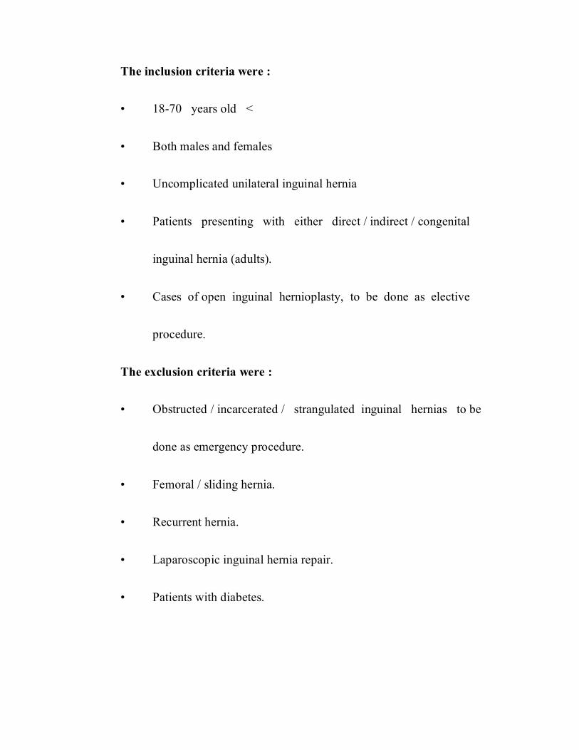
The inclusion criteria were :
• 18-70 years old <
• Both males and females
• Uncomplicated unilateral inguinal hernia
• Patients presenting with either direct / indirect / congenital
inguinal hernia (adults).
• Cases of open inguinal hernioplasty, to be done as elective
procedure.
The exclusion criteria were :
• Obstructed / incarcerated / strangulated inguinal hernias to be
done as emergency procedure.
• Femoral / sliding hernia.
• Recurrent hernia.
• Laparoscopic inguinal hernia repair.
• Patients with diabetes.

Operative procedure :
A single dose of preoperative antibiotic was given and this is
followed for about three days postoperatively. The type of anaesthesia
was spinal anaesthesia.
The procedure was carried out by Lichtenstein technique. The
patient was randomly selected for polypropylene mesh and
parietexprogrip mesh. Both the patient and the observer were double
blinded during the study.
The progrip mesh is made up of light weight macroporous
monofilament polyester material with reabsorbablepolylactic acid
microhooks. These hooks have selfgripping properties to fix in to the
tissues. After complete tissue ingrowth and complete reabsorption of the
polylactic acid hooks (in 15 months ), the low weight (40 gm/m )
polyester mesh, provides the posterior wall reinforcement.
The prolene mesh is made up of knitted construction of
polypropylene fibers. It is low weight (< 45 gm/m ), macroporous (2.4
mm pore size), monofilament material.
In the sutured mesh group, the prolenelight weight mesh was
trimmed to appropriate shape and placed over the posterior wall. The
spermatic cord was encircled through a slit made in the mesh. The mesh

was fixed to the pubic tubercle, with 2-0 prolene suture material. This
suture was continued as a continuous one between the lower edge of the
mesh and inguinal ligament up to a point lateral to the deep inguinal ring.
Medially, the mesh was fixed to the anterior rectus sheath and
internal oblique aponeurosis. Lateral to the spermatic cord the split in the
mesh was sutured and the two tails of the mesh was placed under external
oblique aponeurosis up to at least 5 cm lateral to the deep inguinal ring.


MESH FIXED TO PUBIC TUBERCLE
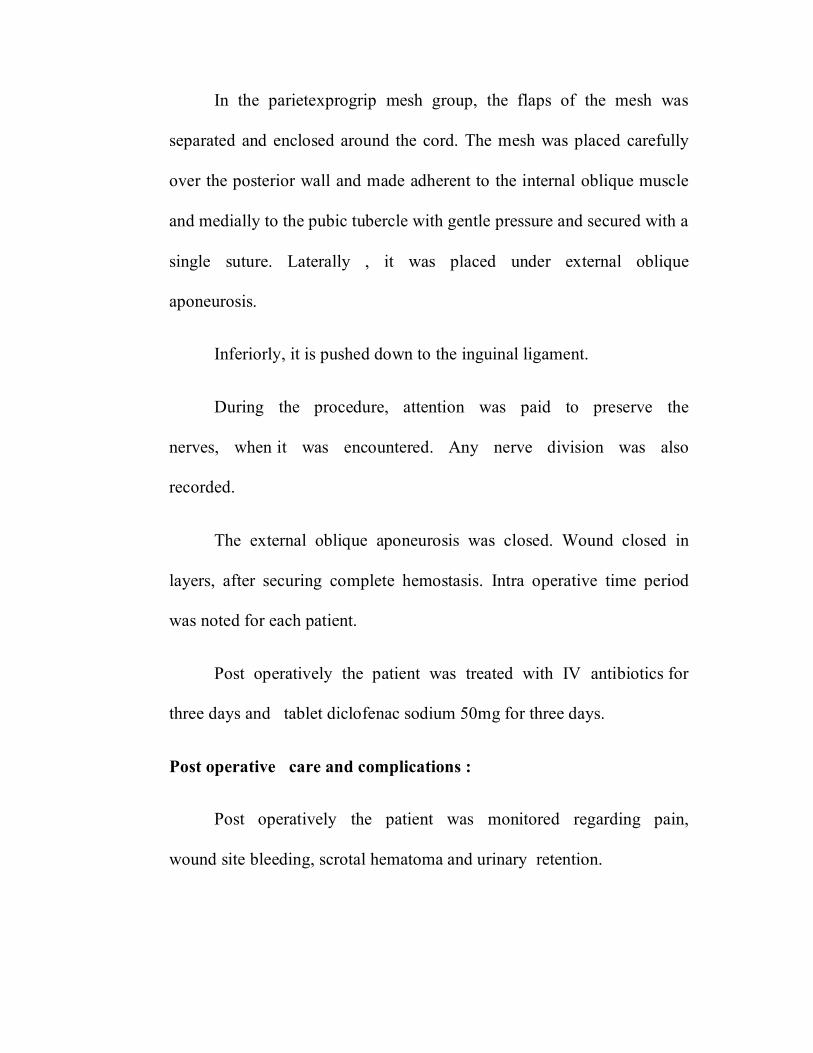
In the parietexprogrip mesh group, the flaps of the mesh was
separated and enclosed around the cord. The mesh was placed carefully
over the posterior wall and made adherent to the internal oblique muscle
and medially to the pubic tubercle with gentle pressure and secured with a
single suture. Laterally , it was placed under external oblique
aponeurosis.
Inferiorly, it is pushed down to the inguinal ligament.
During the procedure, attention was paid to preserve the
nerves, when it was encountered. Any nerve division was also
recorded.
The external oblique aponeurosis was closed. Wound closed in
layers, after securing complete hemostasis. Intra operative time period
was noted for each patient.
Post operatively the patient was treated with IV antibiotics for
three days and tablet diclofenac sodium 50mg for three days.
Post operative care and complications :
Post operatively the patient was monitored regarding pain,
wound site bleeding, scrotal hematoma and urinary retention.

Wound complications , such as wound infection, seroma (>10 ml),
hematoma are taken care. A note of this was also made.
- Urinary retention was treated with catheterization.
- In patients with wound infection the hospital stay
was lengthened, requiring extra doses of antibiotics.
- Seroma with greater tension at wound site was relieved.
- Patients with severe pain, requiring extra dose of
analgesics was noted.
The primary endpoint was to measure the amount of postoperative
pain by, visual analog scale. The secondary end points includes duration
of surgery, the amount of wound infection, seroma, duration of hospital
stay, return to daily activities and recurrence.
The patients were discharged as per their postoperative outcome
and their duration of stay in the hospital was noted.
Follow up :
The patient was advised to follow their, preoperative
lifestyle, except for lifting heavy weight, to avoid cough and
straining at micturition and defecation.

The patient was followed up at first week, 1 month and 3 months
postoperative period to evaluate about the chronic inguinal pain, wound
infection, any disturbance with day today activities, use of analgesics and
recurrence.
PAIN : It was a unpleasant sensory and emotional
thing experienced due to actual or potential tissue damage.
Pain assessment:
It is a subjective experience, both psychological and
physiological, It can be assessed by,
Quantitative pain scale :
Numerical pain rating scale
Wong - baker faces pain rating scale
Visual analog scale
Qualitative pain scale :
Mcgill questionnaire (Ronald melzack in 1975 )
Verbal rating scale :
Here words are used to describe pain.
0 - no pain

1 - mild pain
2 - moderate pain
3 - severe pain
Numerical rating scale :
Here a scale with a range of 0 - 10 was used and a number is
chosen between them.
0 - no pain
10 - worst pain
Wong baker's faces pain rating scale : particularly useful in
children, where, verbal communication is poor.
Face 0 is a happy face (no hurt)
Face 1 is still smiling (hurts a little bit)
Face 2 is not smiling or frowning (hurts a little more)
Face 3 is starting to frown (hurts even more)
Face 4 is definitely frowning (hurts a whole lot)
Face 5 is crying (hurts the worst)
Visual analog scale :

VAS or visual analogue scales use a vertical or horizontal line with
words that convey "no pain" at one end and "worst pain" at the opposite
end. patients are asked to place a mark along the line that indicates your
level of pain.
Mayo clinic chronic pain lists their pain as :
0 - 1 : no pain
1 - 3 : mild pain
4 - 5 : discomforting - moderate pain
6 - 7 : distressing - severe pain
8 - 9 : intense - very severe pain
10 : unbearable pain.

RESULTS AND OBSERVATIONS
TABLE : 1 AGE DISTRIBUTION
Group Statistics
Mesh type N Mean
Std.
Deviation
Std.
Error
Mean Age SM 25 46 8.792 1.758
SGM 25 40 8.421 1.684
In this study, the incidence of hernia was more common in the age
group of 31-60 years.

TABLE 2 : SEX INCIDENCE
SEX No of patients
Male 48
Female 2
In this study, similar to other studies, male gender accounts for 96
% and female gender 4%.
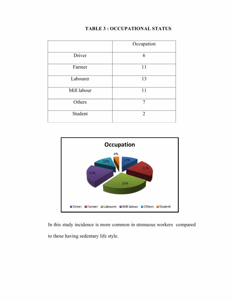
TABLE 3 : OCCUPATIONAL STATUS
Occupation
Driver 6
Farmer 11
Labourer 13
Mill labour 11
Others 7
Student 2
In this study incidence is more common in strenuous workers compared
to those having sedentary life style.

TABLE 4 : PERSONAL HISTORY
Crosstab
Mesh type
Total SM SGM
Smoking NO Count 2 6 8
% within Mesh type 8.0% 24.0% 16.0%
YES Count 23 19 42
% within Mesh type 92.0% 76.0% 84.0%
Total Count 25 25 50
% within Mesh type 100.0% 100.0% 100.0%
In terms of personal life, about 84% of population was found to be
smoking. Thus hernia is more common among smokers,.

TABLE 5: SYMPTOMS
SM SGM
Swelling 88.0% 80.0%
Swelling &
Pain
12.0% 20.0%
Crosstab
Mesh type
Total SM SGM
Symptoms Swelling Count 22 20 42
% within Mesh type 88.0% 80.0% 84.0%
Swelling
& Pain
Count 3 5 8
% within Mesh type 12.0% 20.0% 16.0%
Total Count 25 25 50
% within Mesh type 100.0% 100.0% 100.0%
In this study , most of the patient presents with swelling alone
compared to swelling and pain.

TABLE 6: NATURE OF HERNIA
Diagnosis IH
Left 19
Right 30
R Cong 1
Crosstab
Mesh type
Total SM SGM
Diagnosis IH L Count 11 8 19
% within
Mesh type
44.0% 32.0% 38.0%
R Count 14 16 30
% within
Mesh type
56.0% 64.0% 60.0%
R
CONG
Count 0 1 1
% within
Mesh type
0.0% 4.0% 2.0%
Total Count 25 25 50
% within
Mesh type
100.0% 100.0% 100.0%

Chi-Square Tests
Value df
Asymp. Sig. (2-
sided)
Pearson Chi-
Square
1.607a 2 .448
Likelihood Ratio 1.995 2 .369
N of Valid Cases 50
a. 2 cells (33.3%) have expected count less than 5. The
minimum expected count is .50.

TABLE 7 : TYPE OF REPAIR
Type of repair Number of patients Percentage
Sutured mesh 25 50%
Self gripping mesh 25 50%
Total 50 100%
TABLE 8 : DURATION OF SURGERY
Group Statistics
Mesh type N Mean
time
Std.
Deviation
Std.
Error
Mean
Duration of
Surgery
SM 25 33 2.430 .486
SGM 25 26 .988 .198

Independent Samples Test
Levene's Test for
Equality of
Variances
t-test for Equality of Means
F Sig. t df
Sig.
(2-
tailed)
Mean
Difference
Std. Error
Difference
95%
Confidence
Interval of the
Difference
Lower Upper
Duration of Surgery
Equal variances assumed
3.173 .081 12.044 48 .000 6.320 .525 5.265 7.375
Equal variances not assumed
12.044 31.726 .000 6.320 .525 5.251 7.389

The intraoperative time period was less in self gripping mesh group
with a difference of about 7 Minutes
TABLE 9 : IMMEDIATE POST OPERATIVE PAIN
SM SGM
Mild 28.0% 36.0%
Moderate 44.0% 28.0%
Severe 28.0% 8.0%
No pain 0.0% 28.0%
33
26
0
5
10
15
20
25
30
35
SM SGM
Duration of Surgery / mins

Crosstab
Mesh type
Total SM SGM
Pain
IPO
Mild Count 7 9 16
% within Mesh type 28.0% 36.0% 32.0%
Moderate Count 11 7 18
% within Mesh type 44.0% 28.0% 36.0%
Severe Count 7 2 9
% within Mesh type 28.0% 8.0% 18.0%
No pain Count 0 7 7
% within Mesh type 0.0% 28.0% 14.0%
Total Count 25 25 50
% within Mesh type 100.0% 100.0% 100.0%
0%
20%
40%
60%
80%
100%
SM SGM
28.0% 36.0%
44.0% 28.0%
28.0%
8.0%
0.0%28.0%
Pain IPO
Mild Moderate Severe No pain

In this study the incidence of acute pain was less in self gripping mesh
group. About 28.0% of patients in self gripping mesh group had no pain
in post operative period
TABLE – 10 : POSTOPERATIVE COMPLICATIONS
Crosstab
Mesh type
Total SM SGM
Wound
Complications
Seroma Count 2 0 2
% within Mesh type 8.0% 0.0% 4.0%
Hematoma Count 1 1 2
% within Mesh type 4.0% 4.0% 4.0%
Infection Count 1 1 2
% within Mesh type 4.0% 4.0% 4.0%
Nil Count 21 23 44
SM SGM
Seroma 8.0% 4.0%
Hematoma 4.0%
Infection 4.0% 4.0%
Nil 84.0% 92.0%

% within Mesh type 84.0% 92.0% 88.0%
Total Count 25 25 50
% within Mesh type 100.0% 100.0% 100.0%
In this study, the incidence of postop complications was less
among self gripping mesh group compared to sutured mesh group.
Seroma Hematoma Infection Nil
8.00% 4.00% 4.00%
84.00%
4.00% 4.00%
92.00%
SM SGM

TABLE 11 : PAIN AT 1 MONTH INTERVAL
SM SGM
Mild 28.0% 12.0%
Moderate 12.0% 4.0%
Severe 4.0%
No pain 56.0% 84.0%
Crosstab
Mesh type
Total SM SGM
Pain 1
Month
Mild Count 7 3 10
% within Mesh type 28.0% 12.0% 20.0%
Moderate Count 3 1 4
% within Mesh type 12.0% 4.0% 8.0%
Severe Count 1 0 1
% within Mesh type 4.0% 0.0% 2.0%
No pain Count 14 21 35
% within Mesh type 56.0% 84.0% 70.0%
Total Count 25 25 50
% within Mesh type 100.0% 100.0% 100.0%

In this study, pain at 1 month interval was released to 16% in self
gripping mesh group compared to 44% in sutured mesh group.
TABLE 12: PAIN AT 3 MONTHS INTERVAL
SM SGM
Mild 24.0% 8.0%
Moderate 12.0%
No pain 64.0% 92.0%
Crosstab
Mesh type
Total SM SGM
Pain 3 Mild Count 6 2 8
0%
20%
40%
60%
80%
100%
SM SGM
28.0%12.0%
12.0%
4.0%
4.0%
56.0%84.0%
Pain after 1 month
Mild Moderate Severe No pain

Months % within Mesh type 24.0% 8.0% 16.0%
Moderate Count 3 0 3
% within Mesh type 12.0% 0.0% 6.0%
No pain Count 16 23 39
% within Mesh type 64.0% 92.0% 78.0%
Total Count 25 25 50
% within Mesh type 100.0% 100.0% 100.0%
In this study, the incidence of chronic pain was 8 % in self gripping
mesh group compared to 36 % in sutured mesh group.
0%
20%
40%
60%
80%
100%
SM SGM
24.0%8.0%
12.0%
64.0%92.0%
Pain after 3 months
Mild Moderate No pain

DISCUSSION
In the initial times, tissue repair was employed in the
treatmentof hernias. But this method has been withheld mostly due to
recurrence. Then comes the use of mesh in inguinal hernia repair. For
more than thirty years, there has been widespread use of mesh in inguinal
hernia repair.
In the present, Lichtenstein's tension free hernioplasty is the most
commonly practiced technique. This method utilizes light weight,
synthetic, partially absorbable polypropylene mesh. Fixation of mesh is
done with nonabsorbable suture material and here comes the problem of
post hernioplasty pain syndrome.
Cause of pain after hernia repair is multifactorial. Early post
operative pain is due to tissue damage from preparation of anterior space
and handling of hernial sac. Fixation of mesh to the pubic bone induces
periostitis. The mesh also induces certain inflammatory response and
fixation of mesh to the posterior wall, causes nerve entrapment and
neuropathic pain. Tension due to mesh shrinkage and mesh adherence
contributes to late postoperative pain.

Currently, best outcome in hernia repair is mainly focused towards
the achievement of low incidence of mesh complications such as chronic
inguinodynia.
In this study of 60 patients, half of the study population received
polypropylene mesh and in another half of the study population, self
gripping progrip mesh was used.
Patients were assessed regarding age presentation, life style,
clinical presentation, duration of surgery, postoperative complications
such as postoperative pain immediately after surgery, at one month and
three months interval, seroma, hematoma and wound infection.
The following are the observations :
In our study, the incidence of hernia was included in the age group
of 31-60 years. But according to various studies the incidence of hernia is
found to be more in older age group ie., between age group of 55-65
years.
In regards to the occupation, hernia is more common in labourers
and farmers, ie., in sternous workers accounting to about 75%, when
compared to others like drivers and teachers.

In analyzing the personal life of the study population, about 75% of
the population was found to be smoking.
This early presentation of hernia in middle age group may be due
to increased prevalence of smoking and the heavy working pattern of
living population in this area.
Right sided inguinal hernia is more common due to the late descent
of the right testis and delayed closure of the processusvaginalis.
Similarly, in our study population right side inguinal hernia accounts to
about 60% of all inguinal hernias.
Risk of inguinal hernia is nine times more common in men.
Indirect inguinal hernia is more common in women. In a study by Martin
kurzer of british hernia system, 97% were males and 3% were females.
Similarly in our study population, male gender accounts for about 96 %
of patients and female gender accounts for about 4% of patients.
In a study by Mike S.L.Liem and others about 93% of patients
presents with swelling and about 83% of patients had groin discomfort or
pain. In this study population, 83.3% of patients presents with swelling in
the groin and 16.6% of patients presents with both groin swelling and
pain.

This low level of pain incidence in our study may be due to high
threshold of pain in Indian population.
In regards to the intraoperative procedure, 50% of patients were
randomized to suture mesh and 50% of patients were randomized to self
gripping mesh.
In a study, by L.NJorgensen and others for Danish
multicentreDANGRIP study group, the median duration of surgery was 1
min shorter in patients receiving self gripping mesh. Comparatively, in
our study population the mean duration of surgery in sutured mesh was
33 minutes and the mean duration of surgery in self gripping mesh was
26 minutes and the difference in time period is approximately about 7
minutes.
In our study population, the wound complications in sutured mesh
was about 16% and in self gripping mesh group was about 4%.
This little difference in wound infection may be attributable to the
reduced intra operative time period which reduces the exposure of wound
to the atmosphere. Similarly the difference in incidence of hematoma
may be attributable to the reduced risk of minor vessel injury during
suture fixation of mesh to the posterior wall.

The primary outcome in this study is to evaluate the prevalence of
groin discomfort, numbness, moderate to severe chronic pain. Using a
Visual analog scale in which '0' represents no symptoms and '10'
represents worst degree of symptoms. The patients scores for pain,
numbness, discomfort were classified as mild (1-3), moderate (4-6),
Severe(7-10).
According to a study by, Garcia urena , a multicenteric
observational study, the incidence of acute pain with self gripping mesh
was 27.3% at week one and 7.5% at the end of 1st month and 3.6% at the
end of 3rd month.
In our study population the pain in the immediate post operative
period was reported as mild pain in 28.0% of patients, moderate pain in
44% of patients and severe pain in 28% of patients with sutured mesh.
In self gripping mesh group, about 28% of patients reported no
pain, 36% had mild pain, 28% have moderate pain and only 8% of
patients complained of severe pain. Thus, the pain in self gripping mesh
was comparatively lower since the immediate post operative period.
Similarly, patients reported the pain at discharge was low and also
reduced in group who presented with preoperative pain. In the follow up
study at one month, there was about 46% pain incidence in sutured mesh,

it was reduced to half ie.,16% in self gripping mesh group. In the 3rd
month follow up, the incidence of pain was 36% in sutured mesh group
and it is 8% in self gripping group.

CONCLUSION
In this study, There is a significant reduction in the intra
operative time period in self gripping mesh group, when compared to
sutured group.
There is a significant reduction in acute pain in self gripping mesh group,
with early return to daily activities.
In regards to perioperative outcome, there is a significant
reduction in terms of Seroma, Hematoma and wound infection
There is also a significant reduction in chronic groin pain inself gripping
Mesh group, there by improved quality of life in these patients.
Failure of recurrence in self gripping mesh group reflects the efficient
gripping property of this mesh. Further follow up studies may
better reveal the advantages of self gripping mesh over sutured
mesh in future.

BIBLIOGRAPHY
1. Bull WT. Notes on cases of hernia which have relapsed
aftervarious operations for radical cure. NY Med J. 1891; 53: 615-
617.
2. Bay - Nielsen M, Perkins FM, Kehlet H ; Danish
HerniaDatabase. Pain and functional impairment 1 year after
inguinalherniorrhaphy: A nation wide questionnaire study:
annsurg2001; 233: 1-7.
3. Verstraete L, Swannet H. Long -term follow up
afterLichtenstein Hernioplasty in a general surgical unit.
Hernia2003; 7: 185-190
4. Kumar S, Wilson RG, Nixon SJ, Macintyre IM. Chronic
painafter laparoscopic and open mesh repair of groin hernia.Br
jSurg 2002; 89: 1476-1479.
5. Michael J. Zinner, Stanley W.Ashley - Maingot's
AbdominalOperations 11th edition.
6. Josef E. Fischer's Mastery of surgery 6th edition.
7. Bailey and Love's short practice of surgery - 26th edition.
8. Wantz GE. Abdominal wall hernias In : Seymour I.
Schwartzprinciples of surgery - 9th edn. McGraw Hill,
1999; 1585 -1611.

9. The Washington manual of surgery - 6th edition.
10. Clinical anatomy by regions - Richard S.Snell - 8th edition.
11. Clinically oriented anatomy Moore, Keith L.; Dalley, Arthur F6th
edn.
12. Skandalkis surgical anatomy -2004
13. Grants atlas of anatomy - 12th edition.
14. Maingot's abdominal operations - 10th edition.
15. Marcy HO. Anew use of carbolised catgut sutures.-
BostonMedSurg J.1871; 85:315
16. BassiniE.Nuovometodo per la curaradiccale dell'
erniainguinale. AtticongrAssoc Med Ital 1887; 2:179
17. McEvedy BV. Inguinal hernia: the rectus sheath approach.
WestAfr MedJ.1958 ;7:106
18. Shouldice EE. Surgical treatment of hernia.Ont Med
Rev.1945;12:43
19. Das S. A text book on short cases. Calcutta 2th ed. 1997
20. Russell RCG, Williams NS, BulstrodeCJK. Hernias,
umbilicus and Abdominal Wall, Bailey & Love's - short
practice of surgery, 24th ed. London, Arnold, 2004; 1272 -
1293.

21. Usher FC Arch Surg 1962;84:325
22. Recent advances in surgery 11th ed. - evolution and
evaluation of Newer prosthetic materials. HG Doctor.
23. Gilbert AI :Sutureless repair of inguinal hernia. Am J
Surg163:331,1992.
24. Kugel RD : The Kugel repair for Groin hernias.
SurgClinNorth Ann 83:1119,2003.
25. Randomized clinical trial of self gripping mesh versus sutured
mesh for Lichtenstein inguinal hernia repair by L.N.Jorgensen and
others for the Danish MulticentreDANGRIP Study group - British
journal of surgery 2013; 100: 474-481.
26. Tension free open inguinal hernia repair using an innovative self
Gripping semi-reabsorbable mesh - philippechastan - J Minim
Access Surg. 2006 September; 2(3): 139-143

PROFORMA
Name: DOA
Age/ Sex: DOS
Occupation: DOD
Address:
CHIEF COMPLAINTS:
Swelling
Pain
H/O PRESENT ILLNESS:
Swelling - how did it start?
what was the size and extent?
where did it first appear?
does it disappear spontaneously on lying down?
Pain - dragging and aching type of pain?
Systemic symptoms - colicky abdominal pain
vomiting?
abdominal distension?

absolute constipation?
Other complaints
chronic cough?
Constipation?
frequency/difficulty in micturition?
PAST HISTORY:
H/O Similar episodes before
H/O any Chronic drug use
H/O DM / SHT / BA / TB /Seizure disorder
H/O previous surgery - h/o appendisectomy?
h/o inguinal hernia repair done on same/opposite side.
PERSONAL HISTORY:
Smoker/Alcoholic & Diet
GENERAL PHYSICAL EXAMINATION:
Hydration
Nutritional status
Pallor Icterus

Cyanosis/ clubbing/ edema
Generalized/ regional lymphadenopathy
Pulse rate
Respiratory rate
Blood pressure.
SYSTEMIC EXAMINATION
PER ABDOMEN
Inspection -
o Swelling size, shape, position and extent,visible
peristalsis.
o Skin over the swelling
o Impulse on coughing
o Position of penis
Palpation - Position and extent
o To get above the swelling
o Consistency
o Relation to the testis and spermatic cord
o Impulse on coughing - zieman's technique
o Reducibility

o Finger invagination test
o Ring occlusion test Percussion - dull / resonant Auscultation - for
bowel sounds
o P/R - sphincter tone, fissure in ano, haemorrhoids, prostate
size, growth in rectum.
E/0 testis, epididymis spermatic cord
E/O tone of abdominal muscles
CARDIO VASCULAR SYSTEM RESPIRATORY SYSTEM CENTRAL
NERVOUS SYSTEM DIAGNOSIS:
INVESTIGATIONS:
- Complete hemogram, random blood sugar, blood urea, serum creatinine,
serum electrolytes.
- Electrocardiogram.
- Chest radiograph.
- Ultrasound abdomen
MANAGEMENT:
PRE-OPERATIVE TREATMENT: IV broad spectrum antibiotics
If URI/LRI +

TYPE OF ANAESTHESIA : spinal
THERAPEUTIC PROCEDURE: Lichtenstein's inguinal hemioplasty.
POST-OPERATIVE PERIOD: IV fluids
IV broad spectrum antibiotics
Oral analgesics
Early detection of complications.
FOLLOW UP:
Pain
Wound infection
Seroma formation
Hospital stay
Return of daily activities
Recurrence..

PROFORMA FOR POST OPERATIVE PAIN EVALUATION
NAME : I.P.NO:
AGE/SEX: D.O.A.:
OCCUPATION: D.O.S.:
ADDRESS : D.O.D:
Type of hernia : direct / indirect
Nerves identified : yes / no
Nerves preserved : yes / no
Duration of surgery :
Post op. analgesia used :
Wound complications :seroma / hematoma / infection
Acute pain : groin discomfort / numbness / sharp pins / burning / tearing
Mild / moderate / severe pain / use of analgesics. At one month visit : groin
discomfort / numbness / sharp pins /burning tearing.
Mild / moderate / severe pain / use of analgesics. At three months visit : groin
discomfort / numbness / sharp pins /burning/ tearing Mild / moderate / severe
pain / use of analgesics.
CONSENT FORM
It has been explained to me in my mother tongue and I completely
understand my condition, its related complications and the treatment options
available. I have been explained in detail regarding this study-"
PROSPECTIVE RANDOMIZED TRAIL OF LONG TERM RESULTS
OF INGUINAL HERNIA REPAIR USING AUTOADHESIVE MESH
COMPARED TO CLASSICAL LICHTENSTEIN TECHNIQUE WITH
SUTURES AND POLYPROPYLENE MESH "
I hereby give my consent to participate in the above mentioned study,
DATE:
PLACE:
SIGNATURE OF THE RELATIVE SIGNATURE OF THE PATIENT
WITH NAME WITH NAME
SIGNATURE OF THE WITNESS
WITH NAME

xg;g[jy; gotk;
bgah; -
ghypdk; -
Kfthp - taJ -
muR nfhit kUj;Jtf; fy;Y}hpapy; bghJ mWit rpfpr;ir
Jiwapy;/ gl;lnkw;gog;g[ gapYk; khzth; nkh. b$.uFgjpmth;fs;
nkw;bfhs;Sk; " PROSPECTIVE RANDOMIZED TRAIL OF LONG
TERM RESULTS OF INGUINAL HERNIA REPAIR USING
AUTOADHESIVE MESH COMPARED TO CLASSICAL
LICHTENSTEIN TECHNIQUE WITH SUTURES AND
POLYPROPYLENE MESH "vd;w nrhjidapd; bra;Kiw kw;Wk;
midj;J tpgu';fisa[k; nfl;Lf; bfhz;lJld;/ vdJ midj;J
re;njf';fisa[k; bjspt[g;gLj;jpf; bfhz;nld; vd;gij bjhptpj;Jf;
bfhs;fpnwd;.
ehd; ,e;jMa;tpy; KG rk;kjj;JlDk;/ Ra rpe;jida[lDk; fye;J
bfhs;s rk;kjpf;fpnwd;.
,e;j Ma;tpy; vd;Dila midj;J tpgu';fSk;
ghJfhf;fg;gLtJld; ,jd; Kot[fs; Ma;tpjHpy; btspaplg;gLtjpy; vdf;F
ve;j Ml;nrgida[k; ,y;iy vd;gij bjhptpj;Jf; bfhs;fpnwd;. ve;j

neuj;jpYk; ,e;j Ma;tpy; ,Ue;J tpyfpf; bfhs;s vdf;F chpik cz;L
vd;gija[k; mwpntd;.
,lk;
njjp ifbahg;gk; /nuif

KEYWORDS TO MASTER CHART
SEX:
M- Male
F – Female
SYMPTOMS:
S-swelling
P – Pain
DIAGNOSIS:
(R) – right
(L) – Left
IH – inguinal Hernia
MESH TYPE :
SM – sutured type.
SGM – Self Gripping mesh
OTHERS:
D/O surgery – Duration of surgery
Po/op pain – Post Operative Pain
Wd. compln – Wound complication