2009001madhulikavijayakumar.pdf - EPrints@Tamil Nadu Dr ...
133
“NEOADJUVANT SHORT COURSE RADIOTHERPY FOLLOWED BY SURGERY IN LOCALLY ADVANCED RECTAL CANCERS” A SINGLE ARM PROSPECTIVE STUDY Institution DEPARTMENT OF RADIOTHERAPY MADRAS MEDICAL COLLEGE RAJIV GANDHI GOVERNMENT GENERAL HOSPITAL CHENNAI - 600 003 Dissertation submitted in partial fulfillment of MD BRANCH IX (RADIOTHERAPY) EXAMINATION APRIL 2016 The Tamil Nadu Dr. M. G. R Medical University Chennai - 600032.
-
Upload
khangminh22 -
Category
Documents
-
view
0 -
download
0
Transcript of 2009001madhulikavijayakumar.pdf - EPrints@Tamil Nadu Dr ...

“NEOADJUVANT SHORT COURSE RADIOTHERPY
FOLLOWED BY
SURGERY IN LOCALLY ADVANCED RECTAL CANCERS”
A SINGLE ARM PROSPECTIVE STUDY
Institution
DEPARTMENT OF RADIOTHERAPY
MADRAS MEDICAL COLLEGE
RAJIV GANDHI GOVERNMENT GENERAL HOSPITAL
CHENNAI - 600 003
Dissertation submitted in partial fulfillment of
MD BRANCH IX (RADIOTHERAPY) EXAMINATION APRIL 2016
The Tamil Nadu Dr. M. G. R Medical University
Chennai - 600032.

CERTIFICATE
This is to certify that DR MADHULIKA VIJAYAKUMAR has been a
M.D postgraduate student during the period May 2012 to March 2016 in the
Department of Radiotherapy, Madras Medical College, Government General
Hospital, Chennai.
This Dissertation titled “NEOADJUVANT SHORT COURSE
RADIOTHERPY FOLLOWED BY SURGERY IN LOCALLY
ADVANCED RECTAL CANCERS” is a bonafide work done by her during
her study period and is being submitted to the Tamil Nadu Dr.M.G.R Medical
University in partial fulfillment of the M.D Branch IX Radiotherapy
Examination.
PROF . DR. VIMALA,M.D.,
DEAN,
Madras Medical College &
Rajiv Gandhi Government General
Hospital,
Chennai.

CERTIFICATE
This is to certify that DR. MADHULIKA VIJAYAKUMAR has been a
M.D. postgraduate student during the period May 2012 to March 2016 in the
Department of Radiotherapy, Madras Medical College, Government General
Hospital, Chennai.
This Dissertation titled “NEOADJUVANT SHORT COURSE
RADIOTHERPY FOLLOWED BY SURGERY IN LOCALLY
ADVANCED RECTAL CANCERS” is a bonafide work done by her during
the study period and is being submitted to the Tamil Nadu Dr.M.G.R Medical
University in partial fulfillment of M.D Branch IX Radiotherapy Examination.
Prof. Dr.S. Shanmugakumar, B.Sc., M.D., DMRT,
Professor and Head,
Department of Radiotherapy,
Madras Medical College &
Rajiv Gandhi Government General Hospital,
Chennai.

ACKNOWLEDGEMENT
I express my sincere gratitude to Prof. Dr. R.VIMALA, M.D., Dean,
Madras Medical College, Chennai - 03, who has been a commendable source
of encouragement. I am grateful to her for permitting me to conduct this study.
I express my sincere gratitude to Prof. Dr. SUDHA SESHAIYAN,
M.D., Vice Principal, Madras Medical College, Chennai – 03 for her kind
words of encouragement.
I express my gratitude to Chairman Dr. C.RAJENDRAN M.D. and the
members of the Ethical Committee Council of Madras Medical College and
Rajiv Gandhi Govt. General Hospital, Chennai - 03, affiliated to The Tamil
Nadu Dr. M. G. R Medical University, Chennai - 32 for having approved my
study and for his valuable suggestions.
I express my gratitude to Prof. Dr. M.VANITHA, M.D., Director,
Barnard Institute of Radiology and Oncology, Madras Medical College and
Rajiv Gandhi Govt. General Hospital, Chennai – 03, for her kind words of
encouragement and inspiration.

I am extremely grateful to Prof. Dr. S.SHANMUGA KUMAR, M.D,
D.M.R.T., Professor& Head, Department of Radiation Oncology, Government
General Hospital, Chennai for having devised the study, for his exemplary
guidance, inspiring discussions and valuable suggestions throughout the study
and the prompt help rendered whenever approached.
I am extremely thankful to Prof. Dr .P.BALASUBRAMANIAM, M.D.,
D.M.R.T., EX- Additional Professor, for his periodic monitoring, intellectual
input, kind encouragement and support. Apart from his teaching, his systematic
approach, his dedication, sincerity, the way he guides everyone are lessons I
would like to inculcate for life.
I am gratified to Prof. Dr. Kalaiyarasi` D.CH., M.D., Additional
Professor , for her practical understanding of the subjects, the assignments she
gave which helped me to improve my knowledge about the subject and she
plays a major role in instituting the habit of daily reading. The feedback what
she gives for the assignment and project paved way to explore many areas of the
subject.
I am grateful to Prof., MUTHUVEL., Professor & HOD, Department of
Radiological Physics, for the support, encouragement and motivation rendered
throughout the study period.
I wish to express my sincere gratitude to all the Assistant professors of
our department (past and present) for guiding me during my study period. They

guided me in acquiring the cases, planning the treatment, executing the
chemotherapy and radiotherapy, manage the side effects during the treatment
and much more.
Dr. ANTOINETTE MARY NITHIYA, D.Ch M.D.R.T,
Dr. P. JANARTHINAKANI, M.D.R.T.
Dr. P. K. BASKAR, M.D.R.T
Dr. S. MADHUMATHI, M.D.R.T
Dr. SUNDARESAN, M.D.R.T
Dr. ARUN RAMANAN, M.D.R.T
Dr. PRABHAHARAN, DMRT
Dr. MADHARSHAH, D.M.R.T
Dr. SANJAL KUMAR M.D.R.T
Dr. VIJAY KARTHICK M.D.R.T
I am indebted to the Medical Physicists of The Department of
Radiological Physics who helped me plan the radiotherapy and offered guidance
during difficult situations,
Mr. THIRUMAVALAVAN Msc.,
Mrs. A. KOPPERUNDEVI Msc.,
My words of appreciation and gratitude also extends to our department
radiographers Mr. PURUSHOTAM, Mr. VIVEK and their team of

radiographers and students for their sincere execution of the treatment, kind
support and service rendered for the patients in this study.
I am grateful to Prof. DR. JAGDISH CHANDRA BOSE, M.S., M.Ch,
Professor and Head, Department of Surgical Oncology, and PROF. DR. K.
KALAICHELVI, M.D., D.M, Professor and Head, Department of Medical
Oncology, for their prompt help rendered whenever approached.
My sincere gratitude goes out to my fellow post graduates and friends of
our department for the magnanimous assistance offered to me throughout the
study period. Last but not least I wish to acknowledge the co-operation of my
patients during the study period without whom this study would have been
impossible
I am also thankful to my family especially my husband MR. Vijayakumar
for his continuous support and enduring all the difficulties throughout my post-
graduation.

DECLARATION
I solemnly declare that the dissertation titled “NEOADJUVANT
SHORT COURSE RADIOTHERPY FOLLOWED BY SURGERY IN
LOCALLY ADVANCED RECTAL CANCERS” STUDY was done by
me at the Department of Radiotherapy, Madras Medical College and Rajiv
Gandhi Government General Hospital, Chennai during October2014 to
August2015 under the guidance and supervision of Prof. Dr.
S.SHANMUGAKUMAR.
The Dissertation is submitted to The Tamil Nadu Dr. M. G. R. Medical
University towards the partial fulfilment for the award of M.D. Degree (Branch
IX) in Radiotherapy.
Dr. MADHULIKA VIJAYAKUMAR,
M.D. Radiotherapy,
Post Graduate Student,
Department of Radiotherapy.
Madras Medical College.
Place: Chennai
Date:

INDEX
SL No TITLE PAGE No
1. INTRODUCTION 1
2. LITERATURE REVIEW 19
3. AIM OF THE STUDY 38
4. MATERIALS AND METHODS 39
5. CASE ANALYSIS AND RESULTS 56
6. DISCUSSION 77
7. CONCLUSION 82
8. FUTURE DIRECTIONS 83
9. BIBLIOGRAPHY
10. ANNEXURES
1
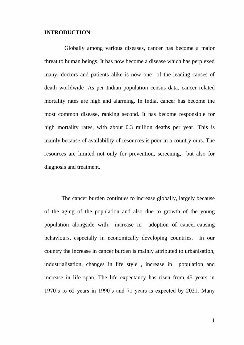
INTRODUCTION:
Globally among various diseases, cancer has become a major
threat to human beings. It has now become a disease which has perplexed
many, doctors and patients alike is now one of the leading causes of
death worldwide .As per Indian population census data, cancer related
mortality rates are high and alarming. In India, cancer has become the
most common disease, ranking second. It has become responsible for
high mortality rates, with about 0.3 million deaths per year. This is
mainly because of availability of resources is poor in a country ours. The
resources are limited not only for prevention, screening, but also for
diagnosis and treatment.
The cancer burden continues to increase globally, largely because
of the aging of the population and also due to growth of the young
population alongside with increase in adoption of cancer-causing
behaviours, especially in economically developing countries. In our
country the increase in cancer burden is mainly attributed to urbanisation,
industrialisation, changes in life style , increase in population and
increase in life span. The life expectancy has risen from 45 years in
1970’s to 62 years in 1990’s and 71 years is expected by 2021. Many

2
cancers are found to have some relation with the diet habits, one among
them are cancers of gastrointestinal tract.
IMPACT OF CANCER IN INDIA:
Impact of cancer is much greater in India, than the mere number of
cases of cancer. The very beginning of diagnosis of cancer causes
immense emotional trauma to the patients and to their families, and its
treatment a major economic burden. The diagnosis of cancer itself is
perceived by most of them as a grave event, a curse with more than one-
third of them suffering from some form of mood disorders (anxiety and
depression). The family members also feel equally distressed. It could
affect both family’s daily functioning and economic situation. The
economic blow often includes both loss of income and increase of
expenses because of treatment and health care. In a country like India
this disease is associated with a lot of fear & despair.
CANCER MANAGEMENT A CHALLENGE IN INDIA:
According to a study conducted by Boston Consulting Group , 70%
cancer cases in India are diagnosed at advanced stages and, most of the
patients do not have access to tertiary care centres. There are not many

3
cancer centers in India to meet the demand of treatment burden. More
radiotherapy centers with adequate number of machines are required to
cover cancer population adequately. Moreover, not all centers are well
equipped with modern facilities. The current Doctor patient ratio is only
1 in 2000 and aim is to achieve, atleast 1 in 1000 by the year 2021.There
is also disproportionate skillful manpower and technology with very few
cancer specialists, trained staff available. And if present, these specialized
cancer centers are available only in very few cities across India. For most
patients in India, treatment cost is so heavy and out of reach. Neither
people do not have insurance cover.Thus delivery of equitable, quality
and affordable cancer care in India is a big challenge.
INCIDENCE OF COLORECTAL CANCERS:
The specialised cancer wing of the world health organisation,
International Agency for Research on Cancer (IARC), released the latest
data on cancer incidence, mortality, and prevalence worldwide in
December 2013.(1) Their online database GLOBOCAN 2012, revealed the
most recent estimates of incidence and prevalence rates of different
cancers. Colorectal cancer has become the third most common cancer
worldwide, with nearly 1.4 million new cases diagnosed in the year
2012. Of these ,majority of patients were men constituting about 7.4

4
million cases and women 6.7 million. This number is expected to rise to
2.4 million cases worldwide by 2035.It is predicted that worldwide, by
the year 2035, the number of colorectal cases will increase to 1.36
million for men and for women 1.08 million. (2)
Amomg both sexes
Lung cancer was the most common cancer worldwide constituting
13% of new cases diagnosed in 2012.
Breast cancer (women only) was the second most common cancer
with nearly 1.7 million new cases in 2012.
Colorectal cancer was the third most common cancer with
nearly 1.4 million new cases in 2012.
GEOGRAPHIC VARIATIONS:
The incidence of colorectal cancers is not uniform throughout the
world. There is a large geographic variation in the global
distribution.colorectal cancer. Colorectal cancer is mainly a disease
of western developed countries with a with ten fold variation
between countries with the highest rates than those with the lowest
rates. It ranges from more than 40 per 100,000 people in the United

5
States, Australia, New Zealand, and Western Europe to less than 5
per 100,000 in Africa and some parts of Asia.(3)
MORTALITY RATES AND TRENDS:
Worldwide mortality due to colorectal cancer is approximately half
of its incidence. Nearly about 530,000 deaths were recorded in the year
2012 , and it contributed to 8% of all cancer related deaths. In the United
States,both in men and women , it has become the second leading cause
of death among other cancers. The incidence rates are appropriate
indicator of trends in disease occurrence. Colorectal cancer incidence is
unaffected by changes in treatment and survival, although it has been
shown to be influenced by improved diagnostic techniques and screening
programs.(4)
CANCER SURVIVAL AND PROGNOSIS:
Survival is highly dependent upon stage of disease at the time of
diagnosis, and 5-year survival rate ranges from 90% for cancers detected
at the localized stage to 70% for regional and to 10% for patients
diagnosed with distant metastasis. With recent improvements in

6
treatment strategies survival for all stages of colorectal cancer at has
increased significantly. The better improvement in 5-year survival is seen
in countries with high life-expectancy and good access to modern
specialized treatment centers. Even then ,large disparities exist in
colorectal cancer survival worldwide and even within same regions. (4)
RISK FACTORS:
There are many number of risk factors associated with increased
incidence of colorectal cancer. Non modifiable risk factors are the ones
that one cannot control, it includes age and inhereted factors. In addition
to this, other factors in environment and lifestyle habits play a significant
role in the development of colorectal cancer.
1. Age:
The likelihood of developing colorectal cancer increases as age
increases above 40 .It sharply rises after 50 years of age. More than 90%
of colorectal cancer cases occur in people aged 50 years or older.
However, it seems to be increasing among younger population too,
mainly because of western life style and dietary habits .
7
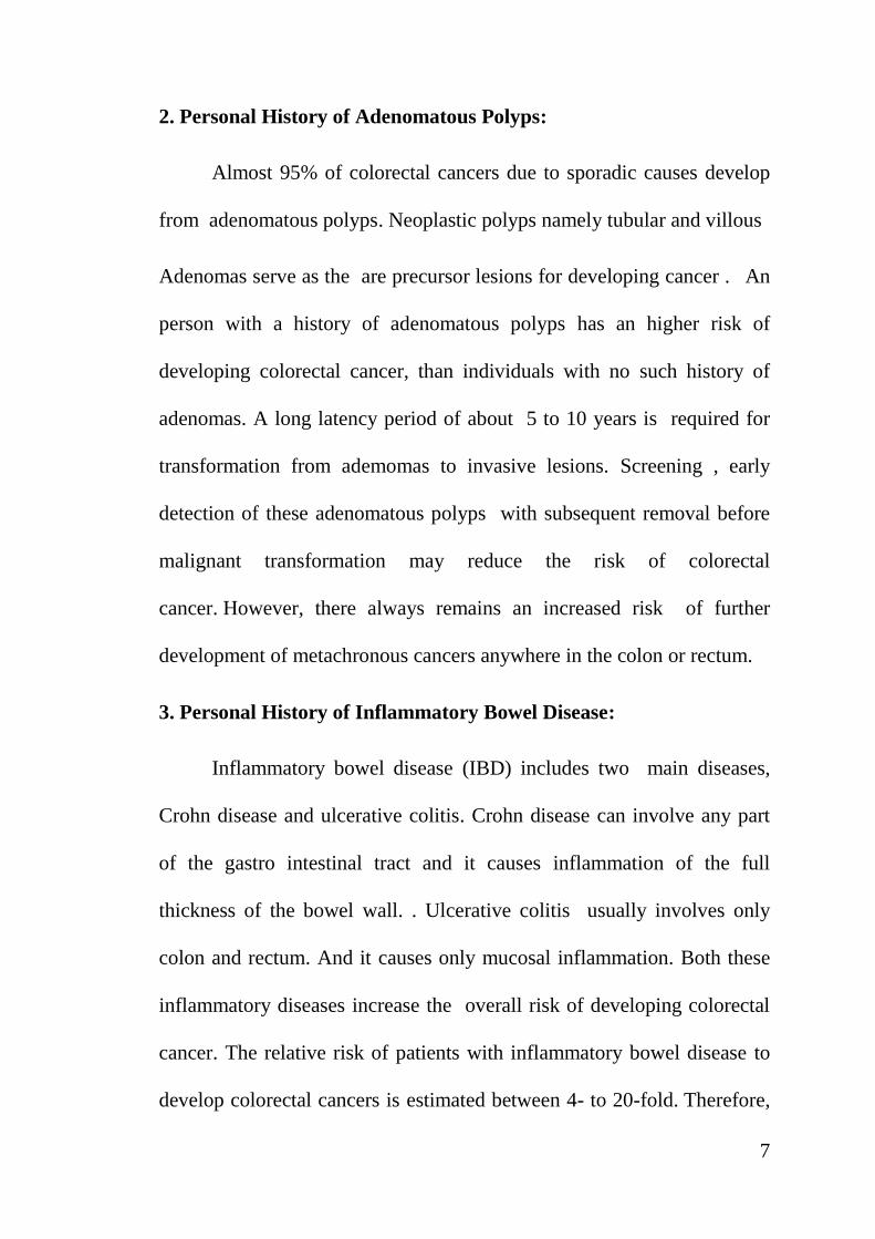
2. Personal History of Adenomatous Polyps:
Almost 95% of colorectal cancers due to sporadic causes develop
from adenomatous polyps. Neoplastic polyps namely tubular and villous
Adenomas serve as the are precursor lesions for developing cancer . An
person with a history of adenomatous polyps has an higher risk of
developing colorectal cancer, than individuals with no such history of
adenomas. A long latency period of about 5 to 10 years is required for
transformation from ademomas to invasive lesions. Screening , early
detection of these adenomatous polyps with subsequent removal before
malignant transformation may reduce the risk of colorectal
cancer. However, there always remains an increased risk of further
development of metachronous cancers anywhere in the colon or rectum.
3. Personal History of Inflammatory Bowel Disease:
Inflammatory bowel disease (IBD) includes two main diseases,
Crohn disease and ulcerative colitis. Crohn disease can involve any part
of the gastro intestinal tract and it causes inflammation of the full
thickness of the bowel wall. . Ulcerative colitis usually involves only
colon and rectum. And it causes only mucosal inflammation. Both these
inflammatory diseases increase the overall risk of developing colorectal
cancer. The relative risk of patients with inflammatory bowel disease to
develop colorectal cancers is estimated between 4- to 20-fold. Therefore,

8
individuals with inflammatory bowel diseases regardless of age are
encouraged to undergo screening for colorectal cancer more frequently
than the general population.
4. Family History of Colorectal Cancer or Adenomatous Polyps :
In about 20% of people who develop colorectal cancer have
positive family history . It is higher in people with affected first degree
relative younger than 60 years of age , or a history of colorectal cancer or
adenomatous polyps in two or more first-degree relatives at any age. The
increased risk is attributed to inherited genes, shared environmental
factors, or combination of these factors.
5. Inherited Genetic Risk:
Nearly 5 to 10% of colorectal cancers are have recognisable
hereditary factors. The most common among the inherited conditions
include familial adenomatous polyposis (FAP) and ,Lynch syndrome
also known as hereditary nonpolyposis colorectal cancer (HNPCC).
Genes causing these forms of inherited colorectal cancer have been
identified. HNPCC is associated with mutations in genes

9
the MLH1 and MSH2 , which are involved in the repair pathway
DNAs. FAP is caused by mutations in APC genes ,which is a tumor
suppressor gene . Lynch syndrome accounts for only 6 % of colorectal
cancers, and individuals have a lifetime risk of developing cancer is as
high as 70 to 80%.The average age at diagnosis is also younger , around
40 years. Patients with genetic mutations in genes involved in DNA
mismatch repair , are also at a higher risk of developing many other
cancers, namely cancer of the genitourinary tract, stomach, small bowel
and pancreas. FAP accounts for only very few cases less than
1%. Unlike people with HNPCC, who develop only a few adenomas,
individuals with FAP characteristically develop hundreds of polyps, at a
relatively younger age, and one or more of these adenomatous polyps
typically undergoes malignant transformation at a very early . All
individuals with FAP, will developed cancer before the age of 40 if the
colon is not removed prophylactically. APC-gene is inherited in an
autosomal dominant fashion. Nearly 80% of people with APC-associated
polyposis have atleast one affected parent. Prenatal testing and
preimplantation genetic testing should be done , if the mutation is
identified in any one of the parent.

10
6. Environmental Risk Factors :
Colorectal cancer develops due to various environmental factors,
which includes cultural, social, and lifestyle modification . Colorectal
cancer has many modifiable causes identified, and a significant
proportion of cases may be theoretically preventable. Some of the
evidence of environmental risk factors as causes comes from studies of
migrants and their offspring. Between migrants from low-risk to high-risk
countries, incidence rates tends to increase toward those of the
populationbelonging to the host country. The incidence of colorectal
cancer in the offspring of migrants from Japan to United States, equal to
or it may surpasses that in naïve American population. This is three or
four times higher than among the Japanese in Japan. Not only migration,
there are other geographic factors influencing incidence rates too. One
among them is living in urban residence. The incident rates are
constantly found to be higher among urban residents. This excess
incidence in urban areas is more apparently seen in males than females ,
and more so for colon cancer than rectal cancer.

11
7. Dietary habits:
Diet strongly influences the risk of colorectal cancer . Several
studies have shown that changes in food habits may reduce to 70% of this
cancer incidence . Diets rich in fat, especially fats of animal origin has
shown to be a major risk. The association of animal fat, as a possible
etiologic factor, is linked to the concept of the Westernization of diet
habits, which favors the growth of gut bacterial flora. These are capable
of degrading the bile salts to potentially carcinogenic N-nitroso
compounds. Consumption of high meat has also shown to an risk factor
in the development of colorectal cancer. This positive association of
cancer with meat consumption is more so for colon cancer than rectal
cancer. It is mainly because of presence ofm heme iron in red meat. In
addition to this ,some meats products are cooked at very high
temperatures, resulting in the production of heterocyclic amines and
polycyclic aromatic hydrocarbons. Both of these compounds are believed
to be carcinogenic .
Some studies have shown that individuals whose food habits includes
less fruits and vegetables , have a relatively higher risk of cancer. Differences
in dietary fiber consumption might be responsible for the geographic variation
in the incidence rates. The basic mechanism is that increased

12
intake of dietary fiber may cause dilution of fecal content thereby
increasing the fecal bulk, and reducing the bowel transit time.
8. Physical Activity and Obesity :
Several habits related to lifestyle modification has been linked to
increase the risk of colorectal cancer. Two modifiable risk factors ,which
are also interrelated to each other , are physical inactivity and excess
body weight. These two factors account for about one fourth to one third
of colorectal cancers. There is also abundant data to support , that higher
levels of physical activity are associated with lowered risk of cancer.
Studies have shown that dose–response effect exist, with frequency and
intensity of physical activity inversely associated with risk of developing
cancer. Regular physical activity and a healthy dietary habits can help
individuals to decrease the risk, although the evidence is much higher
for colon cancers than for rectal cancers. The biologic mechanisms
potentially responsible for the association is that, physical activity raises
the BMR and increases oxygen uptake by the cells. In the long run,
regular periods of physical activity increase the individuals metabolic
efficiency and capacity, it as well as reduces the blood pressure and,
prevents development of insulin resistance. In addition, physical activity

13
also increases gut motility. Physical inactivity in daily routines attributes
to the increased incidence of boby mass index in both males and females,
another risk factor associated with increased incidence.
.
ROLE OF VITAMIN D IN COLORECTAL CANCERS:
Prospective studies has shown that lower levels of vitamin D is
associated with significant increase in mortality among patients with
colorectal cancers. In systemic review and meta-analysis of around 2000
patients with colorectal cancers, compared patients with high level and
low levels of vitamin D. It was found that OS and disease specific
mortality aes were better for patients with higher levels . Yet , no specific
data exist to prove that vitamin D supplementation improves patient
outcomes.
HISTOLOGICAL CLASSIFICATION:
More than 90% of colorectal cancers are adenocarcinomas.
Other histological subtypes include
Adenocarcinoma in situ

14
Medullary carcinoma
Mucinous carcinoma (colloid type) (greater than 50%
mucinous carcinoma)
Signet ring cell carcinoma (greater than 50% signet
ring cell)
Adenosquamous carcinoma
Small cell carcinoma
Undifferentiated carcinoma
Carcinoma, NOS
Squamous cell (epidermoid) carcinoma
PATHWAYS OF SPREAD:
• Lymphatic channels and lymph nodes.
• To liver and lung by hematogenous spread .
• It also has propensity to spread within the bowel wall mainly
longitudinally.
15
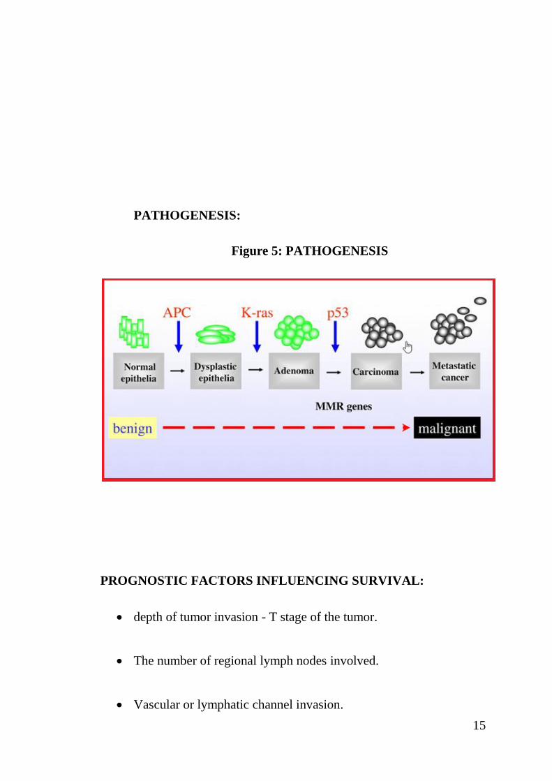
PATHOGENESIS:
Figure 5: PATHOGENESIS
PROGNOSTIC FACTORS INFLUENCING SURVIVAL:
depth of tumor invasion - T stage of the tumor.
The number of regional lymph nodes involved.
Vascular or lymphatic channel invasion.

16
Presence of residual tumor following surgery- it relates to positive
surgical margins.
Grade of the tumor.
Status of Radial margin .
Following Neo adjuvant chemo radiation – presence of residual
tumor.
Histology, and association with microsatellite instability (MSI).
RATIONALE FOR THE PRESENT STUDY:
As seen already, colorectal cancers are very common in our
country. In our institution too they constitute 10% to 15% of the cancers
registered in the OPD. And most of them present in the locally advanced
stages. The presently available standards of the treatment with surgery
and concurrent chemo radiation have a dismal performance in long term
control with overall survival at 2 years hovering around the 50% mark
and less than 20% surviving 5 years. Various modalities are being
devised to overcome this.

17
This is where the intensification of the treatment is considered. The long
course neo adjuvant chemo radiation has its own toxicity profile, leading
to treatment breaks and decreased compliance. The argument for neo
adjuvant radiation before the loco regional treatment is that it results in
reduction of the tumor load, thereby resulting in better loco regional
control. Also it has been alleged that adjuvant radiation has increased risk
of small bowel toxicity and also decreased responsiveness because the
blood supply to the local areas would have been altered. When the same
is given in the neo adjuvant setting, the blood circulation in these areas is
intact and will supposedly result in better results.
With these understandings, there has been a renewed interest in
addressing locally advanced rectal cancers with neo adjuvant radiation in
the form of short course for selected patients. So the present study
justified in addressing this question.

18
LITERATURE REVIEW:
Before 1900, incidence of colon and rectal cancers were negligible.
Since then , following economic development and industrialization the
incidence has been rising dramatically. For all rectal cancers, surgery
remains as the primary treatment modality .Despite these curative
resection, local recurrence remains high. Anatomical confines of pelvis
and the importance of preservation of autonomic nervous system makes
surgery very challenging. This may account for high rates of both local
recurrence and distal relapse.
Many European randomized prospective trials have shown that
multimodality approach results in significant better outcome. Neo
adjuvant treatment has emerged as the standard of care and degree of
tumor regression has become an important prognostic factor. A multi
modality approach including medical oncology , radiation oncology and
colorectal surgery is required for optimal treatment plan.
Multiple randomized trials have shown that addition of
preoperative chemo radiation has shown significant benefit, resulting in
increased pathological response rates and increased local control rates.
Earlier in 1900’s ,the National Institute of Health consensus

19
recommended combined treatment modality for stage II and III rectal
cancer, based on observation from Gastrointestinal tumor study group
and National surgical adjuvant breast and bowel project trials, which
demonstrated chemotherapy along with radiation, following surgical
resection reduces the rates of local recurrence to 33% from 55%.It has
also shown to prolong the disease-free survival rates .
ADVANTAGES OF PREOP CHEMORADIATION:
1. Downsizing the tumor volume, facilitates resection and
increase the likelihood of sphincter saving procedures.
2. Irradiating the tissues before surgery, which has better
oxygenation than the postop tumor bed tissues, and may result in
increased responsiveness to RT.
3. Avoidance of radiotherapy induced injury to small bowels,
which has dropped down into the pelvis by post surgically.
4. The irradiated structures will be removed by surgery,
anastamosis is with healthy Colon.
DISADVANTAGES OF POST OP RT :

20
1. Hypoxic post surgical bed makes radiation & chemotherapy less
effective.
2. Increased small bowels in the radiation field,is a dose limiting
organ, and it increases chances of both acute and late toxicity.
SWEDISH RECTAL TRIAL:
A major shift in preoperative radiotherapy was brought by
Swedish rectal trial in the year 1997. This study had 1168 pts,
randomisied to one of the arms , either single week of RT followed by
surgery, or only surgery. On comparision of results , patients who
received preoperative Radiotherapy had decreased local recurrence rate
11% vs 27%,(5)
and improved overall survival 58% vs 48%. On long term
follow up of 13 years, local recurrence and overall survival was 9% vs
26% , and 38% vs 30%(6).
This study concluded that preoperative
radiotherapy in a single week immediately followed by surgery for rectal
cancer is found to be beneficial in terms of overall survival and cancer-
specific survival and decrease in local recurrence rates(7)
.
Next came the era of TME- Total Mesorectal Exicision, which is a
sharp dissection along the planes of visceral and parietal endopelvic

21
fascia. It also involves removal of regional lymph nodes, while preserving
the autonomic nerves. Multiple cohort and retrospective studies have
shown that Total Mesorectal Exicixion is associated with lowered rates
of pelvic recurrence compared to the less optimal blunt surgical
dissection .
The Dutch TME trial ,in the year 2003 was the first trial to
compare the results of surgey namely Total Mesorectal Exicision (TME)
with and without short course radiation . This trial included 1861 pts, out
of which 924 were randomized to receive either preoperative radiation
followed by TME, 937 were randomised to TME alone. Analysis has
shown that , local recurrence was significantly less in patients, who
received preoperative RT plus TME compared with surgery alone (2.4%
vs 8.2%, P < 0.001),(8)
but there was not much difference in OS between
the two groups. Sub group analysis also proved to be favourable to
preoperative RT arm, showing decrease in local recurrence rates
especially in patients with nodal positivity,(9)
tumors located between 5-10
cm proximally from the anal verge, and patients with negative CRM-
circumferential resection margins.
The German CAO/ARO/AIO 94 trial compared preoperative
short course radiation and postoperative long-course chemo radiation . It

22
included T3 or T4 and/or node-positive tumors in locally advanced rectal
cancers . Chemo radiotherapy consisted of continuous infusional
fluorouracil (100mg/m2 per day for 5 days in the 1st and 5
th week of
radiation) of 50.4 Gy in 1.8 Gy/# in 28fractions.401 and 402 patients
were randomisied to receive preoperative and postoperative chemo
radiotherapy, respectively .Local recurrence in the preoperative group
was 6% and 13% in postoperative group (P = 0.006).(10)
Assessment of
acute and long term toxicities have shown that Grade 3 or higher
toxicity occurred substantially lesser in patients who received pre
operative chemo radiation than post operative group. P value was (P =
0.001 and P = 0.01, respectively). (11)The main inference from the study
has shown that the rates of sphincter preservation, Disease Free Survival
and Overall Survival did not differ much between the two groups.
Another comparision between long and short course radiation
regimen was done by Polish randomized study, which included
patients with T3 and T4 rectal cancer. The results has shown promising
rates of complete pathologic response in the group of patients receiving
long- course chemo radiotherapy: 16% vs 1% of patients had complete
pathologic response in the long-course and short-course arms
respectively.(12)
This study also showed that the sphincter preservation

23
rates were similar in both groups irrespective of the complete
pathological states. Regarding the status of CRM- patients receiving long-
course chemo radiotherapy had only 4% compared with 13% in the short-
course group (P = 0.017).(13)
Inspite of the higher positive CRM rates in
short course arm, there was not much significant differences in rates of
local recurrence, DFS or OS.
The MRC CR07 and NCIC-CTG C016, is a randomized trial in
olving multiple centres. The population study comprised 1350 patients
comparing the outcomes of preoperative short-course RT followed by
surgery , and postoperative chemo radiation in selected patients with
positivity of Cicumferrential Resection Margin. The outcome studied
primarily was local recurrence. It demonstrated a substantial decrease in
rates of local recurrence between patients receiving preoperative short-
course regimen with hazard ratio 0.39( P < 0.0001) (13).
And this was
associated with a 6% absolute improvement in DFS at 3 years (P =
0.03)(14).
These data again demonstrate the superiority of preoperative
chemo radiotherapy.
In a review by Trans-Tasman Radiation Oncology Group Trial
01.04,randomized Trial of Short-Course regimen Versus conventional

24
Long-Course Chemo radiation comparing rates of local recurrence in
Patients With T3 Rectal Cancer has shown that incidence of LR were
7.5%(15)
for Short course RT and 4.4% for long course RT at the end of 3
years . But the p value was not significant. (16)
HYPOFRACTIONATED RADIATION:
The paucity of data regarding the optimal use of hypofractionated
radiotherapy in rectal cancer for a population where 60% to 70% of cases
usually present with locally and very advanced stages has been the idea
behind the study protocol.
Hypofraction implies use of larger ose per fractionation with lesser
number of fractions so as to deliver the equivalent biological effective
dose (BED ) in a shorter duration of time. The increase in dose per
fraction (df) over the reference value of 2Gy , for an isoeffect , the total
dose should be reduced. Due to low ratio curves for late responding
tissues, the curves are steeper than those of early reactions and for
tumors, which have a high ratio. Hence if df is increased to 5 Gy per

25
fraction and considering the ratio for late reacting tissues to be 3Gy ,
then the total dose must be reduced from its reference value.
Hypofractionated schedules have the advantage of being more
convenient for the patient , their care givers and also for the health care
providers by sparing the essential resources.
POST NEOADJUVANT CHEMORADIATION LEVEL:
It has been shown by various studies that after neo adjuvant chemo
radiation, CEA levels of <2.5 ng/mL and 5 ng/ mL(17)
are associated with
improvement in DFS and OS. But few other , studies has discerned this
relation. Many studies focusing on correlation between predictive factors
and long term outcomes do not rely on CEA levels. (18)
So using this
parameter to predict disease recurrence and survival still remains
inconclusive.
RESPONSE ASSESSMENT AFTER NEOADJUVANT
CHEMORADIATION:
The pathological response of tumor to neo adjuvant chemotherapy
has shown to be the most important prognostic factor in terms of rectal

26
cancer recurrence.(19)
Pathological Complete Respone is most strongly
correlated with best outcomes .Many tumor regression grading systems
are proposed and demonstrate a strong association between complete
pathological response and their outcomes. Neo adjuvant chemo radiation
reduces lymph node yield during surgery. The ratio of positive number of
lymph nodes to total lymph nodes dissected is prognostic and presence of
positive lymph node after neoadjuvant treatment is associated with poor
prognosis(20).
The MERCURY TRIAL has studied the diagnosic importance of
predicting the circumferential margin status by MRI. (21)
The high
resolution MRI accurately predicts the surgical margin status involvement
by tumor. This study has shown the importance of identifying the
patients with potentially affected margins and need for treating them with
preoperative chemo radiation (22).
SURGERY:
The concept of total mesorectal excision in rectal cancer surgery
has revolutionised the modern era of treatment . the practice of TME
has reduced the rate of local recurrence and tumour associated mortality

27
and morbidity.The procedure involves the removal of the rectal tumour
along with the entire mesorectum (radial margin). The mesorectum is the
fatty tissue surrounding the rectum that contains the lymph nodes and
main blood vessels that supply the rectum. Most of the local recurrence
is likely to occur in the mesorectum, so removing the entire mesorectum,t
reduces the chance of recurrence(23).
The healthy end of sigmoid colon is attached to the anal sphincter
so normal bowel function can be resumed. Sometimes the anal sphincter
cannot be saved and a permanent colostomy is needed. The location of
the tumour, the size of the tumour and how far away the tumour is from
the anal verge will determine whether the TME operation is done using
either a low anterior resection or an abdominal perineal resection.
LOW ANTERIOR RESECTION (LAR):
A low anterior resection (LAR)is a procedure done to remove
tumours in the upper two-thirds of the rectum. This procedure removes
part of the descending colon, the sigmoid colon, all or part of the rectum
and the mesorectum. A TME approach is used to remove the rectum and
mesorectum.

28
ABDOMINAL PERINEAL RESECTION (APR):
An APR is a procedure done to remove low rectal tumours that
invade the muscles around the anus. APR is also done when it is not
possible to preserve the anal sphincter. This procedure removes part of
the sigmoid colon, rectum, anus, mesorectum and anal muscles. The
surgeon makes 2 incisions – one in the abdomen (using the TME
approach) and one in the perineal region.
Two separate incisions are needed because of the 2 areas that are
being removed. The rectal area is removed through the abdomen and the
anal area is then completely removed and stitched up. Patients will then
have a permanent colostomy. For this reason, APR is only done when
there is no way to leave a margin of healthy tissue below the tumour
margin(24).
LAPAROSCOPIC SURGERY FOR LOCALLY ADVANCED
RECTAL CANCER:
Feasibility of laparoscopic surgery in the management rectal
cancers has not been proven yet. But still, it has not become the standard

29
of care. The United Kingdom Medical Research Council Trial has
compared conventional and laparoscopic assisted surgery in colorectal
cancers. It has demonstrated equivalent results in terms of local control
and survival benefit.(24)
But the operating time is significantly longer and
estimated blood loss is less in laparoscopic surgery as compared to open
surgery. The positivity rates of circumferential resection margin was not
significantly different between the studied groups(25).
The long-term
outcomes of patients in this trial are awaited.
DISTAL RESECTION MARGIN (DRM):
The standard guideline recommendation for distal resected
margin is 4- 5cm, measured from the distal edge of tumour. Many
studies have shown that DRM <2 cm does not increase the recurrence
rates or have a negative impact on the survival(26).
In situations of low
rectal cancers located < 5 cm from the anal verge, 1 – 2 cm may be an
acceptable margin.
Following treatment with neo adjuvant chemo radiation, the
necessity of a margin for distal rectum of 2 to 3 cm is less significant than
CRM.(27)

30
CICUMFERRENTIAL RESECTION MARGIN:
The standard cut- off point for the Circumferrential Resection
Margin is still a matter of debate. Many studies has revealed <= 1mm
margin as an acceptable cut off point(29 -31).
All have reported a
significantly high rates of local recurrence and decreased survival in
patients with inadequate CRM <= 1 mm.(32- 34)
TIMING OF SURGERY AFTER NEOADJUVANT TREATMENT:
After long course neo adjuvant chemo radiation , surgery is done
usually after 6 weeks . This waiting period for 6 weeks is needed for
adequate response to treatment to occur. But in case of short course
radiation with high dose , surgery is usually done within 10 days of
radiation. (35)
Maximum within fourteen days radiation toxicity sets in.
The irradiate bowel is removed during the surgery.
POST OPERATIVE CHEMOTHERAPY:
For locally advanced rectal cancers- stage II and III, the risk of
local recurrence and metastasis remains high if only treated with surgery.

31
So to eradicate the micrometastasis, the role of adjuvant chemotherapy
becomes crucial.
Most of the data on adjuvant chemotherapy regimens comes from
extrapolating trials on colon cancer. The MOSAIC TRIAL was
conducted on stage III colonic cancers .Chemotherapy regimen FOLFOX
4 as compared to 5FU+leucovorin has shown increase in DFS and OS.(36)
In X-ACT study , the efficacy of capecitabine was compared with 5FU+
leucovorin , and it has shown equal efficacy in DFS and OS.(37)
EVALUATION OF MESORECTUM:
The completeness of TME should be evaluated by the pathologist.
The sleeve of mesorectum to be examined completely for its quality of
resection.It is mainly for distal 2/3 rd of rectal cancers.
TREATMENT DURATION TIME:
With long course of radiation , the time period required for neo adjuvant
chemo radiation is 5.3 weeks. Patient has to wait for 6 weeks before

32
surgery. Then adjuvant chemotherapy is given after 3 to 4 weeks
following surgery. So the total treatment time is around 40 weeks till
completion of adjuvant 6 cycles chemotherapy.
With short course radiation, the time required for radiation is one
week. Surgery is done usually within 10 days of radiation. Adjuvant
chemotherapy is given 3 -4 weeks following surgery. So the total
treatment time is around 27 weeks. The significance of short course
radiation is shorter duration of treatment and hence better patients
compliance to treatment schedule.
Treatment breaks which occur during long course chemo radiation
also has negative impact on local control of the disease and long term
survival. Most of the patients default during the course of chemo
radiation too, because of acute toxicities like cystitis and diarrhoea.

33
LONG COURSE CCRT REGIMEN:
Diagnosis , investigation evaluation of treatment plan
2 WEEKS
Concurrent chemo RT 50.4 Gy / 1.8 Gy/ # X 28 #
6WEEKS +
Chemotherapy= Capecitabine/Infustional 5FU/ Bolus 5 FU +
Leucovorin
6WEEKS Waiting period before surgery
Total Mesorectal Excision
LAR/ APR
6WEEKS
Adjuvant chemo therapy 6 cycles
CAPEOX regimen
18 WEEKS for 6 cycles of chemo therapy
Totally : 36 to 40 weeks of treatment.

34
SHORT COURSE RADIATION REGIMEN:
Diagnosis , investigation evaluation of treatment plan
2 WEEKS
Short course radiation hypofraction 5Gy / # X 5 # = 25Gy
7 to 10 days Waiting period before surgery
Total Mesorectal Excision
LAR/ APR
4-5WEEKS
Adjuvant chemo therapy 6 cycles
CAPEOX regimen
18 WEEKS for 6 cycles of chemo therapy
Totally : 25 to 26 weeks of treatment

35
PREOPERATIVE VS POSTOPERATIVE RADIATION:
Main advantages of preoperative radiation , as compared to
radiation given in the postoperative period are related to both response of
tumor tissues and preservation of normal tissues. Although some trials
have indicated that preoperative radiation or chemo RT is asscociated
with increased sphincter preservation rates, but recent meta analysis of
randomisied trials do not support it. (38)This may be due the surgeons
perspective of removing the microscopic disease left behind after tumor
downsizing. It also implies a good oncological practice .
CONCURRENT CHEMOTHERAPY WITH RADIATION:
Many randomized trials have studied the effectiveness of adding
chemotherapy to radiation administrating either preoperatively or
postoperatively. The benefits of adding chemotherapy either with pre or
post operative radiation has increased the , local radiation sensitisation
and systemic control of diseases (ie) the eradication of micro metastasis(39
– 41) .
A recent Cochrane review of six randomizied control studies
showed that adding chemotherapy to preoperative radiation in resectable
stage II/ III rectal cancers enhances the pathologic response rates and

36
improves the local control rates. But it did not have any effect on the
overall survival, 30 day mortality , sphincter preservation rates and late
toxicity(42-45).
KRAS MUTATION:
Approximately 40% of patients with colorectal cancers have
mutations in the genes encoding KRAS. The presence of mutations in
codons 12 and 13 in exon 2 o f KRAS gene predicts the
nonresponsiveness to EGFR inhibition. Hence patients presenting with
metastatic colorectal cancers, being considered for anti EGFR therapy
should have their tumor tissue genotyped for presence of KRAS mutation
before initiation of treatment. Several studies has shown that targeted
agents produce negative impact on survival ,in patients with mutaed
KRAS. So patients with KRAS mutation or NRAS mutation should not
be treated with targeted either cetuximab or panitumumab.
Studies on colorectal cancer formation has shown that KRAS
mutation occurs as an early event. Therefore a strong correlation exists
between the mutation status , primary tumor and metastasis. It has been
found that KIRSTEN RAS ( KRAS) – mutation was associated with poor
survival rates than patients with wild type KRAS . Presence of V600

37
BRAF mutation also signifies poor prognosis too. Data is insufficient to
approve the use of anti EGFR therapy in the first line setting with
systemic chemotherapy.
According to PRIME study , panitumumab was used along with
FOLFOX regimen. It was used as a first line treatment in colorectal
cancers. The results of this study showed that in patients with mutated
KRAS , panitumumab had de trimental effects.
MICRO SATELLITE INSTABILITY:(MSI)
Testing for MSI should be considered for patients <= to 70 years of
age with colorectal cancers.It should also be done on patients who are >
70 years of age , but meeting the Bethesda guidelines.

38
AIMS AND OBJECTIVES:
The aim of this study was to evaluate the efficacy of neo adjuvant
short course Radiotherapy followed by surgery in locally advanced rectal
cancers.
Primary Objectives :
To assess the immediate loco regional response rates of
resectable stage II/III rectal cancers with neo adjuvant short course
radiotherapy followed by surgery within 10 days.
Secondary objectives:
To assess acute toxicity to neo adjuvant short course radiation in
locally advanced rectal cancer.

39
MATERIALS AND METHODS:
STUDY DESIGN:
This was a Single arm prospective study with a Phase III design.
STUDY DURATION:
OCTOBER, 2014 – August, 2015
STUDY CENTRE:
Department of Radiotherapy, Barnard Institute of Radiology &
Oncology, Madras Medical college, Chennai.
SAMPLE SIZE:
30 consecutive patients with histopathologically proven
adenocarcinoma of rectum who fulfilled the inclusion criteria were
recruited in the study from the outpatient department.
The intent of treatment was to be radical, aiming for cure, considering
their disease stage, co- morbidities and performance status.

40
ETHICAL COMMITTEE APPROVAL:
Approval from the institute ethical committee was obtained on
07.10.2014.
INFORMED PATIENT CONSENT:
All patients enrolled in the study were informed about the merits
and demerits of participating in this study and signed an informed consent
form in their regional language, which is Tamil.
METHODOLOGY:
Eligible patients will be treated with hypofractionation 5Gy per
fraction for 5 days in one week, followed by surgery after 2 weeks.
Postoperatively adjuvant chemotherapy is given for 6 cycles. The
response to treatment and acute toxicity is to be assessed periodically.
Inclusion Criteria:
• Biopsy proven newly diagnosed adenocarcinoma of rectum.
• Age 18 - 65 years.
• T3 lesions less than or equal to 5 cm,N0 OR N+ disease.

41
• Performance status ECOG 0-2.
• Medically manageable co-morbidities.
• Signed informed consent prior to initiation of protocol.
Exclusion Criteria:
• Histopathology other than adenocarcinoma .
• Inadequate hepatic and renal functions, bone marrow reserve.
• Uncontrolled co-morbidities.
• Patient not consenting to radiotherapy/ surgery/chemotherapy at
any point in the treatment.
• Previously received treatment for any other malignancy.
Sample Size: 30 patients
Investigation Details:
• . Detailed history elucidation.
• Complete physical examination by inspection, palpation.
• Biopsy from the primary tumour.

42
• Baseline-Complete blood count, liver function test, renal function
test.
• X-ray chest PA view,
• ECG,
• Blood grouping &typing,
• Cardiac evaluation and fitness.
• Anaesthetic fitness for surgery,
• Colonoscopy,
• MRI- Pelvis-Plain &Contrast pretreatment.
• Tumor marker- CEA levels.
• Histopathology of surgical specimen.
Staging was done based on American Joint Committee staging manual
7th edition (for colorectal cancers).
PATHOLOGICAL STAGING OF THE SPECIMEN:
1. Gross description of the tumor and specimen.
2. Grade of the tumor.

43
3. Depth of penetration and extension to adjacent structures.
4. 5. Number of positive Lymph nodes.
6. Status of proximal ,distal and CRM.
7. Neoadjuvant treatment response.
8. LVI
9. Perineural invasion.
10. Number of tumor deposits.
CIRCUMFERRENTIAL RESECTION MARGIN:
It is defined as the closest radial margin , always measured in
millimeters. It is between the deepest penetration of tumor tissue and
resected edge of soft tissue around the rectum. Also from the edge of
positive lymph nodes.CRM is identified by evaluating the outer surface
of the resected specimen , which if often inked at. The examination of the
specimen requires a bread loafing slicing technique.

44
.

45
TREATMENT PROTOCOL:
An informed consent was obtained from all patients. Before
starting treatment, it was ensured that all patients had normal
hematological/renal parameters and normal liver function tests. Adequate
cardiopulmonary function that could tolerate radical surgery was also
assessed before treatment. Comorbid conditions like diabetes and
hypertension were evaluated thoroughly and treated with appropriate
medications.
THERAPEUTIC PROTOCOL:
Short course radiation with hypofractionation 5 Gy / # X 5 #
followed by TME within 10 days for T3 rectal cancers.
EBRT EQUIPMENT Co – 60 Phoenix for Teletherapy

46
SIMULATION AND TREATMENT DELIVERY
The treatment field was verified with PA simulator films in
which the distal extension of the tumor was identified by placing a radio-
opaque marker in the anal verge. The lateral portals were verified by
lateral simulator films.The treatment plan was evaluated with and without
wedges in TPS planning system and optimal treatment plan was selected
from isodose pattern All patients were positioned in the prone position
only with full bladder during external RT to exclude a greater extent of
the small bowel from the treatment field.
SIMULATION FILM USED:
Figure 16: Simulation Film
47
RT SCHEDULE
All patients were treated with hypofractionated dose 500cgy/
# by three field 1 PA portal( 168 cgy) / 2 lateral portals ( 166 cgy each)
to a total dose of 500cgy delivered from Monday, Wednesday to Friday
in a week .
RADIOTHERAPY TECHNIQUE:
Radiotherapy was delivered by three field technique
to the pelvis with a telecobalt machine. 5 # was delivered in 5days in one
week. The clinical target volume including the tumor, internal iliac nodes,
obturator nodes , and the anal canal for adequate clearance received the
calculated dose of 25 Gy. Organ at risk was given due consideration in
treatment planning process. All three fields were treated on all 5 days of
planned radiation with equal dose distribution.

48
RADIATION MARGINS:
POSTERO-ANTERIOR FIELD:
Table 2
RT PORTAL MARGINS FOR EBRT PA PORTAL
SUPERIOR L5 S1 INTERFACE
INFERIOR
2 CM BELOW DISTAL
MARGIN OF TUMOR
LATERAL
2CM LATERAL TO BONY
PELVIS

49
LATERAL FIELD:
Table 3
RT PORTAL MARGINS FOR EBRT LATERAL PORTAL
SUPERIOR L5 S1 INTERFACE
INFERIOR
2 CM BELOW DISTAL
MARGIN OF TUMOR
ANTERIOR
POSTERIOR BORDER OF
PUBIC SYMPHYSIS
POSTERIOR PRESACRAL BAY – S2

50
BED VALUE CALCULATIONS FOR EBRT :
According to the linear –Quadratic model the formula for
calculating the biologically effective dose is
BED =Nxd [1+d/(/)] –K[T-T0]
Where N - no of fractions
d - dose per fraction
/ -dose at which the linear and quadratic cell kills are equal
k -constant (Dose required /day to counter act proliferation)
T - Over all treatment time
T0 – Onset time for proliferation

51
Calculation of biologically equivalent doses to 2 Gy /# for the three most
commonly used fractions.
Table 4
Biologically equivalent doses to 2 Gy / #
25 Gy in 5Gy/# 45 Gy in 25 # 50.4 Gy in 28 #
Tumor control
with
Time correction
35.7 28.1 30.4
Late issue
toxicity
40.0 43.2 48.4
Biologically equivalent doses (2 Gy per fraction) along with a factor
for time correction = biologically equivalent doses (2 Gy per fraction)
– 0.6 Gy ( T-7 ),
Where T = overall treatment time in days.
In this equation ,it is assumed that 0.6 Gy is lost per day due to
tumor repopulation starting after 7 days from the beginning of radiation.

52
RESPONSE EVALUATION AND SURGERY
Within 10 days of last fraction of radiation, patients are taken up
for surgery- TME either low anterior resection or abdomino perineal
resection. Histologic analysis of surgical specimens was done to assess
the extent of CRM-circumferrential resection margin status ,lymph node
status and the involvement of proximal and distal rectal margins.
Operative complications included bladder, small bowel, ureteral or
vascular injuries. Any surgery related adverse event occurring within
30days from the intervention was recorded as postoperative complication.
TOXICITY ASSESSMENT:
Patients were reviewed every day before radiation for any acute
toxic reactions. Reactions like skin desquamation, cystitis, and proctitis
etc. were recorded and graded based on RTOG acute radiation morbidity
criteria. Careful attention was given for maintenance of hydration,
adequate dietary intake and good personal hygiene.
Hematological and renal parameters were assessed before and after
radiation. And before surgery. Anaesthetist opinion was obtained.
53
SIDE EFFECT MANAGEMENT:
In female patients , symptoms of vaginal stenosis can occur. So
they should be instructed to use vaginal dilators .
In case of young males and females, counselimg should be done
regarding infertility risks. They should be given information
regarding sperm and oocyte banking.
STATISTICAL ANALYSIS:
The patient factors, tumor factors, response to treatment, and
toxicities were thoroughly analyzed. The results are expressed in
percentage. Since this study is single armed one and also the sample size
was only 30, the levels of significance cannot be commented on.
FOLLOW UP:
Patients after completion of surgery was discharged from the
hospital and was to review after 4 weeks for adjuvant
chemotheraphy.
CEA levels were done after surgery and there after three monthly.
Imaging was done when indicated clinically

54
Counselling to the patient and attender, rehabilitation, usage of
colostomy bags and cleaning of the same was given.
MANAGEMENT OF LATE SEQUELE OF TREATMENT:
Patients treated with pelvic surgery and radiation can have
symptoms of chrnic diarrhea or incontinence. They should be managed
symptomatically with anti- diarrhea drugs, stool bulk forming agents, diet
manipulation and protective undergarments. Radiation cystitis , increased
frequency and urgency of micturation are managed symptomatically with
bladder irrigation techniques.

55
TREATMENT PROTOCOL
Patient Selection
Based on inclusion criteria
Pretreatment evaluation including blood investigations,imaging ,
biopsy
Treatment administration- neoadjuvant short course RT
5Gy/# X 5# in one week
Regular monitoring of toxicities
Treatment completion
Surgery within 10 days TME
Review with pathology report for response assessment
Adjuvant chemotherapy 6 cycles CAPEOX

56
RESULTS AND ANALYSIS
Out of 30 patients recruited for the study, 29 pts completed their
entire treatment protocol and were available for analysis of results. One
patient underwent diversion colostomy only, followed by palliative
chemotherapy.
PATIENT CHARACTERISTICS:
AGE DISTRIBUTION:
43% of the patients belonged to the age group 51- 60yrs, followed
by 41 -50yrs. The mean age of presentation was 55.5yrs. The youngest
patient age was 35yr and the oldest was 67 yrs.
AGE DISTRIBUTION OF THE STUDY POPULATION
Table 5
AGE GROUP
NUMBER
PERCENTAGE
31- 40yrs
6
20%

57
GENDER:
The gender distribution in the study population is dominated male
population. This study has 16 male patients followed by 14 female
patients.
GENDER DISTRIBUTION OF THE STUDY POPULATION
Table 6
SEX
NO. OF PATIENTS
PERCENTAGE
MALE
16
53.33%
FEMALE
14
46.67%
41 -50yrs
9
30%
51-60yrs
13
43%
61-70yrs
2
7%

58
PERFORMANCE STATUS:
All patients in this study had a general performance status of
ECOG (Eastern Cooperative Oncology Group ) grade 0 or 1.
ECOG PERFORMANCE STATUS
Table 7
ECOG
NO.OF PATIENTS
PERCENTAGE
ECOG 0
18
60%
ECOG 1
12
40%
SYMPTOMS AND SIGNS:
Among the study patients the most common presenting symptom
was bleeding per rectum followed by difficulty in defecation.(figure no:6

59
SYMPTOMS/SIGNS
Table 8
PRESENTING
SYMPTOMS/SIGNS
NUMBER
PERCENTAGE
PAIN DURING
DEFECATION
12
40%
BLEEDING PER
RECTUM
16
53%
DIFFICULTY IN
DEFECATION
18
60%
LOWER ABDOMINAL
PAIN
12
40%
INCREASED
FREQUENCY OF
STOOLS
7
23%
LOSS OF WEIGHT
4
13%

60
HISTOLOGICAL DIFFERENTIATION:
Most of the patients in the study belonged to moderately
differentiated histology followed by well differentiated on
HISTOLOGICAL DIFFERENTIATION
Table 9
HISTOLOGICAL
DIFFERENTIATION
NUMBER OF
PATIENTS
PERCENTAGE
WELL
DIFFERENTIATION
9
30%
MODERATELY
DIFFERENTIATED
16
53.33%
POORLY
DIFFERENTIATED
5
16.67%

61
SIZE OF THE TUMOR:
The estimation of tumor size was by clinical and radiological
examination. Imaging modality used was MRI .
SIZE OF TUMOR
Table 10
.
As per the inclusion criteria, only patients having size of the tumor mass
less than 5 cms were included. 50 % of them had tumor size between 2- 4
cms and 50 % of them between 4 – 5 cms.
SIZE OF THE TUMOR
NO. OF PATIENTS
2-4cm
15 (50%)
4– 5 cm
15 (50%)

62
HISTOLOGIC GRADE:
Most common histological grade was grade II , followed by grade
I tumors.
HISTOLOGICAL GRADE
Table 11
GRADE
NO. OF PATIENTS
GR I
11( 36.67%)
GR II
12 (40%)
GR III
7 ( 23.33%)
TYPE OF SURGERY:
Out of 30 patients , 29 underwent curative resection. One patient
had only diversion colostomy due to adherence to adjacent structures.40
% of patients underwent Low anterior resection.56% of patients had
Abdomino perineal resection. Selection of patients for either of the

63
procedures was based on distance of the tumor from anal verge and also
the oncological principle of achieving adequate distal margin status and
TYPE OF SURGERY
Table 12
TYPE OF SURGERY
NUMBER
PERCENTAGE
Low anterior resection
12
40%
Abdominoperineal
resection
17
56.67%
Hartmans procedure
1
3.33%
STATUS OF CIRCUMFERENTIAL RESECTION MARGIN:
All patients had adequate circumferential resection margin status.
None of them had positivity of CRM less than one centimeter.

64
CIRCUMFERRENTIAL MARGIN STATUS
Table 13
CIRCUMFERRENTIAL
RESECTION MARGIN
NO.OF
PATIENTS
PERCENTAGE
< =1 CM
NONE
O%
>1 CM
29
96.67%
PATHOLOGICAL T STAGE: y p (T) stage
For patients who had minimal involvement of perirectal tissues,
after neoadjuvant radiation they were downstaged to T2 lesion from T3
lesion.

65
Y P( T ) STAGE
Table 14
TUMOR STAGE
NO.OF PATIENTS
PERCENTAGE
T2
9
30%
T3
20
66.67%
DISTANCE OF LOWER BORDER OF TUMOR FROM ANAL
VERGE:
Most of the tumors were 3 to 6 cms from the anal verge, around
46.67 percent. 26.67 5 of the patients had tumors in the upper part of the
rectum , with distal margins more than 6 cms from the analverge.

66
DISTANCE FROM ANAL VERGE
Table 15
DISTANCE FROM
ANALVERGE
NUMBER OF
PATIENTS
PERCENTAGE
0 – 3 cms
8
26.67%
3 – 6 cms
14
46.67%
> 6 cms
8
26.67%
NO OF RETRIEVED MESORECTAL LYMPHNODES:
Yield of lymph nodes in the mesorectum after neoadjuvant
treatment is usually less compared to afferent surgery. Median number of
lymph nodes resected was 7 LNs.

67
NUMBER OF LYMPH NODES DISSECTED
Table 16
NO OF NODES
DISSECTED
NUMBER OF
PATIENTS
PERCENTAGE
0 – 5
11
36.67%
5 – 10
13
43.33%
> 10
5
16.67%

68
NODAL STATUS:
Nodal staging after neoadjuvant radiation is usually difficult. 60%
of the patients had N0 disease.
NODAL STAGING.
Table 17
NODAL STATUS
NO.OF PATIENTS
PERCENTAGE
N0
18
60%
N+
12
40%
INTERVAL BETWEEN RADIATION AND SURGERY:
Most of the patients were taken up for surgery within 10 days.
Median number of days between last day of radiation and surgery is 8
days

69
TUMOR SIZE Vs RESPONSE
Table 18
Tumor size
Response
CRM
1-1.5 CM
CRM
>1.5 CM
T2
DISEASE
2-4 CM
2
4
9
4- 5 CM
10
4
-

70
RESPONSE VS ISTANCE FROM ANAL VERGE
Table 19
DISTANCE FROM ANAL
VERGE
Response
CRM
1-1.5 CM
CRM
>1.5 CM
T2
DISEASE
0 – 3 CM
5
2
0
3 – 6 CM
4
3
7
> 6 CM
3
3
2

71
HISTOLOGICAL DIFFERENTIATION Vs RESPONSE
Table 20
HISTOLOGIC
DIFFERENTIATION
Response
CRM 1-1.5
CM
CRM >1.5
CM
T2
DISEASE
WELL DIFFERENTIATED
3
3
3
MODERATELY
DIFFERENTIATED
5 5
6
POORLY
DIFFERENTIATED
4 -
-

72
PERFORMANCE STATUS Vs RESPONSE:
The ECOG performance status among the study patients did not
show much difference in the response rates, as the study patients are in
the ECOG 0 OR ECOG 1.
GENDER Vs RESPONSE:
As the male population dominated the study 75% of the males had
complete response in contrast to 66% of the females. As the male and
female ratio was not equivalent it cannot be considered as significant.
TREATMENT RELATED ACUTE TOXICITIES:
As expected with pelvic radiation , toxicity to bladder , small bowel
and pelvic bone marrow was not seen in this study. Mainly these type of
complications occur during long course radiation.

73
SYSTEMIC TOXICITY:
The treatment related systemic toxicity was assessed with CTCAE
V 4.03 and treated accordingly. Most of the patients experienced nausea
and diarrhea.
NAUSEA:
80% of the study population developed loss of appetite grade 1
nausea during their treatment course.20% of them developed grade 2
nausea.
VOMITING:
23% of the patients had grade 1(1 or 2 episode) of vomiting during
radiation. Only 6% of the patients had grade 2(3 or 4 episodes) of
vomiting managed by Oral Rehydration Salt and Inj.Ondansetron iv bid
for 3 -5 days. Intravenous fluids were given whenever necessary.

74
DIARRHOEA:
Only 30% of the patients had grade 1 diarrhoea *(many of them
had preexisting diarrhea). Mostly the diarrhea was managed
conservatively.
SYSTEMIC TOXICITY
Table 21
TOXICITY
GRADE I
GRADE II
NAUSEA
24
6
VOMITING
7
2
DIARRHOEA
9
4
75
NAUSEA:
Since the radiation included only the pelvic cavity , and not much
of small bowels into the field , few patients had nausea and vomiting. it
was managed conservatively with
1.IV fluids to correct dehydration, if any.
2.Metoclopramide 40 mg PO every 4–6 hours for 4 days.
3.Ondansetron 4-8 mg IV BD for 4 days.
DIARRHOEA:
Diarrhea is a common complication in any pelvic radiation
regimen. The grade 1 and grade 2 reactions were managed by plenty of
fluid intake, and IV fluids in case of dehydration not corrected by oral
rehydration alone. Antispasmodics and anti-motility agents were used to
reduce the frequency of stools and to manage the abdominal cramps and
pain. Regular monitoring of the biochemical parameters was done.

GLOBOCON 2012
Figure :1- estimated incidence, mortality and prevalence.

Figure: 2 Male and female distribution in various regions.
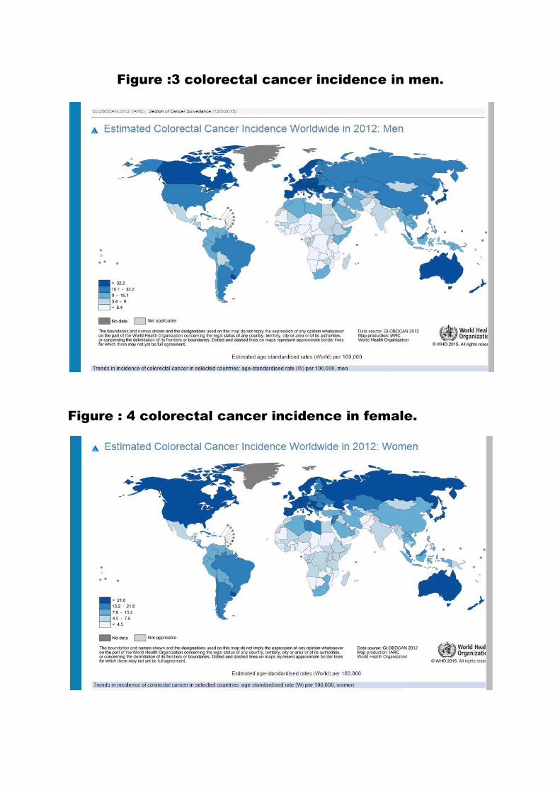
Figure :3 colorectal cancer incidence in men.
Figure : 4 colorectal cancer incidence in female.

Figure: 6 LINEAR QUADRATIC MODEL

Figure : 7 PLANE OF TOTAL MESORECTAL EXCISION

Figure : 8 CIRCUMFERRENTIAL RESECTION MARGIN
Figure : 9 MRI PICTURE SHOWING INVOLVEMENT OF CRM

Figure : 10 KRAS MUTATION

Figure : 11 MOLECULAR PROFILING

Figure : 12 COLOSCOPY SHOWING MASS LESION
Figure : 13 RESECTED SPECIMEN

Figure : 14 INKING OF THE SPECIMEN FOR CRM
Figure : 15 BREAD LOAFING SLICING TECHNIQUE

OPERATIVE PICTURES
Figure : 20 SHARP DISSECTION ALONGMESORECTUM
Figure : 21 IDENTIFYING THE PROXIMAL MARGIN

Figure : 22 RESECTED SPECIMEN
Figure : 23 ORIENTING THE SPECIMEN

EVALUTION OF SPECIMEN FOR CRM
Figure : 24
Figure : 25

Figure: 26
Figure: 27
20%
30%
43%
7%
Age Group
31- 40yrs
41 -50yrs
51-60yrs
61-70yrs
53%
47%
Gender
MALE
FEMALE

Figure: 28
Figure: 29
60%
40%
Performance Status
ECOG 0
ECOG 1
PAIN DURING DEFECATION
BLEEDING PER RECTUM
DIFFICULTY IN DEFECATION
LOWER ABDOMINAL PAIN
INCREASED FREQUENCY OFSTOOLS
LOSS OF WEIGHT
0 5 10 15 20
SYMPTOMS/SIGNS
Number of Patients

Figure: 30
Figure: 31
WELL DIFFERENTIATION
30%
MODERATELY DIFFERENTIATED
53%
POORLY DIFFERENTIATED
17%
HISTOLOGICAL DIFFERENTIATION
50% 50%
Tumor Size
2 - 4cm
4– 5 cm

Figure: 32
Figure: 33
GR I 37%
GR II 40%
GR III 23%
Histological Grade
0
2
4
6
8
10
12
14
16
18
Low anterior resection Abdominoperinealresection
Hartmans procedure
Surgery Type
Surgery Type

Figure: 34
Figure: 35
0%
100%
Margin Status
< =1 CRM
> 1 CRM
31%
69%
Y P( T ) Stage
T2
T3

Figure: 36
Figure: 37
27%
46%
27%
Distance from Anal Verge
0 – 3 cms 3 – 6 cms > 6 cms
38%
45%
17%
Node Dissection
0 – 5
5 – 10
> 10

Figure: 38
Figure: 39
60%
40%
Nodal Staging
N0 N+
2-4 CM 17%
4- 5 CM 83%
Tumor Size vs Response

Figure: 40
Figure: 41
42%
33%
25%
Histological Differentiation vs Reponse
0 – 3 CM 3 – 6 CM > 6 CM
0
5
10
15
20
25
NAUSEA VOMITING DIARRHOEA
Grade I 24 7 9
Grade II 6 2 4
Nu
mb
er
Systemic Toxicity
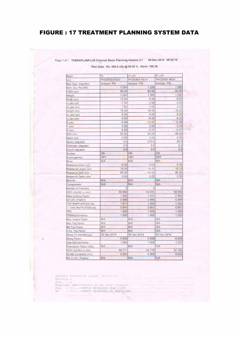
FIGURE : 17 TREATMENT PLANNING SYSTEM DATA

FIGURE : 18 PLANNING WITHOUT WEDGES
FIGURE : 19 PLANNING WITH WEDGES

76
DISCUSSION:
Although radiation has been associated with decreased rates of
local recurrence in rectal cancers, it is also associated with increase in
toxicity, compared to surgery alone. Hence patients with disease at the
lower risk of local recurrence,,like small volume disease, T3 N0 M0 ,
proximal rectal tumors may be adequately treated with short course
radiation.
The results of the present study show that neo adjuvant radiation in
the form of short course therapy is feasible in our setup and should be
considered in select cases of locally advanced rectal cancers. The study
included a wide range of patients across the age groups between the
eligibility ages of 18 years to 70 years. The study population was mostly
males 16 patients with females 14 patients.
All the patients started on the protocol completed the course of
radiation. But only 29 patients went in for curative resection which is
TME .This one single patient had only a diversion procedure. It was
because of adherence of tumor to adjacent structure. This implies
indirectly that it is important to select cases carefully for this regimen.

77
The compliance of the patients to radiation was very good , since
the radiation was only for 5 day. But few patients were reluctant to
undergo permanent colostomy. So both the patients and the family
members were to be counseled for it. Few patients were quite depressed
following colostomy, they were given continuous support and
rehabilitation during the further course of adjuvant treatment.
The patients should be counseled clearly before discharging from
the hospital, the essential part of adjuvant chemo therapy and the right
time patients had to return for the benefits of chemotherapy. Probably
literacy will play a major role in this regard with literate people being
able to grasp the consequences of defaulting adjuvant chemo therapy.
Differentiation which is probably a surrogate for the mitotic rate of the
tumor cells showed a significant correlation with response rates.
Moderately differentiated tumors had a good response in terms of
adequate reduction in tumor size as compared to other degrees of
differentiation. It is a known fact that the T size of the primary tumor will
have an impact on the immediate loco regional control as well as the
recurrence free survival. The present study has confirmed the same fact
with T size smaller tumors achieving more good responses.

78
In the present study too , tumors located more proximal to anal
verge achieved adequate tumor regression , as evidenced by change in the
T stage from T3 to T2. Polish randomisied trial and has shown that Long
course is better to short course for more distally located tumors in terms
of achieving adequate tumor downsizing and negative surgical margins.
So one must be precise in selecting cases for short course radiation.
Patients must be selected on the basis of tumor T stage – T3, no nodal
disease. The location of tumor from anal verge is also important in patient
selection. More distal tumors should be treated with long course and
proximally located tumors with short course. Preoperative assessment of
tumor tethering and fixity should be considered for choosing between
these two radiation regimens. Nodal status at the time of diagnosis plays a
crucial role in deciding the treatment options. Nodal positivity warrants
long course chemo radiation.
The Swedish rectal cancer trial demonstrated that short course
preoperative radiation reduced the risk of local recurrence by half.

79
The Dutch rectal cancer trial showed that short course
preoperative radiotherapy provides a definite benefit when it is combined
with the best surgical approach – Total Mesorectal Exicision.
The German rectal cancer trial which compared pre operative
with poat operative radiation, showed the superior benefit of pre
operative regimen in terms of local control.
The trend is consistent with the common belief that long course is
superior to short course , for a greater downsizing of the T stage, N status.
However the Polish trial did not show any apparent effect on APR rates
for rectal tumors.
In this meticulous study design, the value of short course radiation
in selected patients is proved beyond doubt
STRENGTHS OF THE STUDY:
1. The study delivered the optimal neoadjuvant treatment in the form
of short course radiation for selected patients.

80
2. The definitive part of the treatment, which is the surgery was done
following the neoadjuvant phase.
3. The optimal adjuvant chemotherapy CAPEOX was delivered
following surgery for a period of six months.
4. All the toxicities were graded using standard scale Common
Toxicity Criteria for Adverse Events Version 4.
5. The response assessment was done using a standard scale .
LIMITATIONS OF THE STUDY:
1. Only neo adjuvant radiation was given without chemotherapy.
2. The radiation was delivered using 2D techniques. Delivering with
3D conformal techniques would have been the optimal technique.
3. There was no long term follow up of the patient which would have
given the DFS and OS data for the sequential treatment.
4. The present study was a single arm study. A two arm comparative
randomized study would have been better to settle the question of
whether short course is better to long course chemo radiation.

81
CONCLUSION:
In locally advanced rectal cancers neo adjuvant treatment in the
form of chemo radiation has now become the standard of care . It has
shown to be the effective way in improving the local control rates and
survival of patients. To prevent under treatment or over treatment of
patients, accurate staging of the disease is mandatory for deciding the
treatment policy, which varies with various stages of the disease. MRI
has a major role to play in optimally staging the disease preoperatively.
Following rectal cancer surgery, irrespective of what the
pathological response is adjuvant chemotherapy is indicated. Six months
of peri operative chemotherapy is now the standard of care.
Both short course and long course pre operative regimens have
been used in parallel for more than 15 years, in most European countries.
In our country short course radiation is not widely practised. One reason
for more lenient usage of long course is hypothetical belief that
hypofractionated doses are associated with more late toxicities than long
course conventional fractionation. Secondly for the fear that short course
may not produce adequate tumor downsizing required. Encouragement

82
should be in favour of short course radiation in select patients as
discussed upon, with lower risk of disease.
FUTURE DIRECTIONS:
Future directions would require modulating the various
management strategies, using different drug combinations of
chemotherapy with targeted agents, changing the sequence of treatment
and using newer approaches to radiation therapy. The ultimate treatment
goal is to achieve good response rates and survival. The greatest
challenge will be to individualise care to improve long-term oncologic
outcome, while minimizing the treatment related toxicities and
maintaining good quality of life.

BIBLIOGRAPHY:
1. World Health Organization. Cancer Incidence in Five Continents. Lyon:
The World Health Organization and TheInternational Agency for
Research on Cancer; 2002 2. World Cancer Research Fund and
American Institute for Cancer Research. Food, Nutrition, Physical
Activity, and the Prevention of Cancer: A Global Perspective.
Washington, DC:
2. Ferlay J, Soerjomataram I, Ervik M, Dikshit R, Eser S, Mathers C,
Rebelo M, Parkin DM, Forman D, Bray, F. GLOBOCAN 2012 v1.1,
Cancer Incidence and Mortality Worldwide: IARC CancerBase No. 11
[Internet]. Lyon, France: International Agency for Research on
Cancer; 2014. Available from:http://globocan.iarc.fr
3. 2009 20 o’ Connell jB maggard MA, LIU, JH et al. Rates of colon and
rectal cancers are increasing in young adu;lts. Am surg 2003;2003
(69);866-872 21
4. Livingston EH,YoCK Colorectal cancer in youmg Am j surgery 2004;187
(3) . Davies RJ Miller R, Coleman N. Colorectal cancer screening
prospects for molecular analysis.
5. Improved survival with preoperative radiotherapy in resectable rectal
cancer. Swedish Rectal Cancer Trial. N Engl J Med 1997; 336: 980-987
6. Folkesson J, Birgisson H, Pahlman L, Cedermark B, Glimelius B,
Gunnarsson U. Swedish Rectal Cancer Trial: long lasting benefits from
radiotherapy on survival and local recurrence rate. J Clin Oncol 2005; 23:
5644-5650
7. Birgisson H, Påhlman L, Gunnarsson U, Glimelius B. Adverse effects of
preoperative radiation therapy for rectal cancer: long-term follow-up of
the Swedish Rectal Cancer Trial. J Clin Oncol 2005; 23: 8697-8705
8. Kapiteijn E, Marijnen CA, Nagtegaal ID, Putter H, Steup WH, Wiggers
T, Rutten HJ, Pahlman L, Glimelius B, van Krieken JH, Leer JW, van de
Velde CJ. Preoperative radiotherapy combined with total mesorectal
excision for resectable rectal cancer. N Engl J Med 2001; 345: 638-646
9. Peeters KC, Marijnen CA, Nagtegaal ID, Kranenbarg EK, Putter H,
Wiggers T, Rutten H, Pahlman L, Glimelius B, Leer JW, van de Velde
CJ. The TME trial after a median follow-up of 6 years: increased local
control but no survival benefit in irradiated patients with resectable rectal
carcinoma. Ann Surg 2007; 246: 693-701
10. Sauer R, Becker H, Hohenberger W, Rödel C, Wittekind C, Fietkau R,
Martus P, Tschmelitsch J, Hager E, Hess CF, Karstens JH, Liersch T,
Schmidberger H, Raab R. Preoperative versus postoperative
chemoradiotherapy for rectal cancer N Engl J Med 2004; 351: 1731-174
11. Bujko K, Nowacki MP, Nasierowska-Guttmejer A, Michalski W,
Bebenek M, Kryj M. Long-term results of a randomized trial comparing

preoperative short-course radiotherapy with preoperative conventionally
fractionated chemoradiation for rectal cancer. Br J Surg 2006; 93: 1215-
1223
12. Dusenbery KE, Alul IH, Holland EJ, Khan FM, Levitt SH. Beta
irradiation of recurrent ptergia: results and complications. Int J Radiat
Oncol Biol Phys 1992; 24: 315-320
13. Sebag-Montefiore D, Stephens RJ, Steele R, Monson J,Grieve R, Khanna
S, Quirke P, Couture J, de Metz C, Myint AS, Bessell E, Griffiths G,
Thompson LC, Parmar M. Preoperative radiotherapy versus selective
postoperative chemoradiotherap in patients with rectal cancer (MRC
CR07 and NCIC-CTG C016): a multicentre, randomised trial. Lancet
2009; 373: 811-820
14. Kusters M, Marijnen CA, van de Velde CJ, et al: Patterns of local
recurrence in rectal cancer: A study of the Dutch TME trial. Eur J Surg
Oncol 36:470-476, 2010
15. Bujko K, Nowacki MP, Nasierowska-Guttmejer A, et al: Sphincter
preservation following preoperative radiotherapy for rectal cancer: Report
of a randomised trial comparing short-term radiotherapy vs.
conventionally fractionated radiochemotherapy. Radiother Oncol 72:15-
24, 2004
16. Bujko K, Nowacki MP, Nasierowska-Guttmejer A,et al: Long-term
results of a randomized trial comparing preoperative short-course
radiotherapy with preoperative conventionally fractionated
chemoradiation for rectal cancer. Br J Surg 93:1215-1223, 2006
17. Das P, Skibber JM, Rodriguez-Bigas MA, Feig BW, Chang GJ, Wolff
RA, Eng C, Krishnan S, Janjan NA, Crane CH. Predictors of tumor
response and downstaging in patients Trakarnsanga A et al . Treatment of
locally advanced rectal cancer WJG|www.wjgnet.com 5530 October 21,
2012|Volume 18|Issue 39| who receive preoperative chemoradiation for
rectal cancer. Cancer 2007; 109: 1750-1755
18. Park JW, Lim SB, Kim DY, Jung KH, Hong YS, Chang HJ, Choi HS,
Jeong SY. Carcinoembryonic antigen as a predictor of pathologic
response and a prognostic factor in locally advanced rectal cancer patients
treated with preoperative chemoradiotherapy and surgery. Int J Radiat
Oncol Biol Phys 2009; 74: 810-817
19. Guillem JG, Chessin DB, Shia J, Moore HG, Mazumdar M, Bernard B,
Paty PB, Saltz L, Minsky BD, Weiser MR, Temple LK, Cohen AM,
Wong WD. Clinical examination following preoperative chemoradiation
for rectal cancer is not a reliable surrogate end point. J Clin Oncol 2005;
23: 3475-3479
20. Pomerri F, Pucciarelli S, Maretto I, Zandonà M, Del Bianco P, Amadio L,
Rugge M, Nitti D, Muzzio PC. Prospective assessment of imaging after

preoperative chemoradiotherapy for rectal cancer. Surgery 2011; 149: 56-
64 58 Kim SH, Lee JM, Hong SH, Kim GH, Lee
21. Wibe A, Rendedal PR, Svensson E, Norstein J, Eide TJ, Myrvold HE, et
al. Prognostic significance of the circumferential resection margin
following total mesorectal excision for rectal cancer. Br J Surg
2002;89:327-34.
22. Bipat S, Glas AS, Slors FJ, Zwinderman AH, Bossuyt PM, Stoker J.
Rectal cancer: local staging and assessment of lymph node involvement
with endoluminal US, CT, and MR imaging—a meta-analysis. Radiology
2004;232:773-83.
23. Eriksen MT, Wibe A, Hestvik UE, Haffner J, Wiig JN. Surgical
treatment of primary locally advanced rectal cancer in Norway. Eur J
Surg Oncol 2006;32:174–80.
24. Jayne DG, Thorpe HC, Copeland J, Quirke P, Brown JM, Guillou PJ.
Five-year follow-up of the Medical Research Council CLASICC trial of
laparoscopically assisted versus open surgery for colorectal cancer. Br J
Surg 2010; 97: 1638-1645
25. Kang SB, Park JW, Jeong SY, Nam BH, Choi HS, Kim DW, Lim SB,
Lee TG, Kim DY, Kim JS, Chang HJ, Lee HS, Kim SY, Jung KH, Hong
YS, Kim JH, Sohn DK, Kim DH, Oh JH. Open versus laparoscopic
surgery for mid or low rectal cancer after neoadjuvant chemoradiotherapy
(COREAN trial): short-term outcomes of an open-label randomised
controlled trial. Lancet Oncol 2010; 11: 637-6
26. Kiran RP, Lian L, Lavery IC. Does a subcentimeter distal resection
margin adversely influence oncologic outcomes in patients with rectal
cancer undergoing restorative proctectomy? Dis Colon Rectum 2011; 54:
157-163
27. Nash GM, Weiss A, Dasgupta R, Gonen M, Guillem JG, Wong WD.
Close distal margin and rectal cancer recurrence after sphincter-
preserving rectal resection. Dis Colon Rectum 2010; 53: 1365-1373
28. Quirke P, Durdey P, Dixon MF, Williams NS. Local recurrence of rectal
adenocarcinoma due to inadequate surgical resection. Histopathological
study of lateral tumour spread and surgical excision. Lancet 1986; 2: 996-
999
29. Wibe A, Rendedal PR, Svensson E, Norstein J, Eide TJ,Myrvold HE,
Søreide O. Prognostic significance of the circumferential resection
margin following total mesorectal excision for rectal cancer. Br J Surg
2002; 89: 327-334 Baik SH, Kim NK, Lee YC, Kim H, Lee KY, Sohn
SK, Cho
30. CH. Prognostic significance of circumferential resection margin
following total mesorectal excision and adjuvant chemoradiotherapy in
patients with rectal cancer. Ann Surg Oncol 2007; 14: 462-469

31. Nagtegaal ID, Marijnen CA, Kranenbarg EK, van de VeldeCJ, van
Krieken JH. Circumferential margin involvement is still an important
predictor of local recurrence in rectal carcinoma: not one millimeter but
two millimeters is the limit. Am J Surg Pathol 2002; 26: 350-357
32. Bernstein TE, Endreseth BH, Romundstad P, Wibe A. Circumferential
resection margin as a prognostic factor in rectal cancer. Br J Surg 2009;
96: 1348-1357
33. Gosens MJ, Klaassen RA, Tan-Go I, Rutten HJ, Martijn H,van den Brule
AJ, Nieuwenhuijzen GA, van Krieken JH,Nagtegaal ID. Circumferential
margin involvement is the crucial prognostic factor after multimodality
treatment in patients with locally advanced rectal carcinoma. Clin Cancer
Res 2007; 13: 6617-6623
34. Kelly SB, Mills SJ, Bradburn DM, Ratcliffe AA, BorowskiDW. Effect of
the circumferential resection margin on survival following rectal cancer
surgery. Br J Surg 2011; 98:573-581
35. Guidelines for the management of colorectal cancer. 3rd ed.The
Association of Coloproctology of Great Britain and Ireland,2007
36. André T, Boni C, Mounedji-Boudiaf L, Navarro M, Tabernero J, Hickish
T, Topham C, Zaninelli M, Clingan P Bridgewater J, Tabah-Fisch I, de
Gramont A. Oxaliplatin, fluorouracil, and leucovorin as adjuvant
treatment for colon cancer. N Engl J Med 2004; 350: 2343-2351
37. Twelves C, Wong A, Nowacki MP, Abt M, Burris H, Carrato A, Cassidy
J, Cervantes A, Fagerberg J, Georgoulias V,Husseini F, Jodrell D,
Koralewski P, Kröning H, Maroun J, Marschner N, McKendrick J,
Pawlicki M, Rosso R, SchüllerJ, Seitz JF, Stabuc B, Tujakowski J, Van
Hazel G, Zaluski J, Scheithauer W. Capecitabine as adjuvant treatment
for stage III colon cancer. N Engl J Med 2005; 352: 2696-2704
38. Sauer R, Becker H, Hohenberger W, Rödel C, Wittekind C, Fietkau R,
Martus P, Tschmelitsch J, Hager E, Hess CF,Karstens JH, Liersch T,
Schmidberger H, Raab R. Preoperative versus postoperative
chemoradiotherapy for rectal cancer. N Engl J Med 2004; 351: 1731-1740
39. Bazarbashi S, El-Bassiouni M, Abdelsalam M, Soudy H,Sanea NA,
Jabbar AA, Manji M, Fagih M, Ajarim D. A modern regimen of pre-
operative concurrent chemo-radiation therapy in locally advanced rectal
cancer. J Surg Oncol 2008; 98: 167-174
40. Craven I, Crellin A, Cooper R, Melcher A, Byrne P, Sebag-Montefiore D.
Preoperative radiotherapy combined with 5 days per week capecitabine
chemotherapy in locally advanced rectal cancer. Br J Cancer 2007; 97:
1333-1337
41. Kim JS, Kim JS, Cho MJ, Song KS, Yoon WH. Preoperative
chemoradiation using oral capecitabine in locally advanced rectal cancer.
Int J Radiat Oncol Biol Phys 2002; 54: 403-408

42. Dupuis O, Vie B, Lledo G, Hennequin C, Noirclerc M, BennamounM,
Jacob JH. Preoperative treatment combining capecitabine with radiation
therapy in rectal cancer: a GERCOR Phase II Study. Oncology 2007; 73:
169-176
43. Roh MS. The impact of capecitabine and oxaliplatin in thepreoperative
multimodality treatment in patients with carcinoma of the rectum:
NSABP R-04. Proceedings of ASCO Annual Meeting; 2011: Abstract
No.3053
44. Hofheinz R. Capecitabine versus 5-fluorouracil-based(neo)adjuvant
chemoradiotherapy for locally advanced rectal cancer: Long-term results
of a randomized, phase III trial. Proceedings of ASCO Annual Meeting;
2011: Abstract No.3054
45. Hofheinz R. Capecitabine versus 5-fluorouracil-based(neo)adjuvant
chemoradiotherapy for locally advanced rectal cancer: Long-term results
of a randomized, phase III trial. Proceedings of ASCO Annual Meeting;
2011: AbstractNo.3054

Appendix I – Tables
1 TUMOR REGRESSION GRADING
2 RT PORTAL MARGINS PA PORTAL
3 RT PORTAL MARGIN LATERAL PORTAL
4 BIOLOGICALLY EQUIVALENT DOSE
5 AGE DISTRIBUTION OF THE STUDY POPULATION
6 GENDER DISTRIBUTION OF THE STUDY POPULATION
7 ECOG PERFORMANCE STATUS
8 SYMPTOMS/SIGNS
9 HISTOLOGICAL DIFFERENTIATION
10 SIZE OF TUMOR
11 HISTOLOGICAL GRADE
12 TYPE OF SURGERY
13 CIRCUMFERRENTIAL MARGIN STATUS
14 Y P( T ) STAGE
15 DISTANCE FROM ANAL VERGE
16 NUMBER OF LYMPH NODES DISSECTED
17 NODAL STAGING
18 TUMOR SIZE Vs RESPONSE
19 DISTAL RECTAL MARGIN Vs RESPONSE
20 HISTOLOGICAL DIFFERENTIATION Vs RESPONSE
21 SYSTEMIC TOXICITY

Appendix II – Figures
1 ESTIMATED INCIDENCE, MORTALITY AND PREVALENCE
2 MALE AND FEMALE DISTRIBUTION IN VARIOUS REGIONS
3 COLORECTAL CANCER INCIDENCE IN MEN
4 COLORECTAL CANCER INCIDENCE IN FEMALE
5 PATHOGENISIS
6 LINEAR QUADRATIC MODEL
7 PLANE OF TOTAL MESORECTAL EXCISION
8 CIRCUMFERRENTIAL RESECTION MARGIN
9 MRI PICTURE SHOWING INVOLVEMENT OF CRM
10 KRAS MUTATION
11 MOLECULAR PROFILING
12 COLOSCOPY SHOWING MASS LESION
13 RESECTED SPECIMEN
14 INKING OF THE SPECIMEN FOR CRM
15 BREAD LOAFING SLICING TECHNIQUE
16 BIOLOGICALLY EQUIVALENT DOSE
17 TREATMENT PLANNING
18 TREATMENT PLANNING WITH WEDGES
19 TREATMENT PLANNING WITHOUT WEDGES
20 SHARP DISSECTION ALONGMESORECTUM
21 IDENTIFYING THE PROXIMAL MARGIN
22 RESECTED SPECIMEN

23 ORIENTING THE SPECIMEN
24 EVALUTION OF SPECIMEN FOR CRM
25 EVALUTION OF SPECIMEN FOR CRM
26 AGE GROUP
27 GENDER DISTRIBUTION
28 PERFORMANCE STATUS
29 SIGNS / SYMPTOMS
30 HISTOLOGICAL DIFFERENTIATION
31 TUMOR SIZE
32 GRADE
33 TYPE OF SURGERY
34 MARGIN STATUS
35 PATHOLOGICAL; T STAGE
36 DISTANCE FROM ANAL VERGE
37 NODES DISSECTED
38 NODAL STAGING
39 TUMOR SIZE VS RESPONSE
40 HISTOLOGICAL DIFFERENTIATION VS RESPONSE
41 SYSTEMIC TOXICITY

Annexure I
Information to Participants
Title: “NEOADJUVANT SHORT COURSE RADIOTHERAPY FOLLOWED BY
SURGERY IN LOCALLY ADVANCED RECTAL CANCERS”
Principle Investigator: Dr.Madhulika vijayakumar.
Name of Participant:
Site: Department of Radiotherapy, Madras Medical College & RGGGH,
Chennai-3
You are invited to take part in this research/ study/procedure. The information
in this document is meant to help you decide whether or not to take part.
Please feel free to ask if you have any queries.
What is the purpose of this study?
The incidence of rectal cancers has been increasing worldwide due to
economic development and industrialization. Surgery is the primary modality
of treatment in spite of it, local and distant recurrence rates are high.pre
operative treatment before surgery has shown significant reduction in the local
recurrence and distal failure rates.pre operative treatment in the form of short
course radiation for 5 days followed by surgery has shown promising results in
tumor downstaging and good response rates. We want to assess the
immediate loco regional response rates and treatment related toxicity.
We have obtained permission from the Institutional Ethics Committee.
The study design: Single arm Prospective study.
Study Procedures:
The study involves assessment of response and toxicity in locally
advanced rectal cancers treated with short course pre operative radiotherapy

followed by surgery, which will need complete blood investigations, CT chest,
MRI pelvis and assessment by surgeon &anaesthetist as a part of standard
protocol for any other patients receiving radiotherapy. Following radiotherapy
you will be undergoing surgery after 2 weeks .The tests are essential to
monitor your condition, and to assess the safety and efficacy of treatment. You
may have to come to the hospital for examination and investigations apart
from your scheduled visits, if required.
Possible risks to you: None greater than standard patients receiving
radiotherapy.
Possible benefits to you: Shorter treatment duration during time compared to
patients treated with conventional fractions.
Possible benefits to other people: The results of the research may provide
benefits to the society in terms of advancement of medical knowledge and/or
therapeutic benefit to future patients.
Confidentiality of the information obtained from you:
You have the right to confidentiality regarding the privacy of your
medical information (personal details, results of physical examinations,
investigations, and your medical history).By signing this document, you will be
allowing the research team investigators, other study personnel, sponsors,
Institutional Ethics Committee and any person or agency required by law like
the Drug Controller General of India to view your data, if required. The
information from this study, if published in scientific journals or presented at
scientific meetings, will not reveal your identity.

How will your decision to not participate in the study affect you?
Your decision not to participate in this research study will not affect your
medical care or your relationship with the investigator or the institution. You
will be taken care of and you will not lose any benefits to which you are
entitled.
Can you decide to stop participating in the study once you start?
The participation in this research is purely voluntary and you have the
right to withdraw from this study at any time during the course of the study
without giving any reasons. However, it is advisable that you talk to the
research team prior to stopping the treatment/discontinuing of procedures
etc.
Signature of Investigator Signature of Participant
Date Date

Annexure II
INFORMED CONSENT FORM
TITLE OF THE STUDY: “ NEOADJUVANT SHORT COURSE RADIOTHERAPY FOLLOWED BY SURGERY IN
LOCALLY ADVANCED RECTAL CANCERS.
NAME OF THE PARTICIPANT:
NAME OF THE PRINCIPAL(Co – Investigator ): DR.Madhulika Vijayakumar.
NAME OF THE INSTITUTION: MADRAS MEDICAL COLLEGE
I, _____________________________ have read the information in this form (or it has been read to
me). I was free to ask any questions and they have been answered. I am over 18 years of age and,
exercising my free power of choice, hereby give my consent to be included as a participant in “
NEOADJUVANT SHORT COURSE RADIOTHERAPY FOLLOWED BY SURGERY IN LOCALLY ADVANCED
RECTAL CANCERS ”
1. I have read and understood this consent form and the information provided to me.
2. I have had the consent document explained to me.
3. I have been explained about the nature of the study.
4. I have been explained about my rights and responsibilities by the investigator.
5. I have been informed the investigator of all the treatments I am taking or have taken in
the past 12 months including any native (alternative) treatment.
6. I have been advised about the risks associated with my participation in this study.*
7. I agree to cooperate with the investigator and I will inform him/her immediately if I suffer
unusual symptoms.*
8. I have not participated in any research study within the past 12month(s). *
9. I agree to undergo complete blood count, renal and liver function test, CT scan chest,
MRI pelvis.
10. I am aware of the fact that I can opt out of the study at any time without having to give
any reason and this will not affect my future treatment in this hospital. *
11. I am also aware that the investigator may terminate my participation in the study at any
time, for any reason, without my consent. *
12. I hereby give permission to the investigators to release the information obtained from me
as result of participation in this study to the sponsors, regulatory authorities, Govt.
agencies, and IEC. I understand that they are publicly presented.
13. I have understand that my identity will be kept confidential if my data are publicly
presented.
14. I have had my questions answered to my satisfaction.
15. I have decided to be in the research study.

I am aware that if I have any question during this study, I should contact the investigator. By signing
this consent form I attest that the information given in this document has been clearly explained to
me and understood by me, I will be given a copy of this consent document
Name and signature / thumb impression of the participant (or legal representative if participant
incompetent)
Name ________________ Signature_________________ Date________________
Name and Signature of impartial witness (required for illiterate patients):
Name ________________ Signature_________________ Date________________
Address and contact number of the impartial witness:
Name and Signature of the investigator or his representative obtaining consent
Name ________________ Signature_________________ Date________________

Annexure III
:
.
:
:
. . . .
:
. , . . 5 ,

. , .
.
:
, 5 , . , . , . , . .
:
. .
:
( ,

, ). . .
.
; .
.
: :

Annexure IV
:
.
: : : :
________________________________ , , . . .
. .
. .
, . ,

ANNEXURE V

.
, .
, , . . .
, .
. .
: :