Comparative Proteomics Reveals a Role for Seed Storage Protein AmA1 in Cellular Growth, Development,...
27
Comparative Proteomics Reveals a Role for Seed Storage Protein AmA1 in Cellular Growth, Development, and Nutrient Accumulation Lalit Agrawal, †,§ Kanika Narula, ‡,§ Swaraj Basu, ‡ Shubhendu Shekhar, † Sudip Ghosh, ‡ Asis Datta, ‡ Niranjan Chakraborty,* ,† and Subhra Chakraborty* ,‡ † Laboratory 104 and ‡ Laboratory 105, National Institute of Plant Genome Research, Aruna Asaf Ali Marg, New Delhi 110067, India * S Supporting Information ABSTRACT: Seed storage proteins are known to be utilized as carbon and nitrogen source for growing seedlings and thus are considered as potential candidates for nutritional improvement. However, their precise function remains unknown. We have earlier shown that ectopic expression of a seed storage protein, AmA1, leads to increase in protein besides high tuber yield in potato. To elucidate the AmA1-regulated molecular mechanism affecting increased protein synthesis, reserve accumulation, and enhanced growth, a comparative proteomics approach has been applied to tuber life-cycle between wild-type and AmA1 potato. The differential display of proteomes revealed 150 AmA1-responsive protein spots (ARPs) that change their intensities more than 2.5-fold. The LC−ESI-MS/MS analyses led to the identification of 80 ARPs presumably associated with cell differentiation, regulating diverse functions, viz., protein biogenesis and storage, bioenergy and metabolism, and cell signaling. Metabolome study indicated up-regulation of amino acids paralleling the proteomics analysis. To validate this, we focused our attention on anatomical study that showed differences in cell size in the cortex, premedullary zone and pith of the tuber, coinciding with AmA1 expression and localization. Further, we interrogated the proteome data using one-way analysis of variance, cluster, and partial correlation analysis that identified two significant protein modules and six small correlation groups centered around isoforms of cysteine protease inhibitor, actin, heat shock cognate protein 83 and 14-3-3, pointing toward AmA1-regulated overlapping processes of protein enhancement and cell growth perhaps through a common mechanism of function. A model network was constructed using the protein data sets, which aim to show how target proteins might work in coordinated fashion and attribute to increased protein synthesis and storage reserve accumulation in AmA1 tubers on one hand and organ development on the other. KEYWORDS: seed storage protein, AmA1, potato, comparative proteomics, metabolomics, nutrient accumulation, 2-DE, mass spectrometry, protein network ■ INTRODUCTION Nutritional quality and agricultural productivity are the two key issues to sustainable food production worldwide. This is exemplified several times in the context of an ever-growing population where human health largely depends on plants. Storage organs display diverse nutritional quality and complex multistep development and act as sinks in plants. The composition of nutrients in the storage organs, carbon (C) and nitrogen (N) in particular, greatly influence the organ development and determine the nutritive quality. Both C and N metabolites are known to act as signals that influence many cellular processes, for example, development, metabolism related to N assimilation, and amino acid synthesis, in addition to their essential role as macronutrients in living organisms, including plants. 1−11 Seed storage proteins in plants, rich in essential amino acids, are thought to serve as C and N source for the growing seedling 12 and meet the major dietary protein requirement of over half of the world population. 13 Thus, they presumably play an essential role in productivity and protein quality and have received considerable attention due both to their postulated dual role in growth and importance as a component of the human diet. Intensive research directed toward isolation and characterization of these seed storage proteins not only has allowed advances in our understanding of the synthesis, accumulation, processing, and transcriptional control of their genes 14−17 but also has facilitated the targeted genetic engineering of crop plant’s nutritional status. 18−22 Despite the fact that there are many studies concerning the genetic control and spatial and temporal regulation of seed storage proteins, our current knowledge about their exact functional role and physiological relevance remains unknown. Only recently focus has been given to study crop plants overexpressing storage protein; 23,24 however, the modus operandi of cellular network and physiological consequences toward sensing a storage protein has not been elucidated. Such Special Issue: Agricultural and Environmental Proteomics Received: August 2, 2013 Article pubs.acs.org/jpr © XXXX American Chemical Society A dx.doi.org/10.1021/pr4007987 | J. Proteome Res. XXXX, XXX, XXX−XXX
Transcript of Comparative Proteomics Reveals a Role for Seed Storage Protein AmA1 in Cellular Growth, Development,...

Comparative Proteomics Reveals a Role for Seed Storage ProteinAmA1 in Cellular Growth, Development, and Nutrient AccumulationLalit Agrawal,†,§ Kanika Narula,‡,§ Swaraj Basu,‡ Shubhendu Shekhar,† Sudip Ghosh,‡ Asis Datta,‡
Niranjan Chakraborty,*,† and Subhra Chakraborty*,‡
†Laboratory 104 and ‡Laboratory 105, National Institute of Plant Genome Research, Aruna Asaf Ali Marg, New Delhi 110067, India
*S Supporting Information
ABSTRACT: Seed storage proteins are known to be utilized as carbon andnitrogen source for growing seedlings and thus are considered as potentialcandidates for nutritional improvement. However, their precise functionremains unknown. We have earlier shown that ectopic expression of a seedstorage protein, AmA1, leads to increase in protein besides high tuber yield inpotato. To elucidate the AmA1-regulated molecular mechanism affectingincreased protein synthesis, reserve accumulation, and enhanced growth, acomparative proteomics approach has been applied to tuber life-cyclebetween wild-type and AmA1 potato. The differential display of proteomesrevealed 150 AmA1-responsive protein spots (ARPs) that change theirintensities more than 2.5-fold. The LC−ESI-MS/MS analyses led to theidentification of 80 ARPs presumably associated with cell differentiation,regulating diverse functions, viz., protein biogenesis and storage, bioenergyand metabolism, and cell signaling. Metabolome study indicated up-regulation of amino acids paralleling the proteomics analysis.To validate this, we focused our attention on anatomical study that showed differences in cell size in the cortex, premedullaryzone and pith of the tuber, coinciding with AmA1 expression and localization. Further, we interrogated the proteome data usingone-way analysis of variance, cluster, and partial correlation analysis that identified two significant protein modules and six smallcorrelation groups centered around isoforms of cysteine protease inhibitor, actin, heat shock cognate protein 83 and 14-3-3,pointing toward AmA1-regulated overlapping processes of protein enhancement and cell growth perhaps through a commonmechanism of function. A model network was constructed using the protein data sets, which aim to show how target proteinsmight work in coordinated fashion and attribute to increased protein synthesis and storage reserve accumulation in AmA1 tuberson one hand and organ development on the other.
KEYWORDS: seed storage protein, AmA1, potato, comparative proteomics, metabolomics, nutrient accumulation, 2-DE,mass spectrometry, protein network
■ INTRODUCTION
Nutritional quality and agricultural productivity are the two keyissues to sustainable food production worldwide. This isexemplified several times in the context of an ever-growingpopulation where human health largely depends on plants.Storage organs display diverse nutritional quality and complexmultistep development and act as sinks in plants. Thecomposition of nutrients in the storage organs, carbon (C)and nitrogen (N) in particular, greatly influence the organdevelopment and determine the nutritive quality. Both C and Nmetabolites are known to act as signals that influence manycellular processes, for example, development, metabolismrelated to N assimilation, and amino acid synthesis, in additionto their essential role as macronutrients in living organisms,including plants.1−11 Seed storage proteins in plants, rich inessential amino acids, are thought to serve as C and N sourcefor the growing seedling12 and meet the major dietary proteinrequirement of over half of the world population.13 Thus, theypresumably play an essential role in productivity and proteinquality and have received considerable attention due both to
their postulated dual role in growth and importance as acomponent of the human diet. Intensive research directedtoward isolation and characterization of these seed storageproteins not only has allowed advances in our understanding ofthe synthesis, accumulation, processing, and transcriptionalcontrol of their genes14−17 but also has facilitated the targetedgenetic engineering of crop plant’s nutritional status.18−22
Despite the fact that there are many studies concerning thegenetic control and spatial and temporal regulation of seedstorage proteins, our current knowledge about their exactfunctional role and physiological relevance remains unknown.Only recently focus has been given to study crop plantsoverexpressing storage protein;23,24 however, the modusoperandi of cellular network and physiological consequencestoward sensing a storage protein has not been elucidated. Such
Special Issue: Agricultural and Environmental Proteomics
Received: August 2, 2013
Article
pubs.acs.org/jpr
© XXXX American Chemical Society A dx.doi.org/10.1021/pr4007987 | J. Proteome Res. XXXX, XXX, XXX−XXX

finding will not only impact plant biology but in the near futurewould be useful for identifying biomarkers, prioritizingmolecular targets, and pathway bioengineering for cropimprovement.Cellular physiology is based on the level of proteins, their
activation or deactivation by post-translational modifications,and the metabolite pool. The identification of a protein and itssite of modification can be obtained using MS/MS analyses,while the tandem development of metabolite profiling by gaschromatography−mass spectrometry (GC−MS) paves the wayto a more substantial base of phenome study. Proteomics allowscomprehensive characterization of proteomes, and despite thedevelopment of novel gel-free technologies, two-dimensionalgel electrophoresis (2-DE) coupled with mass spectrometryallows evaluation of expression of hundreds of proteins andtheir isoforms or post-translational modifications and thusappears to be the technique of choice for protein expressionstudies.25−28 Isoforms are highly regulated gene products thatcan differ in their biological activity, regulatory properties,temporal and spatial expression, intracellular location, or anycombination thereof. The significance of their existence can beattributed to their additive and/or differential function, whichultimately culminates in a cumulative and rapid response tophysiological state. Therefore, isoforms are generally consid-ered to diversify the function of a protein. The ability of thisdiscovery approach to produce completely unpredictable andnovel findings by application of a systematic process has beenauthenticated in many studies.29,30 Understanding the bio-logical complexities in a cell upon genetic, epigenetic, andexternal perturbations can be improved and expanded byexploiting proteomic studies that provide comprehensiveevaluation of molecular phenotypes and additional informationabout gene function and cellular pathways.31 Proteomicanalyses of plant organs or tissues have been used to monitordevelopmental changes in seed32,33 and tuber,34,35 environ-mental stress responses,36−38 comparison of plant varieties, andmutant characterization39 in addition to recent study oftransgenic crops.31,40−43 Metabolomics is an imperative toolto recognize new protein function for functional proteomics.When combined with proteome studies, metabolite profilingreveals unanticipated insights into the diverse regulatorypathways to draw a comprehensive and integrated outlook ofcellular metabolism.44,45 The large amount of data generatedfrom such studies requires the use of appropriate statisticalmethods to extract the information of interest. Clusteringalgorithms and principle component analysis (PCA) identifypatterns of expression that may suggest co-regulation, whilePearson partial correlation is one of the ways to verify thebiological relevance of individual protein in the inferredcorrelation network.46 In addition, the emergence of apostgenomic view expands the protein’s role, regarding it asan element in a network of ‘contextual’ or ‘cellular’ functionswithin functional modules.47
AmA1, a storage protein, was reported to be expressed inseed tissue of pseudocereal Amaranthus hypochondriacus anddisappear during germination and seedling growth.15 Theprotein was synthesized during the grain development likeother storage proteins.48 It was one of the first characterizedprotein encoded by a nonallergenic seed storage albumingene.15,20,49,50 In addition, we and others have shown thatAmA1 regulates the biology of nutrition, and its ectopicexpression in storage organs such as tuber and seed helpsincrease protein and amino acid accumulation.20,21 Further,
AmA1 plays an important physiological role during organdevelopment and is crucial for the homoblastic growth ofpotato tuber, the gradual transition cascade from stolon tomature tuber.20,22 Thus we earlier hypothesized that inductionof protein synthesis or mitogenic activity might be the result ofoverexpression of AmA1 or its turned-over products as signalmolecule.20 In a recent study, our preliminary data suggestedthat proteome rebalancing due to AmA1 expression might leadto nutrient enhancement and increased yield.22 It is thusconceivable that AmA1 might play a crucial role during seedgermination and seedling growth as a nutrient source andgrowth-promoting substance. Further, seed storage proteinsapart from their possible role in serving storage function havealso been implicated in defense and environmental stressresponse.51−54 Likewise, we hypothesize that AmA1 might havesimilar functions in Amaranthus hypochondriacus. Despite theabove research linking AmA1 with nutritional enhancement andincreased growth, its biological function remains to be clarified.Following these observations, and given that we have a long-standing interest in understanding the role of the seed storageprotein in plants, we decided to investigate the role of seedprotein AmA1 on the observed homoblastic developmentalchanges and nutrient enhancement.Here, we report the comparative proteome analysis of AmA1
potato and that of the wild-type at four different stages oftuberization. It was reasoned that a differential proteomeanalysis of AmA1 potato might broaden our understanding offunctional protein and signaling networks involved in storageprotein regulated pathways in nutrient signaling and growth inplants. In addition, we provide a global view of how proteinnetworks are modulated in response to AmA1 sensing. Wecombined the quantitative models describing the proteinexpression changes and correlation network of the cell inresponse to AmA1 that allowed us to globally identify a set ofprotein subnetworks affected by the storage protein. Theseresults are discussed in the context of current theories of thecellular and metabolic cues underlying nutrient accumulationand development. The differences in protein expression patternand function appeared to encompass diverse metabolic andsignaling pathways that provide new insights into theunderlying mechanisms, which might contribute to alterednutritive values and increased tuber yield. Furthermore, thepathways identified by comparative proteomics were validatedby analyzing the metabolome.
■ EXPERIMENTAL SECTION
Plant Material and Experimental Design
The transgenic potato genotypes 35S-AmA1 (A16/1) andGBSS-AmA1 (A16/6), overexpressing a seed storage proteinAmA1 constitutively and tuber-specifically, respectively, alongwith the wild-type genotype, A16 described earlier,20 were usedin the present study. A complete randomized design was usedfor growing size normalized seed tubers of wild-type and AmA1potato in three replicate plots with 45 tubers per replicate for100−110 days, until tuber maturation. The wild-type andAmA1 expressing lines were grown side-by-side in replicatedplots at identical conditions to eliminate the environmentalinfluence on the varieties, if any, and therefore should reflectthe effects of the transgene only. Rows were spaced 60 cmapart, and plants were spaced 20 cm apart within each row inthe replication block. Standard cultivation and managementpractices were followed throughout the growing season. Tubers
Journal of Proteome Research Article
dx.doi.org/10.1021/pr4007987 | J. Proteome Res. XXXX, XXX, XXX−XXXB

were planted in the month of October when the temperature isapproximately 20−24 °C in the daytime and 15−18 °C atnight. Tuberization occurred 45 days after planting at lowtemperature (15−18 °C in the daytime and 8−10 °C at night)and short-day photoperiod condition. The following devel-opmental stages were selected and harvested: stolons at 6−8weeks, stage 1 (S1); swollen stolons or initial tubers at 8−10weeks, stage 2 (S2); developing tubers at 10−12 weeks, stage 3(S3); and mature tubers at 14−15 weeks, stage 4 (S4). Tomaintain uniformity among the replicates, tubers wereseparated according to plantation time, morphology, tuberweight, and diameter of the wild-type and AmA1 potatoes forfurther analysis as described earlier.34,35 The average weights ofeach of the aforesaid developmental stages were 0.075, 0.510,0.935, and 2.165 g with average diameter of 3.125, 5.75, 9.125,and 14.00 mm, respectively, in the wild-type tubers. However,the average weight of the AmA1 tubers at these fourdevelopmental stages in 35S-AmA1 and GBSS-AmA1 were0.20 ± 0.025, 1.40 ± 0.151, 5.30 ± 0.512, and 16.0 ± 1.425 gand the average diameters were 6.5 ± 0.635, 12.5 ± 1.170, 22.5± 2.121, and 30.0 ± 3.55 mm, respectively (Figure 1A−C).Tubers were collected from three replicate plots and pooled tonormalize the effect of variation in the biological replicates.
Each biological replicate of a specific developmental stage foreither wild-type or AmA1 potato lines consisted of 30−50tubers for S1, 20−30 tubers for S2, 10−15 tubers for S3, and 10tubers for S4 obtained from 10 different plants (SupportingInformation Figure 1). Tubers were stored at −80 °C forfurther use after quick-freezing in liquid nitrogen.
Physiological and Morphological Characterization
We measured the photosynthetic activity in the transgenic andwild-type plants at 8 weeks of plantation under standardatmospheric (360 ppm CO2) and light (750 μmol/m2/s)conditions. Photosynthetic activity, leaf area, fresh weight anddry weight of wild-type and AmA1 plants were measured asdescribed earlier.22
Isolation of Tuber Proteins and 2-Dimensional GelElectrophoresis
Soluble proteins were isolated from pooled potato tubers fromeach developmental stages of wild-type and AmA1 potato fromthree replicate plots to normalize the effect of variations in thebiological replicates as described earlier.35 Protein concen-tration was determined by Bradford assay (Bio-Rad). The tuberproteins were diluted in dilution buffer [100 mM, Tris-Cl (pH8.5), 20% (v/v) glycerol, 8% (w/v) SDS, 20 mM DTT, 1 mMPMSF] and boiled for 5 min.55 Protein samples were allowed tocool at room temperature (25 °C) and precipitated with 9 volof 100% chilled acetone overnight at −20 °C. The precipitateswere recovered by centrifugation at 10,000g at 4 °C, for 10 min.Protein pellets were washed twice with 80% acetone to removeexcess SDS, air-dried, and resuspended in 2-D rehydrationbuffer [8 M urea, 2 M thiourea, 4% (w/v) CHAPS, 20 mMDTT, 0.5% (v/v) Pharmalyte (pH 4−7) and 0.05% (w/v)bromophenol blue]. Isoelectric focusing was carried out with250 μg of protein. Protein was loaded by in-gel rehydrationmethod onto 13-cm IEF strips (pH 4−7), and electrofocusingwas performed using an IPGphor system (AmershamBiosciences, Bucks, U.K.) at 20 °C for 25,000 Vh. The focusedstrips were subjected to reduction with 1% (w/v) DTT in 10mL of equilibration buffer [6 M urea, 50 mM Tris-HCl (pH8.8), 30% (v/v) glycerol and 2% (w/v) SDS], followed byalkylation with 2.5% (w/v) iodoacetamide in the same buffer.The strips were then loaded on top of 12.5% polyacrylamidegels for SDS-PAGE. The electrophoresed proteins were stainedwith a silver stain plus kit (Bio-Rad).
Image Acquisition and Data Analysis
After two-dimensional gel electrophoresis and gel staining, gelimages were scanned using the Bio-Rad FluorS systemequipped with a 12-bit camera. PDQuest version 7.2.0 (Bio-Rad) was used to assemble the first level match set (masterimage) from three replicate 2-DE gels. For each developmentalstage, at least three 2-DE gels, representing three biologicalreplicates, were used for the data analysis. The detailed dataanalyses were carried out as described previously.35,36 Assess-ment of protein spot quality, molecular mass, and pI ofindividual protein was determined as described previously.35
Each spot included on the standard gel met several criteria: itwas present in at least two of the three gels and wasqualitatively consistent in size and shape in the replicate gels.The low quality spots scoring less than 30 quality score wereeliminated from further analysis. The remaining high-qualityspots were used to calculate the mean value for a given spot,and this value was used as the spot quantity on the standard gel(Supporting Information Document 1). The filtered spot
Figure 1. AmA1-induced growth response in potato tuber. (A)Photographs of various stages of tuber development: stolon, initialtuber, developing tuber, and mature tuber. The developmental stageswere based on tuber weight and diameter as detailed in the Resultssection. The fresh weight (B) and the tuber diameter (C) of theharvested tubers were determined and plotted against each stage oftuberization process. Data represent means ± SD of three measure-ments: (left bar) wild-type, (middle bar) 35S-AmA1, and (right bar)GBSS-AmA1.
Journal of Proteome Research Article
dx.doi.org/10.1021/pr4007987 | J. Proteome Res. XXXX, XXX, XXX−XXXC

quantities from the standard gels were assembled into a datamatrix of high quality spots from four developmental stages forfurther analysis. Protein spot detection and quantification wereobtained using normalized spot volumes given by PDQuestsoftware using the total spot volume normalization procedureto discard experimental variations in 2-DE gels.
Protein Identification and Expression Clustering
The differentially expressed protein spots were excisedmechanically using pipet tips, and in-gel digested with trypsin,and peptides were extracted according to standard techni-ques.35,38 For LC−MS/MS analysis, trypsin-digested peptideswere loaded onto a C18PepMap100 column (3 μm, 100 Å, 75μm i.d., 15 cm) at 300 nL/min (LCPackings), separated with alinear gradient of water/acetonitrile/0.1% formic acid (v/v),and analyzed by electrospray ionization using the ultimate 3000nano HPLC system (Dionex) coupled to either a 4000 Q-TRAP mass spectrometer (Applied Biosystems) or a Q-StarPulsar i time-of-flight mass spectrometer (Applied Biosystems).The peptides were eluted with a gradient of 10−40%acetonitrile (0.1% formic acid) over 60 min. The MS/MSdata were extracted using Analyst software, version 1.5.1(Applied Biosystems). Peptide analysis was performed throughdata-dependent acquisition of MS scan (m/z 400−1800)followed by MS/MS scans. Peptides were identified bysearching the peak-list against the Potato Genome SequenceConsortium (PGSC_DM_v3.4) (56218 sequences, 16895844residues) available at http://potatogenomics.plantbiology.msu.edu/index.html using MASCOT v.2.1 (http://www.matrixsciences.com) search engine. The database search criteriawere as follows: taxonomy, all entries; peptide tolerance, ± 1.2Da; MS/MS tolerance, ± 0.6 Da; peptide charge +1, +2, or +3;maximum allowed missed cleavage, 1; fixed modification,cysteine carbamidomethylation; variable modification, methio-nine oxidation; instrument type, Default. Protein scores werederived from ion scores as a nonprobabilistic basis for rankingprotein hits and as the sum of a series of peptide scores. Thescore threshold to achieve P < 0.05 was set by the Mascotalgorithm and was based on the size of the database used in thesearch. We considered only those protein spots whoseMOWSE score was above the significant threshold leveldetermined by Mascot. Proteins with a confidence intervalpercentage of greater than 95% were considered to represent apositive identification and were also evaluated on the basis ofvarious parameters, such as the number of peptides matched,MOWSE score, and percent coverage of the matched protein.In all of the protein identifications, the probability scores weregreater than the score fixed by MASCOT as significant with a Pvalue <0.05. The abundance of each identified protein wasestimated by determining the protein abundance index (PAI)56
and the emPAI.57 The corresponding protein content in mol %was calculated as described previously.57 For the total numberof observed peptides per protein, the unique sequences werecounted and were imported to Microsoft Excel (SupportingInformation Table 1). Where there was more than oneaccession number for the same peptide, the match wasconsidered in terms of the putative function. The proteinfunctions were assigned using the Pfam (http://pfam.sanger.ac.uk) and InterPro (http://www.ebi.ac.uk/interpro) proteinfunction databases. Self-organizing tree algorithm (SOTA)clustering was performed on the log-transformed fold inductionexpression values across different developmental stages oftuberization using Multi Experiment Viewer (MEV) software.58
The clustering was done with the Pearson correlation asdistance with 10 cycles and a maximum cell diversity of 0.8.59
Isoform Clustal Analysis
The web interface SPECLUST (http://bioinfo.thep.lu.se/speclust.html) was used to construct dendrogram based onLC−MS/MS data for potato proteins. For statistical analysis,the processed mass lists were converted into SPECLUST inputformat files. This program compares m/z peak lists bymeasuring a distance between each pair of lists. The webinterface calculates the mass difference between two peakstaken from different peak lists and determines if the two peaksare identical after taking into account a certain measurementuncertainty (σ) and peak match score (s). The peak matchscore represents the probability that two peaks with measuredmasses m and m′ have a mass difference equal or larger than |m− m′|, given that the mass difference is due only tomeasurement errors. In this online tool, each m/z peak list isinitially assigned to its own cluster. Distances are calculatedbetween each pair of m/z peak lists. The closest pair is foundand merged to a new cluster. Distances between the new clusterand each of the old clusters are calculated. This procedure isrepeated until there is only one single cluster. Then, a score formatching m/z peaks is created, where the similarity betweentwo m/z peak lists is calculated. This is then translated into adistance measure, which varies between 1 for a completelydifferent set of m/z peaks and zero for a perfect match. This isthe starting point in clustering the m/z peak lists and building adendrogram.One-Way ANOVA Analysis
One-way ANOVA (p < 0.05) with Bonferroni post hoccorrection was performed on the stage-specific expressionvalues of both 35S-AmA1 and GBSS-AmA1 potato taking intoconsideration the four stages to identify significantly changedproteins. Consequently these were used in the softwareMultiExperiment Viewer (MeV) package58 (http://www.tm4.org/mev/) for visualization of the data in the heat maps.Protein Correlation Network
The protein expression data across four developmental stagesof potato tubers in both 35S-AmA1 and GBSS-AmA1 weremerged together to generate a partial Pearson correlationmatrix of the first order with 0.01 alpha value cutoff,60 and theresultant SIF file was uploaded in Cytoscape.61 A partialPearson correlation matrix of zero order was preparedseparately for both overexpressor with 0.0001 cutoff. Thecommon correlations between the two matrices were extractedby uploading the resultant SIF files in Cytoscape and findingthe network intersection between the two networks. The initial“combined” network and the “intersection” network were thenmerged to obtain the final correlation network. All self-correlations and duplicate protein entries were removed tomaintain the data nonredundancy. The calculation wasperformed using the binaries available at http://mendes.vbi.vt.edu/tikiindex.php?page=Software.Study of Tuber Cell
Three 1 mm thick pieces of developing tubers of size between10.0 and 22.0 mm in diameter from wild-type and AmA1potato were sampled from 10 weeks of field grown plant andsectioned transversely. The sections were fixed in 2.5%glutaraldehyde and 2.5% PFA in 0.1 M phosphate buffer (pH7.2) for 2 h, washed in the phosphate buffer for 4 × 15 minfollowed by H2O for another 2 × 15 min, dehydrated in a series
Journal of Proteome Research Article
dx.doi.org/10.1021/pr4007987 | J. Proteome Res. XXXX, XXX, XXX−XXXD

of ethanol, and embedded in Technovit 7100. The first set ofthe sections were stained with 0.01% (w/v) safranine tovisualize cell walls. Data collected from cross sections of tubersincluded cell area of the following regions: (a) cortex, (b)perimedullary zone, and (c) pith.
Isolation, Extraction, Derivatization, and GC−MS Analysisof Tuber Metabolites
For metabolite analyses tubers were harvested at mature stagefrom wild-type, 35S-AmA1, and GBSS-AmA1 potato. Thesamples were immediately frozen in liquid nitrogen and storedat −80 °C until further analyses. The experiments wereperformed at least in four replicates. Each replicate consisted ofa pool of nine tubers per genotype. Metabolites were extractedand derivatized as described by Roessner et al.62 In brief, 100mg of tuber was homogenized in 1400 μL of 100% methanolwith 50 μL of ribitol as internal standard (2 mg mL−1) andextracted for 15 min at 70 °C. The extract was mixed with 1 volof water and centrifuged at 2200g. Subsequently, the methanol/water supernatant was aliquoted to 1 mL and dried in vacuo for9−16 h. The dried residue was redissolved and derivatizedusing 80 μL of 20 mg mL−1 methoxyamine hydrochloride inpyridine for 90 min at 30 °C followed by a 30 min treatmentwith 80 μL of MSTFA at 37 °C. Next, 40 μL of retention timestandard mixture was added prior to trimethylsilylation. Thederivatized extracts were diluted 10-fold in n-heptane, and asample volume of 1 μL was injected in splitless mode into aShimadzu GCMS-QP 2010 plus. The mass spectrometer wastuned according to the manufacturer’s recommendations. GCwas performed on an Rtx5MS-30 m column with 0.25 mm i.d.and df 0.25 (Restek). The injection temperature was set at 260°C, interface was set at 270 °C, and ion source was adjusted to230 °C. Helium was used as the carrier gas at a flow rate of 1mL min−1. The analysis was performed using the temperatureprogram described in Roessner et al.62 Mass spectra wererecorded at 2 scan s−1 with an m/z 40−600 scanning range.Peaks were assigned and quantified, and all data werenormalized to the mean response calculated for the wild-typecontrol of each replicate; to allow comparison between thesamples, individual wild-type values were normalized in thesame way as per Roessner et al.63 The recovery of smallrepresentative amounts of each metabolite through theextraction, derivatization, storage, and quantification procedureshas been followed and documented as detailed previously.62
Targeted compounds were analyzed and identified bycomparing their retention times and mass spectra with thosein the NIST or Wiley library.
Gene-Reporter Fusion Construct, Plant Transformation andHistochemical Analysis
The chimeric gene construct pSB3 was made for Agrobacteiummediated gene transfer. In brief, the 3.9 kb HindIII-EcoRIfragment of pSB220 carrying CamV 35S promoter-AmA1-β-GUS-nos terminator cassette was reconstituted into the binaryvector pBI12164 by replacement cloning. The 0.8 kb CamV 35Spromoter from pSB3 was replaced by a 0.8 kb HindIII-XbaIfragment of the GBSS promoter from pPGB1, and the resultingplasmid was named pSB3G. The pSB3G construct wasmobilized into Agrobacterium strain LBA 4404, and potato(Solanum tuberosum L. var. A16) was transformed as describedearlier.20 In situ GUS assays were performed by vacuuminfiltration of potato tuber slices with the colorimetric substrateX-gluc (5-bromo-4-chloro-3-indolyl β-D-glucuronic acid, cyclo-hexylammonium salt) as detailed64 after few modifications.
Infiltrated slices were incubated overnight at 37 °C before theywere cleared with 70% ethanol and visualized using Leicamicroscope.
Statistical Analyses
Analyses of the statistical significance of the data set ofnormalized spot volumes comparing the three sets of proteomicdata (wild-type, 35S-AmA1, and GBSS-AmA1 potato tuber)were performed by unpaired Student’s t tests using Multi-Experiment Viewer (MeV) v4.858 as described previously.65,66
The statistically significant differences with regard to metaboliteanalysis, tuber diameter, and weight measurements weredetermined by unpaired Student’s t tests in Microsoft Excel,while the cell area measurements were performed by ImageJsoftware (http://rsb.info.nih.gov/ij/). P < 0.05 was consideredstatistically significant. The values of all parameters analyzed arefrom three biological replicates per sample except for theanalysis of metabolites where four replicates were used.
■ RESULTS
Monitoring Changes in Plant Growth and Development
Our earlier findings on AmA1 potato revealed that theexpression of the seed storage protein led to a striking increasein tuber yield in addition to the increased nutritive value,particularly protein quantity and quality, irrespective ofpromoters used to drive the expression of the transgene.20
To further characterize, the following detailed analyses wereperformed between field grown wild-type and two independentAmA1 lines, 35S-AmA1 and GBSS-AmA1, expressing thetransgene constitutively and tuber-specifically. The AmA1tubers showed almost a week prior germination, and transgenicpotato plants were phenotypically distinguishable from thewild-type plants. Eight weeks after plantation, AmA1-plantsshowed vigorous aerial growth compared to wild-type plants.The plant height and total fresh weight in 35S-AmA1 andGBSS-AmA1 were found to be 1.5-fold higher compared towild-type (Supporting Information Figure 2A,B). Further, wefound that the leaf area of the AmA1-plant was 50% greaterthan those of the wild-type plants but with no change in thenumber of nodes (Supporting Information Figure 2C,D).These results were in agreement with the correspondingincrease in total biomass by 40% and 60% in the case of 35S-AmA1 and GBSS-AmA1, respectively (Supporting InformationFigure 2E). The status of photosynthetic carbon metabolism isrecognized as the major determinant of crop growth andyield.67 The AmA1 plants displayed up to 35% higherphotosynthetic CO2 fixation (Supporting Information Figure2F) in addition to substantial increase in tuber size and totalyield at maturity (Supporting Information Figure 3).
AmA1 Responsive Changes and 2-DE Analysis
To understand the physiological role of the storage proteinAmA1, we carried out the comparative proteomic analysis ofthe transgenics and their corresponding wild-type at variousdevelopmental stages during the tuber life cycle. For each of thetuber stages three replicate 2-DE gels were run that were thencomputationally combined into a representative standard gel,the first level matchset (Supporting Information Figure 4). Thereplicates had a correlation coefficient of variation (CV) > 0.8and P < 0.05. Further, the gels showed more than 90% highquality protein spots, suggesting high reproducibility among thereplicates (Supporting Information Document 2). For example,248 spots were detected in the wild-type potato from stolon
Journal of Proteome Research Article
dx.doi.org/10.1021/pr4007987 | J. Proteome Res. XXXX, XXX, XXX−XXXE

stage, but 227 were classified as high quality, while in the caseof AmA1 potato 216 spots were classified as high quality spotsfrom 239 and 237 spots, respectively. The normalized volumevalues and standard deviations of all reproducible spots in threeindependent biological replicates for wild-type, 35S-AmA1, andGBSS-AmA1 potato lines at each tuber developmental stageshave been detailed in Supporting Information Document 1. Asecond level matchset was then developed, which allowedcomparison of the standard gels from each of the stage and asecond normalization was done with a set of three unalteredspots identified from across the stages. The filtered spotquantities from the standard gels were assembled into a datamatrix of high-quality spots from all the representative gels forfurther analysis (Figure 2A).
Quantitative image analysis revealed around 53 protein spotsthat changed their intensities by more than 2.5-fold at least inone AmA1 potato line in any of the stages. Figure 2B shows theVenn diagram of the proteins found in different stages. Instolon stage 7 protein spots were exclusive to wild-type potato.The numbers of protein spots exclusive in wild-type were 6, 7,and 12 in the subsequent stages. Twenty-nine protein spotswere commonly differential for both AmA1-expressing linesduring the stolon stage. Eight protein spots were present inwild-type as well as both AmA1-expressing lines in stage 1 andshowed a mixed pattern of either up-regulation or down-regulation. Also, 22, 28, and 24 protein spots in stage 2, stage 3,and stage 4, respectively, were common in wild-type and AmA1tubers. A total of 80 differentially expressed AmA1-responsiveprotein (ARPs) spots from all stages were identified by ion trapLC−MS/MS, which are indicated on the higher level matchsetof each stage independently (Figure 2A). These 80 proteinspots actually account for 45 distinct proteins (Table 1),suggesting 57% unique protein identifications, while theremaining 43% of the identified ARPs either correspond topost-translationally modified forms or may be members ofmultigene families. Of the 80 ARPs, 3 protein spots were clearlyup-regulated and 5 were down-regulated, while 72 of theprotein spots showed a mixed pattern of development stage-dependent expression.
Many seemingly well-resolved 2-DE spots were found tocontain more than a single protein. In an attempt to minimizeco-migration of multiple protein species, we also used arelatively narrow pH range (4−7) for IEF, since the use ofnarrow range IPG strips facilitates higher resolution.28,68 Thetop-ranked hit resulting from LC−MS/MS analysis has beenshown typically to correspond to the most abundant proteinamong multiple proteins present in a spot.69 The spotintensities of different protein constituents were determinedusing the protein abundance index (PAI) and exponentiallymodified protein abundance index (emPAI) (SupportingInformation Table 2), which have been routinely applied inproteomics workflows.57,69−73 Similarly, in this study, if morethan one protein was identified in a spot, the relative abundanceof each protein was determined by calculating the emPAI fromthe MS/MS data, and the assumption was made that the mostabundant protein would account for the observed regulation.Taken together, the effects of co-migrating proteins on theprotein expression ratios observed in 2-DE analyses weredeemed negligible. In cases where more than one protein wasindicated with a significant score for the MS/MS-derivedpeptide sequence, the match was considered in terms of thehighest ranked hit, molecular mass matches, and emPAI, anapproach that has been routinely used for proteomicsstudies.70,74,75 The details of the MS/MS analyses, includingprotein identification, the score of the identified protein,threshold score, number of matched peptides, % sequencecoverage, organism, Potato Genome Sequence Consortium(PGSC_DM_v3.4) http://potatogenomics.plantbiology.msu.edu/index.html) accession number, theoretical and experimen-tal Mr, and pI values for each protein, peptide sequence, andcorresponding peptide score, are provided in SupportingInformation Table 1. This observation is common in 2-DEstudies for several reasons, such as proteolytic processing, theexistence of multiple isoforms, and posttranslational modifica-tion(s) leading to changes in the pI, Mr or both for identicalproteins. If the same protein was identified in multiple spotsthat contained several shared peptides, a differential accumu-lation pattern was observed for each of the protein species, suchas for actin (StC-17, 88, 260, 309, 592, and 625), patatin (StC-39, 42, 45, 86, 471, 512, and 513), and 14-3-3 proteins (StC-11,602, 604, 605, and 606). In such cases these are listed asindependent entities.
Functional Distribution of AmA1-Responsive Proteins
To understand the function of the proteins associated withAmA1-induced changes in the tubers, the differentiallyexpressed ARPs were sorted into different functional categories.Of all the differentially expressed proteins identified, 80 ARPscould be assigned to five functional classes based on theirputative roles in the AmA1-response (Figure 3 and Table1).The differentially expressed proteins appeared to be involvedin different key pathways, viz., amino acid metabolism,glycolysis, sucrose and starch synthesis, and cell signaling.Among the identified ARPs, the major functional categorycorresponded to proteins involved in storage and biogenesis(29%) followed by proteins involved in cell signaling (24%)and protein folding and degradation (24%), while 20% wereinvolved in bioenergy and metabolism (Figure 3).As expected, proteins involved in storage and biogenesis were
found to be the most abundant. Eight ARP spots wereidentified as patatin, of which StC-39, 86, 471, 472, 512, and513 showed low expression, while two (StC-42 and 45) showed
Figure 2. Higher level matchsets and Venn diagrams of AmA1responsive protein spots (ARPs) detected by 2-DE. (A) The matchsetfor each individual tuber developmental stage was created in silico fromthree standard gels of each wild-type and AmA1 potato lines. Thenumbers correspond with the spot ID mentioned in Table 1. (B) Venndiagrams showing the common and exclusive differentially expressedproteins from wild-type and AmA1 potato lines in different stages oftuber development. The areas shown in the diagram are notproportional to the number of proteins in the groups.
Journal of Proteome Research Article
dx.doi.org/10.1021/pr4007987 | J. Proteome Res. XXXX, XXX, XXX−XXXF

Table 1. List of AmA1-Responsive Proteins (ARPs) Identified by MS/MS Analysis
Journal of Proteome Research Article
dx.doi.org/10.1021/pr4007987 | J. Proteome Res. XXXX, XXX, XXX−XXXG
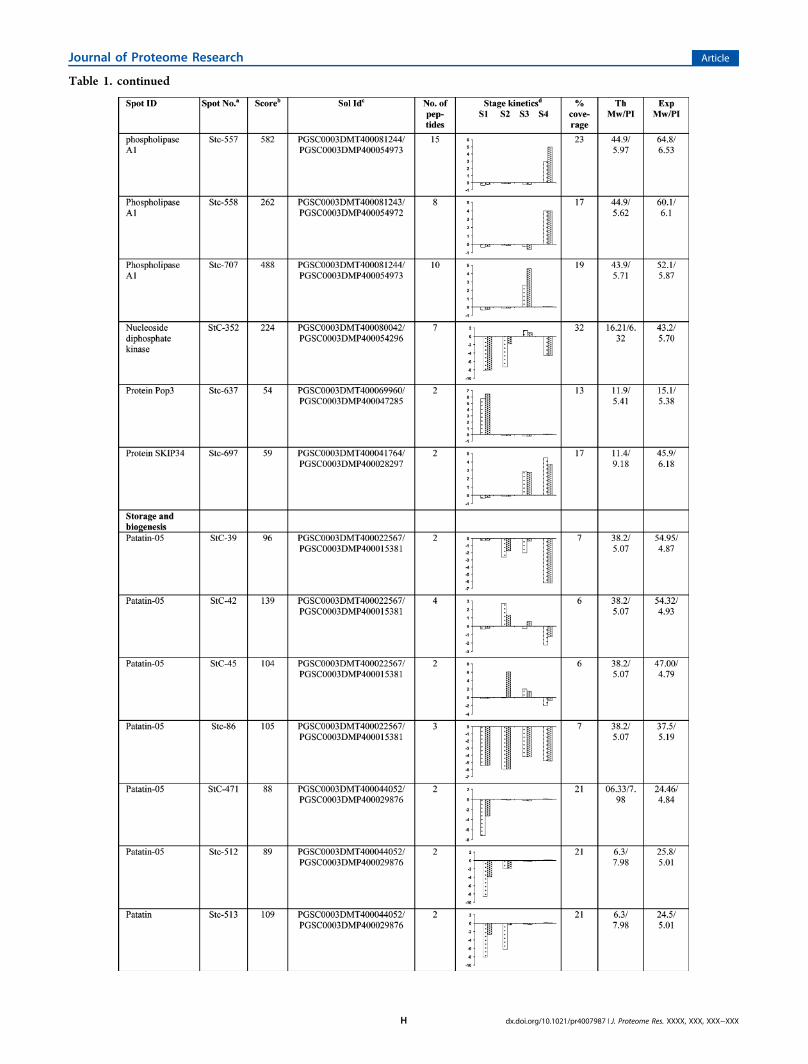
Table 1. continued
Journal of Proteome Research Article
dx.doi.org/10.1021/pr4007987 | J. Proteome Res. XXXX, XXX, XXX−XXXH

Table 1. continued
Journal of Proteome Research Article
dx.doi.org/10.1021/pr4007987 | J. Proteome Res. XXXX, XXX, XXX−XXXI

Table 1. continued
Journal of Proteome Research Article
dx.doi.org/10.1021/pr4007987 | J. Proteome Res. XXXX, XXX, XXX−XXXJ

Table 1. continued
Journal of Proteome Research Article
dx.doi.org/10.1021/pr4007987 | J. Proteome Res. XXXX, XXX, XXX−XXXK

Table 1. continued
Journal of Proteome Research Article
dx.doi.org/10.1021/pr4007987 | J. Proteome Res. XXXX, XXX, XXX−XXXL

mixed pattern. Isoforms of kunitz-type enzyme inhibitors (StC-18, 542, and 609) and cysteine protease inhibitor (StC- 540,612B, 742, and 743) also showed mixed expression. Amongothers, a very high expression was observed in ARPs encodinglipoxygenase proteins (StC-161, 545, and 721) in mature tubersof both AmA1 plants as compared to their wild-typecounterpart. ARPs spots StC-723 and StC-653 were identifiedto be proteosome subunit alpha type and protein disulfideisomerase (PDI). PDI showed an elevated expression alongwithGAPDH (Stc-679) in the stolon stage. A few other interestingARPs such as adenylosuccinate synthase (StC-678) andmethionine synthase (MetE; StC-682) were also found to bedifferentially expressed in AmA1 tubers as compared to wild-type. Acetyl Co-A binding protein (ACBP; StC-648), a highlyconserved cytosolic lipid-binding protein that binds long-chainacyl-CoA esters with high affinity were also identified in thiscategory.The second largest category of differentially regulated ARPs
comprised of proteins involved in cellular signaling. 14-3-3proteins (Stc-11, 602, 604, 605, and 606) were thepredominant proteins found in this category followed byannexin (StC-655), which showed a higher expression in bothtype of AmA1 tubers. We also observed differential expressionof many of the actin isoforms in wild-type and AmA1 tubers.Few of the actins (StC-592 and 625) were highly expressedduring stolon formation and tuber induction, while others(StC-17, 88, and 309) were more actively participating duringtuber maturation (Table 1).
A third important class of ARPs identified is presumablyknown to be involved in protein folding and degradation andincludes HSP80 and small HSPs (sHSPs). In this study, HSP80showed much higher expression in the AmA1 tubers ascompared to wild-type. StC-590 and 591were up-regulated instolons, while the others StC-810, 811, 812, and 813 showed upto 50-fold increase in developing tubers. Besides HSP80, smallHSPS (sHSPs) (StC-272 and 377) also showed differentialexpression in AmA1 tubers. Oligopeptidase A (OpdA) (StC-628, 640, and 641) was among other in this class.Proteins involved in bioenergy and metabolism represent the
fourth set of ARPs that exhibited differential regulation. A highinduction of carbohydrate metabolic enzymes such asfructokinase (FK; StC-618, 644, and 656), aldo/keto reductase(Stc-551 and 553), and glyceraldehyde-3-phosphate dehydro-genase (GAPDH; StC-586 and 679) were observed in AmA1tubers. The expression profile of FKs showed a significantincrease in stolons, while aldo/keto reductase, which showedup to 25-fold increase in AmA1 tubers, is known to catalyze thereduction of aldehyde and carbonyl including monosaccharaideto glucose sugar alcohols. GAPDH, which also showed asignificant increase in expression, reversibly catalyzes theconversion of GAP into 1,3-bis PGA. Aconitase (StC-670) isan enzyme that catalyzes the stereospecific isomerization ofcitrate to isocitrate via cis-aconitate in the tricarboxylic acidcycle along with glucose-1-PO4 adenylyl transferase (StC-128)that belongs to the family of transferases specifically transferringphosphorus from nucleotide to glucose and thus participates instarch metabolism. Another spot was identified as formatedehydrogenase (FDH; StC 584 and 676), a solublemitochondrial enzyme capable of oxidizing formate into CO2
and abundantly found in nongreen tissues and scarce inphotosynthetic tissues showed 20-fold up-regulation in theAmA1 tubers. Another important TCA cycle enzyme, pyruvatedehydrogenase E1 (StC-583), which is the first component ofthe PDC complex that contributes to transferring pyruvate intoacetyl CoA to carry cellular respiration, is up-regulated in themature AmA1 tuber.A few differentially expressed ARPs were also found to be
associated with miscellaneous functions. Protein spot StC-597represent P40, while spot StC-805 was found to be PR10having role in plant protection.
Table 1. continued
aSpot number as given on the two dimensional gel images. The first letters (St) represent the source plant Solanum tuberosum followed by thefraction cytoplasm (c). The numerals indicate the spot numbers corresponding to Figure 2. bThe significance score (P < 0.05) of a protein, asproduced by the Mascot algorithm. cGene identification number as in PGSC. dProtein expression profile represents the average change in spotdensity at various developmental stage kinetics S1 (stolon), S2 (tuber initiation), S3 (developing tuber), S4 (mature tuber) of 35S-AmA1 (left bar)and GBSS-AmA1 (right bar). The data were taken in terms of fold expression with respect to the control value and were log transformed to the base2 in order to level the scale of expression and to reduce the noise.
Figure 3. Functional cataloging of AmA1-responsive proteins (ARPs)in potato tuber. The identified ARPs were assigned a putative functionusing protein function databases and functionally grouped asrepresented in the pie chart.
Journal of Proteome Research Article
dx.doi.org/10.1021/pr4007987 | J. Proteome Res. XXXX, XXX, XXX−XXXM

Categorization of AmA1-Responsive Protein Isoforms
Next, we manually analyzed possible isoforms, i.e., similarproteins present in more than one spot. The multiple spots ofan identical protein are frequently reported in proteomicstudies, presumably due to post translational modification andexistence of different isoforms.76 These modifications oftenintroduce a variation in the molecular mass and net charge ofthe protein. The most efficient technique to separate proteinisoforms thus remains 2-DE.76−78 A closeup of possibleisoforms detected by 2-DE and their expression pattern showed15 unipros appearing as 56 identities. Interestingly, 10 unipros(phospholipase A1, kunitz-type enzyme inhibitor, cysteineproteinase inhibitor, lipoxygenase, formate dehydrogenase,glyceraldehyde3-phosphate dehydrogenase, heat shock cognetprotein 80, 17.6 kD classI sHSP, and oligopeptidaseA1)representing 29 identities (isoforms) showed that each set ofisoforms had the mixed-regulated change in patterns inabundance in response to AmA1, whereas the other 5 unipros(14-3-3 16R, actin, patatin05, HSP 83, fructokinase) appearedas 23 isoforms that exhibited mixed expression patterns oreither up-regulation or down-regulation within each set of
isoforms. In order to confirm whether this set of identifiedproteins could be possible isoforms to each other, a set of massspectra representing these 56 putative isoforms from 15different potato proteins were subjected to cluster analysis bythe web interface SPECLUST.79 The resulted dendrogramrevealed that clusters were dominated by isoforms (SupportingInformation Figure 5), e.g., heat shock protein 80 (cluster Aand C), oligopeptidase A1 (cluster B), heat shock protein(cluster D), actin 101 (cluster E), patatin-05 (cluster F),phosopholipase A1 (cluster G), and lipoxygenase (cluster H).Some of the mass spectra, which were supposed to containisoforms, did not cluster together as nicely as expected due tothe well-known fact that in 2-DE gels multiple proteins arepresent together in a single spot.79 Isoforms are almost alwayseither the products of one gene or of multiple genes thatevolved from a single ancestor gene mostly as splice variants. Itis generally accepted that multiple isoforms result fromsequence-related proteins encoded by distinct genes and/orpolypeptide variants encoded by the same gene [splice variantsand/or posttranslational modifications (PTMs)]. Therefore, anamino acid sequence alignment of the 15 unipros was
Figure 4. Clusterogram of expression profiles of AmA1-responsive potato proteins at various stages of tuberization. The 80 differentially expressedproteins in all four stages were grouped into 11 clusters based on their expression profiles. The self organizing tree algorithm (SOTA) cluster treesare shown at the right side, and the expression profiles are shown in parallel. Each protein is represented by a single row of colored boxes, and eachpotato line is represented by a single column. Induction (or repression) ranges from pale to saturated red (or green). The expression profile of eachindividual protein in a cluster is depicted by gray lines, while the mean expression profile is marked in pink for each cluster. The number of proteins ineach cluster is given in the lef t upper corner, and the cluster number is given below each expression profile. The cluster with n ≥ 5 was taken intoconsideration for the study of co-expression patterns for functionally similar proteins. (A) Stolon, (B) tuber initiation, (C) developing tuber, and (D)mature tuber. Detailed information on proteins within each cluster can be found in Supporting Information Figure 6.
Journal of Proteome Research Article
dx.doi.org/10.1021/pr4007987 | J. Proteome Res. XXXX, XXX, XXX−XXXN

performed using Bioedit to identify the amino acid sequencevariation among the similar proteins present in different spots.The result demonstrated that 8 unipros, namely, 14-3-3, actin,formate dehydrogenase, HSP 17.6, HSP 83, patatin, phospho-lipase A1, oligopeptidase A1, and proteasome, showed eitheramino acid substitution, deletion, addition, or inversion(Supporting Information Document 3). In addition, apartfrom MS/MS peak alignment and amino acid sequencealignment of similar proteins, in silico analysis of possiblepost-translational modifications using different databases wereperformed. Data analyses revealed that phosphorylation siteprediction using Netphos 2.0, kinasephos 2.0, DISPHOS 1.3,pkaPs prediction, pepbasepeptide spectra, and P3DB databasesshowed that phosphorylation sites are present in all the 15unipros, but phosphorylation could occur in 14 unipros exceptkunitz-type enzyme inhibitor with respect to the observed pIamd MW. Further, glycosylation site prediction usingNetNGlyC 2.0, Glymod, IsoGlyP, and NetOGlyC 3.0 showedthat O-glycosylation was absent in all 15 unipros, while N-glycosylation might be present in 14-3-3, phospholipase A1,patatin, kunitz-type enzyme inhibitor, lipoxygenase, HSP 80,HSP 83, and fructokinase. Moreover, only actin variant showedthe putative acetylation site based on the prediction ofacetylation using Automotif server 2. However, no glycophos-pho inosides were detected in any of these proteins whensearched against Big-PI plant predictor (Supporting Informa-tion Table 3). Our results suggest that amino acid sequencevariation, phosphorylation, glycosylation, and acetylation mightbe involved in the generation of these isoforms. Likewise manysimilar phenomena were also observed in other previouslyreported proteomics studies.80,81 Different isoforms of a proteinmay function in either a similar or identical ways but sometimesmay impart different functions.82,83 The function of each of theidentified protein isoform thus was analyzed in view of the roleof the candidate protein.Dynamics of AmA1-Regulated Protein Network
To achieve a comprehensive overview of the comparativeprotein profile during tuber developmental stages, the relativeexpression profiles of the 80 differentially expressed proteinspots were subjected to cluster analysis using the SOTAalgorithm.59 To group relative protein expression profiles onthe basis of similar trends and not of similar expression levels,the Pearson correlation coefficient was used as the distancefunction. The data were taken in terms of fold expression withrespect to the wild-type potato protein expression value.
Furthermore the data sets were log-transformed to base 2 tolevel the scale of expression and to reduce the noise. Figure4A−D shows hierarchical clustering of protein accumulationand different relative expression patterns observed in the AmA1lines versus their wild-type at four tuberization stages. Theanalysis yielded 11 expression clusters in each stage where onlythe clusters with n ≥ 5 were taken into consideration for thestudy of co-expression patterns for functionally similar proteins.The most abundant group in the early stage of tuberdevelopment, viz., the stolon stage, was Cluster 4 with 22proteins. This group consisted primarily of proteins involved inbioenergy and metabolism followed by storage and biogenesisand showed higher expression in AmA1 lines. The proteins incluster 5 in stolon stage included 6 up-regulated proteins thatmay be involved in cell signaling, storage and biogenesis, andbioenergy and metabolism. Clusters 2 and 3 also consist of amajority of proteins of storage and biogenesis class. The largestcluster in stage 2 (cluster 2), stage 3 (cluster 4), and stage 4(cluster 2) consist of 44, 42, and 35 proteins, respectively.Interestingly, in all four developmental stages proteins fromstorage and biogenesis had significant contribution to theclusters, which showed up-regulated proteins in the AmA1lines, although the other classes also had remarkablecontributions to these clusters. Detailed information on theproteins within each cluster can be found in SupportingInformation Figure 6.
Statistical Testing of Protein Expression
We asked which, if any, ARPs exhibited overall differentialexpression during tuberization. We used one-way ANOVA toanswer the question. A heat map and the expression graph ofthe developmental stage significant proteins are shown inFigure 5. Based on the ANOVA, 16 differentially expressedARP spots were found to be significant across the tuberdevelopmental stages that included patatin isomers (StC-86),proteinase inhibitor (StC-472, 542, 609, 612, and 743), andactin isomers (StC-88 and 309).
Correlation Network of AmA1-Responsive Proteins
To elucidate the biological significance of the responses toAmA1, we performed a correlation analysis to assignsignificance level to the proteins whose expression dependson the AmA1 irrespective of its spatial expression due todifferent promoters. To address statistical properties of thesenetworks, we quantified correlations between connectivity ofinteracting nodes using Pearson’s correlation coefficients
Figure 5. Heat map and one-way ANOVA analysis of ARPs. Heat map of a hierarchical cluster of the 16 protein spots that show significantdifferences (P < 0.05) between stages by Student’s t test. The red color represents relatively high expression, and green color represents relatively lowexpression level.
Journal of Proteome Research Article
dx.doi.org/10.1021/pr4007987 | J. Proteome Res. XXXX, XXX, XXX−XXXO

(PCC). We obtained a protein correlation network of 26 nodesand 35 edges. The resultant network incorporated 26 out of 80,45 proteins eligible for the analysis. The network at an alphavalue of 0.01 contains two modules (M) and six smallcorrelation groups (SC) of just 2 and 3 proteins (Figure 6).
We succeeded in extracting functional relationships usingPearson correlation method from the concatenated data setsbased on the physiological role of the AmA1-responsiveproteins that categorized the modules and small correlationgroups into two subnetworks, viz., nutritional network (Figure6A) and growth and development related network (Figure 6B).The nutritional subnetwork was composed of module 1
(M1) and five small correlation groups (SC1−SC5) withcysteine proteinase inhibitor, aldo/keto reductase, ascorbateperoxidase, actin, patatin, acyl-CoA-binding protein, esterase,glyceraldehyde 3-phosphate dehydrogenase, fructokinase, andphospholipase A1. Proteins in M1 are related to storage proteinaccumulation, assembly of fatty acid residue and proteinsrelated to calvin cycle of PSII. In M1, cysteine proteaseinhibitor1 and fructokinase were found to be positivelycorrelated, while both cysteine protease inhibitor and 14-3-316R showed positive correlation with acyl coA binding protein.Interestingly, grouped with these proteins is P40 in SC5 that isnegatively corelated with patatin, indicating its role in storageand biogenesis, and thus we hypothesize that it might havesimilar such function. The proteins in SC1 are involved in fattyacid metabolism and protein storage. Esterase was positivelycorrelated with one of the cysteine protease inhibitor isoform.There is aldo keto reductase, a calvin cycle protein positivelycorrelated with esterase in this group that could thus be co-
regulated. SC2, SC3, and SC4 showed candidates related tostorage protein, cytoskeleton, fatty acid catabolism, and redoxhomeostasis. Phospholipase A1 was positively correlated withaldo/keto reductase, while cysteine proteinase inhibitor wasnegatively correlated with ascorbate peroxidase in an individualassociation.The other subnetwork related to growth and development
including module 2 (M2), and one small correlation group(SC6) consists of heat shock protein 83, 14-3-3 16R, actin, heatshock protein 80, pop3, GAPDH. The cascade in M2 containsproteins involved in cell growth and osmotic regulation, tubermaturity, and development and was positively correlated amongeach other. Proteins found in M2 were positively correlatedwith each other, viz., actin has direct positive correlation withhsp, whereas SC6 contains a cascade of heat shock protein 80involved in protein homeostasis and morphological evolution.
Comparative Analysis of AmA1-Responsive Metabolites
Having found the differential proteome response in AmA1tuber, we further extended the evaluation and analyzed themetabolite pools using GC−MS to understand the impact ofARPs on the primary metabolism in 35S-AmA1 and GBSS-AmA1 tubers. Targeted compounds were identified andanalyzed to corroborate with the enzymatic pathways basedon the proteomic analysis. The majority of the compoundsdetected were found to be altered within AmA1 tubers, inagreement with the data obtained from proteomic study. Sixty-two metabolites were identified in wild-type, and 62 and 56metabolites detected in 35S-AmA1 and GBSS-AmA1 tubers,respectively, showed differential expression with high level ofcertainty. Of the 56 common metabolites between wild-typeand transgenic tubers, 42 (75%) were up-regulated, whereas 14(25%) were down-regulated. The primary metabolites relatedto AmA1-responsive pathways are shown in Figure 7.Consistent with the proteomic data, there was remarkabledifference in metabolite profile with high accumulation ofamino acids in mature AmA1 tubers. Metabolites related toamino acid biosynthesis were up-regulated. A notable trend inthe levels of amino acids was the increase in the concentrationof hydrophobic amino acids such as isoleucine, glycine, leucine,and alanine by a factor of 3.30, 2.74, 4.00, and 1.50 in 35S-AmA1 and 2.12, 1.53, 3.73, and 4.49 in GBSS-AmA1.Furthermore, hydroxyl amino acids displayed a small butsignificant increase in concentration in 35S-AmA1 tubers.Methionine, a sulfur amino acid, showed striking increase of 6−9 times, which indicates the mobilization of protein biosyn-thesis pathway in addition to the accumulation of S-adenosylmethionine (SAM). By contrast, glutamic acid showed atransient accumulation of 1.83-fold, which likely suggests itsrapid mobilization as substrate for acidic amino acid biosyn-thesis. Aspartate represents an important connection betweenamino acid and carbohydrate metabolism. Increase in aspartateshifts the metabolic pathway toward the citric acid cycle by theconversion of aspartic acid to oxaloacetate. Nevertheless, levelof aspartic acid was relatively lower than other amino acids,reflecting the decrease in carbohydrate metabolism (Figure7A). Indeed, metabolites related to carbohydrate biosynthesiswere significantly down-regulated or in steady state level. Asexpected, the levels of monosaccharides or disaccharide(glucose, fructose, and sucrose) were decreased or maintaineda steady state in AmA1 tubers, in accordance with the proteomedata (Figure 7B). Moreover, some metabolites, namely,saturated and unsaturated fatty acids, responded in opposite
Figure 6. Functional correlation network of ARPs. The ARPs weresubjected to partial Pearson correlation analysis using a matrix of thefirst order with 0.01 alpha value cutoff, and the resultant SIF file wasuploaded in Cytoscape for visualization. Red and green edgescorrespond to positive and negative partial correlation, respectively.Boxes indicate modules (M) and small correlation groups (SC) ofknown function. (A) M1 and SC1−SC5 represent the nutritionalsubnetwork, and (B) M2 along with SC6 denote the other subnetworkrelated to growth and development.
Journal of Proteome Research Article
dx.doi.org/10.1021/pr4007987 | J. Proteome Res. XXXX, XXX, XXX−XXXP

directions probably due to conversion of free fatty acid toesterified fatty acids in AmA1 tubers. Unsaturated fatty acidssuch as eicosaenioc acid was down-regulated, while 11,14-eicosadienoic acid and oleic acid showed a significant increaseof 3.69−5.25-fold in both 35S-AmA1 and GBSS-AmA1 tubers.In the case of the saturated fatty acids, undecanoic acid, 9,octadecanoic acid, docosanoic acid, and nonanoic acid showedreduced level in the AmA1 tubers (Figure 7C).
Investigation of Tuber Cell Architecture
To investigate the underlying mechanism that might haveresulted in increased tuber yield in AmA1 plants, the cellarchitectures were studied in different cell layers at tubermaturity and compared with that of wild-type tuber (Figure8A). The cell area was found to be identical in the epidermalregion of the tubers. However, there was an increase in cell areain the cortex and perimedullary region, which was increasedfurther in the pith tissue. The cell area in cortex region is36.21% higher, while in the perimedullary and pith region it was76.62% and 98.03% higher in transgenic tuber (Figure 8B). Tofurther test this observation, we used AmA1 fused to β-glucuronidase reporter gene construct driven by GBSSpromoter (Figure 8C). The AmA1 tubers displayed high levelexpression of AmA1-GUS fusion protein in the cortex,medullary region, and pith (Figure 8D). The in plantalocalization of the AmA1 protein in potato tuber was foundto be correlated with cell growth in this study. Taken together,these results suggest that increase in cell division especially inthe perimedullary region might contribute to more storagetissue in AmA1 tubers.
■ DISCUSSION
Quality and productivity of agricultural crops is a complexfunction of the acquisition of resources and their distributionwithin the plant to harvestable components. Photosynthesisand nutrient capture are the primary elements of overallbiomass production, but it is the allocation of assimilatedresources within the developing plant that determines theproportion of biomass that can be utilized. Plants areprogrammed to maintain a constant rate of photoassimilatesynthesis, translocation, and supply to storage organs, i.e., thesink tissue, which is competitive, and thus the photoassimilate ispartitioned to the active sinks. Sink strength is one of thesignificant factors that determine the direction of photo-assimilate translocation, while sink size represents the totalmass of sink primarily composed of carbon and nitrogencompounds, including storage proteins.84 Storage proteinsaccount for 10−60% of total dry weight in plants.85 Further, itis known that sink strength is closely related to growth and inturn productivity and is influenced by cell turgor andhormones.Our earlier findings and the result in this work showed a
definite role of seed storage protein AmA1 toward nutritionalenhancement and growth. Thus, our aim primarily was toinvestigate the regulatory and functional protein networkoperating in response to AmA1 sensing. We demonstrate thatAmA1-regulated functional protein network and its combina-torial effect cause the protein enhancement and determine theorgan development, tuber in particular. The introduction of theAmA1 gene by means of a constitutive and tuber-specificpromoter may lead to numerous changes within the plantproteome that may be related to many pathways or generalexpression variation of individual proteins. Although the
Figure 7. Comparison of primary metabolite levels in mature AmA1tubers with those in tubers of wild-type. (A) Amino acids, (B) Sugars,and (C) Fatty acids. Data are normalized to the mean responsecalculated for wild-type levels of each replicate (to allow comparisonbetween replicates, individual wild-type values were normalized insame way). Values presented are the mean ± SE of four independentdeterminants.
Journal of Proteome Research Article
dx.doi.org/10.1021/pr4007987 | J. Proteome Res. XXXX, XXX, XXX−XXXQ

comparative proteome analysis revealed the set of AmA1-targetproteins differentially expressed in both 35S-AmA1 and GBSS-AmA1 tubers, the level of expression cannot be related to thepresence of AmA1 with utmost confidence for all. Hence, wechose to interpret the results by taking into consideration theexpression of AmA1 target proteins at four developmentalstages for the two AmA1 potato genotypes together,considering the spots that show a correlation pattern in both,and also separately to check the correlation between spotsindividually in both AmA1 lines to identify the common
correlations. The final correlation network obtained by mergingthe initial “combined” network and the “intersection” networkrevealed a total picture of all sets of protein spots that may becorrelated in both of the data sets with a decent statisticalconfidence. Verifying the biological relevance of the recoverednetworks is difficult, since many interactions between proteinsare currently unknown. These correlation networks are alsoknown to be phenomenological, i.e., many connections do notcorrespond to direct physical interactions between geneproduct and promoter elements, but to a complicated actionthrough more complex regulatory pathways involving theproteome and metabolome.86,87 We verified the biologicalrelevance of the inferred networks by using the Gene OntologyTerm Finder to investigate if the subnetworks contain a highproportion of functionally related proteins. Indeed, the inferrednetworks had high significance scores, implying that theprobability of grouping them by chance is less. Revealing themodular structures in the obtained biological networks helps usto understand cell function. A model representing theregulatory and functional network of AmA1 target proteins isdepicted in Figure 9.
Metabolic and Functional Protein Network Involved inAmA1-Associated Nutritional Enhancement
The synthesis and accumulation of nutrients is a sophisticatedand highly regulated process, wherein many different proteinsact to ensure that a gene is translated and the products areprotected at the right time in a cell. This requires synthesis ofdifferent energy molecules and amino acids, the building blockof proteins, and participation of different molecules with aprotective role in the formation and stabilization of the newlysynthesized proteins.Several ARPs known to be involved in the synthesis of
energy molecule and amino acids were identified in the presentstudy. One of the major regulators MetE (StC-682) involved inde novo synthesis of methionine and regeneration of the methylgroup SAM by catalyzing the transfer of a methyl group frommethyltetrahydrofolate to homocysteine was highly up-regulated in AmA1 tuber at the stolon stage. In plant, it hasbeen estimated that about 20% of the Met is incorporated intoproteins, while 80% is converted to SAM.80,81 Furthermore, itwas interesting to note the increased accumulation ofmethionine in AmA1 mature tubers, which provides furthersupport to our proteome data. From the many changesdetermined following metabolite analysis, the one of particularinterest was the increase in alipahatic amino acids, namely,alanine, leucine, and isoleucine. This small group of branched-chain amino acids is essential for humans.88,89 Aminotransferase(StC-677) has an important role in the biosynthetic pathwaysof these amino acids and the methionine chain elongation cycleof aliphatic glucosinolate formation. Methionine occupies acentral position in cellular metabolism: as a protein constituent,in the initiation of mRNA translation, and as a component ofthe regulatory molecule, SAM. AmA1 being a storage proteinacts as sink to accumulate essential amino acids such asmethionine and cysteine, and thus its expression requires ahigher level of MetE, the key enzyme in methioninebiosynthetic pathway.90 SAM functions as a primary methylgroup donor and most interestingly, as a precursor formetabolites, such as ethylene,91 that activates adenylsuccinatesynthase (StC-678). We observed elevated expression ofadenylsuccinate synthase in stolon, the key enzyme for denovo synthesis of adenosine monophosphate (AMP), suggest-
Figure 8. Cell architectures and accumulation of AmA1 protein.(A)Photomicrographs showing cells in the cortex, perimedulary, andpith regions of the wild-type and AmA1 potato tuber. (B) Graphshowing cell area of different regions along the transverse axis ofpotato tubers. Data represent mean ± SD of three measurements. (C)Schematic representation of the AmA1-β-GUS chimeric gene constructpSB3G. (D) Cross sections of potato tubers of wild-type and AmA1lines. Each cross-section was treated with β-GUS stain forhistochemical analysis. Blue-colored signals indicate accumulation ofAmA1 protein in AmA1 potato. Most of AmA1 was colocalized inmedullary and pith tissue.
Journal of Proteome Research Article
dx.doi.org/10.1021/pr4007987 | J. Proteome Res. XXXX, XXX, XXX−XXXR

ing its role in increasing the tuber primordial formation thatmight lead to increased sink strength. It has been documentedthat increased synthesis of AMP is advantageous for theproduction of high energy donor molecules such as ADP andATP for starch synthesis.92 In AmA1 potato increased level ofglutamate, a key molecule in protein synthesis and the fivecarbon chain precursor of all tetrapyrroles including 5-aminolevulinic acid,93 presumably bind to tRNA muchefficiently, thereby increasing the rate of protein synthesis.As sucrose is the major transport form of fixed photo-
assimilates in developing potato tubers, the mechanismresponsible for the immediate metabolism of sucrose is ofparticular interest. Sucrose cleavage after unloading out of thephloem into the sink organ, such as potato tuber, is known tobe mediated by invertase that cleaves sucrose to glucose andfructose, which are transported to the cytosol for furthermetabolism. The differential proteome data also revealed thepresence of a number of glycolytic and starch biosyntheticpathway enzymes. These proteins are known to havebioenergy-related function. While glucose 1-PO4 adenylyltransferase (StC-128) might be involved in starch synthesisusing glucose and ADP as substrate, fructose is acted upon byfructokinase (FK; StC-618, 644, and 656) to form fructose-6-phosphate to maintain a balance between sucrose synthesis andstarch formation. FK has little impact on glycolysis and
subsequently on metabolites downstream to glycolysis. There-fore, in AmA1 tubers glycolytic proteins such as aldo/ketoreductase (StC-551 and 553), enolase (StC-643), pyruvatedehydrogenase (StC-583), and GAPDH (StC-586 and 679) arehighly induced to supply inorganic phosphate for starchsynthesis. The effect of rigorously modulating fructokinase inpotato tuber has been well established,94 and potato plantsexhibiting reduced FK are characterized by fewer tubernumbers and reduced yield.95 Thus, highly up-regulatedexpression of fructokinase- and fructokinase-like proteins(StC-618, 644, and 656) clearly supports our data of increasedtuber yield. The metabolite profiling of carbohydrate comple-ment provides further support to our claim that concentrationof sucrose and fructose were low in AmA1 tuber, whereasglucose maintained a steady state level. It has long been knownthat reductases regulate glycolysis and starch synthesis, themost important component of carbon assimilation, bydetoxifying glycolysis-derived reactive carbonyl in rapidlymetabolizing cells. Aldo/keto reductase (AKR) superfamily(StC-551, 553) catalyzes mainly the reduction of carbonylgroups or carbon−carbon double bonds of a wide variety ofsubstrates, including steroids, mainly brassinosteroid. Mono-meric cytosolic form of AKR plays a crucial role inosmoregulation, an important process for the acquisition ofdesiccation tolerance in plants. Besides this, members of the
Figure 9. Pathway involved in AmA1-activated regulatory and functional network. Proteins identified in this study are indicated in the yellow boxes.Green circles represent metabolites up-regulated, and orange circles indicate down-regulated metabolites. Graphs are the representatives ofexpression profile of individual protein, and number given below in each graph indicates the protein identification number. ASS, adenylsuccinatesynthase; AscP, ascorbate peroxidase; ACBP, acyl CoA binding protein; HSP80, 80 kDa heat shock protein; HSP17.6, sHsp; HSP83, 83 kDa heatshock protein; PDH, pyruvate dehydrogenase; Glu 1-PA, Glucose 1-PO4 adenylyl transferase; NDK, Nucleotide dikinase; FK, fructokinase; GAPDH,glyceraldehyde 3-phosphate dehydrogenase; PDI, protein disulfide isomerase; 11,14-EA, 11,14-eicosadienoic acid; OA, oleic acid.
Journal of Proteome Research Article
dx.doi.org/10.1021/pr4007987 | J. Proteome Res. XXXX, XXX, XXX−XXXS

AKR family have been shown to be effective in thedetoxification of lipid peroxidation and/or glycolysis-derivedreactive carbonyls.96
While actin filaments may enhance protein synthesis byproviding a scaffold for specific assembly of the translationalmachinery, a recent study indicates that hexokinase (HXK)-dependent glucose signaling might require a previouslyunrecognized interaction with the actin cytoskeleton.97 Ourobservation on differentially regulated actins (StC-88, 260, 592,625) might provide a novel gateway to understand animportant interface between protein synthesis, glucose signal-ing, and tuber development. Actin is known to be encoded by amultigene family with high divergence.98 Further, differentisovariants are likely to be expressed at a particular timedepending on the physiological status of the cell. Therefore,highly regulated expression of actin genes as revealed in thisstudy and their subsequent post-translational modifications areprobably required during tuber development. Although class I(StC-272 and 377) and class II (StC-371) sHSPs were down-regulated at the initial developmental stages of AmA1 tubers,their subsequent up-regulation at tuber maturity implicatestheir role in reserve accumulation as has been reported earlierin pea seeds.99 Among the five conserved families of HSPs,HSP83 in solanaceae, the ortholog of HSP90 are the mostprevalent in plant. Plants synthesize multiple sHSPs encoded bysix nuclear multigene families; each gene family encodesproteins that are destined to a specific cellular compartment(i.e., cytosol, chloroplast, ER, and mitochondria). Diversifica-tion of sHSPs and presence of their isoforms probably reflect amolecular adaptation to stress conditions unique to plants.Although HSP83 chaperones are constitutively expressed inmost organisms, their expression increases in response to stressin both prokaryote and eukaryote. Expression of HSP90isoforms play a substantial role in the subunit−subunitinteractions that stabilize homo- and/or heterotrimers ofdevelopmentally regulated proteins in Arabidopsis.100 Post-translational regulation as opposed to transcriptional ortranslational regulation allows a quick adjustment of proteinactivity. Amino acid sequence variation as revealed in our studymight attribute to the diverse functions of the orthologsbelonging to these multigene family.The 14-3-3 proteins (StC-11, 602, 604, 605, and 606) have
risen to a position of importance in biology, having been shownto regulate many crucial cellular processes because of specificphosphoserine/phosphothreonine binding activity for targetprotein that shuttle from the cytoplasm to the plasmamembrane or nucleus,101 modulating their localization.102,103
The first plant 14-3-3 protein was identified as part of aprotein−DNA complex,104 indicating a role of 14-3-3s in theregulation of transcription. It is well established from thestudies of animal and plant that isoforms have differentialexpression patterns, different subcellular localization, and/orfunctions.83,105−107 We identified isoforms of 14-3-3 proteinknown to be the product of a multigene family, with manyorganisms having 10 or more family members.108 It is knownthat diversity of 14-3-3 is due to alternative splicing.109 Onepossible reason for isoform diversity is simply to ensurefundamental 14-3-3 presence in all cell types, where 14-3-3function is required. Differential expression pattern of 14-3-3and actin isoforms in response to AmA1 might result frompost-translational modification with phosphorylation or de-phosphorylation state.These results suggest that isoforms ofcertain unipros may play the same or different roles in AmA1
tubers. Additional studies showed that 14-3-3 proteins (StC-11,602, 604, 605, and 606) are known to function in regulating theactivities of metabolic enzymes and transcription factors,110
besides their role as osmotic motor.111 Increase in proteincontent presumably elevates the turgor pressure in AmA1expressing cells. Upregulation of 14-3-3 proteins, known to actas osmotic motor might stimulate H+ ATPase pump and cellwall acidification, thereby balancing the turgor pressure thatleads to cell elongation and induction of Ca2+ and G-proteinsignaling in the AmA1 potato. We observed increasedexpression of pop3 (StC-637) in GBSS-AmA1 tuber whosecross-talk with brassinosteroid is reported earlier.112,113 It isinteresting to note BR-induced H2O2 may alter cellular redoxstatus and induce changes in expression of photosyntheticgenes ultimately leading to the increased photoassimilate.Catalase (StC-684) showed higher expression in early stagewith subsequent decrease in the later stage of tuberization,suggesting that this enzyme might try to degrade H2O2.
However, in later stage of tuberization H2O2 is utilized tomaintain redox status of photosynthetic gene like thioredoxin.Expression of kunitz-type enzyme inhibitor (StC-609) knownto inhibit activity of amylase,114 which cleaves starch to glucosethat help in maintaining turgor pressure during tuberdevelopment.One of the major regulations in the process of formation and
stabilization of newly synthesized amino acids and proteins iscarried out by the highly polymorphic low-molecular weightprotease inhibitors, such as cysteine proteinase and kunitz-typeenzyme inhibitors (StC-540, 542, 609, 612B, and 743), thesecond major class of storage proteins in tubers that showedmaximum abundance in AmA1mature tuber and were found toexist as product of multigene family having N-type ofglycoform. They have been documented with multiplefunctions as enzyme inhibitors115,116 or agents of defenseagainst pathogens or insects.117 Protein disulfide isomerase(PDI; StC-653), a multifunctional protein that catalyzes theformation of disulfide bonds during protein biogenesis and isconsidered to be of fundamental importance for stabilizing thetertiary and quaternary structures of many proteins that areprocessed through the endomembrane system,118 showed anelevated expression in stolon. The wheat PDI reported to becolocalized with storage proteins in protein bodies is involvedin the assembly of storage proteins within the ER.119 Also, PDImarkedly increases reactivation of GAPDH and prevents theiraggregation.120 Thus the identical expression profile of PDI andGAPDH in stolon in the transgenics is indicative of theircooperative role in protein enhancement during tuber develop-ment. Oligopeptidase A1 (StC-628, 640, and 641) may play aspecific role in the degradation of signal peptides after they arereleased from precursor forms of secreted proteins.121,122 It is athiol-dependent metallopeptidase, which can cleave andthereby modulate the activity of many proteins and is involvedin seed germination, recycling damaged proteins, regulatingaging processes, and modifying proteins to perform specificpurposes in plant cell.123 Some patatin isoforms (StC-42 and45) showed elevated expression in stages 3 and 4. It is knownthat proteolysis of vegetative proteins facilitates accumulation ofstorage proteins with release of cell wall loosening enzymes forincreased assimilation of C and N macromolecules.
Journal of Proteome Research Article
dx.doi.org/10.1021/pr4007987 | J. Proteome Res. XXXX, XXX, XXX−XXXT

Regulatory and Signaling Network Involved inAmA1-Induced Growth and Organ Development
Numerous studies have shown that the productivity ofagricultural crops is a complex function of the acquisition ofresources and their distribution within the plant to harvestablecomponents. Photosynthesis and nutrient capture are theprimary elements of overall biomass production, but it is theallocation of assimilated resources within the developing plantthat determine the proportion of biomass that can be utilized.Regulation and coordination of the communication within themolecular signaling network is essential for organ growth anddevelopment, including potato tuber. It is an expression of thegenetic program that directs the activities and interaction ofindividual cells. Growth, an irreversible increase in volume andmanifestation of new protoplast, is accomplished by increase infresh and dry weight from stolon to tuber, and this gradualtransition is referred to as homoblastic development.124
Functional sorting of ARPs identified 8 spots as patatin, theprimary storage protein (40% of soluble protein) that isconsidered a critical nutritional component in potato tuber.Patatin, a group of glycoproteins in potato, is encoded by amultigene family.125,126 Isoforms of patatin separated in 2-DEanalysis contained proteins with two predicted molecularmasses of approximately 41.17 and 42.56 kDa, respectively.The size of this difference in the molar mass (1390 Da) is onthe order of one carbohydrate moiety. This clearly emphasizesthe possibility of the existence of a patatin glycoform(Supporting Information Table 3). These are mostly expressedin developing tubers and highly activated after tuber initiation.Patatin has been shown to play an important role in plantdefense and antioxidant activity due to their enzymatic functionof lipid acyl hydrolase that exhibits phospholipase A1 and A2(PLA1 and PLA2) activity.127 Increasing evidence suggests thatpatatin genes exhibit alteration in chromatin state anddifferential transcriptional regulation during the developmentaltransition from stolon to tuber.128 The reaction catalyzed bythis protein generates free fatty acids (FFA) from its substratephospholipids, which can subsequently be converted tojasmonic acid (JA) and its derivatives.129 FFA along withstatolith containing starch and the release of ethylene from theJA-mediated catabolic pathway stimulate dia-gravitropicresponse that results in the growth of stolons at right angleto the pull of gravity, which reduce mutual shading and efficientcapture of photosynthate making even large size tubers capableof capturing sucrose.130 Metabolite profiling of fatty acids alsoconfirmed similar change in stolon, wherein most of thesaturated and unsaturated fatty acids showed up-regulation,implying that FFA is utilized for gravitropic response, while inmature tuber the concentration of fatty acid is less, suggestingthat it is utilized for making fat in plasma membrane. Patatinisoforms (StC-42, 455, 512, 513, 471, and 472) showed down-regulation in AmA1 potato. Growth is an interplay of manyphytohormones, and it is known that down-regulation ofpatatin increases the synthesis of gibberellin’s130 transcriptionfactors and might lead to the observed increase in leaf area,height, and length of internode in AmA1plants (SupportingInformation Figure 2A−F).Lysophosphatidylcholine (LPC), the other product of PLA
activity of patatin (StC-42, 455, 512, 513, 471, and 472), isbiologically active in a number of important cellular signalingpathways involving auxin-induced cell elongation that stim-ulates the H+-ATPase pump in the plasma membrane, which isknown to be a key regulator in the induction of cell wall
acidification leading to elongation.128 Therefore, this activitypossibly is one of the factors in increasing cell division and cellenlargement that contribute to the increased growth of tubersin AmA1 potato. It is widely accepted that the growth of theperimedullary zone produces the largest portion of the tissue inmature tuber131,132 as has been documented in this study(Figure 8A). Of the phospholipase A, PLA2 has a PA2c domainthat contains the highly conserved Ca2+-binding loop that isimportant for Ca2+-dependent membrane translocation.133−135
Ca2+ initiate signal transduction that proceeds in a linearfashion and cross-talk with pathway initiated by POP3(StC637), a Ca2+ receptor and representative of EF-handproteins classified as Ca2+ buffer,136 which act as sensor tointeract with specific targets and stimulate G-protein signaling.The up-regulation of phospholipase A1 (StC-555, 557, 558, and707) in AmA1 tuber clearly indicates its involvement in tubermaturation. It has also been shown to induce brassinosteroidbiosynthetic genes resulting in a hypermorphic phenotype withincreased vegetative growth,134 as was observed in the currentstudy. Further, phospholipase A1 is known to play a pivotal rolein Ca2+ signaling in eukaryotes and is considered to bemultifunctional because of its ability to interact and regulate theactivity of a number of other proteins. Ca2+ is a universal signaland the dynamic changes in its release and entry trigger aplethora of cellular responses. Central to this schema aremembers of the phospholipase superfamily, which relayinformation from the activated receptor to downstream signalcascades by production of second-messenger molecules. Recentstudies reveal that, in addition to its enzymatic activity,phospholipase regulates Ca2+ entry via the formation of anintermolecular lipid-binding domain with canonical transientreceptor potential ion channels. This complex, in turn, controlstrafficking. Thus, ion channels are functionally linked to bothlipase-dependent and -independent activities of phospholi-pase.137 Similarly, annexins, another multigene and multifunc-tional family of soluble proteins, play a crucial role in Ca2+-dependent and Ca2+-independent signaling responses. They areubiquitous soluble proteins capable of binding endomembranesand plasma membranes and have been implicated to function indiverse cellular processes, including signal transduction, channelactivity, vesicular transport and fusion, and DNA replica-tion.138,139 Annexines are found to bind with actin in a Ca2+-and pH-dependent manner. Annexin34 (StC-655), one of thecalcium binding proteins, is also involved in the homeostaticregulation of intracellular Ca2+ in the presence of phospholipidsand thus control structural changes to the membranecytoskeleton, regulation of Ca2+ channels, and stimulation ofG-protein signaling. The up-regulation of annexin (StC-655) inAmA1 potato tubers might cause an overall hypermorphicphenotype along with actin, as it maintains the cell structuralmicroenvironment for survival, turgor, and wall pressure.138
Actin (StC- 592 and 625) depolymerization resulted inalteration in cell division in a very slow manner but reduces thecell elongation dramatically,140 which might lead to a burst ofcell division. H+-ATPase stimulated by auxin is a key player inturgor regulation, since it provides the driving force for ionuptake, followed by water influx through osmosis. 14-3-3protein is emerging as a regulator of many key ion transporterssuch as the plasma membrane H+-ATPase and outwardrectifying K+ channel and functions as osmotic motor inplant cell.110 Therefore, increased expression of 14-3-3 (StC-11,602, 604, 605, and 606) in AmA1 potato presumablytranslocates excess sucrose and simultaneously maintains
Journal of Proteome Research Article
dx.doi.org/10.1021/pr4007987 | J. Proteome Res. XXXX, XXX, XXX−XXXU

osmotic relation in different plant organs. Together with FFAand G protein, 14-3-3 in turn stimulates SKIP 34 (StC-697), akey regulator in auxin signaling. Auxin interacts with various F-box proteins and affects plant morphology, floral development,root and hypocotyls growth.141 Aconitase (StC-670), whichdocumented to prevent senescence and promote growth,142
showed a mixed expression followed by a slight increase in thelater stage of development in AmA1 tuber.Plant lipoxygenases (LOXs: StC-161, 545 and 721), another
lipid degrading enzyme belonging to functionally diverse classof dioxygenases, have been implicated in various physiologicalprocesses, such as growth, senescence, and stress-relatedresponses.143 Several reports have shown the involvement ofLOXs in tuber growth and development in potato,143,144 duringleaf development in soybean,145 seed germination incucumbers,146 seedling development in Arabidopsis,147 andfruit ripening in tomato.148 The involvement of LOXs in tubergrowth and development can be explained by the fact thatLOX-derived products, such as jasmonic acid, methyljasmonate, and tuberonic acid, have strong tuber inducingactivity.149 It is interesting to note individual lipoxygenasegenes were shown to be induced by jasmonates and have a rolein thigmomorphogenesis and gravitropism in wheat.129 Thushigher expression of LOX in AmA1 tuber suggests its possiblerole in tuber growth and development. It might function byregulating the reorientation of microtubules that allows radialcell expansion leading to tuber enlargement.150,151
It is becoming apparent that protein degradation is necessarynot only for the removal of aberrant or damaged proteins but
also for altering the balance of proteins during development orstress adaptation. Proteasome subunit alpha type and subunitbeta type-6 (StC-723 and 680), thought to be present in alleukaryotic cells,152,153 degrade specific proteins that have beentargeted for proteolysis by ubiquitination. In animals and yeast,proteasomes also play a role in regulating the cell cycle andother developmental processes, via control of the levels ofregulatory proteins, such as cyclins and transcription factors. Inaddition, it might also play a role in morphological evolution. Arecent report showed that HSP80 along with sHSPs (StC-371,377) interacts with the 26S proteasome154 and plays a principalrole in its assembly and maintenance. One of the HSP90 familyof proteins, HSP83 (StC-593), is developmentally regulatedand responds to phytohormones.155 It is reported that HSP90family proteins, being molecular chaperones, manage proteinfolding and play a key role in signal-transduction networks,protein degradation, and protein trafficking in Arabidopsis.156
Moreover, HSP83 acts as a ‘buffer’ to sustain the functions ofmutated proteins that participate in the signaling pathways ofdevelopment and morphogenesis. HSP83 is unique in itsfunction. Not only is it required for maturation andmaintenance of most proteins in vivo, but also most of itscellular targets are signal transducer and developmental and cellcycle regulators. HSP83 keep these unstable signaling proteinpoised for activation until stabilized by conformational changes.It also might buffer against developmental noise caused byrandom microenvironmental effect with no genetic basis.Simultaneously, it also acts as genetic capacitor for devel-opmentally regulated genes. In the present study its up-
Figure 10. Model elucidating the summary of cellular pathways involving AmA1-regulated proteins. Proteins indicated in the colored boxes and thepossible pathways depicted by dashed lines were identified in this study.
Journal of Proteome Research Article
dx.doi.org/10.1021/pr4007987 | J. Proteome Res. XXXX, XXX, XXX−XXXV

regulation in AmA1 potato clearly suggests genetic variationmight be expressed to a greater extent affecting manydevelopmental pathways causing morphological evolution,supporting the basis of natural selection for the AmA1-plant.The higher expression of HSP83 might contribute in thedevelopment of AmA1-plant as they might bind to a range oftarget proteins, such steroid hormone receptors, signalingkinases, and a variety of transcription factors157,158 that arecrucial for regulating growth and development. It is known thatHSP83 interacts with the 26S proteasome and plays a principalrole in its assembly and maintenance.159
Acetyl Co-A binding protein (ACBP; StC-648) is a highlyconserved cytosolic lipid-binding protein that binds long-chainacyl-CoA esters with high affinity and is expressed indeveloping embryo, flowers, cotyledons, and other tissues.160
It has been suggested that ACBP may mediate the translocationof acyl-CoA from the source organ to the sink organ duringmembrane synthesis.161 The increase in ACBP expression inAmA1 tubers could be a requirement for increased acyl-CoAtransport during membrane synthesis that might facilitate highrate of cell division. However, under stress FDH (StC-584 and676) accumulate in leaves, and leaf mitochondria acquire theability to use formate as a respiratory substrate.162 FDHexpression was found to be increased in AmA1 tubers, whichmight reflect an alternative source of photorespiration duringthe growth of tuber.
■ CONCLUSIONIn summary, we describe the application of molecularphenotyping to gain new insights into the storage proteinfunction by performing large-scale proteomic analysis onAmA1-expressed potato tubers and analyzing the data setusing a network approach. Data presented here both confirmedour earlier hypothesis and, more importantly, identified novelAmA1-regulated proteins and pathways. We propose a workingmodel on AmA1-mediated regulation involving novel pathwaysand cross-talk between various cellular pathways in nutrient-dependent storage accumulation and organ development. Theresults obtained from the present investigation have beenillustrated to summarize the molecular events in AmA1 potato(Figure 10). The increased sink strength due to thecombinatorial effect of the ARPs discussed might contributeto the concomitant increase in tuber growth. Thus, ourobservation suggests that a nutrient-responsive metabolicpathway overlaps with that of growth and development notonly at the level of protein expression but also functionally inAmA1 potato. The exact function of these proteins can bepredicted only after extensive studies and might be interestingfor future investigations. Such a system based proteomic studyfurther extends our understanding about the storage protein-mediated cellular physiology and may ultimately lead todevelopment of more efficient strategies for plant biotechnol-ogy. In this study, we have identified a number of proteinspreviously unknown as AmA1 targets. Some of them areinvolved in the control of cellular gene expression andphenotype and are probable mediators of protein accumulationand the growth action of AmA1. The resulting lists of proteinswere analyzed for the presence of members of known molecularnetworks. These networks showed substantial overlap andconnectivity with the biological “theme” of nutrient accumu-lation and cell growth. Furthermore, integration of proteomicsdata with metabolomic analysis allowed a comprehensivesystem-level understanding of storage protein mediated
regulation in plant. It is expected that as functional genomicsdata in potato accumulate, the significance of the isoformswould unravel the post-translational regulation toward theAmA1-regulated protein network. It would be interesting todetermine the possible interaction of these differentiallyexpressed proteins among themselves as well as with AmA1,which could contribute to the increased protein and amino acidcontents and tuber yield in the transgenics. Our studyillustrated for the first time the molecular mechanism of aseed storage protein-regulated functional interaction structureof a cellular network involved in increased protein synthesis,storage reserve accumulation, and organ growth leading toenhanced productivity.
■ ASSOCIATED CONTENT*S Supporting Information
Additional figures, tables, and data as described in the text. Thismaterial is available free of charge via the Internet at http://pubs.acs.org.
■ AUTHOR INFORMATIONCorresponding Authors
*Tel: 91-11-26735178. Fax: 91-11-26741658. E-mail:[email protected].*Tel: 91-11-26735186. Fax: 91-11-26741658. E-mail:[email protected] Contributions§These authors contributed equally to this work.Notes
The authors declare no competing financial interest.
■ ACKNOWLEDGMENTSWe thank Dr. Evert Jacobsen for providing the plasmid pPGB1.This work was supported by grants (BT/PR/11676/PBD/16/831/2008) from the Department of Biotechnology (DBT),Government of India and the National Institute of PlantGenome Research, New Delhi, India. L.A. and K.N. are therecipients of predoctoral fellowship from the Council ofScientific and Industrial research (CSIR), Govt. of India.Authors also thank Mr. Jasbeer Singh for illustrations andgraphical representations in the manuscript.
■ REFERENCES(1) Wang, R.; Guegler, K.; LaBrie, S. T.; Crawford, N. M. Genomicanalysis of a nutrient response in Arabidopsis reveals diverse expressionpatterns and novel metabolic and potential regulatory gens that areinduced by nitrate. Plant Cell 2000, 12, 1419−1510.(2) Stitt, M.; Muller, C.; Matt, P.; Gibon, Y.; Carillo, P.; Morcuende,R.; Scheible, W. R.; Krapp, A. Steps towards an integrated view ofnitrogen metabolism. J. Exp. Bot. 2002, 53, 959−970.(3) Wang, R.; Okamoto, M.; Xing, X.; Crawford, N. M. Microarrayanalysis of the nitrate response in Arabidopsis roots and shoots revealsover 1,000 rapidly responding genes and new linkages to glucose,trehalose-6-phosphate, iron, and sulfate metabolism. Plant Physiol.2003, 132, 556−567.(4) Palenchar, P.; Kouranov, A.; Lejay, L.; Coruzzi, G. Genome-widepatterns of carbon and nitrogen regulation of gene expression validatethe combined carbon and nitrogen (CN)-signaling hypothesis inplants. Genome Biol. 2004, 5, R91.(5) Schneper, L.; Duvel, K.; Broach, J. R. Sense and sensibility:nutritional response and signal integration in yeast. Curr. Opin.Microbiol. 2004, 7, 624−630.
Journal of Proteome Research Article
dx.doi.org/10.1021/pr4007987 | J. Proteome Res. XXXX, XXX, XXX−XXXW

(6) Forchhammer, K. Global carbon/nitrogen control by PII signaltransduction in cyanobacteria: from signals to targets. FEMS Microbiol.Rev. 2004, 28, 319−333.(7) Corbesier, L.; Lejeune, P.; Bernier, G. The role of carbohydratesin the induction of flowering in Arabidopsis thaliana: comparisonbetween the wild type and a starchless mutant. Planta 1998, 206, 131−137.(8) Lynch, J. Root architecture and plant productivity. Plant Physiol.1995, 109, 7−13.(9) Forde, B. G. Local and long-range signaling pathways regulatingplant responses to nitrate. Annu. Rev. Plant Biol. 2002, 53, 203−224.(10) Koch, K. E. Carbohydrate-modeulated gene expression in plants.Annu. Rev. Plant. Physiol. Plant Mol. Biol. 1996, 47, 509−540.(11) Stitt, M. Nitrate regulation of metabolism and growth. Curr.Opin. Plant Biol. 1999, 2, 178−186.(12) Schmidt, W.; Barbazuk, B.; Sandford, M.; May, G.; Song, Z.;Zhou, W.; Basil, J.; Herman, E. M. Silencing of soybean seed storageproteins results in a rebalanced protein composition preserving seedprotein content without major collateral changes in the metabolomeand transcriptome. Plant Physiol. 2011, 156, 330−345.(13) Mandal, S.; Mandal, R. K. Seed storage proteins and approachesfor improvement of their nutritional quality by genetic engineering.Curr. Sci. 2000, 79, 576−589.(14) Bechtel, D. B.; Gaines, R. L.; Pomeranz, Y. Changes in proteinbodies of maturing wheat. Cereal Foods World 1980, 25, 516−517.(15) Raina, A.; Datta, A. Molecular cloning of a gene encoding aseed-specific protein with nutritionally balanced amino acidcomposition from Amaranthus. Proc. Natl. Acad. Sci. U.S.A. 1992, 89,11774−11778.(16) Shewry, P. R.; Napier, J. A.; Tatham, A. S. Seed storage proteins:structures and biosynthesis. Plant Cell 1995, 7, 945−956.(17) Crofts, A. J.; Crofts, N.; Whitelegge, J. P.; Okita, T. W. Isolationand identification of cytoskeleton-associated prolamine mRNAbinding proteins from developing rice seeds. Planta 2010, 231,1261−1276.(18) Molvig, L.; Tabe, L. M.; Eggum, B. O.; Moore, A. E.; Craig, S.;Spencer, D.; Higgins, T. J. V. Enhanced methionine levels andincreased nutritive value of seeds of transgenic lupins (Lupinusangustifolius L.) expressing a sunflower seed albumin gene. Proc. Natl.Acad. Sci. U.S.A. 1997, 94, 8393−8398.(19) Bellucci, M.; Lazzari, B.; Viotti, A.; Arcioni, S. Differentialexpression of a g -zein gene in Medicago sativa, Lotus corniculatus andNicotiana tabacum. Plant Sci. 1997, 127, 161−169.(20) Chakraborty, S.; Chakraborty, N.; Datta, A. Increased nutritivevalue of transgenic potato by expressing a nonallergic seed albumingene from Amaranthus hypochondriacus. Proc. Natl. Acad. Sci. U.S.A.2000, 97, 3724−3729.(21) Tamas, C.; Kisgyorgy, B. N.; Rakszegi, M.; Wilkinson, M. D.;Yang, M.-S.; Lang, L.; Tamas, L.; Bedo, Z. Transgenic approach toimprove wheat (Triticum aestivum L.) nutritional quality. Plant CellRep. 2009, 28, 1085−1094.(22) Chakraborty, S.; Chakraborty, N.; Agrawal, L.; Ghosh, S.;Narula, K.; Shekhar, S.; Naik, P. S.; Pande, P. C.; Chakrborti, S. K.;Datta, A. Next generation protein rich potato by expressing a seedprotein gene AmA1 as a result of proteome rebalancing in transgenictuber. Proc. Natl. Acad. Sci. U.S.A 2010, 107, 17533−17538.(23) Oszvald, M.; Balazs, G.; Polya, S.; Tomoskozi, S.; Appels, R.;Bekes, F.; Tamas, L. Wheat storage proteins in transgenic riceendosperm. J. Agric. Food Chem. 2013, 61, 7606−7614.(24) Schmidt, M. A.; Barbazuk, W. B.; Sandford, M.; May, G.; Song,Z.; Zhou, W.; Nikolau, B. J.; Herman, E. M. Silencing of soybean seedstorage proteins results in rebalanced protein composition preservingseed protein content without major collateral changes in themetabolome and transcriptome. Plant Physiol. 2011, 156, 330−345.(25) Burks, C. Molecular biology database list. Nucleic Acids Res.1999, 27, 1−9.(26) Curreem, S. O. T.; Watt, R. M.; Lau, S. K. P.; Woo, P. C. Y.Two-dimensional gel electrophoresis in bacterial proteomics. ProteinCell 2012, 3, 346−363.
(27) Hecker, M.; Antelmann, H.; Buttner, K.; Bernhardt, J. Gel-basedproteomics of Gram-positive bacteria: A powerful tool to addressphysiological questions. Proteomics 2008, 8, 4958−4975.(28) McGregor, E.; Dunn, M. J. Proteomics of the heart: unravelingdisease. Circ. Res. 2006, 98, 309−321.(29) Jungblut, P. R.; Zimny, A. U.; Zeindl-Eberhart, E.; Stulik, J.;Koupilova, K.; Pleibner, K. B.; Otto, A.; Muller, E. C.; Sokolowska-Kohler, W.; Grabber, G.; Stoffler, G. Proteomics in human disease:cancer, heart and infectious diseases. Electrophoresis 1999, 20, 2100−2110.(30) Steppan, C.; Bailey, S.; Bhat, S.; Brown, E.; Bannerjee, R.;Wright, C.; Patel, H.; Ahima, R.; Lazar, M. The hormone resistin linksobesity to diabetes. Nature 2001, 409, 307−312.(31) Ruebelt, M. C.; Leimgruber, N. K.; Lipp, M.; Reynolds, T. L.;Nemeth, M. A.; Astwood, J. D.; Engel, K. H.; Jany, K. D. Applicationof two-dimensional gel electrophoresis to interrogate alterations in theproteome of genetically modified crops. 1. Assessing analyticalvalidation. J. Agric. Food Chem. 2006, 54, 2154−2161.(32) Gallardo, K.; Job, C.; Groot, S. P. C.; Puype, M.; Demol, H.;Vandekerckhove, J.; Job, D. Proteomics analysis of Arabidopsis seedgermination and priming. Plant Physiol. 2001, 126, 835−848.(33) Hajduch, M.; Casteel, J. E.; Hurrelmeyer, K. E.; Song, Z.;Agrawal, G. K.; Thelen, J. J. Proteomic Analysis of Seed Filling inBrassica napus. Developmental characterization of metabolic isozymesusing high-resolution two-dimensional gel electrophoresis. PlantPhysiol. 2006, 141, 32−46.(34) Lehesranta, S. J.; Davies, H. V.; Shepherd, L. V. T.; Koistinen, K.M.; Massot, N.; Nunan, N.; McNicol, J. W.; Karenlampi, S. O.Proteomic analysis of the potato tuber life cycle. Proteomics 2006, 6,6042−6052.(35) Agrawal, L.; Chakraborty, S.; Jaiswal, D.; Gupta, S.; Datta, A.;Chakraborty, N. Comparative proteomics of tuber induction, develop-ment and maturation reveal the complexity of tuberization process inpotato (Solanum tuberosum L.). J. Proteome Res. 2008, 7, 3803−3817.(36) Bhushan, D.; Pandey, A.; Choudhary, M. K.; Datta, A.;Chakraborty, S.; Chakraborty, N. Comparative proteomic analysis ofdifferentially expressed proteins in chickpea extracellular matrix duringdehydration stress. Mol. Cell. Proteomics 2007, 6, 1868−1884.(37) Pandey, A.; Chakraborty, S.; Datta, A.; Chakraborty, N.Proteomics approach to identify dehydration responsive nuclearproteins from chickpea (Cicer arietinum L.). Mol. Cell. Proteomics2008, 7, 88−107.(38) Choudhary, M. K.; Basu, D.; Datta, A.; Chakraborty, N.;Chakraborty, S. Dehydration-responsive nuclear proteome of rice(Oryza sativa L.) illustrates protein network, novel regulators ofcellular adaptation, and evolutionary perspective. Mol. Cell. Proteomics2009, 8, 1579−1598.(39) Herbik, A.; Giritch, A.; Horstmann, C.; Becker, R.; Blazer, H. J.;Baumlein, H.; Stephan, U. W. Iron and copper nutrition dependentchanges in protein expression in a tomato wild type and thenicotinamine-free mutant chloronerva. Plant Physiol. 1996, 111, 533−540.(40) Chakraborty, N.; Ghosh, R.; Ghosh, S.; Narula, K.; Tayal, R.;Datta, A.; Chakraborty, S. Reduction of oxalate levels in tomato fruitand consequent metabolic remodeling following overexpression of afungal oxalate decarboxylase. Plant Physiol. 2013, 162, 364−378.(41) Lehesranta, S. J.; Davies, H. V.; Shepherd, L. V. T.; Nunan, N.;McNicol, J. W.; Aureola, S.; Koistinen, K. M.; Suomalainen, S.; Kokko,K. I.; Karenlampi, S. O. Comparison of tuber proteomes of potatovarieties, landraces and genetically modified lines. Plant Physiol. 2005,138, 1690−1699.(42) Zolla, L.; Rinalducci, S.; Antonioli, P.; Righetti, P. G. Proteomicsas a complementary tool for identifying unintended side effectsoccurring in transgenic maize seeds as a result of genetic modifications.J. Proteome Res. 2008, 7, 1850−1861.(43) Scossa, F.; Chingcuanco, D. L.; Anderson, O. D.; Vensel, W. H.;Lafiandra, D.; D’Ovidio, R.; Masci, S. Comparative proteomic andtranscriptional profiling of a bread wheat cultivar and its derived
Journal of Proteome Research Article
dx.doi.org/10.1021/pr4007987 | J. Proteome Res. XXXX, XXX, XXX−XXXX

transgenic line overexpressing a low molecular weight glutenin subunitgene in the endosperm. Proteomics 2008, 8, 2948−2966.(44) Sobhanian, H.; Motamed, N.; Jazii, F. R.; Nakamura, T.;Komatsu, S. Salt stress induced differential proteome and metabolomeresponse in the shoots of Aeluropus lagopoides (Poaceae), a halophyteC4 plant. J. Proteome Res. 2010, 9, 2882−2897.(45) Lu, J.; Fernandes, E. A.; Cano, A. E. P.; Vinitwatanakhun, J.;Boeren, S.; van Hooijdonk, T.; van Knegsel, A.; Vervoort, J.; Hettinga,K. A. Changes in milk proteome and metabolome associated with dryperiod length, energy balance, and lactation stage in postparturientdairy cows. J. Proteome Res. 2013, 12, 3288−3296.(46) Mutwil, M.; Usadel, B.; Schuttee, M.; Loraine, A.; Ebenhoh, O.;Persson, S. Assembly of an interactive correlation network for theArabidopsis genome using a novel heuristic clustering algorithm. PlantPhysiol. 2010, 152, 29−43.(47) Albert, R. Scale-free networks in cell biology. J. Cell Sci. 2005,118, 4947−4957.(48) Gupta, R. B.; Masci, S.; Lafiandra, D.; Bariana, H. S.;MacRitchie, F. Accumulation of protein subunits and their polymersin developing grains of hexaploid wheats. J. Exp. Bot. 1996, 47, 1377−1385.(49) Chakraborty, S.; Sarmah, B.; Chakraborty, N.; Datta, A.Premature termination of RNA polymerase II mediated transcriptionof a seed protein gene in Schizosaccharomyces pombe. Nucleic Acids. Res.2002, 30, 2940−2949.(50) Sarmah, B.; Chakraborty, N.; Chakraborty, S.; Datta, A. Plantpre-mRNA splicing in fission yeast, Schizosaccharomyces pombe.Biochem. Biophy. Res. Commn. 2002, 293, 1209−1216.(51) Dussert, S.; Davey, M. W.; Laffargue, A.; Doulbeau, S.;Swennen, R.; Etienne, H. Oxidative stress, phospholipid loss and lipidhydrolysis during drying and storage of intermediate seeds. Physiol.Plant. 2006, 127, 192−204.(52) Pernas, M.; Monge, S.; Salcedo, B. Biotic and abiotic stress caninduce cystatin expression in chestnut. FEBS Lett. 2000, 467, 206−210.(53) Grudkowska, M.; Zagdan ska, B. Multifunctional role of plantcysteine proteinases. Acta Biochim. Pol. 2004, 51, 609−624.(54) Chen, Z. Y.; Brown, R. L.; Cleveland, T. E. Evidence for anassociation in corn between stress tolerance and resistance toAspergillus f lavus infection nd aflatoxin contamination. Afr. J.Biotechnol. 2004, 3, 693−699.(55) Hurkman, W. J.; Tanaka, C. K. Solubilization of plantmembrane proteins for analysis by two-dimensional gel electro-phoresis. Plant Physiol. 1986, 81, 802−806.(56) Liu, H.; Sadygov, R. G.; Yates, J. R. A model for randomsampling and estimation of relative protein abundance in shotgunproteomics. Anal. Chem. 2004, 76, 4193−4201.(57) Ishihama, Y.; Oda, Y.; Tabata, T.; Sato, T.; Nagasu, T.;Rappsilber, J.; Mann, M. Exponentially modified protein abundanceindex (emPAI) for estimation of absolute protein amount inproteomics by the number of sequenced peptides per protein. Mol.Cell. Proteomics 2005, 4, 1265−1272.(58) Saeed, A. I.; Sharov, V.; White, J.; Li, J.; Liang, W.; Bhagabati,N.; Braisted, J.; Klapa, M.; Currier, T.; Thiagarajan, M. TM 4: a free,open-source system for microarray data management and analysis.Biotechniques 2003, 34, 374−378.(59) Herrero, M. Ovary signals for directional pollen tube growth.Sex. Plant Reprod. 2001, 14, 3−7.(60) de la Fuente, A.; Bing, N.; Hoeschele, I.; Mendes, P. Discoveryof meaningful associations in genomic data using partial correlationcoefficients. Bioinformatics 2004, 20, 3565−3574.(61) Shannon, P.; Markiel, A.; Ozier, O.; Baliga, N. S.; Wang, J. T.;Ramage, D.; Amin, N.; Schwikowski, B.; Ideker, T. Cytoscape: asoftware environment for integrated models of biomolecularinteraction networks. Genome Res. 2003, 11, 2498−2504.(62) Roessner, U.; Wagner, C.; Kopka, J.; Trethewey, R. N.;Willmitzer, L. Technical advance: simultaneous analysis of metabolitesin potato tuber by gas chromatography-mass spectrometry. Plant J.2000, 23, 131−42.
(63) Roessner, U.; Willmitzer, L.; Fernie, A. R. High-resolutionmetabolic phenotyping of genetically and environmentally diversepotato tuber systems. Identification of phenocopies. Plant Physiol.2001, 127, 749−764.(64) Jefferson, R. A.; Kavanagh, T. A.; Bevan, M. W. GUS fusions:betaglucuronidase as a sensitive and versatile gene fusion marker inhigher plants. EMBO J. 1987, 61, 3901−3907.(65) Chaiyarit, S.; Thongboonkerd, V. Changes in mitochondrialproteome of renal tubular cells induced by calcium oxalatemonohydrate crystal adhesion and internalization are related tomitochondrial dysfunction. J. Proteome Res. 2012, 11, 3269−3280.(66) Sanchez-Bel, P.; Egea, I.; Sanchez-Ballesta, M. T.; Sevillano, L.;Del Carmen Bolarin, M.; Flores, F. B. Proteome changes in tomatofruits prior to visible symptoms of chilling injury are linked todefensive mechanisms, uncoupling of photosynthetic processes andprotein degradation machinery. Plant Cell Physiol. 2012, 53, 470−484.(67) Sweetlove, L. J.; Kossmann, J.; Riesmeier, J. W.; Trethewey, R.N.; Hill, S. A. The control of source to sink carbon flux during tuberdevelopment in potato. Plant J. 1998, 15, 697−706.(68) Sghaier-Hammami, B.; Drira, N.; Jorrín-Novo, J. V. Comparative2-DE proteomic analysis of date palm (Phoenix dactylifera L.) somaticand zygotic embryos. J. Proteomics 2009, 73, 161−177.(69) Yang, Y.; Thannhauser, T. W.; Li, L.; Zhang, S. Development ofan integrated approach for evaluation of 2-D gel image analysis: impactof multiple proteins in single spots on comparative proteomics inconventional 2-D gel/MALDI workflow. Electrophoresis 2007, 28,2080−2094.(70) Perkins, D. N.; Pappin, D. J.; Creasy, D. M.; Cottrell, J. S.Probability-based protein identification by searching sequence data-bases using mass spectrometry data. Electrophoresis 1999, 20, 3551−3567.(71) Zhang, L. J.; Wang, X. E.; Peng, X.; Wei, Y. J.; Cao, R.; Liu, Z.;Xiong, J. X.; Yin, X. F.; Ping, C.; Liang, S. Proteomic analysis of low-abundant integral plasma membrane proteins based on gels. Cell. Mol.Life Sci. 2006, 63, 1790−1804.(72) Cilia, M.; Tamborindeguy, C.; Rolland, M.; Howe, K.;Thannhauser, T. W.; Gray, S. Tangible benefits of the aphidAcyrthosiphon pisum genome sequencing for aphid proteomics:enhancements in protein identification and data validation forhomology-based proteomics. J. Insect Physiol. 2011, 57, 179−190.(73) Taylor, N. L.; Heazlewood, J. L.; Millar, A. H. The Arabidopsisthaliana 2-D gel mitochondrial proteome: refining the value ofreference maps for assessing protein abundance, contaminants andpost-translational modifications. Proteomics 2011, 11, 1720−1733.(74) Poetsch, A.; Wolters, D. Bacterial membrane proteomics.Proteomics 2008, 8, 4100−4122.(75) Yoon, J. H.; Yea, K.; Kim, J.; Choi, Y. S.; Park, S.; Lee, H.; Lee,C. S.; Suh, P.-G.; Ryu, S. H. Comparative proteomic analysis of theinsulin-induced L6 myotube secretome. Proteomics 2009, 9, 51−60.(76) Chevalier, F. Highlights on the capacities of ″Gel-based″proteomics. Proteome Sci. 2010, 8, 23.(77) Jin, M.; Diaz, P. T.; Bourgeois, T.; Eng, C.; Marsh, C. B.; Wu, H.M. Two-dimensional gel proteome reference map of blood monocytes.Proteome Sci. 2006, 4, 16−23.(78) Beckner, M. E.; Chen, X.; An, J.; Day, B. W.; Pollack, I. F.Proteomic characterization of harvested pseudopodia with differentialgel electrophoresis and specific antibodies. Lab. Invest. 2005, 85, 316−327.(79) Alm, R.; Johansson, P.; Hjerno, K.; Emanuelsson, C.; Ringner,M.; Hakkinen, J. Detection and identification of protein isoforms usingcluster analysis of MALDI-MS mass spectra. J. Proteome Res. 2006, 5,785−792.(80) Yan, S. P.; Zhang, Q. Y.; Tang, Z. C.; Su, W. A.; Sun, W. N.Comparative proteomic analysis provides new insights into chillingstress responses in rice. Mol. Cell. Proteomics 2006, 5, 484−496.(81) Ahsan, N.; Lee, D. G.; Lee, S. H.; Kang, K. Y.; Lee, J. J.; Kim, P.J.; Yoon, H. S.; Kim, J. S.; Lee, B. H. Excess copper inducedphysiological and proteomic changes in germinating rice seeds.Chemosphere 2007, 67, 1182−119.
Journal of Proteome Research Article
dx.doi.org/10.1021/pr4007987 | J. Proteome Res. XXXX, XXX, XXX−XXXY

(82) Moreira, J. M. A.; Shen, T.; Ohlsson, G.; Gromov, P.; Gromova,I.; Celis, J. E. A combined proteome and ultrastructural localizationanalysis of 14−3-3 proteins in transformed human amnion (AMA)cells: definition of a framework to study isoform-specific differences.Mol. Cell. Proteomics 2008, 7, 1225−1240.(83) Dai, S. J.; Chen, T. T.; Chong, K.; Xue, Y. B.; Liu, S. Q.; Wang,T. Proteomic identification of differentially expressed proteinsassociated with pollen germination and tube growth reveals character-istics of germinated Oryza sativa pollen. Mol. Cell. Proteomics 2007, 6,207−230.(84) Herbers, H.; Sonnewald, U. Molecular determinants of sinkstrength. Curr. Opin. Plant Biol. 1998, 1, 207−216.(85) Weber, H.; Sreenivasulu, N.; Weschke, W. Molecular Physiologyof Seed Maturation and Seed Storage Protein Biosynthesis. Plant Dev.Biol. 2003, 2, 83−104.(86) Brazhnik, P.; de la Fuente, A.; Mendes, P. Gene networks: howto put the function in genomics. Trends Biotechnol. 2002, 20, 467−472.(87) de la Fuente, A.; Mendes, P. Quantifying gene networks withregulatory strengths. Mol. Biol. Rep. 2002, 29, 73−77.(88) Singh, B. K. Biosynthesis of Valine, Leucine and Isoleucine. InPlant Amino Acids: Biochemistry and Biotechnology; Singh, B. K., Ed.;Marcel Dekker: New York, NY, 1999; pp 227−247.(89) Knill, T.; Schuster, J.; Reichelt, M.; Gershenzon, J.; Binder, S.Arabidopsis branched-chain aminotransferase 3 functions in bothamino acid and glucosinolate biosynthesis. Plant Physiol. 2008, 146,1028−1039.(90) Giovanelli, J.; Mudd, S. H.; Datko, A. H. Quantitative analysis ofpathways of methionine metabolism and their regulation in Lemna.Plant Physiol. 1985, 78, 555−560.(91) Larrainzar, E.; Wienkoop, S.; Weckwerth, W.; Ladrera, R.;Arrese-Igor, C.; Gonzalez, E. M. Medicago truncatula root noduleproteome analysis reveals differential plant and bacteroid responses todrought stress. Plant Physiol. 2007, 144, 1495−1507.(92) Hesse, H.; Kreft, O.; Maimann, S.; Zeh, M.; Hoefgen, R.Current understanding of the regulation of methionine biosynthesis inplants. J. Exp. Bot. 2004, 55, 1799−1808.(93) Do, A.; Yang, S. Methionine metabolism in apple tissue:implication of S-adenosylmethionine as an intermediate in theconversion of methionine to C2H4. Plant Physiol. 1977, 60, 892−896.(94) Li, L.; Modolo, L. V.; Escamilla-Trevino, L. L.; Achnine, L.;Dixon, R. A.; Wang, X. Crystal structure of Medicago truncatulaUGT85H2 - insights into the structural basis of a multifunctional(Iso)flavonoid glycosyltransferase. J. Mol. Biol. 2007, 370, 951−963.(95) Levican, G.; Katz, A.; Valenzuela, P.; Soll, D.; Orellana, O. AtRNAGlu that uncouples protein and tetrapyrrole biosynthesis. FEBSLett. 2005, 579, 6383−6387.(96) Oberschall, A.; Deak, M.; Torok, K.; Sass, L.; Vass, I.; Kovacs, I.;Feher, A.; Dudits, D.; Horvath, G. V. A novel aldose/aldehydereductase protects a transgenic plants against lipid peroxidation underchemical and drought stresses. Plant J. 2000, 24, 437−446.(97) Zrenner, R.; Salanoubat, M.; Willmitzer, L.; Sonnewald, U.Evidence for the crucial role of sucrose synthase for sink strength usingtransgenic potato plants (Solanum tuberosum L.). Plant J. 1995, 7, 97−107.(98) Meagher, R. B.; McKinney, E. C.; Kandasamy, M. K. Isovariantdynamics expand and buffer the responses of complex systems: thediverse plant actin gene family. Plant Cell 1999, 11, 995−1005.(99) Fisher, D. I.; Safrany, S. T.; Strike, P.; McLennan, A. G.;Cartwright, J. L. Nudix hydrolases that degrade dinucleoside anddiphosphoinositol polyphosphates also have 5-phosphoribosyl 1-pyrophosphate (PRPP) pyrophosphatase activity that generates theglycolytic activator ribose 1,5-bisphosphate. J. Biol. Chem. 2002, 277,47313−47317.(100) Sangster, T. A.; Queitsch, C. The HSP90 chaperone complex,an emerging force in plant development and phenotypic plasticity.Curr. Opin. Plant Biol. 2005, 8, 86−92.(101) Balasubramanian, R.; Karve, A.; Kandasamy, M.; Meagher, R.B.; Moore, B. D. A role for F-actin in hexokinase-mediated glucosesignaling. Plant Physiol. 2007, 145, 1423−1434.
(102) DeRocher, A.; Vierling, E. Developmental control of small heatshock protein expression during pea seed maturation. Plant J. 1994, 5,93−102.(103) O’Kelly, I.; Butler, M. H.; Zilberberg, N.; Goldstein, S. A.Forward transport. 14-3-3 binding overcomes retention in endoplas-mic reticulum by dibasic signals. Cell 2002, 111, 577−588.(104) Muslin, A. J.; Tanner, J. W.; Allen, P. M.; Shaw, A. S.Interaction of 14-3-3 with signaling proteins is mediated by therecognition of phosphoserine. Cell 1996, 84, 889−897.(105) Luque, C. M.; Correas, I. A constitutive region is responsiblefor nuclear targeting of 4.1R: modulation by alternative sequencesresults in differential intracellular localization. J. Cell Sci 2000, 113,2485−2495.(106) Lockhart, D. J.; Winzeler, E. A. Genomics, gene expression andDNA arrays. Nature 2000, 405, 827−836.(107) Sanchez-Quiles, V.; Mora, M. I.; Segura, V.; Greco, A.; Epstein,A. L.; Foschini, M. G.; Dayon, L.; Sanchez, J. C.; Prieto, J.; Corrales, F.J.; Santamaria, E. HSV-1 Cgal infection promotes quaking RNAbinding protein production and induces nuclear-cytoplasmic shuttlingof quaking I-5 isoform in human hepatoma cells. Mol. Cell. Proteomics2011, 10, 1−14.(108) Paul, A.-L.; Sehnke, P. C.; Ferl, R. J. Isoform-specificsubcellular localization among 14−3-3 proteins in Arabidopsis seemsto be driven by client interactions. Mol. Biol. Cell 2005, 16, 1735−1743.(109) Philip, N.; Acevedo, S. F.; Skoulakis, E. M. Conditional rescueof olfactory learning and memory defects in mutants of the 14-3-3zetagene leonardo. J. Neurosci. 2001, 21, 8417−8425.(110) Muslin, A. J.; Xing, H. 14−3-3 proteins: regulation ofsubcellular localization by molecular interference. Cell. Signaling 2000,12, 703−709.(111) Lu, G.; DeLisle, A. J.; De Vetten, N. C.; Ferl, R. J. Brainproteins in plants: an Arabidopsis homolog to neurotransmitterpathway activators is part of a DNA binding complex. Proc. Natl.Acad. Sci. U.S.A. 1992, 89, 11490−11494.(112) Sehnke, P. C.; DeLille, J. M.; Ferl, R. J. Consummating signaltransduction: the role of 14−3-3 proteins in the completion of signal-induced transitions in protein activity. Plant Cell 2002, 14, S339−S354.(113) de Boer, A. H. Plant 14−3-3 proteins assist ion channels andpumps. Biochem. Soc. Trans. 2002, 4, 416−420.(114) Bellincampia, D.; Camardellab, L.; Delcourc, J. A.; Desseauxd,V.; D’Ovidioe, R.; Durandf, A.; Elliotf, G.; Gebruersc, K.; Giovaneg, A.;Jugef, N.; Sø renseni, J. F.; Svenssonj, B.; Vairo, D. Potentialphysiological role of plant glycosidase inhibitors. Biochim. Biophy.Acta 2004, 1696, 265−274.(115) Shewry, P. R. Enzyme Inhibitors of Seed: Types andProperties. In Seed Proteins; Shewry, P. R., Casey, R., Eds.; KluwerAcademic Publishers: Dordrecht, 1999; pp 587−615.(116) Glaczinski, H.; Hibges, A.; Salmini, F.; Gebhardt, C. Membersof the Kunitz type inhibitor gene family of potato inhibit soluble tuberinvertase invitro. Potato Res. 2003, 15, 213−218.(117) Schuler, T. H.; Poppy, G. M.; Kerry, B. R.; Denholm, I. Insect-resistant transgenic plants. Trends Biotechnol. 1998, 16, 168−175.(118) Freedman, R. B. Protein disulfide isomerase: multiple roles inthe modification of nascent secretory proteins. Cell 1989, 57, 1069−1072.(119) Shimoni, Y.; Zhu, X.; Levanony, H.; Segal, G.; Galili, G.Purification, characterization and intracellular localization of glycosy-lated protein disulfide isomerase from wheat grains. Plant Physiol.1995, 108, 327−335.(120) Cai, H.; Wan, C.; Tsou, C. Chaperone-like activity of proteindisulfide isomerase in the refolding of a protein with no disulfidebonds. J. Biol. Chem. 1994, 269, 24550−24552.(121) Novak, P.; Dev, I. K. Degradation of a Signal Peptide byProtease IV and Oligopeptidas. J. Bacteriol. 1988, 170, 5067−5075.(122) Conlin, C. A.; Vimr, E. R.; Miller, C. G. Oligopeptidase isrequired for normal phage p22 development. J. Bacteriol. 1992, 174,5869−5880.
Journal of Proteome Research Article
dx.doi.org/10.1021/pr4007987 | J. Proteome Res. XXXX, XXX, XXX−XXXZ

(123) Mc Cool, S.; Pierotti, A. R. Expression of the ThimetOligopeptidase gene is regulated by positively and negatively actingelements. DNA Cell Biol. 2000, 19, 729−738.(124) Sonnewald, U.; Schaewen, A. V.; Willmitzer, L. Expression ofmutant patatin protein in transgenic tobacco plants: role of glycansand lntracellular location. Plant Cell 1999, 2, 345−355.(125) Pots, A. M.; Gruppen, H.; Hessing, M.; van Boekel, M. A. J. S.;Voragen, A. G. J. Isolation and characterization of patatin isoforms. J.Agric. Food Chem. 1999, 47, 4587−4592.(126) Weeda, S. M.; Kumar, G. N. M.; Knowles, N. R.Developmentally linked changes in proteases and protease inhibitorssuggest a role for potato multicystatin in regulating protein content ofpotato tubers. Planta 2009, 230, 73−84.(127) Stupar, R. M.; Beaubien, K. A.; Jin, W.; Song, J.; Lee, M. K.;Wu, C.; Zhang, H. B.; Han, B.; Jiang, J. Structural diversity anddifferential transcription of the patatin multicopy gene family duringpotato tuber development. Genetics 2006, 172, 1263−1275.(128) Ryu, S. B. Phospholipid-derived signaling mediated byphospholipase A in plants. Trends Plant Sci. 2004, 9, 229−235.(129) Mauch, F.; Kmecl, A.; Schaffrath, U.; Volrath, S.; Gorlach, J.;Ward, E.; Ryals, J.; Dudler, R. Mechanosensitive expression of alipoxygenase gene in wheat. Plant Physiol. 1997, 114, 1561−1566.(130) Kloosterman, B.; Vorst, O.; Hall, R. D.; Visser, R. G. F.;Bachem, C. W. Tuber on a chip: differential gene expression duringpotato tuber development. Plant Biotechnol. J. 2005, 3, 505−519.(131) Reeve, R. M.; Hautala, E.; Weaver, M. L. Anatomy andcompositional variations within potatoes. I. Developmental histologyof the tuber. Am. Potato J. 1969, 46, 361−373.(132) Peterson, R. L.; Barker, W. G.; Howarth, M. J. Developmentand Structure of Tubers. In Potato Physiology; Li, P. H., Ed.; AcademicPress: London, 1985; pp 123−147.(133) Du, L.; Poovaiah, B. W. Ca21/calmodulin is critical forbrassinosteroid biosynthesis and plant growth. Nature 2005, 437,741−745.(134) Zhang, L.; Lu, Y.-T. Calmodulin-binding protein kinases inplants. Trends Plant Sci. 2003, 8, 123−128.(135) Rietza, S.; Dermendjievc, G.; Oppermannb, E.; Tafesseb, F. G.;Effendib, Y.; Holkb, A.; Parkera, J. E.; Teigec, M.; Schere, G. F. E.Roles of arabidopsis patatin-related phospholipases A in rootdevelopment are related to auxin responses and phosphate deficiency.Mol. Plant 2010, 3, 524−538.(136) Neer, E. J. Heterotrimeric G proteins: organizers oftransmembrane signals. Cell 1995, 80, 249−257.(137) Pappan, K.; Wang, X. Plant phospholipaseD is an acidicphospholipase active at near-physiological Ca2C concentrations. Arch.Biochem. Biophys. 1999, 368, 347−353.(138) Calvert, C. M.; Gant, S. J.; Bowles, D. J. Tomato annexins p34and p35 bind to F-actin and display nucleotide phosphodiesteraseactivity inhibited by phospholipid binding. Plant Cell 1996, 8, 333−342.(139) Blanchoin, L.; Boujemaa-Paterski, R.; Henty, J. L.; Khurana, P.;Staiger, C. J. Actin dynamics in plant cells: a team effort from multipleproteins orchestrates this very fast-paced game. Curr. Opin. Plant Biol.2010, 13, 714−723.(140) Hussey, P. J.; Ketelaar, T.; Deeks, M. J. Control of the actincytoskeleton in plant cell growth. Annu. Rev. Plant Biol. 2006, 57, 109−125.(141) Hagen, G.; Guilfoyle, T. Auxin-responsive gene, promoters andregulatory factors. Plant Mol. Biol. 2002, 49, 373−385.(142) Luce, K.; Osiewacz, H. D. Increasing organismal healthspan byenhancing mitochondrial protein quality control. Nat. Cell Biol. 2009,11, 852−871.(143) Kolomiets, M. V.; Hannapel, D. J.; Chen, H.; Tymeson, M.;Gladon, R. J. Lipoxygenase is involved in the control of potato tuberdevelopment. Plant Cell 2001, 13, 613−626.(144) Bachem, C. W. B.; van der Hoeven, R. S.; de Bruijn, S. M.;Vreugdenhil, D.; Zabeau, M.; Visser, R. G. F. Visualization ofdifferential gene expression using a novel method of RNA finger-
printing based on AFLP: Analysis of gene expression during potatotuber development. Plant J. 1996, 9, 745−753.(145) Saravitz, D. M.; Siedow, J. N. The lipoxygenase isozymes insoybean [Glycine max (L.) Merr.] leaves: Changes during leafdevelopment, after wounding, and following reproductive sinkremoval. Plant Physiol. 1995, 107, 535−543.(146) Matsui, K.; Irie, M.; Kajiwara, T.; Hatanaka, A. Developmentalchanges in lipoxygenase activity in cotyledons of cucumber seedlings.Plant Sci. 1992, 85, 23−32.(147) Melan, M. A.; Nemhauser, J. L.; Peterman, T. K. Structure andsequence of the Arabidopsis thaliana lipoxygenase 1 gene. Biochim.Biophys. Acta 1994, 1210, 377−380.(148) Kausch, K. D.; Handa, A. K. Molecular cloning of a ripening-specific lipoxygenase and its expression during wild-type and mutanttomato fruit development. Plant Physiol. 1997, 113, 1041−1050.(149) Pelacho, A. M.; Mingo-Castel, A. M. Jasmonic acid inducestuberization of potato stolons cultured in vitro. Plant Physiol. 1991, 97,1253−1255.(150) Matsuki, T.; Tazaki, H.; Fujimori, T.; Hogetsu, T. Theinfluences of jasmonic acid methyl ester on microtubules in potatocells and formation of potato tubers. Biosci. Biotechnol. Biochem. 1992,56, 1329−1330.(151) Fujino, K.; Koda, Y.; Kikuta, Y. Reorientation of corticalmicrotubules in the subapical region during tuberization in singlenodestem segments of potato in culture. Plant Cell Physiol. 1995, 36, 891−895.(152) Palmer, A.; Rivett, A. J.; Thomson, S.; Hendil, K. B.; Butcher,G. W.; Fuertes, G.; Knecht, E. Subpopulations of proteasomes in ratliver nuclei, microsomes and cytosol. Biochem. J. 1996, 316, 401−407.(153) Bahrami, A. R.; Gray, J. E. Expression of a proteasome α-typesubunit gene during tobacco development and senescence. Plant Mol.Biol. 1999, 39, 325−333.(154) Koning, A. J.; Rose, R.; Comai, L. Developmental expression oftomato heat-shock cognate protein 80. Plant Physiol. 1992, 100, 801−811.(155) Rutherford, S. L.; Lindquist, S. Hsp90 as a capacitor formorphological evolution. Nature 1998, 396, 336−342.(156) Wang, W.; Vinocur, B.; Shoseyov, O.; Altman, A. Role of plantheat-shock proteins and molecular chaperones in the abiotic stressresponse. Trends Plant Sci. 2004, 9, 244−252.(157) Young, J. C.; Moarefi, I.; Hartl, F. U. Hsp90: a specialized butessential protein-folding tool. J. Cell Biol. 2001, 154, 267.(158) Neckers, L. Hsp90 inhibitors as novel cancer chemotherapeuticagents. Trends Mol. Med. 2002, 8, S55−61.(159) Imai, J.; Maruya, M.; Yashiroda, H.; Yahara, I.; Tanaka, K. Themolecular chaperone Hsp90 plays a role in the assembly andmaintenance of the 26S proteasome. EMBO J. 2003, 22, 3557−3567.(160) Brown, A. P.; Johnson, P.; Rawsthorne, S.; Hills, M. J.Expression and properties of acyl-CoA binding protein from Brassicanapus. Plant Physiol. Biochem. (Paris, Fr.) 1998, 36, 629−635.(161) Suzui, N.; Nakamura, S.; Fujiwara, T.; Hayashi, H.; Yoneyama,T. A putative acyl-CoA-binding protein is a major phloem sap proteinin rice (Oryza sativa L.). J. Exp. Bot. 2006, 57, 2571−2576.(162) Ambard-Bretteville, F.; Sorin, C.; Rebeille, F.; Hourton-Cabassa, C.; Colas des Francs-Small, C. Repression of formatedehydrogenase in Solanum tuberosum increases steady-state levels offormate and accelerates the accumulation of proline in response toosmotic stress. Plant Mol. Biol. 2003, 52, 1153−1168.
Journal of Proteome Research Article
dx.doi.org/10.1021/pr4007987 | J. Proteome Res. XXXX, XXX, XXX−XXXAA