Comparative genomics of Lactobacillus crispatus suggests novel mechanisms for the competitive...
21
RESEARCH ARTICLE Open Access Comparative genomics of Lactobacillus crispatus suggests novel mechanisms for the competitive exclusion of Gardnerella vaginalis Teija Ojala 1*† , Matti Kankainen 1,2† , Joana Castro 3 , Nuno Cerca 3 , Sanna Edelman 4 , Benita Westerlund-Wikström 5 , Lars Paulin 1 , Liisa Holm 1,6 and Petri Auvinen 1 Abstract Background: Lactobacillus crispatus is a ubiquitous micro-organism encountered in a wide range of host-associated habitats. It can be recovered from the gastrointestinal tract of animals and it is a common constituent of the vaginal microbiota of humans. Moreover, L. crispatus can contribute to the urogenital health of the host through competitive exclusion and the production of antimicrobial agents. In order to investigate the genetic diversity of this important urogenital species, we performed a comparative genomic analysis of L. crispatus. Results: Utilizing the completed genome sequence of a strain ST1 and the draft genome sequences of nine other L. crispatus isolates, we defined the scale and scope of the pan- and core genomic potential of L. crispatus. Our comparative analysis identified 1,224 and 2,705 ortholog groups present in all or only some of the ten strains, respectively. Based on mathematical modeling, sequencing of additional L. crispatus isolates would result in the identification of new genes and functions, whereas the conserved core of the ten strains was a good representation of the final L. crispatus core genome, estimated to level at about 1,116 ortholog groups. Importantly, the current core was observed to encode bacterial components potentially promoting urogenital health. Using antibody fragments specific for one of the conserved L. crispatus adhesins, we demonstrated that the L. crispatus core proteins have a potential to reduce the ability of Gardnerella vaginalis to adhere to epithelial cells. These findings thereby suggest that L. crispatus core proteins could protect the vagina from G. vaginalis and bacterial vaginosis. Conclusions: Our pan-genome analysis provides insights into the intraspecific genome variability and the collective molecular mechanisms of the species L. crispatus. Using this approach, we described the differences and similarities between the genomes and identified features likely to be important for urogenital health. Notably, the conserved genetic backbone of L. crispatus accounted for close to 60% of the ortholog groups of an average L. crispatus strain and included factors for the competitive exclusion of G. vaginalis, providing an explanation on how this urogenital species could improve vaginal health. Keywords: Comparative genomics, Lactobacillus crispatus, Pan-genome, Core genome, Normal flora, Bacterial vaginosis, Gardnerella vaginalis, Competitive exclusion * Correspondence: [email protected] † Equal contributors 1 Institute of Biotechnology, University of Helsinki, Viikinkaari 4, PO Box 56, FI-00014 Helsinki, Finland Full list of author information is available at the end of the article © 2014 Ojala et al.; licensee BioMed Central Ltd. This is an Open Access article distributed under the terms of the Creative Commons Attribution License (http://creativecommons.org/licenses/by/4.0), which permits unrestricted use, distribution, and reproduction in any medium, provided the original work is properly credited. The Creative Commons Public Domain Dedication waiver (http://creativecommons.org/publicdomain/zero/1.0/) applies to the data made available in this article, unless otherwise stated. Ojala et al. BMC Genomics 2014, 15:1070 http://www.biomedcentral.com/1471-2164/15/1070
-
Upload
independent -
Category
Documents
-
view
3 -
download
0
Transcript of Comparative genomics of Lactobacillus crispatus suggests novel mechanisms for the competitive...

Ojala et al. BMC Genomics 2014, 15:1070http://www.biomedcentral.com/1471-2164/15/1070
RESEARCH ARTICLE Open Access
Comparative genomics of Lactobacillus crispatussuggests novel mechanisms for the competitiveexclusion of Gardnerella vaginalisTeija Ojala1*†, Matti Kankainen1,2†, Joana Castro3, Nuno Cerca3, Sanna Edelman4, Benita Westerlund-Wikström5,Lars Paulin1, Liisa Holm1,6 and Petri Auvinen1
Abstract
Background: Lactobacillus crispatus is a ubiquitous micro-organism encountered in a wide range of host-associatedhabitats. It can be recovered from the gastrointestinal tract of animals and it is a common constituent of the vaginalmicrobiota of humans. Moreover, L. crispatus can contribute to the urogenital health of the host through competitiveexclusion and the production of antimicrobial agents. In order to investigate the genetic diversity of this importanturogenital species, we performed a comparative genomic analysis of L. crispatus.
Results: Utilizing the completed genome sequence of a strain ST1 and the draft genome sequences of nine otherL. crispatus isolates, we defined the scale and scope of the pan- and core genomic potential of L. crispatus. Ourcomparative analysis identified 1,224 and 2,705 ortholog groups present in all or only some of the ten strains,respectively. Based on mathematical modeling, sequencing of additional L. crispatus isolates would result in theidentification of new genes and functions, whereas the conserved core of the ten strains was a good representation ofthe final L. crispatus core genome, estimated to level at about 1,116 ortholog groups. Importantly, the current core wasobserved to encode bacterial components potentially promoting urogenital health. Using antibody fragments specificfor one of the conserved L. crispatus adhesins, we demonstrated that the L. crispatus core proteins have a potential toreduce the ability of Gardnerella vaginalis to adhere to epithelial cells. These findings thereby suggest that L. crispatuscore proteins could protect the vagina from G. vaginalis and bacterial vaginosis.
Conclusions: Our pan-genome analysis provides insights into the intraspecific genome variability and the collectivemolecular mechanisms of the species L. crispatus. Using this approach, we described the differences and similaritiesbetween the genomes and identified features likely to be important for urogenital health. Notably, the conservedgenetic backbone of L. crispatus accounted for close to 60% of the ortholog groups of an average L. crispatus strainand included factors for the competitive exclusion of G. vaginalis, providing an explanation on how this urogenitalspecies could improve vaginal health.
Keywords: Comparative genomics, Lactobacillus crispatus, Pan-genome, Core genome, Normal flora, Bacterialvaginosis, Gardnerella vaginalis, Competitive exclusion
* Correspondence: [email protected]†Equal contributors1Institute of Biotechnology, University of Helsinki, Viikinkaari 4, PO Box 56,FI-00014 Helsinki, FinlandFull list of author information is available at the end of the article
© 2014 Ojala et al.; licensee BioMed Central Ltd. This is an Open Access article distributed under the terms of the CreativeCommons Attribution License (http://creativecommons.org/licenses/by/4.0), which permits unrestricted use, distribution, andreproduction in any medium, provided the original work is properly credited. The Creative Commons Public DomainDedication waiver (http://creativecommons.org/publicdomain/zero/1.0/) applies to the data made available in this article,unless otherwise stated.

Ojala et al. BMC Genomics 2014, 15:1070 Page 2 of 21http://www.biomedcentral.com/1471-2164/15/1070
BackgroundLactobacilli are an abundant and heterogeneous groupof lactic acid bacteria which occupies a wide variety ofcarbohydrate-rich niches ranging from plant and dairyenvironments to host-associated habitats. They reside inthe oral cavity, gastrointestinal tract (GIT), and genitouri-nary tract (GUT) of vertebrates. Lately, their occurrenceand activity in the human microbiota as well as theirpotential biotherapeutic effects have gained substantialinterest [1,2]. The healthy human vagina, for instance, ispredominantly colonized by lactobacilli that have a pro-found impact on the health of women by protecting thehost from aberrant urogenital conditions [3-6].Lactobacillus crispatus is an important urogenital spe-
cies that is routinely found in the vaginas of healthywomen [7-9]. It can account for more than 80% of allvaginal bacteria [8] and is considered to be one of themost active species in a healthy vagina [10]. L. crispatusalso contributes to the maintenance of normal vaginalmicrobiota, while its absence has been associated with arange of vaginal abnormalities, especially bacterial vagin-osis (BV) [10-12]. Strains of L. crispatus are even consid-ered as biotherapeutic agents for reducing recurrenturinary tract infections (RUTI) and BV in women [4-6]and have been shown to inhibit in vitro the growth,viability, and adhesion of uropathogens [13-16], suggestinga role for L. crispatus in protecting the vagina from invad-ing pathogens. Specifically, L. crispatus was recently iden-tified to reduce the adhesion of both commensal andpathogenic Gardnerella vaginalis to HeLa cells [17], indi-cating that competitive exclusion of this BV-associatedspecies could be in key role in the health-promotingeffects of L. crispatus. Besides the GUT, L. crispatus hasbeen detected in the GIT of animals. The species is amongthe most profuse lactobacilli in the chicken crop [18] andhas, for example, been isolated from the stratified squa-mous epithelium of the non-secreting portion of the horsestomach [19] and the feces of pigs [20]. L. crispatus hasalso been recovered from human fecal samples [21,22],but this result is best explained by its presence in oralcavity and rectum [23,24]. Intriguingly, the rectal reservoirsof L. crispatus have been associated with a lower prevalenceof BV [24,25], suggesting the role of rectal L. crispatus inthe maintenance of the healthy vaginal flora [25].Recently, the genome sequences of ten L. crispatus
strains have become publicly available [26,27]. Thegenomes are all about 2.0–2.7 Mb in size, with a GCcontent of ~37%. They possess a large number of tRNAmolecules (45 to 64) and are predicted to encode2,022–2,643 proteins, several of which are of potentialimportance to vaginal health. For example, the poten-tial to inhibit harmful microorganisms by direct inhib-ition through lactic acid, hydrogen peroxide, andbacteriocins or by displacing them through competitive
adhesion is supported by the genome annotation data.In addition, these genomes have verified the phylogen-etic position of the species in the Lactobacillusdelbrueckii clade [28,29]. Out of the ten L. crispatusstrains having had their genome defined, nine are vagi-nal isolates and were sequenced as a part of the HumanMicrobiome Project [26], including the strain CTV-05that may have a role in the treatment and prevention ofBV and RUTI [4-6]. The remaining genome belongs tothe chicken-isolated strain ST1 [27], known for itsstrong adherence not only to chicken epithelia but alsoto buccal and vaginal cells of human origin [30-32]. Thestrain ST1 was recently also characterized to produce aLactobacillus epithelium adhesin (LEA) that displaysspecific binding to both crop epithelium and epithelialcells from human vagina [33].Thus far, the genome sequences of different L. crispatus
strains have been studied separately. Unfortunately, asingle genome sequence may not reflect the entire gen-omic complement of a species or provide an under-standing of the biological processes that are peculiar tothe species. Instead, better knowledge of the geneticdiversity of a bacterial species can be gained by com-parative genomics [34-40]. For example, comparativegenomic analyses have established considerable intra-species genetic diversity within the L. delbrueckii clade[34-37], but have also unraveled specific mechanisms ofthe host-microbe interaction that are common for allstrains of the given species [38-40], suggesting species-specific rather than strain-specific host interactionproperties. In the present study, we used comparativegenomics to assess the overall genomic similarity often L. crispatus strains and defined their core andpan-genome. This global view on the gene content of L.crispatus provided an accurate account of features asso-ciated with vaginal health and represents the first effortto describe the genomic potential of this central urogenitalspecies. Specific focus was placed on the molecular mech-anisms governing host-microbe and microbe-microbeinteractions. These mechanisms involve genes encodingor implicated in the production of antimicrobial pep-tides, adhesion-associated compounds, exopolysacchar-ide (EPS), and S-layer proteins forming a paracrystallinestructure on the cell surface [41-43]. In addition, L. cris-patus ortholog data was compared and contrasted withthat of G. vaginalis, a frequent and predominant colon-izer of the vagina of women with BV [44,45], implicatedalso in the development of the disease [46]. These ana-lyses revealed collective molecular factors in L. crispatusantagonistic to G. vaginalis, such as a counterpart to aG. vaginalis major subunit pilin. The detected factors pro-vided an explanation for the previously reported ability ofL. crispatus to reduce the adhesion of G. vaginalis tohost cells [17] and for the inverse association between
Ojala et al. BMC Genomics 2014, 15:1070 Page 3 of 21http://www.biomedcentral.com/1471-2164/15/1070
L. crispatus and G. vaginalis colonization in the vagina[12,44,47]. Overall, this pan-genome study of L. crispa-tus broadens our knowledge of this central vaginalcolonist and sheds light on the molecular mechanismsby which L. crispatus could prevent BV and protect thevagina from pathogens.
Materials and methodsGenome entries and strainsAll available genome sequences of L. crispatus in publicdatabases as of January 2013 were included in this work(Table 1). In addition, all available genome sequences ofG. vaginalis with annotated coding sequences (CDSs) intheir genome files as of May 2014 were included in theG. vaginalis genome analyses (Additional file 1). Toresolve the phylogenetic position of L. crispatus inrespective to closely related lactobacilli, genomes ofLactobacillus helveticus, Lactobacillus acidophilus andBacillus subtilis were downloaded and analyzed togetherwith the L. crispatus genomes. The set of L. helveticus,L. acidophilus and B. subtilis genomes included in thisphylogenetic analysis is listed in Additional file 2. Theannotated genomes were retrieved in GenBank formatfrom GenBank [48] or PATRIC [49]. For the draft ge-nomes, supercontigs were preferred, if available.For adhesion assays, G. vaginalis strain 101 isolated
from a woman with BV [50] and a vaginal Lactobacilluscrispatus strain EX533959VC06 isolated in the scope ofthe project “The Vaginal Microbiome: Disease, Geneticsand the Environment” of the Human Microbiome Pro-ject [26] were used.
Reference-based genome scaffoldingThe draft genomes of L. crispatus (strains 125-2-CHN,214-1, CTV-05, FB049-03, FB077-07, JV-V01, MV-1A-US, MV-3A-US, and SJ-3C-US), L. helveticus (strainsDSM 20075 and MTCC 5463), and L. acidophilus (strainATCC 4796) were subjected to reference-based genomescaffolding using progressive Mauve genome alignmentsoftware with default settings [51]. The genome sequencesof the strains ST1, DPC 4571, and NCFM served as refer-ences for the L. crispatus, L. helveticus, and L. acidophilusdraft genomes, respectively. The contig order was con-firmed through whole genome sequence comparisonsthat were generated using BLASTN [52], and visualizedusing the Artemis Comparison Tool (ACT) [53]. Putativeplasmid-derived contigs among L. crispatus genomes wereseparated from chromosome derived sequence fragmentsusing cBar with default settings [54]. Potential plasmid-derived contigs 2.5 kb or longer were then extracted andaligned to known plasmid sequences using PATRIC’sBLASTN [49]. Contigs that aligned at ≥40% identity over≥70% of their length were considered as plasmid-derived.
Phylogenetic analysesThe organized scaffolds of the 18 strains of L. crispatus,L. helveticus, and L. acidophilus were aligned using MauveProgressive Aligner [51]. Fully conserved columns withsingle nucleotide polymorphism (SNP) were extractedwith Mauve genome alignment software [51], and used forthe construction of the phylogenetic tree using PhyMLwith default settings [55]. Maximum-likelihood trees werevisualized with iTOL [56]. For correct rooting of thephylogenetic tree, a SNP-based phylogenetic tree includingthe B. subtilis genome as an out-group was constructedusing the same approach.
Genome re-annotationIn order to ensure the identical quality standards for allthe investigated genomes, a functional annotation up-date was performed for L. crispatus CDSs. Additionalannotation information for the CDSs was obtained withBlannotator [57], best BLAST, Rast [58], the automaticannotation server (KAAS) [59], COG functional classi-fication system [60], and by searching the predictedprotein products against the PFAM database release26.0 [61]. For Blannotator and best BLAST approach,BLASTP was run with default parameter settings, andhits that aligned with more than 40% amino-acid iden-tity and 80% coverage threshold were retained. The Rast[56] and KAAS [57] and COG [58] annotation wasobtained using the services with default settings. PFAMsearches were performed locally using the HMMer 3.0package [62], relying on the PFAM trusted cut-off forthe score. The EPS gene clusters were identified by manualexamination of the annotation information. The presenceof putative bacteriocin-encoding genes was determinedwith BAGEL3 [63] with default settings. To identify genesassociated with clustered regularly interspaced shortpalindromic repeats (CRISPRs), CDSs were screenedfor the presence of CRISPR-associated (Cas) proteindomains using the hmmscan program from the HMMer3.0 package [62]. Matches having scores exceeding thetrusted cut-off values were considered significant. Casprotein domain models were obtained from the TIGR-FAM database [64,65]. Integration of annotation infor-mation was done using in-house perl scripts producingtab-delimited CDSs information files.Other bioinformatic analyses included identification
of mobile genetic elements and CRISPR loci. Genomicregions potentially obtained by horizontal gene transfer(HGT) were predicted using IslandPick, IslandPath-DIMOB and SIGI-HMM methods with the help ofIslandViewer meta-analysis tool with default settings[66]. Prophage-like gene-clusters were predicted withProhinder using default parameters [67]. Overlappingprophage-like genome regions were merged into singleextended regions spanning a given genomic region and

Table 1 Overview of L. crispatus strains, properties and main findings
Strain & accession Scaffolds Plasmid derivedcontigs
Genomesize (Mb)
Conservedgenome (%)
CDS Orthologgroups
Average CDSlength
CRISPR/Cassystem
Prophageclusters
GIs Adhesins Comments
125-2-CHN[GenBank:ACPV00000000]
30 0 2.31 86 2082 2050 902 Type II 1 6 9 Vaginal isolate from a healthyChinese woman
214-1[GenBank:ADGR00000000]
187 1 2.07 97 2163 2135 847 Type II 2 7 10 Human vaginal isolate
CTV-05[GenBank:ADML00000000]
25 0 2.36 90 2248 2032 838 Type II 6 8 9 Human vaginal isolate; strongadhesion to vaginal cells; healthpromoting use in treatment and
prevention of BV and RUTI
FB049-03[GenBank:AGZF00000000]
5 0 2.46 92 2433 2332 848 Type II 3 8 11 Human vaginal isolate
FB077-07[GenBank:AGZG00000000]
10 1 2.70 84 2643 2516 837 Type II 3 21 13 Human vaginal isolate
JV-V01[GenBank:ACKR00000000]
86 0 2.22 90 2209 2180 839 Type II 2 5 12 Human vaginal isolate
MV-1A-US[GenBank:ACOG0000000]
45 0 2.31 90 2151 2126 877 Type II 6 8 9 Vaginal isolate from a healthyUS woman
MV-3A-US[GenBank:ACQC00000000]
76 0 2.44 89 2330 2275 859 Type II 6 14 9 Vaginal isolate from a healthyUS woman
SJ-3C-US*[PATRIC:ADDT00000000]
201 1 2.09 97 2174 2138 821 Type II 1 15 10 Vaginal isolate from a healthyUS woman
ST1[GenBank:NC_014106]
1 0 2.04 82 2022 1957 896 Type I 0 10 11 Chicken isolate; adheres also tohuman vaginal cells; inhibition
of APEC adhesion
*Annotations are derived from PATRIC.BV: Bacterial vaginosis; RUTI: Recurrent urinary tract infection; APEC: Avian pathogenic Escherichia coli.
Ojala
etal.BM
CGenom
ics2014,15:1070
Page4of
21http://w
ww.biom
edcentral.com/1471-2164/15/1070

Ojala et al. BMC Genomics 2014, 15:1070 Page 5 of 21http://www.biomedcentral.com/1471-2164/15/1070
manually inspected. Putative CRISPR loci were identifiedwith PilerCR run with default settings [68] and manuallyadjusted. MegaBLAST (default parameters) [52] was usedfor similarity searches between CRISPR-spacer sequencesand virus (taxid:10239) and plasmid (taxid:36549) entriesin the GenBank database. Only matches showing 100%identity over the complete CRISPR-spacer were retained.
Annotation of proteinaceous adhesion factorsL. crispatus CDSs potentially involved in binding to thehost were identified by searching the predicted proteinsequences against adhesion associated PFAMs. Adhesionassociated PFAMs were identified by searching the PFAMdatabase release 26.0 [61] entries with various keywordsrelated to adhesion, host tissue components, and bacterialsurface components, and by manual examination ofthe literature. The list of PFAM domains is available inAdditional file 3. In addition, non-adhesion relateddomains for the selected adhesion-related CDSs weredetected by searching the protein sequences against PFAMrelease 27.0 through the PFAM website using gatheringthresholds greater than or equal to the trusted cut-off.
Ortholog predictionOrtholog groups among L. crispatus strains were identi-fied using OrthoMCL [69]. To estimate the developmentof the size of the core and pan-genome as a function ofthe number of sequenced L. crispatus strains, orthologgroups were determined iteratively for an increasingnumbers of sequenced genomes. At each sample size,the analysis was repeated 50 times with different randomsets of L. crispatus genomes. OrthoMCL was run withdefault settings, except for a percent match threshold of35 and BLASTP set to print up to 10,000 alignments.The protein products of the original CDSs were used forthe analysis. The same approach, but without the sam-pling procedure, was used to define the ortholog groupsamong G. vaginalis. Because of the draft quality of mostof the G. vaginalis genomes, ortholog groups presentin ≥ 30G. vaginalis genomes were considered as coregroups.
Estimation of L. crispatus pan- and core genome sizesThe estimation of the L. crispatus core and pan-genomesizes was based on the OrthoMCL results and was per-formed according to previously described approaches[70]. The core genome was extrapolated by fitting anexponential decaying function y = κ exp(−N/τ) +Ω tothe median number of core ortholog groups with aweighted least square regression. In the equation, N isthe number of sequenced strains and κ, τ, and Ω are freeparameters optimized in the regression analysis. The Ωdescribes the estimated core genome size. The power lawy = k Nβ was fitted to pan-genome data with a weighted
least square regression, where y is the median, N is thenumber of genomes, and k and β are free parameters.Regression analyses were done using the nls function ofthe statistical software R [71].
Identification of significant enrichment of genes inCOG-categoriesHypergeometric distribution was used to test the prob-ability of the over-representation of core, strain-specificor variably conserved accessory genes in a given clusterof orthologous groups (COG). The obtained p-valueswere subjected to Bonferroni adjustment to reduce thenumber of false positives introduced by multiple hypoth-esis testing. Only COG categories containing more than20 CDSs were included in the analysis. Statistical testswere performed using the statistical software R [71].
Identification of antagonistic factors against G. vaginalisVirulence-related G. vaginalis CDSs were inferred froma recent comparative genomic analysis [72] and by com-parison to the PFAM database [61]. The PFAM searchwas done using the hmmersearch program from theHMMer 3.0 package. Hits were considered significant iftheir score was above a trusted cut-off value. Virulence-related PFAM models were identified based on a litera-ture review. Following the identification of G. vaginalisvirulence factors, all the members of their orthologgroups were extracted, an alignment built using Musclewith default settings [73], and a hidden Markov model(HMM) constructed using the hmmbuild command.The constructed HMMs were then searched against thepredicted L. crispatus proteomes with the hmmersearchprogram from the HMMer 3.0 package in order to iden-tify counterparts. Hits with E-value greater than or equalto 0.01 were accepted and manually inspected.
Detection of enzymes and metabolic pathwayreconstructionsUsing the automatic annotation server KAAS [59],L. crispatus CDSs were assigned with EC numbersdescribing enzymatic activity. Each strain’s ability toferment carbohydrates and synthetize bio-compoundswas then tested by matching its EC complement againstthe sets of ECs of metabolic reactions providing theconversion of a given starting compound to a particularend product. A route was accepted as intact if at leastone match was found for each enzyme-catalyzed reac-tion. Metabolic routes between two given compoundswere retrieved from the FMM server [74] which con-nects different KEGG reference reaction maps [75] andreconstructs metabolic pathways between metabolites.For the analysis, the amino acids were paired withamino acid synthesis starting materials and with eachother; carbohydrates were paired with selected key
Ojala et al. BMC Genomics 2014, 15:1070 Page 6 of 21http://www.biomedcentral.com/1471-2164/15/1070
intermediates of the central carbon metabolism; theselected central carbon metabolism intermediates werepaired with pyruvate or pyruvate, acetate and ethanol; andpyruvate was paired with various end products. The exactlist of compound pairs screened is available in Additionalfile 4. To determine pathways encoded by the L. crispatuscore genome, the above pathway reconstruction approachwas repeated for the core genome-encoded EC comple-ment. Finally, the mode of carbohydrate fermentationwas studied based on MetaCyc pathways for homolacticand heterolactic fermentation [76]. Hydrogen peroxidegenerating enzymes were detected by screening for ECnumbers of the enzymes having the compound H2O2
(C00027) as a product.
Adhesion assaysBacteria were grown in supplemented brain heart infu-sion (Oxoid) containing 2% (w/w) gelatin (Oxoid), 0.5%yeast extract (Liofilchem), 0.1% starch (Fisher Scientific)and 0.1% glucose (Liofilchem), for 48 h at 37°C, in 10%CO2. Bacterial suspensions were collected by centrifuga-tion at 6,960 g at 4°C for 10 min and washed once withsterile phosphate buffered saline (PBS). Bacteria wereresuspended in PBS and the optical density at 600 nm(OD600) was determined. Correlations between OD600and Colony Forming Units (CFUs) were made prior tothe experiments, and the bacterial suspensions wereadjusted to 1 × 10⁸ CFUs/mL, as optimized before [17].For the adhesion assays, HeLa cells (American Tissue
Culture Collection, ATCC CCL-2) were cultured in DMEMsupplemented with 10% (vol/vol) fetal bovine serum(Sigma-Aldrich) and 1 IU penicillin-streptomycin/mL(Sigma-Aldrich) at 37°C and in 5% CO2. Cells werecultured in chamber slides (Lab-Tek) until they reacheda density of 2 × 10⁵ cells per well (≈ 90% confluence), at37°C in 5% CO2. Before the adhesion assays, cells werewashed twice with 200 μL of PBS to remove non-adherent cells and fixed with cold 4% (w/v) paraformal-dehyde (PFA; Santa Cruz Biotechnology, Inc.) in PBSfor 10 min followed by washing three times with PBS.Fab fragments prepared by papain treatment of purified
IgG against LEA protein of L. crispatus ST1 and flagellumof Escherichia coli strain MG1655 ΔfimA-H were availablefrom a previous study [33]. Fab fragments (final concen-tration 0.7 mg/mL) in PBS supplemented with 5 mMphenylmethylsulfonyl fluoride (PMSF; Sigma-Aldrich)were mixed in independent experiments with G. vaginalisor L. crispatus cells, at room temperature, for 30 min, withrotational agitation at 0.028 g. Mixtures of Fab fragmentsand bacteria or bacteria alone in PBS supplemented with5 mM PMSF were incubated with PFA-fixed HeLa cellsfor 1 hour, at 37°C in 5% CO2. Each well was carefullywashed twice with 200 μL of sterile PBS to remove non-adherent bacteria. Bacterial quantification was done as
previously described [77]. Briefly, after fixing with metha-nol, DAPI (2.5 μg/mL; Sigma-Aldrich) was added to thewells. Microscopic visualization was performed using anOlympus BX51 epifluorescence microscope equipped witha CCD camera (DP72; Olympus) and filters capable ofdetecting the DAPI staining (BP 365–370, FT 400, LP421). The number of adherent bacteria in 20 randomlychosen microscope fields was determined using Image Jsoftware (version 1.41). Results were expressed as thebacteria per HeLa cells, according the mean ± standarddeviation of the two independent experiments, withtechnical duplicates. The data were analyzed using theStudent’s t-test with the statistical software packageSPSS 17.0 (SPSS Inc. Chicago, IL). P-values of less than0.05 were considered significant.
Results and discussionGeneral genomic features of L. crispatusThe genome sequences of ten L. crispatus strains werecompared and analyzed (Table 1). These genomes contain22,455 CDSs, of which 13,774 (61.3%) had an assigned rolein the original genome file. After the annotation update,19,414 CDSs (86.5%) were functionally classified by atleast one of the functional annotation tools. For eachCDS, the results of the different protein classificationanalyses were collected together and analyzed as a group.The obtained annotations are presented in Additionalfile 5. Only one of the genomes (ST1) is in one contigwhereas the rest are in 5–201 super-contigs. Putativeplasmid-derived sequences, each with a length of 2,000bases or more were identified in three vaginal isolates(214-1, FB077-07 and SJ-3C-US), the rest having onlychromosomal-associated super-contigs. Using conservedgenomic synteny, the orientation and order of thechromosomal-associated super-contigs of each draftgenome was determined. Analyses of the resulting archi-tecture revealed that genomes were in general collinear(Figure 1) and shared on average ~90% of each other’scontent, comparable to conservation ratios seen in Lacto-bacillus johnsonii [35], L. helveticus [34], and L. plantarum[78]. The genomes of the strains 214-1 and SJ-3C-US weremost conserved, with ~97% of their sequences conservedin at least one strain, whereas only roughly 82% and 84%of the genomes of strains ST1 and FB077-07 could bealigned against some other L. crispatus genome (Table 1).These data indicate that each assembly presents a nearcomplete chromosome, providing a solid foundation forinter-strain comparisons.
The L. crispatus pan-genomeThe microbial pan-genome is defined as the full comple-ment of genes in a species [79]. In total, this set ofL. crispatus genomes comprised 3,929 ortholog groups,including on average 5.2 orthologs and 0.2 co-orthologs

Figure 1 Whole-genome alignment of the L. crispatus genomes. The contigs of the draft genomes were ordered with MAUVE using the ST1genome as a reference. Matching genome regions were identified with BLASTN and visualized using the Artemis Comparison Tool (ACT). Verticalbands represent the BLASTN matches (bit score≥ 1500). Prophinder-predicted prophage-like genomic regions and IslandViewer predicted GIs arerepresented as blue boxes on the bottom and red boxes on the top strand of each genome, respectively.
Ojala et al. BMC Genomics 2014, 15:1070 Page 7 of 21http://www.biomedcentral.com/1471-2164/15/1070
per group. This current pan-genome was defined usingOrthoMCL and was almost twice the average number ofCDSs (~2,250) and ortholog groups (~2,170) present in asingle L. crispatus strain (Table 1). The ortholog groupaccumulation curve describing the expansion of the pan-genome as a function of genomes added to the analysisfitted well a power law model and was far from saturated(Figure 2A), indicating that the total gene pool accessibleto the species has not yet been fully captured [79] and sug-gesting yet-to-be discovered traits in L. crispatus, similarto that what has previously been reported for Oenococcusoeni [80] or Lactobacillus paracasei [38]. Particularly,the regression model [70] revealed an open pan-genome(positive exponent β = 0.282 ± 0.006) that grows by atleast ten ortholog groups per every additional genomeuntil 285 isolates have had their genome defined.
The L. crispatus core genomeThe core genome is defined as the orthologous genespresent in every strain of a species [79]. We identified
the current L. crispatus core genome to be comprised of1,224 ortholog groups that were conserved across all theten analyzed strains. This common core captured ~57% ofthe ortholog groups of a given genome, which is slightlyless than what orthologous grouping has revealed foranother Lactobacillus species [38]. Based on the examin-ation of the COG functional categories and hypergeo-metric tests (p-value ≤ 0.01), the core was identified tobe significantly enriched with genes belonging to COGcategories J (translation), T (signal transduction), and E(amino acid metabolism and transport) (Figure 2C). Fur-thermore, ~10% of the ortholog groups in the core gen-ome could not be assigned with a descriptive functionalannotation (Additional file 5), and thus may represent pro-teins with yet-to-be discovered housekeeping functions orother functions relevant to the basic aspects of the biologyof the species. We also predicted the core genome tocontain genes encoding features likely to contribute to cellenvelope biogenesis, antimicrobial activity, and host-microbe interaction, as illustrated in detail below.

Figure 2 Pan- and core genomes of L. crispatus. Development of the pan- (A) and core (B) genomes as a function of the number of sequencedL. crispatus strains. The total number of genes found according to the pan- and core genome analysis is shown for increasing numbers of sequencedgenomes. The dashed lines represent least squares fits to the medians and the R2 describes the suitability of the fit. The box plots present median(horizontal line), 25th and 75th percentiles (solid box), with the data extremes shown by whiskers outside the box. C) The distribution of coreand accessory L. crispatus CDSs within COG functional categories. For each category, the top and bottom bars show the percentage of the assignedcore and accessory CDSs relative to the entire core and the accessory L. crispatus CDSs, respectively. The proportion of the strain-specific CDSs ishighlighted (light blue) in the accessory bars. COGs significantly enriched (p-value≤ 0.01, hypergeometric distribution) in core (1), shared accessory (2),or strain-specific (3) CDSs are marked next to the COG identifiers. Only COG functional categories with more than 20 members are shown. The COGcategories are given in the inset at the bottom of the figure. D) Distribution of ortholog groups at different levels of conservation in each strain. TheOrthoMCL-defined ortholog groups were classified into different levels of conservation according to the number of strains they were detected in.Ortholog groups found in all the ten genomes represent the current core (red). Conservation levels are represented by different colors.
Ojala et al. BMC Genomics 2014, 15:1070 Page 8 of 21http://www.biomedcentral.com/1471-2164/15/1070
To estimate the number of ortholog groups present inan infinite number of L. crispatus strains, the number ofshared ortholog groups found on sequential addition ofeach new genome sequence was extrapolated by fitting anexponential decaying function to the medians of coregenome sizes [70]. As expected, the number of orthologgroups in the core genome initially decreased with theaddition of each new genome sequence. The extrapolation
of the curve designated that the core genome plateaus at1116 ± 58 ortholog groups for an infinite number of L.crispatus strains (Figure 2B). Thus, the current L. crispatuscore genome appears to be almost within the estimatederror margin, indicating that the current core is nearly aperfect representation of the final core genome. However,it should be noted that gaps and sequencing errors in draftassemblies might have affected our estimate [81].
Ojala et al. BMC Genomics 2014, 15:1070 Page 9 of 21http://www.biomedcentral.com/1471-2164/15/1070
The L. crispatus variomeWe investigated the distribution of the L. crispatus pan-genome by assessing the number of strains sharing aparticular ortholog group (Figure 2D). In total, 2,705ortholog groups were present in some, but not in all theten L. crispatus strains, forming the current L. crispatusaccessory genome, suggested to provide selective advan-tages for different strain(s) of a species [70,79]. The overallcomposition of the COGs in the core and accessorygenomes was mainly similar (Figure 2C), the most not-able (p-value ≤ 0.01) over-representations of accessorygenome-encoded genes being associated with COGcategories L (replication and repair) and Q (secondarymetabolites biosynthesis, transport and catabolism).Enrichment in the L and Q categories was driven bydiversity in strain-specific transposon-associated classesand ABC-type multidrug transporters, respectively.Included in the accessory genome were also 1,311ortholog groups found only in a single strain. Most ofthese ortholog groups belonged to the genomes of thestrains FB077-77 and ST1 (287 and 264, respectively),which also displayed the smallest (733) and largest (1,292)accessory gene pools, respectively. Fewest strain-specificgroups were present in the genome of the strain MV-1A-US. The mean number of the strain-specific orthologgroups found in the L. crispatus dataset was 131 ± 84,which forms a slightly bigger portion of the genome thanwhat comparative analyses have previously detected inanother Lactobacillus species [82] and less than in someother lactic acid bacteria such as O. oeni [80]. As expected,the strain-specific gene pool is poorly characterized,close to 40% lacking a functional annotation. Interest-ingly, transposase-related genes accounted for ~25% ofall strain-specific genes with an informative functional an-notation. Protein homology searches revealed that ~30%of all strain-specific genes had the highest similarity togenes found in other strains of the L. delbrueckii clade(Additional file 6). The species L. helveticus and Lactoba-cillus kefiranofaciens were deduced to be the two mostnotable reservoirs of genetic variability, providing thebest matching targets for about 10% and 5% (respect-ively) of the strain-specific ortholog groups in L. crispatus.For example, up to 47% of strain-specific ortholog groupsin strain SJ-3C-US had the top match in L. helveticus. Inaddition, more distant Lactobacillus species appear tohave interacted with L. crispatus. Specifically, the strainST1 seems to have received seven strain-specific orthologgroups from Lactobacillus salivarius, which is only dis-tantly related to L. crispatus ST1 but known to exist in thesame ecological niche [18].
Horizontal gene transferHGT is a major force in bacterial evolution and cancontribute to the fitness, metabolic versatility, and
niche-adaptation of bacteria [83]. For example, genomicislands (GI) harboring genes for carbohydrate utilizationreflect to the lifestyle adaptation of Lactobacillus plan-tarum [78]. To determine the presence of GIs andpotentially horizontally acquired genes, the L. crispatusgenomes were interrogated using IslandViewer [66]. Thisanalysis identified between 5 and 21 GIs in each genome(Table 1). Some of these GIs agreed with the observedinterruptions in the genomic synteny whereas others wereconserved (Figure 1), highlighting the imprecision of theprediction methods or indicating the presence of ancientGI acquisition events in L. crispatus. The total span of GIswas longest in L. crispatus 125-2-CHN (~574 kb), shortestin the strain JV-V01 (~47 kb), and on average ~166 kb ina L. crispatus genome. Based on COG and prophage-cluster analysis, over 500 of the total of 1,571 CDSs in theGIs encoded phage-related products or transposases,which is not surprising, given that many of the prophage-like genomic regions co-localize with the GIs (Figure 1).In addition to the mobile elements, the GIs were found tobe rich in metabolism and biosynthesis-related genes.Close to 20% of their gene content was predicted to beinvolved in sugar metabolism and amino acid biosynthesis,pointing a role for HGT in adaptation of L. crispatus tovarying environments. For example, HGT events mayhave contributed to acquisition of cellobiose and fructose-specific transport systems as well as genes implicated insialic acid utilization to certain L. crispatus strains(Additional file 5). On the other hand, the more ancientgene acquisition events in L. crispatus provide anexplanation for the observed presence of an additionalcopy of phosphoketolase genes missing in the closelyrelated L. acidophilus and L. helveticus genomes includedin the phylogenetic analysis. Similarly, the investigatedL. acidophilus and L. helveticus strains also lacked aGI-associated mannosylglycerate hydrolase encodinggenes present in some L. crispatus strains. Moreover,missing from L. acidophilus genomes were also a hydrogenperoxide producing glycolate oxidase (EC:1.1.3.15) genethat was present in all the L. crispatus and most L. hel-veticus genomes, further supporting the role of HGT inenvironmental adaptation. Another hydrogen peroxideproducing enzyme, puryvate oxidase, was in contrastpredicted to be present in all except three L. crispatus,L. helveticus, and L. acidophilus genomes. The L. crispa-tus GIs comprised also several putative EPS biosynthesisgenes in strains ST1, 125-2-CHN, and FB049-03, whichis in accordance with the observation that EPS geneclusters in lactobacilli often have abnormal GC content[84]. Finally, 145 strain-specific genes were associatedwith GIs. Most of these were distributed somewhatrandomly, but it was also possible to define eight long(minimum of five genes) GIs with considerably manystrain-specific genes and probably thus acquired rather
Ojala et al. BMC Genomics 2014, 15:1070 Page 10 of 21http://www.biomedcentral.com/1471-2164/15/1070
recently by HGT. In three of these GIs (EKB62214.1-EKB62134.1, EKB62035.1-EKB62043.1 and LCRIS_01745-LCRIS_01757), the majority of the CDSs did notshow significant similarities to proteins in the NCBI data-bases, suggesting a recent acquisition of yet-undiscoveredtraits.
PhagesTemperate phages are common in vaginal lactobacilliand can form a potential threat for Lactobacillus popula-tions maintaining a healthy vagina [85-87]. Some studieshave even suggested that bacteriophage attack is thecausative agent triggering the breakdown of the protect-ive vaginal microbiota during BV [86,87]. In this study, atotal of 31 prophage-like regions were identified com-prising of 1,636 CDSs and accounting for more than afifth of the ortholog groups in L. crispatus. Markedly,this fraction of prophage-like ortholog groups in L. cris-patus is substantially higher than the 9% reported forL. paracasei [38], indicating a large variation of prophage-related gene contents among different Lactobacillusspecies. Interestingly, the prophage-like clusters wereenriched in the nine vaginal isolates of L. crispatus,whereas there was none in the chicken isolate ST1 (Table 1,Figure 1), possibly reflecting exposure to phage in thehuman vagina. Specifically, the strains 125-2-CHN,SJ-3C-US, 214-1, JV-V01, FB077-07, and FB049-03 eachcontained between one and three prophage-like regionscomposed mostly of CDSs with phage-related or non-informative annotations and with no or limited hom-ology with the genome sequence of other L. crispatus
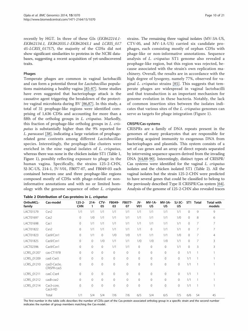
Table 2 Distribution of Cas-proteins in L. crispatus
OrthoMCL-family
Cas-model 125-2-CHN
214-1
CTV-05
FB049-03
FB0
LACT01579 Csn2 1/1 1/1 1/1 1/1 1
LACT01697 Cas2 0 1/0 1/1 1/1 1
LACT01698 Cas1 0 1/1 1/1 1/1 1
LACT01822 Csn2 0 1/1 1/1 1/1 1
LACT01823 Cas9/Csn1 0 1/1 0 1/0 1
LACT01825 Cas9/Csn1 0 0 1/0 1/1 1
LACT02396 Cas9/Csn1 0 0 0 1/1 1
LCRIS_01207 cas-CT1978 0 0 0 0
LCRIS_01209 casE-Cse3 0 0 0 0
LCRIS_01210 casD-Cas5e,CRISPR-cas5
0 0 0 0
LCRIS_01211 casC-Cse4 0 0 0 0
LCRIS_01212 casB-cse2 0 0 0 0
LCRIS_01214 Cas3-core,Cas3-HD
0 0 0 0
Total 1/1 5/4 5/4 7/6 7
The first number in the table cells describes the number of CDSs part of the Cas-prindicates the number of group members matching the Cas-model.
strains. The remaining three vaginal isolates (MV-3A-US,CTV-05, and MV-1A-US) carried six candidate pro-phages, each consisting mostly of orphan CDSs withphage-like or non-informative annotations. Sequenceanalysis of L. crispatus ST1 genome also revealed aprophage-like region, but this region was rejected, be-cause associated with the strain’s own replication ma-chinery. Overall, the results are in accordance with thehigh degree of lysogeny, namely 77%, observed for va-ginal L. crispatus strains [85]. This suggests that tem-perate phages are widespread in vaginal lactobacilliand that transduction is an important mechanism forgenome evolution in these bacteria. Notably, the lackof common insertion sites between the isolates indi-cates that various sites of the L. crispatus genomes canserve as targets for phage integration (Figure 1).
CRISPR/Cas-systemsCRISPRs are a family of DNA repeats present in thegenomes of many prokaryotes that are responsible forproviding acquired immunity to exogenous DNA frombacteriophages and plasmids. This system consists of aset of cas genes and an array of direct repeats separatedby intervening sequence spacers derived from the invadingDNA [64,88-90]. Interestingly, distinct types of CRISPR/Cas systems were identified for the vaginal L. crispatusisolates and the chicken isolated ST1 (Table 2). All thevaginal isolates but the strain 125-2-CHN were predictedto have several genes that could be classified to belong tothe previously described Type II CRISPR/Cas system [64].Analysis of the genome of 125-2-CHN also revealed traces
077-7
JV-V01
MV-1A-US
MV-3A-US
SJ-3C-US
ST1 Total Total withmodels
/1 1/1 1/1 1/1 1/1 0 9 9
/1 1/1 1/1 1/1 1/0 0 8 6
/1 1/1 1/1 1/1 1/1 0 7 7
/1 1/1 0 1/1 1/1 0 7 7
/0 1/1 1/1 1/1 1/0 0 7 4
/1 1/0 1/0 1/0 1/1 0 7 3
/1 0 0 0 1/1 0 3 3
0 0 0 0 0 1/1 1 1
0 0 0 0 0 1/1 1 1
0 0 0 0 0 1/1 1 1
0 0 0 0 0 1/1 1 1
0 0 0 0 0 1/1 1 1
0 0 0 0 0 1/1 1 1
/6 6/5 5/4 6/5 7/5 6/6 54 45
otein associated ortholog group in a specific strain and the second number
Ojala et al. BMC Genomics 2014, 15:1070 Page 11 of 21http://www.biomedcentral.com/1471-2164/15/1070
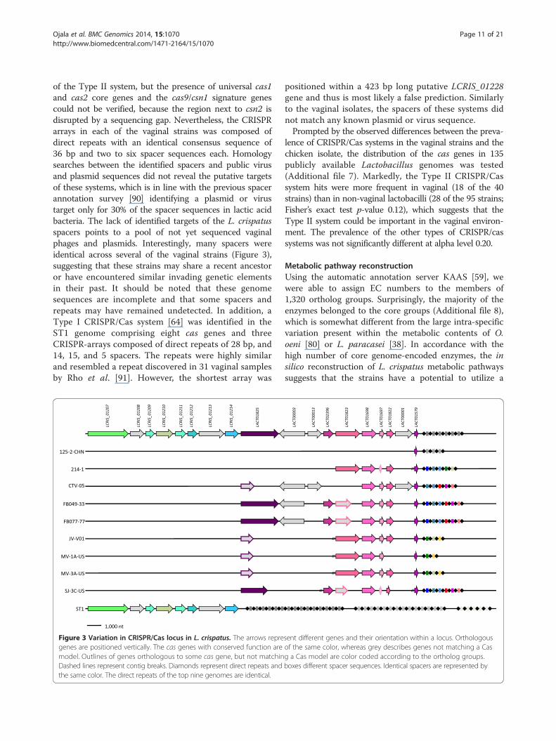
of the Type II system, but the presence of universal cas1and cas2 core genes and the cas9/csn1 signature genescould not be verified, because the region next to csn2 isdisrupted by a sequencing gap. Nevertheless, the CRISPRarrays in each of the vaginal strains was composed ofdirect repeats with an identical consensus sequence of36 bp and two to six spacer sequences each. Homologysearches between the identified spacers and public virusand plasmid sequences did not reveal the putative targetsof these systems, which is in line with the previous spacerannotation survey [90] identifying a plasmid or virustarget only for 30% of the spacer sequences in lactic acidbacteria. The lack of identified targets of the L. crispatusspacers points to a pool of not yet sequenced vaginalphages and plasmids. Interestingly, many spacers wereidentical across several of the vaginal strains (Figure 3),suggesting that these strains may share a recent ancestoror have encountered similar invading genetic elementsin their past. It should be noted that these genomesequences are incomplete and that some spacers andrepeats may have remained undetected. In addition, aType I CRISPR/Cas system [64] was identified in theST1 genome comprising eight cas genes and threeCRISPR-arrays composed of direct repeats of 28 bp, and14, 15, and 5 spacers. The repeats were highly similarand resembled a repeat discovered in 31 vaginal samplesby Rho et al. [91]. However, the shortest array was
Figure 3 Variation in CRISPR/Cas locus in L. crispatus. The arrows repregenes are positioned vertically. The cas genes with conserved function aremodel. Outlines of genes orthologous to some cas gene, but not matchinDashed lines represent contig breaks. Diamonds represent direct repeats andthe same color. The direct repeats of the top nine genomes are identical.
positioned within a 423 bp long putative LCRIS_01228gene and thus is most likely a false prediction. Similarlyto the vaginal isolates, the spacers of these systems didnot match any known plasmid or virus sequence.Prompted by the observed differences between the preva-
lence of CRISPR/Cas systems in the vaginal strains and thechicken isolate, the distribution of the cas genes in 135publicly available Lactobacillus genomes was tested(Additional file 7). Markedly, the Type II CRISPR/Cassystem hits were more frequent in vaginal (18 of the 40strains) than in non-vaginal lactobacilli (28 of the 95 strains;Fisher’s exact test p-value 0.12), which suggests that theType II system could be important in the vaginal environ-ment. The prevalence of the other types of CRISPR/cassystems was not significantly different at alpha level 0.20.
Metabolic pathway reconstructionUsing the automatic annotation server KAAS [59], wewere able to assign EC numbers to the members of1,320 ortholog groups. Surprisingly, the majority of theenzymes belonged to the core groups (Additional file 8),which is somewhat different from the large intra-specificvariation present within the metabolic contents of O.oeni [80] or L. paracasei [38]. In accordance with thehigh number of core genome-encoded enzymes, the insilico reconstruction of L. crispatus metabolic pathwayssuggests that the strains have a potential to utilize a
sent different genes and their orientation within a locus. Orthologousof the same color, whereas grey describes genes not matching a Casg a Cas model are color coded according to the ortholog groups.boxes different spacer sequences. Identical spacers are represented by

Ojala et al. BMC Genomics 2014, 15:1070 Page 12 of 21http://www.biomedcentral.com/1471-2164/15/1070
rather same set of carbohydrates (Additional file 9). Thedata supports the presence of metabolic routes in eachstrain for the conversion of a variety of sugars into thekey intermediates of the pentose phosphate (D-Xylulose5-phosphate), Embden–Meyerhof–Parnas (D-Fructose1,6-bisphosphate), and tagatose-6-phosphate (tagatose-6-phosphate) pathways. Pathways for the conversion ofthe D-Xylulose 5-phosphate and D-Fructose 1,6-bispho-sphate into several of their end products were also anno-tated for nine of the ten strains. The aforementionedindicates the presence of both Embden–Meyerhof–Parnas and pentose phosphate pathways in the ninestrains, which is typical for a heterofermentative speciesand contradictory to the previous classification of L. cris-patus as a homofermentative species [28,92]. The excep-tion is the strain CTV-05 that had only partial pathwaysfor many end product conversions, most likely becauseof sequencing gaps in the corresponding genomic loci.No routes were recorded for the conversion of tagatose-6-phosphate pathway intermediate into pyruvate in anyof the strains. Interestingly, the data also shows evidencefor the presence of strain-specific glycerone conversionsin L. crispatus 125-2-CHN.Regarding urogenital lifestyle, conserved pathways were
annotated for the metabolism glucose and mannose, theformer reported to be the major free monosaccharide andthe latter a minor constituent of the vaginal fluid [93]. Al-though we did not detect complete routes for the metabol-ism of glycogen, seven vaginal strains were discovered tocarry a gene coding for a type I pullulanase debranchingenzyme (LACT01812), which could contribute to the deg-radation of glycogen. Moreover, L. crispatus core appears toencode a sialic acid utilization regulator (RpiR family) andan O-sialoglycoprotein endopeptidase that could contrib-ute to the hydrolysis of O-sialoglycoproteins in the va-ginal mucosa. Notably, the manual examination of theenzyme contents revealed that each strain may generatehydrogen peroxide from pyruvate, of which the formeracts as an antimicrobial compound.We also assessed the range of amino acids that L.
crispatus has a potential to synthetize (Additional file9). Based on the in silico analyses of the biosyntheticcapabilities, all strains can synthesize seven aminoacids either de novo or as derivatives using the samepathways, which is three and four amino acids morethan L helveticus DPC 4571 [94] or L. acidophilusNCFM [84], respectively. Pathways for aspartate bio-synthesis were also annotated in nine isolates, exclud-ing the strain CTV-05 that did not share this property.We again speculate that the lack of biosynthesis routefor aspartate is rather due to the draft nature of thegenome sequence of this strain than a genuine loss.The other differences in amino-acid synthesis relatedto nuances in synthesis routes for cysteine, serine, and
glycine, which seem to vary between isolates. Overall,the in silico analyses predicted a dependency on exter-nal supplies of amino acids for L. crispatus similar tothat described for closely related lactobacilli [84,95]and shows that the strains are rather similar in theirbiosynthetic power. Moreover, none of the detectedconversions was deduced to be strain-specific, furtherhighlighting the similarity.
Proteinaceous adhesinsAdhesion to host tissue has long been considered an im-portant factor and a prerequisite for the long-termcolonization of the human vagina, stimulation of theimmune system, and antagonistic activity against harm-ful pathogens through competitive exclusion [96]. Wescreened the L. crispatus proteomes for adhesion and hostcolonization related domains and identified 103 proteinsgoverning the ability of L. crispatus to colonize and inter-act with the host. These putative adhesins were associatedwith seven distinct types of adhesion-associated domainsbelonging to 21 ortholog groups of which seven are partof the L. crispatus core genome (Table 3, Additional file10). It should be noted, however, that members of thesame ortholog group did not necessarily share adhesiondomains. In addition, six strain-specific adhesins wereidentified, all of which were predicted to be mucus-binding proteins. Interesting examples of the strain-specific adhesins include a sortase-anchored protein(LCRIS_00919) with multiple mucus-binding domains,and LCRIS_01654 being the only member of its orthologgroup (LACT01522) with adhesion-associated domains.One notable core adhesin (LACT00800) was a putative fi-bronectin/fibrinogen-binding protein Fbpa, which has re-cently been proposed to contribute to the fibronectin-binding properties of Lactobacillus iners and to explainthe stronger adhesion of L. iners to human fibronectincompared to other species of Lactobacillus tested in thestudy [97]. Notably, our data does not support this hy-pothesis, since the presence of functional fbpa gene in theL. crispatus core genome should have resulted in equal ad-hesion abilities for the L. crispatus and L. iners strainstested in the study. Markedly, the recently characterizedLEA protein of L. crispatus ST1 [33] belonging toLACT00252 was not identified, indicating that thisadhesin binds to crop epithelium and epithelial cellsfrom human vagina with some novel domain. In additionto the aforementioned putative adhesins, L. crispatuswas predicted to harbor ~30 putative S-layer protein-encoding genes that could potentially contribute tobacterial adhesion. However, these predicted S-layerproteins were different from the S-layer proteins ofother related lactobacilli reportedly implicated in bac-terial adhesion [42,98,99].

Table 3 Distribution of adhesion related proteins in L. crispatus
OrthoMCL- family Adhesion orcolonizationdomain
125-2-CHN
214-1 CTV-05
FB049-03
FB077-07
JV-V01
MV-1A-US
MV-3A-US
SJ-3C-US
ST1 Total Total withdomain
LACT01267 MucBP 1/1 1/0 1/1 1/1 1/1 1/0 0 1/1 1/1 1/1 9 7
LACT01522 MucBP 1/0 1/0 0 1/0 1/0 1/0 1/0 1/0 1/0 1/1 9 1
LACT01644 MucBP 0 1/1 1/1 1/0 1/0 1/1 1/0 1/1 0 1/1 8 5
LACT01663 MucBP 1/1 1/1 0 1/1 1/1 1/1 1/1 1/1 1/1 0 8 8
LACT01712 MucBP 2/0 0 0 1/1 1/1 1/0 1/0 1/0 1/1 0 7 3
LACT01924 MucBP 1/0 1/1 0 1/1 1/1 1/0 1/0 0 0 0 6 3
LACT02281 MucBP 0 0 0 0 1/1 1/0 1/1 0 0 0 3 2
LACT02327 MucBP 1/0 0 0 0 0 1/1 1/0 0 0 0 3 1
LACT02429 MucBP 2/2 0 0 0 0 0 0 0 0 0 1 1
HMPREF9249_02429 MucBP 0 0 0 0 1/1 0 0 0 0 0 1 1
HMPREF0506_0624 MucBP 0 0 0 0 0 1/1 0 0 0 0 1 1
HMPREF0506_0871 MucBP 0 0 0 0 0 1/1 0 0 0 0 1 1
LCRIS_00919 MucBP 0 0 0 0 0 0 0 0 0 1/1 1 1
LACT00524 Big_3 1/1 1/1 1/1 1/1 1/1 1/1 1/1 1/1 1/1 1/1 10 10
LACT00531 Big_3 1/1 1/1 1/1 1/1 1/1 1/1 1/1 1/1 1/1 1/1 10 10
LACT00945 Big_3 1/0 1/0 1/0 1/1 1/1 1/1 1/1 1/1 1/0 1/1 10 6
LACT02389 Big_3 0 1/1 1/1 0 0 0 0 0 1/1 0 3 3
LACT00169 F5_F8_type_C 1/1 1/1 1/1 1/1 1/1 1/1 1/1 1/0 1/1 1/1 10 9
LACT00800 FbpA, DUF814 1/1 1/1 1/1 1/1 1/1 1/1 1/1 1/1 1/1 1/1 10 10
LACT00237 FIVAR 1/0 1/1 1/1 1/1 1/1 1/1 1/1 1/1 1/1 1/1 10 9
LACT00212 fn3 1/1 1/1 1/1 1/1 1/1 1/1 1/1 1/1 1/1 1/1 10 10
Total 16/9 13/10 10/9 13/11 15/13 17/12 14/9 12/9 12/10 11/11 131 102
The first number in the table cells describes the number of CDSs part of the ortholog group in a specific strain and the second the number indicates the numberof group members having the adhesion or colonization related PFAM-domain(s).
Ojala et al. BMC Genomics 2014, 15:1070 Page 13 of 21http://www.biomedcentral.com/1471-2164/15/1070
Cell wall exopolysaccharideIn the L. crispatus genomes, a highly variable genomeregion appears to be associated with EPS biosynthesis.This EPS gene cluster was observed in eight L. crispatusstrains and noted to comprise 37 EPS biosynthesis genes,five of which were present within each operon (Figure 4).The five conserved genes were predicted to encode atranscriptional regulator, a polymerization and chainlength determination protein, a tyrosine-protein kinase,a protein-tyrosine phosphatase, and the priming glyco-syltransferase. The remaining genes coded for proteinswith putative glycosyl transferase functions, indicatingthat the strains produce EPSs with different sugarmonomers and glycosidic linkages. Markedly, EPS geneclusters were not detected in the genomes of L. crispa-tus JV-V01 and 214-1.
Antimicrobial potential in L. crispatusLactobacillus species can maintain the vaginal ecosystemin a healthy condition by the production of antimicrobialsubstances such as lactic acid, hydrogen peroxide andbacteriocin-like substances [9,96]. Lactic acid is the main
end product of the carbohydrate fermentation in lacto-bacilli and can contribute to the vaginal acidity andthereby inhibit the colonization and proliferation ofharmful micro-organisms in the vagina [100]. TheL. crispatus strains studied here appeared to possessbetween three to four L-lactate dehydrogenases for theconversion of puryvate into lactic acid. Interestingly,one specific ldh locus found in five L. crispatus strains wasflanked by a transposase enzyme gene that may affect itsexpression [101]. We also discovered hydrogen peroxideproducing enzymes (EC:1.2.3.3 and EC:1.1.3.15) in eachL. crispatus, which correlates well with the experimentaldata showing that hydrogen peroxide generation iscommon among vaginal L. crispatus [102].Using BAGEL [63], the bacteriocin content of L. cris-
patus was investigated (Table 4). This method was ableclassify several sets of putative bacteriocin gene clustersin each strain, including at least two regions encodingbacteriolysins (similar to enterolysin A [103] and helveti-cin J [104]). In addition, regions implicated in the pro-duction of class II bacteriocins were revealed in thevaginal isolates. A pediocin-like bacteriocin that inhibits

Figure 4 Variation in EPS gene cluster in L. crispatus. The organization and conservation of the exopolysaccharide synthesis regions inL. crispatus. Orthologous genes are represented with the same color and stars indicate genes found in different loci. Dashed lines representcontig breaks in the MV-1A-US and SJ-3C-US clusters.
Ojala et al. BMC Genomics 2014, 15:1070 Page 14 of 21http://www.biomedcentral.com/1471-2164/15/1070
the growth of pathogenic Listeria and Clostridium species[105] was present in five vaginal isolates and all nineencoded a two-component bacteriocin LS2 that inhibits thegrowth of isolates belonging to genera Listeria, Shigella,and Yersinia [106]. Notably, the pediocin-like bacteriocinencoding genes were found in the vicinity of CDSs encod-ing proteins harboring a domain for Enterocin A immunity.
Antagonistic activities against G. vaginalisBV is the most common vaginal disorders, affecting upto a third of women [107]. It has been associated with
Table 4 Distribution of predicted bacteriocin related proteins
Bacteriocinclass
Bacteriocintype
PFAM 125-2-CHN
214-1 CTV-05
Bacteriocinslarger than10kD, Class III
Enterolysin A Peptidase_M23 1/1 2/2 2/2
Bacteriocinhelveticin J
- 1 1 1
Helveticin J - 1 1 1
LAPs Small orfs - 1 0 0
Small unmodifiedbacteriocins,Class II
Penocin A Bacteriocin_II 1/2 0 1/2
LS2, chain A Bacteriocin_IIc 1/1 1/1 1/1
Total 7 5 7
For the bacteriocin-like molecules with PFAM-domain, the first number in the tablestrain, and the second number indicates the number of group members having thein L. helveticus and bacteriocin helveticin J to one identified in L. acidophilus.
increased risk for preterm birth, urinary tract infections,and HIV infection, and represents a condition in whichthe normal protective lactobacilli community is replacedby an overgrowth of anaerobic bacteria [46]. Althoughthe etiology of BV is not known, G. vaginalis is presentin up to 95% of all BV cases [108], indicating that itcould have a role in BV. In our efforts to decipher thegenetic basis of the inhibitory actions of the speciesL. crispatus against G. vaginalis, we performed orthologgrouping of the available G. vaginalis data (Additionalfile 11) and used comparative genomics to identify shared
in L. crispatus
FB049-03
FB077-07
JV-V01
MV-1A-US
MV-3A-US
SJ-3C-US
ST1 Total Total withdomain
2/2 2/2 2/2 2/2 2/2 2/2 2/2 10 10
1 1 1 1 1 1 1 10 -
1 2 1 1 1 1 1 10 -
1 1 0 1 0 0 0 4 -
1/2 1/2 0 0 1/2 1/1 0 6 6
1/1 1/1 1/1 1/1 1/1 1/1 0 9 9
8 9 5 6 7 6 4 49 25
cells describes the number of CDSs part of the ortholog group in a specificPFAM-domain. Helveticin J refers to the antimicrobial molecule first identified

Ojala et al. BMC Genomics 2014, 15:1070 Page 15 of 21http://www.biomedcentral.com/1471-2164/15/1070
common molecular mechanisms between G. vaginalis andL. crispatus. Importantly, our analyses revealed severalcomponents by which L. crispatus could interfere withthe attachment of G. vaginalis in the vagina. Firstly,fibronectin-binding could play a role in this process,given that proteins with FIVAR domains related to hya-luronate or fibronectin-binding were encoded in thecore genomes of both G. vaginalis (GVAG00006) andL. crispatus (LACT00237). Secondly, searching L. cris-patus proteins against the G. vaginalis HMM databasesuggested another L. crispatus protein (LACT01268),which could play a role in preventing the cell adhesionof G. vaginalis to fibronectin. Intriguingly, this counter-part of the G. vaginalis FIVAR-proteins was distributedin nine L. crispatus strains, but had no known adhesiondomains. Another interesting core orholog group ofG. vaginalis was GVAG00055. Many members of thisortholog group contained a bacterial Ig-like domain(PF12245), which is distantly related to the interactiondomains, namely fn3 (PF00041) and Big_3 (PF07523),associated with several L. crispatus core adhesins (Table 3).Moreover, searches against the G. vaginalis HMMsrevealed two additional L. crispatus adhesins (LACT01712and LACT02327) that could act as counterparts ofGVAG00055, although having mucin-binding domains(Table 3). Finally, of the three G. vaginalis pilus-encoding gene clusters that were identified based onthe pilus-encoding genes listed by Yeoman et al. [72],the one associated with most isolates had borderline(E-value ≤ 0.4) counterparts in the L. crispatus coregenome. Its major subunit pilin (GVAG00005) appearsto have two potential antagonists in the L. crispatus coregenome encoding a 12.8-kilodalton protein (LACT00214)and the LEA protein (LACT00252). In addition, thelong CDS (GVAG00017) located next to the major sub-unit component in the cluster and showing similarity toknown adhesins and surface antigens, could be inhibitedby the members of the LACT01712 and LACT02440based on the G. vaginalis HMM searches. Taken to-gether, these findings indicate that L. crispatus couldinterfere with fibronectin-binding and pilus compo-nents of G. vaginalis.Of the other listed virulence-related factors in
G. vaginalis [72], the invasion-associated hydrolase(GVAG00614), protein with two G-related albumin-binding modules (GVAG01097), NLPA lipoprotein(GVAG00181), and endothelin-converting enzyme(GVAG00141) have potential antagonists encoded bythe L. crispatus core based on the G. vaginalis HMMsearches. A noteworthy finding is that the G-relatedalbumin-binding module protein (GVAG01097) presentin 17G. vaginalis isolates shared similarity with 42L. crispatus proteins, including all nine FIVAR-domainassociated proteins of the LACT00237 (Table 3).
Adhesion inhibition assays to HeLa cellsOur comparative analysis described several species-widefactors by which L. crispatus could compete with G.vaginalis in the vagina. For example, the LEA proteinwas identified as a prominent counterpart of one of theG. vaginalis core adhesins and was thereby predicted toparticipate in the adherence inhibition of this pathogen.To validate the role of LEA in the antagonism against G.vaginalis, the adhesion capacity of a vaginal L. crispatusisolate EX533959VC06 and BV-associated G. vaginalis101 to HeLa cells was tested using the previously de-scribed approach [17] with and without the pretreatmentwith Fab fragments prepared against LEA [33]. Markedly,the anti-LEA Fab fragments significantly reduced theadhesion level of both bacterial species to HeLa cellswhereas the unrelated anti-flagellum Fab fragmentsshowed no inhibitory effect (Figure 5). The reduction inadherence was most evident for the strain EX533959VC06;the anti-LEA Fab fragment pretreatment resulting in90.6% (p-value ≤ 0.033) and 89.8% (p-value ≤ 0.024) reduc-tion in adhesion to HeLa cells compared with the un-treated or anti-flagellum Fab fragment pretreatedbacteria, respectively. Intriguingly, pretreating G. vagi-nalis 101 with the anti-LEA Fab fragments caused also asignificant reduction in adherence compared with theuntreated bacterial cells (65.6%; p-value ≤ 0.005) or bac-teria pretreated with the control anti-flagellum Fabfragments (65.1%; p-value ≤ 0.019). These observationsvalidated the predicted competitive character betweenLEA and G. vaginalis, suggesting a role for LEA in thepreviously identified ability of L. crispatus to excludeand displace G. vaginalis from HeLa cells [17]. Theresults also provide an explanation to the inverse associ-ation between L. crispatus and G. vaginalis colonizationin the vagina [12,44,47]. Based on our comparative gen-omic analyses, the LEA protein achieves its inhibitoryeffect by competing with the same attachment sites asthe pili of G. vaginalis. Of note, our adhesion assayprovided a further support for the species-wide distri-bution of LEA among L. crispatus, since the strainEX533959VC06 has not yet been sequenced. Further-more, since LEA has previously been studied only in thechicken isolate ST1 [33], our results serve as the first rec-ord of the functionality of LEA in vaginal L. crispatus.
Phylogenetic relationsPhylogentic relations between the selected L. crispatusstrains and strains of closely related species L. acidophilusand L. helveticus were examined based on a maximum-likelihood tree built from the SNPs of the core genome.Altogether 38,726 conserved polymorphic sites wereidentified from the genome alignments and used for theconstruction of a phylogenetic tree. The phylogenetictree (Figure 6) clearly shows that strains of the same

Figure 5 Inhibition of L. crispatus or G. vaginalis adhesion to HeLa cells by LEA-specific Fab fragments. Cells of L. crispatus EX533959VC06(A) or G. vaginalis 101 (B) were pretreated with LEA-specific IgG Fab fragments or unrelated anti-flagellum Fab fragments or left untreated in PBSsupplemented with 5 mM PMSF before the adhesion assays. The number of adherent bacteria per epithelial cell in 20 randomly chosen microscopicfields was determined. The assay was performed twice with duplicate samples and the results show mean values of adherent bacteria. The asteriskindicates P < 0.05 as calculated by Student’s t test.
Ojala et al. BMC Genomics 2014, 15:1070 Page 16 of 21http://www.biomedcentral.com/1471-2164/15/1070
species cluster together and that each Lactobacillus spe-cies has differentiated as a distinct entity. The species L.crispatus and L. helveticus share the most recent commonancestor and form a sister group to species L. acidophilus,which is accordance with previously reported phylogenetictrees [29,109]. Among the L. crispatus cluster, the chickenisolated ST1 branches off first from the vaginal isolates.
ConclusionsThe rapidly increasing number of complete microbialgenomes offers previously unimaginable possibilities to
Figure 6 Phylogenetic tree. Phylogenetic relations of the selected L. crispon the SNPs of the core genome. The B. subtilis genome was used as the othe branching pattern of the L. crispatus strains is highlighted.
understand the phenotypic and genomic diversity in aparticular species [38,70,79,80]. In this study, we havetaken advantage of publicly available L. crispatus genomesand present the genetic landscape of this importanturogenital lactic acid bacterium [7-12]. We assessed theoverall genomic similarity of ten strains and defined theL. crispatus pan- and core genomes. These analysesdepicted high sequence identity and extensive syntenypunctuated by several GIs, and revealed a current pan-genome that is nearly two times larger than the numberof ortholog groups present in an average L. crispatus
atus (green), L. helveticus (blue) and L. acidophilus (purple) strains basedut-group to root the tree, but is not shown in the figure. In the inset,

Ojala et al. BMC Genomics 2014, 15:1070 Page 17 of 21http://www.biomedcentral.com/1471-2164/15/1070
strain. About one third of all 3,929 ortholog groupswere assigned to all strains, constituting the currentL. crispatus core genome and encoding the basic aspectsof L. crispatus biology. Importantly, these core featurescomprised several CDSs for the production of anti-microbial molecules and competitive exclusion of theBV associated species G. vaginalis, shedding light onthe molecular mechanisms by which L. crispatus couldmaintain vaginal health. The pan-genome analysis alsorevealed 1,311 singleton ortholog groups associatedwith only one strain. The enrichment of functionsrelated to replication and repair among these genesindicates the influence of transposons in genome evolu-tion in this species. A third of the strain-specific ortho-log groups had the highest similarity to genes found inthe other strains of the L. delbrueckii clade, suggestingnotable sequence influx from closely related lactobacilli.Our regression analysis indicates that the genetic diver-sity present within L. crispatus has not yet been com-prehensively captured. Specifically, we estimate thatover ten new ortholog groups will be discovered perevery additional genome until almost 300 L. crispatusstrains have had their genomes defined. This estimationmay be compromised by the uncertainty caused by thedraft genomes that have up to 201 sequence gaps.Nevertheless, the data implies the presence of largerepertoires of undiscovered L. crispatus genes to besequenced in the future. The phylogenetic tree based oncore genome SNPs among the ten isolates revealed thatthe chicken isolated ST1 branches off first from theL. crispatus cluster and that the L. acidophilus clusteris a sister taxon to L. helveticus and L. crispatus, assuggested earlier [29,109].From the perspective of vaginal health, the most inter-
esting genomic diversity regions in L. crispatus includethe loci related to EPS biosynthesis, prophages andadaptive immunity, of which the latter two may play arole in BV. Firstly, the genetic differences in the com-position of the EPS gene region may participate in theL. crispatus adhesion, biofilm formation and competitiveexclusion of pathogens. The EPS-deficient strains JV-V01and 214-1 are particularly interesting, as the deprivationof EPS has been reported to promote bacterial adhesionin other lactobacilli [110,111]. Secondly, the presenceof prophage-like clusters in the vaginal L. crispatusgenomes is in accordance with the previously observed[85] high level of lysogeny in vaginal L. crispatus strains.If truly inducible, the spontaneous release of the pro-phages could contribute to the development of BV [86].Finally, a relationship was depicted between the lifeenvironment of the strains and their adaptive immunitysystems, suggesting that different types CRISPR/Cassystems could be beneficial in different environments.This hypothesis is further supported by the analysis of
the cas gene contents of 135 Lactobacillus genomes thatrevealed higher rates of the Type II CRISPR/Cas sys-tems in vaginal than in non-vaginal lactobacilli. Inaddition, the CRISPR-arrays of the vaginal L. crispatusstrains carry evidence of encounters with common in-vaders, as several of the spacer sequences were identicalbetween several strains.The defined L. crispatus core genome helps to explain
how this species can thrive in the vaginal environmentand benefit vaginal health. In the vaginal epithelium ofreproductive age females, large quantities of glycogenare broken down and then metabolized into lactic acid,which is thought to result in acidification of the vagina[112,113]. Although L. crispatus lacks complete enzym-atic machinery for glycogen degradation, the core gen-ome encodes enzymatic pathways for the utilization of arange of carbohydrates available in the vaginal fluid,which could support the urogenital commensal lifestyleof L. crispatus. Encoded in the core are also severalfeatures potentially governing host-interactions and dis-playing an antagonistic activity against other micro-organisms. Interestingly, the bacteriocin-like moleculesencoded by the L. crispatus genomes could inhibit biofilmintegrated G. vaginalis cells, shown to be more resistant tohydrogen peroxide and lactic acid than the cells in plank-tonic state [114]. Specifically, as G. vaginalis is known todevelop an adherent biofilm on the vaginal epithelium inBV [115] this property could provide attractive means torestore the normal vaginal flora. In addition to the anti-microbial properties, L. crispatus was detected to containseveral proteins that could mediate the previously re-ported [17] competitive exclusion of G. vaginalis from epi-thelial cells and explain the inverse association between L.crispatus and G. vaginalis colonization in the vagina[12,44,47]. Most notably, these specific interference mech-anisms might include blocking the attachment of G. vagi-nalis by disturbing the pilus-mediated adhesion of thepathogen. This mechanism could involve LEA, shownhere to be universally present in all L. crispatus strains,and demonstrated using LEA-specific Fab fragments toinhibit the adhesion of G. vaginalis adhesion to HeLa cells.Although LEA showed sequence similarity to a pilus com-ponent of G. vaginalis, further studies are still needed todecipher whether the counterpart of LEA is indeed thepilin subunit or some other adhesion associated moleculeof G. vaginalis. In addition, we cannot rule out thatsurface molecules other than the ones recognized by theanti-LEA Fab fragments have participated in the contactbetween G. vaginalis and the host cell, since the Fab frag-ments did not abolish the adhesion completely. Neverthe-less, the LEA protein appears to be a key mediator of thecompetitive exclusion of G. vaginalis.In summary, we have presented a comparative analysis
of ten L. crispatus genomes available within the public

Ojala et al. BMC Genomics 2014, 15:1070 Page 18 of 21http://www.biomedcentral.com/1471-2164/15/1070
databases at the time of this study and provided a com-prehensive look on the pan-genomic structure of thisimportant urogenital species. Furthermore, our analysesrevealed a list of core genes implicated in protecting theurogenital tract from G. vaginalis colonization, providingnew insights into the treatment and prevention of BV.
Additional files
Additional file 1: Overview of G. vaginalis strains and properties.In the table, the genomic properties of the G. vaginalis strains used inthis study are given. HMP refers to the Human Microbiome Project.
Additional file 2: List of L. helveticus, L. acidophilus and B. subtilisgenomes included in the phylogenetic analysis. The accession isgiven for each genome.
Additional file 3: List of PFAM domains used in the annotation ofputative L. crispatus adhesins. The accession, ID, and description aregiven for each PFAM matching an adhesion or colonization relatedkeyword. Ones in the remaining columns indicate that the PFAMmatched some L. crispatus CDS, passed the manual curation process andwas included in the final list of adhesion or colonization related domains.
Additional file 4: The start and end compounds used in themetabolism screens. This table describes the compound pairs related tothe de novo synthesis and interconversion of amino acids and carbohydratemetabolism.
Additional file 5: The L. crispatus data table. Results of the differentbioinformatic analyses for each L. crispatus CDS.
Additional file 6: Reservoirs of genetic variability. This table describesthe distribution of best BLAST hits of strain-specific L. crispatus CDSs.
Additional file 7: Prevalence of different types of CRISPR/Cassystems in vaginal and non-vaginal lactobacilli.
Additional file 8: Variation in metabolism related enzymes inL. crispatus. The horizontal lines represent the orthologous groups withassigned EC numbers and the different strains are indicated at the bottomof the picture. The presence of a given ortholog group in a specific strain isindicated with grey (single copy) or dark grey (duplicated genes). Theabsence of a given ortholog group is indicated with light grey. The coloredbar on the left describes the conservation level of the ortholog groups andfollows that of the Figure 2 (red indicates core genome and blue accessorygenome with darkest shade indicating conservation in nine strains andlightest blue strain-specific). The dendrogram was generated using wardlinkage clustering of the presence/absence data.
Additional file 9: Detected metabolic routes between differentcarbohydrate metabolism compounds and between differentamino acid biosynthesis related compounds. The presence of intactmetabolic route from one compound to another is indicated for eachL. crispatus genome. Pathways found in the core genome analysis aremarked with yellow color.
Additional file 10: Domain organization of L. crispatus adhesionand colonization factors. A representative member of eachOrthoMCL-group is presented graphically. The larger colored blocksrepresent adhesion or colonization related PFAM-domains and the thinnerblocks other domains. The names of each color-coded domain are given atthe bottom of the picture.
Additional file 11: Overview of G. vaginalis ortholog groups.For each group, the associated proteins, PFAM domains, matching proteins inL. crispatus, the total number of matching proteins in L. crispatus, and totalnumber of L. crispatus genomes providing the matches are given in the table.
AbbreviationsBV: Bacterial vaginosis; Cas: CRISPR-associated; CDS: Coding sequence;CFUs: Colony forming units; COG: Cluster of orthologous groups;CRISPR: Clustered regularly interspaced short palindromic repeat;EPS: Exopolysaccharide; GI: Genomic Island; GIT: Gastrointestinal tract;
GUT: Genitourinary tract; HGT: Horizontal gene transfer; HMM: HiddenMarkov model; LEA: Lactobacillus epithelium adhesin; OD: Opticaldensity; PBS: Phosphate buffered saline; PFA: Paraformaldehyde;PMSF: Phenylmethylsulfonyl fluoride; RUTI: Recurrent urinary tractinfection; SNP: Single nucleotide polymorphism.
Competing interestsThe authors declare that they have no competing interests.
Authors’ contributionsTO designed experiments and the adhesion experiments, analyzed thegenomic data and prepared the manuscript. MK designed experiments,analyzed the genomic data and prepared the manuscript. JC performed theadhesion assays, analyzed the adhesion data and participated in preparingthe manuscript. NC designed the adhesion experiments, analyzed theadhesion data and participated in preparing the manuscript. SE provided theFab fragments. BWW designed the adhesion experiments and participated inpreparing the manuscript. LP participated in preparing the manuscript. LHparticipated in preparing the manuscript. PA participated in preparing themanuscript. All authors read, commented and approved the final manuscript.
AcknowledgementsWe thank Viikki Doctoral Programme in Molecular Biosciences (VGSB) andIntegrative Life Science Doctoral Program (ILS) for supporting this study,Helmi Pett for useful suggestions for improving the manuscript, andKimberly Jefferson for providing L. crispatus strain EX533959VC06.
Author details1Institute of Biotechnology, University of Helsinki, Viikinkaari 4, PO Box 56,FI-00014 Helsinki, Finland. 2Institute for Molecular Medicine Finland (FIMM),University of Helsinki, Tukholmankatu 8, PO Box 20, FI-00014 Helsinki, Finland.3Centre of Biological Engineering (CEB), Laboratory of Research in BiofilmsRosário Oliveira (LIBRO), University of Minho, Campus de Gualtar, 4710-057Braga, Portugal. 4Functional Foods Forum, University of Turku, Turku FI-20520,Finland. 5Department of Biosciences, Division of General Microbiology,University of Helsinki, Viikinkaari 9c, PO Box 56, FI-00014 Helsinki, Finland.6Department of Biosciences, Division of Genetics, University of Helsinki,Viikinkaari 5, PO Box 56, FI-00014 Helsinki, Finland.
Received: 14 July 2014 Accepted: 24 November 2014Published: 5 December 2014
References1. Siezen RJ, Wilson G: Probiotics genomics. J Microbial Biotechnol 2010, 3(1):1–9.2. Ventura M, O'Flaherty S, Claesson MJ, Turroni F, Klaenhammer TR, van
Sinderen D, O'Toole PW: Genome-scale analyses of health-promotingbacteria: probiogenomics. Nat Rev Microbiol 2009, 7(1):61–71.
3. Uehara S, Monden K, Nomoto K, Seno Y, Kariyama R, Kumon H: A pilotstudy evaluating the safety and effectiveness of Lactobacillus vaginalsuppositories in patients with recurrent urinary tract infection. Int JAntimicrob Agents 2006, 28(Suppl 1):S30–4.
4. Stapleton AE, Au-Yeung M, Hooton TM, Fredricks DN, Roberts PL,Czaja CA, Yarova-Yarovaya Y, Fiedler T, Cox M, Stamm WE: Randomized,placebo-controlled phase 2 trial of a Lactobacillus crispatus probioticgiven intravaginally for prevention of recurrent urinary tract infection.Clin Infect Dis 2011, 52(10):1212–1217.
5. Hemmerling A, Harrison W, Schroeder A, Park J, Korn A, Shiboski S, Cohen CR:Phase 1 dose-ranging safety trial of Lactobacillus crispatus CTV-05 for theprevention of bacterial vaginosis. Sex Transm Dis 2009, 36(9):564–569.
6. Antonio MA, Meyn LA, Murray PJ, Busse B, Hillier SL: Vaginal colonization byprobiotic Lactobacillus crispatus CTV-05 is decreased by sexual activity andendogenous Lactobacilli. J Infect Dis 2009, 199(10):1506–1513.
7. Lamont RF, Sobel JD, Akins RA, Hassan SS, Chaiworapongsa T, Kusanovic JP,Romero R: The vaginal microbiome: new information about genital tractflora using molecular based techniques. BJOG 2011, 118(5):533–549.
8. Ravel J, Gajer P, Abdo Z, Schneider GM, Koenig SS, McCulle SL, Karlebach S,Gorle R, Russell J, Tacket CO, Brotman RM, Davis CC, Ault K, Peralta L, ForneyLJ: Vaginal microbiome of reproductive-age women. Proc Natl Acad SciU S A 2011, 108(Suppl 1):4680–4687.

Ojala et al. BMC Genomics 2014, 15:1070 Page 19 of 21http://www.biomedcentral.com/1471-2164/15/1070
9. Witkin SS, Linhares IM, Giraldo P: Bacterial flora of the female genital tract:function and immune regulation. Best Pract Res Clin Obstet Gynaecol 2007,21(3):347–354.
10. Macklaim J, Fernandes A, Di Bella J, Hammond J, Reid G, Gloor G:Comparative meta-RNA-seq of the vaginal microbiota and differentialexpression by Lactobacillus iners in health and dysbiosis.Microbiome 2013, 1(1):12.
11. Verstraelen H, Verhelst R, Claeys G, De Backer E, Temmerman M,Vaneechoutte M: Longitudinal analysis of the vaginal microflora inpregnancy suggests that L. crispatus promotes the stability of thenormal vaginal microflora and that L. gasseri and/or L. iners are moreconducive to the occurrence of abnormal vaginal microflora. BMC Microbiol2009, 9:116-2180-9-116.
12. Fredricks DN, Fiedler TL, Thomas KK, Oakley BB, Marrazzo JM: Targeted PCRfor detection of vaginal bacteria associated with bacterial vaginosis.J Clin Microbiol 2007, 45(10):3270–3276.
13. Zárate G, Nader-Macias ME: Influence of probiotic vaginal lactobacilli onin vitro adhesion of urogenital pathogens to vaginal epithelial cells.Lett Appl Microbiol 2006, 43(2):174–180.
14. Osset J, Bartolomé RM, García E, Andreu A: Assessment of the capacity ofLactobacillus to inhibit the growth of uropathogens and block theiradhesion to vaginal epithelial cells. J Infect Dis 2001, 183(3):485–491.
15. Atassi F, Brassart D, Grob P, Graf F, Servin AL: Vaginal Lactobacillus isolatesinhibit uropathogenic Escherichia coli. FEMS Microbiol Lett 2006,257(1):132–138.
16. Teixeira GS, Carvalho FP, Arantes RM, Nunes AC, Moreira JL, Mendonca M,Almeida RB, Farias LM, Carvalho MA, Nicoli JR: Characteristics of Lactobacillusand Gardnerella vaginalis from women with or without bacterial vaginosisand their relationships in gnotobiotic mice. J Med Microbiol 2012,61(Pt 8):1074–1081.
17. Castro J, Henriques A, Machado A, Henriques M, Jefferson KK, Cerca N:Reciprocal interference between Lactobacillus spp. and Gardnerellavaginalis on initial adherence to epithelial cells. Int J Med Sci 2013,10(9):1193–1198.
18. Abbas Hilmi HT, Surakka A, Apajalahti J, Saris PE: Identification of the mostabundant lactobacillus species in the crop of 1- and 5-week-old broilerchickens. Appl Environ Microbiol 2007, 73(24):7867–7873.
19. Yuki N, Shimazaki T, Kushiro A, Watanabe K, Uchida K, Yuyama T, Morotomi M:Colonization of the stratified squamous epithelium of the nonsecretingarea of horse stomach by lactobacilli. Appl Environ Microbiol 2000,66(11):5030–5034.
20. De Angelis M, Siragusa S, Berloco M, Caputo L, Settanni L, Alfonsi G,Amerio M, Grandi A, Ragni A, Gobbetti M: Selection of potential probioticlactobacilli from pig feces to be used as additives in pelleted feeding.Res Microbiol 2006, 157(8):792–801.
21. Walter J, Hertel C, Tannock GW, Lis CM, Munro K, Hammes WP: Detection ofLactobacillus, Pediococcus, Leuconostoc, and Weissella species in humanfeces by using group-specific PCR primers and denaturing gradient gelelectrophoresis. Appl Environ Microbiol 2001, 67(6):2578–2585.
22. Kleerebezem M, Vaughan EE: Probiotic and gut lactobacilli and bifidobacteria:molecular approaches to study diversity and activity. Annu Rev Microbiol 2009,63:269–290.
23. Walter J: Ecological role of lactobacilli in the gastrointestinal tract:implications for fundamental and biomedical research. Appl EnvironMicrobiol 2008, 74(16):4985–4996.
24. Marrazzo JM, Fiedler TL, Srinivasan S, Thomas KK, Liu C, Ko D, Xie H, Saracino M,Fredricks DN: Extravaginal reservoirs of vaginal bacteria as risk factors forincident bacterial vaginosis. J Infect Dis 2012, 205(10):1580–1588.
25. Antonio MA, Rabe LK, Hillier SL: Colonization of the rectum byLactobacillus species and decreased risk of bacterial vaginosis. J Infect Dis2005, 192(3):394–398.
26. Nelson KE, Weinstock GM, Highlander SK, Worley KC, Creasy HH, WortmanJR, Rusch DB, Mitreva M, Sodergren E, Chinwalla AT, Feldgarden M, GeversD, Haas BJ, Madupu R, Ward DV, Birren BW, Gibbs RA, Methe B, PetrosinoJF, Strausberg RL, Sutton GG, White OR, Wilson RK, Durkin S, Giglio MG,Gujja S, Howarth C, Kodira CD, Kyrpides N, Mehta T, et al: A catalog ofreference genomes from the human microbiome. Science 2010,328(5981):994–999.
27. Ojala T, Kuparinen V, Koskinen JP, Alatalo E, Holm L, Auvinen P, Edelman S,Westerlund-Wikström B, Korhonen TK, Paulin L, Kankainen M: Genomesequence of Lactobacillus crispatus ST1. J Bacteriol 2010, 192(13):3547–3548.
28. Salvetti E, Torriani S, Felis G: The Genus Lactobacillus: A TaxonomicUpdate. Probiotics Antimicrob Proteins 2012, 4(4):217–226.
29. Kant R, Blom J, Palva A, Siezen RJ, de Vos WM: Comparative genomics ofLactobacillus. Microb Biotechnol 2011, 4(3):323–332.
30. Edelman S, Leskelä S, Ron E, Apajalahti J, Korhonen TK: In vitro adhesion ofan avian pathogenic Escherichia coli O78 strain to surfaces of thechicken intestinal tract and to ileal mucus. Vet Microbiol 2003, 91(1):41–56.
31. Edelman S: Mucosa-Adherent Lactobacilli: Commensal and PathogenicCharacteristics. University of Helsinki: Faculty of Biosciences, Department ofBiological and Environmental Sciences, General Microbiology; 2005.
32. Edelman S, Westerlund-Wikström B, Leskelä S, Kettunen H, Rautonen N,Apajalahti J, Korhonen TK: In vitro adhesion specificity of indigenousLactobacilli within the avian intestinal tract. Appl Environ Microbiol 2002,68(10):5155–5159.
33. Edelman SM, Lehti TA, Kainulainen V, Antikainen J, Kylväjä R, Baumann M,Westerlund-Wikström B, Korhonen TK: Identification of a high-molecular-massLactobacillus epithelium adhesin (LEA) of Lactobacillus crispatus ST1that binds to stratified squamous epithelium. Microbiology 2012,158(Pt 7):1713–1722.
34. Kaleta P, O'Callaghan J, Fitzgerald GF, Beresford TP, Ross RP: Crucial role forinsertion sequence elements in Lactobacillus helveticus evolution asrevealed by interstrain genomic comparison. Appl Environ Microbiol 2010,76(1):212–220.
35. Berger B, Pridmore RD, Barretto C, Delmas-Julien F, Schreiber K, Arigoni F,Brussow H: Similarity and differences in the Lactobacillus acidophilusgroup identified by polyphasic analysis and comparative genomics.J Bacteriol 2007, 189(4):1311–1321.
36. Hao P, Zheng H, Yu Y, Ding G, Gu W, Chen S, Yu Z, Ren S, Oda M, Konno T,Wang S, Li X, Ji ZS, Zhao G: Complete sequencing and pan-genomicanalysis of Lactobacillus delbrueckii subsp. bulgaricus reveal its geneticbasis for industrial yogurt production. PLoS One 2011, 6(1):e15964.
37. Cremonesi P, Chessa S, Castiglioni B: Genome sequence and analysis ofLactobacillus helveticus. Front Microbiol 2012, 3:435.
38. Smokvina T, Wels M, Polka J, Chervaux C, Brisse S, Boekhorst J, vanHylckama Vlieg JE, Siezen RJ: Lactobacillus paracasei comparativegenomics: towards species pan-genome definition and exploitation ofdiversity. PLoS One 2013, 8(7):e68731.
39. Douillard FP, Ribbera A, Kant R, Pietila TE, Jarvinen HM, Messing M,Randazzo CL, Paulin L, Laine P, Ritari J, Caggia C, Lahteinen T, Brouns SJ,Satokari R, von Ossowski I, Reunanen J, Palva A, de Vos WM: Comparativegenomic and functional analysis of 100 Lactobacillus rhamnosus strainsand their comparison with strain GG. PLoS Genet 2013, 9(8):e1003683.
40. Raftis EJ, Salvetti E, Torriani S, Felis GE, O'Toole PW: Genomic diversity ofLactobacillus salivarius. Appl Environ Microbiol 2011, 77(3):954–965.
41. Toba T, Virkola R, Westerlund B, Björkman Y, Sillanpää J, Vartio T, Kalkkinen N,Korhonen TK: A Collagen-Binding S-Layer Protein in Lactobacillus crispatus.Appl Environ Microbiol 1995, 61(7):2467–2471.
42. Sillanpää J, Martinez B, Antikainen J, Toba T, Kalkkinen N, Tankka S,Lounatmaa K, Keränen J, Hook M, Westerlund-Wikström B, Pouwels PH,Korhonen TK: Characterization of the collagen-binding S-layer proteinCbsA of Lactobacillus crispatus. J Bacteriol 2000, 182(22):6440–6450.
43. Antikainen J, Anton L, Sillanpää J, Korhonen TK: Domains in the S-layerprotein CbsA of Lactobacillus crispatus involved in adherence to collagens,laminin and lipoteichoic acids and in self-assembly. Mol Microbiol 2002,46(2):381–394.
44. Shipitsyna E, Roos A, Datcu R, Hallen A, Fredlund H, Jensen JS, Engstrand L,Unemo M: Composition of the vaginal microbiota in women ofreproductive age–sensitive and specific molecular diagnosis of bacterialvaginosis is possible? PLoS One 2013, 8(4):e60670.
45. Fredricks DN, Fiedler TL, Marrazzo JM: Molecular identification ofbacteria associated with bacterial vaginosis. N Engl J Med 2005,353(18):1899–1911.
46. Muzny CA, Schwebke JR: Gardnerella vaginalis: Still a Prime Suspect in thePathogenesis of Bacterial Vaginosis. Curr Infect Dis Rep 2013, 15(2):130–135.
47. Srinivasan S, Hoffman NG, Morgan MT, Matsen FA, Fiedler TL, Hall RW,Ross FJ, McCoy CO, Bumgarner R, Marrazzo JM, Fredricks DN: Bacterialcommunities in women with bacterial vaginosis: high resolutionphylogenetic analyses reveal relationships of microbiota to clinicalcriteria. PLoS One 2012, 7(6):e37818.
48. Benson DA, Clark K, Karsch-Mizrachi I, Lipman DJ, Ostell J, Sayers EW:GenBank. Nucleic Acids Res 2013, 41(Database issue):D36–D42.

Ojala et al. BMC Genomics 2014, 15:1070 Page 20 of 21http://www.biomedcentral.com/1471-2164/15/1070
49. Gillespie JJ, Wattam AR, Cammer SA, Gabbard JL, Shukla MP, Dalay O,Driscoll T, Hix D, Mane SP, Mao C, Nordberg EK, Scott M, Schulman JR,Snyder EE, Sullivan DE, Wang C, Warren A, Williams KP, Xue T, Yoo HS,Zhang C, Zhang Y, Will R, Kenyon RW, Sobral BW: PATRIC: the comprehensivebacterial bioinformatics resource with a focus on human pathogenicspecies. Infect Immun 2011, 79(11):4286–4298.
50. Patterson JL, Stull-Lane A, Girerd PH, Jefferson KK: Analysis of adherence,biofilm formation and cytotoxicity suggests a greater virulence potentialof Gardnerella vaginalis relative to other bacterial-vaginosis-associatedanaerobes. Microbiology 2010, 156(Pt 2):392–399.
51. Rissman AI, Mau B, Biehl BS, Darling AE, Glasner JD, Perna NT: Reorderingcontigs of draft genomes using the Mauve aligner. Bioinformatics 2009,25(16):2071–2073.
52. Altschul SF, Madden TL, Schaffer AA, Zhang J, Zhang Z, Miller W, Lipman DJ:Gapped BLAST and PSI-BLAST: a new generation of protein databasesearch programs. Nucleic Acids Res 1997, 25(17):3389–3402.
53. Carver TJ, Rutherford KM, Berriman M, Rajandream MA, Barrell BG, Parkhill J:ACT: the Artemis Comparison Tool. Bioinformatics 2005, 21(16):3422–3423.
54. Zhou F, Xu Y: cBar: a computer program to distinguish plasmid-derivedfrom chromosome-derived sequence fragments in metagenomics data.Bioinformatics 2010, 26(16):2051–2052.
55. Guindon S, Gascuel O: A simple, fast, and accurate algorithm to estimatelarge phylogenies by maximum likelihood. Syst Biol 2003, 52(5):696–704.
56. Letunic I, Bork P: Interactive Tree Of Life (iTOL): an online tool forphylogenetic tree display and annotation. Bioinformatics 2007, 23(1):127–128.
57. Kankainen M, Ojala T, Holm L: BLANNOTATOR: enhanced homology-basedfunction prediction of bacterial proteins. BMC Bioinformatics 2012,13:33-2105-13-33.
58. Aziz RK, Bartels D, Best AA, DeJongh M, Disz T, Edwards RA, Formsma K,Gerdes S, Glass EM, Kubal M, Meyer F, Olsen GJ, Olson R, Osterman AL,Overbeek RA, McNeil LK, Paarmann D, Paczian T, Parrello B, Pusch GD, ReichC, Stevens R, Vassieva O, Vonstein V, Wilke A, Zagnitko O: The RAST Server:rapid annotations using subsystems technology. BMC Genomics 2008,9:75-2164-9-75.
59. Moriya Y, Itoh M, Okuda S, Yoshizawa AC, Kanehisa M: KAAS: an automaticgenome annotation and pathway reconstruction server. Nucleic Acids Res2007, 35(Web Server issue):W182-5.
60. Tatusov RL, Fedorova ND, Jackson JD, Jacobs AR, Kiryutin B, Koonin EV,Krylov DM, Mazumder R, Mekhedov SL, Nikolskaya AN, Rao BS, Smirnov S,Sverdlov AV, Vasudevan S, Wolf YI, Yin JJ, Natale DA: The COG database: anupdated version includes eukaryotes. BMC Bioinformatics 2003, 4:41.
61. Finn RD, Tate J, Mistry J, Coggill PC, Sammut SJ, Hotz HR, Ceric G, ForslundK, Eddy SR, Sonnhammer EL, Bateman A: The Pfam protein familiesdatabase. Nucleic Acids Res 2008, 36(Database issue):D281–8.
62. Eddy SR: A new generation of homology search tools based onprobabilistic inference. Genome Inform 2009, 23(1):205–211.
63. van Heel AJ, de Jong A, Montalban-Lopez M, Kok J, Kuipers OP: BAGEL3:Automated identification of genes encoding bacteriocins and (non-)bactericidal posttranslationally modified peptides. Nucleic Acids Res 2013,41(Web Server issue):W448–53.
64. Makarova KS, Haft DH, Barrangou R, Brouns SJ, Charpentier E, Horvath P,Moineau S, Mojica FJ, Wolf YI, Yakunin AF, van der Oost J, Koonin EV:Evolution and classification of the CRISPR-Cas systems. Nat Rev Microbiol2011, 9(6):467–477.
65. Haft DH, Selengut J, Mongodin EF, Nelson KE: A guild of 45 CRISPR-associated(Cas) protein families and multiple CRISPR/Cas subtypes exist in prokaryoticgenomes. PLoS Comput Biol 2005, 1(6):e60.
66. Langille MG, Brinkman FS: IslandViewer: an integrated interface forcomputational identification and visualization of genomic islands.Bioinformatics 2009, 25(5):664–665.
67. Lima-Mendez G, Van Helden J, Toussaint A, Leplae R: Prophinder: acomputational tool for prophage prediction in prokaryotic genomes.Bioinformatics 2008, 24(6):863–865.
68. Edgar RC: PILER-CR: fast and accurate identification of CRISPR repeats.BMC Bioinformatics 2007, 8:18.
69. Li L, Stoeckert CJ Jr, Roos DS: OrthoMCL: identification of ortholog groupsfor eukaryotic genomes. Genome Res 2003, 13(9):2178–2189.
70. Tettelin H, Riley D, Cattuto C, Medini D: Comparative genomics: thebacterial pan-genome. Curr Opin Microbiol 2008, 11(5):472–477.
71. Ihaka R, Gentleman R: R: A language for data analysis and graphics.J Comput Graph Stat 1996, 5(3):299–314.
72. Yeoman CJ, Yildirim S, Thomas SM, Durkin AS, Torralba M, Sutton G, Buhay CJ,Ding Y, Dugan-Rocha SP, Muzny DM, Qin X, Gibbs RA, Leigh SR, Stumpf R,White BA, Highlander SK, Nelson KE, Wilson BA: Comparative genomics ofGardnerella vaginalis strains reveals substantial differences in metabolicand virulence potential. PLoS One 2010, 5(8):e12411.
73. Edgar RC: MUSCLE: a multiple sequence alignment method with reducedtime and space complexity. BMC Bioinformatics 2004, 5:113.
74. Chou CH, Chang WC, Chiu CM, Huang CC, Huang HD: FMM: a web serverfor metabolic pathway reconstruction and comparative analysis.Nucleic Acids Res 2009, 37(Web Server issue):W129–34.
75. Kanehisa M, Goto S, Kawashima S, Okuno Y, Hattori M: The KEGG resourcefor deciphering the genome. Nucleic Acids Res 2004, 32(Database issue):D277–80.
76. Karp PD, Riley M, Paley SM, Pellegrini-Toole A: The MetaCyc Database.Nucleic Acids Res 2002, 30(1):59–61.
77. Machado A, Almeida C, Salgueiro D, Henriques A, Vaneechoutte M,Haesebrouck F, Vieira MJ, Rodrigues L, Azevedo NF, Cerca N: Fluorescencein situ Hybridization method using Peptide Nucleic Acid probes forrapid detection of Lactobacillus and Gardnerella spp. BMC Microbiol 2013,13:82-2180-13-82.
78. Siezen RJ, van Hylckama Vlieg JE: Genomic diversity and versatility ofLactobacillus plantarum, a natural metabolic engineer. Microb Cell Fact2011, 10(Suppl 1):S3-2859-10-S1-S3. Epub 2011 Aug 30.
79. Medini D, Donati C, Tettelin H, Masignani V, Rappuoli R: The microbialpan-genome. Curr Opin Genet Dev 2005, 15(6):589–594.
80. Borneman AR, McCarthy JM, Chambers PJ, Bartowsky EJ: Comparativeanalysis of the Oenococcus oeni pan genome reveals genetic diversityin industrially-relevant pathways. BMC Genomics 2012, 13:373-2164-13-373.
81. van Tonder AJ, Mistry S, Bray JE, Hill DM, Cody AJ, Farmer CL, Klugman KP,von Gottberg A, Bentley SD, Parkhill J, Jolley KA, Maiden MC, BrueggemannAB: Defining the estimated core genome of bacterial populations usinga Bayesian decision model. PLoS Comput Biol 2014, 10(8):e1003788.
82. Broadbent JR, Neeno-Eckwall EC, Stahl B, Tandee K, Cai H, Morovic W,Horvath P, Heidenreich J, Perna NT, Barrangou R, Steele JL: Analysis of theLactobacillus casei supragenome and its influence in species evolutionand lifestyle adaptation. BMC Genomics 2012, 13:533-2164-13-533.
83. Lawrence JG: Gene transfer, speciation, and the evolution of bacterialgenomes. Curr Opin Microbiol 1999, 2(5):519–523.
84. Altermann E, Russell WM, Azcarate-Peril MA, Barrangou R, Buck BL, McAuliffeO, Souther N, Dobson A, Duong T, Callanan M, Lick S, Hamrick A, Cano R,Klaenhammer TR: Complete genome sequence of the probiotic lactic acidbacterium Lactobacillus acidophilus NCFM. Proc Natl Acad Sci U S A 2005,102(11):3906–3912.
85. Damelin LH, Paximadis M, Mavri-Damelin D, Birkhead M, Lewis DA,Tiemessen CT: Identification of predominant culturable vaginalLactobacillus species and associated bacteriophages from women withand without vaginal discharge syndrome in South Africa. J Med Microbiol2011, 60(Pt 2):180–183.
86. Kiliç AO, Pavlova SI, Alpay S, Kiliç SS, Tao L: Comparative study of vaginalLactobacillus phages isolated from women in the United States andTurkey: prevalence, morphology, host range, and DNA homology.Clin Diagn Lab Immunol 2001, 8(1):31–39.
87. Pavlova SI, Kiliç AO, Mou SM, Tao L: Phage infection in vaginal lactobacilli:an in vitro study. Infect Dis Obstet Gynecol 1997, 5(1):36–44.
88. Barrangou R, Horvath P: CRISPR: new horizons in phage resistance andstrain identification. Annu Rev Food Sci Technol 2012, 3:143–162.
89. Deveau H, Garneau JE, Moineau S: CRISPR/Cas system and its role inphage-bacteria interactions. Annu Rev Microbiol 2010, 64:475–493.
90. Horvath P, Coute-Monvoisin AC, Romero DA, Boyaval P, Fremaux C,Barrangou R: Comparative analysis of CRISPR loci in lactic acid bacteriagenomes. Int J Food Microbiol 2009, 131(1):62–70.
91. Rho M, Wu YW, Tang H, Doak TG, Ye Y: Diverse CRISPRs evolving inhuman microbiomes. PLoS Genet 2012, 8(6):e1002441.
92. Hammes WP, Vogel RF: The genus Lactobacillus. In The genera of lactic acidbacteria. Edited by Wood BJB, Holzapfel WH. Glasgow: Blackie Academic &Professional; 1995:19–54.
93. Rajan N, Cao Q, Anderson BE, Pruden DL, Sensibar J, Duncan JL, Schaeffer AJ:Roles of Glycoproteins and Oligosaccharides Found in Human Vaginal Fluidin Bacterial Adherence. Infect Immun 1999, 67(10):5027–5032.
94. Callanan M, Kaleta P, O'Callaghan J, O'Sullivan O, Jordan K, McAuliffe O,Sangrador-Vegas A, Slattery L, Fitzgerald GF, Beresford T, Ross RP: Genome

Ojala et al. BMC Genomics 2014, 15:1070 Page 21 of 21http://www.biomedcentral.com/1471-2164/15/1070
sequence of Lactobacillus helveticus, an organism distinguished byselective gene loss and insertion sequence element expansion. J Bacteriol2008, 190(2):727–735.
95. Pridmore RD, Berger B, Desiere F, Vilanova D, Barretto C, Pittet AC, ZwahlenMC, Rouvet M, Altermann E, Barrangou R, Mollet B, Mercenier A, KlaenhammerT, Arigoni F, Schell MA: The genome sequence of the probiotic intestinalbacterium Lactobacillus johnsonii NCC 533. Proc Natl Acad Sci U S A 2004,101(8):2512–2517.
96. Boris S, Barbés C: Role played by lactobacilli in controlling the populationof vaginal pathogens. Microbes Infect 2000, 2(5):543–546.
97. McMillan A, Macklaim JM, Burton JP, Reid G: Adhesion of Lactobacillusiners AB-1 to Human fibronectin: a key mediator for persistence in thevagina? Reprod Sci 2013, 20(7):791–796.
98. Sun Z, Kong J, Hu S, Kong W, Lu W, Liu W: Characterization of a S-layerprotein from Lactobacillus crispatus K313 and the domains responsiblefor binding to cell wall and adherence to collagen. Appl MicrobiolBiotechnol 2013, 97(5):1941–1952.
99. Hynönen U, Westerlund-Wikström B, Palva A, Korhonen TK: Identification byflagellum display of an epithelial cell- and fibronectin-binding functionin the SlpA surface protein of Lactobacillus brevis. J Bacteriol 2002,184(12):3360–3367.
100. O'Hanlon DE, Moench TR, Cone RA: In vaginal fluid, bacteria associatedwith bacterial vaginosis can be suppressed with lactic acid but nothydrogen peroxide. BMC Infect Dis 2011, 11:200-2334-11-200.
101. Poirel L, Decousser JW, Nordmann P: Insertion sequence ISEcp1B isinvolved in expression and mobilization of a bla(CTX-M) beta-lactamasegene. Antimicrob Agents Chemother 2003, 47(9):2938–2945.
102. Antonio MA, Hawes SE, Hillier SL: The identification of vaginal Lactobacillusspecies and the demographic and microbiologic characteristics of womencolonized by these species. J Infect Dis 1999, 180(6):1950–1956.
103. Nilsen T, Nes IF, Holo H: Enterolysin A, a cell wall-degrading bacteriocinfrom Enterococcus faecalis LMG 2333. Appl Environ Microbiol 2003,69(5):2975–2984.
104. Joerger MC, Klaenhammer TR: Cloning, expression, and nucleotidesequence of the Lactobacillus helveticus 481 gene encoding thebacteriocin helveticin J. J Bacteriol 1990, 172(11):6339–6347.
105. Diep DB, Godager L, Brede D, Nes IF: Data mining and characterization of anovel pediocin-like bacteriocin system from the genome of Pediococcuspentosaceus ATCC 25745. Microbiology 2006, 152(Pt 6):1649–1659.
106. Busarcevic M, Dalgalarrondo M: Purification and genetic characterisation ofthe novel bacteriocin LS2 produced by the human oral strain Lactobacillussalivarius BGHO1. Int J Antimicrob Agents 2012, 40(2):127–134.
107. Koumans EH, Sternberg M, Bruce C, McQuillan G, Kendrick J, Sutton M,Markowitz LE: The prevalence of bacterial vaginosis in the United States,2001–2004; associations with symptoms, sexual behaviors, andreproductive health. Sex Transm Dis 2007, 34(11):864–869.
108. Marrazzo JM, Thomas KK, Fiedler TL, Ringwood K, Fredricks DN: Relationshipof specific vaginal bacteria and bacterial vaginosis treatment failure inwomen who have sex with women. Ann Intern Med 2008, 149(1):20–28.
109. Chavagnat F, Haueter M, Jimeno J, Casey MG: Comparison of partial tufgene sequences for the identification of lactobacilli. FEMS Microbiol Lett2002, 217(2):177–183.
110. Lebeer S, Verhoeven TL, Francius G, Schoofs G, Lambrichts I, Dufrene Y,Vanderleyden J, De Keersmaecker SC: Identification of a Gene Cluster forthe Biosynthesis of a Long, Galactose-Rich Exopolysaccharide inLactobacillus rhamnosus GG and Functional Analysis of the PrimingGlycosyltransferase. Appl Environ Microbiol 2009, 75(11):3554–3563.
111. Denou E, Pridmore RD, Berger B, Panoff JM, Arigoni F, Brussow H:Identification of genes associated with the long-gut-persistencephenotype of the probiotic Lactobacillus johnsonii strain NCC533 usinga combination of genomics and transcriptome analysis. J Bacteriol 2008,190(9):3161–3168.
112. Boskey ER, Telsch KM, Whaley KJ, Moench TR, Cone RA: Acid production byvaginal flora in vitro is consistent with the rate and extent of vaginalacidification. Infect Immun 1999, 67(10):5170–5175.
113. Boskey ER, Cone RA, Whaley KJ, Moench TR: Origins of vaginal acidity:high D/L lactate ratio is consistent with bacteria being the primarysource. Hum Reprod 2001, 16(9):1809–1813.
114. Patterson JL, Girerd PH, Karjane NW, Jefferson KK: Effect of biofilmphenotype on resistance of Gardnerella vaginalis to hydrogen peroxideand lactic acid. Am J Obstet Gynecol 2007, 197:170.e1-170.e7.
115. Swidsinski A, Mendling W, Loening-Baucke V, Ladhoff A, Swidsinski S, HaleLP, Lochs H: Adherent biofilms in bacterial vaginosis. Obstet Gynecol 2005,106(5 Pt 1):1013–1023.
doi:10.1186/1471-2164-15-1070Cite this article as: Ojala et al.: Comparative genomics of Lactobacilluscrispatus suggests novel mechanisms for the competitive exclusion ofGardnerella vaginalis. BMC Genomics 2014 15:1070.
Submit your next manuscript to BioMed Centraland take full advantage of:
• Convenient online submission
• Thorough peer review
• No space constraints or color figure charges
• Immediate publication on acceptance
• Inclusion in PubMed, CAS, Scopus and Google Scholar
• Research which is freely available for redistribution
Submit your manuscript at www.biomedcentral.com/submit