Ischemia/Reperfusion Injury in Kidney Transplantation: Mechanisms and Prevention
Upload
independentCategory
view
0download
0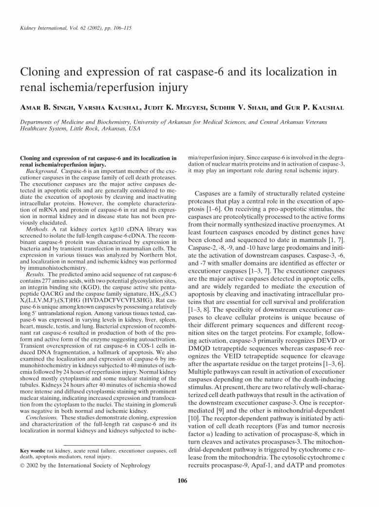
Kidney International, Vol. 62 (2002), pp. 106–115
Cloning and expression of rat caspase-6 and its localization inrenal ischemia/reperfusion injury
AMAR B. SINGH, VARSHA KAUSHAL, JUDIT K. MEGYESI, SUDHIR V. SHAH, and GUR P. KAUSHAL
Departments of Medicine and Biochemistry, University of Arkansas for Medical Sciences, and Central Arkansas VeteransHealthcare System, Little Rock, Arkansas, USA
mia/reperfusion injury. Since caspase-6 is involved in the degra-Cloning and expression of rat caspase-6 and its localization indation of nuclear matrix proteins and in activation of caspase-3,renal ischemia/reperfusion injury.it may play an important role during renal ischemic injury.Background. Caspase-6 is an important member of the exe-
cutioner caspases in the caspase family of cell death proteases.The executioner caspases are the major active caspases de-tected in apoptotic cells and are generally considered to me- Caspases are a family of structurally related cysteinediate the execution of apoptosis by cleaving and inactivating
proteases that play a central role in the execution of apo-intracellular proteins. However, the complete characteriza-ptosis [1–6]. On receiving a pro-apoptotic stimulus, thetion of mRNA and protein of caspase-6 in rat and its expres-
sion in normal kidney and in disease state has not been pre- caspases are proteolytically processed to the active formsviously elucidated. from their normally synthesized inactive proenzymes. At
Methods. A rat kidney cortex �gt10 cDNA library was least fourteen caspases encoded by distinct genes havescreened to isolate the full-length caspase-6 cDNA. The recom-been cloned and sequenced to date in mammals [1, 7].binant caspase-6 protein was characterized by expression inCaspase-2, -8, -9, and -10 have large prodomains and initi-bacteria and by transient transfection in mammalian cells. The
expression in various tissues was analyzed by Northern blot, ate the activation of downstream caspases. Caspase-3, -6,and localization in normal and ischemic kidney was performed and -7 with smaller domains are identified as effecter orby immunohistochemistry. executioner caspases [1–3, 7]. The executioner caspasesResults. The predicted amino acid sequence of rat caspase-6
are the major active caspases detected in apoptotic cells,contains 277 amino acids, with two potential glycosylation sites,and are widely regarded to mediate the execution ofan integrin binding site (KGD), the caspase active site penta-
peptide QACRG and the caspase family signature, HX2-4(S,C) apoptosis by cleaving and inactivating intracellular pro-X4(L,I,V,M,F)2(S,T)HG (HVDADCFVCVFLSHG). Rat cas- teins that are essential for cell survival and proliferationpase-6 is unique among known caspases by possessing a relatively [1–3, 8]. The specificity of downstream executioner cas-long 5� untranslational region. Among various tissues tested, cas-
pases to cleave cellular proteins is unique because ofpase-6 was expressed in varying levels in kidney, liver, spleen,their different primary sequences and different recog-heart, muscle, testis, and lung. Bacterial expression of recombi-
nant rat caspase-6 resulted in production of both of the pro- nition sites on the target proteins. For example, follow-form and active form of the enzyme suggesting autoactivation. ing activation, caspase-3 primarily recognizes DEVD orTransient overexpression of rat caspase-6 in COS-1 cells in- DMQD tetrapeptide sequences whereas caspase-6 rec-duced DNA fragmentation, a hallmark of apoptosis. We also
ognizes the VEID tetrapeptide sequence for cleavageexamined the localization and expression of caspase-6 by im-after the aspartate residue on the target proteins [1–3, 6].munohistochemistry in kidneys subjected to 40 minutes of isch-Multiple pathways can result in activation of executioneremia followed by 24 hours of reperfusion injury. Normal kidney
showed mostly cytoplasmic and some nuclear staining of the caspases depending on the nature of the death-inducingtubules. Kidneys 24 hours after 40 minutes of ischemia showed stimulus. At present, there are two relatively well-charac-more intense and diffused cytoplasmic staining with prominent terized cell death pathways that result in the activation ofnuclear staining, indicating increased expression and transloca-
the downstream executioner caspase-3. One is receptor-tion from the cytoplasm to the nuclei. The staining in glomerulimediated [9] and the other is mitochondrial-dependentwas negative in both normal and ischemic kidney.
Conclusions. These studies demonstrate cloning, expression [10]. The receptor-dependent pathway is initiated by acti-and characterization of the full-length rat caspase-6 and its vation of cell death receptors (Fas and tumor necrosislocalization in normal kidneys and kidneys subjected to ische- factor �) leading to activation of procaspase-8, which in
turn cleaves and activates procaspases-3. The mitochon-drial-dependent pathway is triggered by cytochrome c re-Key words: rat kidney, acute renal failure, executioner caspases, cell
death, apoptosis mediators, renal injury. lease from the mitochondria. The cytosolic cytochrome crecruits procaspase-9, Apaf-1, and dATP and promotes 2002 by the International Society of Nephrology
106

Singh et al: Cloning and localization of rat caspase-6 107
caspase-9 activation. Activated caspase-9 then cleaves 9600 with the following conditions: 94�C for 60 secondsfollowed by 94�C for 5 seconds, 50�C for 30 seconds;and activates procaspase-3 to its active form [1, 9, 10].
Thus, extensive studies have been done on caspase-3 but 68�C for two minutes (30 cycles), and a final extensionat 68�C for seven minutes. The amplified products werevery little information is available on the executioner
caspase, caspase-6, in the apoptotic pathway. resolved by gel electrophoresis on 1% agarose (FMCBioProducts, Rockland, ME, USA) and visualized byMany studies have documented the occurrence of apo-
ptosis during renal ischemia/reperfusion injury and other ethidium bromide staining. The appropriate sized bandwas excised from the gel, purified, and subcloned intorenal diseases [11–15]. Since the executioner caspases are
key intracellular mediators of apoptosis, their activation a pGEMT vector (Promega, Madison, WI, USA). Thecloned cDNA was sequenced using vector primer sites,may play an important role in renal injury. To examine
the specific role of executioner caspases in renal injury T7 and SP6.it is essential to know if these caspases are transcribed
Cloning of rat caspase-6in the kidney. We have recently shown that rat kidneytranscribes the genes for caspase-1, -2, -3, -6, -7, -8, and The cloned cDNA fragment was excised out from the
pGEMTvector, radio-labeled by random priming-9 [16], indicating that all of the executioner caspasesare expressed in the kidney. However, the complete (Amersham Life Science, Arlington Heights, IL, USA)
with [�-32P] dCTP, purified by chromatography on Nuc-characterization of mRNA and proteins of most of thesecaspases in rat kidney remain to be accomplished. At Trap Push Columns (Stratagene, La Jolla, CA, USA),
and used to screen a rat kidney cortex �gt10 cDNApresent, complete identification and characterization ofmRNAs and proteins of only three rat caspases, cas- library (Clontech Inc., Palo Alto, CA, USA). About 5 �
105 plaques were screened using standard protocols [20].pase-1 [17], caspase-2 [18] and caspase-3 [19], have beenreported. To further examine the specific role of the After four cycles of plaque purification six positive clones
were purified and the cDNA inserts were excised usingindividual executioner caspases in renal injury, we fo-cused our present study on caspase-6. This study reports EcoR1 restriction enzyme. The excised cDNAs were gel
purified (Qiagen Inc, Valencia, CA, USA) and subclonedthe cloning and sequencing of the full-length cDNA ofrat caspase-6, and characterization of the recombinant into the vector p ZERO (Invitrogen Inc, Carlsbad, CA,
USA).protein expressed in bacteria and by transient transfec-tion in mammalian cells. We also examined the expres-
Sequencingsion of caspase-6 in various tissues and its localizationby immunohistochemistry in normal kidney as well as Sequencing was performed on both strands using T7
and SP6 primers, and thereafter walking along with gene-in kidneys subjected to ischemia/reperfusion injury.specific primers using the dideoxy DNA cycle sequencingsystem (Gibco BRL, Gaithersburg, MD, USA) and con-
METHODSfirmed by sequencing on an automated DNA sequencer
RT-PCR (Applied Biosystems Inc., Foster City, CA, USA). Thesequence for caspase-6 reported in this article was depos-Poly A� RNA was isolated from normal rat kidney
and reverse transcription-polymerase chain reaction ited in the GenBank database (accession numbersAF025670, NM031775).(RT-PCR) was performed as described previously [16].
Briefly, 1 �g of poly A� was reverse-transcribed usingNorthern blot analysisan oligo (dT) primer and Moloney murine leukemia virus
reverse transcriptase (Perkin-Elmer, Branchburg, NJ, Northern blot analysis was performed as previouslydescribed [20]. In brief, 25 �g of total RNA isolatedUSA). The resulting reverse transcription product was
used as a template for PCR using sense and antisense from different adult rat organs was electrophoresed un-der denaturing condition on a 1.2% agarose gel andprimers based on the human caspase-6 coding sequence
(GenBank accession #U20537). The sequences of prim- blotted to Immobilon N nylon membrane (Millipore,Bedford, MA, USA). The membrane was baked underers used and their locations in the human caspase-6 cod-
ing sequence are: sense, CTAATCTTCAATCACGA vacuum for one hour and hybridized with 32P-labeledfull-length caspase-6 cDNA as probe under standardGAGGTTC (221-243) and antisense, CTCACACAA
ATCTTGAA TGTACCA (780-757) (National Biosci- condition [20]. After washing, the membrane was sub-jected to autoradiography at �70�C to detect mRNAence, Plymouth, MA, USA). The criteria to use these
primers were described in our previous study [16]. At- signals.tempts to amplify rat caspase-6 using degenerate primers
In vitro translationdesigned from the N-terminal initiating domain or fromthe prodomain of human caspase-6 were unsuccessful. The full-length rat caspase-6 cDNA was subcloned
into the vector pZERO. The recombinant plasmid wasPCR was performed in a Perkin-Elmer Thermocycler

Singh et al: Cloning and localization of rat caspase-6108
then linearized on the 3� end of the insert using the NotI of these tags. The resultant plasmid was transformedinto the E. coli strain BL21 (F�ompTr�
B m�B ). The trans-restriction enzyme. In vitro translation was performed
using the TNT T7-coupled reticulocyte lysate system formed bacteria were cultured in the presence of 1mmol/L isopropylthiogalactoside (IPTG; Pharmacia-(Promega Inc.) and [35S]-methionine (Amersham Corp.)
as per the manufacturer’s recommendation. In brief, LLB, Uppsala, Sweden) for three hours. After optimalinduction, the bacterial culture was centrifuged and pel-linear plasmid (1 �g), amino acid mix minus methio-
nine (1 mmol/L), ribonuclease inhibitor, [35S]-methionine lets were suspended in ice-cold buffer containing 20mmol/L HEPES, pH 7.8, 5 mmol/L imidazole, 0.5 mol/L(1000 Ci/mmol), and T7 polymerase were added to a 50
�L reaction mix and incubated at 30�C for two hours. The NaCl, and protease inhibitors (10 �g/mL aprotinin, 1�g/mL antipain, 1 �g/mL pepstatin, 17.5 �g/mL benzam-reaction products were analyzed on 12% sodium dodecyl
sulfate-polyacrylamide gel electrophoresis (SDS-PAGE). idine), and homogenized. The supernatants were firstpurified on columns containing nickel bound resin (5
Expression plasmid and transient transfection mL) using the His-Bind buffer kit supplied by Novagen.The proteins eluted from the columns were dialyzedThe full-length caspase-6 cDNA was subcloned in the
mammalian expression vector pcDNA3.1� (Invitrogen extensively in 20 mmol/L HEPES, pH 7.8,containing pro-tease inhibitors and further purified on a Mono Q columnInc.) downstream of the cytomegalovirus promoter/en-
hancer. Transfection was carried out with Lipofectamine by applying a linear gradient of 0 to 0.5 mol/L NaCl in20 mmol/L Tris-HCl buffer, pH 7.4, using an fast protein(Gibco BRL) according to the manufacturer’s recom-
mendation. In brief, 2.0 � 105 COS-1 cells were tran- liquid chromatography (FPLC) system. Purification ofrat caspase-6 in both proenzyme forms and active formssiently transfected in 2 mL of serum free medium with a
mixture of 1 �g of pCMV -galactosidase plus 6 �g of (P10 and P20 subunits) are shown in Figure 6.test plasmid. Control cells were transfected with empty
Determination of caspase-6 activityvector. After five hours, 4 mL of serum containing growthmedium was added. Forty-eight hours post-transfection, Bacterial cell pellets containing recombinant caspase-6
were lysed with 20 mmol/L HEPES, pH 7.5, containingcells were washed twice with 1� phosphate-buffered sa-line (PBS), scraped, and processed for -galactosidase 10% sucrose, 0.1% CHAPS, 2 mmol/L dithiothreitol
(DTT), 0.1% NP40, 1 mmol/L ethylenediaminetetraace-assay (according to the manufacturer’s recommenda-tion) as well as for DNA damage as previously described tic acid (EDTA), 1 mmol/L phenylmethylsulfonyl fluo-
ride (PMSF), 1 �g/mL leupeptin, and 1 �g/mL pepstatinin our studies [21].A at 4�C. The supernatants obtained after centrifugation
Genomic DNA extraction and electrophoresis were used to determine the enzyme activity. The activityof caspase-6 was determined by fluorometric assay usingTransfected cells were scraped into medium 48 hours
post-transfection and all cells, including the floating cells, the substrate VEID-AMC that is specifically cleaved bythe enzyme at the Asp residue to release the fluorescentwere collected. Genomic DNA was isolated by protein-
ase K digestion, phenol-chloroform extraction, and etha- leaving group amino-4-methyl coumarin (AMC) [16, 22].The enzyme extracts containing 50 �g protein were incu-nol precipitation as described [21]. The DNA samples
were analyzed by electrophoresis on 1.8% agarose gel bated with 100 mmol/L HEPES, pH 7.4, containing 10%sucrose, 0.1% CHAPS, 10 mM DTT, and 50 �mol/L ofand DNA was observed by ethidium bromide staining
of the gel. caspase substrate in a total reaction volume of 0.25 mL.The reaction mixture was incubated for 60 minutes at
Expression of recombinant caspase-6 in bacteria 30�C. At the end of the incubation, the liberated fluores-cent group, AMC, was determined using a fluorescentThe coding sequence of rat caspase-6 was PCR ampli-
fied using 5� and 3� PCR primers (sense: GGTAAGA Spectrofluorometer (Perkin Elmer) with an excitationwavelength of 380 nm and an emission wavelength ofATTCATGACGGAGACAGATGGC and antisense:
TTGGAGCGGCCG CCTACTTACTAGGTTTGGG) 460 nm [22]. AMC was used as a standard. Based on thestandard curve made with fluorescence readings withcontaining Nde and XhoI custom restriction sites, gel
purified, and subcloned in-frame into the NdeI and XhoI free AMC, the data for caspase activity are expressedas nanomoles of AMC liberated when 50 �g of proteinsites of the bacterial expression vector pET-21a (�) (No-
vagen). This subcloning placed the rat caspase-6 se- extract was incubated with 50 �mol/L of substrate for60 minutes at 30�C.quence under the control of the inducible bacteriophage
T7-promoter and introduced an NH2-terminal polyhis-Ischemia-reperfusion modeltidine tag (6 histidines) and S-tag for purification on a
nickel-chelating column and for identification by West- Adult male Sprague-Dawley rats used in the presentstudy were subjected to 40 minutes of ischemia by occlud-ern blot using s-tag antibody, respectively. An entero-
kinase cleavage site also was included to allow removal ing renal pedicles with smooth vascular clamps as pre-

Singh et al: Cloning and localization of rat caspase-6 109
viously described [23] and then allowed to recover for signed from the corresponding human sequences. Our24 hours. Each group consisted of six rats. Blood samples attempts to obtain full-length cDNA by RT-PCR werewere collected for creatinine and urea nitrogen determi- unsuccessful. However, we were able to amplify a 559-bpnation. Animals were anesthetized before tissue harvest- cDNA fragment by RT-PCR using primers based on theing. Rats were perfused through the left ventricle with conserved internal sequences of human caspase-6. Thewarm Hanks’ balanced salt solution (HBSS). Kidney amplified cDNA fragment obtained by RT-PCR was geltissues were fixed in formalin for histology and immuno- purified, and subcloned into the vector pGEMT andhistochemistry. The control group consisted of rats un- sequenced using vector primer sites. Two out of fivedergoing surgical exposure of kidneys without clamping clones sequenced were found to be identical and showedthe renal pedicles. significant homology to the human caspase-6 alpha gene
(we tentatively called this fragment the rat caspase-6Immunohistochemistry cDNA fragment). The remaining cDNA fragments
Representative renal sections (4 to 5 �m thick) of showed no homology to the caspases and were not pur-formalin-fixed paraffin-embedded tissues of control and sued further.ischemic kidney were used. Sections were mounted on We screened a normal rat kidney �gt10 cDNA libraryfrosted micro slides and the paraffin-embedded sections using the subcloned rat caspase-6 cDNA fragment as awere deparaffinized in xylene and re-hydrated through probe to obtain the full-length cDNA. A total of 5 �a graded series of alcohol (100 to 0%). Tissue sections 105 plaques were screened and several clones were puri-were rinsed in PBS and endogenous peroxidase was inac- fied after four rounds of screening. Four clones (clonetivated by incubations with 0.3% H2O2 in methanol. The 1: 1.2 kb; clone 2: 1.5 kb; clone 3: 1.8 kb and clone 4:slides were extensively washed with PBS and preincu- 2.2 kb) were sequenced using vector primer sites andbated with 5% bovine serum albumin (BSA) in PBS for thereafter walking along with primers. Sequencing re-60 minutes to block nonspecific reaction with the antise- vealed that clone 2 contained a total of 1446 nucleotidesrum. Tissue sections were rinsed in PBS, and then incu- and represents the full-length rat caspase-6 cDNA. Itbated with anti-caspase-6 antibody (1:200; Cell Signaling contained the entire 559 bp sequence of the probe, ini-Technology, Inc., Beverly, MA, USA) at 37�C for two tiating methionine, stop codon, and poly adenylationhours, washed in PBS and then incubated with horserad- signal (Fig. 1). Clone 1 was identical to clone 2 but wasish peroxidase-conjugated-goat anti-rabbit IgG for an incomplete. Although clones 3 and 4 did contain thehour. After washing with PBS, the sections were devel- entire sequence of clone 2, the sequence was interruptedoped with DAB and H2O2. For negative control, primary
with small stretches of nucleotide sequences of unknownantibody was omitted and normal rabbit serum was usedhomology suggesting they represented the possibility ofat the same dilution.incompletely processed pre mRNAs, thus these cloneswere not pursued further.Preparation of nuclear and cytosolic fractions
The open reading frame (ORF) of clone 2 encodesNuclei from rat kidney cortex were isolated by homog-277 amino acids (Fig. 1), which is relatively smaller thanenization followed by a sucrose gradient as previouslythat reported for human caspase-6. In vitro translationdescribed [24]. In brief, nuclei were isolated from mincedof clone 2 using TNT T7 coupled reticulocyte lysatekidney cortex by homogenization at 0�C in buffer Asystem resulted in the both proform and the active sub-(20 mmol/L Hepes-NaOH, pH 7.4, 5 mmol/L MgCl2,units (Fig. 2). Formation of smaller subunits from the0.2 mmol/L EGTA, 3 mmol/L 2-mercaptoethanol, 0.1larger precursor represents the processing to form themmol/L PMSF, 1�g/mL leupeptin, 1�g/mL antipain)active enzyme [2–5]. In comparison, rat caspase-3 trans-containing 0.93 mol/L sucrose. The filtered homogenatelation to the proform and the active subunits is alsowas centrifuged over a cushion of 1.61 mol/L sucrose inshown. The published full-length cDNA for mousebuffer A at 20,000 � g at 4�C for 20 minutes. The pelletcaspase-6 and its initiating codon [26] corresponds to thewas collected as a nuclear fraction and supernatant aboveinitiating codon of the rat caspase-6 cDNA suggestingthe sucrose cushion was further centrifuged at 100,000 �that this ATG site is conserved in rodents. To furtherg for 60 minutes, and the resulting supernatant was usedensure that the cloned cDNA is in fact full-length, weas the cytosolic fraction. The nuclear pellets were washedperformed RACE using extreme 5� end of the rat cas-once in 0.25 mol/L sucrose in buffer A and resuspendedpase-6 as primer and obtained the same cDNA sequencein 0.6 mol/L NaCl in buffer A containing 10% glycerol.as in the cloned cDNA obtained from library screening.The initiating codon ATG in the cloned cDNA follows
RESULTS the KOZAK rule and is flanked by purine (A) at �3Isolation and sequencing of rat caspase-6 and �4 position [25]. In addition, an in-frame stop codon
TAA (bp 233–235) is preceded by the initiating codonTo obtain the full-length cDNA for rat caspase-6, wefirst performed RT-PCR using degenerate primers de- ATG in the cloned cDNA, indicating that the initiation

Singh et al: Cloning and localization of rat caspase-6110
Fig. 1. Full-length sequence of rat caspase-6. Features in bold includethe start and stop codon, putative aspartic acid cleavage sites, potentialglycosylation sites (NVTQ, NGSW), and a potential integrin bindingsite (KGD). The linker region is underlined. The active site is in italicand underlined.
Fig. 2. In vitro translation of the full-length cDNA of rat caspase-6. Invitro translation was performed using the TNT T7-coupled reticulocytelysate system (Promega Inc.) and [35S]-methionine (Amersham Corp.)
of caspase-6 expression should begin at this ATG site as per the manufacturer’s recommendation. The reaction products wereanalyzed on 12% SDS-PAGE followed by autoradiography. Translation(bp 239–243). Thus, the occurrence of in-frame stop co-products of caspase-6 and caspase-3 are shown.don further confirms the site of initiation at the ATG
site at bp 239–243.The 5�-UTR in rat caspase-6 is unexpectedly longer
than that reported for other species including human His-104), and those known to form the P1 carboxylatebinding pocket (Arg-47, Gln-144, Arg-200 and Ser-206)and mouse. On the other hand, the 3�-UTR is shorter
than in human caspase-6. Like mouse caspase-6, the loca- were also conserved (Fig. 3). The location of asparticacids at positions 16, 162 and 173 suggest that this mole-tion of the initiating methionine in rat caspase-6 corre-
sponded with the second methionine (19th amino acid) cule is produced as a proenzyme since subunits of p18and p11 are produced after aspartic acid cleavage. Theof human caspase-6 (Fig. 3). Blast analysis of total nucle-
otide sequences revealed 69% identity with human phylogenetic tree analysis showed a tight grouping withother caspase-6 genes. Like the mouse gene, the prodo-caspase-6, however, GAP analysis of the ORF sequence
revealed highest 84% homology at the nucleotide level main in rat is relatively short for a caspase-6 gene, beingjust five amino acids in length, compared to human,and 87.7% homology at the amino acid level with the
human caspase 6, and 40 to 50% identity to the other chicken and trout.known caspases. The predicted amino acid sequence of
Northern analysis and tissue distributionrat caspase-6 showed two potential glycosylation sites, anintegrin binding site (KGD), the conserved pentapeptide Northern analysis of caspase-6 showed a single mRNA
transcript corresponding to a size of approximately 1.6 kb.QACRG sequence of the active site, and the caspasefamily signature HX2-4(S,C)X4(L,I,V,M,F)2(S,T)HG (that To determine the tissue distribution of rat caspase-6,
we performed Northern blot analysis using poly A�is, HVDADCFVCVFLSHG) where X can be any aminoacid. Amino acids involved in catalysis (Cys-146 and RNA from various rat tissues. The results indicated that

Singh et al: Cloning and localization of rat caspase-6 111
Fig. 4. Tissue distribution of rat caspase-6 mRNA. Total RNA fromeach tissue was isolated as described in the text. A total of 25 �g totalRNAs from various tissues of an adult rat were analyzed by Northernblot analysis. The positions of 18S and 28S rRNAs are shown as indi-cated.
the cloned rat caspase-6, we transiently transfected thefull-length cDNA in COS-1 cells. Transfection efficiencywas determined by -gal staining and also by Northernblot analysis for caspase-6 (data not shown). Analysisof genomic DNA from transfected cells showed 200-bpladdering, indicating DNA fragmentation, a hallmark ofapoptosis, whereas no laddering was observed in un-transfected control cells (Fig. 5).
Expression of rat caspase-6 in bacteria anddetermination of its activity
To further characterize the cloned cDNA, the cod-ing sequence was expressed in E. coli as a recombinantprotein. The caspase-6 protein was expressed as a pro-enzyme as well as activated molecule indicating auto-activation (Fig. 6). The activity of caspase-6 also wasconfirmed by activity assay using the VEID-AMC sub-strate (data not shown). This activity increased with in-creasing periods of bacterial induction with IPTG. TheFig. 3. Alignment of the predicted rat caspase-6 amino acid sequencerecombinant protein was purified on nickel-columns us-with other known caspase-6 sequences. Identical and similar residues
(*) identified by the GELLYFISH program are indicated. The active ing the 3� his-tag (Fig. 6). The in vitro expression ofpentapeptide site and caspase family signature are boxed. Residues cloned cDNA in bacteria also provided biologically ac-involved in the P1 carboxylic binding pocket are in bold, the propiece
tive protein, as demonstrated by the activity assay of theis in italics, and residues involved in catalysis are in bold and indicatedwith an arrow. recombinant enzyme (caspase activity in control; empty
vector) in homogenate (0.02 0.01 nmol), and in ex-pressed (vector containing the coding sequence) homog-caspase-6 is expressed to varying levels within differentenate (8 0.6 nmol) where both the pro-form and activetissues including in kidney, liver, spleen, heart, muscle,subunits of caspase-6 were generated (Fig. 6).testis, and lung (Fig. 4), with the highest expression in
heart, lung, kidney, and muscle.Localization and nuclear translocation
Overexpression of rat caspase-6 induces apoptosis During apoptosis caspases have been shown to disas-semble a cell by cleaving a set of proteins and translocat-When overexpressed caspases induce DNA fragmen-
tation and apoptosis of host cells. In order to characterize ing into nucleus. Further, caspase-6 is involved in the

Singh et al: Cloning and localization of rat caspase-6112
Fig. 6. SDS-gel electrophoresis of the rat recombinant caspase-6 ex-pressed in bacteria. The recombinant plasmid, pET-21a (�) containingcoding sequence of ratcaspase-6 and His–tag was transformed into theE. coli strain BL21 (F�ompTr�
B m�B ). Following induction with IPTG
the bacteria were harvested and following lysis the supernatants werepurified on columns containing nickel bound resin. Lanes are: A, molec-ular weight standards; B, homogenate from uninduced bacteria; C,homogenate from 1 mmol/L IPTG-induced bacteria; D, recombinantcaspase-6 purified on nickel column followed by purification on MonoQ column; E, proenzyme; F, p20 subunit; G, p10 subunit. The proformand subunits were first separated on preparative gel electrophoresis.
to a 46 kD lamin A fragment during ischemic injury(Fig. 9). This cleavage is attributed to the action ofcaspase-6 since this caspase not other caspases has beenFig. 5. DNA fragmentation by overexpression of rat caspase-6 in
COS-1 cells. Fragmented DNA was isolated from control and caspase-6 shown to cleave lamin A [27, 28].overexpressing cells and subjected to 1% agarose gel electrophoresis.The DNA was visualized by UV fluorescence after staining with ethid-ium bromide. Lanes are: (A) molecular size markers, (B) control cells DISCUSSIONtransfected with empty vector, and (C) caspase-6 overexpressing cells.
Our studies demonstrate the cloning, sequencing, andcharacterization of the rat caspase-6 cDNA and its ex-pression in the kidney. The cloned cDNA shows highest
degradation of nuclear matrix proteins and in caspase-3homology with the known caspase-6 genes and closeactivation. Therefore, we examined the expression andhomology with other executioner caspases. Like otherlocalization of caspase-6 by immunohistochemistry incaspases, the rat caspase-6 contains the caspase familynormal kidney as well as kidneys subjected to 40 minutessignature and the conserved active site pentapeptideof ischemia followed by 24 hours of reperfusion injury.QACRG required for proteolytic activity. In commonNormal kidney showed mostly cytoplasmic and somewith executioner caspases and caspase-14, caspase-6 hasnuclear staining of the proximal tubules. Kidneys 24a relatively short prodomain [29], and a linker regionhours after 40 minutes of ischemia showed more intensewhere conserved Asp residues allow cleavage by otherand diffused cytoplasmic staining, with nuclear stainingcaspases to release the active heterodimer. Within theprimarily in the tubular segments of the cortex, indicatingprodomain, rat caspase-6 shows close homology at theincreased expression and nuclear translocation (Fig. 7).amino acid level to the mouse caspase-6 but rela-Only occasional staining was observed in the distal tu-tively poor homology to the human, chicken and troutbules within the cortex. The staining in the glomerulicaspase-6. Among the three predicted cleavage sites forwas negative in both normal and ischemic kidney. Thethe human caspase-6 prodomain [30] that occur atcytosolic and nuclear fractions from the control and isch-Asp23-Ala24, Asp32-Pro-33, and Asp40-His 40, only theemic kidneys also were examined for the expression oflast site is conserved within the rat sequence. The ratcaspase-6 by Western blot analysis. As shown in Figure 8,caspase-6 is unique among known caspases in that itthe increased expression of the cytosolic and nuclearpossesses a relatively long 5�-UTR. On long 5�-UTRscaspase-6 in ischemic kidney corresponds very well toCap-dependent ribosomal scanning has been shown tothe immunoperoxidase staining data. Nuclear fractionsbe severely hampered [30, 31]. Long 5�-UTRs are oftenisolated from the control and ischemic kidneys also werefound in mRNAs encoding regulatory proteins likeexamined for the cleavage of nuclear matrix protein,
lamin A. Our data demonstrate that lamin A is cleaved proto-oncogenes, growth factors and their receptors, and

Singh et al: Cloning and localization of rat caspase-6 113
Fig. 7. Immunoperoxidase staining of caspase-6 in renal ischemia/reperfusion injury. Immunohistochemical localization was performed as describedin the text. (A) Tissue sections from normal rat kidney show mostly cytoplasmic and some nuclear staining of the proximal tubules. (B and C )Representative tissue sections from kidneys 24 hours after 40 minutes of ischemia show more intense and diffused cytoplasmic staining withprominent nuclear staining primarily in the proximal tubular segments of the cortex. Glomeruli show negative staining for caspase-6 in both normaland ischemic kidneys.
Fig. 8. Distribution of caspase-6 in cytosolic and nuclear fractions ofischemic kidney. Cytosolic (C) and nuclear (N) fractions were isolatedfrom control and ischemic kidneys as described in the Methods section,and were subjected to Western blot analysis [16] using anti-caspase-6antibody (1:1000, Cell Signaling Technology).
homeodomain proteins and are suspected to provide a Fig. 9. Cleavage of lamin A in ischemic kidney. Nuclear fractions fromcontrol (A) and ischemic (B) kidneys were examined by Western blotregulatory mechanism [31, 32]. However, the mecha-analysis [16] using anti-lamin A antibody (Santa Cruz Biotechnology,nisms involved in 5�-UTR-mediated control are not well Inc.).
understood. It is possible that the long 5�-UTR of ratcaspase-6 is involved in its translational regulation. Aswith all caspases, the rat gene shows conservation ofresidues known to form the P1 carboxylate-binding [28, 33]. Caspase-6, but not caspase-3, is activated duringpocket reflecting the absolute requirement for an Asp neuronal apoptosis and is responsible for the processingat position P1 for substrate cleavage [33]. Residues that of amyloid precursor protein (APP) to generate in-form the P2-P4 binding pocket are less conserved be- creased production of Alzheimer’s amyloid beta peptidetween caspases, accounting for differences in substrate
(A ) [34–36]. However, in sharp contrast, much infor-specificity.
mation is available on the protein targets for caspase-3Although caspase-6 is an important member of theand its role has been extensively studied. Caspase-3 hasexecutioner caspases, the steps involved in its activationbeen reported to degrade many key structural and regu-and its relative contribution in the cell death pathwaylatory proteins including DNA repair enzymes [1, 37–39],are not as well understood as for caspase-3. The identityICAD (inhibitor of caspase-activated DNase) or DNAof some target cellular proteins for caspase-6 in responsefragmentation factor [40, 41], nuclear structural proteinsto various apoptotic stimuli has recently been reported.[1, 42, 43], cytoskeleton proteins [1, 33, 44], transcriptionThe nuclear matrix protein, lamin A, is the only protein
exclusively targeted and cleaved by the activated caspase-6 factors [1, 45–48], and regulatory proteins including the

Singh et al: Cloning and localization of rat caspase-6114
for critical review of the manuscript and valuable discussions, and Ms.proteins involved in the signal transduction pathwayJudy Nagle for secretarial assistance.[1, 49–52].
Recent studies have demonstrated the cleavage of Reprint requests to Gur P. Kaushal, Ph.D., Department of Medicine,University of Arkansas for Medical Sciences, Slot 501, 4301 W. Markhamsome intracellular proteins both by caspase-6 and cas-St., Little Rock, Arkansas 72205, USApase-3. For example, focal adhesion kinase (FAK) isE-mail: [email protected]
cleaved by both caspase-3 and caspase-6 in apoptoticjurkat T cells [53, 54]. The vimentin filament network is REFERENCEScleaved in human macrophages apoptosis induced by
1. Wolf BB, Green Dr: Suicidal tendencies: Apoptotic cell deathoxidized low-density lipoprotein (LDL) [55–57]. AP2�by caspase family proteinases. J Biol Chem 274:20049–20052, 1999[48] is cleaved by both caspase-3 and caspase-6 in a 2. Na T, Lazebnik Y: Caspases: Enemies within. Science 281:1312–
sequence-specific manner at different sites of the protein 1316, 19983. Earnshaw WC, Martins LM, Kaufmann SH: Mammalian cas-during tumor necrosis factor-� (TNF-�)-induced apopto-
pases: structure, activation, substrates, and functions during apo-sis. At present, it is not precisely known whether cas-ptosis. Annu Rev Biochem 68:383–424, 1999
pase-6 activation occurs before, simultaneously, or after 4. Cryns V, Yuan J: Proteases to die for. Genes Develop 12:1551–1570, 1998caspase-3 activation. Some relationship between caspase-3
5. Dl V: Caspases and apoptosis-biology and terminology. Cell Deathand caspase-6 activation has become apparent from re-Differ 6:493–494, 1999
cent studies in cell free systems. Active caspase-3 has 6. Nicholson DW: Caspase structure, proteolytic substrates, andbeen shown to activate caspase-6 [57, 58]; however, the function during apoptotic cell death. Cell Death Differ 6:1028–
1042, 1999relevance of these observations has not been demon-7. Ahmad M, Srinivasula SM, Hegde R, et al: Identification andstrated in an in vivo set up, since ultraviolet or gamma characterization of murine caspase14, a new member of the caspase
irradiation-treated caspase-3 (�/�) hepatocytes showed family. Cancer Res 58:5201–5205, 19988. Faleiro L, Kobayashi R, Fearnhead H, Lazebnik Y: Multiplecaspase-6 activation [59], indicating that caspase-3 is not
species of CPP32 and Mch2 are the major active caspases presentinvolved in caspase-6 activation. On the other hand,in apoptotic cells. EMBO J 16:2271–2281, 1997
some recent studies have demonstrated that caspase-6 9. Strasser A, O’Connor L, Dixit VM: Apoptosis signaling. AnnuRev Biochem 69:217–245, 2000can activate caspase-3 in response to apoptotic stimuli
10. Green DR, Reed JC: Mitochondria and apoptosis. Science 281:[60, 61]. Future studies using caspase-6 null mice will1309–1312, 1998reveal the precise relationship between caspase-3 and 11. Schumer M, Colombel MC, Sawczuk IS, et al: Morphologic, bio-
caspase-6 and the various steps involved in caspase-6 chemical, and molecular evidence of apoptosis during the reperfu-sion phase after brief periods of renal ischemia. Am J Pathol 140:activation in response to an apoptotic stimulus.831–838, 1992At present there is virtually no information on the 12. Daemen MARC, Van’t Veer C, Denecker G, et al: Inhibition of
expression and localization of executioner caspases in apoptosis induced by ischemia-reperfusion prevents inflammation.J Clin Invest 104:541–549, 1999kidney in normal or in disease state. We have previously
13. Sheridan AM, Bonventre JV: Cell biology and molecular mecha-shown distinctive pattern of expression of these caspasesnisms of injury in ischemic acute renal failure. Curr Opin Nephrol
in kidney following induction of ischemia [19], indicating Hypertens 9:427–434, 200014. Lieberthal W, Koh JS, Levine JS: Necrosis and apoptosis in acutethat caspase expression is under strict transcriptional
renal failure. Semin Nephrol 18:505–518, 1998control during ischemia/reperfusion injury. Caspase-615. Rana A, Sathyanarayana P, Lieberthal W: Role of apoptosismRNA was moderately increased during ischemia/re- of renal tubular cells in acute renal failure therapeutic implications.
perfusion injury. Our present study provides the first Apoptosis 6:83–102, 200116. Kaushal GP, Singh AB, Shah SV: Identification of caspase (ICE-demonstration, to our knowledge, of expression of rat
like proteases) gene family in rat kidney and altered expressioncaspase-6 in the kidney, and shows by immunohisto- ischemia/reperfusion injury. Am J Physiol (Renal Physiol 43) 274:chemistry its cytoplasmic localization and some nuclear F587–F595, 1998
17. Keane KM, Giegel DA, Lipinski WJ, et al: Cloning, tissue expres-staining of the tubules in cortex and outer medulla. Fol-sion and regulation of rat interleukin 1 beta converting enzyme.lowing ischemia/reperfusion injury we detected in-Cytokine 7:105–110, 1995
creased localization of caspase-6 in the nuclei, indicating 18. Sato N, Milligan CE, Uchiyama Y, Oppenheim RW: Cloning andsome translocation from the cytoplasm to the nuclei dur- expression of the cDNA encoding rat caspase-2. Gene 202:127–
132, 1997ing the renal injury. In non-renal tissue, a transient in-19. Juan TS, McNiece IK, Jenkins NA, et al: Molecular characteriza-crease in caspase-6 immunoreactivity in astrocytes has tion of mouse and rat CPP32 beta gene encoding a cysteine protease
been reported in permanent focal ischemia [62]. Thus, resembling interleukin-1 beta converting enzyme and CED-3. On-cogene 13:749–755, 1996further studies are required to identify the potential role
20. Sambrook J, Russell DW: Molecular Cloning—A Laboratryof caspase-6, the molecular ordering of the caspase-cas-Manual (3rd ed). New York, Cold Spring Harbor Laboratory
cade as well as the cellular proteins as targets of the Press, 2001caspase-6 in renal injury. 21. Kaushal GP, Ueda N, Shah SV: Role of caspases (ICE/CED
3 proteases) in DNA damage and cell death in response to amitochondrial inhibitor, antimycin A. Kidney Int 52:438–445, 1997ACKNOWLEDGMENTS
22. Stennicke HR, Salvesen GS: Caspase assays. Meth Enzymol 322:91–100, 2000This work was supported by NIH RO1 DK58239 and American
Heart (National) grants to GPK. The authors thank Dr. Randy Haun 23. Baliga R, Ueda N, Shah SV: Increase in bleomycin-detectable

Singh et al: Cloning and localization of rat caspase-6 115
iron in ischaemia/reperfusion injury to rat kidneys. Biochem J the actin-capping protein alpha-adducin at Asp-Asp-Ser-Asp633-Ala by caspase-3 is preceded by its phosphorylation on serine 726291:901–905, 1993
24. Cote J, Renaud J, Ruiz-Carrillo A: Recognition of (dG)n.(dC)n in cisplatin-induced apoptosis of renal epithelial cells. J Biol Chem275:25805–25813, 2000sequences by endonuclease G. Characterization of the calf thymus
nuclease. J Biol Chem 264:3301–3310, 1989 45. Francois F, Godinho MJ, Grimes ML: CREB is cleaved by cas-pases during neural cell apoptosis. FEBS Lett 486:281–284, 200025. Kozak M: An analysis of 5�-noncoding sequences from 699 verte-
brate messenger RNAs. Nucleic Acids Res 15:8125–8148, 1987 46. Ohtsubo T, Kamada S, Mikami T, et al: Identification of NRF2,a member of the NF-E2 family of transcription factors, as a26. Van de Craen M, Vandenabeele P, Declercq W, et al: Character-
ization of seven murine caspase family members. FEBS Lett 403: substrate for caspase-3 (-like) proteases. Cell Death Differ 6:865–872, 199961–69, 1997
27. Takahashi A, Alnemri ES, Lazebnik Y, et al: Cleavage of lamin 47. Rickers A, Peters N, Badock V, et al: Cleavage of transcriptionfactor SP1 by caspases during anti-IgM-induced B-cell apoptosis.A by Mch2 alpha but not CPP32: multiple interleukin 1 beta-
converting enzyme-related proteases with distinct substrate recog- Eur J Biochem 261:269–274, 199948. Nyormoi O, Wang Z, Doan D, et al: Transcription factor ap-nition properties are active in apoptosis. Proc Natl Acad Sci USA
93:8395–8400, 1996 2 alpha is preferentially cleaved by caspase 6 and degraded byproteasome during tumor necrosis factor alpha-induced apoptosis28. Orth K, Chinnaiyan AM, Garg M, et al: The CED-3/ICE-like
protease Mch2 is activated during apoptosis and cleaves the death in breast cancer cells. Mol Cell Biol 21:4856–4867, 200149. Mukerjee N, McGinnis KM, Gnegy ME, Wang KK: Caspase-substrate lamin A. J Biol Chem 271:16443–16446, 1996
29. Hu S, Snipas SJ, Vincenz C, et al: Caspase-14 is a novel develop- mediated calcineurin activation contributes to IL-2 release dur-ing T cell activation. Biochem Biophys Res Commun 285:1192–mentally regulated protease. J Biol Chem 273:29648–29653, 19981199, 200130. Fernandes-Alnemri T, Litwack G, Alnemri ES: Mch2, a new
50. Kang KH, Lee KH, Kim MY, Choi KH: Caspase-3-mediated cleav-member of the apoptotic Ced-3/Ice cysteine protease gene family.age of the NF-kappa B subunit p65 at the NH2 terminus poten-Cancer Res 55:2734–2742, 1995tiates naphthoquinone analog-induced apoptosis. J Biol Chem 276:31. Willis AE: Translational control of growth factor and proto-onco-24638–24644, 2001gene expression. Int J Biochem Cell Biol 31:73–86, 1999
51. Berry DM, Benn SJ, Cheng AM, McGlade CJ: Caspase-32. Van der Velden AW, Thomas AA: The role of the 5� untranslateddependent cleavage of the hematopoietic specific adaptor proteinregion of an mRNA in translation regulation during development.Gads alters signalling from the T cell receptor. Oncogene 20:1203–Int J Biochem Cell Biol 31:87–106, 19991211, 200133. Cohen GM: Caspases: The executioner of apoptosis. Biochem J
52. Ellerby LM, Hackam AS, Propp SS, et al: Kennedy’s disease:326:1–161, 1997Caspase cleavage of the androgen receptor is a crucial event in34. LeBlanc A, Liu H, Goodyer C, et al: Caspase-6 role in apoptosis ofcytotoxicity. J Neurochem 72:185–195, 1999human neurons, amyloidogenesis, and Alzheimer’s disease. J Biol
53. Gervais FG, Thornberry NA, Ruffolo SC, et al: Caspases cleaveChem 274(33):23426–24336, 1999focal adhesion kinase during apoptosis to generate a FRNK-like35. Pellegrini L, Passer BJ, Tabaton M, et al: Alternative, non-polypeptide. J Biol Chem 273:17102–17118, 1998secretase processing of Alzheimer’s beta-amyloid precursor pro-
54. Wen LP, Fahrni JA, Troie S, et al: Cleavage of focal adhe-tein during apoptosis by caspase-6 and -8. J Biol Chem 274:21011–sion kinase by caspases during apoptosis. J Biol Chem 272:26056–21016, 199926061, 199736. Chen Y, McPhie DL, Hirschberg J, Neve RL: The amyloid precur-
55. Muller K, Dulku S, Hardwick SJ, et al: Changes in vimentin insor protein-binding protein APP-BP1 drives the cell cycle throughhuman macrophages during apoptosis induced by oxidised low-the S-M checkpoint and causes apoptosis in neurons. J Biol Chemdensity lipoprotein. Atherosclerosis 156:133–144, 2001275:8929–8935, 2000 56. Yun Y, Chen F, Chang R, et al: Caspase cleavage of vimentin37. Casciola-Rosen LA, Anhalt GJ, Rosen A: DNA-dependent pro- disrupts intermediate filaments and promotes apoptosis. Cell Deathtein kinase is one of a subset of autoantigens specifically cleaved Differ 8:443–5019, 2001
early during apoptosis. J Exp Med 182:1625–1634, 1995 57. Srinivasula SM, Fernandes-Alnemri T, Zangrilli J, et al: The38. Casciola-Rosen LA, Miller DK, Anhalt GJ, Rosen A: Specific Ced-3/interleukin 1beta converting enzyme-like homolog Mch6
cleavage of the 70-kDa protein component of the U1 small nuclear and the lamin-cleaving enzyme Mch2alpha are substrates for theribonucleoprotein is a characteristic biochemical feature of apo- apoptotic mediator CPP32. J Biol Chem 271:27099–27106, 1996ptotic cell death. J Biol Chem 269:30757–30760, 1994 58. Slee EA, Harte MT, Kluck RM, et al: Ordering the cytochrome
39. Lazebnik YA, Kaufmann SH, Desnoyers S, et al: Cleavage of c–initiated caspase cascade: Hierarchical activation of caspases-2,poly(ADP-ribose) polymerase by a proteinase with properties like -3, -6, -7, -8, and -10 in a caspase-9-dependent. J Cell Biol 144:281–ICE. Nature 371:346–347, 1994 292, 1999
40. Enari M, Sakahira H, Yokoyama H, et al: A caspase-activated 59. Zheng TS, Hunot S, Kuida K, et al: Deficiency in caspase-9 orDNase that degrades DNA during apoptosis, and its inhibitor caspase-3 induces compensatory caspase activation. Nat Med 6:ICAD. Nature 391:43–50, 1998 1241–1247, 2000
41. Liu X, Zou H, Slaughter C, Wang X: DFF, a heterodimeric 60. Liu X, Kim CN, Pohl J, Wang X: Purification and characterisa-protein that functions downstream of caspase-3 to trigger DNA tion of an interleukin-1beta-converting enzyme family proteasefragmentation during apoptosis. Cell 89:175–184, 1997 that activates cysteine protease CPP32. J Biol Chem 271:13371–
42. Neamati N, Fernandez A, Wright S, et al: Degradation of lamin 13376, 1996B1 precedes oligonucleosomal DNA fragmentation in apoptotic 61. Allsopp TE, McLuckie J, Kerr LE, et al: Caspase 6 activity initi-thymocytes and isolated thymocyte nuclei. J Immunol 154:3788– ates caspase 3 activation in cerebellar granule cell apoptosis. Cell3795, 1995 Death Differ 7:984–993, 2000
43. Villa P, Kaufmann SH, Earnshaw WC: Caspases and caspase 62. Krupinski J, Lopez E, Marti E, Ferrer I: Expression of caspasesinhibitors. Trends Biochem Sci 22:388–393, 1997 and their substrates in the rat model of focal cerebral ischemia.
Neurobiol Dis 7:332–342, 200044. van de Water B, Tijdens IB, Verbrugge A, et al: Cleavage of
Copyright © 2022 FDOKUMEN