Clinical research with tDCS - Challenges and future directions
21
REVIEW ARTICLE Clinical research with transcranial direct current stimulation (tDCS): Challenges and future directions Andre Russowsky Brunoni, a Michael A. Nitsche, b Nadia Bolognini, c,d Marom Bikson, e Tim Wagner, f Lotfi Merabet, g Dylan J. Edwards, h Antoni Valero-Cabre, i Alexander Rotenberg, j Alvaro Pascual-Leone, k Roberta Ferrucci, l Alberto Priori, l Paulo Sergio Boggio, m Felipe Fregni n a Department of Neurosciences and Behavior, Institute of Psychology, University of S~ ao Paulo, S~ ao Paulo, Brazil b Department of Clinical Neurophysiology, Georg-August University, Goettingen, Germany c Department of Psychology, University of Milano-Bicocca, Milan, Italy d Neuropsychological Laboratory, IRCCS Instituto Auxologico Italiano, Milan, Italy e The City College of City University of New York, New York, New York f Massachusetts Institute of Technology, Boston, Massachusetts g Massachusets Eye and Ear Infirmary, Harvard University, Boston, Massachusetts h Burke Medical Research Institute, White Plains, New York i Boston University School of Medicine, Boston, Massachusetts j Department of Neurology, Children’s Hospital, Harvard Medical School, Boston, Massachusetts k Berenson-Allen Center for Non-invasive Brain Stimulation, Beth Israel Deaconess Medical Center, Harvard Medical School, Boston, Massachusetts l Centro Clinico per la Neurostimolazione, le Neurotecnologie ed i Disordini del Movimento, Fondazione IRCCS Ca’ Granda Ospedale Maggiore Policlinico, Universit a degli Studi di Milano Dipartimento di Scienze Neurologiche, Milan, Italy m Social and Cognitive Neuroscience Laboratory and Developmental Disorders Program, Center for Health and Biological Sciences, Mackenzie Prebyterian University, Sao Paulo, Brazil n Laboratory of Neuromodulation, Department of Physical Medicine and Rehabilitation, Spaulding Rehabilitation Hospital and Massachusetts General Hospital, Harvard Medical School, Boston, Massachusetts Background Transcranial direct current stimulation (tDCS) is a neuromodulatory technique that delivers low- intensity, direct current to cortical areas facilitating or inhibiting spontaneous neuronal activity. In the past 10 years, tDCS physiologic mechanisms of action have been intensively investigated giving support for the investigation of its applications in clinical neuropsychiatry and rehabilitation. However, new methodologic, ethical, and regulatory issues emerge when translating the findings of preclinical Drs. A. Priori and R. Ferrucci have some stock options in the neurostimulation company Newronika srl (Milan, Italy). Correspondence: Felipe Fregni, Laboratory of Neuromodulation, Spaulding Rehabilitation Hospital, 125 Nashua Street #727, Boston, MA 02114. E-mail address: [email protected] Submitted November 7, 2010; revised January 25, 2011. Accepted for publication March 3, 2011. 1935-861X/$ - see front matter Ó 2012 Elsevier Inc. All rights reserved. doi:10.1016/j.brs.2011.03.002 Brain Stimulation (2012) 5, 175–95 www.brainstimjrnl.com
-
Upload
independent -
Category
Documents
-
view
0 -
download
0
Transcript of Clinical research with tDCS - Challenges and future directions
Drs. A. Prio
Corresponde
E-mail addre
Submitted N
1935-861X/$ -
doi:10.1016/j.br
Brain Stimulation (2012) 5, 175–95
www.brainstimjrnl.com
REVIEW ARTICLE
Clinical research with transcranial direct currentstimulation (tDCS): Challenges and future directions
Andre Russowsky Brunoni,a Michael A. Nitsche,b Nadia Bolognini,c,d Marom Bikson,e
Tim Wagner,f Lotfi Merabet,g Dylan J. Edwards,h Antoni Valero-Cabre,i
Alexander Rotenberg,j Alvaro Pascual-Leone,k Roberta Ferrucci,l Alberto Priori,l
Paulo Sergio Boggio,m Felipe Fregnin
aDepartment of Neurosciences and Behavior, Institute of Psychology, University of S~ao Paulo, S~ao Paulo, BrazilbDepartment of Clinical Neurophysiology, Georg-August University, Goettingen, GermanycDepartment of Psychology, University of Milano-Bicocca, Milan, ItalydNeuropsychological Laboratory, IRCCS Instituto Auxologico Italiano, Milan, ItalyeThe City College of City University of New York, New York, New YorkfMassachusetts Institute of Technology, Boston, MassachusettsgMassachusets Eye and Ear Infirmary, Harvard University, Boston, MassachusettshBurke Medical Research Institute, White Plains, New YorkiBoston University School of Medicine, Boston, MassachusettsjDepartment of Neurology, Children’s Hospital, Harvard Medical School, Boston, MassachusettskBerenson-Allen Center for Non-invasive Brain Stimulation, Beth Israel Deaconess Medical Center, Harvard MedicalSchool, Boston, MassachusettslCentro Clinico per la Neurostimolazione, le Neurotecnologie ed i Disordini del Movimento, Fondazione IRCCS Ca’Granda Ospedale Maggiore Policlinico, Universit�a degli Studi di Milano Dipartimento di Scienze Neurologiche, Milan,ItalymSocial and Cognitive Neuroscience Laboratory and Developmental Disorders Program, Center for Health and BiologicalSciences, Mackenzie Prebyterian University, Sao Paulo, BrazilnLaboratory of Neuromodulation, Department of Physical Medicine and Rehabilitation, Spaulding Rehabilitation Hospitaland Massachusetts General Hospital, Harvard Medical School, Boston, Massachusetts
BackgroundTranscranial direct current stimulation (tDCS) is a neuromodulatory technique that delivers low-intensity, direct current to cortical areas facilitating or inhibiting spontaneous neuronal activity. In thepast 10 years, tDCS physiologic mechanisms of action have been intensively investigated givingsupport for the investigation of its applications in clinical neuropsychiatry and rehabilitation. However,new methodologic, ethical, and regulatory issues emerge when translating the findings of preclinical
ri and R. Ferrucci have some stock options in the neurostimulation company Newronika srl (Milan, Italy).
nce: Felipe Fregni, Laboratory of Neuromodulation, Spaulding Rehabilitation Hospital, 125 Nashua Street #727, Boston, MA 02114.
ovember 7, 2010; revised January 25, 2011. Accepted for publication March 3, 2011.
see front matter � 2012 Elsevier Inc. All rights reserved.
s.2011.03.002
176 Brunoni et al
and phase I studies into phase II and III clinical studies. The aim of this comprehensive review is todiscuss the key challenges of this process and possible methods to address them.
MethodsWe convened a workgroup of researchers in the field to review, discuss, and provide updates and keychallenges of tDCS use in clinical research.
Main Findings/DiscussionWe reviewed several basic and clinical studies in the field and identified potential limitations, takinginto account the particularities of the technique. We review and discuss the findings into four topics: (1)mechanisms of action of tDCS, parameters of use and computer-based human brain modelinginvestigating electric current fields and magnitude induced by tDCS; (2) methodologic aspects relatedto the clinical research of tDCS as divided according to study phase (ie, preclinical, phase I, phase II,and phase III studies); (3) ethical and regulatory concerns; and (4) future directions regarding novelapproaches, novel devices, and future studies involving tDCS. Finally, we propose some alternativemethods to facilitate clinical research on tDCS.� 2012 Elsevier Inc. All rights reserved.
Keywords transcranial direct current stimulation; brain stimulation; clinical research; physicalmedicine; neuropsychiatry; medical devices
The effects of uncontrolled electrical stimulation on thebrain have been reported since the distant past. ScriboniusLargus (the physician of the Roman Emperor Claudius),described how placing a live torpedo fish over the scalp todeliver a strong electric current could relieve a headache.1
Galen of Pergamum, the great medical researcher of theancients, and Pliny the Elder also described similar findings.2
In the 11th century, Ibn-Sidah, aMuslimphysician, suggestedusing a live electric catfish for the treatment of epilepsy.2
With the introduction of the electric battery in the 18thcentury, it became possible to evaluate the effect of directtranscranial stimulation systematically. Individuals such asWalsh (1773), Galvani (1791, 1797), and Volta (1792) allrecognized that electrical stimulation of varying durationcould evoke different physiological effects.3 In fact, one ofthe first systematic reports of clinical applications ofgalvanic currents date back to this period, when GiovanniAldini (Galvani’s nephew) and others used transcranial elec-trical stimulation to treat melancholia.4,5 Over the past twocenturies, many other researchers (see Zago et al.3 forfurther references) used galvanic current for the treatmentof mental disorders with varying results. In more recenthistory, the use of electroconvulsive therapy and psycho-pharmacologic drugs and lack of reliable neurophysiologicmarkers have obscured direct current stimulation of thecentral nervous system (CNS) as a therapeutic and researchtool particularly in the field of psychiatry. Nonetheless,galvanic current has been used without interruption for thetreatment of musculoskeletal disorders and peripheral pain.
In fact, a reappraisal of transcranial direct currentstimulation (tDCS) as a form of noninvasive brain stimula-tion took place at the turn of this century. The seminal studiesof Priori and colleagues,6 followed by Nitsche and Paulus7
demonstrated that weak, direct electric currents could bedelivered effectively transcranially as to induce bidirectional,
polarity-dependent changes in cortical. Specifically, anodaldirect current stimulation was shown to increase corticalexcitability, whereas cathodal stimulation decreased it. Inaddition, animal and human studies have provided insightregarding themechanisms underlying tDCS effects on neuro-plasticity8-11 and current distribution according to the brainarea being stimulated.12-15 In addition, several studiesshowed that tDCS could induce specific changes in neuropsy-chologic, psychophysiologic, andmotor activity as a functionof targeted brain areas.16-19 Moreover, certain appealingcharacteristics of tDCS (such as the fact that it is noninvasiveand has mostly well-tolerated, transient, and mild adverseeffects) have sparked an increase in clinical studies particu-larly for neuropsychiatric disorders such as major depressivedisorder, chronic and acute pain, stroke rehabilitation, drugaddiction, and other neurologic and psychiatric condi-tions.20-22 Although reported effects have been heteroge-neous and warrant further clinical studies, studies havebeen generally promising.
As the field of noninvasive brain stimulation movestowards more clinical applications, there are new issues thatemerge. One is methodologic; how to study tDCS inneuropsychiatry that historically has been heavily pharma-cotherapy-based.23 Specifically, what are the optimalapproaches regarding study design (eg, two-arm, three-armversus factorial), study methodology (blinding, use ofplacebo, concomitant use of drugs), sample requirements(ie, sample size, eligibility criteria, sample recruitment),interventions (eg, electrode positioning, dosage, duration,and also comparison against pharmacotherapy), outcomes(eg, clinical versus surrogate outcomes), and safety. Anotherissue is ethical; who should apply tDCS in clinical settings(eg, physicians, neuropsychologists, specialized staff); thetolerable amount of risk for inducing maladaptive, long-term neuroplasticity, and whether tDCS could be used for

Clinical research with tDCS 177
enhancing neuropsychologic performance in healthysubjects; finally, regulatory issues also need to be discussed.In contrast to transcranialmagnetic stimulation (TMS),whichis delivered through a sophisticated device, tDCS can beadministered with devices already manufactured and usedin pain and cosmetic medicine. These devices deliver directcurrent to the joints and/or the skin. Also, contrary to TMS,these devices are affordable and readily accessible and canbe purchased by nontrained individuals, including patients.
The last question is why conducting clinical research ontDCS. Among others, we can identify three main reasons:(1) there is a theoretical clinical basis for tDCS asa substitutive treatment for pharmacotherapy, such aspatients with poor drug tolerability or those with adversepharmacologic interactions (eg, elderly people who useseveral drugs). For instance, one group that would poten-tially benefit from further investigation of tDCS safety ispregnant women with unipolar depression, as there isa lack of acceptable pharmacologic alternatives for thiscondition24; (2) using tDCS as an augmentative treat-mentdfor example, tDCS and restraint therapy for stroke25;or tDCS and pharmacotherapy for chronic pain or majordepression. Again, side effects and noninvasiveness maketDCS an appealing strategy to boost the effects of othertreatments in addition to its neurophysiologic effects onmembrane resting threshold that likely underlie its syner-gistic effects. And, (3) tDCS is inexpensive; being thereforeattractive to areas lacking in resources. If proven effective,tDCS will be an interesting option for developing countries.
The purpose of this review is to assess the current stage oftDCS development and identify its potential limitations incurrent clinical studies as to provide a comprehensiveframework for designing future clinical trials. This reviewis divided in four parts. The first part reviews themechanismsof action of tDCS, parameters of use and computer-basedhuman brain modeling investigating electric current fieldsand magnitude induced by tDCS. Given the conciseness ofthis section, the reader is invited to consult more recentreviews focusing exclusively on the mechanisms of actionand technical development.26,27 The second section coversmethodologic aspects related to the clinical research applica-tion of tDCS. This section is divided according to study phase(ie, preclinical, phase I, phase II, and phase III studies). Thethird section focuses on ethical and regulatory concerns. Thelast section concludes with a presentation of what are ex-pected in the near future regarding novel approaches, noveldevices, and future studies involving tDCS.
The electrophysiology of tDCS
Mechanisms of action
TDCS differs from other noninvasive brain stimulationtechniques such as transcranial electrical stimulation (TES)
and TMS. TDCS does not induce neuronal firing by supra-threshold neuronal membrane depolarization but rathermodulates spontaneous neuronal network activity.27,28 Atthe neuronal level, the primary mechanism of action isa tDCS polarity-dependent shift (polarization) of restingmembrane potential. Although anodal DCS generallyenhances cortical activity and excitability, cathodal DCShas opposite effects.7,29,30 Animal studies have shown thatchanges in excitability are reflected in both spontaneousfiring rates31,32; and responsiveness to afferent synapticinputs.33,34 It is this primary polarization mechanism thatunderlies the acute effects of low-intensity DC currents oncortical excitability in humans.6
However, tDCS elicits after-effects lasting for up to 1hour.9,35 Therefore, its mechanisms of action cannot besolely attributed to changes of the electrical neuronalmembrane potential. In fact, further research showed thattDCS also modifies the synaptic microenvironment, forinstance, by modifying synaptic strength NMDA receptor-dependently or altering GABAergic activity.36-38 TDCSalso interferes with brain excitability through modulationof intracortical and corticospinal neurons.10,39 The effectsof tDCS might be similar to those observed in long-termpotentiation (LTP), as shown by one recent animal studythat applied anodal motor cortex stimulation and showeda lasting increase in postsynaptic excitatory potentials.8
Experiments with peripheral nerve39 and spinal cord40
stimulation showed that DC effects are also nonsynaptic,possibly involving transient changes in the density ofprotein channels localized below the stimulating electrode.
Given that a constant electric field displaces all polarmolecules and most of the neurotransmitters and receptorsin the brain have electrical properties, tDCS might alsoinfluence neuronal function by inducing prolonged neuro-chemical changes.38,40 For instance, magnetic resonancespectroscopy showed that after anodal tDCS brain myoino-sitol significantly increased, whereas n-acetyl-aspartatefailed to change.41
In addition to the ‘‘direct’’ tDCS effects described previ-ously, ‘‘indirect’’ effects are also observed. This is seen inconnectivity-driven alterations of distant cortical and subcor-tical areas.42,43 Interestingly, tDCSmodulates not only singleneuron activity and evoked neuronal activity, but also spon-taneous neuronal oscillations. Ardolino et al.39 found thatbelow the cathodal electrode, the slow EEG activity in thetheta and delta band increases. Animal and modeling studiessuggest that a network of tightly coupled active neurons (eg,oscillations) may be more sensitivity to applied weak currentthan neurons in isolation.44-46
Although most early tDCS studies have been performedin the motor cortex, it should be noticed that tDCS does notonly induce long-lasting alterations of motor-evoked poten-tials, but also affects somatosensory and visual-evokedpotentials. This activity is dependent on the area stimu-lated.47-49 Ferrucci et al.50 and Galea et al.51 providedevidence that tDCS can influence the human cerebellum.
178 Brunoni et al
Cogiamanian et al.40 and Winkler et al.52 demonstrated thattranscutaneous DC stimulation modulates conduction alongthe spinal cord and the segmental reflex pathways.
An important aspect when discussing the mechanisms oftDCS is the magnitude and location of the current inducedin cortical tissues. Several modeling studies have beenconducted to address this issue and will be discussed ina later section.
Finally, constant electrical fields influence several differenttissues (vessels, connective tissue) and pathophysiologicmechanisms (inflammation, cellmigration, vascularmotility);in addition, their effects are observed on multiple cellularstructures (cytoskeleton, mithocondria, membrane).With thatsaid, tDCSmay also influence nonneuronal components of theCNS. Support for this theory is observed below anodal tDCSelectrode as it can induce prolonged brain vasodilatation.53
In conclusion, the mechanisms of action of DCS remainto be completely elucidated, an issue that can haveimportant repercussions for future clinical applications.These mechanisms likely involve different synaptic andnonsynaptic effects on neurons and effects on nonneuronalcells and tissues within the CNS.
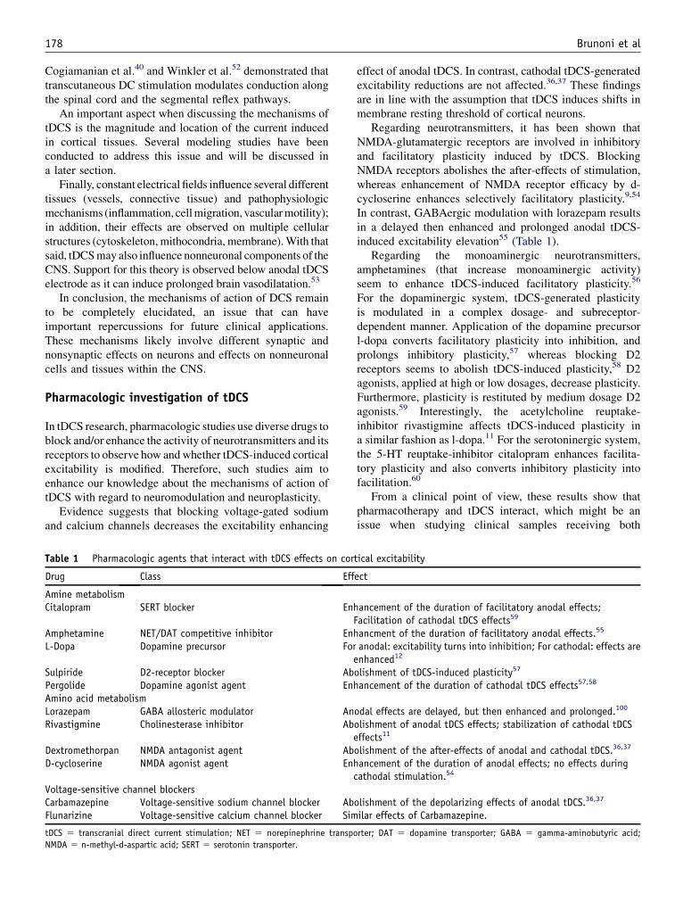
Pharmacologic investigation of tDCS
In tDCS research, pharmacologic studies use diverse drugs toblock and/or enhance the activity of neurotransmitters and itsreceptors to observe how and whether tDCS-induced corticalexcitability is modified. Therefore, such studies aim toenhance our knowledge about the mechanisms of action oftDCS with regard to neuromodulation and neuroplasticity.
Evidence suggests that blocking voltage-gated sodiumand calcium channels decreases the excitability enhancing
Table 1 Pharmacologic agents that interact with tDCS effects on cor
Drug Class Eff
Amine metabolismCitalopram SERT blocker Enh
Amphetamine NET/DAT competitive inhibitor EnhL-Dopa Dopamine precursor For
Sulpiride D2-receptor blocker AbPergolide Dopamine agonist agent EnhAmino acid metabolismLorazepam GABA allosteric modulator AnRivastigmine Cholinesterase inhibitor Ab
Dextromethorpan NMDA antagonist agent AbD-cycloserine NMDA agonist agent Enh
Voltage-sensitive channel blockersCarbamazepine Voltage-sensitive sodium channel blocker AbFlunarizine Voltage-sensitive calcium channel blocker Sim
tDCS 5 transcranial direct current stimulation; NET 5 norepinephrine transpo
NMDA 5 n-methyl-d-aspartic acid; SERT 5 serotonin transporter.
effect of anodal tDCS. In contrast, cathodal tDCS-generatedexcitability reductions are not affected.36,37 These findingsare in line with the assumption that tDCS induces shifts inmembrane resting threshold of cortical neurons.
Regarding neurotransmitters, it has been shown thatNMDA-glutamatergic receptors are involved in inhibitoryand facilitatory plasticity induced by tDCS. BlockingNMDA receptors abolishes the after-effects of stimulation,whereas enhancement of NMDA receptor efficacy by d-cycloserine enhances selectively facilitatory plasticity.9,54
In contrast, GABAergic modulation with lorazepam resultsin a delayed then enhanced and prolonged anodal tDCS-induced excitability elevation55 (Table 1).
Regarding the monoaminergic neurotransmitters,amphetamines (that increase monoaminergic activity)seem to enhance tDCS-induced facilitatory plasticity.56
For the dopaminergic system, tDCS-generated plasticityis modulated in a complex dosage- and subreceptor-dependent manner. Application of the dopamine precursorl-dopa converts facilitatory plasticity into inhibition, andprolongs inhibitory plasticity,57 whereas blocking D2receptors seems to abolish tDCS-induced plasticity,58 D2agonists, applied at high or low dosages, decrease plasticity.Furthermore, plasticity is restituted by medium dosage D2agonists.59 Interestingly, the acetylcholine reuptake-inhibitor rivastigmine affects tDCS-induced plasticity ina similar fashion as l-dopa.11 For the serotoninergic system,the 5-HT reuptake-inhibitor citalopram enhances facilita-tory plasticity and also converts inhibitory plasticity intofacilitation.60
From a clinical point of view, these results show thatpharmacotherapy and tDCS interact, which might be anissue when studying clinical samples receiving both
tical excitability
ect
ancement of the duration of facilitatory anodal effects;Facilitation of cathodal tDCS effects59
ancment of the duration of facilitatory anodal effects.55
anodal: excitability turns into inhibition; For cathodal: effects areenhanced12
olishment of tDCS-induced plasticity57
ancement of the duration of cathodal tDCS effects57,58
odal effects are delayed, but then enhanced and prolonged.100
olishment of anodal tDCS effects; stabilization of cathodal tDCSeffects11
olishment of the after-effects of anodal and cathodal tDCS.36,37
ancement of the duration of anodal effects; no effects duringcathodal stimulation.54
olishment of the depolarizing effects of anodal tDCS.36,37
ilar effects of Carbamazepine.
rter; DAT 5 dopamine transporter; GABA 5 gamma-aminobutyric acid;

Clinical research with tDCS 179
interventions. In fact, the complex nonlinear interactionmakes it difficult to foresee the specific effects ofpathophysiologic alterations or drug application on theamount and direction of tDCS-induced plasticity; thusdemanding further empirical research on this topic.
Parameters of stimulation
TDCS parameters can vary widely and several factors needto be defined. These factors include electrode size andpositioning, intensity, duration of stimulation, number ofsessions per day, and interval between sessions. By varyingthese parameters, different amounts of electric current canbe delivered, thus inducing diverse physiologic and adverseeffects. Another potential concern is that tDCS devices arenot worldwide standardized. These devices can be easilyconstructed using standard equipment and technology inengineering laboratories of colleges and universities. Infact, more than a dozen different tDCS devices can befound throughout neuromodulation laboratories worldwide.
Electrode positioningAlthough tDCS electrical fields are relatively nonfocal,electrode positioning is critical. For instance, a previousstudy showed that changing the electrode reference fromDLPFC to M1 eliminated tDCS effects on workingmemory.16 Other studies have shown that phosphene-thresholds are modulated only during occipital (visualcortex) DCS and not other areas.49,61 Likewise, a tDCS trialfor major depression showed that only DLPFC stimulation(and not occipital stimulation) ameliorated symptoms.62
Although current evidence suggests site-dependent effects,other issues have yet to be exploreddfor instance, oneopen question is whether and how brain stimulation inone area influences adjacent and more distant areas.
TDCS studies usually use one anode and one cathodeelectrode placed over the scalp to modulate a particulararea of the CNS. Electrode positioning is usually deter-mined according to the International EEG 10-20 System.Given the focality of tDCS, this appears appropriate. Forinstance, studies exploring the motor cortex place elec-trodes over C3 or C4; for the visual system, electrodes aretypically placed over O1 or O2 (for a review of tDCSstudies exploring different brain areas see Utz et al.63).
In this study, some terms used to describe tDCSmontages should be discussed: when one electrode is placedbellow the neck, the entire montage is usually described as‘‘unipolar.’’ In contrast, montages with two electrodes on thehead are termed usually ‘‘bipolar.’’ However, this nomen-clature might be inaccurate as technically the DC stimula-tion is always generated via two poles (electrodes)generating an electric dipole between the electrodes.Therefore, an alternative nomenclature of ‘‘mono-cephalic’’and ‘‘bi-cephalic’’ is proposed to differentiate between‘‘unipolar’’ and ‘‘bipolar’’ setups, respectively. Researchersin the field also use the terms ‘‘reference’’ and ‘‘stimulating’’
electrode to refer to the ‘‘neutral’’ and ‘‘active’’ electrode,respectively. However, the term ‘‘reference’’ electrode mayalso be problematic, especially for bicephalic montagesbecause the ‘‘reference’’ electrode is not physiologicallyinert and can contribute to activity modulation as well. Thiscould be a potential confounder depending on the mainstudy question. Nonetheless, researchers use these terms tohighlight that (in their study) they are under the assumptionthat in their particular montage one electrode is beingexplored as the ‘‘stimulating,’’ whereas the other is the‘‘reference.’’
In contrast, having the possibility to increase anddecrease activity in different brain areas simultaneouslymay be advantageous. For instance, this could be useful inconditions involving an imbalanced interhemisphericactivity (ie, in stroke).64 In scenarios whether the referenceelectrode poses a confounding effect, an extracephalicreference electrode could theoretically aid in avoidingthis issue. However, this might increase the risk of shuntingthe electric current through the skin (which would then notreach brain tissue) or displacing the current. Ultimately, thechoice of montage will be application specific; for example,a recent study comparing different tDCS setups showedthat, although bicephalic setups were effective, the monoce-phalic setup was no different than sham stimulation.65
Finally, in a monocephalic setup, using very high currentsthere is the potential risk of influencing brain stem activity,including respiratory control (note that this risk is theoret-ical and was only observed in one historical report).66
Nevertheless, in choosing the extracephalic position, theresearcher must be confident that a significant electric fieldwill be induced on the target brain area.
Moreover, because current flow direction/electrical fieldorientation relative to neuronal orientation might determinethe effects of tDCS,7 it might be that the effects of an ex-tracephalic electrode differs relevantly from that of a bipolarelectrode arrangement. Alternatively, enhancing the size ofone electrode, thus reducing current density, might enablefunctional monocephalic stimulation also with a bicephalicelectrode montage.58
Direct current stimulation can also be delivered overnoncortical brain areas. Ferrucci et al.50 stimulated the cere-bellum showing changes in performance in a cognitive taskfor working memory. Galea et al.51 explored the inhibitoryeffects of the cerebellum on motor-evoked potentials(MEPs) triggeredbyTMSover themotor cortex.This revealedthat tDCS could modify MEPs in a polarity-specific manner.In addition, Cogiamanian et al.40 observed that cathodaltranscutaneous DC over the thoracic spinal cord suppressedtibial somatosensory-evokedpotentials. Furthermore,Winkleret al.52 observed that transcutaneous DCS over the spinal cordmodulates the postactivation depression of the H-reflex.Preliminary data indicates spinal DCS also influences noci-ception67 suggesting that the spinal cord as a target for trans-cutaneous DCS. Challenges for stimulation in this area mustbe considered such as location of induced electrical fields.

180 Brunoni et al
Modeling tDCSDuring tDCS, current is generated across the brain; differentmontages result in distinct current flow through the brain andthus the ability to adjust montage allows customization andoptimization of tDCS for specific applications (see above).Though tDCS montage design often follow basic assump-tions (eg, ‘‘increased/decreased excitability under the anode/cathode’’), computational models of brain current flowduring tDCS (so called ‘‘forward’’ models) provide moreaccurate insight into detailed current flow patterns, and insome cases show that the basic assumptions are not valid.When interpreting the results of such simulations, it isimportant to recognize that the intensity of current flow inany specific brain region does not translate in any simplelinear manner to the degree of brain modulation. However, itseems reasonable to predict that regions with more currentflow are more likely to be affected by stimulation, whereasregions with little or no current flow will be spared the directeffects of stimulation.
Computational models of tDCS range in complexityfrom concentric sphere models to individualized high-resolution models based an individual’s structural magneticresonance imaging (MRI). The appropriate level of detaildepends on the available computational resources and theclinical question being asked (see technical note below).Regardless of complexity, all models share the primaryoutcome of correctly predicting brain current flow duringtranscranial stimulation to guide clinical practice in a mean-ingful manner.
Most clinical studies use tDCS devices that apply directelectric currents via a constant current source, but evenwithin this space there are infinite variations of dosage andmontage that can be leveraged, with the help of models, tooptimize outcomes. The current is sent through patchelectrodes (surface areas typical range from 25 to 35 cm2
but can vary by an order of magnitude) attached to the scalpsurface. Total current injected ranges in magnitude aretypically from 0.5 to 2 mA. Steps taken to improve tDCSspecificity (including the use of larger ‘‘return’’ spongesand extracephalic electrodes) have been proposed but moreanalysis is required to determine the role of electrode-montage in neuromodulation and targeting. Modelingapproaches are instrumental toward this goal. For example,modeling studies have recently predicted a profound roleof the ‘‘return’’ electrode position in modulating overallcurrent flow including under the ‘‘active’’ (or ‘‘stimulating’’)electrode.68 Specifically, for a fixed active electrode positionon the head, changing the position of the return electrode(including cephalic and extracephalic positions) influencescurrent flow through the presumed target region directlyunder the active electrode. Therefore, in addition to consid-ering the role of scalp shunting and action on deep brainstructures (see above) when determining electrode distance,the complete design of electrode montage may subtly modu-late cortical current flow.69 Again, computer modeling canprovide valuable insight into this process.
Recent modeling studies suggest that individual anatom-ical differences may affect current flow through the cortex.In comparison to TMS, which uses MEPs to index itspotency, there is no similar rationale for titrating tDCSdosage. A related issue is the modification of tDCS dosemontages for individuals with skull defects or stroke-related lesions. Such individuals may be candidates fortDCS therapy but defects/lesions are expected to distortcurrent flow. For example, any defect/injury filled withcerebrospinal fluid (CSF), including those related to strokeof traumatic brain injury, is expected to preferentially‘‘shunt’’ current flow.15 Ideally, tDCS would be adjustedin a patient-specific (defect/lesion specific) manner totake advantage of such distortions in guiding current flowto targeted regions, while simultaneously avoiding anysafety concerns (such as current hot spots).
Evidence from modeling studies suggests that for typicaltDCS significant amounts of current can reach broadcortical areas especially between and under the electrodesurface.12,13 Modeling studies also show that electrodemontage is critical to the amount of current shunted throughthe skin.
Electrode montage is critically associated to the amountof current being shunted through the skin, how much isdelivered to the brain, and to what targets. The overalltheme emerging from modeling efforts is that despiteclinical success in applying simplifying rules in dosedesign, all the details and aspects of electrode montagedesign combine to influence current flow such that thesesimplifying rules are applicable but only within a limitedparameter range. For example, average current density(total current/electrode area) at the ‘‘active’’ electrode maybe a useful metric to normalize specific neurophysiologicoutcomes (eg, TMS evoked MEPs), there is no universalrelationship between current density and brain modulationwhen one considers the full spectrum of possible electrodemontages.13,70
Recent modeling data taking into consideration gyri andsulci geometry have shown that electric current canconcentrate on the edge of gyri.71 Therefore, the effectsmight not be homogeneous throughout the stimulatedarea. Increased appreciation of the complexity of currentflow through the head (reflecting the complexity of neuro-anatomy), reinforces the use of applying computationalmodels to assist in tDCS dose design72 rather than simplyrelying on some heuristic rules (eg, ‘‘increased excitabilityunder the anode’’).
In addition to predicting brain current flow, modelingstudies also provide insight into electrode design bypredicting current flow patterns through the skin. Modelingstudies has reinforced that current is not passed uniformlythrough the skin but rather tends to concentrate nearelectrode edges or skin inhomogeneities.13 Electrodedesign can be simple saline-soaked cotton or sponge padsor specifically designed patches with unique shapes andmaterials to maximize stimulation magnitude and focality.

Clinical research with tDCS 181
Modeling confirms that decreasing the salinity of the padsreduces peak current concentration at the edges (even asthe total current applied and average current density isfixed).73
In summary, modeling studies are expected to playa critical role in the development of next-generation tDCStechnologies and approaches. Notably, tDCS devices havenot drastically changed since the time when the battery wasfirst discovered. Thus, conventional technology has certainlimitations. These include focality (area stimulated), depthof penetration, and targeting-location control. To overcomethese and other limitations, technologies using arrays ofelectrodes74 such as ‘‘High Definition’’ tDCS (HD-tDCS)71
and others (eg, simultaneous EEG monitoring during tDCSas to adjust dosage and parameters) have been recentlyproposed. Ultimately, as we begin integrating modern tech-nology with transcranial stimulation techniques, clinicalcontrol and efficacy will undoubtedly improve.
On a final technical note: Though there has been a recentemphasize to develop increasingly accurate and complexmodels,71,72,75 certain universal technical issues should beconsidered for high-precision models, beginning with: (1)high-resolution (eg, 1 mm) anatomic scans so that the entiremodel work flow should preserve precision. Any finite-element human head model is limited by the precisionand accuracy of tissue dimensions (masks) and conductivityvalues incorporated (inhomogeneity and anisotropy). Onehallmark of precision is the cortical surface used in thefinal finite-element mask solver should represent realisticsulci and gyri; (2) Simultaneously, a priori knowledge oftissue anatomy and factors known to shape current floware applied to further refine segmentation. Particularly crit-ical are discontinuities not present in nature that result fromlimited scan resolution; notably both unnatural perforationsin planar tissues (eg, holes in cerebrospinal fluid wherebrain contacts skull) and microstructures (eg, incompleteor voxelized vessels) can produce significant aberrationsin predicted current flow. Addition of complexity withoutproper parameterization can evidently decrease predictionaccuracy. An improper balance between these factors canlead to distortions in brain current flow of an order ofmagnitude or moreduncontrolled additional complexitycan in fact induce distortion. We thus emphasize that themost appropriate methodology (ranging from concentricspheres to individualized models) ultimately depends onthe clinical question being addressed.
The clinical research of tDCS
Studies in nonhumans (Preclinical)
Previous animal studies have assessed safety limits of tDCScurrent intensity. In one study, 58 rats received tDCS withvarying current densities for up to 270 minutes and
histologic evaluation was conducted to assess neuronallesion. Results suggest that brain lesions occurred whencurrent density was at least two orders of magnitude higherthan typically used in humans88 and may reflect increase inbrain temperature never observed using conventional tDCSprotocols.14 Another interesting insight from this study isthat duration of tDCS only becomes a safety issue whenthe intensity of stimulation is near the threshold associatedwith neuronal lesion. Other animals studies conducted withdifferent goals have also shown that tDCS used withcharges similar to human studies do not induce histologicallesions.89
Finally, animal studies are useful for test dosing andexploring physiologic aspects of tDCS mechanisms. Incontrast, such studies are rare, and positioning of theelectrodes as well as different cortical architecture, mightbe critical. Still, animal models might be important foranswering specific questions not possible to be done inhumans.
Studies on healthy volunteers (Phase I)
In drug-based trials, phase I studies are nonrandomized,noncontrolled clinical (human) trials designed to addresssafety and optimal dosage of drugs. This is performed byassessing the adverse effects/safety and dosage or the drugs.In this section, previous tDCS studies that address thesequestions and present issues that remain unsolved (doseparameters was above discussed) are reviewed (Table 2).
Safety/toxicityAlthough tDCS differs in many aspects from othernoninvasive neuromodulatory therapies in that it does notinduce neuronal action potential and uses weak electriccurrents, there are safety concerns that must be addressed.If the electrochemical products generated by these currentscontact the skin, skin irritation may occur; in addition,tissue heating associated with nonintact skin (therefore thisis especially important in people with skin diseases and/orin protocols using daily tDCS applications and/or highelectric currents) may induce skin burning92dalthoughmild redness is more likely related to local, vasodilatationskin changes rather than skin damage.93 In fact, consideringthere is no direct contact between the brain and the elec-trode and also the distance, electrochemical or heatinglesions to the neuronal tissue is less likely. Moreover,experimental and modeling studies suggest no significanttemperature increases for typical tDCS protocols.7,71,73
TDCS has been tested in thousands of subjects world-wide with no evidence of toxic effects to date. In addition tothe hundreds of studies exploring tDCS effects in diversecontexts, some studies have focused specifically on safety.For instance, in a large retrospective study, Poreisz et al.94
reviewed adverse effects in 77 healthy subjects and 25patients who underwent a total of 567 1 mA stimulationsessions. Results show the most common effects were

Table 2 Main issues in the clinical research of tDCS and possible solutions to effectively handle them
Current evidence Key issues Possible solutions
Phase I studiesSafety/adverse effects TDCS is not likely associated to long-term,
deleterious effects. AEs are mild and transientat usual doses.
Safety has not been sufficiently investigated inpeople with skull defects and/or patients withneuropsychiatric disorders.
Further research should actively investigateadverse effects; long-term follow-up; modelingstudies.
Dose-effect curve Higher doses, higher current densities and higherperiods of stimulation seem to be associatedwith effects of larger magnitude and duration.
Great between-subjects variability of effects;using higher doses is limited due to AEs;pharmacotherapy alters dose-effect curve;optimal parameters not yet defined.
Further research addressing pharmacologicalmodification of tDCS effects; increasingduration span of tDCS to avoid skin damage;bayesian approaches and modeling studies todefine optimal dose.
Phase II/III studiesSubjectsRecruitment TDCS is still on its infancy, and few patients and
physicians are aware of this novel technique.Non-referral due to lack of knowledge/suspiciousness of tDCS and time constraints inambulatory settings; ethical issue of receivingplacebo.
Using multiple referral sources; specificneuromodulation ambulatories; building trustwith volunteers and physicians (lectures, websites, explanatory videos).
Eligibility Sample should be homogeneous, especially inphase II studies.
Sources of heterogeneity are: concomitant use ofmedications, incorrect diagnosis ofneuropsychiatric condition, wide spectrum ofseverity and refractoriness.
Stratification during randomization; post-hocanalysis controlling for severity, refractorinessand medications; drug washout.
Attrition High attrition rates might lead to negativefindings; especially if intention-to-treatanalyses are performed.
Daily visits to the research center and skindamage are specific issues related to dropout intDCS trials.
Careful explanation of study objectives andpossible side effects; covering oftransportation costs; flexible schedules; usingrun-in period to identify noncommitters.
MethodsBlinding Blinding is the strongest approach to minimize
bias. Sham TDCS involves applying an electricalcurrent for less than 30 seconds, as to mimicintial side effects.
Several studies suggested that the sham methodis reliable, at least in healthy volunteers, withintermediate-high doses and in one-sessionstudies. TDCS device can be turned offmanually (single-blinded, requiring anotherperson to evaluate subjects) or automatically(double-blinded).
Further studies should explore whether this shammethod is reliable in other contexts, e.g. dailystimulations for 5-10 days, higher doses andnonna€ıve subjects. Staff blinding should alsobe more carefully evaluated.
Approach To induce long-lasting (days to weeks) effects,tDCS must be delivered continuously(usually daily for 5 to 10 days).
Number of sessions and time period betweenstimulations are still undefined as well as theextent of such effects after the initial sessions.
Long follow-up of subjects (months to years);performing specific studies designed toevaluate cumulative changing in corticalexcitability according to the number ofstimulations (and time between them).
Control group In tDCS research, the control group might beeither a sham-group or an active group inwhich polarities are inverted.
The latter approach is an even more reliableblinding method than sham; although it can aswell induce effects.
Studies exploring mechanisms of tDCS could havethree groups; studies using tDCS as treatmentshould prefer using a sham group.
TDCS 5 transcranial direct current stimulation; AE 5 adverse effects.
182
Brunoniet
al
Clinical research with tDCS 183
mild tingling sensations (75%), light itching sensation(30%), moderate fatigue (35%), and headache (11.8%);and most of these effects did not differ from those ofplacebo stimulation. In another study, 164 sessions of stim-ulation were analyzed. Authors found only mild adverseeffects with a low prevalence (0.11% in active and 0.08%in sham stimulation group).95 Other initial studies90,91,96-99
also reported only mild, benign, and transient side effects.In fact, the most severe adverse event reported is skinlesions on the site of electrode placement.92
Historically, the most severe adverse effect was observedin the first study of tDCS. During the 1960s Lippold andRedfearn66 related a brief respiratory and motor paralysis ina bifrontal electrode montage with the current referenceplaced on the leg. No loss of consciousness was reportedand respiration returned to normal when the current wasstopped. This was attributed to the fact that the subjectreceived 10 times the intended intensity, probably 3 mA.27
General exclusion criteria for noninvasive brain stimu-lation also apply for tDCS. Subjects must be free of unstablemedical conditions, or conditions that may increase the riskof stimulation such as uncontrolled epilepsy; althoughepileptic seizures have not been observed in a pilot studywith patients with active epilepsy.100 Also, subjects musthave no metallic implants near the electrodes.
Finally, it should be underscored that most of theseobservations were extracted from single stimulation studiesin healthy subjects without medications. Less is knownabout the adverse effects of daily (or even twice daily) tDCSin patients with neuropsychiatric disorders who use phar-macotherapy. In such conditions, the adverse effects canbe magnified and therefore they should be actively inquiredduring trials. For instance, some single-patient studiesreport that tDCS can induce mania/hypomania in patientswith major depression.101-103 Therefore, we suggest thata medical monitor should supervise tDCS treatment insuch contexts of increased risk of significant adverse effects.
DosageTDCS dosage is defined by the following parameters: (1)current dosage (measured in amperes); (2) duration ofstimulation; and (3) electrode montage (size and position ofall electrodes). Current density (current dose divided byelectrode size) is also an important parameter in consideringdosage; especially for defining safety58; see the followingreview for additional information.104 Themost common elec-trode sizes are of 25-35 cm2 with currents of 1-2 mA (gener-ating densities ranging from 0.28-0.80 A/m2) for up to 20-40minutes. However, the current that effectively reachesneuronal tissue depends on other less controllable factors.These include skin resistance, skull resistance, resistance ofintracranial structures (eg, blood vessels, cerebrospinal fluid,andmeninges) and the resistance of brain tissue, which variesaccording to cell type and structure (eg, glial cells, pyramidalneurons, white matter, and so on). Moreover, patients withskull defects, brain lesions and other conditionswill influence
current amount and delivery. In addition, the baseline corticalexcitability is different in people using pharmacotherapy (eg,benzodiazepines,55 anticonvulsants,105 antidepressants,69
and others106) and/or presenting neuropsychiatric disorders(eg, major depression,107 schizophrenia,108 fibromyalgia,109
migraine,110 and others); an issue that is likely to interferewith the chosen dosage. Finally, other variables influencebaseline cortical excitability such as gender,111 age,112 andsmoking.113 Hence, the same amount of current is likely tohave nonuniformeffects in subjectswith different conditions.For instance, one study showed that low (25 mg) and high(200 mg) doses of l-dopa abolished tDCS-induced effectson cortical excitability, whereas an intermediate (100 mg)dosage increased inhibitory effects.114 Notwithstanding,these studies should be regarded as exploratory and thus repli-cated in other contexts and samples, especially in clinicalpopulations.
Therefore, in the context of clinical research, suchindividual factors are a source of variability and, if importantenough, may result in negative findings. To avoid this, onealternative is to standardize the source of error in the sample.For instance, using saline-soaked sponges to minimize skinresistance (which can also be measured by an ohmmeteradapted in the tDCS devicedsome devices do give theresistance), excluding patients under pharmacotherapy, orcontrolling when it is not feasible (eg, benzodiazepines),avoiding sample heterogeneity using specific diagnosticcriteria, particularly when working with a small, neuropsy-chiatric subject pool. Future studies addressing the interac-tion of tDCS and drugs in psychopharmacologywill continueto explore and identify which drugs do not interferewith tDCS effects and which ones could block or enhancetDCS-excitability effects.10,27
Initial studies measuring brain excitability demonstratethat currents as low as 0.28 A/m2 present depolarizing andhyperpolarizing effects.6,7 In addition, phase I/II studies ad-dressed the effects of varying dose and/or time of stimula-tion on cortical excitability and/or neuropsychologic tasks.Ohn et al.115 tested the effects on working memory during30 minutes of stimulation, showing that performanceincreased in a time-dependent fashion. Other studiesshowed the cognitive effects induced by tDCS are depen-dent on the current intensity; demonstrating effects suchas enhanced verbal fluency improvement at 2 mA (versuslower improvement at 1 mA)18; and working memoryimprovement at 2 mA (versus no improvement at 1 mA)(See Box 1 for other tDCS studies on cognition).116 Never-theless, it remains unclear whether there is a linear (doseversus effect) curve associated with direct current stimula-tion and the influence of each parameter (dose, currentdensity, stimulation duration) on these effects. It is knownthat increasing current densities will increase the depth ofthe electrical field, thus affecting different populations ofneurons. However, at greater intensity tDCS might be pain-ful to the subjects. For these reasons, a more effectiveapproach designed to prolong tDCS effects is to increase

Box 1 Insight from tDCS studies on cognition
TDCS has been increasingly used to transiently modify cognitive functions in the healthy human brain. This fieldpresents an exciting opportunity to extend the application of tDCS from a neuroscience research tool to the potentialtreatment of cognitive impairments. Indeed, the understanding of how to successfully manipulate cortical excitabilityfor the formation of new memories or the acquisition of new skills could fill an important gap between phase I and IIclinical studies. TDCS studies have shown that anodal tDCS delivered over the dorsolateral prefrontal cortex facilitatevisual working memory.16 Conversely, cathodal stimulation had a detrimental effect on short-term auditory memoryperformance.76 Regardless of polarity, tDCS over the cerebellum disrupts practice-dependent improvement duringa modified Sternberg verbal working-memory task,50 whereas intermittent bifrontal tDCS impairs response selectionand preparation in the same task.77 Moreover, anodal tDCS to the anterior temporal lobes delivered before theencoding and retrieval phase was effective in reducing false memories, whereas maintaining veridical memories.78
Finally, the application of anodal tDCS during slow-wave sleep improved declarative memory consolidation.79 Furthereffects of tDCS on cognitive functions in healthy individuals have been shown for decision-making,80,81 probabilisticclassification learning,82 attention,83-85 and language.86,87 Overall, these studies focused on the short-term improve-ments in performance induced by a single session of stimulation, typically delivered online during the task or imme-diately before it. The main limitations are the lack of control conditions over different cortical areas and the lack ofa systematic monitoring of the duration of the effects. The effects of repeated applications of tDCS, their interactionwith specific learning stages and tasks and the extent to which these performance improvements are retained in thelong-term remain to be addressed. Hypothesis-driven behavioral paradigms or stimulation strategies are also necessaryto further explore the functional role of different cortical areas in human learning.
184 Brunoni et al
the stimulation duration as opposed to the currentdensity.7,27,35,37
Short applications (ie, seconds to a few minutes) ofanodal/cathodal tDCS result in excitability shifts duringstimulation but no after-effects. However, no long-termeffects are seen. In contrast, 10 minutes or more ofstimulation can elicit prolonged after-effects, which canbe sustained for over an hour.7,27,39 The exact duration ofeffects depends on the targeted cortical area and on thetype of variable assessed.
For clinical purposes, longer-lasting effects are crucial.Single-dose tDCS interventions have relatively short-livedafter-effects. Multiple stimulation sessions are required toinduce a significant manipulation in synaptic efficacy.117,118
In fact, repeated sessions of tDCS may have cumulativeeffects associated with greater magnitude and duration ofbehavioral effects. For example, cathodal tDCS appliedover 5 consecutive days is associated with cumulativemotor function improvement lasting up to 2 weeks afterthe end of stimulation. This is an effect which is notobserved when sessions are applied weekly (as opposedto daily).98 Whether this approach is appropriate to maxi-mize and stabilize the electrophysiologic effects of tDCSremains under investigation. The optimal repetition rateand duration to promote tDCS-induced plasticity alsoremains to be determined. In animal experiments, repetitionof tDCS during the after-effects of a first stimulationsession has been shown to enhance efficacy.32 However,repeated plasticity induction may result in homeostaticallydriven antagonistic effects.119 Recently, Monte-Silva andcoworkers118 directly compared the effects induced bysingle sessions of cathodal tDCS over the motor cortex tothe effects of repetitive stimulation during or after the
after-effects of the first stimulation. The results showedthat increasing cathodal tDCS duration (1 mA, with nointerstimulation interval) resulted in longer-lasting after-effects, typically over 1 hour (tDCS duration from 9 to18 min prolonged the after-effects from 60 to 90 minutes).Interestingly, when the second stimulation was performedduring the after-effects of the first, a prolongation andenhancement of tDCS-induced effects for up to 120minutes after stimulation was observed. In contrast, whenthe second session was performed 3 or 24 hours after thefirst, tDCS effects on cortical excitability were mixed.This was shown with a primary reduction or abolishmentof the initial effects of cathodal tDCS, followed by a laterreoccurrence of tDCS-induced cortical inhibition. Suchneurophysiologic evidence is indicative of a stimulationtiming-dependent plasticity regulation in the human motorcortex. Understanding the interaction of the consecutivestimulation protocols appears crucial to effectively targetspontaneous changes of cortical activity and excitability(See Box 2 for a discussion on ‘‘offline’’ vs. ‘‘online’’ stim-ulation). Hence, implementing more effective proceduresof plasticity induction procedures in clinical settings iscrucialdin fact these results need to be replicated inclinical populations.
Studies on patients with neuropsychiatricconditions (Phase II/III)
Phases II and III studies relate to using an intervention inclinical samples. Phase II studies are typically small and usetargeted samples to obtain additional information regardingoptimal parameters of stimulation. Phase III are pivotal
Box 2 Two types of study design in tDCS: ‘‘Online’’ versus ‘‘Offline’’
Clinical researchers usually apply tDCS in two main modalities regarding the time point in which the primary outcomevariable is collected. When tDCS and the main outcome are coincident in time (ie, when the variable is collected duringtDCS application) the experiment is said to test the ‘‘online’’ effects of tDCS. The concept is also used when anotherintervention (usually having a similar time span than tDCS such as physical therapy) and tDCS are appliedsimultaneously. The rationale for an ‘‘online’’ approach is to take advantage of the putative property of tDCS toinduce excitability modifications of the brain (which is analogous to TMS) to test neuromodulatory effects on the studyhypothesis, such as alterations of brain functions during tDCS. For instance, an area of investigation that uses thisapproach is transient modulation of moral judgment and decision making during tDCS (see Discussion on ethics in thismanuscript).
On the other hand, when tDCS and the variable being measured can be distinguished in time, it is said that theexperiment is applying tDCS in an ‘‘offline’’ protocol. An ‘‘offline’’ tDCS protocol applies, for instance, when onesurrogate outcome (or clinical parameter) is used before and after stimulation to index tDCS effects (see Discussion onsurrogate outcomes). An ‘‘offline’’ approach is also used in phase II/III tDCS studies. In such cases, tDCS is anexperimental intervention and its long-term, neuroplastic effects are indexed with one or more surrogate and/orclinical outcomes.
Clinical research with tDCS 185
studies, involving larger samples. In the United States, twopositive phase III trials are required for approving a drug ordevice by the Food and Drug Agency (FDA) (See Box 3 forphase II/III studies in major depression).
As mentioned previously, several studies have exploredthe therapeutic application of tDCS in several neuropsy-chiatric disorders. The results of these studies reveal long-lasting tDCS effects and have promoted its use in clinicalsettings. Because clinical development of tDCS is beingconducted mainly in academia, studies are not widelystandardized regarding variables and population samples,therefore limiting conclusions. These findings are alsolimited by small sample sizes and experimental design. Infact, a similar scenario has been observed for TMS 5 to 10years ago.120 This section aims to comprehensively reviewthe main issues of the later phases of clinical trial develop-ment for tDCS.
Issues related to recruitment and eligibilityRecruiting subjects for tDCS clinical trials presents a chal-lenge. Proposing an intervention alternative to the main-stream pharmacotherapy might be seen by prospectivepatients and referring physicians in nonacademic settingsas suspicious. This issue can be particularly important whena large sample size is required and/or if the eligibility criteriaexclude refractory patients who are more prone to enroll inresearch protocols. Likewise, referral physicians, due to timeconstraints in the ambulatory setting, usually prefer to treatdrug-na€ıve patients themselves. Indeed, daily visits for 1-to-2 weeks to research centers might sound unappealing and/orunaffordable even to refractory patients. In such contexts, itis advisable to have multiple referral sources and to usebroad recruitment strategies. Building trust with potentialvolunteers is imperative. One cost-effectiveness approachcould be using explanatory videos in lay language.26
Another issue is sample heterogeneity. In pivotal clinicaltrials comparing tDCS against pharmacotherapy, largesamples are typically required and patient heterogeneitymight be larger than for drug trials for the same condition.This is due to the fact that the severity of the conditionranges from drug-na€ıve to refractory subjects. Targetingonly the former would create difficult enrollment (for thereasons mentioned previously), although targeting only thelatter decreases overall generalizability. Possible solutionsinclude stratification during randomization (refractoryversus nonrefractory), post hoc analysis controlling forrefractoriness, or increasing sample size to address some ofthe issues associated with heterogeneity.
Blinding issuesIt appears easier to conduct sham-controlled trials usingtDCS compared with TMS. TMS induces itching and painsensations over the stimulation site, whereas tDCS inducesa mild tingling sensation that usually rapidly fades.Therefore, sham protocols begin with active tDCS, whichis switched off within a minute. In addition, the tinglingsensation relates to the velocity in which the current iseither increased or decreased. In fact, an increase of currentdelivery from 0.1 to 0.2 mA/s generates no discomfort formost subjects.121 Interestingly, some subjects in the shamgroup continue feeling some tingling even after the currentis discontinued.
These sensations are related to the total amount ofcharge delivered. Although this has yet to be systematicallyevaluated, this relationship can be a potential issue whendelivering relatively high charges (.1.5-2 mA/s) and/orhigher current densities. There is evidence that electrolytesolutions with lower NaCl concentrations (15 mM) areperceived as more comfortable during tDCS than thosesolutions with higher NaCl concentrations (220 mM).

Box 3 Insights from tDCS studies for major depression
In the past 10 years, several trials applied tDCS to subjects with major depressive disorder (MDD). Fregni et al.124
performed a pilot randomized, sham-controlled, double-blind trial in which 10 patients were randomly assigned toreceive either 5 days of active or sham stimulation. Boggio et al.62 also enrolled 40 MDD subjects with differentdegrees of refractoriness (but medication-free) and randomized them to 10 sessions of active dorsolateral prefrontalcortex (DLPFC) tDCS, active occipital tDCS or sham tDCS. The findings suggested that the active DLPFC tDCS group pre-sented a superior, significant improvement in HDRS scores compared with the other groups. Rigonatti et al.90
demonstrated in an open-label study that Fluoxetine 20 mg/d and active tDCS (from patients of Boggio’s study) pre-sented similar scores after 6 weeks of treatment. Ferrucci et al.91 stimulated 14 patients with severe MDD using 2 mA for20 minutes for 5 days twice a day, showing a significant improvement in mood. Such effects seem to be more robust inmore severe patients.125 Loo et al.99 enrolled 40 patients with severe MDD, in a double-blinded, sham-controlled studybut failed to demonstrate significant difference between groups in this phase; tDCS was only more effective during theopen-label phase in which patients received additional five sessions. However, this study has some limitations: thedose applied was relatively low (1 mA), and only five stimulations sessions were held, which were alternated (otherstudies used consecutive sessions). Moreover, patients with axis II disorders were not excluded. Finally, Brunoniet al.126 compared patients with unipolar versus bipolar depression and found that tDCS might be a potentialtreatment for both conditions. However, as with phase II trials, these studies share common characteristics:relatively small sample sizes, heterogeneous sample (eg, refractoriness, medication use), blinding vulnerability(some studies were open-label), absence of primary hypothesis (most of them used several depression rating scales),and presence of ‘‘carryover’’ effects (in crossover studies). These initial trials likely incurred in some false-positive andfalse-negative results; nevertheless, they revealed the potential effectiveness of tDCS for major depressive disorder.Finally, a search made on clinicaltrials.gov in September 2010 revealed that there are at least seven trials exploringthe antidepressive effects of tDCS worldwide; and the design and methods of one of them127 has been recentlypublished.
186 Brunoni et al
Because the ionic strength of deionized water is much lessthan that of all NaCl solutions, there is a significantly largervoltage required to carry current through the skin comparedwith NaCl solutions. Thus, it is recommended the use ofsolutions with relatively low NaCl concentration, in therange 15 mM to 140 mM, as tDCS at these concentrationsis more likely to be perceived as comfortable, requires lowvoltage and still allows good conduction of current.122 Ithas also been proposed to apply topical anesthetics to alle-viate this issue.27
An additional blinding issue is the local vasodilatationafter tDCS. This causes the skin to turn red that might notbe acknowledged by the subject but might be seen by thestaff and other patients. In clinical protocols, such rednesscan be evident after several days of stimulation. This canbecome a logistical issue, demanding stimulated patients toleave the setting immediately, avoiding contact with otherpeople (patients and researchers) as to avoid blindingbreaking. Another approach would be to interview patientsbefore (and not after) being stimulated. If a rater noticesevidence of redness on the scalp of a patient, anotherblinded rater should substitute him/her, although this matteris more important in sham-controlled studies as in studiesusing active groups differing only regarding polarity (andnot scalp site of stimulation) cathodal and anodal stimula-tion cannot be distinguished between each other. Also, 30seconds of active stimulation in the sham protocols mightalso lead to local redness.
Clinical protocols should assess post hoc the effective-ness of blinding; though investigators need to be aware thatpotential differences might occur because active tDCS ismore effective than sham tDCS. It is not easy to detangleunblinding versus response because effectiveness. Otheralternatives are (1) to avoid crossover trials, especiallywhen the crossover happens in the same section, as to avoidsubjects noticing the differences; (2) to apply activeprotocols but switching polarity so that adverse effects donot threaten blinding even if noticed, although the issuewould be whether changing polarity would be an appro-priate control condition, when the reference electrode is notphysiologically inert.
Study designFour approaches in tDCS clinical trials for neuropsychiatricdisorders are possible: (1) to compare active versus tDCSsham in a superiority trial; (2) to compare tDCS versusanother therapy (eg, acupuncture, pharmacotherapy) asa superiority or noninferiority trial; (3) to combine tDCSwith another therapy (eg, physical therapy, pharmaco-therapy) versus sham tDCS and another therapy asa superiority trial; and (4) combination of these approaches.
Two-arm designs are suitable when comparing activeversus tDCS sham, an approach commonly used in pilot,‘‘proof-of-concept’’ studies. This approach is effective instudies exploring the mechanisms of action of tDCS, forexample, with neuroimaging or serum measurements.
Clinical research with tDCS 187
Three-arm and ‘‘double-dummy’’ (ie, placebo pill 1active tDCS versus pharmacotherapy1 sham tDCS) designsare adequate for comparing tDCS against another therapy.The placebo arm is interesting for increasing assay sensi-tivity, although ethical concerns might impede using placebogroups when there are reasons to believe that treatmentefficacy among study arms is imbalanced (principle ofclinical equipoise).123
Another option is a factorial (23 2) design,which could beuseful to test tDCS with and/or against another therapy ofinterest. For instance, in a trial testing tDCS for chronic pain,patients could be randomized to four groups: only tDCS, onlypharmacotherapy, tDCS, and pharmacotherapy and shamplus placebo. In fact, such a design is the most robust as ittests two interventions simultaneously and also one inter-vention against another, making them optimal for pivotalstudies. Although comprehensive, this approach is moredemanding regarding resources, sample size, and logistics.
The n-of-one (n 5 1) trial is a possible approach whenthe researcher is confident that tDCS effects are short-lasting (which is not usually the case for studies usingmultiple sessions of tDCS). In this design, one subject israndomized to receive repeated randomized allocations ofthe tDCS treatment. This is helpful especially to addressdifferent parameters of stimulation for single sessionprotocols.
AttritionAttrition (or ‘‘dropout’’) is the premature discontinuation ofparticipation in a trial occurring either immediately afterthe baseline visit or at any time before endpoint. Thespecific reasons for attrition in tDCS trials should still beinvestigated. Although some might be the same forpharmacotherapy, one reason more specific for tDCS trialsis the difficulty to comply with required daily visits to theresearch center (that usually occur during the first 2 weeksof the study). In intention-to-treat trials, this issue can beparticularly perturbing as such subjects will maintain thesame baseline scores at endpoint and thus diminish theeffect size between groups. To avoid attrition in tDCS trials,some measures can be taken such as: (1) concede one ortwo nonconsecutive missing visits, which are replaced atthe end of the daily stimulation phase and (2) using a ‘‘run-in’’ period, that is, a phase before trial onset in whichsubjects receive either active or placebo/sham treatment(usually for 1 week) as a method to preemptively screenand discard nonadherent subjects. Although the usefulnessof run-in phases is controversial in pharmacotherapy giventhe potential for selection bias, the rationale for using tDCSis to select subjects that can commit to the stimulationprotocol requirements.
Finally, although uncommon, another issue is skin burn.This would prevent further stimulations, breaking blinding,and also forcing the investigators to withdraw treatment,leading to a study dropout. Skin burning can be avoided bydiminishing electric density (ie, increasing electrode size
and/or diminish electric current) and electric resistance (byusing rubber electrodes involved with saline-soakedsponges) over the stimulation site.
Statistical issuesBeing that most tDCS trials are exploratory and using smallsamples, they are particularly vulnerable to type I and typeII errors.
Type I (false-positive) errors occur in exploratory studiesperforming several statistical tests, being the case of manyphase II tDCS trials. In this scenario, investigators need todecide whether to claim findings as exploratory or todetermine a priori the statistical method for the primaryoutcome, differentiating other statistical analyses assecondary.
Type II (false-negative) errors occur in small studies andare related to underpowered trials. Again, most phase IItDCS trials recruit small samples and are prone to this error.To avoid this, researchers must perform sample sizecalculations when designing the trial. Another approach isto use adaptive designs, which allow sample increasingduring the study, although this method may be challengingfor researchers and readers to interpret the data. In thiscontext, given that most of tDCS trials are conducted withlimited resources, the best choice of primary study outcomeis a continuous outcome and two time points so as to increasestatistical power (and consider other analyses as secondary).Although baseline differences are usually not significant intDCS trials probably because trials have a relatively homo-geneous population, one option is to calculate normalizeddifferences from baseline. In this case a simple approach tocalculate sample size is to use independent two-sample t testprovided in most statistical software packages.
Pilot versus pivotal studies for tDCSMost phase II studies are also referred as ‘‘proof-of-concept’’ or ‘‘pilot’’ studies. These studies typically usesmall, high-targeted samples that represent the more severespectrum of a disease to address the efficacy of a giventreatment in optimal conditions. They also use severalsurrogate endpoints and perform many exploratory anal-yses. Exploratory phase II studies are necessary as theyprovide data to be used in subsequent trials. Furthermore,data of small studies can be pooled together in metaanal-yses. However, the validity of these analyses can becontested when approving clinical interventions.128,129
An additional challenge for pilot studies is the explor-atory nature and thus an important degree of risk regardingoutcomes that is normally not seen in animal models inneuromodulation research. This hinders the ability to testthe clinical efficacy of tDCS for a particular condition forthe first time. In such context, a negative finding might bedue to tDCS parameters or a poor neurobiologic model (eg,a negative finding in a pain trial with anodal stimulationover the DLPFC area might represent, besides being a true-negative, either the use of incorrect tDCS parameters,
188 Brunoni et al
a misconception in the neurologic model; thus the DLPFCarea being unrelated to pain pathophysiology). This issueposes an additional challenge in tDCS research.
Therefore, pivotal (phase III) studies are necessary tovalidate tDCS as an effective treatment when proof-of-concept trials showed encouraging results. Future phase IIIstudies should include: (1) sample size estimation based onprior, pilot trials or metaanalyses; (2) robust blindingmethod (eg: using tDCS devices that can be automaticallyturned off as to keep both patients and appliers unaware ofthe intervention delivered) and (3) assessment of sampleheterogeneity, either targeting particular samples (eg,medication-free patients) or identifying potential sourcesof heterogeneity (eg, degree of refractoriness, number ofdepressive episodes, depression severity, and others) andcontrolling for them during study design (stratified random-ization approaches) or statistical analysis.
Surrogate outcomesAlthough several definitions for surrogate (or substitutive)outcomes exist, they are typically understood as laboratorymeasurements that substitute clinically meaningfuloutcomes for being in a prior step in the pathophysiologicpathway of the disease.130 In neuromodulation research,this also includes neuropsychologic tests and neuroimagingscans. The advantage of using surrogate outcomes is avoid-ing long-term, expensive research. This is achieved bysubstituting ‘‘hard’’ outcomes (death or serious events) for‘‘soft’’ measurements that take place earlier. Furthermore,surrogate outcomes must have high accuracy and low vari-ability; otherwise their utility is limited (Table 3).
One surrogate outcome that is often used is TMS-indexed cortical excitability, a neurophysiologic measure-ment. According to the protocol used, it indexes and detectschanges in brain activity.131 For instance, measurement ofmotor thresholddthe lowest intensity to elicit motor-evoked potentials of more than 50 uV in at least 50% oftrialsdis used for studying whether different tDCS proto-cols change motor cortical excitability. Also, measurementof the silent perioddthe period of electromyographicsuppression (or voluntary muscle activity) after one singlesuprathreshold TMS pulsedcan be also used for addressingwhether and how tDCS affects the inhibitory cortical inter-neurons that are recruited during this task. Moreover,paired-pulse TMS is also used for studying inhibitory orexcitatory cortical mechanisms elicited after one supra-threshold pulse and is another method that can be coupledwith tDCS for indexing cortical excitability. Nonetheless,all these methods are limited to the motor cortex and thusmight not necessarily reflect net brain cortical excitabilityand/or cortical excitability of specific brain areas.
Neuropsychologic tests are able to measure brainactivity in some areas, especially those that cannot beindexed through TMS. Moreover, cognitive deficits area common consequence of brain injury, stroke, epilepsy,neurodegenerative, and other neurologic disorders. Hence,
the rehabilitation of cognitive function, such as language,spatial perception, attention, memory, calculation, andpraxis represents an expanding area of neurologicrehabilitation and has recently attracted growing attentionwithin the scientific community. For instance, changes inthe activity of the prefrontal cortex can be measuredusing tests of working memory and attention, whereastemporoparietal stimulation can be evaluated usingworking memory tests. A drawback of several neuro-psychologic tests is the need of a control group to adjustfor learning effects biases. Performance is also influencedby educational level and, therefore, the results of onestudy might not be valid for similar samples in differentcountries.
Neurophysiologic measurements are another possibleapproach to surrogate outcomes. Besides TMS, brainactivity can be measured using electrodes, which can beinterpreted using several methods. These include the qual-itative EEG, which measures spontaneous neuronal firing;the event-related potentials (ERPs), which modifies accord-ing to the brain area provoked; the quantitative EEG(qEEG), which maps brain activity; and, finally, newapproaches that provide a three dimensional brain imagingbased on electromagnetic reconstruction of the brain (whichin fact are not widely accepted due to the ‘‘inverse problemsolution.’’ For a review on this topic, see Pascual-Marquiet al.132). Such measurements lack specificitydsimplepsychological, cognitive, or motor task recruits several brainnetworks and thus the measured ERP can be an epiphenom-enon of another brain region rather than a relevant finding(ie, a ‘‘noise’’ and not a ‘‘signal’’). Another issue is thatthe devices measuring brain activity must be adapted todecrease the electrical noise generated by the tDCS device;or, alternatively, the measurement must be collected eitherbefore or after (but not throughout) tDCS delivery.
Neuroimaging methods are divided into two branches:the first uses radiotracers and is represented by the positron-emission tomography (PET) and the single-photon emis-sion computed tomography (SPECT), which assess brainmetabolism through the emission of gamma rays. Theadvantage of PET/SPECT in tDCS research is that theradiotracer can be injected during brain stimulation, thusproviding ‘‘real-time’’ brain imaging. However, the spatialresolution of such methods is poor. Because they obligatoryrequire using radiotracers, the radiation dose needs to becarefully controlled and monitored. The second branch ofneuroimaging is the MRI. This technique presents highspatial resolution. There are several methodologicapproaches for MRI, which allows evaluation of differentaspects of brain activity. For example, functional MRI(fMRI) explores the paramagnetic properties of hemoglobinto infer brain metabolism (based on blood oxygen satura-tion), whereas magnetic resonance spectroscopy (MRS)analyzes the magnetic fields of relevant molecules (eg,glutamate, GABA) and provides a noninvasive ‘‘chemicalbiopsy’’ of the brain. Some of these techniques such as
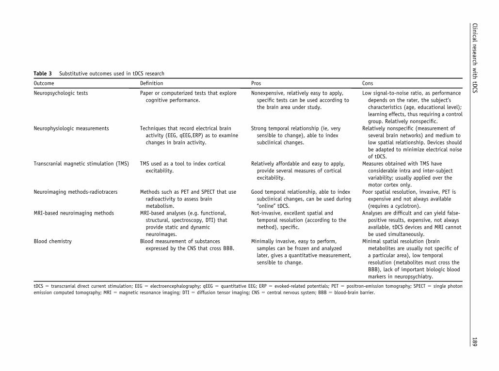
Table 3 Substitutive outcomes used in tDCS research
Outcome Definition Pros Cons
Neuropsychologic tests Paper or computerized tests that explorecognitive performance.
Nonexpensive, relatively easy to apply,specific tests can be used according tothe brain area under study.
Low signal-to-noise ratio, as performancedepends on the rater, the subject’scharacteristics (age, educational level);learning effects, thus requiring a controlgroup. Relatively nonspecific.
Neurophysiologic measurements Techniques that record electrical brainactivity (EEG, qEEG,ERP) as to examinechanges in brain activity.
Strong temporal relationship (ie, verysensible to change), able to indexsubclinical changes.
Relatively nonspecific (measurement ofseveral brain networks) and medium tolow spatial relationship. Devices shouldbe adapted to minimize electrical noiseof tDCS.
Transcranial magnetic stimulation (TMS) TMS used as a tool to index corticalexcitability.
Relatively affordable and easy to apply,provide several measures of corticalexcitability.
Measures obtained with TMS haveconsiderable intra and inter-subjectvariability; usually applied over themotor cortex only.
Neuroimaging methods-radiotracers Methods such as PET and SPECT that useradioactivity to assess brainmetabolism.
Good temporal relationship, able to indexsubclinical changes, can be used during‘‘online’’ tDCS.
Poor spatial resolution, invasive, PET isexpensive and not always available(requires a cyclotron).
MRI-based neuroimaging methods MRI-based analyses (e.g. functional,structural, spectroscopy, DTI) thatprovide static and dynamicneuroimages.
Not-invasive, excellent spatial andtemporal resolution (according to themethod), specific.
Analyses are difficult and can yield false-positive results, expensive, not alwaysavailable, tDCS devices and MRI cannotbe used simultaneously.
Blood chemistry Blood measurement of substancesexpressed by the CNS that cross BBB.
Minimally invasive, easy to perform,samples can be frozen and analyzedlater, gives a quantitative measurement,sensible to change.
Minimal spatial resolution (brainmetabolites are usually not specific ofa particular area), low temporalresolution (metabolites must cross theBBB), lack of important biologic bloodmarkers in neuropsychiatry.
tDCS 5 transcranial direct current stimulation; EEG 5 electroencephalography; qEEG 5 quantitative EEG; ERP 5 evoked-related potentials; PET 5 positron-emission tomography; SPECT 5 single photon
emission computed tomography; MRI 5 magnetic resonance imaging; DTI 5 diffusion tensor imaging; CNS 5 central nervous system; BBB 5 blood-brain barrier.
Clinical
researchwith
tDCS
189
190 Brunoni et al
fMRI lack temporal resolution as it does not measureelectrical activity changes directly (it does indirectly viachanges in cerebral flow). Diffusion tensor imaging (DTI)focuses on the white matter fibers, revealing the neuralconnectivity between brain areas. Finally, voxel-basedmorphometry (VBM) is a computational analysis ofmorphologic images that makes inferences about brainactivity based on the differences of brain tissue concentra-tion among areas. For tDCS, these methods present theadvantage of high spatial resolution; allowing to assesssubtle changes in the stimulated area. For instance, onestudy used VBM to assess neuroplastic changes after 5 daysof TMS over the superior temporal cortex; showingmacroscopic gray matter changes in the region.133 Eventhough, the reliability of some methods of MRI arecurrently under dispute.134 Moreover, tDCS is not usedconcomitantly with MRI yet due to serious risks of over-heating and thus an ‘‘online’’ visualization of the stimulatedarea is not possible although this technical difficulty mightbe resolved in the near future.
Finally, there is a wide range of blood measurementsused in neuropsychiatry research for surrogate outcomes.135
One biomarker under intensive investigation is the brain-derived neurotrophic factor (BDNF). This marker plays animportant role in synaptogenesis and neuroplasticity and
Box 4 Challenges for outcome measures in tDCS clinical research
As neither the full spectrum of clinical efficacy nor the mechanmeasures for tDCS trials ideally will inform both about tDCS clinto clinical data, the common accepted behavioral outcomesfunction. This is particularly relevant for tDCS as it has a modeeffect, particularly for single exposures. Thus in the present eafrom clinical trial designs that incorporate secondary outcomeAmong these are changes in normal function that may be affeffects on chronic pain might add a battery of motor tasks towith treatment. Similarly, an investigator applying tDCS for tmood.
Further, prospects for improving tDCS clinical efficacy improvedate, the common feature in tDCS trials appears to be its caexcitability. Given these data, outcome measures aimed to capmay also be useful additions to ongoing trials. That is, one coand in parallel ask whether an LTP-type or LTD-type change iperhaps in future trials, the tDCS effect may be augmentedbehavioral tasks that facilitate synaptic plasticity. As an exampover an epileptic seizure focus, whether LTD-type suppressionwithin hours of tDCS. However to find out whether seizuressubjects can be stratified into groups that have or have notevaluated separately for subjects that did and did not expersubjects in an epilepsy trial would potentially reduce confoundeffective at the time it was administered. In addition, investigof choosing outcome scales that are not sufficiently sensitive ttDCS strongly changes a component of a larger clinical scale, fuwere found initially.
is thus believed to be linkedwith some neuropsychiatric disor-ders, for instance, BDNF serum levels are low in depressedpatients and increase after antidepressant treatment.136 Arecent study showed BDNF expression also increasesafter tDCS.8 Additional biomarkers used in neuropsychiatryinclude inflammatory proteins such as interleucin-1, interleu-cin-6, and TNF-alpha137; hypothalamic-pituitary-adrenalactivity, which is measured by serum and salivary cortisol138;and oxidative stress proteins such as nitric oxide and otherneuroinflammatory protein markers.139,140 These biomarkerspresent two important drawbacks: first, because of theblood-brain barrier, serum levels might not reflect ‘‘real-time’’ brain activity (or even brain activity at all); second,serum levels can only express the net brain activity, and donot represent a specific area. Therefore, perhaps the mosteffective use in tDCS research is to index disease improvementin phase II/III studies (Also see Box 4 for a discussion onsurrogate outcomes).
TDCS in children
As the brain is under intensive development during child-hood and adolescencedparticularly the prefrontalcortex,141 intensive research is currently being made toexplore how cognition, emotion, behavior, and other
ism of action of tDCS are completely described, outcomeical potency and about the biology of tDCS. With respectmight be insensitive to subtle changes in neurologicst (perhaps subclinical) neuromodulatory and behavioralrly stages of investigation, the field of study may benefits in addition to measures of the patient’s chief symptom.ected by tDCS. For example, an investigator testing tDCSsee whether there is any subtle loss of normal functionreatment of epilepsy may add a questionnaire to assess
if the tDCS mechanism of action is better understood. Topacity to produce a lasting change in regional corticalture the extent to which tDCS induces synaptic plasticityuld ask whether tDCS improved the symptom in question,n regional cortical excitability has occurred. If so, thenby the addition of appropriate pharmacologic agents orle, in future trials in which cathodal tDCS may be appliedhas occurred over the stimulated area can be determinedare reduced in frequency may take days to weeks. Thusundergone regional LTD, and clinical outcomes can beience regional depotentiation. This subclassification ofing results from subjects where tDCS was not biologicallyators would be wise to bear in mind the potential pitfallo capture a relatively modest clinical tDCS effect. Thus, ifrther research can be stimulated, even if negative results

Clinical research with tDCS 191
functions evolve. Having neuromodulatory properties,tDCS would be an interesting tool to explore which brainareas are particularly important in each stage of develop-ment both in healthy and pathologic conditions, such asepilepsy, cerebral palsy, autism, and mental deficiency.However, because of its potential to induce neuroplasticchanges, tDCS should be used carefully especially duringimportant phases of brain development associated withintensive plasticity and also other processes such assynaptic pruning.
A further step would be using tDCS for treatingneuropsychiatric disorders in children, but this has notbeen tested yet. In a review of TMS studies in children, noadverse effects were reported, but its use is still limited forsome reasons, including lack of established safety guide-lines.142 Notwithstanding, tDCS is a promising tool forchildren neurology and psychiatry.
The ethics of tDCS
TDCS is a putative candidate for adjuvant therapy fora range of neuropsychiatric conditions. tDCS is a valuabletool in neuroscience research, as its focality can be usedto explore several brain aspects. Studies regarding tDCSethics reveals its ability to induce changes in behaviorsuch as in moral judgment,143 deception,144,145 and decision-making.146 For instance, one recent study showed tDCSaffected utilitarian behavior. Similarly to other studies intDCS, the polarity-dependent effects resulted in a selfishversus selfless behavior in women.143 Although the effectswere short-lasting (volunteers were not exposed to dailystimulation), the targeted area is similar than used in studiesexploring the long-lasting tDCS effects. Therefore, theethical concern is whether tDCS could induce maladaptivebehavior changes, and if so, to what intensity and extent oftime.
Diverse tDCS studies on healthy subjects have shownpositive changes in attention and memory.84,85,147 From thescope of neuroethics, the issue is whether tDCS enhancescognition in healthy subjects. Can tDCS be used to boostperformance in specific situations (eg, before school tests)?Another issue is that the cognitive effects described(increased attention and memory) from tDCS are in someaspects similar to amphetamines. Despite therapeutic appli-cations, amphetamines are sold illegally as a recreationaland performance enhancer drug (with the suggestivename of ‘‘speed’’). As a tDCS device is easily built andinexpensive (contrary to TMS), it could also be used fornonresearch and nontherapeutic objectives by lay people.In fact, there are online videos in popular web sites suchas Youtube explaining how to build and use a tDCSdevice.148 Although it should be underscored that all theenhancement effects were present for a short period, it ispossible that prolonged daily stimulation could increasethe time span of such effects, thus inducing maladaptive
changes. In contrast, other legal substances such as caffeineare also frequently used as cognitive boosters.
In fact, because applications in these fields are currentlyin the research stage, fixed protocols and safety guidelinesare yet to be defined. Research and development of any newdevices provides an opportunity for brain science andclinical care to advance, and also challenges the medicaland wider communities to address potential dangers andcomplications, ethical and moral quandaries, and issues ofhealthcare economics and distributive justice. For innova-tive neurotechnologies, these are major potential pitfalls tolook out for. Intervening in the brain is always fraught withthe potential for serious consequences. Despite theseconcerns, only by conducting carefully planned clinicaland experimental studies can we provide the impetus toadvance care for people with brain, emotional or psycho-logic, or neuropsychiatric disorders.
Conclusion
The current paper addresses the main aspects of the clinicalresearch of tDCS. This technique has a wide range ofpotential applications and can be used to explore the basicaspects of neurosciences as well as for the treatment ofneuropsychiatric disorders. TDCS has unique characteris-tics such as ability to induce antagonistic effects in corticalexcitability according to the parameters of stimulation;concomitant (‘‘online’’) use with neuropsychologic andpsychophysiologic tests; noninvasiveness and thus absenceof pharmacokinetics interactions, being a putative substi-tutive/augmentative agent in neuropsychiatry; and low-costand portability, making it suitable for increasing access tonovel therapies. However, such characteristics also bringchallenges regarding clinical design, neuroethics and legalissues. In this paper, we aimed to provide an overview oftDCS in clinical research; thereby providing knowledge forconducting proper clinical trials using this promisingapproach.
Acknowledgments
We are thankful to Erin Connors for copyediting thismanuscript. We are also grateful to Scala Institute andMackenzie University (Sao Paulo, Brazil) for the additionalsupport to organize this working group meeting in the IIInternational Symposium in Neuromodulation. Thisworking group meeting was the 2nd International tDCSclub workshop, which took place in Sao Paulo, Brazil, inMarch 2010, at the Social and Cognitive NeuroscienceLaboratory of Mackenzie Presbiterian University. The firstmeeting was held in Milan, Italy in 2008. D.E. wassupported by NIH grant R21HD060999 and F.F. wassupported by NIH grant (5R21DK081773-03).
192 Brunoni et al
References
1. Largus S. De compositionibus medicamentorum. Paris1529.
2. Kellaway P. The part played by the electric fish in the early history of
bioelectricity and electrotherapy. Bull Hist Med 1946;20:112-137.
3. Zago S, Ferrucci R, Fregni F, Priori A. Bartholow, Sciamanna,
Alberti: pioneers in the electrical stimulation of the exposed human
cerebral cortex. Neuroscientist 2008;14(5):521-528.
4. Aldini G. Essai theorique et experimental sur le galvanisme.
Paris1804.
5. Arndt R. Die electricitat in der psychiatrie. Arch Psychiat Nervenk-
rankh 1869;2:259-261.
6. Priori A, Berardelli A, Rona S, Accornero N, Manfredi M. Polariza-
tion of the human motor cortex through the scalp. Neuroreport 1998;
9(10):2257-2260.
7. Nitsche MA, Paulus W. Excitability changes induced in the human
motor cortex by weak transcranial direct current stimulation.
J Physiol 2000;527(Pt 3):633-639.
8. Fritsch B, Reis J, Martinowich K, et al. Direct current stimulation
promotes BDNF-dependent synaptic plasticity: potential implications
for motor learning. Neuron 2010;66(2):198-204.
9. Nitsche MA, Liebetanz D, Antal A, Lang N, Tergau F, Paulus W.
Modulation of cortical excitability by weak direct current stimulation–
technical, safety and functional aspects. Suppl Clin Neurophysiol
2003;56:255-276.
10. Nitsche MA, Seeber A, Frommann K, et al. Modulating parameters
of excitability during and after transcranial direct current stimulation
of the human motor cortex. J Physiol 2005;568(Pt 1):291-303.
11. Kuo MF, Paulus W, Nitsche MA. Boosting focally-induced brain
plasticity by dopamine. Cereb Cortex 2008;18(3):648-651.
12. Wagner T, Fregni F, Fecteau S, Grodzinsky A, Zahn M, Pascual-
Leone A. Transcranial direct current stimulation: a computer-based
human model study. Neuroimage 2007;35(3):1113-1124.
13. Miranda PC, Lomarev M, Hallett M. Modeling the current distribu-
tion during transcranial direct current stimulation. Clin Neurophysiol
2006;117(7):1623-1629.
14. BiksonM,DattaA,ElwassifM.Establishing safety limits for transcranial
direct current stimulation. Clin Neurophysiol 2009;120(6):1033-1034.
15. Datta A, Bikson M, Fregni F. Transcranial direct current stimulation
in patients with skull defects and skull plates: high-resolution compu-
tational FEM study of factors altering cortical current flow. Neuro-
image 2010;52(4):1268-1278.
16. Fregni F, Boggio PS, Nitsche M, et al. Anodal transcranial direct
current stimulation of prefrontal cortex enhances working memory.
Exp Brain Res 2005;166(1):23-30.
17. Boggio PS, Alonso-Alonso M, Mansur CG, et al. Hand function
improvement with low-frequency repetitive transcranial magnetic
stimulation of the unaffected hemisphere in a severe case of stroke.
Am J Phys Med Rehabil 2006;85(11):927-930.
18. Iyer MB, Mattu U, Grafman J, Lomarev M, Sato S, Wassermann EM.
Safety and cognitive effect of frontal DC brain polarization in healthy
individuals. Neurology 2005;64(5):872-875.
19. Fecteau S, Pascual-Leone A, Zald DH, et al. Activation of prefrontal
cortex by transcranial direct current stimulation reduces appetite for
risk during ambiguous decision making. J Neurosci 2007;27(23):
6212-6218.
20. George MS, Aston-Jones G. Noninvasive techniques for probing neu-
rocircuitry and treating illness: vagus nerve stimulation (VNS), trans-
cranial magnetic stimulation (TMS) and transcranial direct current
stimulation (tDCS). Neuropsychopharmacology 2010;35(1):301-316.
21. Fregni F, Pascual-Leone A. Technology insight: noninvasive brain
stimulation in neurology-perspectives on the therapeutic potential
of rTMS and tDCS. Nat Clin Pract Neurol 2007 Jul;3(7):383-393.
22. Nitsche MA, Boggio PS, Fregni F, Pascual-Leone A. Treatment of
depression with transcranial direct current stimulation (tDCS):
a review. Exp Neurol 2009;219(1):14-19.
23. Brunoni AR, Tadini L, Fregni F. Changes in clinical trials method-
ology over time: a systematic review of six decades of research in
psychopharmacology. PLoS One 2010;5(3):e9479.
24. Zhang X, Liu K, Sun J, Zheng Z. Safety and feasibility of repetitive
transcranial magnetic stimulation (rTMS) as a treatment for major
depression during pregnancy. Arch Womens Ment Health 2010;
13(4):369-370.
25. Bolognini N, Pascual-Leone A, Fregni F. Using non-invasive brain
stimulation to augment motor training-induced plasticity.
J Neuroeng Rehabil 2009;6:8.
26. Zaghi S, Acar M, Hultgren B, Boggio PS, Fregni F. Noninvasive
brain stimulation with low-intensity electrical currents: putative
mechanisms of action for direct and alternating current stimulation.
Neuroscientist 2010;16(3):285-307.
27. Nitsche MA, Cohen LG, Wassermann EM, et al. Transcranial direct
current stimulation: state of the art 2008. Brain Stimul 2008;1(3):
206-223.
28. Priori A, Hallett M, Rothwell JC. Repetitive transcranial magnetic
stimulation or transcranial direct current stimulation? Brain Stimul
2009;2(4):241-245.
29. Bindman LJ, Lippold OC, Redfearn JW. Relation between the size
and form of potentials evoked by sensory stimulation and the back-
ground electrical activity in the cerebral cortex of the rat. J Physiol
1964;171:1-25.
30. Purpura DP, McMurtry JG. Intracellular activities and evoked poten-
tial changes during polarization of motor cortex. J Neurophysiol
1965;28:166-185.
31. Creutzfeldt OD, Fromm GH, Kapp H. Influence of transcortical d-c
currents on cortical neuronal activity. Exp Neurol 1962;5:436-452.
32. Bindman LJ, Lippold OC, Redfearn JW. The action of brief polar-
izing currents on the cerebral cortex of the rat (1) during current
flow and (2) in the production of long-lasting after-effects.
J Physiol 1964;172:369-382.
33. Jefferys JG. Influence of electric fields on the excitability of
granule cells in guinea-pig hippocampal slices. J Physiol 1981;
319:143-152.
34. Bikson M, Inoue M, Akiyama H, et al. Effects of uniform extracel-
lular DC electric fields on excitability in rat hippocampal slices in
vitro. J Physiol 2004;557(Pt 1):175-190.
35. Nitsche MA, Paulus W. Sustained excitability elevations induced by
transcranial DC motor cortex stimulation in humans. Neurology
2001;57(10):1899-1901.
36. Liebetanz D, Nitsche MA, Tergau F, Paulus W. Pharmacological
approach to the mechanisms of transcranial DC-stimulation-
induced after-effects of human motor cortex excitability. Brain
2002;125(Pt 10):2238-2247.
37. Nitsche MA, Fricke K, Henschke U, et al. Pharmacological modula-
tion of cortical excitability shifts induced by transcranial direct
current stimulation in humans. J Physiol 2003;553(Pt 1):293-301.
38. Stagg CJ, Best JG, Stephenson MC, et al. Polarity-sensitive modula-
tion of cortical neurotransmitters by transcranial stimulation.
J Neurosci 2009;29(16):5202-5206.
39. Ardolino G, Bossi B, Barbieri S, Priori A. Non-synaptic mecha-
nisms underlie the after-effects of cathodal transcutaneous direct
current stimulation of the human brain. J Physiol 2005;
568(Pt 2):653-663.
40. Cogiamanian F, Vergari M, Pulecchi F, Marceglia S, Priori A. Effect
of spinal transcutaneous direct current stimulation on somatosensory
evoked potentials in humans. Clin Neurophysiol 2008;119(11):
2636-2640.
41. Rango M, Cogiamanian F, Marceglia S, et al. Myoinositol content in
the human brain is modified by transcranial direct current stimulation
in a matter of minutes: a 1H-MRS study. Magn Reson Med 2008;
60(4):782-789.
42. Boros K, Poreisz C, Munchau A, Paulus W, Nitsche MA. Premotor
transcranial direct current stimulation (tDCS) affects primary motor
excitability in humans. Eur J Neurosci 2008;27(5):1292-1300.

Clinical research with tDCS 193
43. Lang N, Siebner HR, Ward NS, et al. How does transcranial DC stim-
ulation of the primary motor cortex alter regional neuronal activity in
the human brain? Eur J Neurosci 2005;22(2):495-504.
44. Parra LC, Bikson M. Model of the effect of extracellular fields on
spike time coherence. Conf Proc IEEE Eng Med Biol Soc 2004;6:
4584-4587.
45. Deans JK, Powell AD, Jefferys JG. Sensitivity of coherent oscilla-
tions in rat hippocampus to AC electric fields. J Physiol 2007;
583(Pt 2):555-565.
46. Frohlich F, McCormick DA. Endogenous electric fields may guide
neocortical network activity. Neuron 2010;67(1):129-143.
47. Accornero N, Li Voti P, La Riccia M, Gregori B. Visual evoked
potentials modulation during direct current cortical polarization.
Exp Brain Res 2007;178(2):261-266.
48. Matsunaga K, Nitsche MA, Tsuji S, Rothwell JC. Effect of transcra-
nial DC sensorimotor cortex stimulation on somatosensory evoked
potentials in humans. Clin Neurophysiol 2004;115(2):456-460.
49. Antal A, Kincses TZ, Nitsche MA, Bartfai O, Paulus W. Excitability
changes induced in the human primary visual cortex by transcranial
direct current stimulation: direct electrophysiological evidence.
Invest Ophthalmol Vis Sci 2004;45(2):702-707.
50. Ferrucci R, Marceglia S, Vergari M, et al. Cerebellar transcranial
direct current stimulation impairs the practice-dependent proficiency
increase in working memory. J Cogn Neurosci 2008;20(9):
1687-1697.
51. Galea JM, Jayaram G, Ajagbe L, Celnik P. Modulation of cerebellar
excitability by polarity-specific noninvasive direct current stimula-
tion. J Neurosci 2009;29(28):9115-9122.
52. Winkler T, Hering P, Straube A. Spinal DC stimulation in humans
modulates post-activation depression of the H-reflex depending on
current polarity. Clin Neurophysiol 2010;121(6):957-961.
53. Merzagora AC, Foffani G, Panyavin I, et al. Prefrontal hemodynamic
changes produced by anodal direct current stimulation. Neuroimage
2010;49(3):2304-2310.
54. Nitsche MA, Jaussi W, Liebetanz D, Lang N, Tergau F, Paulus W.
Consolidation of human motor cortical neuroplasticity by
D-cycloserine. Neuropsychopharmacology 2004;29(8):1573-1578.
55. Nitsche MA, Liebetanz D, Schlitterlau A, et al. GABAergic modula-
tion of DC stimulation-induced motor cortex excitability shifts in hu-
mans. Eur J Neurosci 2004;19(10):2720-2726.
56. Nitsche MA, Grundey J, Liebetanz D, Lang N, Tergau F, Paulus W.
Catecholaminergic consolidation of motor cortical neuroplasticity in
humans. Cereb Cortex 2004;14(11):1240-1245.
57. Kuo MF, Unger M, Liebetanz D, et al. Limited impact of homeostatic
plasticity on motor learning in humans. Neuropsychologia 2008;
46(8):2122-2128.
58. Nitsche MA, Doemkes S, Karakose T, et al. Shaping the effects of
transcranial direct current stimulation of the human motor cortex.
J Neurophysiol 2007;97(4):3109-3117.
59. Monte-Silva K, Kuo MF, Thirugnanasambandam N, Liebetanz D,
Paulus W, Nitsche MA. Dose-dependent inverted U-shaped effect
of dopamine (D2-like) receptor activation on focal and nonfocal plas-
ticity in humans. J Neurosci 2009;29(19):6124-6131.
60. Nitsche MA, Kuo MF, Karrasch R, Wachter B, Liebetanz D,
Paulus W. Serotonin affects transcranial direct current-induced neu-
roplasticity in humans. Biol Psychiatry 2009;66(5):503-508.
61. Antal A, Kincses TZ, Nitsche MA, Paulus W. Manipulation of phos-
phene thresholds by transcranial direct current stimulation in man.
Exp Brain Res 2003;150(3):375-378.
62. Boggio PS, Rigonatti SP, Ribeiro RB, et al. A randomized, double-
blind clinical trial on the efficacy of cortical direct current stimulation
for the treatment of major depression. Int J Neuropsychopharmacol
2008;11(2):249-254.
63. Utz KS, Dimova V, Oppenlander K, Kerkhoff G. Electrified minds:
transcranial direct current stimulation (tDCS) and galvanic vestibular
stimulation (GVS) as methods of non-invasive brain stimulation in
neuropsychology: a review of current data and future implications.
Neuropsychologia 2010;48(10):2789-2810.
64. Williams JA, Pascual-Leone A, Fregni F. Interhemispheric modula-
tion induced by cortical stimulation and motor training. Phys Ther
2010;90(3):398-410.
65. Mahmoudi H, Haghighi AB, Petramfar P, Jahanshahi S, Salehi Z,
Fregni F. Transcranial direct current stimulation: electrode montage
in stroke. Disabil Rehabil 2010 November 26 (Epub ahead of print).
66. Redfearn JW, Lippold OC, Costain R. A preliminary account of the
clinical effects of polarizing the brain in certain psychiatric disorders.
Br J Psychiatry 1964;110:773-785.
67. Cogiamanian F, Vergari M, Ardolino G, et al. Effects of transcuta-
neous spinal cord direct current stimulation (tsDCS) on the lower
limb nociceptive flexion reflex in humans. Proceedings of the XLI
Congress of the Italian Neurological Society; 2010: Neurological
Sciences.
68. Datta A, Rahman A, Scaturro J, Bikson M. Electrode montages for
tDCS and weak transcranial electrical stimulation role of "return"
electrode’s position and size. Clin Neurophysiol 2010;121:
1976-1978.
69. Moliadze V, Antal A, Paulus W. Electrode-distance dependent after-
effects of transcranial direct and random noise stimulation with ex-
tracephalic reference electrodes. Clin Neurophysiol 2010;121(12):
2165-2171.
70. Datta A, Elwassif M, Battaglia F, Bikson M. Transcranial current
stimulation focality using disc and ring electrode configurations:
FEM analysis. J Neural Eng 2008;5(2):163-174.
71. Datta A, Bansal V, Diaz J, Patel J, Reato D, Bikson M. Gyri-precise
head model of transcranial direct current stimulation: Improved
spatial focality using a ring electrode versus conventional rectangular
pad. Brain Stimul 2009;2(4):201-207.
72. Sadleir RJ, Vannorsdall TD, Schretlen DJ, Gordon B. Transcranial
direct current stimulation (tDCS) in a realistic head model. Neuro-
image 2010;51(4):1310-1318.
73. Minhas P, Bansal V, Patel J, et al. Electrodes for high-definition trans-
cutaneous DC stimulation for applications in drug delivery and elec-
trotherapy, including tDCS. J Neurosci Methods 2010;190(2):
188-197.
74. Faria P, Leal A, Miranda PC. Comparing different electrode config-
urations using the 10-10 international system in tDCS: a finite
element model analysis. Conf Proc IEEE Eng Med Biol Soc 2009;
1596-1599.
75. Suh HS, Kim SH, Lee WH, Kim TS. Realistic simulation of transcra-
nial direct current stimulation via 3-d high-resolution finite element
analysis: effect of tissue anisotropy. Conf Proc IEEE Eng Med
Biol Soc 2009;638-641.
76. Elmer S, Burkard M, Renz B, Meyer M, Jancke L. Direct current
induced short-term modulation of the left dorsolateral prefrontal
cortex while learning auditory presented nouns. Behav Brain Funct
2009;5(1):29.
77. Marshall L, Molle M, Siebner HR, Born J. Bifrontal transcranial
direct current stimulation slows reaction time in a working memory
task. BMC Neurosci 2005;6(1):23.
78. Boggio PS, Fregni F, Valasek C, et al. Temporal lobe cortical electri-
cal stimulation during the encoding and retrieval phase reduces false
memories. PLoS One 2009;4(3):e4959.
79. Marshall L, Molle M, Hallschmid M, Born J. Transcranial direct
current stimulation during sleep improves declarative memory.
J Neurosci 2004;24(44):9985-9992.
80. Fecteau S, Knoch D, Fregni F, Sultani N, Boggio P, Pascual-Leone A.
Diminishing risk-taking behavior by modulating activity in the
prefrontal cortex: a direct current stimulation study. J Neurosci
2007;27(46):12500-12505.
81. Hecht D, Walsh V, Lavidor M. Transcranial direct current stimulation
facilitates decision making in a probabilistic guessing task. J Neurosci
2010;30(12):4241-4245.
194 Brunoni et al
82. Kincses TZ, Antal A, Nitsche MA, Bartfai O, Paulus W. Facilitation
of probabilistic classification learning by transcranial direct current
stimulation of the prefrontal cortex in the human. Neuropsychologia
2004;42(1):113-117.
83. Stone DB, Tesche CD. Transcranial direct current stimulation modu-
lates shifts in global/local attention. Neuroreport 2009;20(12):
1115-1119.
84. Sparing R, Thimm M, Hesse MD, Kust J, Karbe H, Fink GR. Bidi-
rectional alterations of interhemispheric parietal balance by non-
invasive cortical stimulation. Brain 2009;132(Pt 11):3011-3020.
85. Bolognini N, Fregni F, Casati C, Olgiati E, Vallar G. Brain polariza-
tion of parietal cortex augments training-induced improvement of
visual exploratory and attentional skills. Brain Res 2010;1349:76-89.
86. Fertonani A, Rosini S, Cotelli M, Rossini PM, Miniussi C. Naming
facilitation induced by transcranial direct current stimulation. Behav
Brain Res 2010;208(2):311-318.
87. Sparing R, Dafotakis M, Meister IG, Thirugnanasambandam N,
Fink GR. Enhancing language performance with non-invasive brain
stimulationda transcranial direct current stimulation study in
healthy humans. Neuropsychologia 2008;46(1):261-268.
88. Liebetanz D, Koch R, Mayenfels S, Konig F, Paulus W, Nitsche MA.
Safety limits of cathodal transcranial direct current stimulation in
rats. Clin Neurophysiol 2009;120(6):1161-1167.
89. Fregni F, Liebetanz D, Monte-Silva KK, et al. Effects of transcranial
direct current stimulation coupled with repetitive electrical stimula-
tion on cortical spreading depression. Exp Neurol 2007;204(1):
462-466.
90. Rigonatti SP, Boggio PS, Myczkowski ML, et al. Transcranial direct
stimulation and fluoxetine for the treatment of depression. Eur
Psychiatry 2008;23(1):74-76.
91. Ferrucci R, Bortolomasi M, Vergari M, et al. Transcranial direct
current stimulation in severe, drug-resistant major depression.
J Affect Disord 2009;118(1-3):215-219.
92. Palm U, Keeser D, Schiller C, et al. Skin lesions after treatment
with transcranial direct current stimulation (tDCS). Brain Stimul
2008;1(4):386-387.
93. Durand S, Fromy B, Bouye P, Saumet JL, Abraham P. Vasodilatation
in response to repeated anodal current application in the human skin
relies on aspirin-sensitive mechanisms. J Physiol 2002;540(Pt 1):
261-269.
94. Poreisz C, Boros K, Antal A, Paulus W. Safety aspects of transcranial
direct current stimulation concerning healthy subjects and patients.
Brain Res Bull 2007;72(4-6):208-214.
95. Tadini L, El-Nazer R, Brunoni AR, et al. Cognitive, mood and EEG
effects of noninvasive cortical stimulation with weak electrical
currents. J ECT 2010 October 5. (Epub ahead of print).
96. Nitsche MA, Liebetanz D, Lang N, Antal A, Tergau F, Paulus W.
Safety criteria for transcranial direct current stimulation (tDCS) in
humans. Clin Neurophysiol 2003;114(11):2220-2222. author reply
2-3.
97. Fregni F, Boggio PS, Nitsche MA, Rigonatti SP, Pascual-Leone A.
Cognitive effects of repeated sessions of transcranial direct current
stimulation in patients with depression. Depress Anxiety 2006;
23(8):482-484.
98. Boggio PS, Nunes A, Rigonatti SP, Nitsche MA, Pascual-Leone A,
Fregni F. Repeated sessions of noninvasive brain DC stimulation is
associated with motor function improvement in stroke patients.
Restor Neurol Neurosci 2007;25(2):123-129.
99. Loo C, Martin D, Pigot M, Arul-Anandam P, Mitchell P, Sachdev P.
Transcranial direct current stimulation priming of therapeutic repet-
itive transcranial magnetic stimulation: a pilot study. J ECT 2009;
25(4):256-260.
100. Fregni F, Thome-Souza S, Nitsche MA, Freedman SD, Valente KD,
Pascual-Leone A. A controlled clinical trial of cathodal DC polariza-
tion in patients with refractory epilepsy. Epilepsia 2006;47(2):
335-342.
101. Arul-Anandam AP, Loo C, Mitchell P. Induction of hypomanic
episode with transcranial direct current stimulation. J ECT 2010;
26(1):68-69.
102. Baccaro A, Brunoni AR, Bensenor IM, Fregni F. Hypomanic episode
in unipolar depression during transcranial direct current stimulation.
Acta Neuropsychiatrica 2010;22(6):316-318.
103. Brunoni AR, Valiengo L, Zanao T, Oliveira J, Bensenor IM, Fregni F.
Manic psychosis following transcranial direct current stimulation and
sertraline. J Neuropsychiatry Clin Neurosci (in press).
104. Miranda PC, Faria P, Hallett M. What does the ratio of injected
current to electrode area tell us about current density in the brain
during tDCS? Clin Neurophysiol 2009;120(6):1183-1187.
105. Fregni F, Boggio PS, Valle AC, et al. Homeostatic effects of plasma
valproate levels on corticospinal excitability changes induced by 1Hz
rTMS in patients with juvenile myoclonic epilepsy. Clin Neurophy-
siol 2006;117(6):1217-1227.
106. Ziemann U. Pharmacology of TMS. Suppl Clin Neurophysiol 2003;
56:226-231.
107. Bajwa S, Bermpohl F, Rigonatti SP, Pascual-Leone A, Boggio PS,
Fregni F. Impaired interhemispheric interactions in patients with
major depression. J Nerv Ment Dis 2008;196(9):671-677.
108. Soubasi E, Chroni E, Gourzis P, Zisis A, Beratis S,
Papathanasopoulos P. Cortical motor neurophysiology of patients
with schizophrenia: a study using transcranial magnetic stimulation.
Psychiatry Res 2010;176(2-3):132-136.
109. Mhalla A, de Andrade DC, Baudic S, Perrot S, Bouhassira D. Alter-
ation of cortical excitability in patients with fibromyalgia. Pain 2010;
149(3):495-500.
110. Brighina F, Palermo A, Fierro B. Cortical inhibition and habituation
to evoked potentials: relevance for pathophysiology of migraine.
J Headache Pain 2009;10(2):77-84.
111. Chaieb L, Antal A, Paulus W. Gender-specific modulation of short-
term neuroplasticity in the visual cortex induced by transcranial
direct current stimulation. Vis Neurosci 2008;25(1):77-81.
112. Ferri R, Del Gracco S, Elia M, Musumeci SA. Age-related changes
of cortical excitability in subjects with sleep-enhanced centrotempo-
ral spikes: a somatosensory evoked potential study. Clin Neurophy-
siol 2000;111(4):591-599.
113. Lang N, Hasan A, Sueske E, Paulus W, Nitsche MA. Cortical hypo-
excitability in chronic smokers? A transcranial magnetic stimulation
study. Neuropsychopharmacol 2008;33(10):2517-2523.
114. Monte-Silva K, Liebetanz D, Grundey J, Paulus W, Nitsche MA.
Dosage-dependent non-linear effect of L-dopa on human motor
cortex plasticity. J Physiol 2010;588(Pt 18):3415-3424.
115. Ohn SH, Park CI, Yoo WK, et al. Time-dependent effect of transcra-
nial direct current stimulation on the enhancement of working
memory. Neuroreport 2008;19(1):43-47.
116. Boggio PS, Ferrucci R, Rigonatti SP, et al. Effects of transcranial
direct current stimulation on working memory in patients with
Parkinson’s disease. J Neurol Sci 2006;249(1):31-38.
117. Froc DJ, Chapman CA, Trepel C, Racine RJ. Long-term depression
and depotentiation in the sensorimotor cortex of the freely moving
rat. J Neurosci 2000;20(1):438-445.
118. Monte-Silva KK, Kuo MF, Liebetanz D, Paulus W, Nitsche MA.
Shaping the optimal repetition interval for cathodal transcranial
direct current stimulation (tDCS). J Neurophysiol 2010;103(4):
1735-1740.
119. Siebner HR, Lang N, Rizzo V, et al. Preconditioning of low-
frequency repetitive transcranial magnetic stimulation with transcra-
nial direct current stimulation: evidence for homeostatic plasticity in
the human motor cortex. J Neurosci 2004;24(13):3379-3385.
120. Brunoni AR, Fregni F. Clinical trial design in noninvasive brain stim-
ulation psychiatric research. Int J Methods Psychiatry Res (in press).
121. Gandiga PC, Hummel FC, Cohen LG. Transcranial DC stimulation
(tDCS): a tool for double-blind sham-controlled clinical studies in
brain stimulation. Clin Neurophysiol 2006;117(4):845-850.
Clinical research with tDCS 195
122. Dundas JE, Thickbroom GW, Mastaglia FL. Perception of comfort
during transcranial DC stimulation: effect of NaCl solution concen-
tration applied to sponge electrodes. Clin Neurophysiol 2007;
118(5):1166-1170.
123. Freedman B. Equipoise and the ethics of clinical research. N Engl
J Med 1987;317(3):141-145.
124. Fregni F, Boggio PS, Nitsche MA, Marcolin MA, Rigonatti SP,
Pascual-Leone A. Treatment of major depression with transcranial
direct current stimulation. Bipolar Disord 2006;8(2):203-204.
125. Ferrucci R, Bortolomasi M, Brunoni AR, et al. Comparative benefits
of Transcranial Direct Current Stimulation (tDCS) treatment in
patients with mild/moderate vs. severe depression. Clin Neuropsychi-
atry 2009;6:246-251.
126. Brunoni AR, Ferrucci R, Bortolomasi M, et al. Transcranial direct
current stimulation (tDCS) in unipolar vs. bipolar depressive disorder.
Prog Neuropsychopharmacol Biol Psychiatry 2011;35(1):96-101.
127. Brunoni AR, Valiengo L, Baccaro A, et al. Sertraline vs. electrical
current therapy for treating depression clinical studyddesign, ratio-
nale and objectives. Contemp Clin Trials 2011;32:90-98.
128. Schutter DJ. Nosce te ipsum: on the efficacy of transcranial magnetic
stimulation in major depressive disorder. Biol Psychiatry 2010;67(5):
e27; reply e9.
129. Yu E, Lurie P. Transcranial magnetic stimulation not proven effec-
tive. Biol Psychiatry 2010;67(2):e13; reply e5-7.
130. D’Agostino RB Jr. Debate: the slippery slope of surrogate outcomes.
Curr Control Trials Cardiovasc Med 2000;1(2):76-78.
131. Kobayashi M, Pascual-Leone A. Transcranial magnetic stimulation
in neurology. Lancet Neurology 2003;2(3):145-156.
132. Pascual-Marqui RD, Esslen M, Kochi K, Lehmann D. Functional
imaging with low-resolution brain electromagnetic tomography
(LORETA): a review. Methods Find Exp Clin Pharmacol 2002;
24(Suppl C):91-95.
133. May A, Hajak G, Ganssbauer S, et al. Structural brain alterations
following 5 days of intervention: dynamic aspects of neuroplasticity.
Cereb Cortex 2007;17(1):205-210.
134. Kriegeskorte N, Lindquist MA, Nichols TE, Poldrack RA, Vul E.
Everything you never wanted to know about circular analysis, but
were afraid to ask. J Cereb Blood Flow Metab 2010;30(9):
1551-1557.
135. Gelenberg AJ, Thase ME, Meyer RE, et al. The history and current
state of antidepressant clinical trial design: a call to action for
proof-of-concept studies. J Clin Psychiatry 2008;69(10):1513-1528.
136. Brunoni AR, Lopes M, Fregni F. A systematic review and meta-
analysis of clinical studies on major depression and BDNF levels:
implications for the role of neuroplasticity in depression. Int J Neuro-
psychopharmacol 2008;11(8):1169-1180.
137. Dowlati Y, Herrmann N, Swardfager W, et al. A meta-analysis of
cytokines in major depression. Biol Psychiatry 2010;67(5):446-457.
138. Quinones MP, Kaddurah-Daouk R. Metabolomics tools for identi-
fying biomarkers for neuropsychiatric diseases. Neurobiol Dis
2009;35(2):165-176.
139. Marques AH, Cizza G, Sternberg E. [Brain-immune interactions
and implications in psychiatric disorders]. Rev Bras Psiquiatr 2007;
29(Suppl. 1):S27-S32.
140. Maes M, Yirmyia R, Noraberg J, et al. The inflammatory & neurode-
generative (I&ND) hypothesis of depression: leads for future
research and new drug developments in depression. Metab Brain
Dis 2009;24(1):27-53.
141. Spencer-Smith M, Anderson V. Healthy and abnormal development
of the prefrontal cortex. Dev Neurorehabil 2009;12(5):279-297.
142. Frye RE, Rotenberg A, Ousley M, Pascual-Leone A. Transcranial
magnetic stimulation in child neurology: current and future direc-
tions. J Child Neurol 2008;23(1):79-96.
143. Fumagalli M, Vergari M, Pasqualetti P, et al. Brain switches utili-
tarian behavior: does gender make the difference? PLoS One 2010;
5(1):e8865.
144. Luber B, Fisher C, Appelbaum PS, Ploesser M, Lisanby SH. Non-
invasive brain stimulation in the detection of deception: scientific
challenges and ethical consequences. Behav Sci Law 2009;27(2):
191-208.
145. Mameli F, Mrakic-Sposta S, Vergari M, et al. Dorsolateral prefrontal
cortex specifically processes generaldbut not personaldknowledge
deception: multiple brain networks for lying. Behav Brain Res 2010;
211(2):164-168.
146. Boggio PS, Campanha C, Valasek CA, Fecteau S, Pascual-Leone A,
Fregni F. Modulation of decision-making in a gambling task in older
adults with transcranial direct current stimulation. Eur J Neurosci
2010;31(3):593-597.
147. Reis J, Robertson E, Krakauer JW, et al. Consensus: ‘‘Can tDCS and
TMS enhance motor learning and memory formation?’’. Brain Stimul
2008;1(4):363-369.
148. Anonymous. 2010 [cited 2010 September 25th]; Available at: http://
www.youtube.com/verify_age?next_url5http://www.youtube.com/
watch%3Fv%3DSymQqZYiVyA.