
Clavicula amit final 2013JW-CA##22288
12
ORIGINAL COMMUNICATIONS The Anatomy of the Clavicle: A Three-dimensional Cadaveric Study AMIT BERNAT 1 , TOON HUYSMANS 2 , FRANCIS VAN GLABBEEK 1 , JAN SIJBERS 2 , JAN GIELEN 3 , ALEXANDER VAN TONGEL 4 1 Department of Orthopedic Surgery and Traumatology, University Hospital of Antwerp, Antwerp, Belgium 2 iMinds-Vision Lab, Department of physics, University of Antwerp, Antwerp, Belgium 3 Department of Radiology, University Hospital of Antwerp, Antwerp, Belgium 4 Department of Orthopedic Surgery and Traumatology, University Hospital of Ghent, Ghent, Belgium The clavicle has a complex osteologic structure that makes morphological analy- sis extremely difficult. A three-dimensional study was conducted to examine the anatomical variations and characteristics of the bone. Sixty-eight human cadaver clavicles were dissected, CAT-scanned, and reconstructed. An auto- mated parameterization and correspondence shape analysis system was devel- oped. A new length, designated as centerline (CL) length, was defined and measured. This length represents the true length of the clavicle. The endpoint length was measured as the distance between two endpoints. The width and curvature were measured in the axial (AX) and frontal (FR) plane and defined along the CL. Next gender and side characteristics and variations were exam- ined. The mean CL length was 159.0 6 11.0 mm. The mean endpoint length was 149.4 6 10.3 mm, which was statistically significantly shorter than the CL. The male clavicle was significantly longer (166.8 6 7.3 mm vs. 151.0 6 8.2 mm), wider (14.6 6 1.5 mm vs. 12.7 6 1.3 mm lateral FR plane and 24.7 6 2.8 mm vs. 22.8 6 2.8 mm medial AX plane), and more curved (10.8 6 2.8 mm vs. 8.6 6 2.3 mm medial and 10.5 6 3.3 mm vs. 9.1 6 2.5 mm lateral) than the female one. Left clavicles were significant longer (159.8 6 10.9 mm vs. 158.0 6 11.2 mm) than right clavicles. A novel three-dimensional system was developed, used and tested in order to explore the anatomical variations and characteristics of the human clavicle. This information, together with the automated system, can be applied to future clavicle populations and to the design of fixation plates for clavicle fractures. Clin. Anat. 00:000–000, 2013. V C 2013 Wiley Periodicals, Inc. Key words: clavicle anatomy; three-dimensional analysis; true length; width; curvature INTRODUCTION The clavicle serves as the sole bone structure connecting the axial skeleton to the shoulder girdle through the sternoclavicular joint medially and the acromioclavicular joint laterally. It is a complex struc- ture that plays an important role in the stability, movement, and cosmetic aspect of the shoulder girdle. The close relationship between the clavicle, its shape, the muscles and ligaments forms the basic platform for the full range of motion of the shoulder (Lazarus and Seon, 2006). Clavicular morphology has developed in recent years a great interest among researchers. Archeolo- gists use the clavicle to explain evolutionary proc- esses (Voisin, 2006, 2008), anatomic and forensic pathology use the clavicle for explaining handedness, *Correspondence to: Amit Bernat, M.D., Department of Ortho- pedic Surgery and Traumatology, University Hospital of Antwerp, Wilrijkstraat 10, 2650 Edegem, Belgium. E-mail: [email protected] Received 28 June 2012; Revised 3 June 2013; Accepted 5 June 2013 Published online 00 Month 2013 in Wiley Online Library (wileyonlinelibrary.com). DOI: 10.1002/ca.2228810.1002/ ca.22288 V V C 2013 Wiley Periodicals, Inc. Clinical Anatomy 00:00–00 (2013) J_ID: za9 Customer A_ID: CA22288 Cadmus Art: CA22288 Ed. Ref. No.: 12-0310.R3 Date: 2-September-13 Stage: Page: 1 ID: jwweb3b2server Time: 13:09 I Path: D:/JW/Support/Printer_Autopdf/3D_IN/JW-CA##130065
Transcript of Clavicula amit final 2013JW-CA##22288
ORIGINAL COMMUNICATIONS
The Anatomy of the Clavicle:A Three-dimensional Cadaveric Study
AMIT BERNAT1, TOON HUYSMANS2, FRANCIS VAN GLABBEEK1, JAN SIJBERS2,JAN GIELEN3, ALEXANDER VAN TONGEL4
1Department of Orthopedic Surgery and Traumatology, University Hospital of Antwerp, Antwerp, Belgium2iMinds-Vision Lab, Department of physics, University of Antwerp, Antwerp, Belgium
3Department of Radiology, University Hospital of Antwerp, Antwerp, Belgium4Department of Orthopedic Surgery and Traumatology, University Hospital of Ghent, Ghent, Belgium
The clavicle has a complex osteologic structure that makes morphological analy-sis extremely difficult. A three-dimensional study was conducted to examine theanatomical variations and characteristics of the bone. Sixty-eight humancadaver clavicles were dissected, CAT-scanned, and reconstructed. An auto-mated parameterization and correspondence shape analysis system was devel-oped. A new length, designated as centerline (CL) length, was defined andmeasured. This length represents the true length of the clavicle. The endpointlength was measured as the distance between two endpoints. The width andcurvature were measured in the axial (AX) and frontal (FR) plane and definedalong the CL. Next gender and side characteristics and variations were exam-ined. The mean CL length was 159.0 6 11.0 mm. The mean endpoint lengthwas 149.4 6 10.3 mm, which was statistically significantly shorter than the CL.The male clavicle was significantly longer (166.8 6 7.3 mm vs. 151.0 6 8.2mm), wider (14.6 6 1.5 mm vs. 12.7 6 1.3 mm lateral FR plane and 24.7 6 2.8mm vs. 22.8 6 2.8 mm medial AX plane), and more curved (10.8 6 2.8 mm vs.8.6 6 2.3 mm medial and 10.5 6 3.3 mm vs. 9.1 6 2.5 mm lateral) than thefemale one. Left clavicles were significant longer (159.8 6 10.9 mm vs. 158.0 611.2 mm) than right clavicles. A novel three-dimensional system was developed,used and tested in order to explore the anatomical variations and characteristicsof the human clavicle. This information, together with the automated system,can be applied to future clavicle populations and to the design of fixation platesfor clavicle fractures. Clin. Anat. 00:000–000, 2013. VC 2013 Wiley Periodicals, Inc.
Key words: clavicle anatomy; three-dimensional analysis; true length; width;curvature
INTRODUCTION
The clavicle serves as the sole bone structureconnecting the axial skeleton to the shoulder girdlethrough the sternoclavicular joint medially and theacromioclavicular joint laterally. It is a complex struc-ture that plays an important role in the stability,movement, and cosmetic aspect of the shouldergirdle. The close relationship between the clavicle, itsshape, the muscles and ligaments forms the basicplatform for the full range of motion of the shoulder(Lazarus and Seon, 2006).
Clavicular morphology has developed in recentyears a great interest among researchers. Archeolo-
gists use the clavicle to explain evolutionary proc-esses (Voisin, 2006, 2008), anatomic and forensicpathology use the clavicle for explaining handedness,
*Correspondence to: Amit Bernat, M.D., Department of Ortho-pedic Surgery and Traumatology, University Hospital ofAntwerp, Wilrijkstraat 10, 2650 Edegem, Belgium.E-mail: [email protected]
Received 28 June 2012; Revised 3 June 2013; Accepted 5 June2013
Published online 00 Month 2013 in Wiley Online Library(wileyonlinelibrary.com). DOI: 10.1002/ca.2228810.1002/ca.22288
VVC 2013 Wiley Periodicals, Inc.
Clinical Anatomy 00:00–00 (2013)
J_ID: za9 Customer A_ID: CA22288 Cadmus Art: CA22288 Ed. Ref. No.: 12-0310.R3 Date: 2-September-13 Stage: Page: 1
ID: jwweb3b2server Time: 13:09 I Path: D:/JW/Support/Printer_Autopdf/3D_IN/JW-CA##130065
gender, bone and muscle development (McCormicket al., 1991; Mays et al., 1999; Auerbach and Raxter,2008; Danforth and Thompson, 2008; Ludewig et al.,2009).
Orthopedic surgeons research the clavicle for bettertreatment of fractures and complications (Nowaket al., 2004; Andermahr et al., 2007; Huang et al.,2007; VanBeek et al., 2011; Hillen et al., 2012).
Clavicle fractures account for 2.6%–12% of allfractures and for 44–66% of fractures about theshoulder. Middle-third fractures account for 80% of allclavicular fractures; whereas, fractures of the lateraland medial third of the clavicle account for 15% and5%, respectively (Egol et al., 2010).
Injuries at the diaphysis of the clavicle are morelikely to be displaced as compared with medial- andlateral third fractures. Recent evidence suggests thesespecific subsets of patients may be at high risk fornonunion, shoulder dysfunction, or residual pain afternonsurgical management (Altamimi and McKee,2008; McKee et al., 2006). In these patients, acutesurgical intervention may minimize suboptimaloutcomes (Canadian Orthopaedic Trauma Society,2007; Althausen et al., 2013).
Altamimi and McKee (2008) have advocated theuse of anatomical precontoured plates for thedecrease in soft tissue irritation and increase inpatient satisfaction. VanBeek et al. (2011) have com-pared the outcome after precontoured and noncon-toured superior plating of the clavicle and showed asignificant decrease of hardware prominence and 50%less reoperation rate for hardware removal in the pre-contoured group.
Several studies have been conducted to define itscomplex shape and variability of this double-curvedbone structure. Most of these studies used atwo-dimensional (2D) analysis (Parsons, 1916; Olivier1951–1956; McCormick et al., 1991; Jit and Singh,1996; Mays et al., 1999; Andermahr et al., 2007;Huang et al., 2007; Auerbach and Raxter, 2008; Dan-forth and Thompson, 2008; Voisin, 2008).
Recently, it has been described that 3D morpho-metric analysis can provide more exact, correct, andconsistent results (Fitzpatrick et al., 2007). Daruwallaet al. (2010a,b) were the first to measure the lengthand width of the clavicle using 3D reconstruction.However, their study had several weaknesses. First,only 27 clavicles were evaluated, which is too small anumber taking the high variability of the bone intoaccount. Secondly, the population was not balanced(9 men and 18 women, 15 left and 12 right). Thesecan influence the average length, width, and curva-ture results. Thirdly, a coordinate system was createdbased on axes that were defined on each individualclavicle with a user interface. This process was donemanually on each clavicle. One of the axes was basedon the selection of two end points. As the variationrate at the edges of the clavicle is high, selectingthese points can give a false representativeevaluation.
In the present study, we used a newly developedtechnique for the mathematical evaluation of3D-reconstructed material to evaluate the length(defined as the line which crosses through the center
of the clavicle and as the line between two extremitypoints), width, and curvature (in the frontal and axialplane). All the measurements were repeated for thecomparison between sides and gender.
These anatomical characteristics will provide abetter understanding of the complexity of theclavicle and act as a steppingstone in the creationand development of an anatomical precontouredplate.
MATERIAL AND METHODS
Study Population
In this study, 68 human clavicles (34 pairs) fromhuman Caucasian cadavers were dissected and investi-gated. This set of clavicles represented 17 male and17 female specimens with a mean age of 77 years(range: 43–99 years). The population consisted of 34(17 males, 17 females) right and 34 (17 males,17 females) left clavicles.
Data Acquisition
Preparation of the clavicles was done in theanatomy lab at the University of Antwerp. Afterresection of the clavicles, the soft-tissue envelope wascompletely removed.
All clavicles were scanned with a GE LightSpeedVolume CT (GE Medical Systems, Milwaukee WI) witha spatial resolution of 500 3 500 3 600 lm3 at theUniversity Hospital of Antwerp. The scans wereperformed by placing the clavicles in a standardizedfashion with their flat superior surface of the lateralend facing down to prevent movement of the boneduring the scan.
Surface Generation
The computed tomography (CT) reconstructionsfrom the GE Lightspeed Volume CT system were auto-matically segmented by morphological image-processing operations. Where necessary, manualcorrections were made using Avizo (VisualizationSciences Group, Burlington MA). From the obtainedsegmented images, the outer boundary surface ofeach clavicle was extracted using the marching cubesalgorithm (Lorensen and Cline, 1987).
For each clavicle surface, a point on both the ster-nal and acromial end was selected using custom-made manual annotation software. Then, each surfacewas punctured at the selected ends, effectively con-verting it to cylindrical topology, which is a necessarystep in our analysis procedure (Fig. F11). Repeating thisstep twice by two orthopedic surgeons in 10 selectedclavicles at random tested inter- and intra-observerdiscrepancy.
Finally, all left clavicles were mirrored withrespect to the sagittal plane, and thereby broughtinto the coordinate space of the right clavicle. Thisallowed us to compare the geometry of all claviclesin the population, both left and right, in a meaning-ful way.
J_ID: za9 Customer A_ID: CA22288 Cadmus Art: CA22288 Ed. Ref. No.: 12-0310.R3 Date: 2-September-13 Stage: Page: 2
ID: jwweb3b2server Time: 13:09 I Path: D:/JW/Support/Printer_Autopdf/3D_IN/JW-CA##130065
2 Bernat et al.

Surface Parameterization (The Process ofTransforming the Clavicle into theCylinder)
Each clavicle in the population was parameterizedonto the cylinder. This allowed us to calculate thecenterline (CL) of each clavicle and to construct acorrespondence between the surfaces of the clavicles.
The parameterization defined a mapping from eachpoint on the surface of the clavicle onto the cylinder,much like a cylindrical map projection in cartographywhere the surface of the earth is projected on thesurface of a cylinder. We used the cylindrical parame-terization technique of Huysmans et al. (2005), whichprovides a map between the clavicle surface and thecylinder that introduces the least deformation, i.e.areas and angles are preserved as much as possible.
Secondly, it automatically estimates the optimallength of the cylinder so as to introduce the least pos-sible deformation with the mapping. This techniquealso guarantees that the mapping is one-to-one, i.e.each point of the clavicle surface exactly correspondsto one point on the cylinder and vice versa (Fig. 1).
The parameterization of a clavicle can be visualizedon the surface of the clavicle using a texture. Theaxial lines of the cylinder were mapped to blue curvesalong the clavicle and the circumferential lines of thecylinder were mapped to red cross-sections of theclavicle (Fig. 1). Based on the parameterization of theclavicle, by averaging the points on each circumferen-tial line of the parameterized clavicle the central pointcan be calculated, called the center points.
Next, these center points were connected into acurve, called the CL (Fig. F22). As a final step in theparameterization, we modified the parameterizationsuch that an equal distance measured along the CLseparated the positions of the centers of the circum-ferential lines. In this way, the circumferential linesdivided the clavicle in 100 equal parts.
Correspondence Construction
To compare the geometry of the clavicles in thepopulation with each other, a correspondence wasdefined. Such a correspondence (a) spatially alignsthe clavicles with each other bringing them in a stand-ard position, and (b) aligns the parameterizations with
COLOR
Fig. 1. Parameterization of a clavicle to the cylinder.Left: gradual deformation of the clavicle into a cylinder ofoptimal length and with minimal deformation. Right: theblue axial lines of the cylinder are mapped by the parame-terization to curves along the clavicle and the red circum-ferential lines are mapped to cross-sections of theclavicle.
COLOR
Fig. 2. Visualization of some (23) circumferentiallines of the parameterization on the cylinder togetherwith the CL that connects the centers of the circumferen-tial lines.
COLOR
Fig. 3. Transparent overlay of all 68 clavicles. Bothspatial alignment (position and orientation) and parame-terization alignment (blue axial and red circumferentiallines) has been achieved.
COLOR
Fig. 4. The FR (green)–AX (red) coordinate systemfor the evaluation of width and curvature. The average CLis shown in black and the average clavicle is shown ingray.
COLOR
Fig. 5. Visualization of the feature measurement: CL(blue curved line) and EP (black straight line). A cross-section at the acromial end, viewed in the AX and FRplane. The curvature measured by perpendicular linesviewed in the AX and FR plane.
J_ID: za9 Customer A_ID: CA22288 Cadmus Art: CA22288 Ed. Ref. No.: 12-0310.R3 Date: 2-September-13 Stage: Page: 3
ID: jwweb3b2server Time: 13:09 I Path: D:/JW/Support/Printer_Autopdf/3D_IN/JW-CA##130065
The Anatomy of the Clavicle 3
each other such that the axial and circumferentialparameterization lines of different clavicles line up asmuch as possible (Fig.F3 3).
To obtain such a correspondence, we followed theapproach of Huysmans et al. (2010), which deter-mines the optimal values for the position and orienta-tion of each clavicle and the optimal rotation of theparameterization for each clavicle (Huysmans et al.,2010). The method finds the optimal values for thesedegrees of freedom by minimizing the variance forcorresponding points.
Reference Coordinate System
To consistently measure and compare the geomet-ric features of the clavicle, we used the obtainedcorrespondence and introduced a standard referencecoordinate system. This coordinate system providedus with two perpendicular planes, in which the widthand curvature of the clavicles will be reported.
The orientation of the coordinate system wasderived from the 3D-averaged CL of all clavicles usingprincipal component analysis (Fig.F4 4) (Pearson,1901): (1) the origin of the coordinate system islocated at the centroid (average of all center points)of the average CL; (2) the first axis is oriented in thedirection that yields the largest variance of the CLpoints projected on this direction; (3) the second axisis oriented perpendicular on the first axis and again inthe direction that yields the largest variance of theprojected CL points. The first and the second axesform the axial plane, also known as the transversalplane (AX); (4) the third axis is perpendicular to boththe first and the second axis. The first and the thirdaxis form the frontal plane, also known as the coronalplane (FR).
Feature Measurement
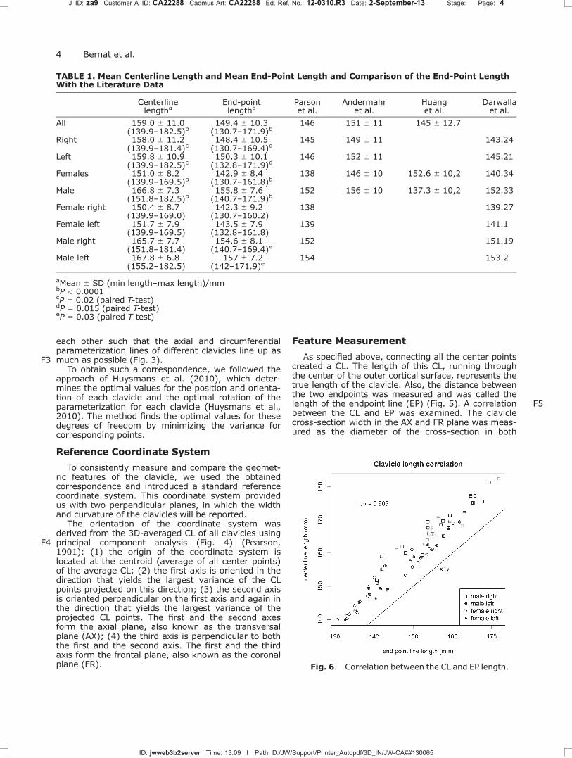
As specified above, connecting all the center pointscreated a CL. The length of this CL, running throughthe center of the outer cortical surface, represents thetrue length of the clavicle. Also, the distance betweenthe two endpoints was measured and was called thelength of the endpoint line (EP) (Fig. F55). A correlationbetween the CL and EP was examined. The claviclecross-section width in the AX and FR plane was meas-ured as the diameter of the cross-section in both
TABLE 1. Mean Centerline Length and Mean End-Point Length and Comparison of the End-Point LengthWith the Literature Data
Centerlinelengtha
End-pointlengtha
Parsonet al.
Andermahret al.
Huanget al.
Darwallaet al.
All 159.0 6 11.0(139.9–182.5)b
149.4 6 10.3(130.7–171.9)b
146 151 6 11 145 6 12.7
Right 158.0 6 11.2(139.9–181.4)c
148.4 6 10.5(130.7–169.4)d
145 149 6 11 143.24
Left 159.8 6 10.9(139.9–182.5)c
150.3 6 10.1(132.8–171.9)d
146 152 6 11 145.21
Females 151.0 6 8.2(139.9–169.5)b
142.9 6 8.4(130.7–161.8)b
138 146 6 10 152.6 6 10,2 140.34
Male 166.8 6 7.3(151.8–182.5)b
155.8 6 7.6(140.7–171.9)b
152 156 6 10 137.3 6 10,2 152.33
Female right 150.4 6 8.7(139.9–169.0)
142.3 6 9.2(130.7–160.2)
138 139.27
Female left 151.7 6 7.9(139.9–169.5)
143.5 6 7.9(132.8–161.8)
139 141.1
Male right 165.7 6 7.7(151.8–181.4)
154.6 6 8.1(140.7–169.4)e
152 151.19
Male left 167.8 6 6.8(155.2–182.5)
157 6 7.2(142–171.9)e
154 153.2
aMean 6 SD (min length–max length)/mmbP < 0.0001cP 5 0.02 (paired T-test)dP 5 0.015 (paired T-test)eP 5 0.03 (paired T-test)
Fig. 6. Correlation between the CL and EP length.
J_ID: za9 Customer A_ID: CA22288 Cadmus Art: CA22288 Ed. Ref. No.: 12-0310.R3 Date: 2-September-13 Stage: Page: 4
ID: jwweb3b2server Time: 13:09 I Path: D:/JW/Support/Printer_Autopdf/3D_IN/JW-CA##130065
4 Bernat et al.

planes. The largest diameter at the sternal end and atthe acromial end in both planes was evaluated. Thesmallest diameter of the clavicle in both planes wasalso measured. The location of these diameters wasmeasured and defined as a percentage of the CLlength of the clavicle starting at the sternal end(Fig. 5). The curvature at each cross-section centerwas measured as the distance to the closest point onthe EP and was described as depth. This was per-formed in the two planes, resulting in an AX curvatureand an FR curvature. The depth was described as pos-itive if going anterior and negative if going posterior inthe AX plane, and positive if going superior and nega-
tive if going inferior in the FR plane. Also, the inflec-tion point (point on a curve at which the convexity orconcavity changes sign) in the AX plane wasmeasured.
All the measurements were done in five groups:average, male, female, left, and right. A comparisonwas made between the male–female group and theleft–right group.
Hypothesis testing was performed using theunpaired two-sample Student t-test. For the left–rightanalysis a paired Student T-test was used. Resultswith a P-value less or equal than 0.05 were consid-ered to be significant.
Fig. 7. Mean width as a function of the length, starting at the sternal end in the AXplane.
J_ID: za9 Customer A_ID: CA22288 Cadmus Art: CA22288 Ed. Ref. No.: 12-0310.R3 Date: 2-September-13 Stage: Page: 5
ID: jwweb3b2server Time: 13:09 I Path: D:/JW/Support/Printer_Autopdf/3D_IN/JW-CA##130065
The Anatomy of the Clavicle 5
RESULTS
Interclass Correlation
The EP length interobserver difference mean was0.25 6 0.37 mm for the first observer and 0.47 6 0.75mm for the second observer. The CL length interob-server difference mean was 0.26 6 0.25 mm for thefirst observer and 0.29 6 0.34 mm for the secondobserver. The intraobserver difference mean was 0.476 0.58 mm for the EP length and 0.42 6 0.33 mm forthe CL length. The two-way Intraclass Correlation(ICC) consistency was 0.9998 for the EP and 0.9996
for the CL length and the standardized error of mea-surement was 0.15 mm for the EP length and 0.21 mmfor the CL length, which can be consider as excellent.
Length Centerline 5 True Length Clavicle
The mean CL length of the clavicles was 159.0 611.0 mm (139.9–182.5) for the general population(Table T11). The mean CL length of the male clavicleswas significantly larger (15.8 mm) than that of thefemale clavicles (P < 0.0001). The mean of the CLlength of the left clavicles was 1.8 mm larger than the
Fig. 8. Mean width as a function of the length, starting at the sternal end in the FRplane.
J_ID: za9 Customer A_ID: CA22288 Cadmus Art: CA22288 Ed. Ref. No.: 12-0310.R3 Date: 2-September-13 Stage: Page: 6
ID: jwweb3b2server Time: 13:09 I Path: D:/JW/Support/Printer_Autopdf/3D_IN/JW-CA##130065
6 Bernat et al.
length of the right clavicles (P 5 0.02). The mean CLlength of the left male clavicle was 1.9 mm largerthan the right CL length (P 5 0.09). Also, the meanCL length of the left female clavicle was 1.3 mm largerthan the right CL length (P 5 0.11).
Length “Endpoints”
The mean EP length of the clavicles was 149.4 610.3 mm (130.7–171.9) for the general population(Table 1). The mean EP length of the male clavicles wassignificantly larger (12.9 mm) than that of the femaleclavicles (P < 0.0001). Again the mean EP length of theleft clavicles was significant larger (1.9 mm) than thelength of the right clavicle (P 5 0.015). The mean EPlength of the left male clavicle was significant larger(2.4 mm) than the right EP length (P 5 0.03). Also, themean EP length of the left female clavicle was 1.2 mmlarger than the right EP length (P 5 0.25).
CL Length vs EP Length
The mean CL length was statistically significantly(P < 0.0001) larger than the mean EP length, 9.6 mmrespectively (Table 1) (Fig.F6 6).
Correlation between the CL and EP length is seen inFigure 6. Parson correlation coefficient is 0.968, whichindicates a strong correlation. Straight clavicles situatedon the line, curved clavicles have a longer CL length andtherefore placed at the upper side of the graph.
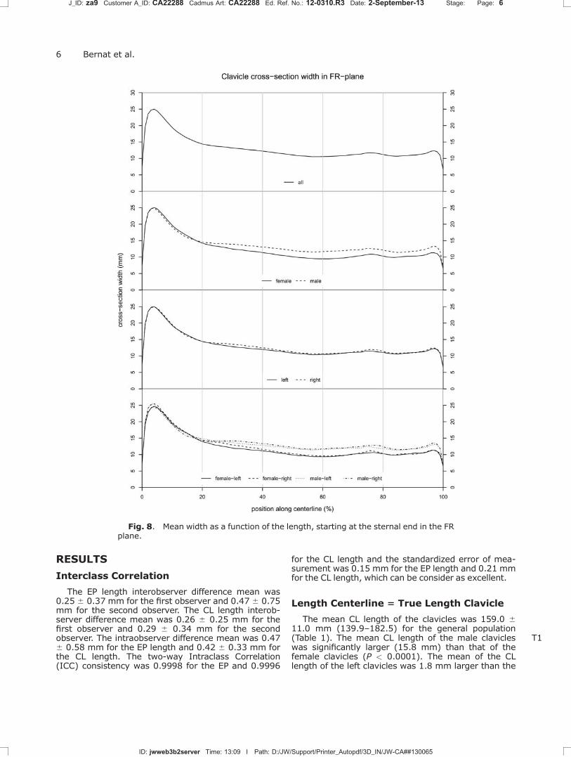
Width AX–FR Plane
The thickest mean width at the sternal end and atthe acromial end was located in the AX and FR plane
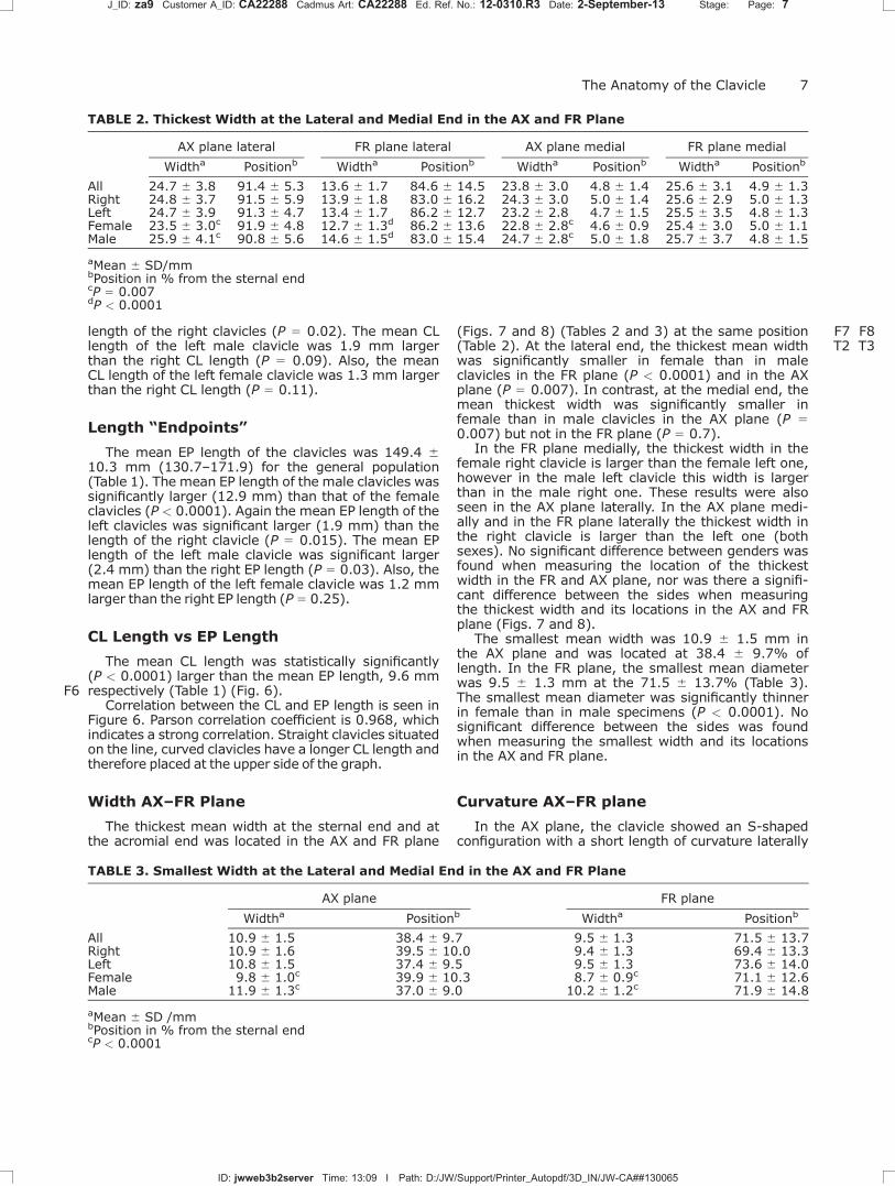
(Figs. F77 and 8) F8(Tables 2 and 3) at the same position(Table 2). At the lateral end, the thickest T2 T3mean widthwas significantly smaller in female than in maleclavicles in the FR plane (P < 0.0001) and in the AXplane (P 5 0.007). In contrast, at the medial end, themean thickest width was significantly smaller infemale than in male clavicles in the AX plane (P 50.007) but not in the FR plane (P 5 0.7).
In the FR plane medially, the thickest width in thefemale right clavicle is larger than the female left one,however in the male left clavicle this width is largerthan in the male right one. These results were alsoseen in the AX plane laterally. In the AX plane medi-ally and in the FR plane laterally the thickest width inthe right clavicle is larger than the left one (bothsexes). No significant difference between genders wasfound when measuring the location of the thickestwidth in the FR and AX plane, nor was there a signifi-cant difference between the sides when measuringthe thickest width and its locations in the AX and FRplane (Figs. 7 and 8).
The smallest mean width was 10.9 6 1.5 mm inthe AX plane and was located at 38.4 6 9.7% oflength. In the FR plane, the smallest mean diameterwas 9.5 6 1.3 mm at the 71.5 6 13.7% (Table 3).The smallest mean diameter was significantly thinnerin female than in male specimens (P < 0.0001). Nosignificant difference between the sides was foundwhen measuring the smallest width and its locationsin the AX and FR plane.
Curvature AX–FR plane
In the AX plane, the clavicle showed an S-shapedconfiguration with a short length of curvature laterally
TABLE 2. Thickest Width at the Lateral and Medial End in the AX and FR Plane
AX plane lateral FR plane lateral AX plane medial FR plane medial
Widtha Positionb Widtha Positionb Widtha Positionb Widtha Positionb
All 24.7 6 3.8 91.4 6 5.3 13.6 6 1.7 84.6 6 14.5 23.8 6 3.0 4.8 6 1.4 25.6 6 3.1 4.9 6 1.3Right 24.8 6 3.7 91.5 6 5.9 13.9 6 1.8 83.0 6 16.2 24.3 6 3.0 5.0 6 1.4 25.6 6 2.9 5.0 6 1.3Left 24.7 6 3.9 91.3 6 4.7 13.4 6 1.7 86.2 6 12.7 23.2 6 2.8 4.7 6 1.5 25.5 6 3.5 4.8 6 1.3Female 23.5 6 3.0c 91.9 6 4.8 12.7 6 1.3d 86.2 6 13.6 22.8 6 2.8c 4.6 6 0.9 25.4 6 3.0 5.0 6 1.1Male 25.9 6 4.1c 90.8 6 5.6 14.6 6 1.5d 83.0 6 15.4 24.7 6 2.8c 5.0 6 1.8 25.7 6 3.7 4.8 6 1.5
aMean 6 SD/mmbPosition in % from the sternal endcP 5 0.007dP < 0.0001
TABLE 3. Smallest Width at the Lateral and Medial End in the AX and FR Plane
AX plane FR plane
Widtha Positionb Widtha Positionb
All 10.9 6 1.5 38.4 6 9.7 9.5 6 1.3 71.5 6 13.7Right 10.9 6 1.6 39.5 6 10.0 9.4 6 1.3 69.4 6 13.3Left 10.8 6 1.5 37.4 6 9.5 9.5 6 1.3 73.6 6 14.0Female 9.8 6 1.0c 39.9 6 10.3 8.7 6 0.9c 71.1 6 12.6Male 11.9 6 1.3c 37.0 6 9.0 10.2 6 1.2c 71.9 6 14.8
aMean 6 SD /mmbPosition in % from the sternal endcP < 0.0001
J_ID: za9 Customer A_ID: CA22288 Cadmus Art: CA22288 Ed. Ref. No.: 12-0310.R3 Date: 2-September-13 Stage: Page: 7
ID: jwweb3b2server Time: 13:09 I Path: D:/JW/Support/Printer_Autopdf/3D_IN/JW-CA##130065
The Anatomy of the Clavicle 7
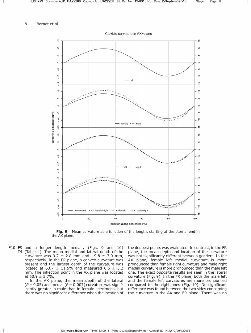
and a longer length medially (Figs.F9 9 andF10 10)(TableT4 4). The mean medial and lateral depth of thecurvature was 9.7 6 2.8 mm and 29.8 6 3.0 mm,respectively. In the FR plane, a convex curvature waspresent and the largest depth of the curvature waslocated at 63.7 6 11.5% and measured 6.6 6 3.2mm. The inflection point in the AX plane was locatedat 60.9 6 5.7%.
In the AX plane, the mean depth of the lateral(P 5 0.05) and medial (P 5 0.007) curvature was signif-icantly greater in male than in female specimens, butthere was no significant difference when the location of
the deepest points was evaluated. In contrast, in the FRplane, the mean depth and location of the curvaturewas not significantly different between genders. In theAX plane, female left medial curvature is morepronounced than female right curvature and male rightmedial curvature is more pronounced than themale leftone. The exact opposite results are seen in the lateralcurvature (Fig. 9). In the FR plane, both the male leftand the female left curvatures are more pronouncedcompared to the right ones (Fig. 10). No significantdifference was found between the two sides concerningthe curvature in the AX and FR plane. There was no
Fig. 9. Mean curvature as a function of the length, starting at the sternal end inthe AX plane.
J_ID: za9 Customer A_ID: CA22288 Cadmus Art: CA22288 Ed. Ref. No.: 12-0310.R3 Date: 2-September-13 Stage: Page: 8
ID: jwweb3b2server Time: 13:09 I Path: D:/JW/Support/Printer_Autopdf/3D_IN/JW-CA##130065
8 Bernat et al.
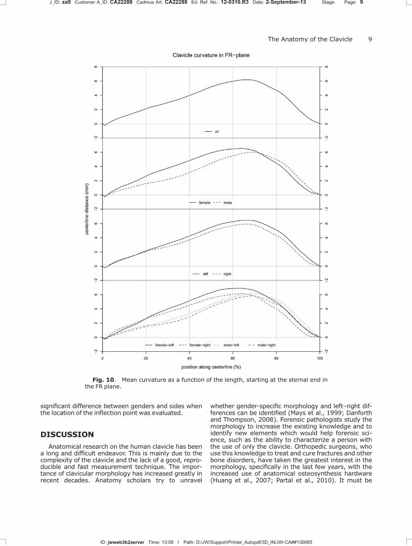
significant difference between genders and sides whenthe location of the inflection point was evaluated.
DISCUSSION
Anatomical research on the human clavicle has beena long and difficult endeavor. This is mainly due to thecomplexity of the clavicle and the lack of a good, repro-ducible and fast measurement technique. The impor-tance of clavicular morphology has increased greatly inrecent decades. Anatomy scholars try to unravel
whether gender-specific morphology and left–right dif-ferences can be identified (Mays et al., 1999; Danforthand Thompson, 2008). Forensic pathologists study themorphology to increase the existing knowledge and toidentify new elements which would help forensic sci-ence, such as the ability to characterize a person withthe use of only the clavicle. Orthopedic surgeons, whouse this knowledge to treat and cure fractures and otherbone disorders, have taken the greatest interest in themorphology, specifically in the last few years, with theincreased use of anatomical osteosynthesis hardware(Huang et al., 2007; Partal et al., 2010). It must be
Fig. 10. Mean curvature as a function of the length, starting at the sternal end inthe FR plane.
J_ID: za9 Customer A_ID: CA22288 Cadmus Art: CA22288 Ed. Ref. No.: 12-0310.R3 Date: 2-September-13 Stage: Page: 9
ID: jwweb3b2server Time: 13:09 I Path: D:/JW/Support/Printer_Autopdf/3D_IN/JW-CA##130065
The Anatomy of the Clavicle 9

noted that while intramedullary and plate fixation areaccepted and widely used methods of treatment forfractures of the clavicle, current clavicular implantsoverlook the variations in geometry of the bone(Daruwalla et al., 2010b).
To our knowledge, our study represents the largestpopulation of fresh frozen cadaver clavicles that werescanned, computed, and analyzed. Furthermore, ourcorrespondence technique provided us with a singleglobal coordinate system in which all clavicles werealigned andmeasured. The coordinate system, togetherwith the AX and FR planes, was derived from the popula-tion average CL. Taking into account information of allthe clavicles in the construction of the coordinatesystem increases the robustness and consistency of themeasurements. The measurement process was alsohighly automated, requiring only two points to beselected for each clavicle. Extending it to larger popula-tions of clavicles requires minimal effort. Also, thecross-sections in our measurement technique wereperpendicular to the CL, providing a more physiologicalevaluation of the width and the curvature of the claviclecompared to axis-aligned cross-sections.
Length
Our technique enabled us to not only measure thetrue length of the clavicle (CL), but also the endpointlength, which was compared with the literature data.
Hillen et al. (2012) showed significant orientationchanges with clavicle shortening. These changes wereprogressive with the amount of shortening. The scap-ula was changed into a more “winging” position, thesternoclavicular and acromiclavicular joint was moreprotracted and axially rotated. Restoration of clavicu-lar length is considered a determining factor for theoptimal restoration of function after a clavicle fracture(Hill et al., 1997; Andermahr et al., 2006). Until now,the true length of the clavicle has not yet beendescribed. In all studies, the length of the clavicle wasmeasured as the length between two extremity points(end-point length) and not as the true length(Parsons, 1916; Mays et al., 1999; Andermahr et al.,2007; Daruwalla et al., 2010a). The EP length is anunderestimation of the true length of the clavicle.
We defined the true length of the clavicle as thelength of the line connecting the center of the cross-sections of the clavicle. As already mentioned, a
significant difference was found between CL lengthand EP length. The mean true length was 9.6 mmlarger than the mean EP length.
The average EP length in our population was 149.9mm, which is consistent with previous findings byAndermahr et al. (2007) (Table 1). Huang et al. (2007)and Parsons (1916) reported different lengths in theirpopulation. Two reasons may account for these differ-ences. A first explanationmay be the variation in demo-graphics and evolution (Garcia and Quintana-Domeque, 2007). Our study was based on a Caucasianpopulation from the 20th century. This correspondswith the population described by Andermahr et al.(2007). Parsons (1916) and Huang et al. (2007) used acollection of bones from the previous century andanother geographic location. Second, the definition anddetermination of length also vary between studies.
As was reported in previous literature (Parsons,1916; Andermahr et al., 2007; Haung et al., 2007; Dar-uwalla et al., 2010a), the male clavicle is longer than thefemale one. We found significant differences in lengthfor both the EP and CL length (12.9 and 15.8 mm,respectively).
As described in literature (Parsons, 1916; Mayset al., 1999; Andermahr et al., 2007; Auerbach andRaxter, 2008; Danforth and Thompson, 2008; Daru-walla et al., 2010a), we found a difference betweensides. This was found significant for both the EP and theCL length (1.8 and 1.9 mm, respectively). Most authorsfound the mean left clavicle to be larger than the rightone, but only two authors reported a significant differ-ence (Mays et al., 1999; Auerbach and Raxter, 2008).The other authors either did not perform this measure-ment or did not find a significant difference. When eval-uating the length in both genders there was only astatistical difference in themale EP length.
On the basis of our data, a new set of anatomicalplates can be developed that take into account clavic-ular center length, though allowing for correct ana-tomical reconstruction and fit, which may beassociated with better clinical result. In regard to cur-vature and other morphometric elements, the use ofthe CL can help to restore the correct length andalignment of the fractured clavicle.
Width
Anatomically, the lungs and vascular structures arein close proximity to the clavicle. Placing a screw
TABLE 4. Deepest Curvature Position and Depth in the AX and FR Plane and the InflectionPoint in the AX Plane
FR AX
Deptha Positionb Med deptha Med positionb Inflexion positionb Lat deptha Lat positionb
All 6.6 6 3.2 63.7 6 11.5 9.7 6 2.8 31.6 6 3.4 60.9 6 5.7 29.8 6 3.0 79.4 6 3.3Right 6.4 6 3.6 62.5 6 14.7 9.7 6 2.9 32.1 6 2.9 60.8 6 6.0 29.7 6 2.6 79.1 6 2.8Left 6.8 6 2.8 62.5 6 7.0 9.7 6 2.7 31.1 6 3.7 60.4 6 5.5 29.9 6 3.4 79.7 6 3.8Female 6.8 6 3.7 61.4 6 9.8 8.6 6 2.3c 31.7 6 3.6 61.1 6 5.3 29.1 6 2.5d 79.3 6 3.8Male 6.4 6 2.6 66.0 6 12.7 10.8 6 2.8c 31.4 6 3.2 60.0 6 6.1 210.5 6 3.3d 79.5 6 2.8
aMean 6 SD/mmbPosition in % from the sternal endcP 5 0.007dP 5 0.05
J_ID: za9 Customer A_ID: CA22288 Cadmus Art: CA22288 Ed. Ref. No.: 12-0310.R3 Date: 2-September-13 Stage: Page: 10
ID: jwweb3b2server Time: 13:09 I Path: D:/JW/Support/Printer_Autopdf/3D_IN/JW-CA##130065
10 Bernat et al.

longer than required can damage these structures.Knowing the thickness in each parts of the claviclecan be helpful to assess the screw length when ante-roposterior or superoinferior plate and screw fixationis used.
Width was measured in two planes, FR and AX.Several different measurement protocols have beendescribed. Mays et al. (1999) and Danforth et al.(2008) measured the width only at the midshaft.Andermahr et al. (2007) measured the diameter inthree locations and Parsons in five locations.Daruwalla et al. (2010a) measured the width at 10%intervals of the length. We were the first to measurethe diameter at 100 different cross-sections. In thisway, a graph could be constructed to describe thewidth along the clavicle. The thickest width both in theAX as in the FR plane is located at the same position.Females have a significantly lees wide clavicle thanmales, but only in the FR plane laterally and in the AXplane medially. In females the thinnest width is againsmaller compared to male clavicles in both planes.The location of the thinnest width in males is signifi-cantly more lateral than in females. There were nosignificant differences between sides regarding thewidth of the clavicle.
Mays et al. (1999) and Auerbach and Raxter(2008) have used the diameter at the midshaft tostudy the asymmetry and handedness of the clavicle.With our results the width can be measured in eachpoint and directly applied for the further research ofasymmetry.
Curvature
The curvature of the clavicle is extremely difficultto describe and different methods have been used tothis end. Parsons (1916) was the first to measure thecurvature with the description of angles but hismethod had a low interobserver reliability. Severalother authors measured the depth of the curvature bymeasuring the distance between the outer surfaceand the EP line (Mays et al., 1999; Andermahr et al.,2007; Huang et al., 2007; Daruwalla et al., 2010a). Inthe present study, the curvature was measured as thedepth between the CL and the EP line in the FR andAX plane at all cross-sections. In our opinion, thisprovides a more physiological representation of thecurvature medially, laterally, and inferiorly in the twoplanes. As far as we know, this is the first representa-tion of the curvature in the frontal plane in thisconfiguration.
In the FR plane, the clavicle was found to have asuperior curvature with the largest depth at 63.7% ofthe clavicle. This is more medial than what Huanget al. (2007) described, namely a mean location at75% of the clavicle. Specimens from Caucasiansdonors also had a bow apex that was significantlymore medial compared with those from donors ofAfrican ancestry and a trend toward a larger bowmagnitude that was not statistically significant (Huanget al., 2007). Our population consisted only of whiteCaucasian specimens. We found a significantdifference between male and female clavicles in both
curvatures. This was also the first time the location ofthe inflection point was described. The location wasfound not to change between sides and genders. Thefindings relating to the curvature in both planes andthe location of the inflection point in both genders canbe important in the design of new anatomical clavicu-lar plates. A clear S-shaped curvature is present inthe AX plane and has already been applied in thecurrent anatomical clavicular plate. As previouslyreported by Voisin (2006, 2008) and Olivier (1951),we observed a second inferior curvature in the FRplane, which is not compensated for in the currentcommercially available plates. Altamimi and McKee(2008) have advocated to fit the plate perfectly onthe clavicle to avoid soft tissue irritation and patientdissatisfaction, we believe that the inferior curvatureis one of the missing pieces in achieving a perfect fit.
Limitations
Our study has some limitations. Firstly, ourpopulation consisted of only 64 samples but wasoptimally balanced (34 right vs. 34 left).
Secondly, because the body length of the cadaverswas not known, the ratio between body length andclavicular length could not be evaluated. Also, data onthe handedness in the samples were unavailable.Such information could have been useful for theleft–right analysis.
CONCLUSION
The goal of this study was to examine the 3Danatomical characteristics and variations of thehuman clavicle, using a fully automated 3D analysis.This technique allowed the true length of the clavicleto be measured for the first time. Also, the curvaturein the AX and FR plane was described more accu-rately. A significant difference between genders inlength, width, and curvature and the length betweenthe sides was found.
ACKNOWLEDGMENTS
Authors thank Iris Wojtowicz, Department ofOrthopedic Surgery and Traumatology, GhentUniversity Hospital, for her linguistic support and ourcolleagues at the anatomy lab, Department of Anatomyand Embryology, Antwerp University, for their help withthe preparation of the clavicles. Last, authors wouldlike to thank the donors and their families.
REFERENCES
Altamimi SA, McKee MD. 2008. Nonoperative treatment comparewith plate fixation of displaced midshaft clavicular fractures:Surgical technique. J Bone Joint Surg Am 90:1–8.
Althausen PL, Shannon S, Lu M, O’Mara TJ, Bray TJ. 2013. Clinicaland financial comparison of operative and nonoperative treat-ment of displaced clavicle fractures. J Shoulder Elbow Surg22:608–611.
J_ID: za9 Customer A_ID: CA22288 Cadmus Art: CA22288 Ed. Ref. No.: 12-0310.R3 Date: 2-September-13 Stage: Page: 11
ID: jwweb3b2server Time: 13:09 I Path: D:/JW/Support/Printer_Autopdf/3D_IN/JW-CA##130065
The Anatomy of the Clavicle 11
Andermahr J, Jubel A, Elsner A, Prokop A, Tsikaras P, Jupiter J,Koebke J. 2006. Malunion of the clavicle causes significant gle-noid malposition: A quantitative anatomic investigation. SurgRadiol Anat 28:447–456.
Andermahr J, Jubel A, Elsner A, Johann J, Prokop A, Rehm KE,Koebke J. 2007. Anatomy of the clavicle and the intramedullarynailing of midclavicular fractures. Clin Anat 20:48–56.
Auerbach BM, Raxter MH. 2008. Patterns of clavicular bilateralasymmetry in relation to the humerus: Variation among humans.J Hum Evol 54:663–674.
Canadian Orthopaedic Trauma Society. 2007. Nonoperative treat-ment compared with plate fixation of displaced midshaft clavicu-lar fractures. a multicenter, randomized clinical trial. J Bone JointSurg Am 89:1–10.
Danforth ME, Thompson A. 2008. An evaluation of determination ofhandedness using standard osteological measurements. J Foren-sic Sci 53:777–781.
Daruwalla ZJ, Courtis P, Fitzpatrick C, Fitzpatrick D, Mullett H.2010a. Anatomic variation of the clavicle: A novel three-dimensional study. Clin Anat 23:199–209.
Daruwalla ZJ, Courtis P, Fitzpatrick C, Fitzpatrick D, Mullett H.2010b. An application of principal component analysis to theclavicle and clavicle fixation devices. J Orthop Surg Res 5:21.
Egol KA, Koval KJ, Zuckerman JD. 2010. Handbook of fractures. 4thEd. Philadelphia: Lippincott, Williams & Wilkins. p 141–149.ISBN: 978–1605477602.
Fitzpatrick C, FitzPatrick D, Auger D, Lee J. 2007. A tibial-basedcoordinate system for three-dimensional data. Knee 14:133–137.
Garcia J, Quintana-Domeque C. 2007. The evolution of adult heightin Europe: A brief note. Econ Hum Biol 5:340–349.
Hill JM, McGuire MH, Crosby LA. 1997. Closed treatment of displacedmiddle-third fractures of the clavicle gives poor results. J BoneJoint Surg Br 79:537–539.
Hillen RJ, Burger BJ, P€oll RG, van Dijk CN, Veeger DH. 2012. Theeffect of experimental shortening of the clavicle on shoulderkinematics. Clin Biomech (Bristol, Avon) 27:777–781.
Huang JI, Toogood P, Chen MR, Wilber JH, Cooperman DR. 2007.Clavicular anatomy and the applicability of precontoured plates. JBone Joint Surg Am 89:2260–2265.
Huysmans T, Sijbers J, Verdonk B. 2005. Parameterization of tubularsurfaces on the cylinder. J Winter School Comput Graph 13:97–104. (ISSN No. 1213–6964).
Huysmans T, Sijbers J, Verdonk B. 2010. Automatic construction ofcorrespondences for tubular surfaces. IEEE Trans Pattern AnalMach Intell 32:636–651.
Jit I, Singh S. 1996. The sexing of the adult clavicles. Indian J MedRes 54:551–571.
Lazarus M, Seon C. 2006. In: Bucholz R, Heckman J, Court-BrownC, editors. Chapter 32: Fractures of the Clavicle. 6th Ed. Philide-phia: Lippincott Williams Wilkins. p 1212–1213. (ISBN No.978–0781746366).
Lorensen WE, Cline HE. 1987. Marching cubes: A high resolution 3Dsurface construction algorithm. SIGGRAPH Comput Graph 21:163–169.
Ludewig PM, Phadke V, Braman JP, Hassett DR, Cieminski CJ,LaPrade RF. 2009. Motion of the shoulder complex duringmultiplanar humeral elevation. J Bone Joint Surg Am 91:378–389.
Mays S, Steele J, Ford M. 1999. Directional asymmetry in the humanclavicle. Int J Osteoarchaeol 9:18–28.
McCormick WF, Stewart JH, Greene H. 1991. Sexing of humanclavicles using length and circumference measurements. Am JForensic Med Pathol 12:175–181.
McKee MD, Pedersen EM, Jones C, Stephen DJ, Kreder HJ,Schemitsch EH. 2006. Deficits following nonoperative treatmentof displaced midshaft clavicular fractures. J Bone Joint Surg Am88:35–40.
Nowak J, Holgersson M, Larsson S. 2004. Can we predict long-termsequelae after fractures of the clavicle based on initial finding? Aprospective study with nine to ten years of follow-up. J shoulderElbow Surg 13:479–486.
Olivier G. (1951–1956). Anthropologie de la clavicule. Bull MemSoc Anthropolo Paris S�erie 10:47–56 (VII), 67–99 (I), 144–153(VIII), 269–279 (IV), 282–289 (IX), 290–302 (X), 553–561(V).
Parsons FG. 1916. On the proportions and characteristics of themodern english clavicle. J Anat 51(Part 1):71–93.
Partal G, Meyers KN, Sama N, Pagenkopf E, Lewis PB, Goldman A,Wright TM, Helfet DL. 2010. Superior versus anteroinferior plat-ing of the clavicle revisited: A mechanical study. J Orthop Trauma24:420–425.
Pearson K. 1901. On lines and planes of closest fit to systems ofpoints in space. Philos Mag 2:559–572.
VanBeek C, Boselli KJ, Cadet ER, Ahmad CS, Levine WN. 2011.Precontoured plating of clavicle fractures: Decreasedhardware-related complications? Clin Orthop Relat Res 469:3337–3343.
Voisin JL. 2006. Clavicle, a neglected bone: Morphology and relationto arm movements and shoulder architecture in primates. AnatRec A Discov Mol Cell Evol Biol 288:944–953.
Voisin JL. 2008. The Omo I hominin clavicle: Archaic or modern? JHum Evol 55:438–443.
J_ID: za9 Customer A_ID: CA22288 Cadmus Art: CA22288 Ed. Ref. No.: 12-0310.R3 Date: 2-September-13 Stage: Page: 12
ID: jwweb3b2server Time: 13:09 I Path: D:/JW/Support/Printer_Autopdf/3D_IN/JW-CA##130065
12 Bernat et al.