Circadian Gating of Cell Division in Cyanobacteria Growing with Average Doubling Times of Less than...
6
Proc. Natl. Acad. Sci. USA Vol. 93, pp. 10183-10188, September 1996 Cell Biology Circadian gating of cell division in cyanobacteria growing with average doubling times of less than 24 hours (biological clockISynechococcus/flow cytometry) TETSUYA MORI*, BRIAN BINDERt, AND CARL HIRSCHIE JOHNSON*t *Department of Biology, Vanderbilt University, Nashville, TN 37235; and tDepartment of Marine Sciences, University of Georgia, Athens, GA 30602 Communicated by Shinya Inoue, Marine Biology Laboratory, Woods Hole, MA, March 29, 1996 (received for review February 9, 1996) ABSTRACT To ascertain whether the circadian oscillator in the prokaryotic cyanobacterium Synechococcus PCC 7942 regulates the timing of cell division in rapidly growing cul- tures, we measured the rate of cell division, DNA content, cell size, and gene expression (monitored by luminescence of the PpsbAJ::luxAIB reporter) in cultures that were continuously diluted to maintain an approximately equal cell density. We found that populations dividing at rates as rapid as once per 10 h manifest circadian gating of cell division, since phases in which cell division slows or stops recur with a circadian periodicity. The data clearly show that Synechococcus cells growing with doubling times that are considerably faster than once per 24 h nonetheless express robust circadian rhythms of cell division and gene expression. Apparently Synechococcus cells are able to simultaneously sustain two timing circuits that express significantly different periods. Circadian rhythms and cell division cycles (CDCs) constitute two important cyclic biological systems. These rhythmic phe- nomena are usually not independent in unicellular eukaryotic organisms, where the circadian system often controls the timing of the cell division cycle (1-3). The nature of this control appears to be via "gating" of cell division such that the circadian oscillator specifies certain phases in which cell divi- sion is "allowed" to occur and other phases in which it is "forbidden," even if the cells have attained sufficient size (1). Thus, the circadian oscillator acts in addition to other "check- points" in determining when cells divide. Recently, it has been discovered that prokaryotic cyanobac- teria express authentic circadian rhythms of gene expression (4-10). We wondered whether these prokaryotes would exhibit circadian control over the timing of cell division, especially in cases where the doubling times are much shorter than 24 h. In unicellular eukaryotic organisms, the relationship between cell division and circadian expression has come to be encapsulated in the so-called "circadian-infradian rule," which states that circadian rhythms are expressed only in cells that are dividing once per day or more slowly, i.e., in the "infradian" mode; in cells dividing more rapidly than once per day, cellular pro- cesses are thought to become uncoupled from circadian os- cillator control (1, 11). This rule implies that there is an interdependency between these two timing circuits such that when cell division is more rapid than once per day, the cells are unable to maintain an independent circadian oscillation. It has been suggested therefore that a circadian clock that provides temporal programming is only adaptive to organisms whose generation time is as long or longer than a day (12). There have been a few investigations with Synechococcus species of cyanobacteria indicating circadian rhythms of cell division (4, 13), but these have been limited to strains and/or growth conditions that allowed only relatively slow growth (doubling times slower than once per 24 h). We decided to reexamine these issues using a strain (Synechococcus sp. strain PCC 7942) that exhibits bona fide circadian rhythms (6) and can grow rapidly. Our goal was to test whether cyanobacteria that are dividing more rapidly than once per 24 h nevertheless exhibit circadian regulation over cell division. We achieved this objective using Synechococcus cultures that were continuously diluted with fresh medium to attain cultures that were main- tained at an approximately equal cell density. The results we report herein indicate that the circadian clock controls the timing of cell division, cell size, and the average cellular DNA content, even in cyanobacterial cultures that are growing with doubling times that are much faster than once per 24 h. MATERIALS AND METHODS Synechococcus sp. PCC 7942 Strains. The strains used in this study were wild-type (PCC 7942), AMC149, SP22, and LP27. The strain AMC149 is a bacterial luciferase reporter strain that contains a PpsbAI::luxAB translational fusion integrated into a neutral site of chromosome of the wild-type strain (6). The psbAI gene encodes the Dl protein of photosystem II. Strains LP27 and SP22 are circadian period mutants isolated after chemical mutagenesis of AMC149 (14). Culture Conditions. Cells were cultured photoautotrophi- cally at 30 ± 0.5°C in BG-11 medium in 1-liter bottles as described (6). For growth of strains AMC149, SP22, and LP27, the culture medium was supplemented with spectinomycin (40 gg/ml). For growth of wild-type Synechococcus cells, BG-11 medium was used without spectinomycin. Cell suspensions were bubbled with air and stirred. Illumination was provided from the side of the culture bottle by a cool-white fluorescent light bulb standing parallel to the culture bottle. The light intensity in all experiments reported herein was 125 ,tE per m2 per s. Cultures were synchronized with light-dark cycles of 12 h of light followed by 12 h of darkness (LD 12:12) and then released into free-running conditions of continuous light (LL). For continuously diluted cultures, cells were initially grown in LD 12:12 in a batch culture. Cultures were then released into LL and continuous dilution of culture was started. Cell cultures were maintained at an approximately constant cell density and volume by replacing cell suspension at a continuous rate with fresh medium. The fresh medium was continuously supplied to 500 ml of cell suspension in a culture bottle using a peristaltic pump at a flow rate of about 30 ml/h. Therefore, approxi- mately 30 ml of cell suspension was withdrawn from the culture each hour and replaced with 30 ml of fresh medium, resulting in a rate of dilution that was almost constant at about 30/500 per h (0.06 h-1). A portion of the cell suspension that was removed from the culture was automatically saved into one of Abbreviations: CDC, cell division cycle; LD, light-dark cycle; LL, continuous light; FALS, forward-angle light scatter; DT, doubling time. *To whom reprint requests should be addressed at: Department of Biology, Box 1812-B, Vanderbilt University, Nashville, TN 37235. e-mail: [email protected]. 10183 The publication costs of this article were defrayed in part by page charge payment. This article must therefore be hereby marked "advertisement" in accordance with 18 U.S.C. §1734 solely to indicate this fact.
-
Upload
independent -
Category
Documents
-
view
2 -
download
0
Transcript of Circadian Gating of Cell Division in Cyanobacteria Growing with Average Doubling Times of Less than...

Proc. Natl. Acad. Sci. USAVol. 93, pp. 10183-10188, September 1996Cell Biology
Circadian gating of cell division in cyanobacteria growing withaverage doubling times of less than 24 hours
(biological clockISynechococcus/flow cytometry)
TETSUYA MORI*, BRIAN BINDERt, AND CARL HIRSCHIE JOHNSON*t*Department of Biology, Vanderbilt University, Nashville, TN 37235; and tDepartment of Marine Sciences, University of Georgia, Athens, GA 30602
Communicated by Shinya Inoue, Marine Biology Laboratory, Woods Hole, MA, March 29, 1996 (received for review February 9, 1996)
ABSTRACT To ascertain whether the circadian oscillatorin the prokaryotic cyanobacterium Synechococcus PCC 7942regulates the timing of cell division in rapidly growing cul-tures, we measured the rate of cell division, DNA content, cellsize, and gene expression (monitored by luminescence of thePpsbAJ::luxAIB reporter) in cultures that were continuouslydiluted to maintain an approximately equal cell density. Wefound that populations dividing at rates as rapid as once per10 h manifest circadian gating of cell division, since phases inwhich cell division slows or stops recur with a circadianperiodicity. The data clearly show that Synechococcus cellsgrowing with doubling times that are considerably faster thanonce per 24 h nonetheless express robust circadian rhythms ofcell division and gene expression. Apparently Synechococcuscells are able to simultaneously sustain two timing circuitsthat express significantly different periods.
Circadian rhythms and cell division cycles (CDCs) constitutetwo important cyclic biological systems. These rhythmic phe-nomena are usually not independent in unicellular eukaryoticorganisms, where the circadian system often controls thetiming of the cell division cycle (1-3). The nature of thiscontrol appears to be via "gating" of cell division such that thecircadian oscillator specifies certain phases in which cell divi-sion is "allowed" to occur and other phases in which it is"forbidden," even if the cells have attained sufficient size (1).Thus, the circadian oscillator acts in addition to other "check-points" in determining when cells divide.
Recently, it has been discovered that prokaryotic cyanobac-teria express authentic circadian rhythms of gene expression(4-10). Wewondered whether these prokaryotes would exhibitcircadian control over the timing of cell division, especially incases where the doubling times are much shorter than 24 h. Inunicellular eukaryotic organisms, the relationship between celldivision and circadian expression has come to be encapsulatedin the so-called "circadian-infradian rule," which states thatcircadian rhythms are expressed only in cells that are dividingonce per day or more slowly, i.e., in the "infradian" mode; incells dividing more rapidly than once per day, cellular pro-cesses are thought to become uncoupled from circadian os-cillator control (1, 11). This rule implies that there is aninterdependency between these two timing circuits such thatwhen cell division is more rapid than once per day, the cells areunable to maintain an independent circadian oscillation. It hasbeen suggested therefore that a circadian clock that providestemporal programming is only adaptive to organisms whosegeneration time is as long or longer than a day (12).There have been a few investigations with Synechococcus
species of cyanobacteria indicating circadian rhythms of celldivision (4, 13), but these have been limited to strains and/orgrowth conditions that allowed only relatively slow growth(doubling times slower than once per 24 h). We decided to
reexamine these issues using a strain (Synechococcus sp. strainPCC 7942) that exhibits bona fide circadian rhythms (6) andcan grow rapidly. Our goal was to test whether cyanobacteriathat are dividing more rapidly than once per 24 h neverthelessexhibit circadian regulation over cell division. We achieved thisobjective using Synechococcus cultures that were continuouslydiluted with fresh medium to attain cultures that were main-tained at an approximately equal cell density. The results wereport herein indicate that the circadian clock controls thetiming of cell division, cell size, and the average cellular DNAcontent, even in cyanobacterial cultures that are growing withdoubling times that are much faster than once per 24 h.
MATERIALS AND METHODSSynechococcus sp. PCC 7942 Strains. The strains used in this
study were wild-type (PCC 7942), AMC149, SP22, and LP27.The strain AMC149 is a bacterial luciferase reporter strain thatcontains a PpsbAI::luxAB translational fusion integrated intoa neutral site of chromosome of the wild-type strain (6). ThepsbAI gene encodes the Dl protein of photosystem II. StrainsLP27 and SP22 are circadian period mutants isolated afterchemical mutagenesis of AMC149 (14).
Culture Conditions. Cells were cultured photoautotrophi-cally at 30 ± 0.5°C in BG-11 medium in 1-liter bottles asdescribed (6). For growth of strains AMC149, SP22, and LP27,the culture medium was supplemented with spectinomycin (40gg/ml). For growth of wild-type Synechococcus cells, BG-11medium was used without spectinomycin. Cell suspensionswere bubbled with air and stirred. Illumination was providedfrom the side of the culture bottle by a cool-white fluorescentlight bulb standing parallel to the culture bottle. The lightintensity in all experiments reported herein was 125 ,tE per m2per s. Cultures were synchronized with light-dark cycles of 12 hof light followed by 12 h of darkness (LD 12:12) and thenreleased into free-running conditions of continuous light (LL).For continuously diluted cultures, cells were initially grown
in LD 12:12 in a batch culture. Cultureswere then released intoLL and continuous dilution of culture was started. Cell cultureswere maintained at an approximately constant cell density andvolume by replacing cell suspension at a continuous rate withfresh medium. The fresh medium was continuously supplied to500 ml of cell suspension in a culture bottle using a peristalticpump at a flow rate of about 30 ml/h. Therefore, approxi-mately 30 ml of cell suspension was withdrawn from the cultureeach hour and replaced with 30 ml of fresh medium, resultingin a rate of dilution that was almost constant at about 30/500per h (0.06 h-1). A portion of the cell suspension that wasremoved from the culture was automatically saved into one of
Abbreviations: CDC, cell division cycle; LD, light-dark cycle; LL,continuous light; FALS, forward-angle light scatter; DT, doublingtime.*To whom reprint requests should be addressed at: Department ofBiology, Box 1812-B, Vanderbilt University, Nashville, TN 37235.e-mail: [email protected].
10183
The publication costs of this article were defrayed in part by page chargepayment. This article must therefore be hereby marked "advertisement" inaccordance with 18 U.S.C. §1734 solely to indicate this fact.

10184 Cell Biology: Mon et al. ~~~~~Proc. Nati. Acad. Sci. USA 93 (1996)
a series of test tubes in a fraction collector for measurementsof cell' number and luminescence and for flow cytometricanalyses (see below).Monitoring Cell Division. The portion of the cells for
determilnation of cell number was automatically collected anddiluted 1:1 with iodine solution (iodine at 2' mg/ml andpotassium iodine at 1 mg/ml) so that cells were fixed imme-di'ately. The samples were kept at room temperature or at 40C.The cell numbers in the suspensions were me'asured with anelectronic particle counter (model ZF, Coulter) with a 30-gLmdiameter aperture tube. Periods of cell division rhythms werecalculated by the maximum entropy .method (15, 16). Micro-scopical examinations of cells from rapidly dividing culturesindicated that each mother cell divided into only two daughtercells (i.e., clusters of two, but not of three' or more, daughtercells were observed from dividing cultures).Flow Cytometry. For flow cytometry, a portion of the
culture (3 ml) was saved every 3 h and centrifuged at 20,000 xg (Sorvall SS-34, 12,700 rpm) for to0 min at 40C. The pellet wasresuspended in 5 ml of 70% ethanol and then centrifuged againas above. The resulting pellet was resuspended in 3 ml ofabsolute ethanol and stored at - 150C. Aliquots of the ethanol-fixed samples were resuspende,d in phosphate-buffered saline(pH 7.5) and stained with the DNA-specific fluorochromeHoechst 33342 (final concentration = 0.5 ~tg/ml). Stainedsamples were analyzed on a Coulter EPICS-753 flow cytom-eter using 150- to 300-mW UV excitation and a "Biosense"flow cell. Hoechst fluorescence, which is'proportional to DNAcontent, was measured between 408 and 470 nm; phycocyaninfluorescence, used to unambiguously identify cyanobacterialcells, was measured through a 680-nm bandpass filter (40-nmband width); and forward-angle light scatter (FALS) wasmeasured with a custom-installed photomultiplier tubethrough a 320-nm bandpass filter. All parameters were col-lected as linear pulse-integrated values. A control sample,derived from a stationary-'phase Synechococcus PCC 6301culture, was repeatedly analyzed along with each batch ofexperimental samples and served a's a staining standard forthose samples. Approxima'tely 30,000 cells were measured foreach determination of DNA content and FALS. Genomenumbers were assigned to peaks in the DNA frequencydistributions as described (17). FALS data were normalized tothe control sample and expressed as relative values. Therelationship between FALS and cell size is complex anddepends on the refractive index of the cells and the opticalconfiguration of the instrument (18). For a given cell typeanalyzed on a given instrum'ent, however, it is reasonable toassume that changes in FALS reflect changes in cell size, atleast qualitatively (19), as we confirmed microscopically.Measurement of Luminescence. Every 3 h, 1 ml of culture
was transferred into a 20-ml glass scintillation vial, and then acapless 1.5-ml microcentrifuge tube containing 300 pAl of 10%n-decanal in soybean oil was placed in the vial. The vial wassealed to allow the n-decanal vapor to equilibrate and thenimmediately placed into a luminometer apparatus and incu-bated at room temperature (21 ± 1L50C in darkness. After a30-mmn incubation, light emission from the culture was mea-sured with the photomultiplier tube (model 931B, HamamatsuPhotonics, Hamamatsu' City, Japan) of the luminometer. Lightemission from samples were calibrated to a standard curvedetermined with C-14 standards (20).
RESULTSCircadian Rhythms of Cell Division and Luminescence in
PCC 7942 and AM(3149. As shownm in Figs. 1 and 2, Synecho-coccus cells growing with doubling times less than 24 hdisplayed circadian rhythms of cell division in continuous-dilution cultures. Fig. 1 shows three cultures diluted continu-ously with fresh medium to maintain the culture in exponential
~0S.
Qz
0
0.2
r,2
0
DT=11.8h
) 2 3 4 5 6 7 8
DaysFIG. 1. Cell division rhythms in continuously diluted cultures of
AMC149 and PCC 7942. (A) Cell number data for PCC 7942 andAMC149 cultures. The uppermost trace is for wild-type SynechococcusPCC 7942; the two AMC149 traces are from cultures that werepreviously entrained to LD cycles that were 12 h out of phase with eachother compared with laboratory clock time (Central Standard Time).Abscissa: the last LD cycle preceding LL is illustrated by the bars onthe, upper abscissa (upper bar for top and middle traces, lower bar forlowest trace; 'White = light, black = dark, grey = subjective nightphases of LL). Ordinates: the leftmost ordinate is for the bottom trace,the middle ordinate is for the middle trace, and the rightmost ordinateis for the top trace. After the entrainment, the cultures were releasedinto LL and continuously diluted. The dilution rates of the cultureswere 0.0658 h-1 (upper trace), 0.057 h-1 (middle trace), and 0.0618h-1 (lower trace). The periods of the cultures estimated by themaximum entropy method were 24.0 h (upper trace), 25.2 h (middletrace), and 24.2 h (lower trace). (B) The data inA for the middle traceare replotted as a cumulative increase in cell number (i.e., a logisticgrowth curve, calculated from the rate of dilution and the cell numberdata ofA). The diagonal line indicates a doubling time (DT) of 11.8 h.(C) The middle trace from A has been replotted as an instantaneousrate of increase in cell number compensated for the rate of mediumdilution ["Divisions/hour" = ln(change of cell number per h)]. Thepoints in C are the rate of change between each successive pair ofpoints in the data of A, and the line is a three-point moving average.On this graph, a value of zero means that the cell number did notincrease at that time.
growth. The doubling times for these cultures, as calculatedfrom the cell numbers and the dilution rate ar'e 10.3 h for thePCC 7942 culture, 11.8 h for the upper AMC149 trace, and10.9 h for the lower AMC149 trace'. Under these conditions,cell division occurred in the subjective day. and late subjectivenight (increasing or constant cell numbers in the face ofdilution), while the cells slowed or stopped.dividing early in thesubjective night (decreasing cell numbers). The periods of thecell division rhythms in these cultures were 24.0 h (uppertrace), 25.2 h (middle trace), and 24.2 h (lower trace). The two
10184 Cell Biology: Mori et al.

Proc. Natl. Acad. Sci. USA 93 (1996) 10185
aL)
i2
Q:j
0-
)
0
~r2
+4
-24 0 24 48 72
Time in LL (hours)96 120
FIG. 2. Cell number, luminescence, cell size (as FALS), and DNAcontent in continuously diluted cultures of AMC149. Cells wereentrained to LD 12:12 in batch cultures and then released into LL attime 0, at which time continuous dilution began and was maintaineduntil h 96. The dilution rate was 0.0608 h-1 (DT = 11.0 h). (A)Luminescence expressed by luciferase reporter construct. The maxi-mal luminescence was 8.6 to 11.8 x 10⁷ quanta per s per ml of culture(or 2.76 to 2.91 quanta per s per cell), and the minimum activities were0.7 to 1.1 X 10⁷ quanta per s per ml of culture (or 0.15 to 0.25 quantaper s per cell); the ratio of maximum to minimum was between 8 and13. (B) Number of cells per ml of culture (raw data). Estimation ofperiod in LL by the maximum entropy method was 23.3 h. (C)Instantaneous rate of increase in cell number (in divisions per h)compensated for the rate of medium dilution and plotted as in C of Fig.1. (D) Average FALS per cell. (E) Average number of genomes percell (DNA content per cell). (F) Rate of DNA synthesis, expressed asthe DNA-specific rate of increase in DNA in the culture (units =
1/time). For a given time point t, this rate was calculated as the sumof the dilution rate and the observed rate of change in total DNA(between times t and t + 1) divided by total DNA at time t; total DNAwas calculated as the product of the cells per ml and mean DNA percell (B and E, respectively). A slope of zero means that the specific rateof DNA synthesis is constant over time.
AMC149 traces are from cultures that had been previouslyentrained to LD 12:12 cycles that were in reverse-phaserelationship relative to laboratory clock time (Central Stan-dard Time). The antiphase relationship of cell division in thesecultures shows that the rhythms of cell division in LL were
entrained by the prior LD cycles, a diagnostic characteristic ofcircadian rhythms. The upper AMC149 trace has also beenplotted as a logistic growth curve (Fig. 1B) and as an instan-taneous rate of increase in cell number as compensated for therate of medium dilution (Fig. 1C).Because AMC149 is a transformed strain that carries a
foreign gene set in its chromosome, it is conceivable that itsgrowth characteristics could be altered. A previous studyfound no evidence for such an effect (9), and the clearcircadian rhythm expressed in the wild-type strain Synecho-coccus sp. PCC 7942 in this experiment likewise indicates thatthe AMC149 rhythms reported in this paper are not artifactsof the genetic alteration in AMC149.
Fig. 2 illustrates another continuous-dilution experiment inwhich cell number, luminescence, cell size (as FALS) andDNA content per AMC149 cell were monitored for severaldays. After growth in LD 12:12 (the last 1.5 days are shown inFig. 2), the AMC149 culture was placed in LL and continu-ously diluted to maintain cell concentration between 2.6 and4.5 x 107 cells per ml for more than 96 h. As in the experimentsshown in Fig. 1, this continuous-dilution culture maintains acircadian rhythm of the timing of cell division in LL (period =23.3 h), with maximal division rates in the subjective day/latesubjective night and slow or arrested division early in thesubjective night (Fig. 2B). The raw data in Fig. 2B have beenrecalculated in Fig. 2C to include the rate of dilution so as topresent an instantaneous rate of increase in cell number.Luminescence and DNA content per cell in the continuous-
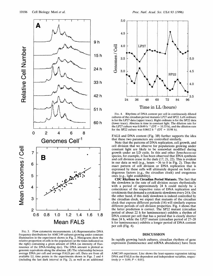
dilution culture of Fig. 2 also exhibited circadian rhythmicity.Lumlinescence cycled with peaks in the early subjective day andtroughs in the early subjective night (Fig. 2A), indicating arhythm of psbAI gene expression as has been observed (9).Average DNA content per cell also showed a distinct circadianrhythm (Fig. 2E). Synechococcus cells are known to containmultiple copies of the chromosome (17). These changes inmean cellular DNA content reflect changes in the relativeproportion of cells in the population containing 2, 3, 4, 5, etc.genome equivalents (Fig. 3A). In this experiment, the averagenumber of chromosomes per cell oscillated between 3.9 and 5.4genomes per cell, with peaks in the early subjective night phaseand troughs in the late subjective-day phase (Fig. 2E).Average cell size as reflected by average FALS showed an
oscillation that paralleled that of cellular DNA content (Fig.2D). We obtained similar results by microscopically measuringcell size directly (data not shown). In addition, there was asignificant positive correlation between mean FALS and DNAcontent in this and other experiments (Fig. 3B), indicating thatlarger cells do indeed have higher DNA content.To determine whether the rate of new DNA synthesis also
exhibited a circadian rhythm, the DNA content per cell and therate of medium dilution were used to calculate the instanta-neous specific rate of increase of DNA and plotted in Fig. 2F.The data have an average slope of zero, suggesting that DNAsynthesis initiates and proceeds at a constant rate, even thoughthe cells are dividing rhythmically. Such uncoupling of DNAsynthesis and cell division has been hypothesized to occur inother species of Synechococcus as well (17, 21). One interpre-tation of these data is that the cells are attempting to maintaina constant cellular chromosome content under conditions inwhich the rate of cytokinesis is slightly faster than the rate ofDNA replication. In such a case, there must be an interval inwhich cytokinesis is slowed to allow the DNA content per cellto catch up. Apparently the circadian clock controls the timingof this slowdown interval. This same logic may apply to cellgrowth as well: if new biomass is produced at a constant rate,we would expect cell size to oscillate such that the cells will belargest at the end of the slowdown interval, just beforecytokinesis begins again. This prediction is upheld by theFALS data in Figs. 2 and 4. The strong correlation between
Cell Biology: Mori et aL
Proc. Natl. Acad. Sci. USA 93 (1996)
5.0
4.5
9 h
15 h
a)0
a)
0
024 h
4.0
3.5
3.0
33 h
42 h24 36 48 60 72 84 96
5.0
4.5
4.0
3.5
Time in LL (hours)51 h
60 h
0 2 4 6 8
Genomes Cell-1
6 0.8 1.0 1.2 1.4 1.6 1.8
Mean FALSFIG. 3. Flow cytometric measurements. (A) Representative DNA
frequency distributions for AMC149 cultures growing under constantillumination in the experiment shown in Fig. 2. Histograms show therelative proportion of cells in the population (at the times indicated onthe right) containing a given amount of DNA (as intensity of fluo-rescence of the DNA-binding dye). The DNA amount is plotted as
genomic equivalents along the abscissa. (B) The relationship betweenaverage DNA per cell and average FALS per cell. Data are from allavailable LL time points in the experiments shown in Figs. 2 and 4(including the last dark interval in Fig. 2), as well as an additional
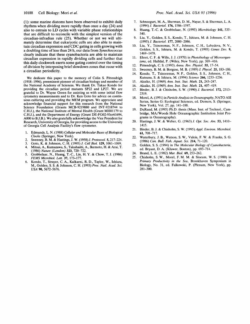
FIG. 4. Rhythms of DNA content per cell in continuously dilutedcultures of the circadian period mutants LP27 and SP22. Left ordinateis for the LP27 data (upper trace). Right ordinate is for the SP22 data(lower trace). Abscissa is time in constant light. The dilution rate forthe LP27 culture was 0.0649 h-I (DT = 10.33 h), and the dilution ratefor the SP22 culture was 0.0612 h-1 (DT = 10.98 h).
FALS and DNA content (Fig. 3B) further supports the ideathat these two parameters are controlled similarly.Note that the patterns of DNA replication, cell growth, and
cell division that we observe for populations growing underconstant light are likely to be somewhat modified duringgrowth under an LD cycle. In this and other Synechococcusspecies, for example, it has been observed that DNA synthesisand cell division cease in the dark (17, 21, 22). This is evidentin our data as well (e.g., hours -36 to 0 in Fig. 2). Thus theexact pattern of cell division or DNA replication that isexpressed by these cells will ultimately depend on both en-dogenous factors (e.g., the circadian clock) and exogenousones (e.g., light availability).CDC Rhythms in Circadian Period Mutants. The fact that
the slowdown in the rate of cell division occurs rhythmicallywith a period of approximately 24 h could merely be acoincidence of the respective rates of DNA replication andcytokinesis that demand a cytokinetic slowdown every 24 h. Onthe other hand, if this daily slowdown is indeed controlled bythe circadian clock, we expect that mutants of the circadianclock that express different periods (14) will similarly expressdifferent periods of cell division properties. Fig. 4 shows thatthe latter prediction is correct. The SP22 mutant (circadianperiod of about 22 h for luminescence) exhibits a rhythm ofDNA content per cell that has a period that is clearly shorterthan 24 h, while the LP27 mutant (circadian period of 27-28h for luminescence) exhibits a longer period of DNA contentper cell (Fig. 4).
DISCUSSIONIn rapidly growing batch cultures, circadian rhythms of geneexpression (luminescence and mRNA abundance) have been
AMC149 experiment. Line shows the least-squares regression takingDNA and FALS as the dependent and independent variables, respec-tively (r = 0.89; P < 0.01).
a)-0Ez
a1)
a)
a1)
B
*0X: *@~~~~~~1
0a)
E00
0a)
CZn
7
6
5-
4
3
20.(
10186 Cell Biology: Mori et al.

Proc. Natl. Acad. Sci. USA 93 (1996) 10187
observed in cultures of the AMC149 strain of Synechococcusthat correspond with the expectation of a circadian rhythm ofluminescence in each cell and an exponential increase in thenumber of cells in the culture (T. Kondo, T.M., N. V. Leb-edeva, S. Aoki, M. Ishiura & S. S. Golden, unpublished data).We wanted to determine whether that result could be obtainedwith continuous cultures in which the cell concentrations wereprecisely measured to discern circadian gating. In this study, weused continuously diluted cultures to maintain a constantgrowth environment. In batch cultures, nutrient levels areconstantly depleted over the course of the experiment. Moreimportantly, the growth of batch cultures entails a continualdecrease in the effective light intensity as the heavily pig-mented cells "self-shade" each other. This self-shading can bevery significant in Synechococcus cultures at cell densitiesgreater than an OD750 of 0.1 (23), which corresponds to a cellconcentration of about 3 x 10⁷ cells per ml. Consequently, theeffective light intensity decreases progressively in batch cul-tures at densities above 3 x 10⁷ cells per ml, which in turnprogressively restricts the only source of energy available tothese cells.Our data clearly show that Synechococcus PCC 7942 cells
growing with doubling times that are considerably faster thanonce per 24 h nonetheless express robust circadian rhythms ofcell division (exemplified by "slowdowns" of division rate),DNA content, and psbAI gene expression (monitored byluminescence of the PpsbAI::luxAB reporter). A previous study(10) reported that the circadian clock globally regulatespromoter activity in Synechococcus PCC 7942. Possibly someof the promoters that we have observed to express circadianrhythms of gene expression are most directly related to celldivision; in other words, that gene expression which might bemost directly involved in cytokinesis exhibits circadian prop-erties because the timing of cell division is dictated by thecircadian oscillator.
Apparently Synechococcus cells are able to keep track of twotiming circuits that are partially independent of each other.More specifically, the circadian clock-as gauged by the timingof cell division and luminescence-seems completely indepen-dent of the cell division cycle, since its period is essentially thesame in cultures with quite different doubling times (T.M.,unpublished observations). On the other hand, the cell divisioncycle does not seem to be completely independent of thecircadian clock, since there are phases of the circadian cycle inwhich cell division slows or stops, implying some inhibitorygating control of the circadian clock over the CDC timer.These observations contradict the usual formulation of the
circadian-infradian rule. What does this mean? It might beargued that the relationship between the circadian clock andthe CDC timer is fundamentally different in eukaryotic uni-cells versus cyanobacteria. On the other hand, the propositionsof the circadian-infradian rule have only been adequatelytested in a few eukaryotic organisms (11). It has therefore beeninstructive to test the implications of the rule in a different typeof organism-in this case, a prokaryote-to show that someorganisms are able to maintain circadian timing in the face ofrapid cell division cycling.
Fig. 5 illustrates a perspective on cell division timing thatsuggests that the regulation in Synechococcus might not befundamentally different from that in eukaryotic unicells. Thefigure illustrates that each of three organisms have phasesduring their circadian cycle in which division is "allowed" or"forbidden." The allowed phases are those during which a"gate is open," or in other nomenclature, during which thecircadian checkpoint is satisfied. The forbidden phases arethose in which the "gate is closed," or in which the circadiancheckpoint is not satisfied. In the case of Euglena, there is onlya brief allowed "gate," and during this time, a single mothercell may divide into only two daughter cells in each cycle. Thegating in Synechococcus is not so very different conceptually
Single-Fission Eukaryotic Cell: Euglena(maximum of 1 division per mother cell each circadian cycle)
onset of division
0 12 24
Forbidden P'hases Allowed Forbidden Phases(1 divisionper cell)
Multiple-Fission Eukaryotic Cell: Chllamydwnomnas(maximum of 4 divisions per mother cell each circadian cycle)
onset of division
0 12 24
Forbidden Phases Allowed (multipledivisions per cell)
Multiple-Fission Prokaryotic Cell: Syntechococcus(maximum of 4 divisions per mother cell each circadian cycle)
0 12 24--- - pK 77A4 4
Allowed Phases(multiple divisionsper cell)
Forbidden Allowed PhaseslPhases (multiple divisions
per cell)
FIG. 5. Model for circadian gating of cell division in the photoau-totrophic unicellular organisms Euglena, Chlamydomonas, and Syn-echococcus. The cell division timing of all three organisms is charac-terized by a "forbidden phase" in which cell division is not allowed toproceed, and an "allowed phase," in which cells of sufficient size areable to divide. In each panel, the white/grey bar indicates the circadiantime course: white for subjective day, grey for subjective night.Numbers under the bar (0, 12, 24) indicate circadian time (O and 24 =dawn; 12 = dusk).
except that it is the forbidden zone (the phase of division"slowdowns") that is brief. In Synechococcus PCC 7942, divi-sion of the mother cell may occur up to four times in a dailycycle (for a doubling time of 6 h). The eukaryotic multiple-fission alga Chlamydomonas is an intermediate case; theallowed zone is brief, but several cycles of nuclear replicationand cytokinesis can occur during this brief interval. Duringthat allowed zone, the period of the nuclear replication cyclemay be as rapid as 0.5-1 h. A single mother cell can divide intoas many as 32 daughter cells per cycle. Consequently, eventhough the cell division cycle is gated by a circadian clock inthese cells (3), the data from Chlamydomonas show that theperiod of the circadian cycle is not required to mesh with theperiod of a single round of nuclear replication. Thus, whilethere may be environmental factors that act to limit celldivision to only certain phases of the daily cycle (12), there maynot be mechanistic constraints that prevent cells from keepingtrack of two timing circuits with different periods.These considerations suggest that perhaps the circadian-
infradian rule is not as accurate a descriptor of the relationshipbetween the circadian oscillator and the cell division cycle aswe once thought, even in eukaryotic unicells. In addition to thecase of Chlamydomonas, which was discussed in the precedingparagraph, there has been other evidence that the circadian-infradian rule might not always apply to eukaryotic unicells
Cell Biology: Mori et aL
Proc. Natl. Acad. Sci. USA 93 (1996)
(1): some marine diatoms have been observed to exhibit dailyrhythms when dividing more rapidly than once a day (24) andalso to entrain to LD cycles with variable phase relationshipsthat are difficult to reconcile with the simplest version of thecircadian-infradian rule (25). Whether or not we will ulti-mately determine that eukaryotic cells are also able to main-tain circadian expression and CDC gating in cells growing witha doubling time of less than 24 h, our data from Synechococcusclearly indicate that these cyanobacteria are able to maintaincircadian expression in rapidly dividing cells and further thatthis daily clockwork exerts some gating control over the timingof division by interposing brief slowdown zones that recur witha circadian periodicity.
We dedicate this paper to the memory of Colin S. Pittendrigh(1918-1996), preeminent pioneer of circadian biology and member ofthe National Academy of Sciences. We thank Dr. Takao Kondo forproviding the circadian period mutants SP22 and LP27. We aregrateful to Dr. Wayne Green for assisting us with some initial flowcytometry measurements and to Dr. Ken Goto for advice on contin-uous culturing and providing the MEM program. We appreciate andacknowledge financial support for this research from the NationalScience Foundation (Grants MCB-9219880 and INT-9218744 toC.H.J.), the National Institute of Mental Health (Grant MHO1179 toC.H.J.), and the Department of Energy (Grant DE-FG02-93er61694,AOOO to B.J.B.). We also gratefully acknowledge the Vice President forResearch, University of Georgia, for providing access to the Universityof Georgia Cell Analysis Facility's flow cytometer.
1. Edmunds, L. N. (1988) Cellular and Molecular Bases ofBiologicalClocks (Springer, New York).
2. Sweeney, B. M. & Hastings, J. W. (1958) J. Protozool. 5, 217-224.3. Goto, K. & Johnson, C. H. (1995) J. Cell Biol. 129, 1061-1069.4. Mitsui, A., Kumazawa, S., Takahashi, A., Ikemoto, H. & Arai, T.
(1986) Nature (London) 323, 720-722.5. Grobbelaar, N., Huang, T.-C., Lin, H. Y. & Chow, T. J. (1986)
FEMS Microbiol. Lett. 37, 173-177.6. Kondo, T., Strayer, C. A., Kulkarni, R. D., Taylor, W., Ishiura,
M., Golden, S. S. & Johnson, C. H. (1993) Proc. Natl. Acad. Sci.USA 90, 5672-5676.
7. Schneegurt, M. A., Sherman, D. M., Nayar, S. & Sherman, L. A.(1994) J. Bacteriol. 176, 1586-1597.
8. Huang, T.-C. & Grobbelaar, N. (1995) Microbiology 141, 535-540.
9. Liu, Y., Golden, S. S., Kondo, T., Ishiura, M. & Johnson, C. H.(1995) J. Bacteriol. 177, 2080-2086.
10. Liu, Y., Tsinoremas, N. F., Johnson, C. H., Lebedeva, N. V.,Golden, S. S., Ishiura, M. & Kondo, T. (1995) Genes Dev. 9,1469-1478.
11. Ehret, C. F. & Wille, J. J. (1970) in Photobiology ofMicroorgan-isms, ed. Halldal, P. (Wiley, New York), pp. 369-416.
12. Pittendrigh, C. S. (1993) Annu. Rev. Physiol. 55, 17-54.13. Sweeney, B. M. & Borgese, M. B. (1989) J. Phycol. 25, 183-186.14. Kondo, T., Tsinoremas, N. F., Golden, S. S., Johnson, C. H.,
Kutsuna, S. & Ishiura, M. (1994) Science 266, 1233-1236.15. Akaike, H. (1969) Ann. Inst. Stat. Math. 21, 243-247.16. Akaike, H. (1969) Ann. Inst. Stat. Math. 21, 407-419.17. Binder, B. J. & Chisholm, S. W. (1990) J. Bacteriol. 172, 2313-
2319.18. Morel, A. (1991) in ParticleAnalysis in Oceanography, NATO ASI
Series, Series G: Ecological Sciences, ed. Demers, S. (Springer,New York), Vol. 27, pp. 141-188.
19. DuRand, M. (1995) Ph.D. thesis (Mass. Inst. of Technol., Cam-bridge, MA/Woods Hole Oceanographic Institution Joint Pro-grams in Oceanography).
20. Hastings, J. W. & Weber, G. (1963) J. Opt. Soc. Am. 53, 1410-1415.
21. Binder, B. J. & Chisholm, S. W. (1995) Appl. Environ. Microbiol.61, 708-717.
22. Waterbury, J. B., Watson, S. W., Valois, F. W. & Franks, S. G.(1986) Can. Bull. Fish. Aquat. Sci. 214, 71-120.
23. Golden, S. S. (1994) in The Molecular Biology of Cyanobacteria,ed. Bryant, D. A. (Kluwer, Boston), pp. 693-714.
24. Brand, L. E. (1982) Mar. Biol. 69, 253-262.25. Chisholm, S. W., Morel, F. M. M. & Slocum, W. S. (1980) in
Primary Productivity in the Sea, Brookhaven Symposium inBiology, No. 31, ed. Falkowski, P. (Plenum, New York), pp.281-300.
10188 Cell Biology: Mori et al.