Effects of inosine on reperfusion injury after heart transplantation☆
Upload
independentCategory
view
0download
0
Cinnamic Anilides as New Mitochondrial Permeability TransitionPore Inhibitors Endowed with Ischemia-Reperfusion Injury ProtectiveEffect in VivoDaniele Fancelli,*,†,○ Agnese Abate,‡,◆ Raffaella Amici,†,○ Paolo Bernardi,⊥ Marco Ballarini,†,§
Anna Cappa,‡,○ Giacomo Carenzi,‡,¶ Andrea Colombo,∇,+ Cristina Contursi,†,▲ Fabio Di Lisa,#
Giulio Dondio,∇, □ Stefania Gagliardi,∇,◇ Eva Milanesi,† Saverio Minucci,§,∥ Gilles Pain,†,△
Pier Giuseppe Pelicci,§ Alessandra Saccani,†,■ Mariangela Storto,†,§ Florian Thaler,†,○ Mario Varasi,‡,○
Manuela Villa,†,○ and Simon Plyte†,●
†Genextra Group, Congenia s.r.l., Via Adamello 16, 20139 Milan, Italy‡Genextra Group, DAC s.r.l., , Via Adamello 16, 20139 Milan, Italy§Department of Experimental Oncology, European Institute of Oncology IEO, Via Adamello 16, 20139 Milan, Italy∥Department of Biosciences, University of Milan, 20100 Milan, Italy⊥Department of Biomedical Sciences, University of Padua, Via Ugo Bassi 58/B, 35121 Padua, Italy#Department of Biomedical Sciences, University of Padua, Viale G. Colombo 3, 35131 Padua, Italy∇NiKem Research s.r.l., Via Zambeletti 25, 20021 Baranzate, MI, Italy
*S Supporting Information
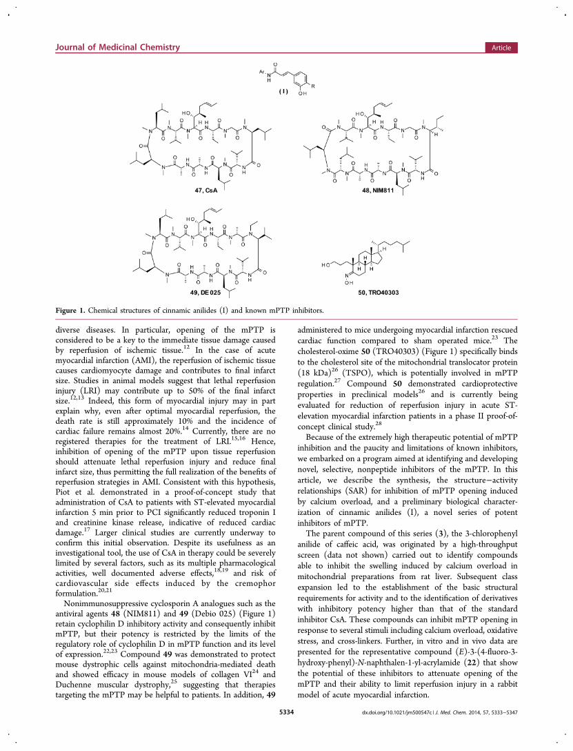
ABSTRACT: In this account, we report the development of a seriesof substituted cinnamic anilides that represents a novel class ofmitochondrial permeability transition pore (mPTP) inhibitors. Initialclass expansion led to the establishment of the basic structuralrequirements for activity and to the identification of derivatives withinhibitory potency higher than that of the standard inhibitorcyclosporine-A (CsA). These compounds can inhibit mPTP openingin response to several stimuli including calcium overload, oxidativestress, and thiol cross-linkers. The activity of the cinnamic anilidemPTP inhibitors turned out to be additive with that of CsA, suggestingfor these inhibitors a molecular target different from cyclophylin-D. Invitro and in vivo data are presented for (E)-3-(4-fluoro-3-hydroxy-phenyl)-N-naphthalen-1-yl-acrylamide 22, one of the most interestingcompounds in this series, able to attenuate opening of the mPTP andlimit reperfusion injury in a rabbit model of acute myocardial infarction.
■ INTRODUCTION
The mitochondrial permeability transition (mPT) is aphenomenon whereby the inner mitochondrial membranebecomes permeable to solutes. This induces cessation ofmitochondrial respiration and energy production, mitochon-drial swelling and outer membrane rupture, release of apoptoticfactors, and cell death leading to organ damage. The mPT ismediated by opening of a nonselective pore within themitochondria: the mitochondrial permeability transition pore(mPTP). Even after intensive investigation, the exact molecularnature of the mPTP is still not known.1 Several proteins havebeen implicated in facilitating the mPTP: adenine nucleotidetransporter, phosphate carrier protein, cyclophilin D, andrecently the F1Fo ATP synthase complex, but no single proteinhas been unambiguously demonstrated to be the pore-formingunit.2−5 However, strong evidence now points to pore
formation by dimers of the ATP synthase.4 Much of ourunderstanding of the functioning of the mPTP comes fromstudies using the immunosuppressive agent 47 (cyclosporine A,CsA)6 (Figure 1), which in the late 1980s was discovered toinhibit the mPTP, as well as from cyclophilin D knockoutmice.7 Opening of the mPTP can be induced by calcium (Ca2+)overload and reactive oxygen species (ROS). These conditionsare common to several diseases including reperfusion injury(angioplasty after a heart attack, transplantation surgery, andstroke), neurological diseases such as amyloid lateral sclerosisand Alzheimer’s disease, traumatic brain injury, dystrophies,and myopathies.8−11 In essence, prolonged opening of themPTP can have catastrophic effects on organ function in many
Received: March 13, 2014Published: June 11, 2014
Article
pubs.acs.org/jmc
© 2014 American Chemical Society 5333 dx.doi.org/10.1021/jm500547c | J. Med. Chem. 2014, 57, 5333−5347
diverse diseases. In particular, opening of the mPTP isconsidered to be a key to the immediate tissue damage causedby reperfusion of ischemic tissue.12 In the case of acutemyocardial infarction (AMI), the reperfusion of ischemic tissuecauses cardiomyocyte damage and contributes to final infarctsize. Studies in animal models suggest that lethal reperfusioninjury (LRI) may contribute up to 50% of the final infarctsize.12,13 Indeed, this form of myocardial injury may in partexplain why, even after optimal myocardial reperfusion, thedeath rate is still approximately 10% and the incidence ofcardiac failure remains almost 20%.14 Currently, there are noregistered therapies for the treatment of LRI.15,16 Hence,inhibition of opening of the mPTP upon tissue reperfusionshould attenuate lethal reperfusion injury and reduce finalinfarct size, thus permitting the full realization of the benefits ofreperfusion strategies in AMI. Consistent with this hypothesis,Piot et al. demonstrated in a proof-of-concept study thatadministration of CsA to patients with ST-elevated myocardialinfarction 5 min prior to PCI significantly reduced troponin Iand creatinine kinase release, indicative of reduced cardiacdamage.17 Larger clinical studies are currently underway toconfirm this initial observation. Despite its usefulness as aninvestigational tool, the use of CsA in therapy could be severelylimited by several factors, such as its multiple pharmacologicalactivities, well documented adverse effects,18,19 and risk ofcardiovascular side effects induced by the cremophorformulation.20,21
Nonimmunosuppressive cyclosporin A analogues such as theantiviral agents 48 (NIM811) and 49 (Debio 025) (Figure 1)retain cyclophilin D inhibitory activity and consequently inhibitmPTP, but their potency is restricted by the limits of theregulatory role of cyclophilin D in mPTP function and its levelof expression.22,23 Compound 49 was demonstrated to protectmouse dystrophic cells against mitochondria-mediated deathand showed efficacy in mouse models of collagen VI24 andDuchenne muscular dystrophy,25 suggesting that therapiestargeting the mPTP may be helpful to patients. In addition, 49
administered to mice undergoing myocardial infarction rescuedcardiac function compared to sham operated mice.23 Thecholesterol-oxime 50 (TRO40303) (Figure 1) specifically bindsto the cholesterol site of the mitochondrial translocator protein(18 kDa)26 (TSPO), which is potentially involved in mPTPregulation.27 Compound 50 demonstrated cardioprotectiveproperties in preclinical models26 and is currently beingevaluated for reduction of reperfusion injury in acute ST-elevation myocardial infarction patients in a phase II proof-of-concept clinical study.28
Because of the extremely high therapeutic potential of mPTPinhibition and the paucity and limitations of known inhibitors,we embarked on a program aimed at identifying and developingnovel, selective, nonpeptide inhibitors of the mPTP. In thisarticle, we describe the synthesis, the structure−activityrelationships (SAR) for inhibition of mPTP opening inducedby calcium overload, and a preliminary biological character-ization of cinnamic anilides (I), a novel series of potentinhibitors of mPTP.The parent compound of this series (3), the 3-chlorophenyl
anilide of caffeic acid, was originated by a high-throughputscreen (data not shown) carried out to identify compoundsable to inhibit the swelling induced by calcium overload inmitochondrial preparations from rat liver. Subsequent classexpansion led to the establishment of the basic structuralrequirements for activity and to the identification of derivativeswith inhibitory potency higher than that of the standardinhibitor CsA. These compounds can inhibit mPTP opening inresponse to several stimuli including calcium overload, oxidativestress, and cross-linkers. Further, in vitro and in vivo data arepresented for the representative compound (E)-3-(4-fluoro-3-hydroxy-phenyl)-N-naphthalen-1-yl-acrylamide (22) that showthe potential of these inhibitors to attenuate opening of themPTP and their ability to limit reperfusion injury in a rabbitmodel of acute myocardial infarction.
Figure 1. Chemical structures of cinnamic anilides (I) and known mPTP inhibitors.
Journal of Medicinal Chemistry Article
dx.doi.org/10.1021/jm500547c | J. Med. Chem. 2014, 57, 5333−53475334

■ CHEMISTRY
Most of the cinnamic amides 3−34 were directly obtained bystandard coupling reactions (Scheme 1) carried out on thecommercially available carboxylic acids 1a−m and anilines 2a−t. Specifically, the carboxylic acids were either activated withSOCl2 (method B) or (COCl)2 (method C) to form their acylchlorides and then coupled with the anilines or the acids weredirectly reacted with the anilines in the presence of DCC(method A) or EDC/HOBt (method D). In some cases, thephenolic moiety of the 3-hydroxy-4-methoxy-cinnamic acid 1dwas first protected as an acetate (Scheme 2).Compounds 10, 14, and 18 were obtained by reduction of
the corresponding nitro derivatives 10a, 14a, and 18a,respectively (Scheme 3). Attempts to prepare compound 37
by direct coupling of the Weinreb amide of 2-(3-hydroxy-4-methoxyphenyl)cyclopropanecarboxylic acid (36) were un-successful, thus the amide was hydrolyzed to the correspondingcarboxylic acid, activated with EDC and HOBt, and finallycoupled with 3-chloroaniline 2a (Scheme 4).N-3-Acetoxy-4-methoxyphenyl-N′-3-Cl-benzoylthiourea 40,
obtained by the reaction of the 3-Cl-aniline 2a and 3-acetyl-4-methoxy benzoyl isothiocyanate 39, was converted into theamino-triazole derivative 41 by condensation with hydrazineand a concomitant loss of the acetyl protecting group (Scheme5).The 1,3,4-oxadiazole analogue 44 was prepared in two steps
by coupling 3-chlorobenzoic acid hydrazide 42 and 3-acetyl-4-methoxy-cinnamic acid 1n followed by alkaline hydrolysis of
Scheme 1a
aReagents and conditions: coupling method A, ArNH2, DCC, THF, reflux; coupling method B, (i) SOCl2, THF, 50 °C, (ii) ArNH2, NEt3, THF, 0°C to rt; coupling method C, (i) (COCl)2, DCM, reflux, (ii) ArNH2, NEt3, DCM, 0 °C to rt; coupling method D, ArNH2, EDC, HOBt, DCM, 0 °Cto rt; coupling method E, (i) SOCl2, reflux, (ii) ArNH2, pyridine, 85 °C.
Journal of Medicinal Chemistry Article
dx.doi.org/10.1021/jm500547c | J. Med. Chem. 2014, 57, 5333−53475335

the acetate protecting group (Scheme 6). Finally, thephenylpropanoic analogue 46 was obtained by coupling thecorresponding 3-hydroxy-4-methoxy-phenylpropanoic acid 45that was activated with SOCl2 and then treated with 3-chloro-aniline 2a.
■ SAR
The mPTP inhibitory activity of cinnamic anilides 3−34 wasinitially evaluated by measuring their ability to protect murinemitochondria from mPTP opening induced by the exposure toincreasing concentrations of Ca2+ (calcium retention capacityassay (CRC)). This method allows measuring of the propensityof mitochondria to open the mPTP after calcium uptake. In thepresence of extra-mitochondrial calcium, isolated mitochondriatake up calcium into the matrix via the calcium uniporter.Continued addition of extra-mitochondrial calcium andsubsequent uptake leads to the calcium-induced opening ofthe mPTP, mitochondrial depolarization, and release of storedcalcium. The total amount of calcium that can be retained untilcalcium-induced mPTP opening occurs is termed the calciumretention capacity.29 An example of the calcium retention
Scheme 2a
aReagents and conditions: (a) (i) NaH, THF, 0 °C, (ii) Ac2O reflux; (b) (i) SOCl2, THF, reflux, (ii) ArNHR3, DIPEA, THF, reflux; (c) HCl,MeOH, THF, rt; (d) (i) NaOHaq MeOH, reflux, (ii) HClaq.
Scheme 3a
aReagents and conditions: (a) SnCl2·2H2O, EtOH, EtOAc, reflux,1.5−7 h.
Journal of Medicinal Chemistry Article
dx.doi.org/10.1021/jm500547c | J. Med. Chem. 2014, 57, 5333−53475336

capacity assay using the well-characterized mPTP inhibitor CsAis shown in Figure 2.Measuring the increase of calcium needed to induce the
mPTP in mitochondria treated with mPTP inhibitor vsuntreated mitochondria (Ca2+ overloading, μM) is a convenientand sensitive assay for assessing the ability of the compounds to
inhibit mPTP opening. The ratio between the amount of
calcium required to trigger mPT in the presence of the
compound (CRCi) with respect to that required to induce
mPT in the absence of the compound (CRCo) is a measure of
the inhibitory effect of the compound on the mPTP.
Scheme 4. Synthesis of Racemic trans-Cyclopropane Analoguea
aReagents and conditions: (a) Me-NH-OMe·HCl, EDC, HOBt, DCM, rt, 16 h; (b) (i) NaH, Me3SO+I−, DMSO, rt, 16 h, (ii) HClaq; (c) (i) tBuOK,
H2O, Et2O, rt, 20 h, (ii) 3-ClPhNH2, EDC, HOBt, DCM, rt, 36 h.
Scheme 5a
aReagents and conditions: (a) (i) SOCl2, DMF, THF, reflux, 2 h, (ii) KSCN, MeCN, reflux, 2 h; (b) 3-Cl-aniline, MeCN, 0.5 h rt; (c) NH2NH2,CHCl3, reflux, 2 h.
Scheme 6a
aReagents and conditions: (a) POCl3, 1,4-dioxane, reflux, 1 h; (b) 4N HCl, 1,4-dioxane, MeOH, rt.
Journal of Medicinal Chemistry Article
dx.doi.org/10.1021/jm500547c | J. Med. Chem. 2014, 57, 5333−53475337

All the cinnamic anilides here described were tested in theCRC assay in a dose escalation series at 0.1, 0.5, 1, and 5 μM.Because no significant differences were observed in the potencyranking of the compounds tested at different concentrations(see Supporting Information for the complete result data set),only the CRC values measured at 1 μM inhibitor concentrationare reported in the following SAR tables.In the absence of structural information on the binding mode
of these cinnamic anilides to their target mPTP component, wefirst performed a limited SAR investigation to identify thestructural elements of the hit compound 3 that are essential formPTP inhibition.The structure of 3 was divided into three regions, namely the
cinnamic phenyl ring, the central linker, and the aniline arylring (Figure 3), and systematic variations were carried out ineach region.
For this purpose and as the first attempt, the influence of thevicinal hydroxy moieties at the cinnamic phenyl ring wasinvestigated (Table 1). The comparison of the activities ofcompounds 4 vs 5 and 6 vs 7 immediately suggests a crucialrole for the hydroxyl group at position 3. This was furtherconfirmed by the complete inactivity of compounds 9−12,wherein the 3-hydroxyl is replaced by potential bioisosters suchas fluorine, amino, or sulfonamido groups. In contrast,replacement of the hydroxyl at position 4 with differentmoieties (compounds 4, 6, 13, 14, and 23) was better toleratedand the modifications variously modulated the potency. Themost active example in this series resulted to be the 3-OH-4-OMe derivative 6, which demonstrated potency comparable to
that of the reference standard CsA and was used for the veryinitial characterization of the mPTP inhibitory properties of theclass. Thus, we decided to fix the right-hand side of compound6 with the 3-OH-4-OMe moiety while exploring differentcentral linkers and anilines.The impact of the central linker on the activity of the
compounds was explored starting from analogue 6, but all themodifications introduced in this region such as N-methylation(34), double bond reduction (46), cyclopropanation (37), orcyclization (41, 44) completely abolished activity (Table 2).Finally, the effect of different anilines was investigated (Table
3, 15−33). For this purpose, the 3-hydroxy-4-methoxy-cinnamic moiety was kept fixed. Far from being exhaustive,this initial exploration suggests that, while the chlorine presentin the initial hit turned out to be the best among the residues
Figure 2. Calcium retention capacity assay. CaCl2 (1 μL of 2 mMstock solution) was added every minute to mouse liver mitochondria(200 μL of a 0.5 mg/mL suspension) in the presence of thefluorescent Ca2+ indicator Calcium Green 5N. Calcium uptake wasfollowed by measuring extra-mitochondrial calcium green fluorescenceuntil mPTP opening was achieved. CsA (1 μM) was added to themitochondria immediately prior to the start of the experiment.
Figure 3. Regions of the structure of the hit compound 3 subjected toseparate SAR investigation.
Table 1. N-(3-Chlorophenyl)cinnamic Anilides
compd R1 R2 CRCi/CRC0 (%)a SDb
CsA 224 123 OH OH 155 124 OH H 136 125 H OH 87 8.96 OH OMe 186 147 OMe OH 93 118 OMe OMe 75 6.29 F H 90 1010 NH2 H 94 8.611 SO2NHMe H 91 8.112 −NHCHCH− 87 8.413 OH Me 133 1114 OH NH2 102 1023 OH F 175 16
aIncrease of the calcium retention capacity of mouse livermitochondria after incubation with 1 μM inhibitor. bn = 3.
Table 2. Modification of the Central Linker
aIncrease of the calcium retention capacity of mouse livermitochondria after incubation with 1 μM inhibitor. bn = 3.
Journal of Medicinal Chemistry Article
dx.doi.org/10.1021/jm500547c | J. Med. Chem. 2014, 57, 5333−53475338

explored at position 3 of the phenyl ring (6, 15−19, 24), thisregion of the scaffold tolerates a wide range of modifications. Infact, phenyl substitution at positions 2 and 4 (20, 21), phenyldisubstitution (25−27), and replacement of the phenyl withbicyclic moieties (28−33) led to compounds with maintainedor improved potency.In summary, the above findings allowed us to identify the
phenol at position 3 of the cinnamic phenyl ring and theacrylamido linker as the structural determinants for the activityof this novel class of mPTP inhibitors, while modifications inthe aniline region of the scaffold as well as position 4 of thecinnamic phenyl showed to be well tolerated and thus usablefor the optimization of potency and properties.Searching for a lead compound more potent than the initial
lead 6, we drew our attention to the high potency showed bythe naphthyl derivative 30 and investigated the effect of theintroduction of heteroatoms in the bicyclic ring (31−33) as
well as the replacement of the methoxy moiety with fluorine(22). In the latter case, the aim was to reduce lipophilicity andpotential metabolic liabilities of the molecule. While theheterobicyclic derivatives were less active, the fluorine analogue22 showed the highest potency in the series and was selected asa class representative compound for a deeper biologicalcharacterization.
■ BIOLOGY
Typically, opening of the mPTP in isolated mitochondria canbe achieved by challenging the mitochondria with high Ca2+
concentrations and can be assayed spectrophotometrically bymonitoring the decrease of absorbance at 540 nm (A540),which indicates mitochondrial swelling as a result of soluteinflux into the mitochondrial matrix via the open mPTP.In addition, the mPTP opening can be induced by
challenging isolated mitochondria with oxidizing agents (e.g.,diamide or menadione) or uncouplers of electron transport(e.g., trifluorocarbonylcyanide phenylhydrazone (FCCP)). In apreliminary assessment of the mechanism of action of this novelclass of inhibitors, compound 6 was tested in this swelling assayfor its ability to prevent mPTP opening in response to thedifferent stimuli mentioned above in isolated mouse livermitochondria. The results reported in Figure 4 show thatcinnamic anilides prevent mPTP opening induced by stimuliindependent of changes in calcium flux, thus suggesting thatthey behave as genuine inhibitors of the mPTP and not ofcalcium homeostasis (Figure 4B−D).Because the cinnamide skeleton is present in diverse
compounds with a wide range of pharmacological activ-ities,30−34 we profiled our initial lead molecule in a Cerepdiversity profile. Thus, compound 6 was tested at aconcentration of 10 μM versus a panel of 69 receptors andion channels and 14 enzymes (see complete list in theSupporting Information). Affinity for each target is measuredby displacement of a radiolabeled ligand specific for eachparticular receptor or enzyme. The results, expressed in termsof percentage of displaced ligand, confirmed a notably cleanprofile for 6: in fact, for just 1 (norepinephrine transporter) outof 83 assessed targets was found a displacement higher than50%. These initial results, positive in terms of mechanism ofaction and selectivity, encouraged us in further developing thisclass.After the initial round of class expansion, compound 22 was
selected for wider biological characterization. Prompted by thepresence of an α,β-unsaturated amide group in the structure,the electrophilic behavior of compound 22 was first examined.The compound was incubated at a test concentration of 100μM with glutathione (100 μM) or N-acetyl-cysteine (100 μM)at 25 °C. HPLC analysis carried out after 24 h incubationshowed quantitative recovery of 22 without detection of anythiol adducts (see Supporting Information).A more extensive assessment of the potency in the CRC
assay was then carried out in comparison to the standard CsA.The CRC of freshly prepared mouse liver mitochondria wasdetermined for 22 and CsA in a dose escalation series from 0.1to 5.0 μM (Figure 5). A clear dose response was observed with22 and the compound enabled mitochondria to withstand veryhigh levels of Ca2+, significantly superior to CsA. In addition,the CRC of CsA reached a plateau around 0.5 μM of CsA anddid not increase at higher doses. This is probably due thetitration of cyclophillin D, its primary mitochondrial target.
Table 3. 3-Hydroxycinnamic Anilides
aIncrease of the calcium retention capacity of mouse livermitochondria after incubation with 1 μM inhibitor. bn = 3.
Journal of Medicinal Chemistry Article
dx.doi.org/10.1021/jm500547c | J. Med. Chem. 2014, 57, 5333−53475339

The observation that the CRC of CsA reached a plateau atrelatively low concentrations while the cinnamic anilidescontinued to increase the CRC was the first indication thatthe molecular target of this new class might not be cyclophilinD. To further investigate this point, we measured the CRC of
CsA in combination with our inhibitors. Figure 6 shows thatthere is a clear additivity between CsA and 22. The protectiveeffect of the combination is significantly higher than themaximum effect obtainable with CsA alone, which furtherconfirms the hypothesis that the cinnamic anilides do not targetcyclophilin D.
Figure 4. The cinnamic anilides inhibit opening of the mPTP in isolated mitochondria in response to various stimuli. Compound 6 (1 μM) inhibitsswelling of mouse liver mitochondria exposed to (A) calcium overload (CaCl2 150 μM), (B) uncoupling of electron transport chain (FCCP 50 nM),(C,D) oxidative stress (menadione 100 μM and diamide 300 μM, respectively).
Figure 5. CRC of mouse liver mitochondria treated with increasingconcentrations of 22 or CsA.
Figure 6. CRC of mouse liver mitochondria treated with combinationof 22 and CsA; the results are the mean of three experiments ±standard deviation.
Journal of Medicinal Chemistry Article
dx.doi.org/10.1021/jm500547c | J. Med. Chem. 2014, 57, 5333−53475340

The potential use of mPTP inhibitors in preventing LRI afterPCI requires that the inhibitor can reach the cardiacmitochondria very rapidly due to the fact that the mPTPopens within the first few minutes of reperfusion. To this end,we assessed the CRC of cardiac mitochondria after wholehearts had been perfused with the compound for just 2 min.Freshly prepared beating mouse hearts, immobilized on aLangendorff reperfusion apparatus, were perfused withcompound 22 at a 5 μM concentration or the correspondingamount of DMSO for 2 min. Hearts were then flushed withbuffer, the mitochondria prepared, and the CRC measured(Figure 7A).
Mitochondria from hearts perfused with 22 showed asignificantly higher CRC than those treated with the vehicle(DMSO). These data clearly show that 22 can reach the cardiacmitochondria in whole organs in as little as 2 min.Further, similar CRC experiments performed ex vivo on
cardiac mitochondria prepared 5 min after iv injection of 22(15 mg/kg) into the tail vein of mice confirmed that thecompound, after systemic administration, is able to reachrapidly cardiac mitochondria and exert its protecting effect(Figure 7B).On the basis of these results, we decided to assess the efficacy
of 22 in a rabbit model of acute myocardial infarction.New Zealand white rabbits were subjected to 30 min of left
anterior descending (LAD) coronary artery ligation followed by4 h of reperfusion. Animals were then sacrificed, and the heartswere analyzed to determine the area that had been ischemic(area at risk, AAR) during the ligation and the area that wasnecrotic (infarct size, IS) at the end of the reperfusion period.Animals were given either vehicle, 22, or CsA via iv bolusinjection 5 min prior to unligation of the LAD coronary artery.The ratio of the area at risk to infarct area (AAR/IS) is ameasure of the extent of the final damage to the heart with
respect to the amount of tissue exposed to the ischemic insult.To correlate the pharmacodynamics response with plasmaconcentration of 22, a satellite group of animals (n = 5) wastreated with the same dose of 22 and the test item quantified inplasma up to 240 min.The data in Figure 8 clearly show that 22, when given just
prior to organ reperfusion, is cardioprotective in this animal
model of acute myocardial infarction and can reduce infarct sizeby almost 50%. Compound 22 showed a plasma concentrationafter 5 min of 4.8 μM that reduced to 0.02 μM after 240 minfrom the iv injection. The compound was cleared very rapidlyin rabbit plasma, showing a t1/2 of about 30 min. However, asexpected for an agent targeting I/R injury, this short-termedplasma exposure was able to induce a significant pharmacody-namic effect in this model of acute myocardial infarction.Further, the reduction in infarct size was similar to that seenwith CsA, which works primarily by attenuating the mPTP,thus confirming the validity of targeting the mPTP.Finally, to exclude potential cardiovascular side effects that
could hamper the development of these compounds as noveltherapeutics to treat acute ischemia-reperfusion injury,compound 22 was tested in vitro on the cardiac hERG channeland in vivo in a standard rat telemetry study. Compound 22 didnot show any significant binding to hERG up to theconcentration of 30 μM, while no significant alterations ofany of the monitored parameters (body temperature, heart rate,systolic and diastolic blood pressure) were observed in the ratsafety study up to a dose of 30 mg/kg given iv, whichcorrespond to plasma levels well above those observed in theefficacy experiments (see Supporting Information).Taken together, these experiments strongly support the
notion that administration of our cinnamic anilide mPTP
Figure 7. (A) Calcium retention capacity of mitochondria isolatedfrom mouse hearts after perfusion with compound 22 (Langendorffapparatus) at 5 μM for 2 min. Data expressed as absolute quantity ofcalcium retained. Vehicle (DMSO) treated group CRC = 80 μM, SD =6.8, n = 6; treated group CRC = 125 μM, SD = 6.0 (n = 6); p < 0.01.(B) Calcium retention capacity of mitochondria isolated from mousehearts after 5 min from iv administration of compound 22 (15 mg/kg).Data expressed as absolute quantity of calcium retained. Vehicle(DMSO) treated group CRC = 69.8 μM, SD = 15.7, n = 16; treatedgroup CRC = 108.4 μM, SD = 20.2 (n = 6); p < 0.01.
Figure 8. Compound 22 is cardioprotective in a rabbit model of acutemyocardial infarction. Rabbits were subjected to left anteriordescending (LAD) coronary artery occlusion for 30 min followed by4 h of reperfusion. Compound 22 (5 mg/kg in 20% DMSO, 40%PEG400) and CsA (10 mg/kg as Sandimmune) were administered byiv bolus 5 min prior to reperfusion. Infarct size (IS) and area at risk(AAR) were determined by measuring TTC and Evans blue-stainedheart slices. Vehicle treated group AAR/IS = 57.3%, SD = 18.1 n = 8;compound 22 treated group AAR/IS = 30.9%, SD = 7.8 (n = 8) p <0.01; CsA treated group group AAR/IS = 32.6%, SD = 14.1 (n = 8) p< 0.01.
Journal of Medicinal Chemistry Article
dx.doi.org/10.1021/jm500547c | J. Med. Chem. 2014, 57, 5333−53475341

inhibitors during acute myocardial infarction and just prior toreperfusion therapy should prevent mPTP opening, attenuatelethal reperfusion injury, and reduce infarct size. Indeed, a drugcandidate from this chemical series is currently undergoingclinical evaluation for the eventual treatment of lethalreperfusion injury in the setting of acute myocardial infarction.
■ CONCLUSIONSWe identified a series of novel mPTP inhibitors based on thecinnamic anilido scaffold that are characterized by simplicity instructure, low molecular weight, and mechanism of actiondifferent from cyclophilin D inhibition.On the basis of initial SAR studies, we identified the
following structural requirements to gain mPTP inhibitoryactivity: (i) a phenol moiety at position 3 of the cinnamicphenyl ring, (ii) an unsubstituted acrylamido linker joining thecinnamic phenyl ring and the anilinic aryl moiety.The high potential of cinnamic anilides is exemplified by the
rapid identification of compound 22, which demonstratedpotency equal or higher than the gold standard, CsA, in a seriesof in vitro and in vivo experiments.Taken together, all these findings highlight that this series of
cinnamic anilides offer the possibility of studying the biology ofthe mPTP and the therapeutic potential of mPTP inhibitionusing potent, low molecular weight inhibitors.
■ EXPERIMENTAL SECTIONReagents and solvents used, unless stated otherwise, were ofcommercially available reagent grade quality and were used withoutfurther purification. Flash chromatography purifications wereperformed on Merck silica gel 60 (0.04−0.063 mm). Nuclear magneticresonance spectra (1H NMR) were recorded on a Bruker 400 MHzspectrometer at 300 K and are referenced in ppm (δ) relative to TMS.Coupling constants (J) are expressed in hertz (Hz). HPLC−MSexperiments were performed either on an Acquity UPLC apparatus,equipped with a diode array and a Micromass SQD single quadruple(Waters) or on an Agilent 1100, equipped with a diode array and aBruker ion−trap Esquire 3000+. Purity was monitored at 254 nm, andthe purities of the compounds used for biological tests were found tobe at least 95% with the exception of 10 (90%) and 11 (90%) (seeSupporting Information).3-Acetoxy-4-hydroxycinnamic Acid (1n). NaH (60%; 4.5 g,
113.29 mmol) was added portionwise to a solution of 3-hydroxy-4-methoxycinnamic acid (10 g, 51.5 mmol) in dry THF (220 mL) whilestirring at 0 °C under a nitrogen atmosphere. Stirring was continued at0 °C for about 40 min, then acetic anhydride (7.3 mL, 77.25 mmol)was added dropwise at 0 °C. The reaction mixture was heated at refluxfor about 3 h, further acetic anhydride (1.46 mL, 15.4 mmol) wasadded, and heating continued for additional 6 h. After cooling to rt,solvents were evaporated; the residue was taken up in 2 N HCl andwater, extracted with EtOAc. The combined organic layers werewashed with water and brine, dried over sodium sulfate, andconcentrated to dryness. The resulting colorless powder was trituratedin EtOAc, filtered, and dried under vacuum at 40 °C to afford 3-acetoxy-4-hydroxycinnamic acid 1n (10 g, 82%) as a colorless powder.1H NMR (500 MHz, DMSO-d6) δ 12.27 (s, 1H), 7.58−7.55 (m, 1H),7.54−7.49 (m, 2H), 7.15 (m, 1H), 6.40 (d, J = 16.1 Hz, 1H), 3.81 (s,3H), 2.26 (s, 3H). m/z (ES+), (M + Na)+ = 259.(E)-N-(3-Chlorophenyl)-3-(3,4-dihydroxyphenyl)-prop-2-en-
amide (3). 3-Chloroaniline 2a (23.6 μL, 2.22 mmol) and N,N′-dicyclohexylcarbodiimide (504 mg, 2.44 mmol) were added to asolution of caffeic acid 1a (400 mg, 2.22 mmol) in THF (10 mL), andthe resulting mixture was stirred at reflux for 7 h. After cooling toroom temperature, the solid residue was filtered off and the remainingsolution was evaporated. The residue was purified by flash silicachromatography (elution gradient: 20−50% EtOAc in n-hexane) toafford (E)-N-(3-chlorophenyl)-3-(3,4-dihydroxyphenyl)-prop-2-enam-
ide 3 (270 mg 48%) as a white powder. 1H NMR (400 MHz, DMSO-d6) δ 10.27 (bs, 1H), 9.38 (bs, 2H), 7.96 (m, 1H), 7.54 (m, 1H), 7.46(d, J = 15.6 Hz, 1H), 7.38 (m, 1H), 7.14 (m, 1H), 7.05 (m, 1H), 6.96(m, 1H), 6.82 (m, 1H), 6.54 (d, J = 15.6 Hz, 1H). m/z (ES+), (M +H)+ = 290.
(E)-N-(3-Chlorophenyl)-3-(3-hydroxyphenyl)-prop-2-enam-ide (4). A solution of 3-hydroxycinnamic acid 1b (1.0 g, 6.1 mmol)and thionyl chloride (0.53 mL, 7.32 mmol) in dry THF (15 mL) wasstirred at 55 °C for 3 h. Then a further aliquot of thionyl chloride (0.1mL, 1.38 mmol) was added, and the mixture was stirred at refluxtemperature for additional 1.5 h. After cooling to about 5 °C, asolution of 3-chloroaniline 2a (0.65 mL, 6.1 mmol) and triethylamine(3.4 mL, 24.4 mmol) in dry THF (5 mL) was added dropwise. Afterstirring at rt for 16 h, the reaction mixture was diluted with DCM andwashed with water, 0.5N aqueous hydrochloric acid, and brine.Organic layer was dried over sodium sulfate, concentrated underreduced pressure, and purified by flash silica chromatography(petroleum ether/EtOAc 45:55) to afford (E)-N-(3-chlorophenyl)-3-(3-hydroxyphenyl)-prop-2-enamide 4 (671 mg, 40%) as a beige solid.1H NMR (400 MHz, DMSO-d6) δ 10.37 (s, 1H), 9.64 (s, 1H), 7.93(m, 1H), 7.53−7.49 (m, 2H), 7.37 (m, 1H), 7.25 (m, 1H), 7.13 (m,1H), 7.05 (m, 1H), 6.70 (m, 1H), 6.83 (m, 1H), 6.72 (d, J = 15.6 Hz,1H). m/z (ES+), (M + H)+ = 274.
(E)- 3-(3-Aminophenyl)-N-(3-chlorophenyl)-prop-2-enamide(10). SnCl2·2H2O (530 mg, 2.35 mmol) was added to a solution of(E)-3-(3-nitrophenyl)-N-(3-chlorophenyl)-prop-2-enamide 10a (142mg, 0.47 mmol) in EtOH/EtOAc 1/1 (6 mL). After stirring at refluxfor 7 h, the reaction mixture was cooled to rt and poured onto ice/water; pH was adjusted to 8 with NaHCO3 (satd aq), and the resultingaqueous solution was extracted with ethyl acetate. The combinedorganic layers were washed with brine, dried over sodium sulfate, andconcentrated under reduced pressure to afford (E)-3-(3-amino-phenyl)-N-(3-chlorophenyl)-prop-2-enamide 10 (88 mg, 69%) as anorange solid. 1H NMR (400 MHz, DMSO-d6) δ 10.37 (s, 1H), 7.94(m, 1H), 7.52 (m, 1H), 7.44 (d, J = 15.67 Hz, 1H), 7.36 (m, 1H),7.14−7.07 (m, 2H), 6.78−6.75 (m, 2H), 6.66 (d, J = 15.67 Hz, 1H),6.61 (m, 1H), 5.25 (s, 2H). m/z (ES+), (M + H)+ = 273.
(E)-N-(3-Chlorophenyl)-3-[3-[(methylsulfonyl)amino]-phenyl]-prop-2-enamide (11). A solution of 3-[(methylsulfonyl)-amino]cinnamic acid 1i (329 mg, 1.36 mmol), oxalyl chloride (0.35mL, 4.1 mmol), and catalytic dry DMF in dry DCM (15 mL) wasstirred at reflux for 1.5 h. After cooling to rt, solvent and excess oxalylchloride were evaporated under reduced pressure and the residue wastaken up in dry toluene and concentrated. A solution of 3-chloroaniline 2a (0.144 mL, 1.36 mmol) and triethylamine (0.284mL, 2.04 mmol) in dry DCM (8 mL) was added dropwise at 0 °C tothe raw acyl chloride in toluene. After stirring at rt for 16 h, thereaction mixture was diluted with DCM and washed with water, 0.5Naqueous hydrochloric acid, NaHCO3 (satd aq), and brine. Organiclayer was dried over sodium sulfate, concentrated under reducedpressure, and purified by flash silica chromatography (DCM/acetone97:3) and trituration with diisopropyl ether/methanol to afford (E)-N-(3-chlorophenyl)-3-[3-[(methylsulfonyl)amino]phenyl]-prop-2-enam-ide 11 (50 mg, 14%) as a beige solid. 1H NMR (400 MHz, DMSO-d6)δ 11.5 (s, 1H), 9.92 (s, 1H), 7.94 (m,bs, 1H), 7.60−7.34 (m, 6H),7.25−7.12 (m, 2H), 6.77 (d, J = 15.65 Hz, 1H), 3.35 (s, 3H). m/z (ES+), (M + H)+ = 351.
(E)-N-(3-Chlorophenyl)-3-(1H-indol-6-yl)-prop-2-enamide(12). EDC hydrochloride (245 mg, 1.28 mmol) and HOBt (173 mg,1.28 mmol) were added to a solution of (E)-3-(1H-indol-6-yl)-prop-2-enoic acid 1j (120 mg, 0.64 mmol) in dry DCM (6 mL) while coolingat 0 °C. The mixture was allowed to warm to room temperature andstirred for 45 min. 3-Chloroaniline 2a (0.082 mL, 0.77 mmol) wasadded, and the mixture was stirred at room temperature for 4 h, thenat reflux for 7 h. After cooling, the mixture was diluted with DCM,washed with NaHCO3 (satd aq), dried over sodium sulfate, filtered,and concentrated. The residue was purified by flash silicachromatography (elution gradient: DCM/MeOH 100:0.5) to afford(E)-N-(3-chlorophenyl)- 3-(1H-indol-6-yl)-prop-2-enamide 12 (82mg, 43%) as a pale-yellow powder. 1H NMR (400 MHz, DMSO-d6)
Journal of Medicinal Chemistry Article
dx.doi.org/10.1021/jm500547c | J. Med. Chem. 2014, 57, 5333−53475342

δ 11.35 (s, 1H), 10.30 (s, 1H), 7.95 (t, J = 2, 1H), 7.70 (d, J = 15.6,1H), 7.64 (s, 1H), 7.59 (d, J = 8.4, 1H), 7.54−7.52 (m, 1H), 7.46 (t, J= 2.6, 1H), 7.36 (t, J = 8, 1H), 7.30 (dd, J = 8.4, J = 1.2, 1H), 7.11 (dd,J = 8, J = 1.2, 1H), 6.75 (d, J = 15.6, 1H), 6.47 (m, 1H). m/z (ES+),(2M + Na)+ = 615.(E)-N-(3-Chlorophenyl)-3-(4-amino-3-hydroxyphenyl)-prop-
2-enamide (14). SnCl2·2H2O (562 mg, 2.5 mmol) was added to asolution of (E)-N-(3-chlorophenyl)-3-(3-hydroxy-4-nitrophenyl)-prop-2-enamide 14a (159 mg, 0.5 mmol) in EtOH (6 mL). Themixture was stirred at reflux for about 1.5 h, cooled to roomtemperature, and concentrated. The residue was taken up withNaHCO3 (satd aq) and Rochelle’s salt solution and extracted withEtOAc. The combined organic layers were washed with brine, driedover sodium sulfate, and concentrated under reduced pressure toafford (E)-N-(3-chlorophenyl)-3-(4-amino-3-hydroxyphenyl)-prop-2-enamide 14 (89 mg, 62%) as a yellow powder. 1H NMR (400MHz, DMSO-d6) δ 10.16 (s, 1H), 9.34 (s, 1H), 7.93 (t, J = 1.6 Hz,1H), 7.50 (m, 1H), 7.34 (m, 2H), 7.08 (m, 1H), 6.92 (s, 1H), 6.87 (m,1H), 6.59 (d, J = 8.0 Hz, 1H), 6.38 (t, J = 15.6 Hz, 1H), 5.15 (s, 2H).m/z (ES+), (M + H)+ = 289.(E)-N-(3-Aminophenyl)-3-(3-hydroxy-4-methoxyphenyl)-
prop-2-enamide (18). A suspension of (E)-3-(3-hydroxy-4-methox-yphenyl)-N-(3-nitrophenyl)-prop-2-enamide 18a (314 mg, 1.00mmol) and SnCl2·2H2O (1.128 g, 5.00 mmol) in EtOH (20 mL)was heated at reflux for 1.5 h. The resulting solution was cooled toroom temperature and concentrated. The residue was taken up withNaHCO3 (satd aq) and Rochelle’s salt solution and extracted withEtOAc. The combined organic layers were dried over sodium sulfate,filtered, and concentrated. Purification by flash silica chromatography(DCM/MeOH 98:2) afforded (E)-N-(3-aminophenyl)-3-(3-hydroxy-4-methoxyphenyl)-prop-2-enamide 18 (110 mg, 39%) as an off-whitepowder. 1H NMR (400 MHz, DMSO-d6) δ 9.75 (s, 1H), 9.19 (s, 1H),7.38 (d, J = 15.6 Hz, 1H), 7.02−6.90 (m, 5H), 6.79 (d, J = 8.0 Hz,1H), 6.60 (d, J = 15.6 Hz, 1H), 6.26 (dd, J = 8.0 Hz, J = 1.2 Hz, 1H),5.05 (s, 2H), 3.81 (s, 3H). m/z (ES+), (2M + Na)+ = 591.( E ) -N - ( 3 - C a rboxam idopheny l ) - 3 - ( 3 - a c e t o xy - 4 -
methoxyphenyl)prop-2-enamide (24a). A solution of 3-acetoxy-4-methoxycinnamic acid 1n (0.45 g, 1.9 mmol), thionyl chloride (0.14mL, 2.66 mmol) ,and 3 drops of DMF in dry THF (10 mL) wasstirred at reflux for 2 h. Then a further aliquot of thionyl chloride (0.05mL, 0.76 mmol) was added, and the mixture was stirred at refluxtemperature for additional 2 h. After cooling to about 5 °C, a solutionof 3-aminobenzamide 2j (0.259 g, 1.9 mmol) and triethylamine (0.53mL, 3.8 mmol) in dry DCM (5 mL) was added dropwise. After stirringat rt overnight, THF was removed under vacuum and the residue wastriturated in DCM to give a first aliquot of the target cinnamic anilide24a as a beige solid (185 mg). After filtration, the solution was washedwith 1N aqueous hydrochloric acid and NaHCO3 (satd aq), dried oversodium sulfate, and evaporated to afford a second aliquot of 24a. Thetwo solids were mixed and triturated in DCM to yield (E)-N-(3-carboxamidophenyl)-3-(3-acetoxy-4-methoxyphenyl)prop-2-enamide24a (671 mg, 79%). m/z (ES+), (M + Na)+ = 377.( E ) -N - ( 3 -C a rboxam idopheny l ) - 3 - ( 3 - h yd roxy - 4 -
methoxyphenyl)prop-2-enamide (24). A suspension of (E)-N-(3-carboxamidophenyl)-3-(3-acetoxy-4-methoxyphenyl)prop-2-enamide24a (535 mg, 1.5 mmol) and NaOH (50% in water) (0.16 mL, 3mmol) in MeOH (5 mL) was stirred at reflux for 1 h. After cooling toroom temperature, MeOH was evaporated, the reaction mixture wasdiluted with water, and pH was adjusted to 6 with 2 N HCl. Theresulting precipitate was filtered off, washed with water, and driedunder vacuum to afford (E)-N-(3-carboxamidophenyl)-3-(3-hydroxy-4-methoxyphenyl)prop-2-enamide 24 (252 mg, 54%) as a white solid.1H NMR (400 MHz, DMSO-d6) δ 10.21 (s, 1H), 9.23 (s, 1H), 8.10 (s,1H), 7.92−7.87 (m, 2H), 7.54 (d, J = 7.6 Hz, 1H), 7.45 (d, J = 15.6Hz, 1H), 7.39 (t, J = 8.0 Hz, 1H), 7.32 (s, 1H), 7.05−7.03 (m, 2H),6.97 (d, J = 8.4 Hz, 1H), 6.60 (d, J = 15.6 Hz, 1H), 3.81 (s, 3H). m/z(ES+), (2M + H)+ = 625.(E)-3-(3-Hydroxy-4-methoxyphenyl)-N-indan-1-yl-acryla-
mide (28). (E)-3-(3-Acetoxy-4-methoxyphenyl)-N-indan-1-yl-acryla-mide 28a (267 mg, 0.76 mmol) was treated with 3 N HCl in MeOH
(5 mL, 1.67 mmol), at room temperature for about 1.5 h. Solventswere evaporated, and the residue was taken up in MeOH andconcentrated (twice), then triturated with diethyl ether and filtered togive (E)-3-(3-hydroxy-4-methoxyphenyl)-N-indan-1-yl-acrylamide 28(237 mg, 100%) as a green powder. 1H NMR (400 MHz, DMSO-d6)δ 8.39 (m, 1H), 7.35 (d, J = 15.6 Hz, 1H), 7.27−7.17 (m, 4H), 6.98−6.93 (m, 3H), 6.44 (d, J = 16.0 Hz, 1H), 5.39 (q, J = 7.6 Hz, 1H), 3.79(s, 3H), 2.99−2.92 (m, 1H), 2.86−2.78 (m, 1H), 2.47−2.39 (m, 1H),1.86−1.80 (m, 1H). m/z (ES+), (2M + Na)+ = 641.
(E)-3-(3-Hydroxy-4-methoxy-phenyl)-N-(2-naphthyl)-prop-2-enamide (29). K2CO3 (61.4 mg, 0.44 mmol) was added to a solutionof (E)-3-(3-acetoxy-4-methoxyphenyl)-N-(2-naphthyl)-prop-2-enam-ide 29a (80 mg, 0.22 mmol) in MeOH:THF:water 10:1:1 (4.8 mL).After stirring for 2 h at rt, solvents were evaporated, the residue waspartitioned between EtOAc, and water and the aqueous phase wasextracted with EtOAc. The combined organic layers were dried oversodium sulfate, filtered, and concentrated to dryness to afford (E)-3-(3-hydroxy-4-methoxyphenyl)-N-(2-naphthyl)-prop-2-enamide 29 (70mg, 100%) as a yellow powder. 1H NMR (500 MHz, DMSO-d6) δ10.39 (s, 1H), 9.02−8.97 (m, 1H), 8.41 (s, 1H), 7.91−7.77 (m, 3H),7.72−7.64 (m, 1H), 7.43−7.42 (m, 1H), 7.50−7.32 (m, 3H), 7.09−7.03 (m, 1H), 7.00−6.88 (m, 2H), 6.66 (d, J = 15.7 Hz, 1H), 3.80 (s,3H). m/z (ES+), (M + H)+ = 318.
(E)-N-Benzoxazol-4-yl-3-(3-acetoxy-4-methoxyphenyl)prop-2-enamide (31a). A solution of 3-acetoxy-4-hydroxycinnamic acid 1n(236 mg, 1 mmol) and thionyl chloride (0.09 mL, 1.2 mmol) in dryTHF (5 mL) was stirred at 55 °C for 3 h. Then a further aliquot ofthionyl chloride (0.07 mL, 1 mmol) was added, and the mixture wasstirred at reflux for additional 2 h. After cooling to rt, solvents wereevaporated, and dry THF (3 mL) was added, followed by a solution of4-amino-benzoxazole 2q (168 mg, 1.25 mmol) and triethylamine (0.42mL, 3 mmol) in dry THF (2 mL). After stirring at rt for 2 h, solventwas evaporated, and the residue was taken up in EtOAc and washedwith water, NaHCO3 (satd aq), and brine. The organic layer was driedover sodium sulfate, filtered, and evaporated. The residue wastriturated in acetone and then DCM to afford (E)-N-benzoxazol-4-yl-3-(3-acetoxy-4-methoxyphenyl)prop-2-enamide 31a (66 mg, 19%)as a light-yellow powder. 1H NMR (500 MHz, DMSO-d6) δ 2.28 (s,3H) 3.82 (s, 3H) 7.11−8.34 (m, 8H) 8.79 (s, 1H) 10.26 (s, 1H). m/z(ES+), (M + H)+ = 353.
(E)-3-(3-Hydroxy-4-methoxyphenyl)-N-(1-methyl-1H-inda-zol-4-yl)prop-2-enamide hydrochloride (32). A solution of 3-acetoxy-4-hydroxycinnamic acid 1n (236 mg, 1 mmol) and thionylchloride (0.08 mL, 1.1 mmol) in dry THF (5 mL) was stirred at 55 °Cfor 2 h. Then a further aliquot of thionyl chloride (0.03 mL, 0.4 mmol)was added, and the mixture was stirred at reflux for additional 2 h.After cooling to rt, a solution of 4-amino-1-methyl-indazole 2r (147mg, 1 mmol) and triethylamine (0.42 mL, 3 mmol) in dry THF (2mL) was added dropwise. After stirring at rt for 22 h, solvent wasevaporated, and the residue was taken up in EtOAc and washed withwater, NaHCO3 (satd aq), and brine. The organic layer was dried oversodium sulfate, concentrated under reduced pressure, and purified byflash silica chromatography (DCM/EtOAc 90:10), followed bytrituration with diethyl ether. The resulting solid (30 mg, 0.08mmol) was suspended in 3 N HCl in MeOH (2 mL) and stirred at rtfor 2 h. Solvents were evaporated, and the residue was taken up inMeOH and concentrated (twice) and finally triturated in diethyl etherto afford (E)-3-(3-hydroxy-4-methoxyphenyl)-N-(1-methyl-1H-inda-zol-4-yl)prop-2-enamide hydrochloride 32 (20 mg, 6%) as a beigepowder. 1H NMR (400 MHz, DMSO-d6) δ 10.10 (s, 1H), 9.24 (s,1H), 8.31 (s, 1H), 7.88 (m, 1H), 7.50 (d, J = 15.6 Hz, 1H), 7.37−7.32(m, 2H), 7.09−7.06 (m, 2H), 6.99 (d, J = 8.0 Hz, 1H), 6.86 (d, J =15.6 Hz, 1H), 4.03 (s, 3H), 3.82 (s, 3H). m/z (ES+), (M + H)+ = 324.
(E)-3-(3-Hydroxy-4-methoxyphenyl)-N-methoxy-N-methyl-prop-2-enamide (35). N,O-Dimethylhydroxylamine hydrochloride(1.2 g, 12.4 mmol), EDC hydrochloride (2.38 g, 12.4 mmol), andHOBt (837 mg, 6.2 mmol) were added to a suspension of 3-hydroxy-4-methoxycinnamic acid 1d (2 g, 10.3 mmol) in DCM (10 mL). Afterstirring at room temperature for 16 h, the reaction mixture was dilutedwith DCM and washed with water. Purification by flash silica
Journal of Medicinal Chemistry Article
dx.doi.org/10.1021/jm500547c | J. Med. Chem. 2014, 57, 5333−53475343

chromatography (n-hexane/EtOAc 1:1) afforded (E)-3-(3-hydroxy-4-methoxyphenyl)-N-methoxy-N-methyl-prop-2-enamide 35 (607 mg,25%) as an off-white powder. 1H NMR (400 MHz, CDCl3) δ 7.66 (d,1H, J = 15.7), 7.23 (d, 1H, J = 2.1), 7.07 (dd, 1H, J = 8.3, 2.1), 6.91 (d,1H, J = 15.7), 6.86 (d, 1H, J = 8.3), 3.94 (s, 3H), 3.78 (s, 3H), 3.32 (s,3H). m/z (ES+), (M + H)+ = 238.t r ans - ra c -N -Methoxy -N -methy l -2 - (3 -hyd roxy -4 -
methoxyphenyl)cyclopropanecarboxamide (36). DMSO (7mL) was added dropwise to a mixture of NaH (274 mg, 6.88mmol) and trimethylsulfoxonium iodide (1.5 g, 6.88 mmol). Afterstirring at room temperature for 30 min, a solution of (E)-3-(3-hydroxy-4-methoxyphenyl)-N-methoxy-N-methyl-prop-2-enamide 35(407 mg, 1.72 mmol) in DMSO (3 mL) was added, and the resultingmixture was stirred overnight. The reaction mixture was poured intowater, acidified to pH 3 with 2 N HCl, and extracted with diethylether. The combined organic layers were dried over sodium sulfate andconcentrated under reduced pressure. Purification by flash silicachromatography afforded trans-rac-N-methoxy-N-methyl-2-(3-hy-droxy-4-methoxyphenyl)cyclopropane-carboxamide 36 (331 mg,77%) as an off-white powder. 1H NMR (400 MHz, CDCl3) δ6.79−6.69 (m, 3H), 3.89 (s, 3H), 3.72 (s, 3H), 3.25 (s, 3H), 2.48−2.41 (m, 1H), 2.36 (m, 1H), 1.62−1.58 (m, 2H), 1.28−1.25 (m, 1H) .m/z (ES+), (M + H)+ = 252.t r a n s - r a c -N - ( 3 - Ch l o r opheny l ) - 2 - ( 3 - h yd ro xy - 4 -
methoxyphenyl)cyclopropanecarboxamide (37). To a suspen-s ion of t rans - rac -N -methoxy-N -methy l -2 -(3-hydroxy-4-methoxyphenyl)cyclopropane-carboxamide 36 (330 mg, 1.31 mmol)in diethyl ether (8 mL) was added t-BuOK (884 mg, 7.88 mmol)followed by water (47 mL). The reaction mixture was stirred for 20 h,then poured onto crushed ice and extracted twice with diethyl ether.The aqueous phase was cooled to 0 °C and acidified to pH 1 with 1 NHCl. Extraction with diethyl ether afforded raw trans-rac-2-(3-hydroxy-4-methoxyphenyl)cyclopropanecarboxylic acid as a yellow oil that wasused in the next step without any further purification. 2-(3-Hydroxy-4-methoxyphenyl)cyclopropanecarboxylic (130 mg, 0.62 mmol), 3-chloroaniline 2a (86 μL, 0.81 mmol), and a catalytic amount of HOBtwere added to a suspension of EDC hydrochloride (144 mg, 0.75mmol) in DCM (5 mL). The resulting mixture was stirred at roomtemperature for 36 h, poured into water, and the organic phase waswashed twice with water, dried over sodium sulfate, filtered, andconcentrated. Purification by flash silica chromatography (n-hexane/EtOAc 3:1) afforded trans-rac-N-(3-chlorophenyl)-2-(3-hydroxy-4-methoxyphenyl)cyclopropanecarboxamide 37 (57 mg, 29%) as anoff-white powder. 1H NMR (400 MHz, DMSO-d6) δ 10.39 (s, 1H),8.90 (s, 1H), 7.83 (t, 1H, J = 2 Hz), 7.44−7.43 (m, 1H), 7.32 (m, 1H,J = 8.1 Hz), 7.10−7.07 (m, 1H), 6.83 (d, 1H, J = 8.1), 6.60−6.56 (m,2H), 3.73 (s, 3H), 2.25 (m,1H), 1.95 (m, 1H), 1.43 (m, 1H), 1.24 (m,1H). m/z (ES+), (M + H)+ = 318.N-[(3-Chlorophenyl)carbamothioyl]-3-acetoxy-4-methoxy-
benzamide (40). A solution of 3-acetoxy-4-methoxybenzoic acid 38(0.5 g, 2.38 mmol), thionyl chloride (0.19 mL, 2.62 mmol), and 3drops of DMF in dry THF (10 mL) was stirred at reflux for 2 h. Themixture was then concentrated in vacuo, treated twice with toluene,and dried. The crude was taken up with CH3CN (5 mL), KSCN (231mg, 2.38 mmol) was added, and the resulting mixture was heated atreflux for additional 2 h. After cooling to room temperature, KCl wasfiltered off, 3-chloroaniline (225 μL, 2.14 mmoL) in CH3CN (2 mL)was added, and the solution was stirried for 0.5 h at rt. Filtration of theresulting solid afforded N-[(3-chlorophenyl)carbamothioyl]-3-acetoxy-4-methoxybenzamide 40 (704 mg, 87%) as a white powder. 1H NMR(300 MHz, DMSO-d6) δ 12.58 (br s, 1H), 11.49 (br s, 1H), 8.00 (dd, J= 8.6, 2.2 Hz, 1H), 7.95 (s, 1H), 7.84 (d, J = 2.3 Hz, 1H), 7.65−7.53(m, 1H), 7.45 (t, J = 7.9, 1H), 7.33 (ddd, J = 7.9, 2.0, 0.9 Hz, 1H), 7.29(d, J = 8.8 Hz, 1H), 3.89 (s, 3H), 2.30 (s, 3H). m/z (ES+), (M + H)+
= 379.5-[5-(3-Chloroanilino)-4H-1,2,4-triazol-3-yl]-2-methoxyphe-
nol (41). A solution of N-[(3-chlorophenyl)carbamothioyl]-3-acetoxy-4-methoxybenzamide 40 (0.3 g, 0.79 mmol) and hydrazine hydrate(0.19 μL, 3.95 mmol) in CHCl3 (8 mL) was stirred at reflux for 2 h.After cooling to room temperature, the resulting solid was filtered and
purified by flash silica chromatography (elution gradient: 2−5%MeOH in DCM) to afford 5-[5-(3-chloroanilino)-4H-1,2,4-triazol-3-yl]-2-methoxyphenol 41 (81 mg, 32%) as a white powder. 1H NMR(400 MHz, DMSO-d6) δ 9.46 (s, 1H), 9.28 (br s, 1H), 7.76 (t, J = 2.05Hz, 1H), 7.45 (dd, J = 8.22, 2.05 Hz, 1H), 7.36−7.42 (m, 2H), 7.24 (t,J = 8.07 Hz, 1H), 7.06 (d, J = 9.10 Hz, 1H), 6.83 (dd, J = 7.63, 1.47Hz, 1H), 3.83 (s, 3H). m/z (ES+), (M + H)+ = 317.
5-[(E)-2-[5-(3-Chlorophenyl)-1,3,4-oxadiazol-2-yl]ethenyl]-2-methoxyphenyl acetate (43). A mixture of 3-acetoxy-4-hydrox-ycinnamic acid 1n (210 mg, 0.9 mmol), 3-chlorobenzoic acidhydrazide 42 (157.5 mg, 0.9 mmol), and POCl3 (2.5 mL) was heatedat reflux for 1 h. After cooling to rt, the reaction mixture was pouredonto ice/water and extracted with EtOAc. The combined organiclayers were washed with NaHCO3 (satd aq) and brine, dried oversodium sulfate, filtered, and concentrated to dryness. Purification byflash silica chromatography (n-hexane/EtOAc 80:20) afforded 5-[(E)-2-[5-(3-chlorophenyl)-1,3,4-oxadiazol-2-yl]ethenyl]-2-methoxyphenylacetate 43 (100 mg, 30%) as a colorless powder. 1H NMR (500 MHz,DMSO-d6) δ 8.11 (m, 1H), 8.08−8.05 (m, 1H), 7.77 (d, J = 16.4 Hz,1H), 7.75−7.65 (m,4H), 7.28 (d, J = 16.4 Hz, 1H), 7.22 (m, 1H), 3.84(s, 3H), 2.30 (s, 3H). m/z (ES+), (M + H)+ = 371.
5-[(E)-2-[5-(3-Chlorophenyl)-1,3,4-oxadiazol-2-yl]ethenyl]-2-methoxyphenol (44). A solution of 5-[(E)-2-[5-(3-chlorophenyl)-1,3,4-oxadiazol-2-yl]ethenyl]-2-methoxyphenyl acetate 43 (100 mg,0.27 mmol) in MeOH (2 mL) was treated with 4 N HCl in 1,4-dioxane (1.1 mL, 0.27 mmol) at rt for 6 h. Solvents were evaporatedand the residue taken up in diethyl ether and filtered. Purification byflash silica chromatography (elution gradient: 10−25% acetone in n-hexane) afforded 5-[(E)-2-[5-(3-chlorophenyl)-1,3,4-oxadiazol-2-yl]-ethenyl]-2-methoxyphenol 44 (20 mg, 22%) as a colorless powder. 1HNMR (500 MHz, DMSO-d6) δ 9.21 (s, 1H), 8.13 (m, 1H), 8.07 (m,1H), 7.74−7.64 (m, 3H), 7.22 (m, 2H), 7.09 (d, J = 16.4 Hz, 1H),7.01 (m, 1H), 3.83 (s, 3H). m/z (ES+), (M + H)+ = 329.
N-(3-Chlorophenyl)-3-(3-hydroxy-4-methoxy-phenyl)-propanamide (46). A solution of 3-(3-hydroxy-4-methoxy-phenyl)-propanoic acid (0.5 g, 2.57 mmol), thionyl chloride (0.2 mL, 2.83mmol), and 3 drops of DMF in dry THF (15 mL) was stirred at refluxfor 2 h. Then a further aliquot of thionyl chloride (50 μL, 0.69 mmol)was added, and the mixture was stirred at reflux temperature foradditional 2 h. After cooling to about 5 °C, a solution of 3-Cl-aniline(0.27 mL, 2.57 mmol) and triethylamine (0.71 mL, 5.14 mmol) in dryDCM (5 mL) was added dropwise. After stirring at rt for 16 h, thereaction mixture was concentrated under vacuum, diluted with DCM,and washed with water, 0.5 N aqueous hydrochloric acid, and brine.Organic layer was dried over sodium sulfate, concentrated underreduced pressure, and purified by flash silica chromatography (hexane/EtOAc 6:4) to afford N-(3-chlorophenyl)-3-(3-hydroxy-4-methoxy-phenyl)propanamide 46 (430 mg, 55%) as an off-white powder. 1HNMR (400 MHz, DMSO-d6) δ 10.05 (s, 1H), 8.79 (s, 1H), 7.81 (t, J =2.0 Hz, 1H), 7.43−7.40 (m, 1H), 7.31 (t, J = 8.0 Hz, 1H), 7.09−7.06(m, 1H), 6.80 (d, J = 8.0 Hz, 1H), 6.66 (d, J = 2.0 Hz, 1H), 6.60 (dd, J= 8.4 Hz, J = 2.0 Hz, 1H), 3.71 (s, 3H), 2.76 (t, J = 8.0 Hz, 2H), 2.55(t, J = 8.0 Hz, 2H). m/z (ES+), (2M + Na)+ = 633.
Induction of mPTP Opening in Isolated Mitochondria.Mitochondria were freshly prepared from C57/bl6 male mouse liversas previously described.35 Briefly, each mouse liver was excised aftercervical dislocation and placed in ice-cold “mitochondria buffer” (250mM sucrose, 10 mM Tris-HCl, 0.1 mM EGTA, pH 7.4). Liver wasrinsed 3−4 times with ice-cold mitochondria buffer, minced withscissors, and passed through a 7 mL Dounce homogenizer (Wheaton)using three strokes with a “loose” pestle in ice. The homogenate wasdiluted to 50 mL and centrifuged at 900g for 10 min at 4 °C (BeckmanAvanti J-25 refrigerated). The supernatant was carefully decanted andcentrifuged at 7000g for 10 min at 4 °C. The mitochondrial pellet wascarefully washed with 2 mL of ice-cold mitochondria buffer, diluted to50 mL, and spun at 7000g for 10 min at 4 °C. The resultingmitochondrial pellet was resuspended in 500 μL of mitochondriabuffer and stored in ice. Protein concentration was determined usingthe Biuret assay.
Journal of Medicinal Chemistry Article
dx.doi.org/10.1021/jm500547c | J. Med. Chem. 2014, 57, 5333−53475344

Isolation of Mitochondria from Mouse Heart. Hearts wereminced with scissors (into very small pieces) in 3 mL of mitochondriabuffer (250 mM Sucrose, 10 mM Tris-HCl, 0.1 mM EGTA, 0.1% BSA;pH 7.4) and homogenized with a 1 mL Dounce homogenizer(Wheaton) using four strokes of a “loose” pestle and two strokes of a“tight” pestle. The homogenates were centrifuged in 1.5 mL tubes at3100 rpm (Heraeus Biofuge Fresco centrifuge) for 10 min at 4 °C.The supernatant was then centrifuged at 8700 rpm for 10 min, and theresulting pellet was washed with 1 mL of mitochondria buffer (withoutBSA) followed by another centrifugation at 8700 rpm for 10 min.Pellet was then resuspended in CRC buffer (see CRC assaydescription).Mitochondrial Swelling Assay. For mitochondrial swelling
experiments, 100 μL of assay buffer (120 mM KCl, 1 mM H2KPO4,10 mM MOPS, 20 μM EGTA, 5 mM glutamate, 2.5 mM malate, pH7.4) containing mitochondrial solution at 1 mg/mL was placed in eachwell of a 96-well plate. Then 100 μL of assay buffer containing 300 μMCaCl2 was then added to the solution with a final concentration of 0.5mg/mL for mitochondria and 150 μM CaCl2. Mitochondrial swellingwas then followed by measuring the change in absorbance at 540 nmusing a spectrophotometer (Spectramax Plus-384, Molecular Devices,Sunnyvale, CA, USA), with a reading every 9 s/well for a total time of10 min. Then 2 μL of the compounds at the desired concentrationswere added before the addition of the mitochondrial solution.Mitochondrial Calcium Retention Capacity (CRC) Assay.29
Mitochondrial CRC was assessed fluorimetrically in the presence ofthe fluorescent Ca2+ indicator Calcium Green 5N (Molecular ProbesC3737; ex-em 505−535 nm) using a temperature-controlledPerkinElmer LS 55 spectrofluorimeter. Mitochondria (0.5 mg) werediluted in 1 mL of CRC buffer (120 mM KCl, 1 mM H2KPO4, 10 mMMOPS, 20 μM EGTA, 5 mM glutamate, 2.5 mM malate, pH 7.4)containing 1 μM Calcium Green. CaCl2 (1 μL of 2 mM stock) wasadded every minute to 200 μL of the mitochondrial suspension, and itsuptake was followed by measuring extra-mitochondrial calcium greenfluorescence (PerkinElmer spectrofluorimeter at 25 °C) until mPTPopening was achieved. Compounds were added to the mitochondriaimmediately prior to the start of the experiment. CsA was used aspositive control at a final concentration of 1 μM.In Vivo Studies. All aspects of animal care, use, and welfare for all
animals used for in vivo studies were performed in strict compliancewith the U.S. Department of Agriculture’s (USDA) Animal Welfare(Rabbit experiment) or with EU and Italian Guidelines for LaboratoryAnimal Welfare (mouse and rat experiments).Ex Vivo CRC. Mice hearts were excised, flushed with perfusion
buffer (118 mM NaCl, 25 mM NaHCO3, 4.7 mM KCl, 2.15 KH2PO4,0.6 mM MgSO4, 1.69 mM CaCl2, 11.1 mM glucose), and connected toa Langendorff reperfusion apparatus. Beating hearts were first perfusedwith perfusion buffer for 2 min prior to treatment and then withcompound 22 at 5 μM concentration with 0.1% DMSO in theperfusion buffer or DMSO (0.1% in perfusion buffer) for 2 min.Hearts were then immediately placed in ice followed by mitochondriapreparation, and CRC evaluation was performed as described above.In Vivo Ischemia Reperfusion Injury in Rabbits. New Zealand
white rabbits were subjected to left anterior descending coronaryartery ligation followed by reperfusion. Briefly, animals wereanesthetized (30 mg/kg sodium pentobarbitol iv), and a sternotomywas performed to visualize the left anterior descending artery (LAD).The LAD was temporarily ligated and maintained occluded for 30 min.At 5 min prior to reperfusion, the animals were administeredcompound 22 (5 mg/kg in 20% DMSO; 40% PEG400) or vehiclevia iv bolus. At the end of the 30 min occlusion, the knot was loosenedleaving the ligature in place, and the vessel was allowed to reperfuse for4 h. At the end of the reperfusion period, each animal was injectedintravenously (iv) with 8−10 mL of 0.5% Fast Green. The animalswere euthanized, and the hearts were flushed with heparinized salineuntil cleared of blood. The hearts were cut into four slices which werephotographed (cranial surface and caudal surface), along with acalibrated measurement block. The slices were then incubated in a 1%solution of triphenyltetrazolium chloride (TTC) in sodium phosphatebuffer (pH 7.4) for 20 min at 37 °C. The heart rings were weighed and
photographed again. The sections of the heart were evaluated for areaat risk (area of the heart perfused by the occluded artery) and infarctsize using the photographs taken during necropsy. The imagesincluding the ruler were analyzed with ImageJ 1.32j software from theNational Institutes of Health (NIH) and evaluated by group pairwiseanalysis (ANOVA).
hERG Binding Assay. Interaction with the hERG channel wasassessed by displacement of the radioligand [3H]-astemizole asdescribed by Chiu et al.36
Assessment of Cardiovascular Parameters in the ConsciousRat. Cardiovascular parameters were measured in conscious female ratfollowing a single intravenous administration of compound 22 bymeans of a telemetry system (Data Science International, St. Paul,MN, USA). Animals used in this study had been instrumented about 1week before the start of the study. Telemetry transmitters (modelTL11M2-C50-PXT) implanted in the abdominal cavity allowedrecording of blood pressure (via femoral artery) and body temper-ature.
Prior to the start of the study, animals were checked for generalhealth status and for the functionality of the radio transmitters(readable and normal blood pressure waveform).
Cardiovascular parameters were collected from at least 1 h beforetreatment to about 24 h after treatment. The following parameterswere calculated from the recorded blood pressure and bodytemperature waveforms: heart rate (bpm), systolic blood pressure(mmHg), diastolic blood pressure (mmHg), average blood pressure(mmHg), and body temperature (°C).
Plasma Exposure in the Rabbit. Blood samples were collectedfrom the jugular vein of New Zealand white rabbits (n = 5). Sampleswere collected predose and at 5, 15, 30, 60, and 240 min postreperfusion and placed in tubes containing K2EDTA as anticoagulant.The blood samples were stored on an ice block until centrifuged. Theplasma was collected and temporarily stored on dry ice until storedfrozen at −50 to −90 °C until the analysis. Briefly, 100 μL of plasmawere added to a Sirocco filter plate (Waters) containing a mixture ofACN (300 μL) and 25% H3PO4 (10 μL). The plate was shaken for 20min and filtered under vacuum (15 mmHg) for 3 min. Sample analysiswas performed on an Acquity UPLC injecting 5 μL of the resultingsolution on a Acquity HSS T3 column (50 mm × 2.1 mm × 1.8 μm, T= 40 °C; eluent, water, ACN, 0.1% HCOOH gradient from 2% B to100% B in 1.3 min flow 0.45 mL/min), coupled with a sampleorganizer and interfaced to a triple quadrupole Premiere XE (Waters,Milford, MA). The mass spectrometer was operated using electrosprayinterface (ESI) with a capillary voltage of 3.5 kV, cone voltage of 23 V,collision energy of 17 V, extractor 5 V, source temperature of 120 °C,desolvation gas flow of 800 L/h, and desolvation temperature of 480°C. Collision energy was optimized for each compound. LC-MS/MSanalyses were carried out using a positive electrospray ionization(ESI(+)) interface in MRM (multiple reaction monitoring) mode.
Plasma concentrations of compound 22 were extrapolated on aneight-point calibration curve (2.5−1000 ng/mL). QC samples of thetest compound at three different concentrations (high, medium, andlow) were considered for acceptance of the analytical runs with anaccuracy within ±15% except at the LLOQ of 2.5 ng/mL (lowest limitof quantification) where ±20% was accepted.
Pharmacokinetic parameters were calculated by a noncompartmen-tal method using WinNonLin 5.1 software (Pharsight, Mountain View,CA).
Plasma Exposure in the Rat. Blood samples were collected fromthe retro-orbital plexus of WI(Glx/BRL/Han)IGS rats. Samples werecollected at 0.083, 0.332, 1, 2, 4, 7, and 24 h and placed in tubescontaining K2EDTA as anticoagulant. The blood samples were storedon an ice block until centrifuged. The plasma was collected andtemporarily stored on dry ice until stored frozen at −50 to −90 °Cuntil the analysis. The same sample preparation and analytical methoddeveloped for the rabbit (see above) was used to quantify compound22 in the rat plasma.
Journal of Medicinal Chemistry Article
dx.doi.org/10.1021/jm500547c | J. Med. Chem. 2014, 57, 5333−53475345

■ ASSOCIATED CONTENT*S Supporting InformationCRC of mouse liver mitochondria after incubation with 0.1, 0.5,and 5 μM inhibitor; synthesis and characterization ofcompounds 5−9, 13, 15−17, 19−23, 25−27, 30, 31, 33, 34,10a, 14a, 18a, 25a, 26a, 27a−30a, 33a, and 34a; purity of keycompounds as determined by HPLC; 1H NMR and UPLCtraces of compounds 6 and 22; summary results of in vitroselectivity assays (compound 6); reactivity assay of compound22 with N-acetyl cysteine or glutathione; plasma levels of 22 inthe rat telemetry study. This material is available free of chargevia the Internet at http://pubs.acs.org.
■ AUTHOR INFORMATIONCorresponding Author*Phone: +39 02 9437 5128. Fax: +39 02 9437 5990. E-mail:[email protected] Addresses○Drug Discovery Program, Experimental Oncology Depart-ment, European Institute of Oncology IEO, Via Adamello 16,20139 Milan, Italy.◆Sbarro Health Research Organization−Center for Biotech-nology, Temple University, 1900 North 12th Street, Phila-delphia, Pennsylvania 19122−6099, United States.¶Fondazione Istituto Insubrico Ricerca per la Vita, Via R.Lepetit 34, 21040 Gerenzano, VA, Italy.+Teva Pharmaceuticals Fine Chemicals, Strada Statale Briantea,23892 Bulciago, LC, Italy.▲Translational Center for Regenerative Medicine, FreseniusMedical Care Italia SpA, Via Crema 8, Palazzo Pignano, CR,Italy.□ Aphad srl, Via della Resistanza 65, 20090 Buccinasco, MI,Italy.◇Flamma SpA, Via Bedeschi 22, 24040 Chignolo d’Isola, BG,Italy.△Sigma-Tau Industrie Farmaceutiche Riunite SpA, ViaPontina, 00040 Pomezia, RM, Italy.■Fondazione IRCCS, Istituto Neurologico Carlo Besta atIFOM Fondazione Istituto FIRC di Oncologia Molecolare, ViaAdamello 16, 20139 Milan, Italy.●Boehringer Ingelheim Pharma GmbH & Co. KG, Birkendor-fer Strasse 65, 88397 Biberach an der Riss, Germany.NotesThe authors declare the following competing financial interests:SM and PGP are cofounder and stock holder of Genextra, aholding company that owns 100% of Congenia shares. SM andPGP are cofounder of Congenia. SM has been chief scientificofficer of Congenia from 2004 to 2009; PB and MVa are stockholders of Genextra; most of the authors are former employeesof Genextra, as indicated in the affiliations.
■ ACKNOWLEDGMENTSWe acknowledge the contribution of Mark Johnson at MPIResearch (Mattawan, MI, US) for the in vivo rabbit study,Carlo Arrigoni at Accelera (Nerviano MI, Italy) for the rattelemetry study, and Michel Ovize at Lyon University (Lyon,France) for helpful discussion and advice.
■ ABBREVIATIONS USEDAAR, area at risk; ACN, acetonitrile; CRC, calcium retentioncapacity; CsA, cyclosporine A; EDC, N-(3-(dimethylamino)-propyl)-N′-ethylcarbodiimide hydrochloride; EGTA, ethylene
glycol-bis(2-aminoethyl ether)-N,N,N′,N′-tetraacetic acid;EtOAc, ethyl acetate; FCCP, trifluorocarbonylcyanide phenyl-hydrazone; HOBt, 1-hydroxybenzotriazole; IS, infarct size;LAD, left anterior descending; LRI, lethal reperfusion injury;MOPS, 3-(N-morpholino)propanesulfonic acid; mPT, mito-chondrial permeability transition; mPTP, mitochondrial per-meability transition pore; PCI, percutaneous coronary inter-vention; TTC, triphenyltetrazolium chloride
■ REFERENCES(1) Siemen, D.; Ziemer, M. What Is the Nature of the MitochondrialPermeability Transition Pore and What Is It Not? IUBMB Life 2013,65, 255−262.(2) Bernardi, P. The Mitochondrial Permeability Transition Pore: AMystery Solved? Front. Physiol. 2013, 4, 95.(3) Halestrap, A. P. What Is the Mitochondrial PermeabilityTransition Pore? J. Mol. Cell. Cardiol. 2009, 46, 821−831.(4) Giorgio, V.; von Stockum, S.; Antoniel, M.; Fabbro, A.; Fogolari,F.; Forte, M.; Glick, G. D.; Petronilli, V.; Zoratti, M.; Szabo, I.; Lippe,G.; Bernardi, P. Dimers of Mitochondrial ATP Synthase Form thePermeability Transition Pore. Proc. Natl. Acad. Sci. U. S. A. 2013, 110,5887−5892.(5) Azzolin, L.; von Stockum, S.; Basso, E.; Petronilli, V.; Forte, M.A.; Bernardi, P. The Mitochondrial Permeability Transition from Yeastto Mammals. FEBS Lett. 2010, 584, 2504−2509.(6) Hausenloy, D. J.; Boston-Griffiths, E. A.; Yellon, D. M.Cyclosporin A and Cardioprotection: From Investigative Tool toTherapeutic Agent. Br. J. Pharmacol. 2012, 165, 1235−1245.(7) Baines, C. P.; Kaiser, R. A.; Purcell, N. H.; Blair, N. S.; Osinska,H.; Hambleton, M. A.; Brunskill, E. W.; Sayen, M. R.; Gottlieb, R. A.;Dorn, G. W.; Robbins, J.; Molkentin, J. D. Loss of Cyclophilin DReveals a Critical Role for Mitochondrial Permeability Transition inCell Death. Nature 2005, 434, 658−662.(8) Martin, L. J. The Mitochondrial Permeability Transition Pore: AMolecular Target for Amyotrophic Lateral Sclerosis Therapy. Biochim.Biophys. Acta 2010, 1802, 186−197.(9) Muirhead, K. E.; Borger, E.; Aitken, L.; Conway, S. J.; Gunn-Moore, F. J. The Consequences of Mitochondrial Amyloid Beta-Peptide in Alzheimer’s Disease. Biochem. J. 2010, 426, 255−270.(10) Du, H.; Guo, L.; Zhang, W.; Rydzewska, M.; Yan, S.;Cyclophilin, D. Deficiency Improves Mitochondrial Function andLearning/Memory in Aging Alzheimer Disease Mouse Model.Neurobiol. Aging 2011, 32, 398−406.(11) Mazzeo, A. T.; Beat, A.; Singh, A.; Bullock, M. R. The Role ofMitochondrial Transition Pore, and Its Modulation, in TraumaticBrain Injury and Delayed Neurodegeneration after TBI. Exp. Neurol.2009, 218, 363−370.(12) Yellon, D. M.; Hausenloy, D. J. Myocardial Reperfusion Injury.N. Engl. J. Med. 2007, 357, 1121−1135.(13) Dirksen, M. T.; Laarman, G. J.; Simoons, M. L.; Duncker, D. J.Reperfusion Injury in Humans: A Review of Clinical Trials onReperfusion Injury Inhibitory Strategies. Cardiovasc. Res. 2007, 74,343−355.(14) Jhund, P. S.; McMurray, J. J. Heart Failure after AcuteMyocardial Infarction: A Lost Battle in the War on Heart Failure?Circulation 2008, 118, 2019−2021.(15) Morel, O.; Perret, T.; Delarche, N.; Labeque, J. N.; Jouve, B.;Elbaz, M.; Piot, C.; Ovize, M. Pharmacological Approaches toReperfusion Therapy. Cardiovasc. Res. 2012, 94, 246−252.(16) Sharma, V.; Bell, R. M.; Yellon, D. M. Targeting ReperfusionInjury in Acute Myocardial Infarction: A Review of Reperfusion InjuryPharmacotherapy. Expert Opin. Pharmacother. 2012, 13, 1153−1175.(17) Piot, C.; Croisille, P.; Staat, P.; Thibault, H.; Rioufol, G.;Mewton, N.; Elbelghiti, R.; Cung, T. T.; Bonnefoy, E.; Angoulvant, D.;Macia, C.; Raczka, F.; Sportouch, C.; Gahide, G.; Finet, G.; Andre-Fouet, X.; Revel, D.; Kirkorian, G.; Monassier, J.-P.; Derumeaux, G.;Ovize, M. Effect of Cyclosporine on Reperfusion Injury in AcuteMyocardial Infarction. N. Engl. J. Med. 2008, 359, 473−481.
Journal of Medicinal Chemistry Article
dx.doi.org/10.1021/jm500547c | J. Med. Chem. 2014, 57, 5333−53475346

(18) Rezzani, R. Cyclosporine A and Adverse Effects on Organs:Histochemical Studies. Prog. Histochem. Cytochem. 2004, 39, 85−128.(19) Freeman, D. J. Pharmacology and Pharmacokinetics ofCyclosporine. Clin. Biochem. 1991, 24, 9−14.(20) Mankad, P.; Spatenka, J.; Slavik, Z.; O’Neil, G.; Chester, A.;Yacoub, M. Acute Effects of Cyclosporin and Cremophor EL onEndothelial Function and Vascular Smooth Muscle in the Isolated RatHeart. Cardiovasc. Drugs Ther. 1992, 6, 77−83.(21) N’guessan, B. B.; Sanchez, H.; Zoll, J.; Ribera, F.; Dufour, S.;Lampert, E.; Kindo, M.; Geny, B.; Ventura-Clapier, R.; Mettauer, B.Oxidative Capacities of Cardiac and Skeletal Muscles of HeartTransplant Recipients: Mitochondrial Effects of Cyclosporin-A and ItsVehicle Cremophor-EL. Fundam. Clin. Pharmacol. 2012, 1−10.(22) Waldmeier, P. C. Inhibition of the Mitochondrial PermeabilityTransition by the Nonimmunosuppressive Cyclosporin DerivativeNIM811. Mol. Pharmacol. 2002, 62, 22−29.(23) Gomez, L.; Thibault, H.; Gharib, A.; Dumont, J.-M.; Vuagniaux,G.; Scalfaro, P.; Derumeaux, G.; Ovize, M. Inhibition of MitochondrialPermeability Transition Improves Functional Recovery and ReducesMortality Following Acute Myocardial Infarction in Mice. Am. J.Physiol.: Heart Circ. Physiol. 2007, 293, H1654−H1661.(24) Tiepolo, T.; Angelin, A.; Palma, E.; Sabatelli, P.; Merlini, L.;Nicolosi, L.; Finetti, F.; Braghetta, P.; Vuagniaux, G.; Dumont, J.-M.;Baldari, C. T.; Bonaldo, P.; Bernardi, P. The Cyclophilin InhibitorDebio 025 Normalizes Mitochondrial Function, Muscle Apoptosis andUltrastructural Defects in Col6a1−/− Myopathic Mice. Br. J.Pharmacol. 2009, 157, 1045−1052.(25) Reutenauer, J.; Dorchies, O. M.; Patthey-Vuadens, O.;Vuagniaux, G.; Ruegg, U. T. Investigation of Debio 025, a CyclophilinInhibitor, in the Dystrophic Mdx Mouse, a Model for DuchenneMuscular Dystrophy. Br. J. Pharmacol. 2008, 155, 574−584.(26) Schaller, S.; Paradis, S.; Ngoh, G. TRO40303, a NewCardioprotective Compound, Inhibits Mitochondrial PermeabilityTransition. J. Pharmacol. Exp. Ther. 2010, 33, 696−706.(27) Ricchelli, F.; Sileikyte, J.; Bernardi, P. Shedding Light on theMitochondrial Permeability Transition. Biochim. Biophys. Acta 2011,1807, 482−490.(28) Le Lamer, S.; Paradis, S.; Rahmouni, H.; Chaimbault, C.;Michaud, M.; Culcasi, M.; Afxantidis, J.; Latreille, M.; Berna, P.;Berdeaux, A.; Pietri, S.; Morin, D.; Donazzolo, Y.; Abitbol, J.-L.; Pruss,R. M.; Schaller, S. Translation of TRO40303 from MyocardialInfarction Models to Demonstration of Safety and Tolerance in aRandomized Phase I Trial. J. Transl. Med. 2014, 12, 38.(29) Chem, J. B.; Fontaine, E.; Bernardi, P. A Ubiquinone-BindingSite Regulates the Mitochondrial Permeability Transition Pore. J. Biol.Chem. 1998, 273, 25734−25740.(30) Doherty, E. M.; Fotsch, C.; Bo, Y.; Chakrabarti, P. P.; Chen, N.;Gavva, N.; Han, N.; Kelly, M. G.; Kincaid, J.; Klionsky, L.; Liu, Q.;Ognyanov, V. I.; Tamir, R.; Wang, X.; Zhu, J.; Norman, M. H.;Treanor, J. J. S. Discovery of Potent, Orally Available VanilloidReceptor-1 Antagonists. Structure−Activity Relationship of N-ArylCinnamides. J. Med. Chem. 2005, 48, 71−90.(31) Germain, A. R.; Carmody, L. C.; Nag, P. P.; Morgan, B.;Verplank, L.; Fernandez, C.; Donckele, E.; Feng, Y.; Perez, J. R.;Dandapani, S.; Palmer, M.; Lander, E. S.; Gupta, P. B.; Schreiber, S. L.;Munoz, B. Cinnamides as Selective Small-Molecule Inhibitors of aCellular Model of Breast Cancer Stem Cells. Bioorg. Med. Chem. Lett.2013, 23, 1834−1838.(32) Tamiz, a P.; Cai, S. X.; Zhou, Z. L.; Yuen, P. W.; Schelkun, R.M.; Whittemore, E. R.; Weber, E.; Woodward, R. M.; Keana, J. F.Structure−Activity Relationship of N-(Phenylalkyl)cinnamides asNovel NR2B Subtype-Selective NMDA Receptor Antagonists. J.Med. Chem. 1999, 42, 3412−3420.(33) Xiao, Y.; Yang, X.; Li, B.; Yuan, H.; Wan, S.; Xu, Y.; Qin, Z.Design, Synthesis and Antifungal/Insecticidal Evaluation of NovelCinnamide Derivatives. Molecules 2011, 16, 8945−8957.(34) Yoya, G. K.; Bedos-Belval, F.; Constant, P.; Duran, H.; Daffe,M.; Baltas, M. Synthesis and Evaluation of a Novel Series of Pseudo-
Cinnamic Derivatives as Antituberculosis Agents. Bioorg. Med. Chem.Lett. 2009, 19, 341−343.(35) Basso, E.; Fante, L.; Fowlkes, J.; Petronilli, V.; Forte, M. A.;Bernardi, P. Properties of the Permeability Transition Pore inMitochondria Devoid of Cyclophilin D. J. Biol. Chem. 2005, 280,18558−18561.(36) Chiu, P. J. S.; Marcoe, K. F.; Bounds, S. E.; Lin, C.-H.; Feng, J.-J.; Lin, A.; Cheng, F.-C.; Crumb, W. J.; Mitchell, R. Validation of a[3H]Astemizole Binding Assay in HEK293 Cells Expressing HERG K+ Channels. J. Pharmacol. Sci. 2004, 95, 311−319.
Journal of Medicinal Chemistry Article
dx.doi.org/10.1021/jm500547c | J. Med. Chem. 2014, 57, 5333−53475347
Copyright © 2022 FDOKUMEN